Embed Size (px)
Citation preview

3/15/2011
1
METABOLIC BONE DISEASE
DEGENERATIVE BONE DISEASE
AUTOIMMUNE AND INFLAMMATORY DISORDERS
INFECTIOUS DISORDERS
CONNECTIVE TISSUE DISORDER
Lemone and Burke Chap 42
Objectives
� Discuss etiology, pathophysiology, clinical
manifestations, and collaborative management of:
� Osteoporosis, gout, osteopenia, Paget’s disease,
osteomalacia and osteomyelitis
� Osteoarthritis, rheumatoid arthritis, septic arthritis, Sjogren’s syndrome, and scleroderma
Metabolic Bone Disease
� Osteoporosis
� Gout
� Paget’s Disease
� Osteomalacia

3/15/2011
2
Osteoporosis
� Porous bone
� Low bone mass
� Structural deterioration of
bone tissue
� Increased bone fragility
� Known as the silent thief
� Robs the skeleton of it’s banked resources
� Associated with aging
Osteoporosis
� Risk factors
� Family history
� Female
� Low bone mass at age 25-35
� Caucasian or Asian
� Small build
� Life style� Insufficient calcium intake
� Inactivity
� Smoking
� Excessive alcohol
� Chronic diseases
Osteoporosis:Etiology and Pathophysiology
� Exact patho unclear
� Bone resorption exceeds bone deposition
� Bone mass loss
� Older women – 35-50%
� Older men – 20-35%
� Osteoporosis most commonly in the bones of the spine, hips, and wrists

3/15/2011
3
Osteoporosis - Clinical Manifestations
� Back pain or spontaneous fracture
� Fracture from minimal trauma
� Hip, vertebral or wrist fracture
� Collapsed vertebrae resulting in loss of height and
kyphosis
� Spinal deformities
� Severely stooped posture
Osteoporosis - Diagnosis
� H&P
� Bone density scan
� Lab tests
� Alkaline Phosphatase (ASP)
� Serum bone Glaprotein
� Serum Calcium
� Thyroid function test
Osteoporosis – Collaborative Management
� Preventative� Health promotion
� Nutrition
� Medication� HRT
� Calcium supplements
� Vitamin D
� Biphosphonates
� Androgens
� Pain management
� Fall prevention
� Exercise

3/15/2011
4
Osteoporosis - Nursing Diagnoses
� Risk for injury
� Impaired physical mobility
� Acute pain or chronic pain
� Impaired nutrition – less than body requirements
� Health seeking behavior
Osteopenia
� What is osteopenia?
� Bone mineral density (BMD) that is lower than normal peak BMD, but not low enough to be classified as osteoporosis
� Can be a precursor to osteoporosis
Gout
� Inflammatory response to high uric acid level
� Deposites of urates in connective tissue
� Inflammation causes nodules – tophi
� Primary or secondary disorder
� Affects >84% of all Americans

3/15/2011
5
Gout - Clinical Manifestations
� Pain, swelling, redness, warmness, stiffness in affected joint
� Inflammation of tissues around joint causes skin to be swollen, tender - sore if even slightly touched
� Usually attacks the big toe first (75% of first attacks)
� Acute onset and usually occurs at night
Gout - Manifestations
� Three stages:
� Asymptomatic hyperuricemia
� Acute gouty arthritis
� Chronic (tophaceous) gout
Gout -Diagnosis
� By clinical symptoms
� Serum uric acid levels
� Urinary uric acid levels
� Evaluation of fluid aspirated from acutely inflamed joint or material aspirated from a tophus
� This is the most definitive test for gout
� CBC (elevated WBC)
� Elevated ESR during acute attack

3/15/2011
6
Gout –Interdisciplinary Care
� H&P
� Medication� Colchinine
� Allopurinol
� NSAIDs
� Corticosteroids
� Diet� Vit E
� Amino Acids
� Dark berries
� Low purine diet
� Weight loss for obese patients
� Liberal fluid intake
� Rest
Gout - Nursing Diagnosis
� Acute pain
� Assess affected areas
� Position affected joint for comfort
� Protect joint from pressure
� Take NSAIDs and anti-gout meds as prescribed� Watch for side effects of medication
� Bedrest
� Knowledge deficit
� Disease and manifestation
� Rationale for meds
� Importance of increase fluids
� Alcohol abstinence
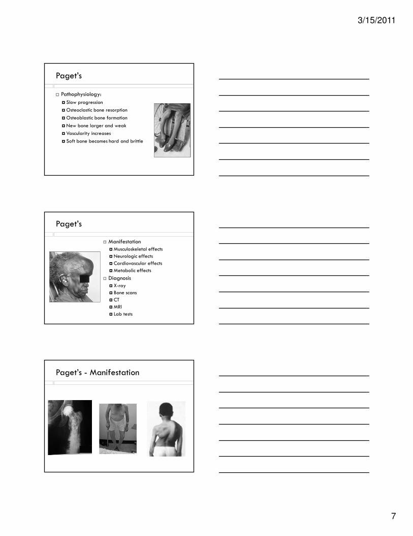
Paget’s Disease (Osteitis Deforma)
� An excess of bone
destruction and unorganized bone
formation
� Cause is unknown
� Average age at dx is 50-
60 yrs
� Affects the axial skeleton
3/15/2011
7
Paget’s
� Pathophysiology:
� Slow progression
�Osteoclastic bone resorption
�Osteoblastic bone formation
� New bone larger and weak
� Vascularity increases
� Soft bone becomes hard and brittle
Paget’s
� Manifestation
�Musculoskeletal effects
� Neurologic effects
� Cardiovascular effects
�Metabolic effects
� Diagnosis
� X-ray
� Bone scans
� CT
�MRI
� Lab tests
Paget’s - Manifestation

3/15/2011
8
Paget’s Collaborative Management
� Relieve pain
� Prevent or minimize complications
� Medication
� Pain relieve
� Biphosphonates
� Calcium supplement
� Surgery
Paget’s Nursing Diagnosis
� Chronic pain
� Assess location and quality
� Heat therapy and massage
� Teach – NSAID, placement of brace/corset
� Impaired physical mobility
� Assitive device when ambulating
� Teach – placement of brace/corset, good body mechanics
Osteomalacia (Adult Rickets)
� Vitamin D deficiency resulting in decalcification and
softening of the bone
� Not enough Vitamin D in diet
� Not enough exposure to sunlight
� Impaired intestinal absorption of fats
� Increased renal loss or decreased absorption of phosphate
� Same as Rickets in children

3/15/2011
9
Osteomalacia -
� Pathophysiology
� Vitamin D deficiency
� Lack of intake
� Lack of sunlight
� Phosphate depletion
� Acidosis
� Bone mineralization inhibitors
� CRF
� Calcium malabsorption
Osteomalacia - diagnosis
� Health history
� X-ray
� Lab tests
� Calcium
� Alk Phos
� Thyroid function
Osteomalacia -Collaborative management
� Correct Vitamin D deficiency
� Increase diet intake
� Expose to sunlight
� Calcium and Phosphate supplement
� Safety measures to prevent falls
� Encourage exercise
� Teach use of assistive devices

3/15/2011
10
Degenerative Bone Disease
� Osteoarthritis (OA)�Most common of all arthritis�Leading cause of pain and disability in elderly
�Loss of articular cartilage in joints�90% people has x-ray evidence of OA by age 40
�Gender and ethnicity effects�Localized�generalized
OA - pathophysiology
� Articular cartilage
loss
� Bone exposed
� Bone thickens
� Bone spurs develop
� inflammation
OA- risk factors
� Increasing age
� Genetic
� Trauma
� Overweight
� Inactivity
� Hormonal

3/15/2011
11
OA - Clinical Manifestations
� Joint involvement� Joint pain
� Joint stiffness
� Crepitus
� Joint enlargement
� Decreased ROM
� Flexion contractures
� Rarely does joint appear to be hot and inflamed (secondary synovitis)
OA- manifestation – (cont)
� Heberden’s nodes
�Most common
� Distal joint
� Bouchard’s nodes
� Less common
� Proximal joint
OA - Diagnosis
� H&P
� X-ray
� Lab test
� HA – hyaloronic acid

3/15/2011
12
OA - Management
� Conservative
� ROM
� Ice and heat
� Medication� Analgesics
� Topical
� Corticosteroids
�Muscle relaxants
� Surgery� Arthroscopy
� arthroplasty
OA – nursing Diagnosis
� Chronic pain r/t muscle spasms and cartilage
deterioration
� Impaired physical mobility r/t pain and
degenerative changes
� Self care deficit
Autoimmune and Inflammatory Disorder
Rheumatoid Arthritis
� Systemic disease
� Causes inflammation of the connective tissue
� 3 times as likely in women
� Onset age 20-40
� Cause unknown
� Genetic link?
� Infectious link?
� Environmental link?
� Hormonal link?

3/15/2011
13
Rheumatoid Arthritis (RA) Patho
� Auto-antibodies form - attack healthy tissue,
� Inflammation first in synovial membrane
� Inflammation spreads:� articular cartilage,
� joint capsule,
� ligaments and tendons
� Synovium thickens creating pannus:
RA (Pathophysiology)
RA - Manifestation
� Fatigue
� Loss of appetite
� Low grade fever
� Muscle and joint aches
� Stiffness
� Most notable in the morning
� Multiple joints inflamed in symmetrical pattern
� Joints - red, swollen, painful, and tender

3/15/2011
14
Systemic Symptoms of RA
� Sjogren’s syndrome
� Pleuritis
� Pericarditis
� Anemia
� Vaculitis
Diagnosis of RA
� History and physical examination
� Abnormal blood antibodies called:
�Rheumatoid factor (RF) found in 80% of patients
�Antinuclear antibody (ANA)
� Erythrocyte Sedimentation Rate (ESR)
� CBC
� Joint X-rays: swelling of the soft tissue
� Bone scanning: can show inflamed joints
� CCP
� Examination of the synovial fluid
RA - Management
� Relieve pain
� Reduce inflammation
� Rest and exercise
� Plasmapherises
� Alternative treatments
� Medication
� NSAIDs
� Corticosteroids (oral)
� Antirheumatic
� Corticosteroids (injection)

3/15/2011
15
RA – Nursing Diagnosis
� Chronic pain
� Fatigue
� Ineffective role performance
� Disturbed body image
Infectious Disorder: Osteomyelitis
� Bacterial infection of bone� Cause - fungus, parasites, virus, and bacteria
(Staphylococcus Aureus most common)
� Acute: new bone infection lasting < 6 weeks
� Chronic: bone infection present > 6 weeks or recurring bone infection
Osteomyelitis - Patho
� Most common cause direct contamination of bone
� Invasion from adjacent soft tissue infection
� Peripheral artery disease
� Bacteria lodge and multiply in bone

3/15/2011
16
Osteomyelitis - Patho
� Phagocytosis
� Pus
� Periosteum lifts
� Ischemia and necrosis
Etiology
� Hematogenous Osteomyelitis
� Sources of pathogens: UTI, soft tissue infections, endocarditis, and infected IV sites
� Spine is common site of infection in adults
�Affects older adults, IV drug abusers, sickle cell anemia
� Surgical prosthesis
�Hip and knee replacements
Etiology (continued)
� Osteomyelitis from a contiguous infection
� Infection from adjacent soft tissues
�Most common cause of osteomyelitis in adults
�Often due to:
� Direct penetrating wounds
� Decubitus ulcers
� Neurosurgery
� Osteomyelitis associated with vascular insufficiency
� Those with DM and PVD are at risk
� Neuropathy exposes foot to trauma and pressure ulcers
� Infection can spread to bone

3/15/2011
17
Manifestations of Osteomyelitis
� Low grade fever, malaise
� Cardiovascular effects -
� Tachycardia
� GI effects
� Nausea and vomiting, Anorexia
� MS effects
� Limp , Localized tenderness
� Integumentary effects
� Drainage and ulceration
� Swelling, erythema, and warmth
� Lymph node involvement
Osteomyelitis
� Diagnosis
� Bone scans
�MRI and CT scan
� Biopsy
� Blood tests
� Erythrocyte sedimentation rate (ESR) will be elevated
� Elevated C-Reactive protein
� CBC (WBC will be elevated)
� Blood cultures
Osteomylitis - Management
� Medication
� Antibiotic therapy
� Analgesics
� Surgery
� Debridement

3/15/2011
18
Osteomyelitis – Nursing Diagnosis
� Risk for infection
� Hyperthermia
� Impaired physical mobility
� Acute pain
Septic Arthritis
� Joint space invaded by pathogen
� Risk factors include bacteremia, RA
� Manifestation
� Abrupt onset
� Joint hot, swollen, painful, fluid filled
� Fever chills
� Medical emergency
� Aspirate fluids
� Abx
� Immobilize
Connective Tissue Disorder
� Scleroderma
� Sjogren’s Syndrome

3/15/2011
19
Scleroderma - Etiology
� A chronic autoimmune disease
� 300,000 people in the US
� Ages affected 25-55 (Female
> male)
� No known cause
� 2 Types
� Localized
� Systemic
SclerodermaLocalized vs systemic
� LOCALIZED
� Thickened, hardened skin and scarring
� Skin appears tight, reddish, or scaly.
� Extreme itching
� Can be limited around fingers or in large areas
such as limbs.
� Disabling but not fatal
� SYSTEMIC
� All skin symptoms
� CREST
� Complications
�Musculoskeletal
� Lungs
� Heart
� Digestive tract
� Kidneys
Scleroderma - diagnosis
� Diagnosis is usually due to clinical suspicion.
� ANA – id autoimmune process
� ESR – up in inflammatory process
� CBC – anemia
� Bone biopsy – confirm dx

3/15/2011
20
SclerodermaCollaborative Management
� Treatment based on symptoms
� Medication
� Calcium channel blocker (Raynaud’s)
� ACE inhibitors
� H2 receptor blocker
� Physical therapy
� Stretching of muscles important
� Dialysis
Sjogren’s Syndrome
� Causes inflammation of
exocrine glands
� Mucosal dryness
�Mouth
� Eyes
� Throat
� Lungs
� Vagina
� Skin
Sjorgen’s
� Diagnosis� H&P
� Schirmer’s test
� Treatment
� Supportive
� Artificial tears
� Increased fluid intake
� Avoid med that dry mucous membranes (i.e. decongestants)

3/15/2011
21
NCLEX
� The nurse administers Allopurinal to a client with
gout. The nurse explains that the goal of this therapy is to:
� A. Increase bone density
� B. Decrease synovial swelling
� C. Decrease uric acid production
� D. Prevent crystallization of uric acid
NCLEX
� A client with RA asks the nurse why the MD is going
to inject hydrocortisone into the knee joint. The nurse explains that the most important reason for the
injection is to:
� A. Lubricate the joint
� B. Reduce inflammation
� C. Provide physiotherapy
� D. Prevent ankylosis of the joint
NCLEX
� Which medication should the nurse anticipate being
perscribed to relieve the pain experienced by a client with RA?
� A. Xanax, 0.5 mg PO TID
� B. Ibuprofen 400 mg PO every 4 hours
� C. Codeine 30 mg PO every 4 hours
� D. Meperidine 30 mg PO every 4 hours