Embed Size (px)
Citation preview

UNIVERSIDADE FEDERAL DO ACRE
LEONARDO KOURI ALBUQUERQUE
MODELOS ANATÔMICOS DE LUXAÇÃO PATELAR 3D ESTUDO RADIOLÓGICO E AVALIAÇÃO NO ENSINO
DA ANATOMIA ANIMAL APLICADA
RIO BRANCO ACRE – BRASIL
ABRIL – 2018

LEONARDO KOURI ALBUQUERQUE
MODELOS ANATÔMICOS DE LUXAÇÃO PATELAR 3D ESTUDO RADIOLÓGICO E AVALIAÇÃO NO ENSINO
DA ANATOMIA ANIMAL APLICADA
Dissertação apresentada à Universidade Federal do Acre, como parte das exigências do Programa de Pós-Graduação em Sanidade e Produção Animal Sustentável na Amazônia Ocidental, para a obtenção do título de Mestre em Ciência Animal.
RIO BRANCO ACRE – BRASIL
ABRIL – 2018


LEONARDO KOURI ALBUQUERQUE
MODELOS ANATÔMICOS DE LUXAÇÃO PATELAR 3D ESTUDO RADIOLÓGICO E AVALIAÇÃO NO ENSINO
DA ANATOMIA ANIMAL APLICADA
Dissertação apresentada à Universidade Federal do Acre, como parte das exigências do Programa de Pós-Graduação em Sanidade e Produção Animal Sustentável na Amazônia Ocidental, para a obtenção do título de Mestre em Ciência Animal.
APROVADA: 30 de abril de 2018.
______________________________ ______________________________
__________________________________
Prof. Dr.Yuri Karaccas de Carvalho UFAC
(Orientador)
Prof.ª Dr.ª Soraia F. de Souza UFAC
Prof. Dr. Marcello Machado UFPR

Ao meu pai, Aldemar Holanda Albuquerque Filho. À minha mãe, Zenaida Kouri Albuquerque.
Dedico.
AGRADECIMENTOS
Agradeço primeiramente а Deus, que iluminou meu caminho durante esta caminhada.
À Universidade Federal do Acre (UFAC) e ao Programa de Pós-graduação em Sanidade e Produção Animal Sustentável na Amazônia Ocidental (PPGESPA) pelas oportunidades oferecidas.
A todos os docentes do PPGESPA que contribuem para qualificação dos futuros mestres e com o desenvolvimento da região norte. Em especial ao meu orientador, Prof. Dr. Yuri Karaccas de Carvalho, por toda a paciência e empenho com que sempre me orientou neste trabalho e em todos aqueles que realizei durante o mestrado.
À Coordenação de Aperfeiçoamento de Pessoal de Nível Superior (CAPES) pela concessão de bolsa de estudo.
Agradeço aos meus pais Aldemar Holanda Albuquerque e Zenaida Kouri Albuquerque e aos meus irmãos Luana Kouri Albuquerque e Leandro Kouri Albuquerque, por todo carinho recebido o longo da minha vida, e pela confiança e incentivo que me foram dados durante essa trajetória. A vocês expresso o meu maior agradecimento.
Agradeço a minha namorada Juliana Macedo Lage por ter me apoiado e ficado ao meu lado nas horas que mais precisei.
E a todos aqueles que de alguma maneira contribuíram para a realização desse trabalho.

“Você vence. Todos os dias. Quando não desiste, você vence. Quando levanta e
segue, você vence.”
Ana Nunes
CERTIFICADO DO COMITÊ DE ÉTICA NO USO DE ANIMAIS –UFAC
Título do projeto: Confecção de Modelo 3D de Luxação Patelar Canina e sua
Aplicação como método Alternativo no Ensino Prático da
Medicina Veterinária
Processo número: 23107.007926/2017-90
Protocolo número: 18/2017
Responsável: Prof. Dr. Yuri Karaccas de Carvalho
Data de aprovação: 06/06/2017

RESUMO
ALBUQUERQUE. Leonardo Kouri. Universidade Federal do Acre, abril de 2018 Modelos Anatômicos de Luxação Patelar 3D: Estudo Radiológico e Avaliação no Ensino da Anatomia Animal Aplicada. Orientador: Yuri Karaccas de Carvalho. As características da impressão 3D fazem da tecnologia uma ferramenta útil no Ensino da Anatomia Animal Aplicada, favorecendo relação direta com a anatomia real e com grande potencial para fornecer materiais didáticos de alta qualidade. A finalidade do estudo foi a confecção e avaliação de Modelos Anatômicos de Luxação Patelar 3D (MALP3D) como material didático no Ensino da Anatomia Animal Aplicada. Seu desenvolvimento partiu da necessidade de representação dos diferentes graus de severidade da Luxação Patelar (LP) durante aulas práticas. Foram criados cinco MALP3D, correspondentes aos diferentes graus da doença. A criação baseou-se na digitalização por scanner 3D de ossos In natura correspondentes ao fêmur, tíbia, fíbula e patela, edição em software de modelagem 3D e confecção em impressora 3D. Posteriormente, foram realizadas radiografias dos MALP3D em equipamento de raio X digital para ilustrar nos diferentes posicionamentos os graus da doença. Após a criação foi realizada avaliação dos modelos e radiografias dos MALP3D em uma turma de 36 discentes distribuídos aleatoriamente em dois grupos (Tradicional e MALP3D). Todas as estruturas impressas mostraram grande semelhança com as peças naturais, os MALP3D quando radiografados e mensurados foram capazes de representar os diferentes graus da doença de forma adequada e quando aplicados em aula proporcionaram acréscimos no conhecimento dos discentes. Os MALP3D mostraram ser uma alternativa educacional importante, capazes de suprir a falta de materiais representativos para o Ensino aplicado da Luxação Patelar (LP) e contribuir para a formação dos discentes.
Palavras chaves: Impressão 3D, Prototipagem Rápida, FDM, Radiografia, Modelo
didático
ABSTRACT
ALBUQUERQUE. Leonardo Kouri. Federal University of Acre, April 2018 Anatomical Models of Patellar Dislocation 3D: Radiological Study and Evaluation in the Teaching of Applied Animal Anatomy. Advisor: Yuri Karaccas de Carvalho. The characteristics of 3D printing technology make it a useful tool in the teaching of Applied Veterinary Anatomy and foster a direct relationship with real anatomy. Moreover, 3D printing has great potential as a source of high-quality teaching materials. The purpose of the present study was to design and evaluate 3D anatomical models of patellar luxation (3DAMPL) as a resource material for the teaching of Applied Veterinary Anatomy. The models were developed because there was a need to represent different grades of severity of patellar luxation (LP) during practical classes. Five 3DAMPL models were designed; they correspond to the different grades of the disease. To create the models, natural bones (corresponding to the femur, tibia, fibula and patella) were scanned in a 3D scanner, edited in 3D modeling software and printed in a 3D printer. Subsequently, the 3DAMPL models were X-rayed in a digital X-ray machine to illustrate the grades of the disease in different positions. The 3DAMPL models and their respective x-ray images were then evaluated in a class of 36 students randomly distributed into two groups (Traditional and 3DAMPL). All the printed structures were very similar to their natural counterparts; when the MALP3D models were X-rayed and measured, they were able to represent the different grades of the disease appropriately and when they were applied in the classroom, they helped students gain further knowledge of the disease. The 3DAMPL models were found to be an important alternative educational resource because they compensated for the lack of representative materials for the applied teaching of patellar luxation (PL) as well as enhanced the learning of the students. Keywords: 3D printing, Rapid Prototyping, FDM, Radiography, Physical models for
teaching

SUMÁRIO
págs.
RESUMO ABSTRACT 1 ARTIGO ................................................................................................................ 1
1.1 Artigo ............................................................................................................. 1 APÊNDICES .......................................................................................................... 25
1
1 ARTIGO
1 Artigo 1
3D Anatomical Models of Patellar Luxation: Radiological research and assessment
for the teaching of applied veterinary anatomy
Leonardo Kouri Albuquerque, Ricardo Ysaac Garcia Nunez, Patrícia Peruquetti, Rita
Cássia Ribeiro Pereira, Romeu Paulo Martins Silva, Yuri Karaccas de Carvalho.
Submetido à Anatomical Sciences Education em maio de 2018.

1
INTRODUCTION 1
2
Patellar luxation (PL) in dogs is a highly-prevalent orthopedic disease (Lara, 3
2013). It results from the development of bone abnormalities which affect hind limb 4
alignment and cause the loss of the normal anatomical relationship between the 5
trochlear groove of the femur and the patella at four different grades (Piermattei et al., 6
2006). Thus, learning about the disease is important in the education of future 7
veterinary physicians. 8
PL in dogs is addressed in the advanced cycle of the Veterinary Medicine 9
Undergraduate Program through books, images, and mainly in the dependence of 10
clinical cases (Davis et al., 2014). Although PL is commonplace in routine veterinary 11
care, teaching about this disease is a challenging task; for example, students lack basic 12
knowledge of anatomy; there is not enough support material for practical classes and 13
there are not enough clinical cases covering all grades of the disease. As a result, there 14
is a gap in veterinary education (Smith et al., 2017). 15
Furthermore, medical education and research institutions are currently 16
undergoing a process of adaptation as a result of several pedagogical, ethical and 17
economic limitations in practical teaching. Eventually, these limitations further hinder 18
the transmission of that type of knowledge and, hence, have a direct influence on the 19
education of students (Sugand et al, 2010; Vaccarezza and Papa, 2014). 20
Among the innovations that have emerged to resolve this problem, 3D printing 21
technology is seen as capable of favoring a direct relationship with real anatomy in 22
enough detail to provide an alternative to the use of animals. Also, it has great potential 23
to provide a source of high-quality teaching materials which, in turn, can help 24
educational institutions cope with limitations (Mcmenamin et al., 2014; Lim et al., 25
2016). 26
By using this technique, students can handle 3D anatomical models regardless 27
of their location. Because digital files are easy to reproduce and distribute, they offer 28
access to models which do not exist in a particular region or educational institution, 29
thus favoring the large-scale replication and widespread use of these models. 30
In turn, 3D printing is characterized as an important tool to develop practical 31
classes, as it can foster authentic and detailed understanding of the disease as well as 32

2
enable demonstration and experimentation in the classroom. As a result, theoretical 1
learning can be deepened (Abouhashem et al., 2015; O'Reilly et al., 2016). 2
Therefore, the objective of the present study was to design and evaluate 3D 3
anatomical models of patellar luxation (3DAMPL) as possible materials for the 4
teaching of applied veterinary anatomy. 5
6
7
8
9
10
11
12
13
14
15
16
17
18
19
20
21
22
23
24
25
26
3
MATERIAL AND METHODS 1
2
The study was developed in two steps at Federal University of Acre (UFAC), 3
Rio Branco, AC, Brazil. The first step was performed at the Laboratory of 3D 4
Technology. It consisted in scanning and printing the selected anatomical parts. The 5
second step was performed during the Applied Veterinary Anatomy course. The most 6
relevant aspects of patellar luxation (PL) were addressed and 3D anatomical models 7
of patellar luxation (3DAMPL) were evaluated. The research was registered and 8
approved (Protocol no. 23107.007926/2017-90) by the Animal Research Ethics 9
Committee (CEUA- UFAC). Patent application No. xxxx. 10
11
Creation of 3D anatomical models of patellar luxation (3DAMPL) 12
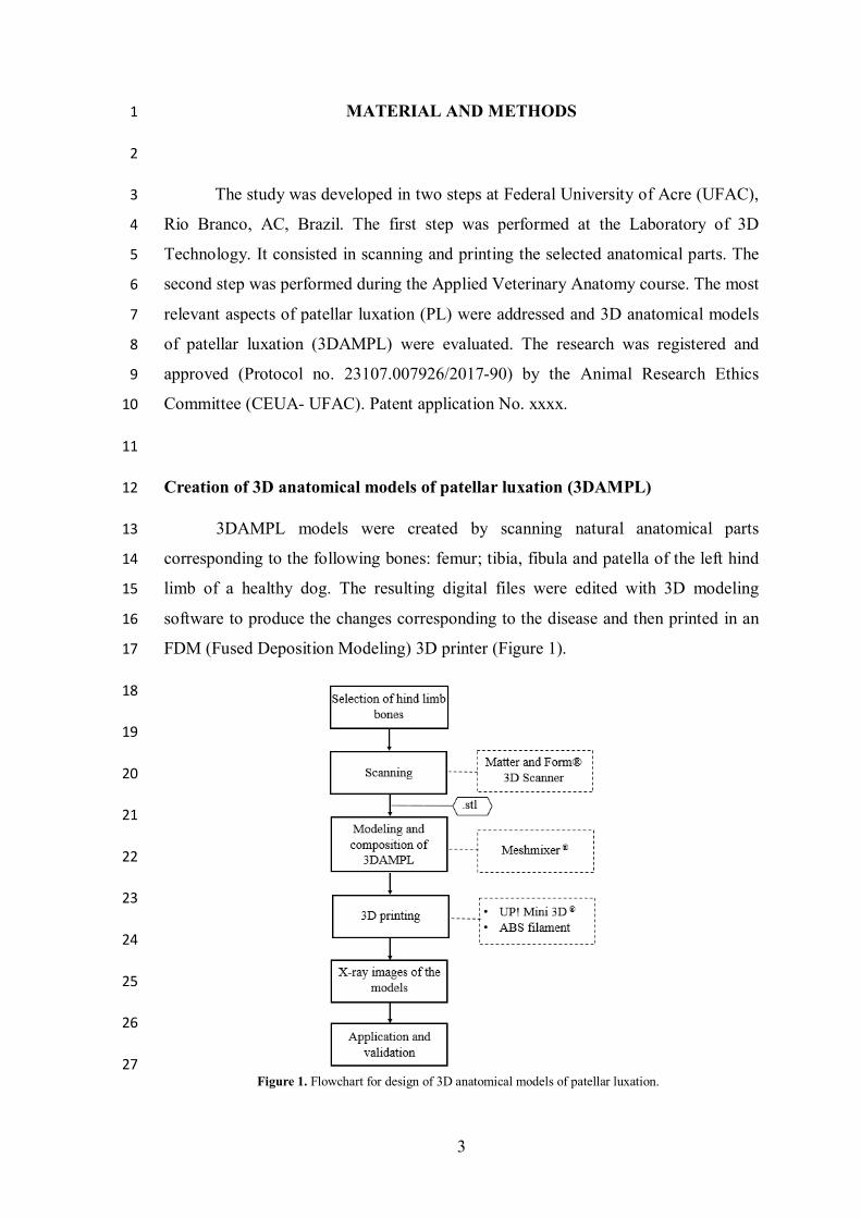
3DAMPL models were created by scanning natural anatomical parts 13
corresponding to the following bones: femur; tibia, fibula and patella of the left hind 14
limb of a healthy dog. The resulting digital files were edited with 3D modeling 15
software to produce the changes corresponding to the disease and then printed in an 16
FDM (Fused Deposition Modeling) 3D printer (Figure 1). 17
18
19
20
21
22
23
24
25
26
27 Figure 1. Flowchart for design of 3D anatomical models of patellar luxation.

4
The anatomical parts were scanned in a Matter and Form© 3D scanner 1
(Toronto, Canada) with a CMOS HD sensor and two lasers with scanning accuracy of 2
0.43 mm (capture of small details) and capture size within ± 0.25 mm. With the aid of 3
the included software “Matter and Form Scan®”, each bone was scanned individually. 4
The resulting virtual models were saved in .stl format and stored in a database. 5
Later, they were imported into a 3D design software program (Autodesk Meshmixer®, 6
version 3.1, Autodesk Inc. ©, California, United States) for modeling and composition 7
of 3DAMPL models by means of tools that have enabled the reproduction of the main 8
bone deformities corresponding to each of the four grades of PL. 9
All the characteristics of the disease reproduced in the 3DAMPL models were 10
based on studies reported by Singleton (1969); Piermattei et al., (2006); Netto et al., 11
(2012); Oliveira and Tudury, (2016); OFA (2017) (Table 1). 12
13
14
15
16
17
18
19
20
21
The portions of the 3DAMPL models were constructed separately in an UP! 22
Mini® 3D printer (Beijing Tiertime Technology Co. Ltd. ©, Beijing, China), in high-23
quality ABS (Acrylonitrile Butadiene Styrene) thermoplastic filament with 99% infill 24
and layer thickness of 0.2 mm. A mechanism composed of an articulating pin and hole 25
was inserted between the bones of the femur and tibia in order to enable the 26
reproduction of movements similarly to the movements of the knee joint and also allow 27
users to assemble and disassemble the parts. 28
29
Grades Characteristics
I Tibial tubercle deviation is minimal or non-existent;
Minimal changes in the patellofemoral joint.
II Mild deformities in the patellofemoral joint;
Tibial tubercle deviation from 15º to 30º.
III
Permanent patellar luxation;
Tibial tubercle deviation from 30º to 60º;
Shallow trochlear groove;
Angular deformities of the femur and the tibia are frequent.
IV
Permanent patellar luxation;
Tibial tubercle deviation from 60º to 90º;
Trochlear groove commonly absent or with convex trochlear surface;
Large angular deformities in the femur and the tibia.
Source: Table modified from (Piermattei et al., 2006).
Table 1. Anatomical changes visible in different grades of patellar luxation in dogs.
5
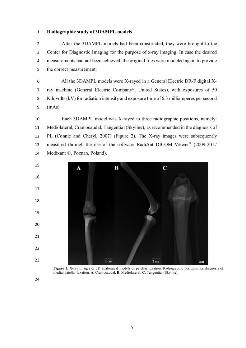
Radiographic study of 3DAMPL models 1
After the 3DAMPL models had been constructed, they were brought to the 2
Center for Diagnostic Imaging for the purpose of x-ray imaging. In case the desired 3
measurements had not been achieved, the original files were modeled again to provide 4
the correct measurement. 5
All the 3DAMPL models were X-rayed in a General Electric DR-F digital X-6
ray machine (General Electric Company©, United States), with exposures of 50 7
Kilovolts (kV) for radiation intensity and exposure time of 6.3 milliamperes per second 8
(mAs). 9
Each 3DAMPL model was X-rayed in three radiographic positions, namely: 10
Mediolateral; Craniocaudal; Tangential (Skyline), as recommended in the diagnosis of 11
PL (Connie and Cheryl, 2007) (Figure 2). The X-ray images were subsequently 12
measured through the use of the software RadiAnt DICOM Viewer® (2009-2017 13
Medixant ©, Poznan, Poland). 14
15
16
17
18
19
20
21
22
23
24
Figure 2. X-ray images of 3D anatomical models of patellar luxation. Radiographic positions for diagnosis of medial patellar luxation. A. Craniocaudal. B. Mediolateral. C. Tangential (Skyline).
6
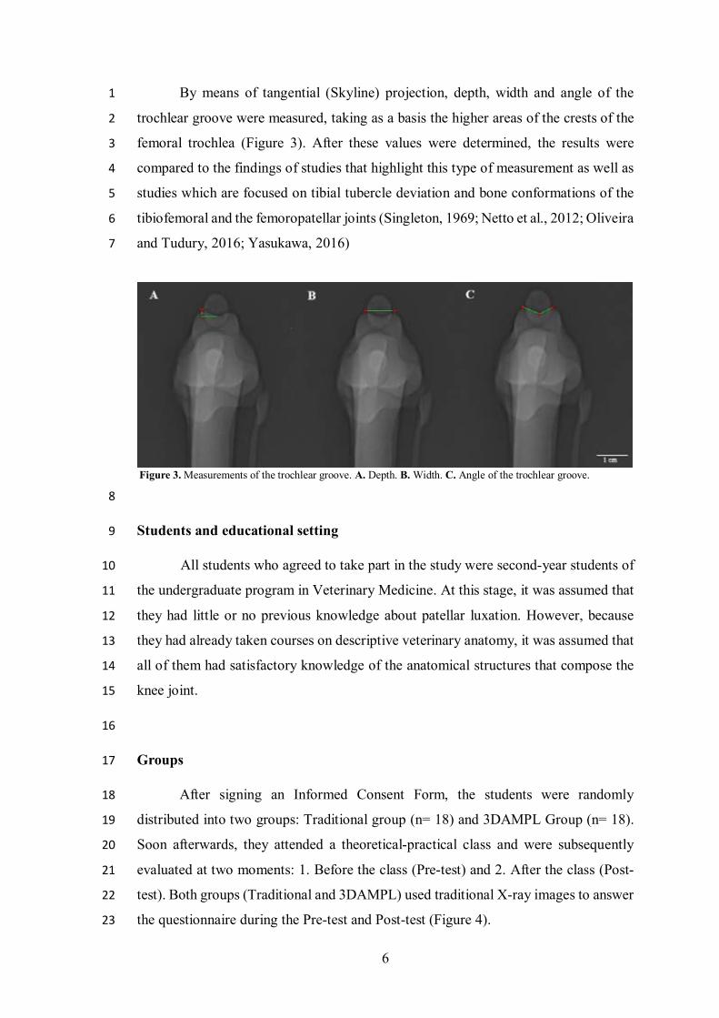
By means of tangential (Skyline) projection, depth, width and angle of the 1
trochlear groove were measured, taking as a basis the higher areas of the crests of the 2
femoral trochlea (Figure 3). After these values were determined, the results were 3
compared to the findings of studies that highlight this type of measurement as well as 4
studies which are focused on tibial tubercle deviation and bone conformations of the 5
tibiofemoral and the femoropatellar joints (Singleton, 1969; Netto et al., 2012; Oliveira 6
and Tudury, 2016; Yasukawa, 2016) 7
8
Students and educational setting 9
All students who agreed to take part in the study were second-year students of 10
the undergraduate program in Veterinary Medicine. At this stage, it was assumed that 11
they had little or no previous knowledge about patellar luxation. However, because 12
they had already taken courses on descriptive veterinary anatomy, it was assumed that 13
all of them had satisfactory knowledge of the anatomical structures that compose the 14
knee joint. 15
16
Groups 17
After signing an Informed Consent Form, the students were randomly 18
distributed into two groups: Traditional group (n= 18) and 3DAMPL Group (n= 18). 19
Soon afterwards, they attended a theoretical-practical class and were subsequently 20
evaluated at two moments: 1. Before the class (Pre-test) and 2. After the class (Post-21
test). Both groups (Traditional and 3DAMPL) used traditional X-ray images to answer 22
the questionnaire during the Pre-test and Post-test (Figure 4). 23
Figure 3. Measurements of the trochlear groove. A. Depth. B. Width. C. Angle of the trochlear groove.

7
1
2
3
4
5
6
7
8
9
10
11
Application and assessment of the 3DAMPL models 12
The lessons were taught by the same lecturer and lasted for 40 minutes. At that 13
time, the lecturer explained the most important anatomical and radiographic aspects of 14
PL and its main measurements as a tool for classification of the disease. The first group 15
(Traditional) received the lesson in a conventional form, i.e., using X-ray images of 16
clinical cases, while the second group (3DAMPL) was taught with the use of the 17
3DAMPL models and the respective X-ray images. At the end of each lesson, the 18
students were given 10 minutes to discuss the subject and to evaluate the specimens 19
by comparing them to the contents that had been taught. 20
The test for evaluation of the 3DAMPL models was composed of an 21
anonymous questionnaire with 29 affirmative questions, whose possible answer 22
choices were true or false (T or F). The questionnaire was divided into three sections 23
linked to specific knowledge of the disease, namely: 1. Radiographic anatomy of the 24
knee joint; 2. Radiographic findings for patellar luxation; 3. Classification of patellar 25
luxation. 26
27
28
Figure 4. Flowchart for assessment of the 3D anatomical models of patellar luxation.
8
Statistical Analysis 1
To check if the groups had the same level of knowledge, an analysis was 2
performed of the answers given in the Pre-test by the groups that were taught the 3
traditional lesson in comparison with the lesson taught with the 3DAMPL models. To 4
check what students had learned when using the models, the correct answers of the 5
Post-test were submitted to the non-paired t-test with a significance level of 5% (p< 6
0.05). To assess improvements in learning after use of the 3DAMPL models, the 7
answers were tested separately between the three topics covered in the questionnaire 8
(Radiographic anatomy of the knee joint, Radiographic findings for patellar luxation 9
and Classification of patellar luxation). 10
11
12
13
14
15
16
17
18
19
20
21
22
23
24
25
26
27

9
RESULTS 1
2
Evaluation of the 3DAMPL models 3
The 3DAMPL models showed great accuracy in the replication of all the bone 4
structures involved. The printed models maintained the same length, width and depth 5
of the bones. Also, each part of the bones was represented in detail (Figure 5). 6
7
The digital file which resulted from scanning of natural bones was used as the 8
basis for the creation of all other 3DAMPL models. It was named Grade 0, because it 9
did not have signs of anatomical deformities. The 3D-printed replica showed perfectly 10
visible femur and trochlear crest of the femur and similar conformation to that of the 11
original bone; the oval shape, width and thickness of the patella have been preserved 12
and followed the original aspect ratio; the tibia retained the same smooth and defined 13
lines that delimit tibial tuberosity. 14
The presence of the articulated mechanism between the bones of the femur and 15
tibia gave the 3DAMPL models the ability to reproduce movements of extension, 16
flexion and rotation similarly to the movements of the knee joint. Thus, radiographic 17
Figure 5. Natural bones (Left) and 3D-printed bones (Right) that compose the tibiofemoral and femoropatellar joints. A. Femoral Head (FH), Femur (Fe), Medial trochlear crest (MTC), Lateral trochlear crest (LTC), Trochlear Groove (TG), Patella (Pa). B. Tibial tubercle (TB), Tibia (Ti), Fibula (Fi).

10
positions suitable for the study could be replicated, and the users could assemble and 1
disassemble the parts. 2
Four 3DAMPL models were printed, each representing one of the different 3
grades of the disease. Each model was composed of four structures, namely: femur 4
(femur + articulating pin); tibia, fibula and patella. The structures were printed 5
separately in ABS thermoplastic material with 99% infill, layer thickness of 0.2 mm 6
and fine quality that ensures reduced print speed, in order to offer good quality of the 7
final product. 8
At the end of the production of all the 3DAMPL models, approximate values 9
were estimated for development time, printing time, material expenditure and cost per 10
anatomical structure and per printed 3DAMPL model. The values are shown in Table 11
1. 12
13
Table 1. Values for creation and printing of 3D anatomical models of patellar luxation. Time (Hours), Material 14 expenditure (grams) and cost (US dollar) per part and MALP3D model. 15
16
For Grade I, small changes were made on the surface of the femoral trochlea 17
to achieve the anatomical incongruence which is compatible with the grade. For 18
Grades II, III and IV, bigger changes were made with the purpose of replicating the 19
corresponding signs of osteoarthritis. The deformations significantly affected the 20
femoral trochlea and the trochlear groove and were necessary to achieve the 21
Development time (h)
Printing time (h)
Material expenditure (g)
Cost (US$)
Anatomical structures
Femur 2.5 3.5 28.8 0.87
Tibia 1.5 2.0 23.3 0.70
Fibula 0.33 0.45 2.8 0.08
Patella 0.16 0.13 0.7 0.02
3DAMPL
1 Unit 4.49 6.08 55.6 1.67
4 Units 6.49 24.32 222.4 6.68
*Value of the dollar = BRL 3.30 *ABS Filament Value = $30.30

11
measurements equivalent to each grade of the disease, and to allow a macroscopic 1
view of the anatomical changes (Figure 6). 2
3
The x-ray images made of the 3DAMPL models were very similar to traditional 4
ones, clearly revealing radiopaque and radiolucent areas. In the X-ray images, each 5
one of the bones and their corresponding anatomical structures could be observed and 6
determined. As a result of the addition of the articulating mechanism between the 7
bones of the femur and tibia, the presence of the socket pin was clearly visible in the 8
X-ray images made for the study. However, their presence did not influence the 9
evaluation of the pathological aspects of the disease (Figure 7). 10
11
12
13
14
15
16
17
18
19
FIGURE 7. X-ray images of the knee joint. Craniocaudal and tangential (Skyline) projection. 1. Femur; 2. Tibia; 3. Fibula; 4. Patella; 5. Femoral trochlea; 6. Tibial tubercle; 7. Trochlear groove. 8. Socket pin.
Figure 6. 3D Anatomical Models of Patellar Luxation. Detail of the deformities produced in the trochlear groove, tibial tubercle deviation and patellar luxation. A. Grade I; B. Grade II; C. Grade III; D. Grade IV.
12
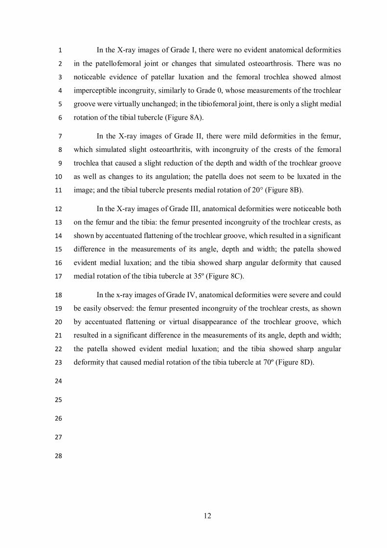
In the X-ray images of Grade I, there were no evident anatomical deformities 1
in the patellofemoral joint or changes that simulated osteoarthrosis. There was no 2
noticeable evidence of patellar luxation and the femoral trochlea showed almost 3
imperceptible incongruity, similarly to Grade 0, whose measurements of the trochlear 4
groove were virtually unchanged; in the tibiofemoral joint, there is only a slight medial 5
rotation of the tibial tubercle (Figure 8A). 6
In the X-ray images of Grade II, there were mild deformities in the femur, 7
which simulated slight osteoarthritis, with incongruity of the crests of the femoral 8
trochlea that caused a slight reduction of the depth and width of the trochlear groove 9
as well as changes to its angulation; the patella does not seem to be luxated in the 10
image; and the tibial tubercle presents medial rotation of 20° (Figure 8B). 11
In the X-ray images of Grade III, anatomical deformities were noticeable both 12
on the femur and the tibia: the femur presented incongruity of the trochlear crests, as 13
shown by accentuated flattening of the trochlear groove, which resulted in a significant 14
difference in the measurements of its angle, depth and width; the patella showed 15
evident medial luxation; and the tibia showed sharp angular deformity that caused 16
medial rotation of the tibia tubercle at 35º (Figure 8C). 17
In the x-ray images of Grade IV, anatomical deformities were severe and could 18
be easily observed: the femur presented incongruity of the trochlear crests, as shown 19
by accentuated flattening or virtual disappearance of the trochlear groove, which 20
resulted in a significant difference in the measurements of its angle, depth and width; 21
the patella showed evident medial luxation; and the tibia showed sharp angular 22
deformity that caused medial rotation of the tibia tubercle at 70º (Figure 8D). 23
24
25
26
27
28

13
1
2
3
4
5
6
7
8
9
10
11
12
Figure 8. X-ray images of 3D anatomical models of patellar luxation, Craniocaudal and tangential projections, respectively. A. Grade I; B. Grade II; C. Grade III; D. Grade IV.

14
Table 2 shows the measurements made of the 3DAMPLmodels for width, depth 1
and angle of the trochlear groove, as well as the values of tibial tuberosity deviation. 2
3
4
Evaluation of learning with the 3DAMPL models 5
The general analysis of the questionnaires showed that the mean number of 6
correct answers between the two groups (Traditional and 3DAMPL) was statistically 7
similar in both the Pre-test (p= 0.148) and the Post-test (p= 0.529). When analyzing 8
the means (Pre-test and Post-test) within the same group, a significant difference (p< 9
0.05) was found in both groups (Figure 9A). 10
In the comparative analysis between the groups (Traditional and 3DAMPL) in 11
the Pre-test of the item “Radiographic anatomy of the knee Joint”, it was found that 12
the mean number of correct answers of the 3DAMPL group was higher than the mean 13
of correct answers in the traditional group, hence there was a significant difference for 14
this test. By contrast, in the Post-test for the same item, no significant results were 15
found (p= 0.643). To analyze the means for correct answers in the questionnaire (Pre-16
test and Post-test) within the same group, there was a significant increase in the number 17
of correct answers for both the Traditional group and the 3DAMPL group (Figure 9B). 18
For the item “Radiographic findings for patellar luxation”, no significant 19
differences were found between the groups (Traditional and 3DAMPL) in the Pre-test 20
(p= 0.916) and the Post-test (p= 0.645). When analyzing each individual group, despite 21
the increase in the mean number of correct answers for this item in both groups, the 22
difference between the Pre-test and the Post-test was not significant (p>0.05) (Figure 23
9C). 24
25
26
Depth Width Angle TTD Grade 0 1.97 9.95 128.8° - Grade I 1.95 9.95 130.3° - Grade II 1.87 9.22 132.6° 20°
Grade III 1.13 8.98 149.1° 35° Grade IV - 8.67 155.7° 70°
Table 2. Measurements of the trochlear groove (depth and width in millimeters) and tibial tubercle deviation (TTD) in different grades of the 3DAMPL models.

15
For the item “Classification of patellar luxation”, there were no significant 1
differences in the analysis of the Pre-test (p= 0.644) and the Post-test (p= 0.843) when 2
comparing the means of the groups (Traditional and 3DAMPL). In the individual 3
analysis per group, despite the increase in the means, there were no significant 4
differences (p>0.05) (Figure 9D). 5
6
7
8
9
10
11
12
Figure 9. Graphs of the mean number of correct answers in the questionnaire. A. General Results. B. Radiographic anatomy of the knee joint. C. Radiographic findings for patellar luxation. D. Classification of patellar luxation.
16
DISCUSSION 1
2
In several studies, 3D printing has been described as a useful complement in 3
human and veterinary medical education and particularly associated with issues such 4
as surgery (Castilho et al., 2014; Harrysson et al., 2015; Kim et al., 2018) and anatomy 5
(Preece et al., 2013; Thomas et al., 2016; Lim et al., 2016; Adams et al., 2016 Li et al., 6
2018). 7
The development of 3DAMPL models arose from the need to represent 8
different grades of severity of PL during practical classes. Although it is a highly 9
prevalent disease in veterinary clinical practice, it is often addressed in the classroom 10
in a limited manner. There are not enough medical centers and few cases of this disease 11
are treated in educational institutions; therefore, students do not have enough clinical 12
experience about the disease or have fewer possibilities of visualizing the different 13
grades of severity of the disease during their undergraduate education. 14
The initial scanning of natural bones with the 3D scanner was a fundamental 15
step in the development of the research, since it provided digital files with excellent 16
quality and resolution that were used for the design of the 3DAMPL models. This 17
corroborated other studies that also reported the accuracy of 3D scanning in the process 18
of development of anatomical models (Cantín; Muños and Olate, 2015; Thomas et al., 19
2016; Li et al., 2018). 20
The stage of design of the 3DAMPL models required a total time estimated at 21
approximately 6.49 hours. It should be noted that prior knowledge of details during 22
reproduction of the models and, especially, mastery of the tools used in the 3D 23
modeling software were crucial to replicate bone deformities accurately and to achieve 24
the desired results in a shorter time frame. 25
When a comparison was made of the time spent in the design of the 3DAMPL 26
models based on the use of diagnostic imaging equipment, it was found that the two 27
techniques have a similar length. However, if an analysis is made of the advantages 28
and the cost of each method, 3D printing is more feasible for use in places that do not 29
have financial resources to rely on medical equipment (Mcmenamin et al., 2014; 30
Mitsopoulou et al., 2015). 31

17
The scanned structures that served as a basis (Grade 0) for the design of the 1
3DAMPL models showed great precision when printed. They represent the bone 2
conformations and the main anatomical structures of the natural bones with great 3
similarity. This was an important factor for the modeling process, because it was an 4
anatomical reference during reproduction of the deformities corresponding to each of 5
the grades of PL. 6
As shown in a study by Chung et al., (2018), the settings of the print parameters 7
in use (such as infill of the model, layer thickness, temperature and speed of printing) 8
are critical factors to ensure accuracy and good print quality. These aspects are also 9
closely related to the quantity of material used in the process, which directly affects 10
the cost and time of printing. 11
When the values of time and cost of printing of 3DAMPL models were 12
compared with those of similar studies that also used printers for extrusion of plastic 13
material (Li et al., 2018; Chung et al., 2018), it was found that the values were similar 14
in structures which have similar proportions, whereas there was a slight variation of 15
values in larger structures. This may be explained by the size ratio of the object being 16
printed, position on the printing surface, quantity of support filament, print parameters 17
in use or even type of printer in use (Aguiar and Yonezawa, 2014). 18
As pointed out, the use of 3D-printed replicas has the potential to provide a 19
readily available source of high-quality teaching materials. As shown in other studies 20
(Liew et al., 2015), the use of technology allows a better assessment of pathological 21
aspects of a disease when they are examined in tangible models of bone anatomy, 22
allowing manipulation and visualization in different perspectives, a fact that provides 23
learners with greater accessibility to different forms of learning. 24
The digital files of the 3DAMPL models as well as those of other studies may 25
contribute to the creation of databases, thus favoring the universalization of models, 26
i.e., any person or institution can have access to these models regardless of where they 27
are. Also, students can learn at their own pace and review the materials as many times 28
as necessary (Abouhashem et al., 2015; O'Reilly et al., 2016). 29
However, in order for models to appropriately contribute to the education of 30
professionals, experimental evidence is required to prove the efficiency of their 31
application (Mcmenamin et al., 2014; Abudayyeh et al., 2017). Thus, in the present 32

18
study, 3DAMPL models and their X-ray images were applied in order to prove that 1
they are a reliable source of teaching material for the proposed theme. 2
The x-ray images of the 3DAMPL models were very similar to traditional x-3
ray images; they showed each of the bones and their corresponding anatomical 4
structures, as well as changes that simulated osteoarthrosis and anatomical deformities 5
in the patellofemoral joint, caused by the disease (Piermattei et al., 2006; Oliveira and 6
Tudury, 2016). This was extremely important for measurements and validation of the 7
models. 8
The presence of an articulated mechanism between the bones of the femur and 9
tibia made the 3DAMPL models more realistic and interesting for learners, because 10
their parts could be assembled and disassembled as well as handled to take different 11
positions. This mechanism was also essential at the time of reproduction of X-ray 12
images, because the ability of motion (extension, flexion and rotation) allowed the 13
3DAMPL models to take the most appropriate positions for X-ray imaging. 14
There are no studies to date that have cited X-ray imaging of 3D anatomical 15
models. Therefore, this study proposed the use of 3DAMPL models for the creation of 16
X-ray images, and this is a valuable step in the demonstration of pathological aspects 17
that are usually not often addressed in Applied Veterinary Anatomy classes. 18
It should be noted that the procedure in use has a distinctive approach, because 19
it takes the opposite path as advocated in most studies published previously, i.e., most 20
of them have reported the use of medical images for the production of 3D-printed 21
models (Ebert et al., 2011; Haspel et al., 2014; Kondo et al., 2015). Moreover, the 22
procedures described in the present study can be used as a valuable resource for 23
creating collections of teaching materials and, thus, provide educational institutions 24
with the possibility of compensating for possible deficiencies, or use them as an 25
additional resource in the classroom, hence improving the education of students and 26
the study of different diseases. 27
As cited by Preece et al. (2013), in several studies that have focused on the 28
development of new models for teaching, evaluations are usually based only on the 29
satisfaction of end users, but they do not present quantitative evidence to support the 30
real effectiveness of new models in the improvement of knowledge. 31

19
The general results of the evaluation provided evidence that the printed 1
3DAMPL models can be used as a complementary resource to teach Applied 2
Veterinary Anatomy effectively and increase the knowledge of learners, thus 3
corroborating the findings of the study by Lim et al. (2016), who also found a 4
significant effect on learning in a group of students who used 3D-printed materials. 5
Because students had already taken descriptive veterinary anatomy classes but 6
had not been, so far, acquainted with clinical medicine courses, they were expected to 7
have little or no previous knowledge about patellar luxation during the application of 8
the Pre-test and satisfactory knowledge of the anatomical structures that compose the 9
knee joint. However, the assumption about the students’ knowledge of anatomy was 10
not confirmed, because the analysis showed a significant difference between the 11
groups during the Pre-test. 12
However, such difference may be due to the fact that the students had never 13
seen X-ray images and, therefore, they did not feel confident to identify anatomical 14
structures. This became clear because of the significant increase in the average number 15
of correct answers in the questionnaire and the balance represented by the similarity 16
of results between the groups after the classes. 17
The low mean number of correct answers for the items of radiographic findings 18
and classification of PL in both groups during the Pre-test confirmed the fact that the 19
students had little knowledge about the disease. This inference was further reinforced 20
during a survey carried out at the time of class, when most learners reported that they 21
were unaware of the disease. 22
After the classes were taught, it was found that the students gained some 23
knowledge about the disease, although not significantly. However, this was indicative 24
of increased knowledge about the subject. Therefore, it can be stated that the students 25
learned the content in a similar manner in the two groups. 26
The replacement of traditional X-ray images with 3DAMPL images did not 27
interfere in the way that the students learned about the subject. After the classes, there 28
were no significant differences between the groups, i.e., the students can study the 29
subject by looking at 3DAMPL images and then identify the disease in traditional X-30
ray images. 31
20
The present results highlight the importance of the experiment conducted in 1
this study for the teaching and learning of Applied Veterinary Anatomy. It can be 2
concluded that highly-accurate teaching materials can be developed to bridge the gaps 3
found in medical and veterinary education which may make it difficult to explain 4
certain diseases. These materials can help students enter the labor market with a better 5
understanding of the morphological and functional aspects relative to the conditions 6
and organic abnormalities of the disease. 7
8
9
10
11
12
13
14
15
16
17
18
19
20
21
22
23
24
25
26
21
CONCLUSION 1
2
The development of the 3DAMPL models showed that economically viable 3
teaching materials can be designed for educational institutions, and they can represent 4
the main pathological aspects of a disease in a similar manner to traditional materials. 5
This radiological study and the 3DAMPL models provide an important educational 6
alternative, because they not only compensate for the lack of suitable physical models 7
for applied teaching but also ensure greater accessibility to different individuals, who 8
have their own way of perceiving and storing new information. Thus, better results are 9
achieved during the learning process. 10
11
12
Acknowledgments 13
The authors are thankful to the Coordination for the Improvement of Higher Education 14
Personnel (CAPES) for granting a Master's scholarship. 15
16
17
18
19
20
21
22
23
24
25
26
27

22
LITERATURE CITED 1
2
Abouhashem Y, Dayal M, Savanah S, Štrkalj G. 2015. The application of 3D printing 3 in anatomy education. Med Educ Online 20:1–3. 4
Abudayyeh I, Gordon B, Ansari MM, Jutzy K, Stoletniy L, Hilliard A. 2017. A 5 practical guide to cardiovascular 3D printing in clinical practice: Overview and 6 examples. J Interven Cardiol 1–9. 7 8 Adams JW, Paxton L, Dawes K, Burlak K, Quayle M, Mcmenamin PG. 2015. 3D 9 printed reproductions of orbital dissections : a novel mode of visualising anatomy for 10 trainees in ophthalmology or optometry. Br J Ophthalmol 99:1162–1167. 11
Aguiar LDCD, Yonezawa WM. 2014. Construção de Instrumentos Didáticos com 12 Impressoras 3D. IV Simpósio Nacional de ensino de Ciência e Tecnologia. 13 Anais...Ponta Grossa – PR. 14
Castilho M, Dias M, Vorndran E, Gbureck U, Gouveia B, Arm H, Fernandes P, 15 Gouveia B, Armés H, Pires E, Rodrigues J. 2014. Application of a 3D printed 16 customized implant for canine cruciate ligament treatment by tibial tuberosity 17 advancement. Biofabrication 6:1–13. 18
Cantín M, Muñoz M, Olate S. 2015.Generation of 3D Tooth Models Based on Three-19 dimensional Scanning to Study the Morphology of Permanent Teeth. Int J Morphol 20 33:782–787. 21
Connie MH, Cheryl DH. 2007. Diagnóstico por imagem para a prática veterinária. 3rd 22 Ed. São Paulo: Roca, 296 p. 23
Chung M, Radacsi N, Robert C, Mccarthy ED, Callanan A, Conlisk N, Hoskins PR, 24 Koutsos V. 2018. On the optimization of low-cost FDM 3D printers for accurate 25 replication of patient-specific abdominal aortic aneurysm geometry. 3D Printing in 26 Medicine 4:1–10. 27
Davis CR, Bates AS, Ellis H, Roberts AM., 2014. Human Anatomy: Let the Students 28 Tell Us How to Teach. Anat Sci Educ 7:262-272 29
Ebert LC, Thali MJ, Ross S. 2011. Getting in touch — 3D printing in Forensic 30 Imaging. Forensic Sci Int 211:1–6. 31
Harrysson OLA, Marcellin-Little DJ, Horn TJ. 2015. Applications of Metal Additive 32 Manufacturing in Veterinary Orthopedic Surgery. The Minerals, Metals & Materials 33 Society Applications 67:647–654. 34
Hespel AM, Wilhite R, Hudson J. 2014. Invited review-applications for 3d printers in 35 veterinary medicine. Vet Radiol Ultrasound 55:347–358. 36
Kim SE, Shim KM, Jang K, Shim J, Kang SS. 2018. Three-Dimensional Printing-37 based Reconstruction of a Maxillary Bone Defect in a Dog Following Tumor Removal. 38 In Vivo 32:63–70. 39

23
Kondo K, Nemoto M, Masuda H, Okonogi S, Nomoto J, Harada N, Sugo N, Miyazaki 1 C, Chikao M. 2015. Anatomical Reproducibility of a Model Molded by a Three-2 dimensional Printer. Neurol Med Chir (Tokyo) 55:592-596. 3
Lara JS, Oliveira HP, Alves EGL, Silva RF, Resende CMF. 2013. Aspectos clínicos, 4 cirúrgicos e epidemiológicos da luxação patelar em cães atendidos no Hospital 5 Veterinário, no período de janeiro de 2000 a julho de 2010: estudo retrospectivo. Arq 6 Bras Med Vet Zootec 65:1274-1280. 7
Li F, Liu C, Song X, Huan Y, Gao S, Jiang Z. 2018. Production of Accurate Skeletal 8 Models of Domestic Animals Using Three-Dimensional Scanning and Printing 9 Technology. Anat Sci Educ 11:73–80. 10 11 Lim KHA, Loo ZY, Goldie SJ, Adams JW, McMenamin PG. 2016. Use of 3D printed 12 models in medical education: A randomized control trial comparing 3D prints versus 13 cadaveric materials for learning external cardiac anatomy. Anat Sci Educ 9:213–221. 14 15 Liew Y, Beveridge E, Demetriades AK, Hughes MA. 2015. 3D printing of patient-16 specific anatomy : A tool to improve patient consent and enhance imaging 17 interpretation by trainees. Br J Neurosurg 29:712–714. 18 19 Mcmenamin PG, Quayle MR, Mchenry CR, Adams JW. 2014. The Production of 20 Anatomical Teaching Resources Using Three-Dimensional (3D) Printing Technology. 21 Anat Sci Educ 7:479-486. 22 23 Mitsopoulou V, Michailidis D, Theodorou E, Polydoras S, Kaisarlis G, Spitas V, 24 Stathopoulou E, Provatidis C, Theodorou G. 2015. Digitizing, modelling and 3D 25 printing of skeletal digital models of Palaeoloxodon tiliensis (Tilos , Dodecanese , 26 Greece). Quatern Int 379:4–13. 27
Netto AS, Brito MBS, Severino FR, Campos LRA, Nico MAC, Oliveira VM, Severino 28 NR. 2012. Estudo da articulação patelofemoral por ressonância magnética: a variação 29 da morfologia do ligamento patelofemoral medial. Rev Bras Ortop 47:204–209. 30
Oliveira GK, Tudury EA. 2016. Estudo radiográfico das medidas da patela e do sulco 31 troclear em cães toys hígidos e portadores de luxação patelar medial grau II e III. 32 Universidade Federal Rural de Pernambuco: Recife. Doutorado em Ciência Animal. 33 Tese. 51 p. 34
O'Reilly MK, Reese S, Herlihy T, Geoghegan T, Cantwell CP, Feeney RNM, Jones 35 JFX. 2016. Fabrication and assessment of 3D printed anatomical models of the lower 36 limb for anatomical teaching and femoral vessel access training in medicine. Anat Sci 37 Educ 9:71–79. 38
Piermattei D, Flo G, Decamp C. 2006. Handbook of small animal orthopedics and 39 fracture. 4th Ed. Philadelphia: Saunders, 818 p. 40
Preece D, Williams SB, Lam R, Weller R. 2013. “Let’s Get Physical”: Advantages of 41 a Physical Model Over 3D Computer Models and Textbooks in Learning Imaging 42 Anatomy. Anat Sci Educ 224:216–224. 43

24
Singleton B. 1969. The Surgical Correction of Stifle Deformities in the Dog. J Small 1 Anim Pract 10:59–69. 2
Smith CF, Tollemache N, Covill C, Johnston M. 2017. Take Away Body Parts! An 3 Investigation into the Use of 3D-Printed Anatomical Models in Undergraduate 4 Anatomy Education. Anat Sci Educ 11:44–53. 5
Sugand K, Abrahams P, Khurana A. 2010.The Anatomy of Anatomy: A Review for 6 Its Modernization. Anat Sci Educ 3:83–93. 7
The Orthopedic Foundation for Animals (OFA). 2017. Application for Patellar 8 Luxation Database. E Nifong Boulevard, Columbia, Missouri. 9
Thomas DB, Hiscox JD, Dixon BJ, Potgieter J. 2016. 3D scanning and printing 10 skeletal tissues for anatomy education. J Anat 229:473–481. 11 12 Vaccarezza M, Papa V. 2015. 3D printing: a valuable resource in human anatomy 13 education. Anat Sci Int 90:64–65. 14
Yasukawa S, Edamura K, Tanegashima K, Seki M, Teshima K, Asano K, Nakayama 15 T, Hayash T. 2016. Evaluation of bone deformities of the femur, tibia, and patella in 16 Toy Poodles with medial patellar luxation using computed tomography. Vet Comp 17 Orthop Traumatol. 29:29–38. 18
19
20

25
APÊNDICES

26
Apêndice A – Termo de consentimento livre e esclarecido – TCLE. modelos anatômicos 3D de luxação patelar e sua aplicação no ensino da medicina veterinária.
1. Apresentação A pesquisa “Modelos Anatômicos 3D de Luxação Patelar e sua Aplicação no Ensino da Medicina Veterinária”, tem por objetivo “avaliar a utilização dos modelos anatômicos 3D da Luxação Patelar no processo de Ensino e aprendizagem em substituição aos métodos tradicionais utilizados nas disciplinas de Anatomia Patológica, Diagnóstico por Imagem e Clínica Cirúrgica do curso de Medicina Veterinária”. A população alvo é constituída por alunos de ambos os sexos, do Curso de Medicina Veterinária da Universidade Federal do Acre do município de Rio Branco, estado do Acre. Trata-se de uma pesquisa em nível de Dissertação de Mestrado, realizado pelo pesquisador Leonardo Kouri Albuquerque e seu orientador Professor Doutor Yuri Karaccas de Carvalho.
2. Esclarecimento
Esclarecemos que a sua participação, na pesquisa “Modelos Anatômicos 3D de Luxação Patelar e sua Aplicação no Ensino da Medicina Veterinária”, consiste em participar de aula teórico-prática e responder questionário pré e pós aula teórico-prática sobre a Luxação Patelar.
A participação do (a) aluno (a) é voluntária podendo desistir a qualquer momento, não havendo custos materiais ou financeiros para você ou para o (a) aluno (a), bem como não haverá remuneração pela participação do (a) aluno (a). Você poderá retirar seu consentimento em qualquer momento da realização da pesquisa, sem ter que justificar sua desistência e sem que sofra quaisquer tipos de coação ou penalidade por parte de seu professor e/ou dos pesquisadores.
Os riscos da pesquisa são mínimos, podendo ocorrer possíveis desconfortos emocionais por parte do (a) aluno (a). Esses desconfortos poderão ocorrer por ocasião da emissão das respostas às questões dos questionários ou em decorrência da participação na aplicação dos modelos, visto que podem sentir receio de externar suas percepções sobre a funcionalidade do modelo, utilizado como recursos didáticos durante os processos de ensino e aprendizagem dos conteúdos para os quais foram produzidos. Para minimizar e/ou excluir tais desconfortos, será solicitado ao (a) aluno (a) que não responda o questionário na sala de aula, leve-o para casa, não se identifique ao responder o questionário para garantir o anonimato da resposta e deposite o questionário respondido numa urna deixada no laboratório da pesquisa. Garantimos manter o mais amplo, absoluto e irrestrito sigilo profissional sobre a identidade do (a) aluno (a), durante e após o término da pesquisa. Desse modo, a identidade pessoal do (a) aluno (a) será excluída de todos e quaisquer produtos da pesquisa para fins de publicação científica.
27
Os possíveis benefícios o que o (a) aluno (a), terá com a pesquisa são que, ao utilizar o modelo anatômico experimentalmente, desenvolva aprendizagens significativas dos conteúdos curriculares para os quais o modelo anatômico foi elaborado.
Esclarecemos que os dados coletados por meio do questionário serão utilizados única e exclusivamente para produção do Relatório de Pesquisa e seus resultados serão publicados em meios de comunicação científica, tais como eventos científicos, livro e/ou revista acadêmica, sempre resguardando sua identidade.
Você receberá uma via deste Termo de Consentimento Livre e Esclarecido (TCLE), o qual terá as duas primeiras páginas rubricadas pelo pesquisador responsável e por você e a última página será assinada pelo pesquisador responsável e por você.
Para maiores informações e esclarecimentos sobre a pesquisa e/ou seus procedimentos, você poderá entrar em contato com o pesquisador responsável Leonardo Kouri Albuquerque, pelo telefone nº (68) 99220 4145 e e-mail [email protected].
Por fim, eu, Leonardo Kouri Albuquerque, pesquisador responsável, declaro cumprir todas as exigências éticas contidas nos itens IV. 3, da Resolução CNS Nº 466/2012, durante e após a realização da pesquisa. 3. Consentimento Eu, ___________________________________________________________________, RG Nº _________________, CPF Nº ____.____.____-___, declaro que: (a) li e compreendi o Termo de Consentimento Livre e Esclarecido (TCLE); (b) que a minha participação na pesquisa “Modelos Anatômicos 3D de Luxação Patelar e sua Aplicação no Ensino da Medicina Veterinária” é livre e espontânea; (c), não terei nenhum custo e nem serei remunerado pela participação; (d) posso retirar o consentimento e desistir a qualquer momento como participante da pesquisa, sem ter que justificar minha desistência e não sofrer qualquer tipo de coação ou punição. Diante do exposto, aponho minha rubrica nas duas primeiras páginas do TCLE e minha assinatura abaixo, como prova do meu Consentimento Livre e Esclarecido em permitir minha participação na pesquisa.
Rio Branco - Acre, _______ de ___________________ 2017.
______________________________________________
Participante da Pesquisa
______________________________________________ Leonardo Kouri Albuquerque
Pesquisador Responsável

28
Apêndice B – Avaliação da aplicação do modelo anatômico de luxação patelar. Assinale (V) para afirmações verdadeiras e (F) para afirmações falsas:
ANATOMIA E ASPECTOS RADIOGRÁFICOS DO JOELHO 1. ( ) Na imagem radiográfica A há o mau alinhamento de uma ou mais estruturas ósseas da
articulação do joelho. 2. ( ) Na imagem radiográfica E não é possível visualizar a Patela. 3. ( ) A imagem radiográfica B corresponde ao posicionamento Craniocaudal. 4. ( ) Na imagem radiográfica A é possível visualizar a Tuberosidade da Tíbia. 5. ( ) A imagem radiográfica D corresponde ao posicionamento Tangencial (Skyline). 6. ( ) A imagem radiográfica F corresponde ao membro pélvico esquerdo. 7. ( ) Na imagem radiográfica C é possível visualizar o Sulco Troclear. 8. ( ) A imagem radiográfica C corresponde ao posicionamento Médio-lateral. 9. ( ) A imagem radiográfica A corresponde ao membro pélvico direito. 10. ( ) A imagem radiográfica F apresenta a Tuberosidade da Tíbia em seu posicionamento
anatômico normal. ACHADOS RADIOGRÁFICOS DA LUXAÇÃO PATELAR 11. ( ) Na imagem radiográfica D é possível visualizar a Patela luxada. 12. ( ) Na imagem radiográfica B é possível visualizar grave deformidade no Sulco Troclear. 13. ( ) As imagens radiográficas B e E apresentam alterações anatômicas evidentes. 14. ( ) A imagem radiográfica A apresenta Luxação Patelar Lateral. 15. ( ) As imagens radiográficas A e D indicam Luxações reduzíveis. 16. ( ) A imagem radiográfica B apresenta Sulco Troclear pouco profundo. 17. ( ) As imagens radiográficas B e F indicam Luxações permanentes. 18. ( ) A imagem radiográfica B apresenta Sulco Troclear com deformidades leves. 19. ( ) A imagem radiográfica F apresenta Luxação Patelar Medial.
CLASSIFICAÇÃO DA LUXAÇÃO PATELAR 20. ( ) Na imagem radiográfica D o desvio da Tuberosidade da Tíbia pode estar entre 30° e 60°. 21. ( ) A imagem radiográfica C permite avaliar a profundidade do Sulco Troclear. 22. ( ) A imagem radiográfica B permite avaliar a largura do Sulco Troclear. 23. ( ) Na imagem radiográfica A o desvio da Tuberosidade da Tíbia pode estar entre 60° e 90°. 24. ( ) A imagem radiográfica C permite avaliar a angulação do Sulco Troclear. 25. ( ) As alterações anatômicas apresentadas na imagem radiográfica D corresponde ao Grau I
da Luxação Patelar. 26. ( ) O desvio da Tuberosidade da Tíbia apresentado na imagem radiográfica F corresponde ao
Grau III da Luxação Patelar. 27. ( ) Na imagem radiográfica A o desvio da Tuberosidade da Tíbia pode estar entre 15° e 30°. 28. ( ) A imagem radiográfica F corresponde ao Grau IV da Luxação Patelar. 29. ( ) A imagem radiográfica A corresponde ao Grau II da Luxação Patelar