Embed Size (px)
Citation preview
University of Birmingham
Diagnosis, treatment-monitoring and follow-up ofchildren and adolescents with X-linkedhypophosphatemia (XLH)Rothenbuhler, Anya; Schnabel, Dirk; Högler, Wolfgang; Linglart, Agnes
DOI:10.1016/j.metabol.2019.03.009
License:Creative Commons: Attribution-NonCommercial-NoDerivs (CC BY-NC-ND)
Document VersionPeer reviewed version
Citation for published version (Harvard):Rothenbuhler, A, Schnabel, D, Högler, W & Linglart, A 2019, 'Diagnosis, treatment-monitoring and follow-up ofchildren and adolescents with X-linked hypophosphatemia (XLH)', Metabolism.https://doi.org/10.1016/j.metabol.2019.03.009
Link to publication on Research at Birmingham portal
General rightsUnless a licence is specified above, all rights (including copyright and moral rights) in this document are retained by the authors and/or thecopyright holders. The express permission of the copyright holder must be obtained for any use of this material other than for purposespermitted by law.
•Users may freely distribute the URL that is used to identify this publication.•Users may download and/or print one copy of the publication from the University of Birmingham research portal for the purpose of privatestudy or non-commercial research.•User may use extracts from the document in line with the concept of ‘fair dealing’ under the Copyright, Designs and Patents Act 1988 (?)•Users may not further distribute the material nor use it for the purposes of commercial gain.
Where a licence is displayed above, please note the terms and conditions of the licence govern your use of this document.
When citing, please reference the published version.
Take down policyWhile the University of Birmingham exercises care and attention in making items available there are rare occasions when an item has beenuploaded in error or has been deemed to be commercially or otherwise sensitive.
If you believe that this is the case for this document, please contact [email protected] providing details and we will remove access tothe work immediately and investigate.
Download date: 09. Nov. 2021

1
Diagnosis, treatment-monitoring and follow-up of children and adolescents with X-linked hypophosphatemia (XLH) 1
2
Anya Rothenbuhler1, 2, 3 3
Dirk Schnabel4 4
Wolfgang Högler5, 6 5
Agnes Linglart1, 2, 3 6
7
Affiliations 8
1 APHP, Endocrinology and Diabetology for Children, Bicêtre Paris Sud Hospital, Le Kremlin-Bicêtre, France 9
2 APHP, Reference Center for Rare Disorders of Calcium and Phosphate Metabolism, filière OSCAR, Paris, France 10
3 APHP, Platform of Expertise for Rare Disorders Paris-Sud, Bicêtre Paris Sud Hospital, Le Kremlin-Bicêtre, France 11
4 Center for Chronic Sick Children, Pediatric Endocrinology, Charité, University Medicine Berlin, Germany 12
5 Institute of Metabolism and Systems Research, University of Birmingham, Birmingham, United Kingdom 13
6 Department of Pediatrics and Adolescent Medicine, Johannes Kepler University Linz, Linz, Austria 14
15
Corresponding author 16
Anya Rothenbuhler 17
Endocrinology and Diabetology for Children 18
Bicêtre Paris Sud Hospital, Le Kremlin-Bicêtre, France 19
21
22
23
24
2
Abstract 25
Early diagnosis, optimal therapeutic management and regular follow up of children with X-linked hypophosphatemia (XLH) determine their long 26
term outcomes and future quality of life. Biochemical screening of potentially affected newborns in familial cases and improving physician’s 27
knowledge on clinical signs, symptoms and biochemical characteristics of XLH for de novo cases should lead to earlier diagnosis and treatment 28
initiation. The follow-up of children with XLH includes clinical, biochemical and radiological monitoring of treatment (efficacy and complications) 29
and screening for XLH-related dental, neurosurgical, rheumatological, cardiovascular, renal and ENT complications. In 2018, the European 30
Union approved the use of burosumab, a humanized monoclonal anti-FGF23 antibody, as an alternative therapy to conventional therapy (active 31
vitamin D analogues and phosphate supplements) in growing children with XLH and insufficiently controlled disease. 32
Diagnostic criteria of XLH and the principles of disease management with conventional treatment or with burosumab are reviewed in this paper. 33
34
Keywords 35
X-linked hypophosphatemia (XLH), alfacalcidol, burosumab, osteomalacia, rickets 36
37
38
39
3
1. INTRODUCTION 40
41
42
X-linked hypophosphatemic rickets (XLHR, OMIM 307800), a rare genetic disease due to inactivating mutations in the PHEX gene (Phosphate 43
Regulating Gene with Homologies to the Endopeptidase on the X chromosome MIM #300550) is the most common form of hypophosphatemic 44
rickets with an incidence of 1:20000(1,2). 45
PHEX is expressed in osteocytes and odontoblasts and inactivating PHEX mutations result in increased synthesis and secretion of fibroblast 46
growth factor 23 (FGF23). Part of the pathophysiological mechanism that underlies XLHR is impaired proximal renal phosphate reabsorption 47
and reduced 1-hydroxylation of 25-OH vitamin D due to the excess action of FGF23(3,4). 48
49
Children affected by XLH present with rickets, severely impaired mineralization of bone (osteomalacia) and teeth, and other signs and 50
symptoms ultimately caused by excess FGF23, with the typical biochemical profile of hypophosphatemia, renal phosphate wasting and reduced 51
calcitriol [1,25(OH)2 vitamin D] concentration. 52
Early diagnosis and optimal management and follow-up of children and adolescents with XLH are the keys to successful outcomes, which 53
determine the future quality of life of these patients. To date, large-scale natural history studies of XLH are lacking, which makes it is difficult to 54
distinguish possibly inevitable long-term complications due to the underlying condition from sequelae of inadequate management. The burden 55
of disease observed in today’s adult XLH patients(5,6) suggests that late diagnosis and inadequate management contribute to adverse 56
outcomes. 57
The aim of this review is to highlight the need for early diagnosis and optimal management so that children with XLH become healthier future 58
adults. 59
60
61
62
63
4
2. DIAGNOSIS OF XLH 64
65
Disease awareness of physicians and affected family members leads to early diagnosis and thus early treatment initiation. Early diagnosis of 66
XLH is of major importance since early treatment initiation leads to better outcomes such as improved linear growth and final height, bone mass 67
accrual, fewer bone deformations and better dental health(7–11). 68
Two diagnostic settings need to be distinguished. 69
70
2.1 Diagnosis of familial cases of XLH 71
About 85-90% of familial cases of hypophosphatemic rickets are associated with PHEX gene mutations(10,12–19). XLH due to PHEX 72
mutations follows an X-linked dominant inheritance pattern(2). Thus, affected fathers transmit the disease to all of their daughters and none of 73
their sons. Affected mothers have a 50% risk of having an affected daughter or son. In the setting of familial XLH, potentially affected newborns 74
should be biochemically screened and treatment should be initiated as soon as the diagnosis is made in order to prevent rachitic changes, leg 75
bowing and short stature. If appropriately managed, it is unlikely that these patients develop active rickets and consequent orthopedic 76
complications (figure 1). 77
However, even in familial XLH cases diagnosis can be delayed. Unfortunately, adult patients are often lost to follow-up and may not have been 78
informed or may have incompletely understood the inheritance pattern. This is illustrated by the median age at diagnosis of XLH in familial 79
cases of 1.3 years, ranging from 0.1 to 14.3 years (n=58; unpublished data obtained from patients followed at the French reference center of 80
Bicêtre, Paris, and(20)). During the transition from pediatric to adult care, it is crucial to explain the inheritance pattern to adolescents and 81
young adults with childbearing potential. 82
83
In babies born to parents affected with XLH, biochemical screening should be performed as soon as possible after the 1st week of life or 84
certainly at first presentation to their family doctor. Screening includes serum phosphate, creatinine and alkaline phosphatase (ALP), and 85
urinary phosphate and creatinine. Diagnosis of XLH is suspected if the serum phosphate level is below the normal range for newborns and if 86
renal phosphate wasting is documented using the calculated renal phosphate reabsorption rate(21–23). It is essential that serum phosphate 87
5
and ALP concentrations are interpreted based on reference ranges for newborns and infants as these are physiologically higher than those for 88
adults(24,25). Although clinical and radiological signs of rickets (figure 1) are often lacking in those babies, ALP may be found at the upper level 89
of normal. Once diagnosis is made, the patient needs to be referred to a pediatrician specialized in bone disease (e.g. a pediatric 90
endocrinologist) and treatment should be initiated immediately. The genetic diagnosis, i.e. PHEX sequencing, confirms the diagnosis; it may be 91
done on cord blood or on a sample drawn after birth. Waiting for genetic results should not delay the start of treatment. Of note, a serum 92
phosphate level within the normal range during the first months of life does not rule out the diagnosis and, in the absence of genetic diagnosis, 93
the biochemical screening should be repeated including serum phosphate and ALP. 94
95
2.2 Diagnosis of de novo cases of XLH 96
Children with XLH due to de novo PHEX mutations, i.e. one third of the patients, are usually diagnosed after a diagnostic odyssey. Mean age at 97
diagnosis is 3.9 ± 3.1 years, ranging from 0.9 to 13.1 years (unpublished data, Bicêtre, Paris) (figure 2a). 98
99
2.2.1 Revealing symptoms 100
Diverse clinical presentations may lead to the diagnosis of XLH. The most frequent and typical presentation is rickets (figure 1) which manifests 101
as long bone deformities, especially leg bowing, delayed walking, waddling gait and bone/joint pain developing progressively once toddlers start 102
standing and walking. In those patients, pediatricians and/or orthopedic surgeons are often the first specialists that are consulted because of leg 103
deformities. As a certain degree of leg bowing in toddlers is considered physiological (26) (figure 2b), the first consultation may not always lead 104
to a diagnostic work-up and therefore diagnosis is delayed until symptoms worsen. Stunted growth may be the revealing symptom in de novo 105
XLH children (14% of cases in Bicêtre center; unpublished data); noteworthy, growth velocity is always poor at the time of diagnosis in those 106
children. Rarely, the diagnosis of rickets is made from radiographs taken for other reasons, e.g. systematic screening of hip dysplasia in 107
France. 108
109
2.2.2 Diagnostic criteria 110
6
Any leg bowing (genu varum or valgum) whether or not associated with poor statural growth, and widening of the metaphysis (ankles and 111
wrists) should lead to a radiological and biochemical work-up. Tooth abscesses or facial cellulitis occurring on apparently healthy teeth suggest 112
poor dentin mineralization(9). Radiological signs of XLH are detailed in this issue by C. Adamsbaum and colleagues. Briefly, radiographs of the 113
hand, knees and lower limbs show the long bone deformities, abnormal growth plates with widened and frayed metaphyses. In contrast to other 114
forms of rickets, bone cortices appear dense(20,27). At the time of diagnosis, fractures are uncommon in children and adolescents. 115
Biochemical criteria (table 1) for the diagnosis of XLH include: 116
serum phosphate below the normal threshold for age(28,29) associated with renal phosphate wasting, e.g. reduced calculated maximal 117
tubular reabsorption of phosphate as a function of glomerular filtration rate (TmP/GFR)(21). Of note, the fractional tubular resorption of 118
phosphate (TRP) value may be within the normal range in children with XLH, and in the presence of hypophosphatemia only the 119
TmP/GFR is diagnostic; 120
ALP levels above the upper limit of normal for age, indicating rickets/osteomalacia. In children, the measure of total ALP is used, in 121
contrast to adults, in whom bone ALP should be measured preferably(20,30). Although ALP levels are elevated in XLH children and 122
adolescents, the increase is not in the order of magnitude as seen in vitamin D deficiency rickets, defects in calcitriol synthesis or 123
calcitriol receptor mutations (commonly called the vitamin D receptor) (figure 2c); 124
parathyroid hormone (PTH) levels in the normal or upper normal range; any mild increase in PTH may be caused by underlying 125
additional vitamin D or dietary calcium deficiency; 126
normal serum calcium, and low urinary calcium excretion; 127
exclusion of other proximal or distal tubular wasting disorders; 128
exclusion, and otherwise prior correction, of vitamin D or dietary calcium deficiency. 129
In summary, the key to correct diagnosis of de novo XLH cases is good knowledge of the clinical signs and symptoms and the correct use of 130
age-adjusted biochemical investigations to distinguish various forms of rickets. 131
132
7
The diagnosis of XLH may be confirmed by the measurement of elevated levels of intact FGF23. However, FGF23 concentrations may be 133
inappropriately normal and this does not exclude the diagnosis. Patients with XLH produce levels of FGF23 that are well below those of patients 134
with oncogenic osteomalacia (31,32). FGF23 levels are influenced by several factors including phosphate intake(33,34). 135
136
2.3 Genetic confirmation of XLH 137
The final confirmation of XLH is obtained through genetic analysis which identifies mutations in the PHEX gene in ~70% of patients with 138
hypophosphatemic rickets, and 85-90% of patients when the disease is familial (2,12,16,19,35–46). Whenever possible, genetic analysis is 139
recommended. Different types of PHEX mutations exist including point mutations, splice-site mutations, small and large deletions, deletions of 140
pseudo-exons, and mosaicism, suggesting that several techniques or strategies may be necessary to reach a final diagnosis(18,39,47–51). 141
142
3. SEVERITY OF DISEASE AND COMPLICATIONS 143
We now have enough evidence to inform patients that XLH is a multisystemic disorder that may be associated with several complications 144
including 145
tooth abscesses, taurodontism (enlarged pulp chambers and body of tooth), facial cellulitis and periodontitis(52,53); 146
premature fusion of cranial sutures leading to dolichocephaly and/or craniosynostosis; in some cases, patients may present with 147
increased intracranial pressure, Chiari 1 malformation, syringomyelia, papillary oedema or neurological signs(54–61) ; 148
hearing impairment(20,62–65); 149
short stature: final height below -2SD is found in ~50% of patients adequately treated by conventional therapy(10,11,20,24,66–74); 150
reduced muscle function due to hypophosphatemia(75,76); 151
joint and bone pain 152
153
At the time of diagnosis, XLH children should undergo a thorough work-up to assess the severity/extent of the disease(20,30) including: 154
8
measuring rickets severity, judged by inter-malleolar and intercondylar distances, 6MWT (6-minute walking test) as a global dynamic 155
measure, serum ALP and PTH levels, serum and urinary calcium and phosphate concentrations, and hand, standing long leg and/or 156
knee X-rays; 157
assessing possible complications of the disease, i.e. craniosynostosis and its neurological complications(55,60), hearing impairment(62), 158
abnormal dental mineralization(9), growth retardation(69), and reduced muscle function(76); 159
measuring renal function (glomerular and tubular) and morphology before the start of therapy through kidney ultrasound and detailed 160
biochemical work-up. 161
162
4. DIFFERENTIAL DIAGNOSIS 163
164
Once the diagnosis of rickets is confirmed by clinical, biochemical and radiological criteria, and the diagnosis of nutritional rickets and vitamin D 165
resistant rickets (VDDR1-3), all of which are associated with secondary phosphate wasting due to high PTH levels, have been ruled out, other 166
causes of hypophosphatemic rickets should be considered in patients who do not carry a PHEX variant, even if they display an elevated FGF23 167
level. The different causes of hypophosphatemic rickets are described in table 3. 168
Several rickets-like diseases that may lead to progressive bone deformities, abnormal gait and metaphyseal irregularities need to be excluded. 169
These conditions may be found in the presence of low ALP levels, e.g. hypophosphatasia(77) or normal levels of ALP, e.g. healed nutritional 170
rickets, Blount’s disease or Schmid type metaphyseal dysplasia(78). 171
172
5. DISEASE MANAGEMENT 173
5.1 Principles of disease management 174
Once the diagnosis of XLH is established, the objective of the treatment is to restore the lower limb biomechanic axis and gait, improve growth, 175
bone and teeth mineralization and muscular function. Disease management should also include social aspects, patient/family education and 176
support. In addition, during follow up, the multidisciplinary team will aim at preventing the development of endocrine, orthopedic, rheumatologic, 177
9
metabolic, cardiovascular and renal complications. So far, international recommendations that could guide physicians in the management of 178
these rare patients are lacking. However, some reports have been published including extensive physicians’ expertise(20,30). 179
The patient pathway will involve different health and social disciplines throughout infancy, childhood and adolescence. We suggest that patients 180
are seen at regular intervals by multidisciplinary teams lead by a pediatric expert in bone diseases, who will liaise with the patient’s local 181
healthcare providers (general practitioners/ pediatricians), a pediatric radiologist, orthopedic surgeon, physiotherapist, dentist and orthodontist. 182
Additional professions may be required, e.g. pediatric neurosurgeon, ear, nose and throat (ENT) specialist, ophthalmologist, dietician, social 183
worker and psychologist. 184
185
Two different therapies are currently available for XLH: active vitamin D analogues combined with phosphate supplements, and burosumab, the 186
monoclonal fully human anti-FGF23 antibody. These treatments have different therapeutic objectives and outcomes, and therefore require 187
different management as highlighted in table 2. 188
189
5.2 Conventional treatment with vitamin D analogues and phosphate supplements 190
For decades, the association of active vitamin D analogues (alfacalcidol or calcitriol) and phosphate supplements using multiple daily dosing 191
was the only treatment option for children with XLH. The objective of this therapy is to counteract the calcitriol deficiency secondary to FGF23 192
exces and to compensate renal phosphate wasting. Medication doses reported in the literature, most of which date back over 20 years, vary 193
widely, from 10-80 ng/kg/day of calcitriol and 30-180 mg/kg/day of elemental phosphate(8,20,67,79–83). Advice on treatment, based on recent 194
reviews(20,30) and the authors’ expertise is shown in table 2. 195
196
This therapy has demonstrated its efficacy to: 197
decrease ALP concentrations to the upper limit of normal in ~ one year(20); 198
improve bone deformity, bone pain and gait in 30 to 60% of patients(11,66,67,80,82,84–86); 199
improve growth velocity in the magnitude of ~ 1 standard deviation(8,10,11,20,66–72,79,84,87); 200
10
significantly improve dentin mineralization and therefore decrease teeth abscesses and oral complications in affected 201
children(7,9,20,88,89). 202
Improvement in some of these outcomes, i.e. linear growth, final height, radiological features of rickets and oral health(7,10,11,90), has been 203
associated with early treatment initiation and longer duration of treatment. 204
Many limitations to this therapy have been identified over the years and should be known by the caring physician, including: 205
the absence of correction of the phosphate wasting with continued hypophosphatemia(20,30,80,85); 206
the risk of nephrocalcinosis and/or urolithiasis; large doses of active vitamin D and oral phosphate supplements have both been 207
associated with an increased rate of nephrocalcinosis in children(67,91–95); 208
the risk of hyperparathyroidism; large oral doses of phosphate supplements are associated with the development of secondary and 209
tertiary hyperparathyroidism by yet unknown mechanisms (8,86,91,92,96–100) 210
the insufficient, or lack of, response of some children, leading to corrective surgeries of lower limbs(101); 211
and the incomplete correction of muscle function deficits(75,76). 212
In addition to these major issues, we are lacking large scale studies to evaluate the impact of this conventional therapy on the quality of life and 213
on the development of several disease complications such as craniosynostosis and hearing problems, enthesopathy, chronic pain and fatigue. 214
215
Dose Adjustment for conventional therapy (table 2) 216
The daily dose of phosphate supplements and vitamin D analogues is adjusted to serum ALP and PTH and urinary calcium/creatinine 217
concentrations, clinical measures (leg bowing, growth velocity) and the patient’s weight. The goal is to maintain normal ALP, PTH and urinary 218
calcium/creatinine levels but not to normalise serum phosphate levels. During the first months of treatment, consistently elevated ALP levels 219
without hypercalciuria should lead to an increase in active vitamin D analogue and/or phosphate dose. Vice versa, normalized ALP in the 220
presence of hypercalciuria may require a reduction in the dose of active vitamin D analogues. If PTH level increases, one must consider 221
lowering phosphate supplementation and/or increasing the dose of active vitamin D analogues. In all cases, strict adherence to medication, in 222
particular the multiple daily dosing of phosphate is essential. 223
11
224
5.3 Novel therapy with anti-FGF23 antibody 225
As for today, the alternative therapy is burosumab, the humanized monoclonal anti-FGF23 antibody which was recently approved in the 226
European Union for the treatment of XLH children over 1 year of age and adolescents who are still growing, and in the US for the treatment of 227
all patients affected by XLH over 1 year of age(102,103). The main objective of this treatment is to counteract excess FGF23, thereby restoring 228
phosphate reabsorption and endogenous 1,25(OH)2 vitamin D synthesis. 229
In children with severe XLH aged 5 to 12 years, the treatment with burosumab, given subcutaneously every 2 weeks was found to result 230
in(104): 231
a steady increase in serum phosphate concentration to a range between 1.1 and 1.6 mmol/l due to an increase in TmP/GFR; 232
an increase in the calcitriol levels; 233
an improvement in the radiographic rickets severity scores after 40 and 64 weeks of treatment; 234
an improvement of physical function as shown by the increase in the distance walked during the 6MWT. 235
In contrast to conventional therapy, the burosumab dose is adjusted to the serum phosphate concentration as described in table 2. The 236
recommended starting dose in Europe is 0.4 mg/kg body weight (0.8 mg/kg in US), followed by a titration period to reach a serum phosphate 237
level in the low normal range for age, through dose increments every 4 weeks (maximum dose 2.0 mg/kg body weight or 90 mg every 15 days). 238
239
The limitations known to this treatment are(104–106): 240
injections site reactions, headache and muscular pain; 241
gain in growth velocity appears limited; 242
the therapy is recent and therefore data on any long-term outcomes, e.g. hyperparathyroidism, nephrocalcinosis, surgery, body 243
disproportion and adult complications such as enthesopathy are not yet available. 244
245
12
Given the available evidence for both therapies, we propose that children born into families affected with XLH, as well as children with a de 246
novo diagnosis of XLH, be started on conventional therapy except if the diagnosis was delayed for several years, thus rickets considered as 247
severe. In our view, treatment with burosumab should be offered (unless of course injections are refused), preferably with rigorous 248
documentation and follow-up, to XLH children aged 1 year or older and in adolescents with growing skeletons if: 249
- they have radiographic evidence of rickets; 250
they are refractory to conventional therapy; 251
they experience complications related to conventional therapy. 252
253
6. DISEASE FOLLOW-UP (table 2) 254
Adequate follow-up of XLH patients includes clinical, biochemical and radiological monitoring of treatment (efficacy and complications) and 255
screening for XLH-related dental, neurosurgical, rheumatological, cardiovascular, renal and/or ENT complications(20,30). 256
The parameters, as well as their frequency of assessment vary depending on age, disease severity and existing XLH-related complications. 257
258
6.1 Treatment monitoring 259
Clinical follow-up includes measuring intercondylar distance, intermalleolar distance, and if possible tibial torsion(107), height and growth 260
velocity. An annual 6MWT can also be helpful in older children (from 5-6 years of age)(104,108). The number of dental abscesses and 261
episodes of acute oral infections are recorded. The rachitic/osteomalacic, insufficiently treated bone, is associated with elevated ALP and low 262
urinary calcium. In contrast, when rickets is healing, ALP tends to normalize, and urinary calcium to increase. The FGF23 level is not used as a 263
tool for treatment monitoring in XLH children(33,109,110). 264
The efficacy and safety of conventional therapy, i.e. phosphate supplements and vitamin D analogues, is monitored by measuring ALP, the 265
biomarker of rickets activity and osteomalacia(20,66,82,85). PTH is measured regularly as hyperparathyroidism is promoted by oral phosphate 266
supplementation, especially during adolescence(8,86,91,111). Serum and specifically urine calcium measurements are necessary to evaluate 267
the safety of vitamin D analogues. In children younger than 5 years of age, the 24 hours urine collection is quite difficult, and spot urine samples 268
13
are preferred. In children older than 5 years of age, 24 hours urine collections are advised if the urinary calcium is above the upper limit of 269
normal (Uca/cr > 0.7 mmol/mmol)(112). 270
The follow-up of children with rickets may include radiographs. Once the diagnosis is made, radiological techniques using small amounts of X-271
rays, such as EOS, may be used whenever possible. Radiographs of lower limbs or knees (done not more than every 2 years) may be useful 272
during follow-up. Radiographs may be indicated if patients are refractory to therapy, if orthopedic surgery is indicated, in cases of unexplained 273
bone pain and before transition to adult care. 274
After initial treatment initiation and during physiological periods of rapid growth (infancy and puberty) it is useful to evaluate the patient every 275
three months clinically and biochemically. This is also the case for patients of all ages with unsatisfactory results, e.g. ALP remaining high, 276
worsening of leg bowing, or pain. Otherwise 6-monthly evaluation is sufficient. 277
278
The questions that should be asked if treatment objectives under conventional therapy are not achieved are: 279
Is treatment correctly prescribed? 280
Are the doses appropriate? Adjusted to weight and growth velocity? Adjusted to biochemical markers? 281
Are phosphate supplements prescribed in multiple daily doses? 282
Is treatment correctly given? Are phosphate supplements given separately from calcium intakes such as milk and yogurt? 283
Is compliance correct? As in many chronic diseases that require multiple daily doses of medication, poor compliance often explains poor 284
metabolic control and poor clinical outcome. 285
286
6.2 Particular aspects concerning patients treated with burosumab 287
In patients treated with burosumab, serum phosphate is a strong biomarker of efficacy and is monitored for treatment titration and follow-up. In 288
clinical trials, the phosphate target ranges from 1.1 to 1.6 mmol/l. Measurements are performed every 2 later every 4 weeks (104) for dose 289
adjustment. We do not yet know the optimal serum phosphate target for children. During treatment, TmP/GFR and ALP require monitoring 290
since they act as short-term and long-term biochemical markers of burosumab efficacy, respectively(104). The 1,25(OH)2 vitamin D 291
14
concentration increases rapidly upon burosumab therapy during 64 weeks of observation ; its value as a biomarker to adjust the therapy has 292
not been evaluated(105,113,114). 293
294
7. SCREENING FOR XLH COMPLICATIONS 295
Craniosynostosis and premature fusion of cranial sutures are complications of XLH. Craniosynostosis should be screened for by 296
assessing head circumference, skull shape and also neurological signs (fundoscopy, headaches). To date, there are no clear guidelines 297
regarding MRI evaluations of children with XLH. However, recent data demonstrated the high frequency of Chiari 1 malformation and 298
syringomyelia in XLH children raising the question whether systematic evaluation of the brain through MRI during childhood is indicated 299
(55,60,115). In case of neurological symptoms, a CT scan and/or brain MRI should be done. 300
Hearing should be evaluated by audiometry during childhood; however data to identify the best time period or at risk patients is scarce(62). 301
Dental examination should be performed at least once a year in children(9). For details refer to the dedicated article on dental issues. 302
Growth should be monitored at least twice a year; bone age evaluation may be included in short children or in case of decreased growth 303
velocity, a sign of active rickets which may require an increase in therapy(10,11,20,41,68,69). Growth hormone (GH) is not a standard 304
treatment or indication for children with XLH. Studies have shown that GH increases short-term linear growth in short XLH children before 305
puberty(116–123). Data on only 5 patients treated until final height showed no significant gain(124). 306
Nephrocalcinosis and nephrolithiasis are screened for using kidney ultrasounds and should be done at the start of therapy and 307
approximately every 1-2 years depending on the urinary excretion of calcium(125). 308
Cardiovascular screening. To date, cardiac complications of XLH, on conventional therapy, are not commonly reported. This is surprising 309
given that FGF23 has long been known to be associated with cardiovascular risk and the development of pathological hypertrophy that can lead 310
to congestive heart failure(126). Only very recently was left ventricular hypertrophy and hypertension described in a subset of XLH patients in a 311
prospective clinical study(127). Given the sparse evidence, it is difficult to come up with firm recommendations. We have decided to 312
recommend cardiac echography every 5 years until further evidence emerges. Regular cardiac ultrasound measurements were part of the 313
safety features of the burosumab clinical trials with no evidence of complications. Given the limited long-term safety data, we feel that regular 314
echocardiography is required until more evidence emerges. 315
15
316
8. SOCIAL AND PATIENT EDUCATION FOLLOW-UP 317
Education of patients and families is crucial at the time of diagnosis. Regular updates are necessary to assess and encourage adherence to 318
treatments, provide service contacts and information about patients’ association groups, inform patients of scientific discoveries, including new 319
therapies, support school and professional achievement and provide adequate social support (e.g. XLHnetwork.com; (1); http:// 320
phosphatdiabetes.de). 321
322
9. PERSPECTIVES 323
XLH is a multisystemic disorder that may manifest in children only once they start standing and walking. Untreated, or insufficiently treated, the 324
disease leads to severe handicaps including bone pain, bone deformities, dental complications with abscesses and missing teeth, and short 325
stature. The current conventional therapy, based on phosphate supplements and vitamin D analogues allows improvement in bone deformities, 326
growth velocity and bone and dentin mineralization. However, this treatment does not restore normal phosphate levels and many patients do 327
not respond fully/adequately to this therapy. Burosumab counteracts FGF23 excess thus restores renal phosphate reabsorption in treated 328
children and adolescents. These convincing results have led to the approval of burosumab by EMA and FDA for the use in Europe and the 329
USA. However, long-term data are needed, especially on growth, renal calcium excretion, PTH secretion and bone disease overall. When 330
children evolve through adolescence and then adulthood, complications of their disease such as hearing impairment, hyperparathyroidism, 331
cardiovascular and renal complications, rheumatological issues and enthesopathy may occur. These complications, which may be modified by 332
the disease’s therapy, render difficult the decision to stop or pursue the daily conventional treatment through adulthood once growth is 333
complete. Since osteomalacia will return after any of the two therapies is stopped, adult bone specialists will need to gather further long-term 334
treatment data. 335
336
Contributors 337
AR and AL wrote the initial draft of the article.338
All authors contributed equally in the construction and the revision of the article. 339
16
All authors read and approved the final manuscript. 340
341
Acknowledgements 342
Publication of this article was supported by Kyowa Kirin Pharma. The company did not take part in writing, editing and revision of the 343
manuscript. 344
345
Funding 346
This research did not receive any specific grant from funding agencies in the public, commercial, or not-for-profit sectors. 347
348
Conflict of interest 349
AR and AL received honoraria and travel grants from Kiowa Kirin and Ultragenyx. 350
DS reports honoraria, consulting fees and non-financial support from Kyowa Kirin, outside the submitted work. 351
WH received honoraria, research and travel support from Kyowa Kirin and honoraria and consulting fees from Ultragenyx. 352
353
354
355
17
Figure 1: Radiographic features of rickets in XLH children 356
A. Radiographs of lower limbs in a baby girl with an affected XLH father carrying a PHEX mutation. The baby is also affected; there are almost 357
no signs of rickets on radiographs at birth. Treatment was started on day 7 with alfacalcidol 1ug/day (27 ng/kg/d) and phosphate supplements 358
(Phosphoneuros®) 80 mg, 4 times per day (60 mg/kg/d) given twenty minutes after breast feeding. On radiographs at age 2.5 years, there were 359
no signs of rickets (mild features at year 1). B. Different features of rickets, highlighting the variability in XLH disease severity, at the time of 360
diagnosis in three children diagnosed late with de novo PHEX mutations. 361
362
Figure 2: Diagnosis of XLH in children 363
A. Age at diagnosis in children affected with XLH; we selected only the de novo cases (n=36 out of 94). The cohort of patients is followed at the 364
Bicêtre reference center, Paris, France (unpublished). The mean age (+/- SD) at diagnosis (dotted line) was 3.9 ± 3.1 yrs, the median was 2.7 365
yrs [min: 0.9 - max:13.7] yrs. Each bar represents a case. B. Physiological distance between knees and ankles in children, adapted from. The 366
upper part of the graph represents the 2SD intercondylar distance (positive) and the lower part the 2 SD intermalleolar distance (negative) and 367
the mean values are represented in the middle of the graph. C. Serum ALP concentrations in 21 children with XLH at the time of diagnosis 368
compared with 7 children affected by a molecular defect in the calcitriol (VDR) receptor. Median and 5th - 95th percentile of ALP are given. 369
370
371
372
18
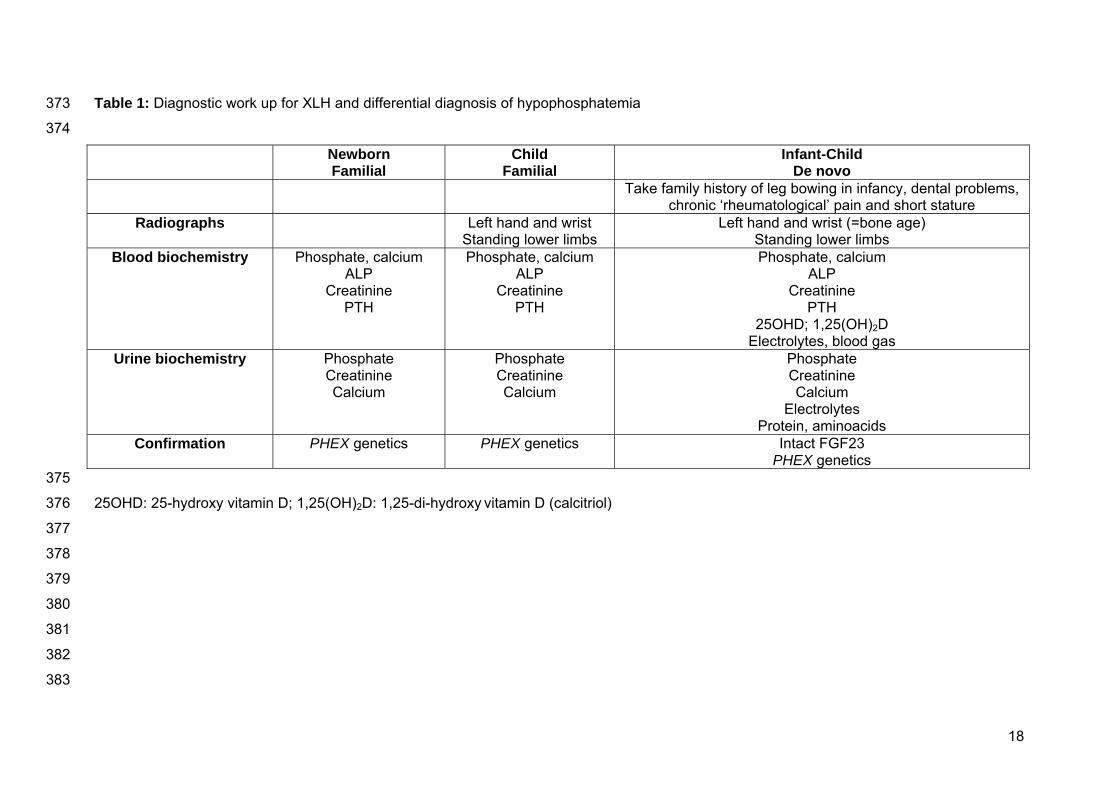
Table 1: Diagnostic work up for XLH and differential diagnosis of hypophosphatemia 373
374
Newborn Familial
Child Familial
Infant-Child De novo
Take family history of leg bowing in infancy, dental problems, chronic ‘rheumatological’ pain and short stature
Radiographs Left hand and wrist Standing lower limbs
Left hand and wrist (=bone age) Standing lower limbs
Blood biochemistry Phosphate, calcium ALP
Creatinine PTH
Phosphate, calcium ALP
Creatinine PTH
Phosphate, calcium ALP
Creatinine PTH
25OHD; 1,25(OH)2D Electrolytes, blood gas
Urine biochemistry Phosphate Creatinine Calcium
Phosphate Creatinine Calcium
Phosphate Creatinine Calcium
Electrolytes Protein, aminoacids
Confirmation PHEX genetics PHEX genetics Intact FGF23 PHEX genetics
375
25OHD: 25-hydroxy vitamin D; 1,25(OH)2D: 1,25-di-hydroxy vitamin D (calcitriol) 376
377
378
379
380
381
382
383
19
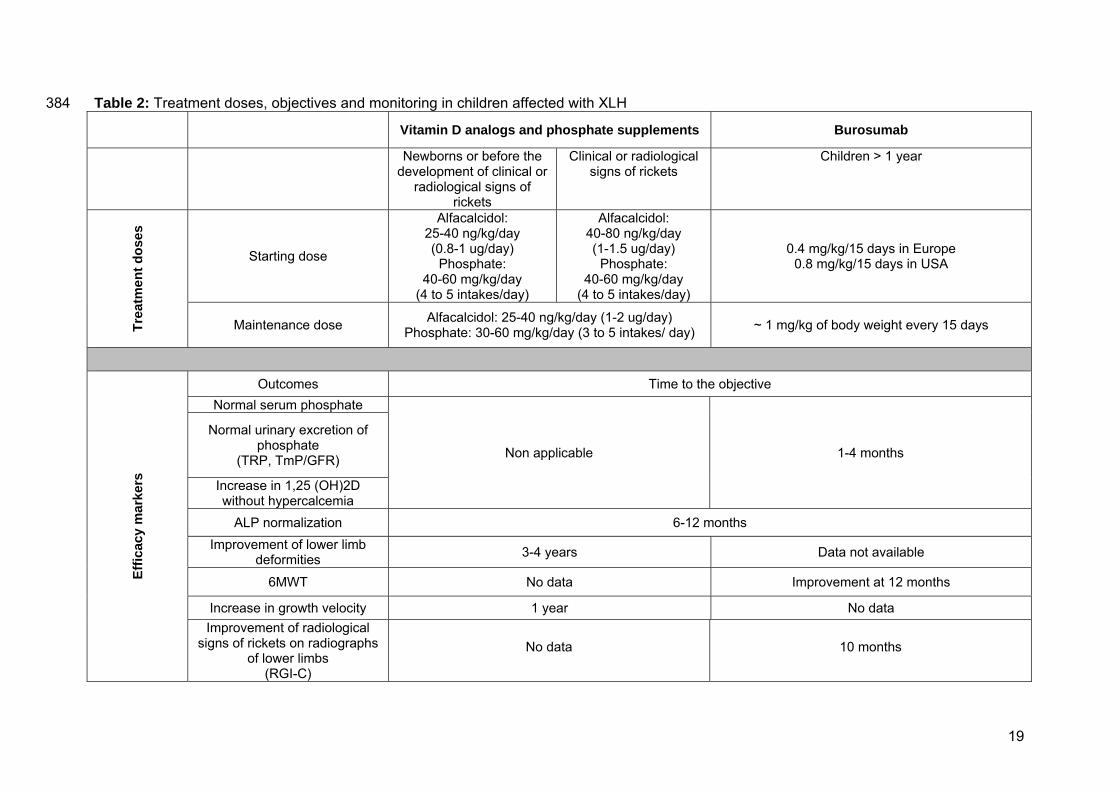
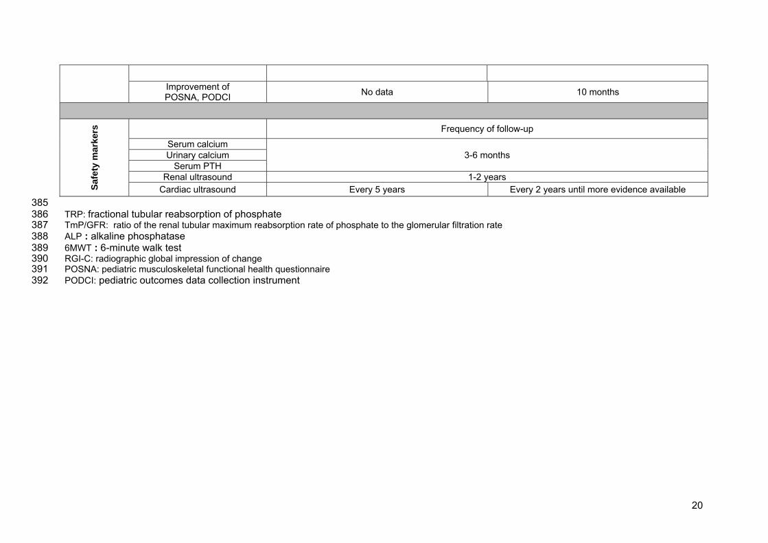
Table 2: Treatment doses, objectives and monitoring in children affected with XLH 384 Vitamin D analogs and phosphate supplements Burosumab
Newborns or before the development of clinical or
radiological signs of rickets
Clinical or radiological signs of rickets
Children > 1 year Tr
eatm
ent d
oses
Starting dose
Alfacalcidol: 25-40 ng/kg/day (0.8-1 ug/day)
Phosphate: 40-60 mg/kg/day
(4 to 5 intakes/day)
Alfacalcidol: 40-80 ng/kg/day (1-1.5 ug/day)
Phosphate: 40-60 mg/kg/day
(4 to 5 intakes/day)
0.4 mg/kg/15 days in Europe 0.8 mg/kg/15 days in USA
Maintenance dose Alfacalcidol: 25-40 ng/kg/day (1-2 ug/day) Phosphate: 30-60 mg/kg/day (3 to 5 intakes/ day) ~ 1 mg/kg of body weight every 15 days
Effic
acy
mar
kers
Outcomes Time to the objective Normal serum phosphate
Non applicable 1-4 months
Normal urinary excretion of phosphate
(TRP, TmP/GFR)
Increase in 1,25 (OH)2D without hypercalcemia
ALP normalization 6-12 months Improvement of lower limb
deformities 3-4 years Data not available
6MWT No data Improvement at 12 months
Increase in growth velocity 1 year No data Improvement of radiological
signs of rickets on radiographs of lower limbs
(RGI-C)
No data 10 months
20
Improvement of POSNA, PODCI No data 10 months
Sa
fety
mar
kers
Frequency of follow-up
Serum calcium 3-6 months Urinary calcium
Serum PTH Renal ultrasound 1-2 years
Cardiac ultrasound Every 5 years Every 2 years until more evidence available 385 TRP: fractional tubular reabsorption of phosphate 386 TmP/GFR: ratio of the renal tubular maximum reabsorption rate of phosphate to the glomerular filtration rate 387 ALP : alkaline phosphatase 388 6MWT : 6-minute walk test 389 RGI-C: radiographic global impression of change 390 POSNA: pediatric musculoskeletal functional health questionnaire 391 PODCI: pediatric outcomes data collection instrument 392
21
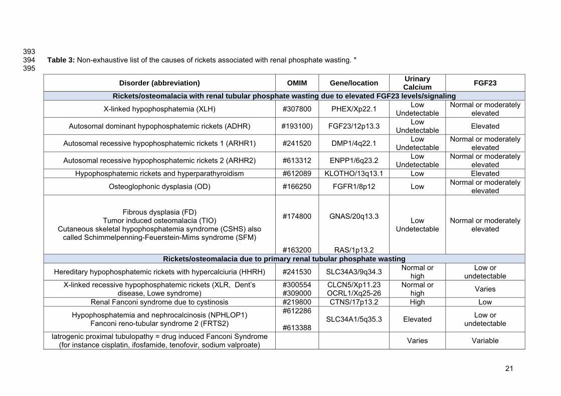
393 Table 3: Non-exhaustive list of the causes of rickets associated with renal phosphate wasting. * 394 395
Disorder (abbreviation) OMIM Gene/location Urinary Calcium FGF23
Rickets/osteomalacia with renal tubular phosphate wasting due to elevated FGF23 levels/signaling
X-linked hypophosphatemia (XLH) #307800 PHEX/Xp22.1 Low Undetectable
Normal or moderately elevated
Autosomal dominant hypophosphatemic rickets (ADHR) #193100) FGF23/12p13.3 Low Undetectable Elevated
Autosomal recessive hypophosphatemic rickets 1 (ARHR1) #241520 DMP1/4q22.1 Low Undetectable
Normal or moderately elevated
Autosomal recessive hypophosphatemic rickets 2 (ARHR2) #613312 ENPP1/6q23.2 Low Undetectable
Normal or moderately elevated
Hypophosphatemic rickets and hyperparathyroidism #612089 KLOTHO/13q13.1 Low Elevated
Osteoglophonic dysplasia (OD) #166250 FGFR1/8p12 Low Normal or moderately elevated
Fibrous dysplasia (FD) Tumor induced osteomalacia (TIO)
Cutaneous skeletal hypophosphatemia syndrome (CSHS) also called Schimmelpenning-Feuerstein-Mims syndrome (SFM)
#174800
#163200
GNAS/20q13.3
RAS/1p13.2
Low Undetectable
Normal or moderately elevated
Rickets/osteomalacia due to primary renal tubular phosphate wasting
Hereditary hypophosphatemic rickets with hypercalciuria (HHRH) #241530 SLC34A3/9q34.3 Normal or high
Low or undetectable
X-linked recessive hypophosphatemic rickets (XLR, Dent’s disease, Lowe syndrome)
#300554 #309000
CLCN5/Xp11.23 OCRL1/Xq25-26
Normal or high Varies
Renal Fanconi syndrome due to cystinosis #219800 CTNS/17p13.2 High Low
Hypophosphatemia and nephrocalcinosis (NPHLOP1) Fanconi reno-tubular syndrome 2 (FRTS2)
#612286
#613388 SLC34A1/5q35.3 Elevated Low or
undetectable
Iatrogenic proximal tubulopathy = drug induced Fanconi Syndrome (for instance cisplatin, ifosfamide, tenofovir, sodium valproate) Varies Variable
22
* excluding causes of rickets with secondary phosphate wasting due to high PTH levels such as nutritional rickets, and rickets due to vitamin D 396
deficiency or resistance. 397
398
23
399 400 401 402 403 404 405 406 407 408 References 409
1. Tenenhouse HS. X-linked hypophosphataemia: a homologous disorder in humans and mice. Nephrol Dial Transplant Off Publ Eur Dial 410
Transpl Assoc - Eur Ren Assoc. 1999 Feb;14(2):333–41. 411
2. A gene (PEX) with homologies to endopeptidases is mutated in patients with X-linked hypophosphatemic rickets. The HYP Consortium. 412
Nat Genet. 1995 Oct;11(2):130–6. 413
3. Liu S, Tang W, Zhou J, Stubbs JR, Luo Q, Pi M, et al. Fibroblast growth factor 23 is a counter-regulatory phosphaturic hormone for 414
vitamin D. J Am Soc Nephrol JASN. 2006 May;17(5):1305–15. 415
4. Shimada T, Mizutani S, Muto T, Yoneya T, Hino R, Takeda S, et al. Cloning and characterization of FGF23 as a causative factor of 416
tumor-induced osteomalacia. Proc Natl Acad Sci U S A. 2001 May 22;98(11):6500–5. 417
5. Che H, Roux C, Etcheto A, Rothenbuhler A, Kamenicky P, Linglart A, et al. Impaired quality of life in adults with X-linked 418
hypophosphatemia and skeletal symptoms. Eur J Endocrinol. 2016 Mar;174(3):325–33. 419
6. Beck-Nielsen SS, Brusgaard K, Rasmussen LM, Brixen K, Brock-Jacobsen B, Poulsen MR, et al. Phenotype presentation of 420
hypophosphatemic rickets in adults. Calcif Tissue Int. 2010 Aug;87(2):108–19. 421
7. Biosse Duplan M, Coyac BR, Bardet C, Zadikian C, Rothenbuhler A, Kamenicky P, et al. Phosphate and Vitamin D Prevent Periodontitis 422
in X-Linked Hypophosphatemia. J Dent Res. 2017 Apr;96(4):388–95. 423
8. Rasmussen H, Pechet M, Anast C, Mazur A, Gertner J, Broadus AE. Long-term treatment of familial hypophosphatemic rickets with oral 424
phosphate and 1 alpha-hydroxyvitamin D3. J Pediatr. 1981 Jul;99(1):16–25. 425
24
9. Chaussain-Miller C, Sinding C, Wolikow M, Lasfargues J-J, Godeau G, Garabédian M. Dental abnormalities in patients with familial 426
hypophosphatemic vitamin D-resistant rickets: prevention by early treatment with 1-hydroxyvitamin D. J Pediatr. 2003 Mar;142(3):324–31. 427
10. Quinlan C, Guegan K, Offiah A, Neill RO, Hiorns MP, Ellard S, et al. Growth in PHEX-associated X-linked hypophosphatemic rickets: the 428
importance of early treatment. Pediatr Nephrol Berl Ger. 2012 Apr;27(4):581–8. 429
11. Mäkitie O, Doria A, Kooh SW, Cole WG, Daneman A, Sochett E. Early treatment improves growth and biochemical and radiographic 430
outcome in X-linked hypophosphatemic rickets. J Clin Endocrinol Metab. 2003 Aug;88(8):3591–7. 431
12. Gaucher C, Walrant-Debray O, Nguyen TM, Esterle L, Garabedian M, Jehan F. PHEX analysis in 118 pedigrees reveals new genetic 432
clues in hypophosphatemic rickets. Hum Genet. 2009 May;125(4):401–11. 433
13. Holm IA, Nelson AE, Robinson BG, Mason RS, Marsh DJ, Cowell CT, et al. Mutational analysis and genotype-phenotype correlation of 434
the PHEX gene in X-linked hypophosphatemic rickets. J Clin Endocrinol Metab. 2001 Aug;86(8):3889–99. 435
14. Li S-S, Gu J-M, Yu W-J, He J-W, Fu W-Z, Zhang Z-L. Seven novel and six de novo PHEX gene mutations in patients with 436
hypophosphatemic rickets. Int J Mol Med. 2016 Dec 1;38(6):1703–14. 437
15. Morey M, Castro-Feijóo L, Barreiro J, Cabanas P, Pombo M, Gil M, et al. Genetic diagnosis of X-linked dominant hypophosphatemic 438
rickets in a cohort study: Tubular reabsorption of phosphate and 1,25(OH) 2 D serum levels are associated with PHEX mutation type. BMC Med 439
Genet. 2011 Sep 8;12(1):116. 440
16. Ruppe MD, Brosnan PG, Au KS, Tran PX, Dominguez BW, Northrup H. Mutational analysis of PHEX, FGF23 and DMP1 in a cohort of 441
patients with hypophosphatemic rickets. Clin Endocrinol (Oxf). 2011 Mar;74(3):312–8. 442
17. Beck-Nielsen SS, Brixen K, Gram J, Brusgaard K. Mutational analysis of PHEX, FGF23, DMP1, SLC34A3 and CLCN5 in patients with 443
hypophosphatemic rickets. J Hum Genet. 2012 Jul;57(7):453–8. 444
18. Ma SL, Vega-Warner V, Gillies C, Sampson MG, Kher V, Sethi SK, et al. Whole Exome Sequencing Reveals Novel PHEX Splice Site 445
Mutations in Patients with Hypophosphatemic Rickets. PloS One. 2015;10(6):e0130729. 446
19. Ichikawa S, Traxler EA, Estwick SA, Curry LR, Johnson ML, Sorenson AH, et al. Mutational survey of the PHEX gene in patients with X-447
linked hypophosphatemic rickets. Bone. 2008 Oct;43(4):663–6. 448
20. Linglart A, Biosse-Duplan M, Briot K, Chaussain C, Esterle L, Guillaume-Czitrom S, et al. Therapeutic management of 449
25
hypophosphatemic rickets from infancy to adulthood. Endocr Connect. 2014;3(1):R13-30. 450
21. Brodehl J. Assessment and interpretation of the tubular threshold for phosphate in infants and children. Pediatr Nephrol Berl Ger. 1994 451
Oct;8(5):645. 452
22. Brodehl J, Krause A, Hoyer PF. Assessment of maximal tubular phosphate reabsorption: comparison of direct measurement with the 453
nomogram of Bijvoet. Pediatr Nephrol Berl Ger. 1988 Apr;2(2):183–9. 454
23. Mian AN, Schwartz GJ. Measurement and Estimation of Glomerular Filtration Rate in Children. Adv Chronic Kidney Dis. 2017 455
Nov;24(6):348–56. 456
24. Santos F, Fuente R, Mejia N, Mantecon L, Gil-Peña H, Ordoñez FA. Hypophosphatemia and growth. Pediatr Nephrol Berl Ger. 2013 457
Apr;28(4):595–603. 458
25. Turan S, Topcu B, Gökçe İ, Güran T, Atay Z, Omar A, et al. Serum alkaline phosphatase levels in healthy children and evaluation of 459
alkaline phosphatase z-scores in different types of rickets. J Clin Res Pediatr Endocrinol. 2011;3(1):7–11. 460
26. Greene WB. Genu varum and genu valgum in children: differential diagnosis and guidelines for evaluation. Compr Ther. 1996 461
Jan;22(1):22–9. 462
27. Cheung M, Roschger P, Klaushofer K, Veilleux L-N, Roughley P, Glorieux FH, et al. Cortical and trabecular bone density in X-linked 463
hypophosphatemic rickets. J Clin Endocrinol Metab. 2013 May;98(5):E954-961. 464
28. Clayton BE, Jenkins, P. Clinical tests and refernce ranges. In: Paediatric chemical pathology. Blackwell Scientific Publications. Oxford; 465
1960. 466
29. Greenberg BG, Winters RW, Graham JB. The normal range of serum inorganic phosphorus and its utility as a discriminant in the 467
diagnosis of congenital hypophosphatemia. J Clin Endocrinol Metab. 1960 Mar;20:364–79. 468
30. Carpenter TO, Imel EA, Holm IA, Jan de Beur SM, Insogna KL. A clinician’s guide to X-linked hypophosphatemia. J Bone Miner Res Off 469
J Am Soc Bone Miner Res. 2011 Jul;26(7):1381–8. 470
31. Jonsson KB, Zahradnik R, Larsson T, White KE, Sugimoto T, Imanishi Y, et al. Fibroblast growth factor 23 in oncogenic osteomalacia 471
and X-linked hypophosphatemia. N Engl J Med. 2003 Apr 24;348(17):1656–63. 472
32. Nagata Y, Imanishi Y, Ishii A, Kurajoh M, Motoyama K, Morioka T, et al. Evaluation of bone markers in hypophosphatemic 473
26
rickets/osteomalacia. Endocrine. 2011 Oct 1;40(2):315. 474
33. Endo I, Fukumoto S, Ozono K, Namba N, Tanaka H, Inoue D, et al. Clinical usefulness of measurement of fibroblast growth factor 23 475
(FGF23) in hypophosphatemic patients: proposal of diagnostic criteria using FGF23 measurement. Bone. 2008 Jun;42(6):1235–9. 476
34. Souberbielle J-C, Prié D, Piketty M-L, Rothenbuhler A, Delanaye P, Chanson P, et al. Evaluation of a New Fully Automated Assay for 477
Plasma Intact FGF23. Calcif Tissue Int. 2017;101(5):510–8. 478
35. Dixon PH, Christie PT, Wooding C, Trump D, Grieff M, Holm I, et al. Mutational analysis of PHEX gene in X-linked hypophosphatemia. J 479
Clin Endocrinol Metab. 1998 Oct;83(10):3615–23. 480
36. Tyynismaa H, Kaitila I, Näntö-Salonen K, Ala-Houhala M, Alitalo T. Identification of fifteen novel PHEX gene mutations in Finnish 481
patients with hypophosphatemic rickets. Hum Mutat. 2000 Apr;15(4):383–4. 482
37. Guven A, Al-Rijjal RA, BinEssa HA, Dogan D, Kor Y, Zou M, et al. Mutational analysis of PHEX, FGF23 and CLCN5 in patients with 483
hypophosphataemic rickets. Clin Endocrinol (Oxf). 2017 Jul;87(1):103–12. 484
38. Song HR, Park JW, Cho DY, Yang JH, Yoon HR, Jung SC. PHEX gene mutations and genotype-phenotype analysis of Korean patients 485
with hypophosphatemic rickets. J Korean Med Sci. 2007 Dec;22(6):981–6. 486
39. Sabbagh Y, Jones AO, Tenenhouse HS. PHEXdb, a locus-specific database for mutations causing X-linked hypophosphatemia. Hum 487
Mutat. 2000;16(1):1–6. 488
40. Razali NN, Hwu TT, Thilakavathy K. Phosphate homeostasis and genetic mutations of familial hypophosphatemic rickets. J Pediatr 489
Endocrinol Metab JPEM. 2015 Sep;28(9–10):1009–17. 490
41. Rafaelsen S, Johansson S, Ræder H, Bjerknes R. Hereditary hypophosphatemia in Norway: a retrospective population-based study of 491
genotypes, phenotypes, and treatment complications. Eur J Endocrinol. 2016 Feb;174(2):125–36. 492
42. Popowska E, Pronicka E, Sulek A, Jurkiewicz D, Rowe P, Rowinska E, et al. X-linked hypophosphatemia in Polish patients. 1. Mutations 493
in the PHEX gene. J Appl Genet. 2000;41. 494
43. Popowska E, Pronicka E, Sulek A, Jurkiewicz D, Rowinska E, Sykut-Cegielska J, et al. X-linked hypophosphatemia in Polish patients. 2. 495
Analysis of clinical features and genotype-phenotype correlation. J Appl Genet. 2001;42(1):73–88. 496
44. Clausmeyer S, Hesse V, Clemens PC, Engelbach M, Kreuzer M, Becker-Rose P, et al. Mutational analysis of the PHEX gene: novel 497
27
point mutations and detection of large deletions by MLPA in patients with X-linked hypophosphatemic rickets. Calcif Tissue Int. 2009 498
Sep;85(3):211–20. 499
45. Cho HY, Lee BH, Kang JH, Ha IS, Cheong HI, Choi Y. A clinical and molecular genetic study of hypophosphatemic rickets in children. 500
Pediatr Res. 2005 Aug;58(2):329–33. 501
46. Capelli S, Donghi V, Maruca K, Vezzoli G, Corbetta S, Brandi ML, et al. Clinical and molecular heterogeneity in a large series of patients 502
with hypophosphatemic rickets. Bone. 2015 Oct;79:143–9. 503
47. Saito T, Nishii Y, Yasuda T, Ito N, Suzuki H, Igarashi T, et al. Familial hypophosphatemic rickets caused by a large deletion in PHEX 504
gene. Eur J Endocrinol. 2009 Oct;161(4):647–51. 505
48. Pekkarinen T, Lorenz-Depiereux B, Lohman M, Mäkitie O. Unusually severe hypophosphatemic rickets caused by a novel and complex 506
re-arrangement of the PHEX gene. Am J Med Genet A. 2014 Nov;164A(11):2931–7. 507
49. Zou M, Buluş D, Al-Rijjal RA, Andıran N, BinEssa H, Kattan WE, et al. Hypophosphatemic rickets caused by a novel splice donor site 508
mutation and activation of two cryptic splice donor sites in the PHEX gene. J Pediatr Endocrinol Metab JPEM. 2015 Jan;28(1–2):211–6. 509
50. Goji K, Ozaki K, Sadewa AH, Nishio H, Matsuo M. Somatic and germline mosaicism for a mutation of the PHEX gene can lead to 510
genetic transmission of X-linked hypophosphatemic rickets that mimics an autosomal dominant trait. J Clin Endocrinol Metab. 2006 511
Feb;91(2):365–70. 512
51. Christie PT, Harding B, Nesbit MA, Whyte MP, Thakker RV. X-linked hypophosphatemia attributable to pseudoexons of the PHEX gene. 513
J Clin Endocrinol Metab. 2001 Aug;86(8):3840–4. 514
52. Schwartz S, Scriver CR, Reade TM, Shields ED. Oral findings in patients with autosomal dominant hypophosphatemic bone disease 515
and X-linked hypophosphatemia: further evidence that they are different diseases. Oral Surg Oral Med Oral Pathol. 1988 Sep;66(3):310–4. 516
53. Pereira CM, de Andrade CR, Vargas PA, Coletta RD, de Almeida OP, Lopes MA. Dental alterations associated with X-linked 517
hypophosphatemic rickets. J Endod. 2004 Apr;30(4):241–5. 518
54. Caldemeyer KS, Boaz JC, Wappner RS, Moran CC, Smith RR, Quets JP. Chiari I malformation: association with hypophosphatemic 519
rickets and MR imaging appearance. Radiology. 1995 Jun 1;195(3):733–8. 520
55. Rothenbuhler A, Fadel N, Debza Y, Bacchetta J, Diallo MT, Adamsbaum C, et al. High Incidence of Cranial Synostosis and Chiari I 521
28
Malformation in Children With X-Linked Hypophosphatemic Rickets (XLHR). J Bone Miner Res Off J Am Soc Bone Miner Res. 2019 522
Mar;34(3):490–6. 523
56. Watts L, Wordsworth P. Chiari malformation, syringomyelia and bulbar palsy in X linked hypophosphataemia. BMJ Case Rep. 2015 Nov 524
11;2015. 525
57. Glass LRD, Dagi TF, Dagi LR. Papilledema in the setting of x-linked hypophosphatemic rickets with craniosynostosis. Case Rep 526
Ophthalmol. 2011 Sep;2(3):376–81. 527
58. Jaszczuk P, Rogers GF, Guzman R, Proctor MR. X-linked hypophosphatemic rickets and sagittal craniosynostosis: three patients 528
requiring operative cranial expansion: case series and literature review. Childs Nerv Syst ChNS Off J Int Soc Pediatr Neurosurg. 2016 529
May;32(5):887–91. 530
59. Murthy AS. X-linked hypophosphatemic rickets and craniosynostosis. J Craniofac Surg. 2009 Mar;20(2):439–42. 531
60. Vega RA, Opalak C, Harshbarger RJ, Fearon JA, Ritter AM, Collins JJ, et al. Hypophosphatemic rickets and craniosynostosis: a 532
multicenter case series. J Neurosurg Pediatr. 2016 Jun;17(6):694–700. 533
61. Willis FR, Beattie TJ. Craniosynostosis in X-linked hypophosphataemic rickets. J Paediatr Child Health. 1997 Feb;33(1):78–9. 534
62. Pantel G, Probst R, Podvinec M, Gurtler N. Hearing loss and fluctuating hearing levels in X-linked hypophosphataemic osteomalacia. J 535
Laryngol Otol. 2009 Jan;123(1):136–40. 536
63. Meister M, Johnson A, Popelka GR, Kim GS, Whyte MP. Audiologic findings in young patients with hypophosphatemic bone disease. 537
Ann Otol Rhinol Laryngol. 1986 Jul;95(4 Pt 1):415–20. 538
64. Fishman G, Miller-Hansen D, Jacobsen C, Singhal VK, Alon US. Hearing impairment in familial X-linked hypophosphatemic rickets. Eur 539
J Pediatr. 2004 Oct 1;163(10):622–3. 540
65. Davies M, Kane R, Valentine J. Impaired hearing in X-linked hypophosphataemic (vitamin-D-resistant) osteomalacia. Ann Intern Med. 541
1984 Feb;100(2):230–2. 542
66. Tsuru N, Chan JC, Chinchilli VM. Renal hypophosphatemic rickets. Growth and mineral metabolism after treatment with calcitriol (1,25-543
dihydroxyvitamin D3) and phosphate supplementation. Am J Child. 1987 Jan;141(1):108–10. 544
67. Verge CF, Lam A, Simpson JM, Cowell CT, Howard NJ, Silink M. Effects of therapy in X-linked hypophosphatemic rickets. N Engl J 545
29
Med. 1991 Dec 26;325(26):1843–8. 546
68. Fuente R, Gil-Peña H, Claramunt-Taberner D, Hernández O, Fernández-Iglesias A, Alonso-Durán L, et al. X-linked hypophosphatemia 547
and growth. Rev Endocr Metab Disord. 2017;18(1):107–15. 548
69. Zivicnjak M, Schnabel D, Billing H, Staude H, Filler G, Querfeld U, et al. Age-related stature and linear body segments in children with X-549
linked hypophosphatemic rickets. Pediatr Nephrol. 2011 Feb;26(2):223–31. 550
70. Seikaly MG, Browne RH, Baum M. The Effect of Phosphate Supplementation on Linear Growth in Children with X-Linked 551
Hypophosphatemia. Pediatrics. 1994 Oct 1;94(4):478–81. 552
71. Friedman NE, Lobaugh B, Drezner MK. Effects of calcitriol and phosphorus therapy on the growth of patients with X-linked 553
hypophosphatemia. J Clin Endocrinol Metab. 1993 Apr;76(4):839–44. 554
72. Ariceta G, Langman CB. Growth in X-linked hypophosphatemic rickets. Eur J Pediatr. 2007 Apr;166(4):303–9. 555
73. Evans GA, Arulanantham K, Gage JR. Primary hypophosphatemic rickets. Effect of oral phosphate and vitamin D on growth and 556
surgical treatment. J Bone Joint Surg Am. 1980 Oct;62(7):1130–8. 557
74. Steendijk R, Hauspie RC. The pattern of growth and growth retardation of patients with hypophosphataemic vitamin D-resistant rickets: 558
a longitudinal study. Eur J Pediatr. 1992 Jun;151(6):422–7. 559
75. Veilleux L-N, Cheung MS, Glorieux FH, Rauch F. The muscle-bone relationship in X-linked hypophosphatemic rickets. J Clin Endocrinol 560
Metab. 2013 May;98(5):E990-995. 561
76. Veilleux L-N, Cheung M, Ben Amor M, Rauch F. Abnormalities in muscle density and muscle function in hypophosphatemic rickets. J 562
Clin Endocrinol Metab. 2012 Aug;97(8):E1492-1498. 563
77. Whyte MP. Hypophosphatasia and the role of alkaline phosphatase in skeletal mineralization. Endocr Rev. 1994 Aug;15(4):439–61. 564
78. Bonaventure J, Chaminade F, Maroteaux P. Mutations in three subdomains of the carboxy-terminal region of collagen type X account 565
for most of the Schmid metaphyseal dysplasias. Hum Genet. 1995 Jul;96(1):58–64. 566
79. Petersen DJ, Boniface AM, Schranck FW, Rupich RC, Whyte MP. X-linked hypophosphatemic rickets: a study (with literature review) of 567
linear growth response to calcitriol and phosphate therapy. J Bone Min Res. 1992 Jun;7(6):583–97. 568
80. Glorieux FH, Marie PJ, Pettifor JM, Delvin EE. Bone Response to Phosphate Salts, Ergocalciferol, and Calcitriol in Hypophosphatemic 569
30
Vitamin D-Resistant Rickets. N Engl J Med. 1980;303(18):1023–31. 570
81. Harrell RM, Lyles KW, Harrelson JM, Friedman NE, Drezner MK. Healing of bone disease in X-linked hypophosphatemic 571
rickets/osteomalacia. Induction and maintenance with phosphorus and calcitriol. J Clin Invest. 1985 Jun;75(6):1858–68. 572
82. Chesney RW, Mazess RB, Rose P, Hamstra AJ, DeLuca HF, Breed AL. Long-Term Influence of Calcitriol (1,25-Dihydroxyvitamin D) and 573
Supplemental Phosphate in X-Linked Hypophosphatemic Rickets. Pediatrics. 1983;71(4):559–67. 574
83. Scriver CR, Reade T, Halal F, Costa T, Cole DE. Autosomal hypophosphataemic bone disease responds to 1,25-(OH)2D3. Arch Child. 575
1981 Mar;56(3):203–7. 576
84. Balsan S, Tieder M. Linear growth in patients with hypophosphatemic vitamin D-resistant rickets: influence of treatment regimen and 577
parental height. J Pediatr. 1990 Mar;116(3):365–71. 578
85. Costa T, Marie PJ, Scriver CR, Cole DE, Reade TM, Nogrady B, et al. X-linked hypophosphatemia: effect of calcitriol on renal handling 579
of phosphate, serum phosphate, and bone mineralization. J Clin Endocrinol Metab. 1981 Mar;52(3):463–72. 580
86. Makitie O, Kooh SW, Sochett E. Prolonged high-dose phosphate treatment: a risk factor for tertiary hyperparathyroidism in X-linked 581
hypophosphatemic rickets. Clin Endocrinol Oxf. 2003 Feb;58(2):163–8. 582
87. Sochett E, Doria AS, Henriques F, Kooh SW, Daneman A, Mäkitie O. Growth and metabolic control during puberty in girls with X-linked 583
hypophosphataemic rickets. Horm Res. 2004;61(5):252–6. 584
88. Lee B-N, Jung H-Y, Chang H-S, Hwang Y-C, Oh W-M. Dental management of patients with X-linked hypophosphatemia. Restor Dent 585
Endod. 2017 May;42(2):146–51. 586
89. Douyere D, Joseph C, Gaucher C, Chaussain C, Courson F. Familial hypophosphatemic vitamin D-resistant rickets--prevention of 587
spontaneous dental abscesses on primary teeth: a case report. Oral Surg Oral Med Oral Pathol Oral Radiol Endod. 2009 Apr;107(4):525–30. 588
90. Connor J, Olear EA, Insogna KL, Katz L, Baker S, Kaur R, et al. Conventional Therapy in Adults With X-Linked Hypophosphatemia: 589
Effects on Enthesopathy and Dental Disease. J Clin Endocrinol Metab. 2015 Oct;100(10):3625–32. 590
91. Alon U, Lovell HB, Donaldson DL. Nephrocalcinosis, Hyperparathyroidism, and Renal Failure in Familial Hypophosphatemic Rickets. 591
Clin Pediatr (Phila). 1992 Mar 1;31(3):180–3. 592
92. Nielsen LH, Rahbek ET, Beck-Nielsen SS, Christesen HT. Treatment of hypophosphataemic rickets in children remains a challenge. 593
31
Dan Med J. 2014 Jul;61(7):A4874. 594
93. Taylor A, Sherman NH, Norman ME. Nephrocalcinosis in X-linked hypophosphatemia: effect of treatment versus disease. Pediatr 595
Nephrol. 1995 Apr;9(2):173–5. 596
94. Seikaly M, Browne R, Baum M. Nephrocalcinosis Is Associated With Renal Tubular Acidosis in Children With X-Linked 597
Hypophosphatemia. Pediatrics. 1996 Jan 1;97(1):91–3. 598
95. Goodyer PR, Kronick JB, Jequier S, Reade TM, Scriver CR. Nephrocalcinosis and its relationship to treatment of hereditary rickets. J 599
Pediatr. 1987 Nov;111(5):700–4. 600
96. Yavropoulou MP, Kotsa K, Gotzamani Psarrakou A, Papazisi A, Tranga T, Ventis S, et al. Cinacalcet in hyperparathyroidism secondary 601
to X-linked hypophosphatemic rickets: case report and brief literature review. Horm Athens Greece. 2010 Sep;9(3):274–8. 602
97. McHenry CR, Mostafavi K, Murphy TA. Tertiary hyperparathyroidism attributable to long-term oral phosphate therapy. Endocr Pract. 603
2006 May 1;12(3):294–8. 604
98. Rivkees SA, el-Hajj-Fuleihan G, Brown EM, Crawford JD. Tertiary hyperparathyroidism during high phosphate therapy of familial 605
hypophosphatemic rickets. J Clin Endocrinol Metab. 1992 Dec;75(6):1514–8. 606
99. Carpenter TO, Olear EA, Zhang JH, Ellis BK, Simpson CA, Cheng D, et al. Effect of Paricalcitol on Circulating Parathyroid Hormone in 607
X-Linked Hypophosphatemia: A Randomized, Double-Blind, Placebo-Controlled Study. J Clin Endocrinol Metab. 2014 Sep;99(9):3103–11. 608
100. Alon US, Levy-Olomucki R, Moore WV, Stubbs J, Liu S, Quarles LD. Calcimimetics as an Adjuvant Treatment for Familial 609
Hypophosphatemic Rickets. Clin J Am Soc Nephrol. 2008 Jan 5;3(3):658–64. 610
101. Horn A, Wright J, Bockenhauer D, Van’t Hoff W, Eastwood DM. The orthopaedic management of lower limb deformity in 611
hypophosphataemic rickets. J Child Orthop. 2017 Aug 1;11(4):298–305. 612
102. New medicine for rare bone disease | European Medicines Agency [Internet]. [cited 2018 Oct 4]. Available from: 613
https://www.ema.europa.eu/en/news/new-medicine-rare-bone-disease 614
103. Commissioner O of the. Press Announcements - FDA approves first therapy for rare inherited form of rickets, x-linked 615
hypophosphatemia [Internet]. [cited 2018 Oct 4]. Available from: 616
https://www.fda.gov/newsevents/newsroom/pressannouncements/ucm604810.htm 617
32
104. Carpenter TO, Whyte MP, Imel EA, Boot AM, Högler W, Linglart A, et al. Burosumab Therapy in Children with X-Linked 618
Hypophosphatemia. N Engl J Med. 2018 24;378(21):1987–98. 619
105. Carpenter TO, Imel EA, Ruppe MD, Weber TJ, Klausner MA, Wooddell MM, et al. Randomized trial of the anti-FGF23 antibody KRN23 620
in X-linked hypophosphatemia. J Clin Invest. 2014 Apr;124(4):1587–97. 621
106. Insogna KL, Briot K, Imel EA, Kamenický P, Ruppe MD, Portale AA, et al. A Randomized, Double-Blind, Placebo-Controlled, Phase 3 622
Trial Evaluating the Efficacy of Burosumab, an Anti-FGF23 Antibody, in Adults With X-Linked Hypophosphatemia: Week 24 Primary Analysis. J 623
Bone Miner Res Off J Am Soc Bone Miner Res. 2018 Jun 26; 624
107. Yeo A, James K, Ramachandran M. Normal lower limb variants in children. BMJ. 2015 Jul 7;350:h3394. 625
108. Saraff V, Schneider J, Colleselli V, Ruepp M, Rauchenzauner M, Neururer S, et al. Sex-, age-, and height-specific reference curves for 626
the 6-min walk test in healthy children and adolescents. Eur J Pediatr. 2015 Jun;174(6):837–40. 627
109. Imel EA, DiMeglio LA, Hui SL, Carpenter TO, Econs MJ. Treatment of X-linked hypophosphatemia with calcitriol and phosphate 628
increases circulating fibroblast growth factor 23 concentrations. J Clin Endocrinol Metab. 2010 Apr;95(4):1846–50. 629
110. Carpenter TO, Insogna KL, Zhang JH, Ellis B, Nieman S, Simpson C, et al. Circulating levels of soluble klotho and FGF23 in X-linked 630
hypophosphatemia: circadian variance, effects of treatment, and relationship to parathyroid status. J Clin Endocrinol Metab. 2010 631
Nov;95(11):E352-357. 632
111. Schmitt CP, Mehls O. The enigma of hyperparathyroidism in hypophosphatemic rickets. Pediatr Nephrol Berl Ger. 2004 May;19(5):473–633
7. 634
112. Matos V, van Melle G, Boulat O, Markert M, Bachmann C, Guignard JP. Urinary phosphate/creatinine, calcium/creatinine, and 635
magnesium/creatinine ratios in a healthy pediatric population. J Pediatr. 1997 Aug;131(2):252–7. 636
113. Ruppe MD, Zhang X, Imel EA, Weber TJ, Klausner MA, Ito T, et al. Effect of four monthly doses of a human monoclonal anti-FGF23 637
antibody (KRN23) on quality of life in X-linked hypophosphatemia. Bone Rep. 2016 Dec;5:158–62. 638
114. Zhang X, Imel EA, Ruppe MD, Weber TJ, Klausner MA, Ito T, et al. Pharmacokinetics and pharmacodynamics of a human monoclonal 639
anti-FGF23 antibody (KRN23) in the first multiple ascending-dose trial treating adults with X-linked hypophosphatemia. J Clin Pharmacol. 640
2016;56(2):176–85. 641
33
115. Currarino G. Sagittal synostosis in X-linked hypophosphatemic rickets and related diseases. Pediatr Radiol. 2007 Aug;37(8):805–12. 642
116. Haffner D, Nissel R, Wuhl E, Mehls O. Effects of growth hormone treatment on body proportions and final height among small children 643
with X-linked hypophosphatemic rickets. Pediatrics. 2004 Jun;113(6):e593-6. 644
117. Živičnjak M, Schnabel D, Staude H, Even G, Marx M, Beetz R, et al. Three-Year Growth Hormone Treatment in Short Children with X-645
Linked Hypophosphatemic Rickets: Effects on Linear Growth and Body Disproportion. J Clin Endocrinol Metab. 2011 Dec 1;96(12):E2097–105. 646
118. Makitie O, Toiviainen-Salo S, Marttinen E, Kaitila I, Sochett E, Sipila I. Metabolic control and growth during exclusive growth hormone 647
treatment in X-linked hypophosphatemic rickets. Horm Res. 2008;69(4):212–20. 648
119. Huiming Y, Chaomin W. Recombinant growth hormone therapy for X-linked hypophosphatemia in children. Cochrane Database Syst 649
Rev. 2005;(1):CD004447. 650
120. Reusz GS, Miltenyi G, Stubnya G, Szabo A, Horvath C, Byrd DJ, et al. X-linked hypophosphatemia: effects of treatment with 651
recombinant human growth hormone. Pediatr Nephrol. 1997 Oct;11(5):573–7. 652
121. Saggese G, Baroncelli GI, Bertelloni S, Perri G. Long-term growth hormone treatment in children with renal hypophosphatemic rickets: 653
effects on growth, mineral metabolism, and bone density. J Pediatr. 1995 Sep;127(3):395–402. 654
122. Seikaly MG, Brown R, Baum M. The effect of recombinant human growth hormone in children with X-linked hypophosphatemia. 655
Pediatrics. 1997 Nov;100(5):879–84. 656
123. Rothenbuhler A, Esterle L, Gueorguieva I, Salles J-P, Mignot B, Colle M, et al. Two-year recombinant human growth hormone (rhGH) 657
treatment is more effective in pre-pubertal compared to pubertal short children with X-linked hypophosphatemic rickets (XLHR). Growth Horm 658
IGF Res Off J Growth Horm Res Soc Int IGF Res Soc. 2017;36:11–5. 659
124. Meyerhoff N, Haffner D, Staude H, Wühl E, Marx M, Beetz R, et al. Effects of growth hormone treatment on adult height in severely 660
short children with X-linked hypophosphatemic rickets. Pediatr Nephrol Berl Ger. 2018 Mar;33(3):447–56. 661
125. Keskin M, Savaş-Erdeve Ş, Sağsak E, Çetinkaya S, Aycan Z. Risk factors affecting the development of nephrocalcinosis, the most 662
common complication of hypophosphatemic rickets. J Pediatr Endocrinol Metab JPEM. 2015 Nov 1;28(11–12):1333–7. 663
126. Vervloet M. Renal and extrarenal effects of fibroblast growth factor 23. Nat Rev Nephrol. 2019 Feb;15(2):109–20. 664
127. Hernández-Frías O, Gil-Peña H, Pérez-Roldán JM, González-Sanchez S, Ariceta G, Chocrón S, et al. Risk of cardiovascular 665
34
involvement in pediatric patients with X-linked hypophosphatemia. Pediatr Nephrol Berl Ger. 2019 Jan 4; 666
667
668


![Laboratory Diagnosis and Monitoring of Diabetes Mellitus 2002 · Laboratory diagnosis and monitoring of diabetes mellitus / Hans Reinauer ... [et al.] 1.Diabetes mellitus - diagnosis](https://img.pdfslide.net/doc/110x75/5ec89e79cc5c715c612bb0f3/laboratory-diagnosis-and-monitoring-of-diabetes-mellitus-2002-laboratory-diagnosis.jpg)