Embed Size (px)
Citation preview

UNIVERSITY OF CAPE COAST
PHYTOCHEMISTRY, ANTI-INFLAMMATORY AND ANTIOXIDANT ACTIVITIES
OF THE ROOT BARK OF ANTHOSTEMA AUBRYANUM (BAILL)
PATRICK MALCOLM FYNN
2016
Digitized by UCC, Library

© Patrick Malcolm Fynn
University of Cape Coast
Digitized by UCC, Library

ii
DECLARATION
Candidate’s Declaration
I hereby declare that this thesis is the result of my own original research and
that no part of it has been presented for another degree in this university or
elsewhere.
Candidate’s Signature:.................................................... Date:...........................
Name: Patrick Malcolm Fynn
Supervisors’ Declaration
We hereby declare that the preparation and presentation of the thesis were
supervised in accordance with the guidelines on supervision of thesis laid
down by the University of Cape Coast.
Principal Supervisor’s Signature:.................................... Date:.........................
Name: Prof. Yaw Opoku-Boahen
Co-Supervisor’s Signature: ........................................... Date:.........................
Name: Dr. (Mrs) Genevieve Adukpo
Digitized by UCC, Library

iii
ABSTRACT
The work presented in this thesis involves the scientific investigation
into the traditional uses of the root bark of Anthostema aubryanum (Baill.,
family, Euphorbiaceae) as an anti-inflammatory and antioxidant agent. It also
describes the isolation and characterization of two compounds from the alkaloid
extract of the root bark of Anthostema aubryanum Baill. The anti-inflammatory
activity was investigated using the acute carrageenan – induced foot pad edema
model in six weeks old rats. The extracts were given orally to the rats at 30, 100
and 300 mg/kg body weight, 1 hour after induction of oedema with carrageenan
using diclofenac as the reference drug. All extracts of the root bark were
demonstrated to display a time-and dose-dependent anti-inflammatory effects in
rats with methanolic extract showing the highest activity (ED50 = 5.29± 0.02
BDW) compared to the standard drug, diclofenac (ED50 = 1.99± 0.01). The
antioxidant properties of the extracts were investigated using three assays; total
antioxidant capacity, total phenolic content and DPPH scavenging activity. The
antioxidant activity of the methanolic crude extract (IC50=8.84±0.02 µg/ml) was
equivalent to the standard vitamin E (IC50=8.61±0.01 µg/ml) with total phenolic
content of 74.53±0.004. Comprehensive chromatographic and spectroscopic
analyses of the alkaloid extract led to the isolation and characterization of two
major anti-inflammatory and antioxidant agent as 5-methoxycanthin-6-one and
canthin-6-one with the former showing the highest pharmacological activity
(ED50=60.84±0.01, IC50=27.62±0.010 and ED50=96.64±0.01, IC50=33.60±0.01
respectively). This is the first report of the isolation of these compounds from
the family Euphorbiaceae.
Digitized by UCC, Library

iv
ACKNOWLEDGEMENTS
I would like to express my sincere gratitude to my supervisors,
Professor Yaw Opoku-Boahen and Dr (Mrs) Genevieve Adukpo, both of the
Department of Chemistry, for their professional guidance, advice,
encouragement and the goodwill with which they guided this work. I am really
very grateful.
I am again grateful to my good friend Dr Francis Armah for providing
us with the plant sample and assisting in the pharmacological activities.
I also express my appreciation to the laboratory technicians of the
Departments of Chemistry, University of Cape Coast, Biomedical and
Forensic Sciences, University of Cape Coast and Pharmacognosy, Kwame
Nkrumah University of Science and Technology, Kumasi, for their excellent
technical assistance. I am forever grateful.
I would like to thank Professors Solomon Habtemariam of the
Department of Pharmacognosy Research Laboratories, Medway School of
Science, University of Greenwich, United Kingdom and Baldwyn Torto,
Chemical and Behavioral Ecology Department, International Centre for Insect
Physiology and Ecology, Kenya for generously running and providing us with
the NMR and MS spectra of the isolated compounds.
I would like to thank Rev. Sr. Elizabeth Amoako-Arhen, the Principal
of OLA College of Education, Cape Coast for her unflinching support
throughout the programme. The sponsorship from the Ghana Education Trust
Fund (GETFUND) is gratefully acknowledged.
Finally, I wish to thank my family and friends for their support,
especially, my friend, Justice Owuraku Addo.
Digitized by UCC, Library

v
DEDICATION
To my lovely wife, Naomi Arthur Fynn (Mrs) and children, Nhyiraba,
Nyameyie, Judalyn and Jedida
Digitized by UCC, Library

vi
TABLE OF CONTENTS
Page
DECLARATION ii
ABSTRACT iii
ACKNOWLEDGEMENTS iv
DEDICATION v
TABLE OF CONTENTS vi
LIST OF TABLES xiii
LIST OF FIGURES xiv
LIST OF ABBREVIATIONS xviii
CHAPTER ONE: INTRODUCTION
Background to the Study 1
The Plant Anthostema aubryanum (Baill) 3
Botanical Description of Plant Species 4
Ethnomedicinal Uses 5
Statement of the Problem 6
Justification of the Study 8
Main Objectives of the Study 11
Specific Objectives of the Study 11
CHAPTER TWO: LITERATURE REVIEW
Introduction 12
The Family Euphorbiaceae 12
Ethnomedicinal Uses of Euphorbiaceae 14
Phytochemistry of Euphorbiaceae 16
Diterpenes 17
Digitized by UCC, Library

vii
Triterpenes 22
Alkaloids 23
Flavonoids and other phenolic compounds 25
Tannins 28
Coumarins 30
Cyanogenic Glycosides 31
Fatty Alcohols 33
Other Classes of Compounds 34
Alkaloids 35
Properties of Alkaloids 36
Structure and Classification of Alkaloids 37
Biosynthetic Classification 37
Chemical Classification 38
Pharmacological Classification 39
Taxonomic Classification 39
Types of Alkaloids 40
True Alkaloids 40
Protoalkaloids 42
Pseudoalkaloids 42
Nomenclature of Alkaloids 43
Pharmacological Uses of Alkaloids 44
Distribution of Alkaloids 44
The Family Euphorbiaceae 46
The Family Apocynaceae 47
The Family Asteraceae 48
Digitized by UCC, Library

viii
The Family Loganiaceae 49
The Papaveraceae Family 50
The Family Rutaceae 51
The Family Solanaceae 53
The Family Erythroxylaceae 54
The Family Boraginaceae 55
The Family Fabaceae 56
The Family Menispermaceae 57
The Family Berberidaceae 59
The Family Ranunculaceae 60
The Family Liliaceae 61
The Family Rubiaceae 62
The Family Amaryllidaceae 64
The Family Elaeagnaceae 65
The Family Zygophyllaceae 65
Mushroom 66
Moss 67
Fungi and Bacteria 68
Animals 69
Tests for Alkaloids 73
Extraction and Isolation of Alkaloids 76
Acidic Water Extraction 76
Aqueous-Alcohol Extraction 77
Organic Solvent Extraction 77
Beta-carboline Alkaloids 78
Digitized by UCC, Library
ix
Nomenclature of Beta-carboline Alkaloids 78
Distribution of Beta-carboline Alkaloids 78
Biosynthesis of Beta-carboline Alkaloids 82
Synthesis of Beta-carboline Alkaloids 82
Pharmacological Uses of Beta-carboline Alkaloids 86
Inflammation 94
Inflammatory Pathway 98
Experimental Models of Inflammation 99
Models of Acute Inflammation 99
Carrageenan-induced Paw Edema 100
Oxidative Stress 101
Antioxidants 103
Determination of Antioxidant Properties 104
Total Antioxidant Capacity 105
DPPH radical scavenging activity
Total Antioxidant Activity by the Phosphomolybdenum Method
106
107
Total Phenolic Activity by Folin-ciocalteau Method 107
CHAPTER THREE: MATERIALS AND METHODS
Chemicals 109
General Experimental Procedures 109
Collection and Authentication of Plant Sample 110
Processing of Plant Material 110
Phytochemicals Screening of Crude Plant Extract 110
Extraction of Plant Material 116
Anti-Inflammatory Assay of Extracts 117
Digitized by UCC, Library

x
Experimental Animals 117
Carrageenan-Induced Edema in Rats 117
Anti-inflammatory Assay of Crude Methanolic Extract 118
Anti-inflammatory Assay of Crude Alkaloid Extract 119
Antioxidant Assay of Extracts 119
Total Phenolic Content Assay 119
Total Antioxidant Capacity Assay 119
In Vitro Qualitative DPPH Test 120
Quantitative Antioxidant Assays of Extracts 120
Statistical Analysis of Data 121
Fractionation of Alkaloid Extract 122
Chromatographic Materials 122
Detection for Analytical thin Layer Chromatography 122
Column Chromatography 123
Preparative-Layer Chromatography 123
Development of Thin Layer Chromatogram 124
Isolation of Compounds from the Crude Alkaloid Extract
Column chromatographic separation 0f the crude alkaloid extract
125
125
Isolation of Compound M1 128
Isolation of Compound M2 and M3 128
Isolation of Compounds M4 and M5 129
Anti-inflammatory Activity of Isolated Compounds 130
In Vitro DPPH Radical Scavenging Activity of Isolated Compounds 130
CHAPTER FOUR: RESULTS AND DISCUSION
Introduction
131
Digitized by UCC, Library

xi
Characterization and Identification of Isolated Compounds 133
Identification of M1 as 5-Methoxy-Canthin-6-one (1) 133
Identification of M5 as Canthin-6-one (2) 138
Bioassays 142
Anti-inflammatory Activity of Root Bark Extract 142
Anti-inflammatory Activity of Crude Alkaloid Extract 147
Anti-inflammatory Activity of the Isolated Compounds 147
Antioxidant Activity of Extracts 151
Antioxidant Activity of Crude Extracts and Isolated compounds 151
Quantitative Antioxidant Assay of Extracts 152
Total Phenolic Content 152
Total Antioxidant Capacity 153
DPPH Radical Scavenging Activity of Extracts of A. Aubryanum 156
Antioxidant Activity of Isolated Compounds 157
Quantitative DPPH Radical Scavenging Test 157
CHAPTER FIVE: SUMMARY, CONCLUSIONS AND
RECOMMENDATIONS
Introduction 161
Summary 161
Conclusions 163
Recommendations 165
Suggestions For Further Research 167
REFERENCES 168
Digitized by UCC, Library

xii
APPENDIX A: 1H-NMR of M1 in MeOD at 500 MHz
APPENDIX B: Integrated 1H-NMR of M1 in MeOD at 500 MHz APPENDIX C: 13C-NMR of M1 in MeOD at 500 MHz
APPENDIX D: Expanded 13C-NMR of M1 in MeOD at 500 MHz
APPENDIX E: Mass spectrum of M1 APPENDIX F: Elemental analysis of M1
APPENDIX G: 1H-NMR of M5 in MeOD at 500 MHz APPENDIX H: Integrated 1H-NMR of M5 in MeOD at 500 MHz APPENDIX I: 13C-NMR of M5 in MeOD at 500 MHz
APPENDIX J: Expanded 13C-NMR of M5 in MeOD at 500 MHz APPENDIX K: Mass spectrum of M5
CURRICULUM VITAE
LIST OF PUBLICATIONS
197
198
199
200
201
202
203
204
205
206
207
Digitized by UCC, Library
xiii
LIST OF TABLES
Table Page
1 Phytochemical Analysis of A. aubryanum 132
2 1H-NMR and 13C-NMR spectral data and 1H-13C long-range
correlations of M1 in MeOD at 500 MHz
137
3 13C-NMR Chemical shifts (ppm) of Canthin-6-one and
Compound M5
140
4 1H-NMR and 13C-NMR spectral data and 1H-13C long-range
correlations of M5 in MeOD at 500 MHz
141
5 Effect of Crude Extracts and Standard Drug on Carrageenan-
induced Edema
143
6 Effect of M1 and M5 on Carrageenan-induced Edema 148
7 Total Phenolic Content of Root Extract 152
8 Total Antioxidant Capacity of Root Extract 153
9 DPPH Scavenging Activity of Root Extract 156
10 DPPH Scavenging Activity of M1 and M5 158
Digitized by UCC, Library

xiv
LIST OF FIGURES
Figure Page
1 Diseases with Chronic Inflammation 2
2 Photograph of A. aubryanum 5
3 Examples of Diterpenoids Isolated from the Family
Euphorbiaceae
21
4 Examples of Triterpenoids Isolated from the Family
Euphorbiaceae
22
5 Examples of Alkaloids Isolated from the Family Euphorbiaceae 25
6 Examples of Flavonoids Isolated from the Family
Euphorbiaceae
27
7 Examples of Tannins Isolated from the Family Euphorbiaceae 29
8 Examples of Coumarins Isolated from the Family Euphorbiaceae 31
9 Examples of Cyanogenic Glycosides Isolated from the Family
Euphorbiaceae
33
10 Examples of Fatty Alcohols Isolated from the Family
Euphorbiaceae
34
11 Examples of Phenylbutanoid isolated from the Family
Euphorbiaceae
35
12 Examples of True Alkaloids 41
13 Examples of Protoalkaloids 42
14 Examples of Pseudoalkaloids 43
15 Alkaloids of Euphorbiaceae 47
16 Alkaloids of Asteraceae 49
Digitized by UCC, Library

xv
17 Alkaloids of Loganiaceae 50
18 Alkaloids of Papaveraceae 51
19 Alkaloids of Rutaceae 53
20 Alkaloids of Solanaceae 54
21 Alkaloids of Erythroxylaceae 55
22 Alkaloids of Boraginaceae 56
23 Alkaloids of Fabaceae 57
24 Alkaloids of Menispermaceae 59
25 Alkaloids of Berberidaceae 60
26 Alkaloids of Ranunculaceae 61
27 Alkaloids of Liliaceae 62
28 Alkaloids of Rubiaceae 63
29 Alkaloids of Amaryllidaceae 65
30 Alkaloids of Elaeagnaceae 65
31 Alkaloids of Zygophyllaceae 66
32 Alkaloids of Mushroom 67
33 Alkaloids of Moss 68
34 Alkaloids of Fungi and Bacteria 69
35 Alkaloids of Animals 73
36 Biosynthesis of Simple Beta-carboline Alkaloids 82
37
38
Thermolysis of Tryptophan (1) to Form Tryptamine (2)
By-products of the thermolysis of tryptophan to form tryptamine
85
86
39 Pathways for the Generation of the Various Mediators of
Inflammation
99
40 Pathway for the Detoxification of Reactive Oxygen species by
Digitized by UCC, Library
xvi
Superoxide Dismutase, Catalase and Peroxidases 104
41 Schematic Representation of the Isolation of Alkaloid 126
42 TLC Analysis of Crude Alkaloid Extract 127
43 Schematic Representation of the Isolation of M1 128
44 Schematic Representation of the Isolation of M2 and M3 129
45 Schematic Representation of the Isolation of M4 and M5 130
46
47
Fragmentation Pattern of Compound M1
The structure of compound M5
136
139
48 Time-course Oedema Development Following Carrageenan
Injection into Rat Paws and Dose (mg/Kg-)-dependent anti-
inflammatory Effect of the Standard Positive Controls,
Diclofenac
144
49 Effect of the Methanol Root Bark Extract (30-300 mg/kg Oral),
on Time Course Curve (a) and Total OedemaResponse
(Expressed as AUC, b) for 5 Hours, in Carrageenan –Induced
Paw Edema in Rats. .***p<0.0001; ***p<0.001; ***p<0.01
compared to vehicle-treated group
145
50 Effect of crude alkaloidal extract (30-300 mg/Kg oral), on time
course curve (a) and the total oedema response (expressed as
AUC, b) for 5 hours, in carrageenan-induced paw oedema in rats.
***p<0.0001; ***p<0.001; ***p<0.01 compared to vehicle-
treated group.
146
51 Effect of 5-methoxy-canthin-6-one (3-30mg/Kg; i.p) on time
course curve (a) and the total edema response (expressed as AUC,
b) in carrageenan-induced paw oedema in rats. ***p<0.0001;
Digitized by UCC, Library

xvii
***p<0.001; ***p<0.01 compared to vehicle treated group 149
52 Effect of Canthin-6-one (3-30 mg/kg; i.p) on time Course
Curve (a) and the total edema Response (Expressed as AUC,
b) in Carrageenan-induced Paw Edema in Rats.*** p<0.0001;
***p<0.001; ***p<0.01compared to vehicle-treated group
53 Dose Response Curves for Crude, Alkaloidal, M1, M5 and
Diclofenac on Carrageenan-induced Foot Edema in Rats
150
151
54 Absorbance against Concentration of Vitamin E Used in the
calibration curve
152
55 Concentration Response Curves for Standard Drug, Extracts and
Isolated Compounds
158
56 Plot of Percent Inhibition Against Concentration of Extracts and Isolated Compounds 159
57 DPPH Absorption Spectra of Extracts and Isolated Compounds 159
Digitized by UCC, Library
xviii
LIST OF ABBREVIATIONS
ANOVA Analysis of variance
ASTM American Standard Test Method
C Carbon
CC Column Chromatography
13C-NMR Carbon-13 Nuclear Magnetic Resonance
COX Cyclooxygenase
COSY Correlation Spectroscopy
DEPT Distortionless Enhancement by Polarization Transfer
DMARDS Disease modifying antirheumatic drugs
DMSO Dimethyl sulfoxide
DPPH 2, 2-diphenyl-1-picrylhydrazyl
EI Electron Impact ionization
eV Electron Volt
GC Gas Chromatography
H Proton
Hz Hertz
1H-NMR Proton Nuclear Magnetic Resonance
HMBC Heteronuclear Multiple Bond Correlation
HPLC High Performance Liquid Chromatography
HSQC Heteronuclear Single Quantum Coherence
IR Infrared
J Coupling constant
LT Leukotrienes
MS Mass Spectrometry
Digitized by UCC, Library
xix
m/z Mass-to-Charge Ratio
NMR Nuclear Magnetic Resonance
NOE Nuclear Overhauser Effect
NOESY Nuclear Overhauser Enhancement Spectroscopy
NSAIDS Non-steroidal anti-inflammatory drugs
PAF Platelet Activation Factor
PGs Prostaglandins
ppm Parts Per Million
PTLC Preparative Thin Layer Chromatography
Rf Retardation factor
s Singlet
t Triplet
TLC Thin layer chromatography
2D Two Dimensional
UV Ultraviolet
WHO World Health Organization
Digitized by UCC, Library

1
CHAPTER ONE
INTRODUCTION
Plant-based remedies have proved to be useful in the treatment and
management of diseases and are used extensively in ethnomedical and
ethnoveterinary practices (Dangarembizi et al., 2013). The prohibitive cost of
conventional medicines and their limited availability especially to rural
communities in Africa and other developing countries have driven the continued
dependence on traditional therapeutics. About 75-90% of the world population
still relies on plant and plant extracts as a source of primary health care (Bruno,
2012). This widespread use of plant derived extracts in disease management has
led to an interest in the identification and characterization of the active
compounds which give the extracts their therapeutic potential. The active
compounds have provided significant leads in the development of more
effective synthetic molecules.
Background to the Study
Pain and inflammation are the major conditions associated with various
diseases (Agnihotri et al., 2010). Typical inflammatory diseases such as
meningitis, rheumatoid arthritis, asthma, colitis and hepatitis are the leading
cause of disability and death (Amponsah, 2012) and chronic inflammation has
been implicated in the pathogenesis of cancer, cardiovascular, pulmonary and
neurodegenerative diseases (Amponsah, 2012). Inflammation activates
neutrophils and macrophages to produce free radicals such as reactive oxygen
species and reactive nitrogen (ROS/RNS species as well nitric oxide (NO)
Digitized by UCC, Library

2
which deregulate cellular function causing tissue damage leading to chronic
inflammatory diseases (Wu et al., 2006) and also inhibit wound healing.
Various molecules have been isolated from plant drugs which have been proven
to be effective in such conditions. For example; aspirin, a potent anti-
inflammatory analgesic molecule was developed from salicin, a compound
isolated from the bark of Salix alba Linn (Agnihotri et al., 2010).
Figure 1: Diseases with chronic inflammation
About 25 % of the drugs prescribed worldwide come from plants, with
121 of such active compounds being in current use (Rates, 2001). Of the 252
drugs considered as basic and essential by the World Health Organization
(WHO), 11 % are exclusively of plant origin and a significant number are
semi-synthetic drugs obtained from natural precursors and that about 60% of
INFLAMATION
Cancer
Cardiovascular
Alzheimer’s diseases
Pulmonary diseases
Arthritis
Autoimmune diseases
Neurological diseases
Diabetes
Digitized by UCC, Library

3
anti-tumor and anti-infectious drugs in use or under clinical trials are of natural
origin (Amponsah, 2012).
The vast majority of these drugs cannot be synthesized and are still
obtained from wild or cultivated plants. Natural compounds can thus be lead
compounds, allowing the design, development and the discovery of new
therapeutic agents (Hamburger & Hostettmann, 1991). A search in the natural
product alert data base suggest that only about 15% of all plant species had been
studied to some extent for their phytochemistry and only about 5% for one or
more biological activities (Amponsah, 2012). Although extensive research on
medicinal plants is published every year, only a few plants have been
comprehensively studied for their pharmacological properties. Thus traditional
medicines and medicinal plants obviously represent a great source of novel
medicines and leads for drug development.
The Plant Anthostema aubryanum (Baill)
Anthostema aubryanum (Baill,) is a flowering plant in the family
Euphorbiaceae (Spurge family) and Anthostema was first described as a genus
in 1824 (A. Juss 1824). The genus is native to Africa and consists of only three
species, Anthostema aubryanum (Baill), Anthostema senegalense (A. juss) and
Anthostema madagascariense (Baill). The genus is related to the genus
Dichostema. The genus can be found in humid evergreen forest from sea level
up to 900-1700 metres high in altitude, sometimes in swamps. Geographically,
Anthostema aubryanum (Baill) can be found in Gabon, Guinea-Bissau, Ghana,
Cote d’Ivoire, DR Congo and Madagascar.
Digitized by UCC, Library

4
In Ghana, it is found in the swampy surroundings of Axim and Abora, all in the
Western region.
Botanical Description of Plant Species
Anthostema aubryanum (Baill) is an evergreen monoecious shrub to
medium-sized tree up to 30 metres tall with succulent white latex in all parts
(Hawthorne & Jongkind, 2006). Branches have layers with evenly spaced
leaves. The leaves are rounded at the base; young leaves reddish which are ten
in pairs and laterally meeting near the margin. The leaves have finer veins
which tend to run parallel. The leaves are alternate, simple and entire. Stipules
are small, petiole up to 1.50 cm long and groved. Bole is branchless, up to 15
metres high, 50 cm in diameter and is cylindrical. The bark surface is densely
fissured or smooth, reddish to blackish. The blade is elliptical to oborate, 5-13
cm x 2.5-5 cm, cuneate at base, acuminate to obtuse at apex, leathery,
glaborous, pinnately veined with 10-15 pairs of lateral veins. Inflorescence on
axillary cyme with apex of each cyme-branch having common involucres
composed of four small partly fused bracts with glandular margins enclosing a
female flower surrounded by involucres, each containing several male flowers.
Flowers are unisexual. Male flowers have short pedicel, 3-4 toothed perianth
with a single stamen. Female flowers have short, stout-pedicel, 3-4 lobed
perianth, ovary superior and glaborous, 3-celled, styles short and spreading.
Fruit has 3-lobed capsule, 3 cm in diameter, and green turning brown at
dehiscence with persistent style, 3-seeded. Seeds are ovoid, 12 mm long,
laterally compressed, brownish and shiny (Govaerts et al., 2000).
Digitized by UCC, Library

5
Figure 2: Photograph of A. aubryanum
Ethnomedicinal Uses
Anthostema aubryanum Baill (Euphorbiaceae) is a tropical wild plant
which is commonly used in African ethnomedicine for treating a number of
disease conditions which include inflammation, malaria, urinary tract infections,
mental illness, wounds (especially post abortion or after delivery) and other
disease conditions like pregnancy troubles (Abbiw, 1990;Muganza et al., 2012).
In Democratic Republic of Congo, it is used to treat infections of the
gastrointestinal tract, constipation, diarrhoea and dysentery (Muganza et al.,
2012; Bruno, 2012). In DRC, it is called Assogo. In Ghana, the Nzemas called it
“Sese” and the Ahantas called it “kyirikasa” (hate talking).
In Senegal, a bark maceration is drunk to treat and manage intestinal infection,
kidney problems, edema, impotence and as a laxative (Bruno, 2012). The bark
Digitized by UCC, Library

6
is also used as a fish poison to catch small fish in Senegal. Just like Anthostema
senegalense, it used to treat leprosy, menstrual problems and help with the
expulsion of the afterbirth (Abreu et al., 1999).
The latex is toxic, acrid and vesicant and can cause blindness. The latex is used
as a drastic purgative and is applied externally to sores. The latex is used in
traditional medicine as glue and the smoke from the wood is reportedly used to
drive away animals
Like many woody trees, A. aubryanum is commonly used in homesteads for
fencing, firewood and construction.
Statement of the Problem
The stem and root bark of Anthostema aubryanum are routinely employed
in the West African ethnomedicine to treat inflammation and a variety of other
disease conditions. Although the chemistry and pharmacology of different
classes of phytochemicals from the family Euphorbiaceae are fairly established,
the plant has not yet been investigated phytochemically.
Majority of human population worldwide is getting affected by the
inflammation related disorders. The excessive production of free radicals by
phagocytic leucocytes during the inflammatory process, as part of host defence,
deregulates cellular function causing tissue injury which in turn augments the
state of inflammation leading to chronic inflammatory diseases (Amponsah,
2012). Known treatments against inflammation include the use of
corticosteroids, non-steroidal anti-inflammatory drugs (NSAIDs), disease
modifying anti-rheumatic drugs (DMARDS) and the opiates (Amponsah, 2012).
Digitized by UCC, Library

7
However, common side effects of these synthetic drugs include
gastrointestinal ulceration, haemorrhage, erectile dysfunction, kidney
dysfunction (nephrotoxicity), hypertension, liver toxicity and liver failure
(hepatotoxicity), etc. There is also tolerance and dependence induced by the
opiates. The use of these drugs also produces free radicals which cause tissue
damage. A number of immuno-suppressing agents have been developed based
on their inhibition of cyclooxygenase-1 (COX-1), but they cause detrimental
side effects on long term administration. Accordingly, selective inhibitors of
cyclooxygenase-2 (COX-2) were developed to avoid side effects of COX-1
inhibitors. However, one of these inhibitors has been reported to increase the
risk of myocardial infarction and atherothrombotic conditions. Thus, it is likely
that COX-2 inhibitors will not be suitable for the treatment of chronic
inflammatory diseases, such as rheumatoid arthritis (Agnihotri et al., 2010).
Drug therapy for rheumatoid arthritis is based on the principal approaches of
symptomatic treatment with non-steroidal anti-inflammatory drugs (NSAIDs)
and disease modifying antirheumatic drugs (DMARDs). However, most of the
currently available drugs primarily target the control of pain and/or the
inflammation associated with joint synovitis, but do little to interfere with the
underlying immuno-inflammatory condition, and hence do little to block the
disease progression and reduce cartilage and bone destruction of joints
(Agnihotri et al., 2010). As a result, therapeutic agents suitable for the treatment
of chronic inflammatory diseases are highly desirable, which has led to an
increased interest in complementary and alternative medicines
Digitized by UCC, Library

8
Many synthetic antioxidants such as butylated hydroxyanisole (BHA),
butylated hydroxytoulene (BHT), tertiary hydroquinone (TBHQ), etc are
commonly used as additives or preservatives by the pharmaceutical, cosmetic
and food industries (Esa, et al., 2013).These antioxidants have toxic and/or
carcinogenic and mutagenic effects..
Therefore, new drugs are needed to augment or replace the currently available
therapeutics.
The crude water extract of the stem bark of Anthostema senegalense
showed strong anthelmintic activity against the larvae of Haemonchus contortus
in vitro (Abreu et al., 1999). A crude stem bark extract exhibited significant
activity against Leishmania donovani with IC50 of 9.10 μg/mL as well as
moderate antibacterial and antifungal activities in vitro (Tandon et al., 2011).
Scientific research has thus validated the ethnomedicinal claims that the genus
Anthostema is useful in disease management. Therefore, Anthostema
aubryanum (Baill) was selected to isolate, characterize, identify and quantify
the active compounds and possibly determine the mechanisms underlying its
curative properties.
Justification of the Study
Ghana is an area of high biodiversity, holding a tremendous richness of
as yet uninvestigated plant species. In this contemporary world, indigenous
people in Ghana still rely mainly on their herbal traditional medicine. Currently
there has been an increased interest globally to identify natural products from
plant sources which are pharmacologically potent and have low or no side
Digitized by UCC, Library

9
effects for use in protective medicine and the food industry. These plants can
promote good health and alleviate illness and have proven to be safe, better
patience tolerance, relatively less expensive and globally competitive. These
plants represent a potential source of new compounds with antioxidant
properties. Free radicals play a role in the health of the modern era and the
diseases caused by them are becoming a part of normal life. Herbal medicine
and their phytoconstituents are important in managing pathological conditions
of those diseases caused by free radicals such as wound. Antioxidants, which
scavenge these free radicals, have been found to complement the anti-
inflammatory process, promote tissue repair and wound healing. Wound is one
disease condition that is causing havoc to the world population but seems to be
forgotten or neglected. Wound infection is a major complication of injury and it
accounts for 50-70% of hospitalized death (Barku, 2015). For instance, in
Ghana 273,346 (1.64%) of the general population suffer one or more forms of
open wounds (Barku, 2015). Wound healing disorders present a serious clinical
problem of medical health care in Africa and in Ghana and are associated with
diseases such as diabetes, hypertension and obesity as a result of poor hygienic
conditions and malnutrition (Barku, 2015). Most of these disorders lead to
complications, high morbidity and mortality rates.
A number of medicinal plants have been used in treating inflammation and its
related disorders. Many of them have been studied scientifically and proved to
be beneficial anti-inflammatory agents and are in clinical use such as aspirin,
berberine and colchicine (Agnihotri, et al., 2010). Also, Quercetin, kaempferol
Digitized by UCC, Library

10
and their derivatives have been isolated from Cistus laurifolius Linn. These
natural products exhibit potent anti-inflammatory and antinociceptive activities
(Agnihotri, et al., 2010). The potency of these flavonoids was found to be equal
to that of indomethacin, a well-known anti-inflammatory drug, without inducing
any apparent acute toxicity or gastric damage. These compounds also possess
potent antihepatotoxic activity against acetaminophen-induced liver damage in
mice.
Alchornea cordifolia has been widely used throughout Africa to treat diseases
like asthma, hepatitis, colitis, metritis, vaginitis, splenomegaly and dermatitis.
These reported activities are due to the presence of guanidine alkaloids and
flavonoids. These natural products have been found to inhibit human neutrophil
elastase (HNE), matrix metalloproteinases (MMP-2 and -9) and arachidonic
acid metabolism which are associated with anti-inflammatory process in vitro
studies. Curcumin isolated from turmeric is very effective in treating
postsurgical inflammation and is a potent antioxidant (Agnihotri, et al., 2010).
To date, no bleeding disorders have been reported with curcumin
supplementation.
In Ghana Anthostema aubryanum (Baill) is rare and near extinct due to
deforestation and there is therefore the need for documentation. Hence this
research sought to evaluate the biological potential of A. aubryanum that can
help prevent diseases, lower health problems and probably meet man’s demands
for primary healthcare.
Digitized by UCC, Library

11
Main Objectives of the Study
The research primarily seeks to evaluate the anti-inflammatory and
antioxidant activities of methanolic extract of the root bark of A. aubryanum.
The study therefore seeks to achieve the following specific objectives.
Specific Objectives of the Study
1. to screen the root bark of A. aubryanum for phytochemical constituents
2. to evaluate in vivo anti-inflammatory activity of the root bark of A.
aubryanum using the acute carrageenan-induced foot edema in rats.
3. to evaluate the antioxidant activity of the root bark of A. aubryanum.
4. to isolate and purify the alkaloids present in the root bark using various
chromatographic methods.
5. to characterize and identify the isolated alkaloids using spectroscopic
methods.
6. to evaluate the anti-inflammatory and antioxidant activities of the isolated
alkaloids.
Digitized by UCC, Library
12
CHAPTER TWO
LITERATURE REVIEW
INTRODUCTION
Phytochemical screening and pharmacological activity studies on the
root bark of Anthostema aubryanum (Baill) followed by comprehensive
chromatographic and spectroscopic analyses of the alkaloid extract led to the
isolation and characterization of two major anti-inflammatory and antioxidant
β-carboline alkaloids. In this review, we present a brief, yet comprehensive, up-
to-date summary including the biochemical and pharmacological importance of
β-carboline alkaloids.
THE FAMILY EUPHORBIACEAE
The family Euphorbiaceae is the sixth largest and one of the most
diversified families of angiosperms, consisting of about 300 genera and over
8000 species (Volken, 1999). The largest genus is Euphorbia consisting of over
1600 species followed by the genus Croton with nearly 700 species. Thirteen
other genera contain over 100 species. These include for example Phyllanthus
(480 species), Acalypha (430 species), Glochidon (280 species), Macaranga
(240 species), Manihot (160 species), Jatropha (150 species) and Tragia (140
species). The smallest genus is the Anthostema with only three species. The
Euphorbiaceae display an extraordinary range of growth forms, ranging from
large desert succulents to trees and even small herbaceous types (Volken, 1999).
The family Euphorbiaceae has provided many problems for botanists and
taxonomists due to the great variation of forms exhibited. Several systematists
Digitized by UCC, Library

13
studied the classification of the Euphorbiaceae in the last 180 years. The first
major milestone in the history of the taxonomy of the Euphorbiaceae was the
classification of Jussieu (1824), who identified the major series of genera that
(after much later revision) correspond roughly to the current subfamilies.
Afterwards Muller provided the first detailed classification of the family into
subfamilies, tribes and subtribes. Pax and Hoffmann (cited by Volken)
recognized four subfamilies of very different size, the Phyllanthoideae with 65
genera, the Crotonoideae with 209, the Porantheroideae with 34, and the
Ricinocarpoideae with 5 (Volken, 1999). In all of the classifications of the
Euphorbiaceae proposed before 1975, the major criteria were drawn from
details of gross morphology observable with the naked eye or a dissecting lens
(Volken, 1999).
Webster presented in 1975 a classification, grouping the 300 genera of
Euphorbiaceae into 52 tribes in the following five subfamilies: Phyllanthoideae
Oldfieldioideae, Acalyphoideae, Crotonoideae and Euphorbioideae, with several
of the tribes divided into subtribes (Volken 1999). In 1994 Webster published a
revised classification, suggesting five subfamilies, 49 tribes and 317 genera
(Volken, 1999). Although the taxonomic classification of Webster from 1994 is
considered the actual systematic classification, critical remarks showed the
difficulties in the classification of infrafamiliar relationships in the
Euphorbiaceae (Volken, 1999). It can thus be assumed, that the classification of
the Euphorbiaceae has not yet been accomplished nor will be for the next future.
Digitized by UCC, Library
14
Although present worldwide, the family Euphorbiaceae is a predominantly
tropical family. There are only a few exclusively extratropical genera, e.g.
Crotonopsis (North America), Mercurialis (temperate and warm temperate
Eurasia), Seidelia (South Africa), Dysopsis (temperate and Andean South
America). Only one genus, the genus Euphorbia, is cosmopolitan. In Papua
New Guinea there are only two endemic genera, namely Annesijoa and
Neomphalea (Volken, 1999).
Characteristic of the family Euphorbiaceae are the so called cyathia; mostly
greenish-yellow, single flower type formations, which represent inflorescences.
Although looking like a hermaphrodite flower, male and female flowers are
separate. The male flowers consist of a single petiolate stamen. They are
arranged around a single, female flower, consisting of a three-celled ovary,
protruding from the cyathium. The fruit is composed by a small capsule, made
up of three fruitlets or “coccae" (Euphorbiaceae are therefore also known as
Tricoccae), which split explosively to release the seed (Volken, 1999).
Ethnomedicinal Uses of Euphorbiaceae
Ethnomedicinal uses of Euphorbiaceae are based on their medicinal,
toxic or economically interesting properties. Medicinal purposes for
euphorbiaceous plants range from treatment of tumours, migraine, parasite
infestations, bacterial infections, anti inflammation, pregnancy related problems,
venereal diseases, skin conditions, purgatives to their use as abortifacients
(Volken, 1999). In 1966 Farnsworth published a review on antitumor effects of
traditionally used plants, mentioning 12 species of Euphorbiaceae with
Digitized by UCC, Library

15
antitumor activity, including Acalypha phleidos, Croton monanthogynos,
Euphorbia amygdaloides, and Macaranga triloba (Volken, 1999). In a survey
on the medicinal use of plants Hartwell mentioned 26 different active genera of
Euphorbiaceae for the treatment of tumours, growths and warts (Volken, 1999).
Several Euphorbiaceae are used traditionally as remedies against parasite
infections. Macaranga kilimandscharica and Ormocarpum trichocarpum are
used against bilharziasis. Anthostema senegalense A. juss, Anthostema
aubryanum Baill as well as Mercurialis annua and Acalypha indica are
traditionally used as anthelmintics and as remedies against scabies (Watt and
Breyer-Brandwijk 1962). Bacterial infections such as lepra are treated by
natives in Polynesia with a wood decoction of Excoecaria agallocha or leaves
of Homalanthus populneus. Many euphorbiaceous plants are reported as
traditional remedies against venereal diseases. Jatropha curcas is used against
syphilis, Phyllanthus virgatus and Aleurites moluccana are used against
gonorrhoea (Volken, 1999). Traditional uses of euphorbiaceous plants as
abortifacients or purgatives are widespread. Leaves of Croton lobatus are
reported to act as abortifacient. The most drastic of all purgatives known comes
from the seeds of Croton tiglium. It is now generally out of use, being too toxic.
Causing violent evacuation in minutest doses, it may also cause sloughing of the
intestinal lining (Volken, 1999).
Different species of this family have been noted for their toxicological
effects, for example induction of inflammation of skin and mucous membranes,
conjunctivitis, and strong purgative activity. Also some species such as those of
Digitized by UCC, Library

16
the Anthostema genus are used as fish poisons and as ingredients of arrow
poisons. Ricinus communis (castor oil plant) is employed in medicine as a
cathartic and in industry in the manufacturing processes of greases and other
lubricants. It is also used in the tanning industry to preserve both the flexibility
and the impermeability of leather; and it is also used in the production of soaps,
glycerine, paints, enamels, varnishes, dyes, plastics, rubber, linoleum, polishes,
waxes, carbon-paper, and crayons. The most well known economic plant of the
Euphorbiaceae is the rubber tree, Hevea brasiliensis, which is the main source
of natural rubber. Moreover, Manioc, cassava, or tapioca plant, Manihot
esculenta, is a source of a staple foodstuff for many people in many African
countries. It originated from South America and from there it has been
introduced into every part of the world’s tropics. A serious drawback of cassava
cultivation is that it exhausts the soil in which it grows.
Phytochemistry of Euphorbiaceae
The diverse nature of this plant family is also exhibited by its secondary
metabolism. The chemistry of the Euphorbiaceae is among the most diverse and
interesting of flowering plant families. Many compounds from many different
chemical classes have been reported from members of the Euphorbiaceae. An
intense chemical work has been done largely on the genera Euphorbia (Seigler
1994) and Croton (Salatino and Negri, 2007). Most genera contain characteristic
milky latex which consists of mineral salts, proteins, amino acids, terpenes and
cautchouc. The composition of these latexes shows a big chemical heterogeneity
and is mainly responsible for the toxic effects and biological activities
Digitized by UCC, Library

17
(Hegnauer 1989). Terpenoids are the predominant secondary metabolite
constituents in Euphorbiaceae (Salatino and Negri, 2007), chiefly diterpenoids,
which may belong to the cembranoid, clerodane, neoclerodane, halimane,
isopimarane, kaurene, secokaurane, labdane, phorbol and trachyobane skeletal
types. Triterpenoids, either pentacyclic or steroidal, have frequently been
reported for Euphorbiaceae species. Volatile oils containing mono and
sesquitepenoids, and sometimes shikimate-derived compounds are also common
in the family. Several species have been reported as sources of different classes
of alkaloids. Phenolic compounds have frequently been reported, among which
flavonoids, lignoids, glycosides and proanthocyanidins predominate.
Diterpenes
Clerodane diterpenes, an extremely diverse group of terpenoids with
more than 800 known compounds, seem to be one of the prevalent classes of
compounds in the family, especially the Croton genus (Salatino, et al., 2007).
The furane clerodanes with a lactone ring trans-crotonin and trans-
dehydrocrotonin have been isolated from the stem bark of C. cajucara, which
yielded also the nor-clerodanes cajucarin A and B, cajucarin-β, cajucarinolide
and sacarin (Maciel et al., 2000). Trans-crotonin and trans-dehydrocrotonin
were obtained from the aerial parts of the same plant. Other sources of furano
clerodanes are the stem barks of C. eluteria and C. urucurana (Salatino and
Negri, 2007). C. urucurana yielded cascallin, cascarillone, cascarillins A-D,
cascarillins E-I, cascarilldione, eluterin K and pseudoeluterin B (Salatino and
Negri, 2007). Also, ten new clerodanes (eluterins A-J) have been isolated from
Digitized by UCC, Library

18
C. eluteria. The stem bark of C. urucurana yielded sonderianin, 15,16-epoxy-
3,13(16)-clerodatriene-2-one and 12-epi-methyl-barbascoate Clerodanes were
obtained from the bark of C. lechleri; crolechinol and crolechinic acid, and the
lactone clerodanes korberin A and B. Methylbarbascoate is a trans-clerodane
found as major diterpene in leaves of C. californicus (Salatino and Negri, 2007).
From shoots of C. schiedeana, Puebia et al., (2005) isolated cis- and trans-
dehydrocrotonin and the new neo-clerodanes 5β-hydroxy-cis-dehydrocrotonin
and (12R)-12-hydroxy-cascarillone. The acid fraction of shoot extracts of C.
schiedeanus yielded two new cis-clerodanes(-)-methyl-16-hydroxy-19-nor-2-
oxo-cis-cleroda-3,13-dien-15,16-olide-20-oate and (+)-15-methoxyfloridolide A
(Palmeira et al., 2005). The same authors isolated the new clerodanes
crotobrasilins A and B from leaves and stems of C. brasiliensis (spreng.) Mull
Arg. The labdane crotonadiol was obtained from the stem bark of C. zambesicus
(Ngadjui et al., 2002). From the same plant, the clerodanes crotocorylifuran and
crotozambefuran A-C were isolated together with the trachylobanes, 7β-
acetoxy-trachyloban-18—oic acid and trachyloban-7β,18-diol (Salatino, et al.,
2007). Two clerodanes, 3α,4β-dihydroxy-15,16-epoxy-12-oxocleroda-
13(16),14-dien-9-al and 3α,4β-dihydroxy-15,16-epoxy-12-oxocleroda-
13(16)14-diene were isolated from bark of the Madagascarian C. hovarum
Leandri (Krebs et al., 1996). From leaves of the same plant, the clerodanes
3,12-dioxo-15,16-epoxy-cleroda-13(16),14-dien-9-al and 3α,4β-dihydroxy-
15,16-epoxy-nor-12-oxo-cleroda-5(10),13(16),14-triene were isolated (Krebs
and Ramiarantsoa, 1997). From leaves of C. zambesicus the trachylobane, ent-
Digitized by UCC, Library

19
trachyloban--3β-ol was obtained (Thongtan et al., 2003). From the same plant,
ent-18-hydroxy-trachyloban-3-one and the isopimarane-type diterpene, isomara-
7,15-dien-3β-ol were also obtained (Block et al, 2004). C. tonkinensis, a species
native to Vietnam, has been a prolific source of ent-kaurane-type diterpenes.
From the leaves of this species, ent-7β-hydroxy-15-oxokaur-16-en-18-yl-acetate
and ent-1α-acetoxy-7β,14α-dihydroxy-kaur-16-en-15-one were isolated (Minh
et al., 2003). From the same source, the known ent--kauranes ent-7α,14β-
dihydroxykaur-16-en-15-one and ent-18-acetoxy-7α-hydroxykaur-16-en-15-one
plus the new compounds ent-1β-acetoxy-7α,14β-dihroxy-kaur-16-en-15-one
and ent-18-acetoxy-7α,14β-dihroxykaur-16-en-15-one were isolated together
with four new ent-kauranes (Salatino and Negri, 2007). Also, Giang et al.,
(2005) isolated six new ent-kauranes from the leaves of C. tonkinensis. Besides
clerodane and kaurane derivatives, the leaves of C. sublyratus contain the
acyclic diterpene alcohol plaunotol (Vongchareonsathit and De-Eknamkul,
1998). Leaves of this plant are the main source of this compound though it may
be found in the leaf chloroplasts of C. stellatopilosus Ohba (Wungsintaweekul
and De-Eknamkul, 2005). C. oblongifolius has been a prolific source of
diterpenes including: (i) the clerodane 11-dehydro (-) hardwickiic acid (ii) the
labanes, labda-7,12 (E),14-tiene, labda-7,12(E),14-trien-17-al (iii) the
cembranoid diterpenes, crotocembranoic acid and neocrotocembranal (iv) the
cytotoxic labdane diterpenoids, 2-acetoxy-3-hydroxy-labda-8(17),12(E)-14-
triene and 2,3-dihydroxy-labda-8(17)12(E),14-triene were isolated
(Roengsumran et al., 1999) (v) the labdane nidorellol, the furoclerodane
Digitized by UCC, Library

20
croblongifolin and the clerodane crovatin (Roengsumran et al., 2002); (vi) the
halimanes crotohalimaneic acid and 12-benzoyloxycrotohalimaneic acid; (vii)
new labdane-type diterpenoids were isolated from C. californicus, C. draco and
C. aromaticus L., a species with red latex native in Sri Lanka (Bandara et al.,
1987). Secokauranes have been isolated from the leaves of C. kongensis
(Thongtan et al., 2003). A prenylbisabolone diterpene with insecticidal effect
was isolated from the Jamaican C. linearis Jacq (Alexander et al., 1991). In
addition to yucalexins B-6 and P-4, roots of C. sarcopetalus, a shrub native to
Bolivia and central and north-western Argentina, contain diterpenes bearing the
novel skeleton sarcopetalane: sarcopetaloic acid and two sarcopetalolides (De
Heluani et al., 2000). The same plant contains junceic acid and stress
metabolites. Salatino and Negri, (2007) reported of the isolation of secolabdane
diterpene- saudinolide from Cluytia species. A clerodane diterpenoid,
cromiargyne has also been isolated from Croton hemiargyreus (Amaral and
Barnes 1997). Many genera contain phorbol esters, tri- or tetracyclic diterpene
esters, with three different structure subtypes, known as tigliane, daphnane and
ingenane. A new jatrophane polyesters and 4-deoxyphorbol diesters have been
isolated from Euphorbia semiperfoliata (Salatino and Negri, 2007).
Although most Euphorbiaceae are plants not known as aromatic, some
Croton species contain volatile oils. Other species have not been reported as
bearing volatile oils, though they were shown to possess sesquiterpenes
commonly found in volatile oils. The volatile oils of several species contain
phenylpropanoids and terpenoids (mono and sesquiterpenes), while from other
Digitized by UCC, Library

21
species only terpenoids have been isolated. The volatile oils of the leaves and
stem bark of C. aepetaefolius contains mono and sesquiterpenes such as 1,8-
cineole and terpineol, bicyclogermacrene, respectively, and volatile
phenylpropanoids (such as methylleugenol), and the acetophenone xanthoxylin
(Magalhaes et al., 1998). A volatile oil was isolated from the roots of C.
sarcopetalus with trans-methylisoeugenol as the main constituent (De Heluani et
al., 2000). Linalool and cineol are monoterpenoids seemingly relatively
frequent in Croton. Linalool is among the major constituents of the volatile oil
of C. stellulifer Hutch, an edemic species of S. Tome and Principe ; this oil
contains kessane, a sesquiterpenoid oxide not found elsewhere in Croton
(Viasberg et al., 1989).
OCOOCH3
OH
O
O
O
OO H
OHH
O
OH
O
RO
OH
HH
OH
H
O
OiBu
iBu :
Saudinolide
Cromiargyne
O
4-Deoxyphorbol diesters
AcO
R2O
O
OAcOR1
H
HO
BzO
OBz : R1: Ac
R2 : AC
Jatrophane polyesters
Figure 3: Examples of diterpenoids isolated from the family Euphorbiaceae
Digitized by UCC, Library

22
Triterpenes
Triterpenoids are derived biosynthetically from squalene (Harbone,
2008) and produce several pharmacologically active groups such as steroids,
saponins and cardiac glycosides (Ramawat et al., 2009). These terpenes are
active against bacteria, viruses, fungi and protozoa (Cowan, 1999). Many of
them find applications in industries, e.g. in perfumes, mosquito repellants,
starting materials for the synthesis of vitamin A, antimalarial compounds
(Artemisinin), anticancer compounds (Taxol), insect hormones, insect
antifeedant and growth inhibitors, plant growth stimulators, etc.
Figure 4: Examples of triterpenoids isolated from the family Euphorbiaceae
Most species of Euphorbiaceae contain triterpenes. The major triterpenes
are derivatives of cycloartenol and tetracyclic triterpenes, example boeticol,
(Volken, 1999) and securinegins. There are also pentacyclic triterpenes,
example kamaladiolacetate, which was isolated from a Mallotus species
H H
H
OAc
OH
RO
HO
Boeticol Kamaladiol-3-acetate
Cis/ trans Securinegin
R= p-coumaroyl
Digitized by UCC, Library

23
(Volken, 1999). Also, cucurbitacines and cucurbitacin-derivatives have been
reported from several Euphorbiaceae species. The aerial parts of C. draco
contains β-sitosterol, stigmasterol and the new sterol ergasterol-5α-8α-
endoperoxide (Salatino et al., 2007).
Alkaloids
Alkaloids are low molecular weight nitrogen containing compounds that
have remarkable physiological effects (Ramawat et al., 2009). This has led to
their use as pharmaceuticals, stimulants, and narcotics. They are cyclic organic
compound containing nitrogen in a negative oxidation state which is of limited
distribution among living organisms (Bhat, et al., 2007)
Different classes of alkaloids have been isolated from a number of
Euphorbiaceae, especially from the genera Croton, Phyllanthus and Securinega
(Volken, 1999) and the lesser known genus ; Trigonostemon. In 1970
Yamaguchi reported on the isolation of Benzylisoquinoline alkaloids aporphine
and crotonosine from Croton linearis. Yamaguchi also reported of the isolation
of Securinine alkaloids, a small group of compounds which only occurs in the
subfamily Phyllanthoideae, example virosecurinine from Securinega virosa.
Imidazole alkaloids have been isolated from the genera Glochidion and
Alchornea. There has also been report of isolation of alkaloids derived from
nicotinic acid such as ricine, isolated from Ricinus communis (Rizk and El-
Missiry, 1986).
Attioua et al., (2012) reported of the isolation of onosmin A and B, N-(2-
hydroxy-1-phenylpropyl) benzamide and aurentiamide from the aerial part of
Digitized by UCC, Library

24
Croton lobatus. Glutarimide alkaloids and a new class of sesquiterpenes
guaiane-type alkaloids have recently been isolated from Croton species.
Taspine, an unusual alkaloid with a dilactone structure resembling elagic acid
and one nitrogen atom not included in a heterocyclic ring, was found in the red
latex of three species, C. draco (Murillo et al., 2001), C. lechleri (Risco et al.,
2003) and C. palanostigma (Itokawa et al., 1991). Taspine has also been
obtained from plant sources of benzylisoquinolines and biogenetically related
alkaloids, such as Berberidaceae and Magnoliaceae. From the leaves of C.
lechleri other alkaloids, probably related biogenetically to Taspine, have also
been isolated such as glaucine, isoboldine, magnoflorine, norisoboldine
thaliporphine and sinoacutine (Salatino and Negri, 2007).
Tetrahydroprotoberberine alkaloids have been reported from C. hemiargyreus
Mull. Arg, and C. flavens L. From the leaves and stems of C. hemiargyreus,
Amaral and Barnes (1998) isolated 2,10-dihydro-3,10-dimethoxy-8β-
methyldibenzo[a,g]-quinolizidine (hemiargyrine), in addition to glaucine,
oxoglaucine, salutaridine and norsalutaridine. The Tetrahydroprotoberberine
alkaloids scoulerine and coreximine and the morphinanedienone alkaloids
salutaridine and salutarine, in addition to sebiferine, norsinoacutine and
flavinantine, were isolated from plants from Barbados of C. flavens by
Eisenreich et al., (2003). From shoots of C. salutaris, Barnes and Soeiro (1981)
isolated salutarine and salutaridine, the latter a biosynthetically precursor of
morphine. Isoboldine and laudanine were found in the ethanolic extracts of
leaves and twigs of C. celtidifolius (Amaral and Barnes, 1997). Stuart and
Digitized by UCC, Library

25
Graham (1973) verified that C. linearis synthesizes crotonosine through
linearisine. The β-carboline alkaloids 2-ethoxycarbonyltetrahydroharman and 6-
hydroxy-2-methyltetrahydroharman were obtained from C. moritibensis, a
species from north-eastern Brazil (Araujo-junior et al., 2004). Hu et al., (2009)
also reported of the isolation of six β-carboline alkaloids from Trigonostemon
lii. The aerial parts of C. cuneatus yielded the new glutarimidine alkaloids
julocrotol, isojulocrotol and julocrotone in addition to julocrotonine (Suarez et
al., 2004). Anabasine and the new guaiane-type alkaloids muscicapines A, B
and C were obtained from the roots of the north-eastern Brazilian C. muscicapa
Mull. Arg. (Araujo-junior et al., 2004).
Figure 5: Examples of alkaloids isolated from the family Euphorbiaceae
Flavonoids and other Phenolic Compounds
Flavones are phenolic compounds containing benzo-γ-pyrone ring with
phenyl substitution at position 2 of the pyrone ring. Flavonol is a 3-hydroxy
derivative of flavone. Flavonoids are also hydroxylated phenolic compounds
that occur as C6-C3 unit linked to an aromatic ring. Flavonoids are known to be
synthesized by plants in response to microbial infection (Cowan, 1999).
Flavonoid compounds are effective antimicrobial (Tsuchiya et al., 1996),
N
H
O
O
H
Virosecurinine
NHH3CO
H3CO
H
Crotonosine
N
OCH3
O
Onosmin A
N
OH
H
O
H
Onosmin B
NH
O
HN
O
O O
Aurentiamide acetate
O
NH
CH3
O
OH
N-(2-hydroxy-1-phenylpropyl) benzamide
Digitized by UCC, Library

26
antibacterial (Borris, 1996), antiviral including HIV (Critchfield et al., 1996)
and antischistosomal (Perrett et al., 1995) agents. Flavonoid compounds are
also the major anti-inflammatory agents and can inhibit both cyclooxygenase
and lipooxygenase pathways of the arachidonic metabolism depending upon
their chemical structures (Chi et al., 2001). Flavonoids are good antioxidants
which scavenge and reduce free radical formation (Grassi et al., 2010).
Flavonoids also possess cardio-suppressant and hypotensive properties
(Ramawat et al., 2009). Flavonoids have many other biological activities
including: mitochondrial-adhesion inhibition, antiulcer, estrogenic, estrogen
receptor binding, antiangiogenic, anticancer, protein kinase inhibition,
prostaglandin-synthesis inhibition, DNA synthesis/cell cycle arrest and
topoisomerase inhibition (Bhat et al., 2007).
Flavonoids, particularly flavones and flavonols occur in the family
Euphorbiaceae. They occur as O- and C-glycosides and their methyl ethers. The
two most common flavonols, kaempferol and quercetin and their glycosides are
widespread in different genera of the family (Rizk 1987). From the red latex of
C. draco and C. panamensis, myricithin was isolated (Kostova et al., 1999;
Tsacheva et al., 2004). Leaves of C. cajucara yielded kaempferol-3,7-dimethyl
ether and 3,4,7-trimethyl ether (Maciel et al, 2000), while shoots of C.
schiedeanus contain quercetin-3,7-dimethyl ether (Guerrero et al., 2002). The
leaves of C. betulaster yielded 5-hydroxy-7,4’-dimethoxyflavone (Barbosa et
al., 2004) and from the C. hovarum Leandri, Krebs and Ramiarantsoa (1997)
isolated the flavone C-glycoside vitexin. The n-hexane extracts of C.
Digitized by UCC, Library
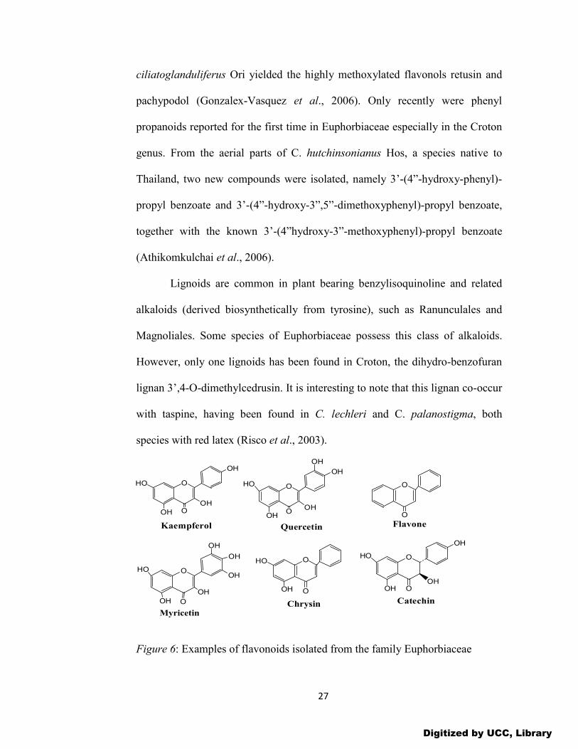
27
ciliatoglanduliferus Ori yielded the highly methoxylated flavonols retusin and
pachypodol (Gonzalex-Vasquez et al., 2006). Only recently were phenyl
propanoids reported for the first time in Euphorbiaceae especially in the Croton
genus. From the aerial parts of C. hutchinsonianus Hos, a species native to
Thailand, two new compounds were isolated, namely 3’-(4”-hydroxy-phenyl)-
propyl benzoate and 3’-(4”-hydroxy-3”,5”-dimethoxyphenyl)-propyl benzoate,
together with the known 3’-(4”hydroxy-3”-methoxyphenyl)-propyl benzoate
(Athikomkulchai et al., 2006).
Lignoids are common in plant bearing benzylisoquinoline and related
alkaloids (derived biosynthetically from tyrosine), such as Ranunculales and
Magnoliales. Some species of Euphorbiaceae possess this class of alkaloids.
However, only one lignoids has been found in Croton, the dihydro-benzofuran
lignan 3’,4-O-dimethylcedrusin. It is interesting to note that this lignan co-occur
with taspine, having been found in C. lechleri and C. palanostigma, both
species with red latex (Risco et al., 2003).
Figure 6: Examples of flavonoids isolated from the family Euphorbiaceae
O O
O
OH
OH
HO
OH
OHOH
HO
OH
OH
OH
OH OH
HO
O
OH
OH
OO
Kaempferol Quercetin
Myricetin
O
O
Flavone
O
O
O
OOH
HO
OH
HO
OH
OH
Chrysin Catechin
Digitized by UCC, Library

28
Tannins
“Tannin” is a general descriptive name for a group of polymeric
phenolic compounds capable of tanning leather or precipitating gelatin from
solution (astringency). The term tannin can therefore be defined as chemical
structure or group of chemical compounds that have tannin properties. They are
found in almost every plant part: bark, wood, leaves, fruits, and roots (Scalbert,
1991). They are divided into three groups, hydrolyzable, condensed tannins and
pseudotannins. Hydrolyzable tannins are based on gallic acid, usually as
multiple esters with D-glucose; while the more numerous condensed tannins
(proanthocyanidins) are derived from flavonoid monomers. Pseudotannins are
simpler phenolic compounds of low molecular weight co-occurring with
tannins. These compounds do not give the standard test for tannins
(Goldbeater’s skin test), e.g. gallic acid, catechins, chlorogenic acid, etc.
Tannins may be formed by condensations of flavan derivatives which have been
transported to woody tissues of plants or by polymerization of quinine units
(Geissman, 1963). They may also be formed by the combination of catechins
monomers (the so-called proanthocyanidins), or by ester bounded units of
glucose, gallic and/or elagic acid (hydrolysable tannins). So far, only
proanthocyanidins have been characterized in Croton species.
Proanthocyanidins have been reported as important active principles of species
containing red latex (Pieters et al., 1995).
Many human physiological activities such as stimulation of phagocytic
cells, host-mediated tumor activity and a wide range of anti-infective
Digitized by UCC, Library

29
activities have been assigned to tannins (Haslam, 1996). Tannins can be
toxic to filamentous fungi, yeasts and bacteria (Scalbert, 1991). Tannins
also possess antidiarrheal and anti-inflammatory activities (Njoronge and
Kibunga, 2007). Tannins and tannic acid reduce secretion by denaturing
proteins of the intestinal mucosa forming protein tannates which make the
mucosa more resistant to chemical alteration (Dangarembizi et al., 2013).
Compounds with anti-diarrheal properties also act by decreasing intestinal
motility, stimulating water absorption and reducing electrolyte secretion
(Njoronge and Bussman, 2006). Monomers such as (+)-catechin, (-)-
epicatechin, (+)-gallocatechin, (-)-epigallocatechin and dimeric
procyanidins B-1 and B-4 have been isolated (Salatino and Negri, 2007).
Dimers and trimers have also been isolated and characterized. The fruits of
Phyllantus emblica contain corilagen, gallic acid and elagic acid (Singh et
al., 2011).
Figure 7: Examples of tannins isolated from the family Euphorbiaceae.
O
O
O
O
OH
HO
HO OH
Ellagic acid
OH
O
OH
HO
HO
Gallic acid
O
OH
OH
OH
OH
HO
OH
Gallocatechin
CO
CHOMe
O
O
O
OH
HO
OH
OH
OHOH
HO
HO
Corilagin O
O
HO
HO OHOH
OH
O
O O
OO
HOH2C
Furosin
Digitized by UCC, Library

30
Coumarins Coumarins are phenolic compounds made up of fused benzene and α-
pyrone rings, i.e. they are 5,6-benzo-2-pyrone compounds (Bhat et al., 2007).
Coumarins are responsible for the characteristic odor of hay. More than 1350
coumarins have been isolated till 1997 (Bhat et al., 2007). They are well known
for their antithrombotic (Thastrup et al., 1985), anti-inflammatory and
vasodilatory (Namba et al., 1988) activities. Coumarins are known to be highly
toxic to rodents especially warfarin which is used as an oral anticoagulant and a
rodenticide (Keating and O’Kennedy, 1997) and may also have antiviral effects
(Berkada, 1978). Several other coumarins have antimicrobial and estrogenic
activities (Cowan, 1999). Coumarins have been used to prevent recurrences of
cold sores caused by HSV-1 in humans (Berkada, 1978) but are ineffective
against leprosy. Also, phytoalexins, which are hydroxylated derivatives of
coumarins, are produced in carrots in response to fungal infection and can be
presumed to have antifungal activity (Hoult and Paya, 1996).
Coumarins particularly of the furanocoumarin type abound in
Euphorbiaceae (Seigler, 1994). Scopoletin was obtained from the wood extract
of E. tirucalli and C. draco (Murillo et al., 2001). The fruits of Phyllantus
emblica yielded umbelliferone and seselin (Singh et al., 2011). Daphnehtin and
psolaren have been isolated from Daphnehtin tangutica and Euphorbia buxoides
respectively (Pan et al., 2010).
Digitized by UCC, Library

31
Figure 8: Examples of coumarins isolated from the family Euphorbiaceae
Cyanogenic Glycosides
Cyanogenic glycosides (CGs) or cyanoglycosides account for
approximately 90% of the plant toxins known as cyanogenes. The key
characteristic of these toxins is cyanogenesis, the formation of free hydrogen
cyanide, and is associated with cyanohydrins that have been stabilized by
glycosylation to form the cyanogenic glycosides (FSANZ, 2004). The CGs are
O-β-glycosidic derivatives of α-hydroxynitriles (Poulton, 1990). Depending on
their precursor amino acid, they may be aromatic, aliphatic or cyclopentenoid in
nature. Most CGs are cyanogenic monosaccharides, though cyanogenic
oligosaccharides also exist. Sulphated, malonylated and acylated derivatives of
CGs are also known (Poulton, 1990). The major edible plants in which CGs
occur are cassava, lima beans, sorghum, almonds, stone fruits and bamboo
shoots. In small quantities these glycosides do exhibit expectorant, sedative and
digestive properties. However, many of these edible plants are highly
cyanogenic and have caused numerous cases of acute cyanide poisoning of
animals including man. Cases of acute cyanide poisoning have been associated
O O
Coumarin
O O
O O O O
O O
O O
O O
HO
H3CO
HO
H3CO
H3CO
Umbelliferone
ScoparoneScopoletin
OHHO
Daphnethin
OPsoralen
O
Seselin
Digitized by UCC, Library

32
with misuse, particularly of preparations from apricot pits, bitter almonds and
cyanide rich apple seeds. In areas of the world where these cyanogenic plants
are the staple food, chronic cyanide poisoning and associated pathological
conditions exist (Poulton, 1989). Goitre and cretinisim due to iodine deficiency
can be exacerbated by chronic consumption of insufficiently processed cassava.
Neurologically, there has been report of Konzo or spastic paraparesis in children
and woman of child-bearing age in East Africa in times of food shortage and is
associated with a high and sustained intake of cassava in combination with a
low intake of protein (Davis, 1991). Also, tropical ataxic neuropathy (TAN),
which is attributed to cyanide exposure from the chronic consumption of food
derived from cassava, has been reported. CGs are widely distributed among 100
families of flowering plants. They are also found in some species of ferns,
fungi, bacteria and animals especially arthropods.
The family Euphorbiaceae is rich in cyanogenic glycosides, especially
the genera Euphorbia and Croton. Seven cyanopyridone derivatives and one
seco compound have been isolated from a methanolic extract of the
inflorescences and leaves extract of Acalypha indica (Salatino and Negri, 2007).
Digitized by UCC, Library

33
Figure 9: Examples of cyanogenic glycosides isolated from the family Euphorbiaceae
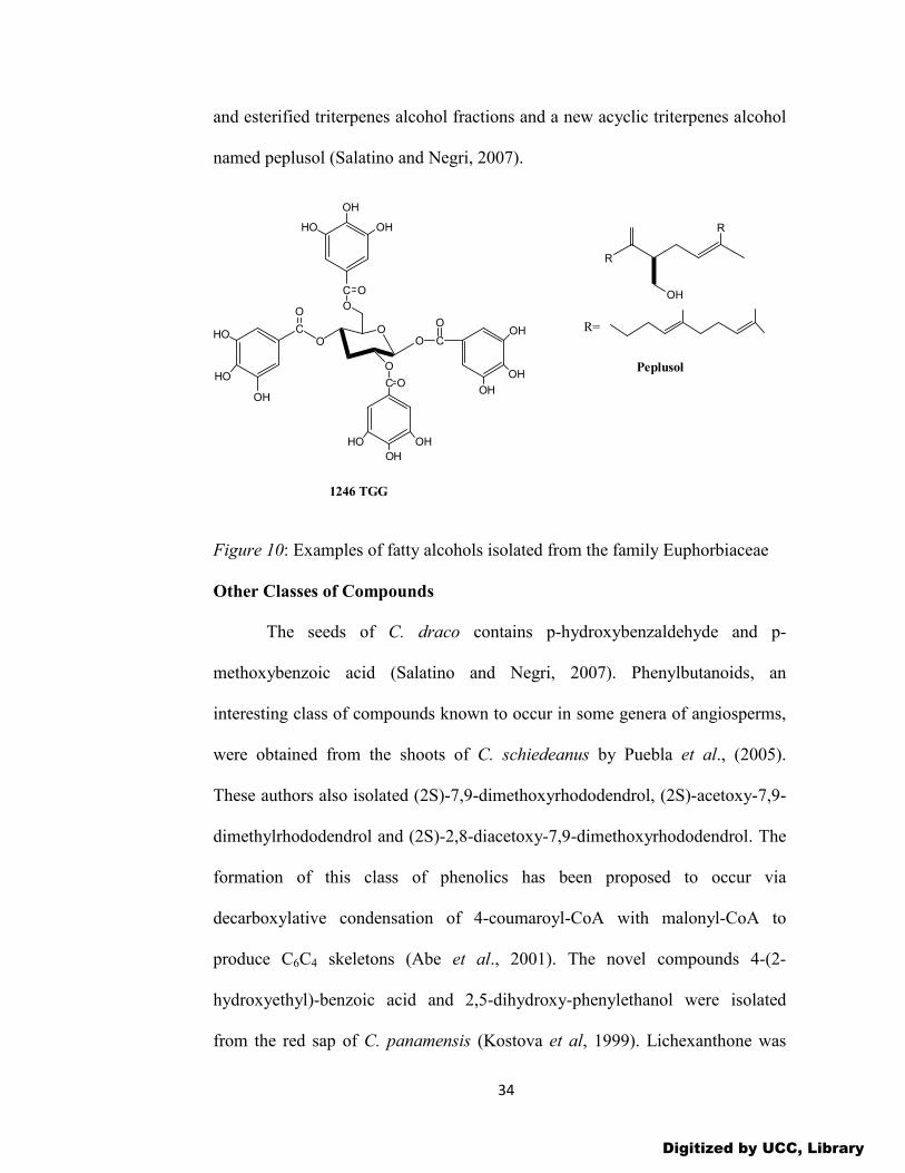
Fatty Alcohols
Different genera of Euphorbiaceae contain Long-chain fatty alcohols
(particularly n-octacosanol and n-hexacosanol) and hydrocarbons, especially the
genus Euphorbia yielded a considerable amount of hydrocarbons and alcohols
(Rizk 1987). The dried sap of C. draco yielded 3,4,5-trimethoxycinnamic
alcohol (Salatino and Negri, 2007). The polyalcohols IL-1-O-myo-inositol and
neo-inositol were isolated from C. celtidifolius (Salatino et al, 2007). From the
roots of the traditional Chinese medicinal plant; Phyllantus emblica L, 1,2,4,6-
tetra-O-galloyl-β-D-glucose (1246 TGG) has been isolated. The less polar
fractions of the latex of E. peplus were found to contain obtusifoliol,
cycloartenol, 24-methylenecycloartenol and 24-methylenelanosterol in the free
NO
O O
OH
OH
OH
OH
R3
OCH3
R1
R2
R4
HN
CN
O
O
O
OH
OH
HOOH
H3CONO
O
O
HO
OH
OH
OH
OCH3
CH3
R1= CH3; R2= OH; R3= H; R4= CN- AcalyphinR1= CH3; R2= H; R3= OH; R4= CN-EpiacalyphinR1= H; R2= OH; R3= H; R4= CN- NoracalyphinR1= H; R2= H; R3= OH; R4= CN-EpinoracalyphinR1= CH3; R2= OH; R3= H; R4= CONH2- Acalypin amide
ar-Acalyphidone CH3
Seco-Acalyphin
N
O
OO
CH3
O
HOOH
OH
OCH3
CONH2
H
Epiacalyphin amide ycloside
Digitized by UCC, Library
34
and esterified triterpenes alcohol fractions and a new acyclic triterpenes alcohol
named peplusol (Salatino and Negri, 2007).
Figure 10: Examples of fatty alcohols isolated from the family Euphorbiaceae
Other Classes of Compounds
The seeds of C. draco contains p-hydroxybenzaldehyde and p-
methoxybenzoic acid (Salatino and Negri, 2007). Phenylbutanoids, an
interesting class of compounds known to occur in some genera of angiosperms,
were obtained from the shoots of C. schiedeanus by Puebla et al., (2005).
These authors also isolated (2S)-7,9-dimethoxyrhododendrol, (2S)-acetoxy-7,9-
dimethylrhododendrol and (2S)-2,8-diacetoxy-7,9-dimethoxyrhododendrol. The
formation of this class of phenolics has been proposed to occur via
decarboxylative condensation of 4-coumaroyl-CoA with malonyl-CoA to
produce C6C4 skeletons (Abe et al., 2001). The novel compounds 4-(2-
hydroxyethyl)-benzoic acid and 2,5-dihydroxy-phenylethanol were isolated
from the red sap of C. panamensis (Kostova et al, 1999). Lichexanthone was
CO O C
OHOH
HO
HO
O
OO
OHO
C O
HO
OH
OH
O
C O
OH
OH
HO
OH
1246 TGG
R
R
OH
Peplusol
R=
Digitized by UCC, Library
35
obtained from the aerial parts of C. cuneatus (Suarez et al., 2004). From the
same plant, Hernandez and Delgado (1992) isolated a mixture of polyprenols
with castaprenol-II being the major compound. Simiarenol (a high molecular
mass triterpenoid) and esters of amyrine with fatty acids containing carbon
chains above 20 atoms have been isolated from the shoots of C. hemiargyreus.
Benzoyl-methylpolyols were isolated from C. betulaster and C. luetzelburgii
(Barbosa et al., 2004). Furanoarabinoid-gallactan, a polysaccharide, is the main
compound found in the gum exudates of C. urucurana (Milo et al., 2002). The
peptide derivatives aurentiamide acetate and N-benzoylphenylalanine were
isolated from shoots of C. hieronyini (Catalan et al., 2003). Cyclopeptides were
reported from the red latex of C. draco (Tsacheva et al., 2004).
OH
MeO
HO
MeO
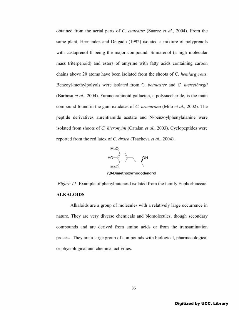
7,9-Dimethoxyrhododendrol
Figure 11: Example of phenylbutanoid isolated from the family Euphorbiaceae
ALKALOIDS
Alkaloids are a group of molecules with a relatively large occurrence in
nature. They are very diverse chemicals and biomolecules, though secondary
compounds and are derived from amino acids or from the transamination
process. They are a large group of compounds with biological, pharmacological
or physiological and chemical activities.
Digitized by UCC, Library

36
Properties of Alkaloids
In plants, alkaloids because of their basic nature occur largely as salts of
organic acids like acetic, oxalic, citric, malic, lactic, tannic, aconitic, quinic
acids, etc with well-defined crystalline structures. Some basic pyridine alkaloids
such as nicotine, myosmine, anabasine, etc occur in free state or as N-oxides. A
few alkaloids are present as glycosides of common sugars such as glucose,
rhamnose, galactose (Solanum and Veratrum alkaloids), or as esters of organic
acids (e.g. reserpine, hyoscyamine, cocaine). Some alkaloids are present as
quaternary salts (tubocurarine hydrochloride, muscarine chloride or as tertiary
amine oxides. Many neutral compounds where the nitrogen is involved in an
amide group are now included as alkaloids. Examples are colchicine and
piperine. In addition to the elements carbon, hydrogen and nitrogen, most
alkaloids contain oxygen. A few, such as coniine and nicotine, are oxygen-free
and are liquids. Although coloured alkaloids are very rare, berberine is yellow
and the salts of sanguinarine are copper-red. Knowledge of the solubility of
alkaloids and their salts is of considerable pharmaceutical importance. Not only
are alkaloidal substances administered in solution, but also the differences in
solubility between alkaloids and their salts provide methods for the isolation of
alkaloids from plants and their separation from the non-alkaloidal substances
also present. While the solubilities of different alkaloids and their salts show
considerable variation due to their varied structures, free bases are frequently
sparingly soluble in water but soluble in organic solvents; with salts the reverse
is often the case. However, there are exceptions to this generalization.
Digitized by UCC, Library
37
Structure and Classification of Alkaloids
Alkaloids show great variety in their botanical and biochemical origin,
in chemical structure and in pharmacological action. Consequently, many
different systems of classification are possible. They may be classified
according to their:
(1) biological and ecological activity
(2) chemical structures
(3) biosynthetic pathway
(4) common molecular precursor used to construct the molecule.
Biosynthetic Classification
This classification is based on the types of molecular precursors or
building block compounds used by living organisms from which the alkaloids
are produced biosynthetically. It is therefore convenient and also logical to
group all alkaloids having been derived from the same precursor but possessing
different taxonomic distribution and pharmacological activities together.
Examples:
(a) Indole alkaloids derived from tryptophan
(b) Piperidine alkaloids derived from lysine
(c) Pyrrolidine alkaloids derived from ornithine,
(d) Phenylethylamine alkaloids derived from tyrosine
(e) Imidazole alkaloids derived from histidine.
The major drawback of this method is that the relationship of alkaloids to each
other and to their precursors is not always apparent.
Digitized by UCC, Library

38
Chemical Classification
Chemical classification is the most widely accepted and common mode
of classification of alkaloids and it depends on the type of heterocyclic ring
structure present. There are two broad divisions:
1. Non-heterocyclic sometimes called ’protoalkaloids’ or biological amines.
2. Heterocyclic or typical alkaloids, divided into 14 groups according to their
ring structure. These groups are as follows:
(a) alkaloids derived from amination reactions such as acetate-derived
alkaloids, phenylalanine-derived Alkaloids, terpenoid alkaloids and steroidal
Alkaloid, (b) alkaloids derived from anthranilic acid e.g. quinazoline alkaloids,
quinoline alkaloids and acridine alkaloids
(c) alkaloids derived from histidine, e.g. imidazole alkaloids
(d) alkaloids derived from lysine, e.g. piperidine Alkaloids, quinolizidine
alkaloids and indolizidine Alkaloids
(e) alkaloids derived from nicotinic acid such as pyridine alkaloids,
(f) alkaloids derived from ornithine, e.g. pyrrolidine alkaloids, tropane alkaloids
and pyrrolizidine alkaloids
(g) alkaloids derived from tyrosine such as phenylethylamine alkaloids, simple
tetrahydro iso-quinoline alkaloids and modified benzyl tetrahydro iso-quinoline
alkaloids
(h) alkaloids derived from tryptophan which include; simple indole alkaloids,
simple β-carboline alkaloids, terpenoid indole alkaloids, quinoline Alkaloids,
pyrroloindole alkaloids and ergot Alkaloids.
Digitized by UCC, Library

39
Pharmacological Classification
Alkaloids exhibit a broad range of very specific pharmacological
characteristics which are used as a strong basis for the general classification of
the wide-spectrum of alkaloids derived from the kingdom of the living
organisms. These pharmacological properties include: analgesics, cardio-
vascular drugs, central nervous system stimulants and depressants, dilation of
pupil of eye, mydriatics, anticholinergics, sympathomimetics, antimalarials,
purgatives, etc. It must be emphasized that this classification is not quite
common and widely known. Examples:
(i) morphine as narcotic analgesic,
(ii) quinine as antimalarial,
(iii) strychnine as reflex excitability,
(iv) lobeline as respiratory stimulant,
(v) boldine as choleretics and laxatives,
(vi) aconitine as neuralgia,
(vii) pilocarpine as antiglaucoma agent and miotic,
(viii) ergonovine as oxytocic,
(ix) ephedrine as bronchodilator
(x) narceine as analgesic (narcotic) and antitussive, etc.
Taxonomic Classification
This classification deals with the source of compounds or alkaloids
based on the taxonomy or family of the organisms. These taxa are the genus,
subgenus, species, subspecies and variety. Thus, the taxonomic classification
Digitized by UCC, Library

40
deals with a large group of alkaloids based mainly on their respective
distribution in a variety of plant families or ‘natural order’. Invariably, they are
grouped together according to the name of the genus wherein they belong to,
such as: coca, cinchona, ephedra. Examples include the following:
(i) Cannabinaceous alkaloids e.g. Cannabis sativa Linn, (hemp, marijuana),
(ii) Rubiaceous alkaloids e.g. Cinchona sp. (quinine)
(iii) Solanaceous alkaloids: e.g., Atropa belladona L. (Deadly Nightshade).
Some phytochemists also classify alkaloids based on their chemotaxonomic
properties.
Types of Alkaloids
In general there are three main types of alkaloids: true alkaloids,
protoalkaloids and pseudoalkaloids (Aniszewski, 1994; Jakubke, 1994).
True alkaloids and protoalkaloids are derived from amino acids while
pseudoalkaloids are derived from the precursors or postcursors of amino acids
(Dewick, 2002; Hu et al., 2003).
True Alkaloids
These are alkaloids which are derived from amino acid and they share a
heterocyclic ring with nitrogen (Aniszewski, 2007). They are highly reactive
substances with biological activities even in low doses. They are basic, contain
one or more nitrogen atoms and have a marked physiological action on man or
other animals.
They generally have bitter taste and appear as white solid, except nicotine
which is a brown liquid. True alkaloids form water-soluble salts and most of
Digitized by UCC, Library
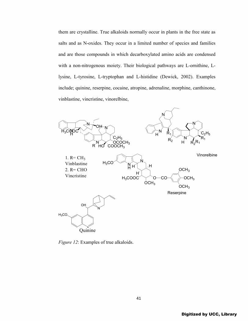
41
them are crystalline. True alkaloids normally occur in plants in the free state as
salts and as N-oxides. They occur in a limited number of species and families
and are those compounds in which decarboxylated amino acids are condensed
with a non-nitrogenous moiety. Their biological pathways are L-ornithine, L-
lysine, L-tyrosine, L-tryptophan and L-histidine (Dewick, 2002). Examples
include; quinine, reserpine, cocaine, atropine, adrenaline, morphine, canthinone,
vinblastine, vincristine, vinorelbine,
N
N
N
N
C2H5
R1R1H
R1
R2
Vinorelbine
NH
N
OOCH3
H3COOC
H3CO
H
HH
Reserpine
CO
OCH3
OCH3
OCH3
H
R2
NH
NOH
N
N
C2H5
OCOCH3COOCH3HOR
H3COOC
1. R= CH3
Vinblastine
2. R= CHO
Vincristine
N
N
H3CO
OH
Quinine
Figure 12: Examples of true alkaloids.
Digitized by UCC, Library

42
Protoalkaloids
Protoalkaloids are alkaloid-like amines in which the nitrogen atom
derived from an amino acid is not a part of the heterocyclic structure (Jakubke,
1994). They are not restricted to any particular class of alkaloids and are often
classified according to the amino acids from which they are derived. They lack
one or more of the properties of typical alkaloids.
They are normally derived from L-tyrosine and L-tryptophan. They have a
closed ring, being perfect but structurally they are simple alkaloids. Examples
are yohimbine, mescaline, hordenine, protoberberine, tryptamine and the new
alkaloids- stachydrine and 4-hydroxystachydrine (Aniszewski, 2007).
Figure 13: Examples of protoalkaloids
Pseudoalkaloids
In pseudoalkaloids, the basic carbon skeletons are not derived from
amino acids (Jakubke, 1994). They are actually connected with amino acid
pathways. Pseudoalkaloids are derived from the precursors or postcursors
(derivatives in the degradation process) of amino acids. They can also result
from the amination and transamination reactions of the different pathways
connected with precursors or postcursors of amino acids (Dewick, 2002). In the
case of steroidal or terpenoid alkaloid skeletons, the nitrogen atom is inserted
Digitized by UCC, Library
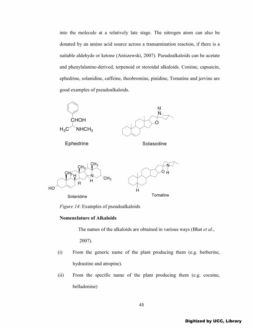
43
into the molecule at a relatively late stage. The nitrogen atom can also be
donated by an amino acid source across a transamination reaction, if there is a
suitable aldehyde or ketone (Aniszewski, 2007). Pseudoalkaloids can be acetate
and phenylalanine-derived, terpenoid or steroidal alkaloids. Coniine, capsaicin,
ephedrine, solanidine, caffeine, theobromine, pinidine, Tomatine and jervine are
good examples of pseudoalkaloids.
Figure 14: Examples of pseudoalkaloids
Nomenclature of Alkaloids
The names of the alkaloids are obtained in various ways (Bhat et al.,
2007).
(i) From the generic name of the plant producing them (e.g. berberine,
hydrastine and atropine).
(ii) From the specific name of the plant producing them (e.g. cocaine,
belladonine)
O
NH
Solasodine
NCH3
CH3
CH3
HO
CH3
HH
H
Solanidine
O
N
H
HTomatine
CHOH
NHCH3H3C
Ephedrine
Digitized by UCC, Library

44
(iii) From the physiological activity (emetine, morphine)
(iv) Occasionally from the person who discovered it (e.g. pelletierine)
Many at times a prefix or suffix is added to the name of the principal alkaloid
to designate another alkaloid from the same source (e.g. quinine, quinidine,
hydroquinine). By convention, the names of all alkaloids must end in ‘ine’.
Pharmacological Uses of Alkaloids
Almost all alkaloids possess curative properties. Alkaloids possess a
variety of pharmacological activities (Bhat et al., 2007). These activities include
the following: analgesic potentiator (cocaine), antiambic (emetine),
anticholinergics (atropine, hyoscyamine, scopolamine, and galanthamine),
antimalarial (quinine), antihypertensive (reserpine, protoveratrine), antitussive
(codeine, noscapine), cardiac depressant (quinidine), central nervous stimulant
(caffeine), diuretic (theophylline, theobromine), gout suppressant (colchicine),
local anesthetic (cocaine), narcotic analgesic (codeine, morphine), antitumor
(vinblastine, vincristine), antiglaucoma (pilocarpine), oxytocic (ergonovine),
skeletal muscle relaxant (methyl lycoconitine, tubocurarine), smooth muscle
relaxant (papaverine, theophylline), sympathomimetic (ephedrine), tranquilizer
(reserpine), etc.
Distribution of Alkaloids
Alkaloid-containing plants constitute an extremely varied group both
taxonomically and chemically, a basic nitrogen being the only unifying factor
for the various classes. For this reason, questions of the physiological role of
alkaloids in the plant, their importance in taxonomy, and biogenesis are often
Digitized by UCC, Library

45
more satisfactorily discussed at the level of a particular class of alkaloid. A
similar situation pertains to the therapeutic and pharmacological activities of
alkaloids. As most alkaloids are extremely toxic, plants containing them do not
feature strongly in herbal medicine but they have always been important in the
allopathic system where dosage is strictly controlled and in homoeopathy where
the dose-rate is so low as to be harmless. Some 150 years of alkaloid chemistry
had resulted by the mid-1940s in the isolation of about 800 alkaloids; the new
technology of the next 50 years increased this figure to the order of 10 000. In
practice, those substances present in plants and giving the standard qualitative
tests outlined below are termed alkaloids, and frequently in plant surveys this
evidence alone is used to classify a particular plant as ‘alkaloid-containing’.
Alkaloids are most abundant in higher plants. At least 25% of higher plants
contain these molecules which belong to more than 150 families. They are
widely distributed in higher plants particularly the dicotyledonous of the
families Euphorbiaceae, Apocynaceae, Asteraceae, Loganiaceae, Papaveraceae,
Rutaceae, Solanaceae, Erythroxylaceae, Boraginaceae, Fabaceae,
Menispermaceae, Berberidaceae, Ranunculaceae, Liliaceae, Rubiaceae,
Amaryllidaceae, Elaeagnaceae and Zygophyllaceae. Usually the occurrence of a
particular alkaloid is localized to the seeds, leaves, bark or roots of the plant and
each site may contain closely related alkaloids. Both the total alkaloid content
and the relative proportion of the component bases may vary considerably with
the stage of growth of the plant and its locality. Different species of the same
family of plants may contain the same or structurally related alkaloids. For
Digitized by UCC, Library
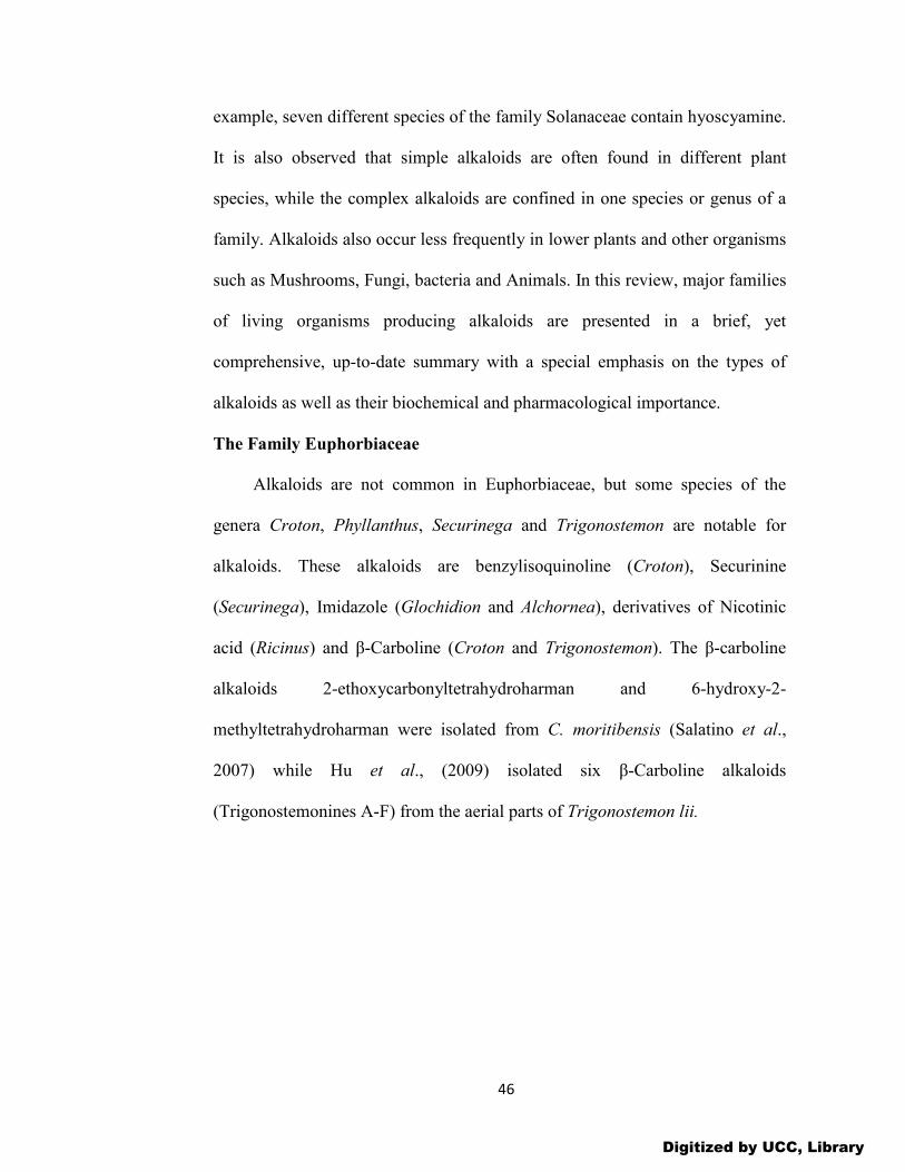
46
example, seven different species of the family Solanaceae contain hyoscyamine.
It is also observed that simple alkaloids are often found in different plant
species, while the complex alkaloids are confined in one species or genus of a
family. Alkaloids also occur less frequently in lower plants and other organisms
such as Mushrooms, Fungi, bacteria and Animals. In this review, major families
of living organisms producing alkaloids are presented in a brief, yet
comprehensive, up-to-date summary with a special emphasis on the types of
alkaloids as well as their biochemical and pharmacological importance.
The Family Euphorbiaceae
Alkaloids are not common in Euphorbiaceae, but some species of the
genera Croton, Phyllanthus, Securinega and Trigonostemon are notable for
alkaloids. These alkaloids are benzylisoquinoline (Croton), Securinine
(Securinega), Imidazole (Glochidion and Alchornea), derivatives of Nicotinic
acid (Ricinus) and β-Carboline (Croton and Trigonostemon). The β-carboline
alkaloids 2-ethoxycarbonyltetrahydroharman and 6-hydroxy-2-
methyltetrahydroharman were isolated from C. moritibensis (Salatino et al.,
2007) while Hu et al., (2009) isolated six β-Carboline alkaloids
(Trigonostemonines A-F) from the aerial parts of Trigonostemon lii.
Digitized by UCC, Library

47
NH
N
O O
H2NR2
R1
A, R1=H, R2=OCH3B,R1=OCH3 R2=H
NH
N
NR2
R1
C,R1=OCH3 R2=H
D,R1=H R2=OCH3
NH
N
NH3CO
E
NH
N
HN
H3CO
F
Figure 15: Trigonostemonines A-F alkaloids of Euphorbiaceae
The Family Apocynaceae
The Apocynaceae family (Lindl. juss) is distributed worldwide, especially
in tropical and sub-tropical areas (Aniszewski, 2007). It is large botanical taxa
containing at least 150 genera and 1700 species (Blundell, 1987). Alkaloids are
especially abundant in the following genera: Rauvolfia (devil’s-pepper),
Catharanthus G. Don (periwinkle), Tabernaemontana (milkwood), Strophantus
DC (Strophantus), Voacanga U (voacanga) and Alstoni R. Br. (alstonia)
(Endress et al., 1996). The species in these genera contain indole, terpenoid,
quinoline, pyrroloindole and ergot alkaloids. Rauwolfia serpentina contains
reserpine and rescinnamine, the quinine tree (R. capra) yielded quinine, and T.
iboga contains iboganine. Cinchona contains around 25 closely related
quinoline alkaloids, of which the most important are quinine, quinidine and
cinchonidine (Bhat et al., 2007). Cinchona and its alkaloids have been used in
the treatment of malaria for many years. The structure of quinine has provided
Digitized by UCC, Library

48
lead to important synthetic antimalarial drugs including chloroquine and
mefloquine.
Reserpine has been isolated from the roots of Rauvolfia canescens (Bhat et al.,
2005). This alkaloid gas been employed in clinical practice for the treatment of
hypertension and as a tranquilizer and also as a controller of other cardiac
disorders. It is known that 180 biologically active alkaloids have been isolated
from the genus Alstonia and this makes it one of the most important in terms of
alkaloid use (Macabeo et al., 2005; Keawpradub et al., 1999). The periwinkle
(e.g., Catharanthus roseus and Vinca spp.) have yielded potent anticancer
alkaloids-vinblastine, vincristine, vindesine, vinorelbine, vindoline, vindolinine,
leurosine, ajmalicine, etc. (refer to page 42 for examples drawn). All alkaloids
from Apocynaceae have a strong biological and medical effect and many of
them are used in cancer chemotherapy (Aniszewski, 2007).
The Family Asteraceae
This plant family is very large, containing over 900 genera and more than
20 000 species (Judd et al., 1999). They are distributed worldwide and the
species are found everywhere. The genus Senecio L., (ragwort) is especially rich
in pyrrolidine, tropane and pyrrolizidine alkaloid (senecivernine, sencionine,
retrorsine, retronecine, senecivernine, seneciphylline, spartioidine, jaconine,
adonifoline, sekirkine, jacoline, etc (Pelser et al., 2005). The genus Centaurea
L. is also rich in indole, terpenoid, quinoline and pyrroloindole alkaloids, for
example afzelin and apigenin. Other alkaloids have been isolated from Senecio
triangularis (9-0-acetyl-7-0-angelyl-retronecine, 7-0-angelyl-, 9-0-angelyl-, and
Digitized by UCC, Library

49
7-0-angelyl-9-0-sarracinylretronicine (Aniszewski, 2007). Cheng and Roeder
(1986) have isolated two pyrrolizidine alkaloids (senkirkine and doronine) from
Emilia sonchifolia.
O
N
O
O
O O
N
O
O
OO
N
O
O
O
OH
O
N
O
O
OO
N
O
O
O
H
Senecovernine
HOHO
Senecionine
HO
Retrorsine
HO
O O
HO
N
HOH
Senkirkine Dehydrosekirkine Retronecine
HO
Figure 16: Alkaloids of Asteraceae
The Family Loganiaceae
Thirty genera and more than 500 species belong to this family although
new systematic research has proposed that Loganiaceae should be divided into
several families (Struwe et al., 1994).
The Logan plant family contains plant species which are rich in pyrrolidine,
tropane and pyrrolizidine alkaloids. The genus Strychnos is especially rich in
alkaloids such as strychnine, brucine and curare (Frederich et al., 2000). This
genus contains 190 species and more than 300 different alkaloids have been
isolated. These alkaloids have important biological activities and strong medical
applications (Lansiaux et al., 2002, Frederich et al., 2004). They are also used in
exterminating rodents and for trapping fur-bearing animal (Bhat et al., 2007).
Digitized by UCC, Library

50
Sungucine and isosungucine have been isolated from S. icaja (Lansiaux et al.,
2002). These alkaloids interact with DNA, inhibit the synthesis of nucleic acid
and induce apoptosis in HL-60 leukaemia cells. The alkaloids strychnogucine A
and strychnogucine B have also been isolated from the stem bark of Strychnos
mellodora, a tree growing in the mountainous rain forests of Tanzania and
Zimbabwe (Aniszewski, 2007).
N
O
N
N
O
N
N
O
N
N
O
N
Sungucine
O
N
O
N
ON
O
N
O
H3CO
H3CO
StrychnineBrucine
N
O
N
N
O
N
O
OH
Strychnogucine A
Strychnogucine B
Figure 17: Alkaloids of Loganiaceae
The Family Papaveraceae
This poppy plant family is relatively large, comprising 26 genera and about
250 species (Judd et al., 1999). The opium poppy (Papaver somniferum L.,) is
a known source of opium from its latex. The family contains mainly
phenylethylamino- and iso-quinoline alkaloids such as morphine, codeine,
thebanine, papaverine, narcotine, Narceine, isoboldine and salsolinol. These
alkaloids are strong narcotics and have strong medicinal applications
Digitized by UCC, Library

51
(Aniszewski, 2007). Many new alkaloids have also been isolated from this
family. Alkaloids such as sanquinarine, cholidonine, hydrastine, berberine and
chelerythine have been isolated from Chelidonium majus (Vavreckova et al.,
1996). Twenty-three iso-quinoline alkaloids have isolated from Corydalis
bulleyana Diels (Hao and Qicheng, 1986). Examples of such alkaloids include
protopine, corydamine, allocryptopine, corycavanine, bulleyamine, spallidamine
etc. These alkaloids are well known for their biological activity and
spallidamine has been found to display fungitoxic activity (Ma, et al., 1999).
N
OCH3
OCH3
H3CO
H3CO
Papaverine
ON CH3
ON CH3
HO
HO
H3CO
HO
Morphine Codeine
ON
O
O
Thebaine
NO
OCH3
OCH3
O
OO
CH3
Hydrastine
N
OCH3
OCH3
O
O
Berberine
Figure 18: Alkaloids of Papaveraceae
The Family Rutaceae
The Citrus botanical family contains more than 150 genera and 900
species (Purseglove, 1979). Many species contain quinazoline, quinoline,
acridine, canthinone and imidazole alkaloids (Aniszewski, 2007). Species such
as Dictamus albus or Skimmia japonica contain quinazoline, quinoline and
acridine alkaloids such as dictamine, skimmianine and also acronycine
(Acronychia baueri), melicopticine (Melicope fareana) and rutacridone (Ruta
Digitized by UCC, Library

52
graveolens). Many alkaloids with potential estrogenic activity have been
reported in Haplophyllum A.juss (Nazrullaev, et al., 2001). These alkaloids
include acutine, toddaliopin A, acetylfolifidine, bucharidine, fagaronine,
dubinidine, dubinine, glycoperine, evoxine, ϒ-fagarine, folifidine, linarinic acid,
perfamine and skimmianine. Recently, fagaronine has been isolated from
Fagara zanthoxyloides and this alkaloid induces erythroleukemic cell
differentiation by gene activation (Dupont et al., 2005). Bioassay-guided
fractionation has led to the isolation of three indolopyridoquinazoline alkaloids,
1-hydroxy rutaecarpine, rutaecarpine and 1-methoxyrutaecarpine from the fruit
of Z. integrifolium (Sheen et al., 1996). Galipea officinalis (Hancock) is a shrub
growing in tropical America and used in folk medicine as an antispasmodic,
antipyretic, astringent and tonic. Nine quinoline alkaloids have been isolated
from this plant, of which galipine, cusparine, demethoxycusparine and
galipinine are active (Rakotoson et al., 1998). Moreover, a new carbazole
alkaloid, Clausine Z, has been isolated from the stems and leaves of Clausena
excavata Burm by Potterat et al (2005). This alkaloid exhibited inhibitory
activity against cyclin-dependent kinase 5 (CDK 5) and showed protective
effects on cerebellar granule neurons in vitro (Potterat et al., 2005). Cebrian-
Torrjon et al., (2015) have reported of the isolation of an antifungal alkaloid-
canthin-6-one from the leaves of Zanthoxylum chiloperone.
Digitized by UCC, Library

53
Figure 19: Alkaloids of Rutaceae
The Family Solanaceae
The Nightshade plant family contains 90 genera and more than 2000
species distributed in all continents (Purseglove, 1979). The family contains
pyrrolidine, tropane, steroidal and pyrrolizidine alkaloids especially the genus
Atropa L. (Nightshade) which contains hyoscyamine, hyoscine and
cuscohygrine (Aniszewski, 2007). The genera Jimsweed (Thornapple), Datura
L. (Pitura plants) and the species Atropa belladona L. (deadly nightshade)
contain tropane alkaloids (Ylinen et al, 1986). The genera Mandragora L. and
Scopolia L. also contain this type of alkaloids. Moreover, the Solanaceae
family also contains both nicotinic acid- derived and phenylalanine-derived
alkaloids such as anabasine, nornicotine, ricine, nicotine, arecoline, cathine,
cathionine, ephedrine, etc. The Nicotiana L. genus (the tobacco plant genus)
with about 45 species contains such alkaloids as nicotine and anabasine.
Capsicum L. (paprika plant) which has about 50 species and native to Central
Digitized by UCC, Library

54
and Southern America contains capsaicin as its main alkaloid. Steroidal
alkaloids, such as solanidine are well known in the potato genus (Solanum L.).
The unripe fruits of Solanum lycocarpum St. Hill, also contains steroidal
alkaloids as solamargine and solasodine (Schwarz et al., 2005). Solasodine is
shown to penetrate animal body, the placental and hematoencephalical barrier
and impact the foetuses. Tomatine, another steroidal alkaloid is common in the
genus Lycopersicon L. (tomato plant genus).
Figure 20: Alkaloids of Solanaceae
The Family Erythroxylaceae
The Erythroxylaceae family is distributed in the tropics and is endemic to
South America, especially in the regions of Peru and Bolivia, where
Erythroxylum coca (coca bush) has been known for over 5000 years
(Aniszewski, 2005). The family contains pyrrolidine, tropane and pyrrolizidine
alkaloids and three dominant species, E. coca, E. truxilense and E.
NCH3
CH3
CH3
HO
CH3
HH
H
Solanidine
O
NH
HTomatine
OCH2OH
H
O
NCH3
Hyoscyamine
N
NCH3
N
NH N
NH
Nicotine Myosmine Anabasine
Digitized by UCC, Library

55
novagranatense contain cocaine, ecgonine, cinnamylcocaine, α-truxilline,
truxilline, methylecgonine, tropine, hygrine, hygroline and cuscohygrine. These
alkaloids are used as drugs in main stream medicine and are also the object of
pathological and criminal activity (Aniszewski, 2007). New tropane alkaloids
have been isolated from the root bark of Erythroxylum vacciniifolium
(catuabines H-I, three hydroxyl derivatives and vaccinines A and B). These
tropane alkaloids are interesting for their ester moieties (Zanolari et al., 2005). It
must be emphasized that the genus Erythroxylum contains about 250 species
and apart from the cocaine-producing species, has not been examined
systematically by modern analytical methods (Aniszewski, 2007)
N COOCH3
OCOC6H5H
H
CH3N COOH
OHH
H
CH3
CocaineEcgonine
OHN CH3
Tropine
N CH3
O
CH3
Hygrine
Figure 21: Alkaloids of Erythroxylaceae
The Family Boraginaceae
The Boraginaceae plant family (Forget-me-not family) contains
pyrrolidine, tropane and pyrrolizidine alkaloids especially indicine-N-oxide in
Heliotropium indicum (heliotrope) and Cynoglosum creticum (Hound’s tongue)
species. New alkaloids, europine, ilamine and their N-oxides have been isolated
from another heliotrope species, Heliotropium crassifolium (Farsam et al.,
2000). These alkaloids have strong toxic effects. Bracca et al (2003) have
Digitized by UCC, Library
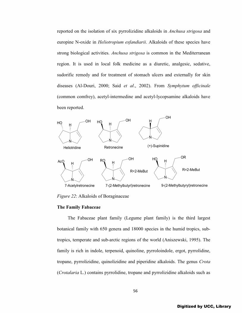
56
reported on the isolation of six pyrrolizidine alkaloids in Anchusa strigosa and
europine N-oxide in Heliotropium esfandiarii. Alkaloids of these species have
strong biological activities. Anchusa strigosa is common in the Mediterranean
region. It is used in local folk medicine as a diuretic, analgesic, sedative,
sudorific remedy and for treatment of stomach ulcers and externally for skin
diseases (Al-Douri, 2000; Said et al., 2002). From Symphytum officinale
(common comfrey), acetyl-intermedine and acetyl-lycopsamine alkaloids have
been reported.
N
HOH
OH
N
HOH
OH
N
AcOH
OH
N
ROH
OH
R=2-MeBut
7-(2-Methylbutyrl)retronecine
N
HOH
OR
R=2-MeBut
9-(2-Methylbutyryl)retronecine
Retronecine
7-Acetylretronecine
Heliotridine
N
HOH
(+)-Supinidine
Figure 22: Alkaloids of Boraginaceae
The Family Fabaceae
The Fabaceae plant family (Legume plant family) is the third largest
botanical family with 650 genera and 18000 species in the humid tropics, sub-
tropics, temperate and sub-arctic regions of the world (Aniszewski, 1995). The
family is rich in indole, terpenoid, quinoline, pyrroloindole, ergot, pyrrolidine,
tropane, pyrrolizidine, quinolizidine and piperidine alkaloids. The genus Crota
(Crotalaria L.) contains pyrrolidine, tropane and pyrrolizidine alkaloids such as
Digitized by UCC, Library

57
senecionine. Many species within this family are rich in quinolizidine alkaloids,
including; lupinine, lupanine, angustifoline, epilupinine, anagyrine, etc.
Przybylak et al., (2005) have detected 46 compounds from six Mexican species
and have been able to identify 24 of them as alkaloids from the lupanine group:
sparteine, ammodendrine, epiaphyllidine, epiaphylline, tetrahydrorhombifoline,
angustifoline, multiflorine, etc. The Calabar bean (Physostigma venenosum L.)
contains indole, quinoline and ergot alkaloids such as eserine, eseramine,
physovenine and geneserine. Lou et al., (2001) have isolated two new alkaloids;
2-methoxyl-3-(3-indolyl)-propionic and 2-hydroxyl-3-[3-(1-N-methyl)-indolyl]
propionic acid from peanut skins (Arachis hypogeae L.). These alkaloids had
not previously been isolated from natural sources. All alkaloids from this plant
family have both biological and ecological importance.
N
OHH
Lupine
NN
OH
HLupanine
NNH
OH
H
Angustifoline
N
N
O
Anagyrine
O
O
HN
NH
NH3C
H
H3C
CH3
Eserine
Figure 23: Alkaloids of Fabaceae
The Family Menispermaceae
This plant family is large containing about 70 genera and 450 species and
is found throughout the tropics (Thanikaimoni, 1986). The family contains
Digitized by UCC, Library

58
isoquinoline and phenylethylamino alkaloids. The genus Stephania is rich in
tetrandrine and stephanine and the Curare genus (Chondrodendron) contains
curare and tubocurarine. All these alkaloids are of important medicinal value.
More than 150 different alkaloids have been isolated from the Stephania genus
(Camacho, 2000). Some of these alkaloids include liriodenine, isocorydine,
atherospermidine, stephalagine and dehydrostephalagine. Liriodenine showed
strong cytotoxic activity while corydine and atherospermidine are able to
damage DNA (Goren et al., 2003). Chen et al., (2000) have isolated tetrandrine
from the root of a Chinese herb Stephania tetrandra S. Moore. This alkaloid
showed inhibition to both culture-activation and TGF-beta (1)-stimulated
activation of quiescent rat hepatic stellate cells (HSCs) in vitro (Chen et al.,
2005). The species Stephania cepharantha Hayata has yielded cepharanthine,
cepharanoline, isotetrandrine and berbamine (Nakaoji et al., 1997).
Cepharanthine is an active component of hair growth (Aniszewski, 2007).
Epinetrum villosum is a twining liana found in Congo and Angola and is used
traditionally for the treatment of fever, malaria and dysentery (Otshudi et al.,
2000). From this plant, cycleanine, cycleanine N-oxide, isochondodendrine,
cocsoline, troupin, and quinine have been isolated. These alkaloids exhibit both
antimicrobial and antiplasmodial activities (Otshudi et al., 2005). The genus
Cissampelos contains cissampareine, which has potential medicinal uses, but
it’s also psychoactive (Aniszewski, 2007).
Digitized by UCC, Library

59
N
O
N
O
H
O
O
O
O
H
Cepharantine
N
O
O
O
N
O
O
O
OCH3
AtherospermidineLiriodenine
N
H3CO
HOCH3
H3CO
H3COCorydine
N
H3CO
H3COCH3
HO
H3CO
N
H3CO
H3COH
HO
H3CO
Isocorydine Norisocorydine
H
Figure 24: Alkaloids of Menispermaceae
The Family Berberidaceae
The Berberry botanical family (Berberidaceae Torr., Gray Juss) contains
isoquinoline and phenylethylamino alkaloids especially berberine. Other
alkaloids such as glaucine, hydroxyacanthin and berbamine have also been
isolated from this family (Guo and Fu, 2005). Berbamine has shown to possess
anti-arrhythmia, anti-myocardial, ischemia and anti-thrombosis activities
(Aniszewski, 2007). Extracts of Nandina domestica T., are widely used in
Japanese folk medicine for the treatment of whooping cough, asthma, pharynx
tumours, uterine bleeding and diabetes (Aniszewski, 2007). Orallo (2004) has
isolated (+)-nantenine from the extract of this plant and this natural alkaloid was
first isolated by Takase and Ohasi in 1926.
Digitized by UCC, Library

60
NO
N
O
OOO
OH
H
Berbamine
N
OO
O
O
Glaucine
N
OCH3
H3CO
O
O
CH3
Nantenine
N
O
OOCH3
OCH3
Berberine
H
Figure 25: Alkaloids of Berberidaceae
The Family Ranunculaceae
The Buttercup plant family, which has 50 genera and about 2000 species,
is found in the temperate regions (Judd et al., 1999). It contains isoquinoline,
phenylethylamino and terpenoid alkaloids (Aniszewski, 2007). The genus
Hydrastis L., is rich in isoquinoline and phenylethylamino alkaloids such as
berberine and hydrastine and the genus Aconitum L., contains terpenoid
alkaloids as aconitine, aconine, benzoylaconitine and sinomontanine.
Fangcholine and fuzitine have been isolated from the genus Thalictrum
orientale, growing in Turkey (Erdemgil et al., 2000). Many other alkaloids have
been found in this genus. For instance, karacoline, karakanine, songorine,
nepelline, cammaconine and secokaraconitine have been isolated from
Aconitum karacolicum (Rupaics) from Kyrgyzstan (Sudtankhodzhaev et al.,
2002). A new alkaloid, arcutin with antibacterial and medicinal activities has
been isolated from Aconitum arcuatum Maxim (Sudtankhodzhaev et al., 200).
Digitized by UCC, Library

61
OCH3
OH
OH
N
C2H5
OCH3
H3CH2CO
OCOC6H5
OCH3
COCH3
OH
OH
N
C2H5
OCH3
H3CH2CO
OCOC6H5
OCH3
OH
OH
OH
N
C2H5
OCH3
H2CH3CO
Aconine Aconitine
OH
OH
Benzoylacotinine
Figure 26: Alkaloids of Ranunculaceae
The Family Liliaceae
This plant family contains more than 200 genera and about 3500 species
and it is distributed worldwide (Judd et al., 1999). It contains both isoquinoline
and steroidal alkaloids. The isoquinoline and phenylethylamino alkaloids are
found in the genera Kreysigia which yielded autumnaline, floramultine and
kreysigine, and Colchicum L., which produced colchicine. Steroidal alkaloids
are common in the Hellebore genus, example, jervine, cyclopamine,
cycloposine and protoveratrine A and B (Veratrum album) and O-acetyljervine
(Veratrum lobelianum) (Suladze and Vachnadez, 2002). Four new steroid
alkaloids- puqienine A and B, N-demethylpuqietinone and puqietinonoside have
been isolated from Fritillaria species. The bulb of this plant is used traditionally
in China as an antitussive and expectorant. All four alkaloids had established the
scientific basis for the ethnomedicinal uses of the plant (Aniszewski, 2007).
Digitized by UCC, Library

62
NCH3
H
H
CH3
HO
O
O
Jervin
O
HN CH3
H
H
H3C
H
CH3
HO
H3C
H
H
H
CyclopamineN
O
HO OCOOCH3
OH OCOCHCH3
OHOHCH3
CH3
OCH3
H
COOCH3 CH3
Protoveratrine
HO
CH3
H
Figure 27: Alkaloids of Liliaceae
The Family Rubiaceae
The Rubiaceae (Coffee family) contains more than 400 genera and over
6000 species (Judd et al., 1999). It is distributed in the tropics and the sub-
tropics (Purseglove, 1979). Species in this family are trees, bushes and liane
(Blundell, 1987). The family contains indole, pyrrolidinoindoline, quinoline and
benzoquinolizidine alkaloids (Aniszewski, 2007).
The coffee family is especially rich in purine alkaloids such as caffeine,
theophylline and theobromine. Other plant families like the tea (Theaceae), the
guarana (Sapinidaceae) and the cola (Sterculiaceae) contain the same or similar
purine alkaloids. Purine alkaloids (especially caffeine) have positive biological
and prophylactic effect in decreasing the risk of Parkinson’s disease
(unpublished). Tryptophan-derived alkaloids with important biological activities
also exist in the cola family (Hoelzel et al., 2005).
The genus Psychotria is one of the largest genera of flowering plants and the
largest within the Rubiaceae, with estimated 2000 species distributed worldwide
Digitized by UCC, Library

63
(Fynn, 2011). The indole alkaloids are the predominant groups of alkaloids
isolated from Psychotria species. For example, the leaves of Psychotria
forsteriana contains quadrigemine A and B, psychotridine and isopsychotridine
C with high cytotoxic activity on cultured rat hepatoma cells (HTC line) (Roth
et al., 1986). Also, Staerk et al., (2000) reported of the isolation of
corynantheidine derivatives and α-yohimbine from the bark of Corynanthe
pachyceras K. Schum. All these alkaloids demonstrated powerful
leishmanicidal, antiplasmodial and cytotoxic activity. Many indole alkaloids
such as emetine, calycosidine and cephaeline with potent pharmacological
activity occur in the Rubiaceae family.
N
N N
NH
CH3
CH3O
O
N
N N
NH3C
CH3
CH3O
O
N
N N
NH
CH3
HO
O
Theobromine Caffeine Theophylline
NH
N
OCH3
OCH3
H3CO
H3CO
H
Emetine
NH
N
OCH3
OCH3
HO
H3CO
H
Cephaeline
Calycosidine
H3CH2C H3CH2C
Figure 28: Alkaloids of Rubiaceae
Digitized by UCC, Library

64
The Family Amaryllidaceae
The amaryllidaceae botanical family is a large family consisting of 50
genera and 850 species and is distributed throughout the world (Judd et al.,
1999). The family is rich in isoquinoline and phenylethylamino alkaloids
(Aniszewski, 2007). The genus Lycorus L., (Spider lily genus) contains lycorine
and Galanthus L.(Snowdrop genus) is rich in galanthamine and galanthindole
(Unver et al., 1999, 2003) . Galanthine, haemanthine, lycorine and lycorenine
have been isolated from zephyranthes citrine Baker (Aniszewski, 2007). Herrera
et al., (2001) have also isolated oxomaritidine, maritidine and vittatine from the
same plant species. Alkaloids of Zephyranthes citrina especially
haemanthamine have inhibitory effects on the growth of HeLa cells and protein
synthesis, as well as being a cytotoxic against both MOLT 4 and various
human tumoural cell lines (Weniger et al., 1995). Maritidine exhibits
antineoplastic activity and galanthine has a high inhibitory capacity with
ascorbic acid biosynthesis in the potato (Evidente et al., 1983). Alkaloids
having antiviral, antitumoural, analgesic and insecticidal activities have been
isolated from Pancratium sickenbergi (Lewis, 2000; Abou-Donia et al., 2002).
These alkaloids are hippadine, pseudolycorine, tris-pheridine, norgalanthamine,
haemanthidine, vittatine, pancracine, 11-hydroxyvittatine, ent-6α-6β-
hydroxybuphasine, and (-)-8-demethylmaritidine. From the bulbs Leucojum
vernum, two new alkaloids, leucoverine and acetylleicoverine have been
isolated (Forgo and Hohmann 2005). Shihunine and dihydroshihunine exist in
Behria tenuiflora and these alkaloids have been shown to be inhibitors of
Digitized by UCC, Library

65
Na+/K+ ATPase in the rat kidney (Bastida et al., 1996). It must be emphasized
that all alkaloids from Amaryllidaceae display antiviral activity (Aniszewski,
2007).
Figure 29: Alkaloids of Amaryllidaceae
The Family Elaeagnaceae
The Oleaster botanical family is one of the smallest families comprising
3 genera and 50 species (Aniszewski, 2007). It is found mostly in the temperate
regions of the world. It contains indole (β-carboline) alkaloids especially
elaeagine which is predominantly found in the Russian olive Elaeagnus
angustifolia (Oleaster genus) (Aniszewski, 2007) together with harman,
harmine, harmol and harmalol.
Figure 30: Alkaloids of Elaeagnaceae
The Family Zygophyllaceae
The Zygophyllaceae (the Caltrop plant family) consists of nearly 30
genera and more than 230 species, grows in the tropic, subtropics and warm
regions of the world (Judd et al., 1999; Blundell, 1987). The family is very rich
N
HO
O
O
Lycorine
N
O
O
OCH3
Haemanthamine
HN
O
CH3
OH
H3CO
Galanthamine
OHH
OH
NH
NH
Elaeagine
Digitized by UCC, Library

66
in β-carboline alkaloids especially harman and harmine which is normally found
in the Pegan genus (Peganum harmala L.). The genus Nitraria (Nitraria sibirica
Pall) contains alkaloids derived from acetate, dihydroschoberine and nitrabirine
N-oxide (Tulyaganov et al., 2001). Komavine and acetylkomavine have been
isolated from Nitraria komarovii (Tulyaganov et al., 2001).
Figure 31: Alkaloids of Zygophyllaceae
Mushroom
Apart from the plant botanical family, alkaloids occur in many other
botanical families including the mushroom (Aniszewski, 2007). The mushroom
genera Psilocybe, Conocybe, Panaeolus and Stoparia are rich in the β-carboline
alkaloids serotonin, psilocin and psilocybin. These alkaloids are powerful
psychoactive and neurotransmitter compounds. These compounds also
demonstrated a broad spectrum of pharmacological properties including
NHO
H
Nitramine
NHN
O
Nitraramine
N
NH
Nitrarine
NH
NH
Komavine
NH
N
Acetylkomavine
NH
NH3CO
CH3
Harmaline
NH
NHO
CH3NH
N
CH3
HO
Harmol Harmolol
O
Digitized by UCC, Library
67
sedative, anxiolytic, hypnotic anticonvulsant as well as antimicrobial activities
(Cao et al., 2007)
Figure 32: Alkaloids of Mushroom
Moss
The moss (Lycopodiaceae family) contains indole and isoquinoline
alkaloids and the genus Lycopodium L., is a rich source of annotinine,
lycopodine and cernuine (Aniszewski, 2007). The genus Huperzia contains
huperzine J, K, L, A and its derivatives (Ayer and Trifonov, 1993). These
alkaloids have potential effects on Alzheimer’s disease (Aniszewski, 2007). Tan
et al (2002) have isolated phlegmariurine, 11α-hydroxy-phlegmariurine B, 7α-
hydroxyphlegmariurine B, fawcettimine and 7α11α-dihydroxyphlegmariurine
from H. serrata (Thumb.).
NH
NH2HONH
NHO
NH
N
Serotonin Psilocin
POH
OO
HO
Psilocybin
CH3
CH3
CH3
CH3
Digitized by UCC, Library

68
Figure 33: Alkaloids of Moss
Fungi and Bacteria
The fungus botanical family contains ergot alkaloids and the fungi
Aspergillus, Rhizopus, Penicillium and Claviceps produce parasitic ergoline and
ergotamine alkaloids (Aniszewski, 2007). The ergot alkaloids derived from
indole in the fungus Claviceps purpurea, are highly toxic and have been used in
the development of lysergic acid diethylamine, LSD, which is hallucinogenic
and, in small doses, is used in the treatment of schizophrenia (Li et al., 2005). A
new alkaloid, asterrelenin, together with terretonin, territem A and B have been
isolated from Aspergillus terreus (Li et al., 2005). Two new diastereomeric
quinoline alkaloids have been isolated from Penicillium janczewskii obtained
from a marine sample (He et al., 2005). These compounds showed a low to
moderate general toxicity (Aniszewski, 2007). From the new species
Penicillium rivulum Frisvad, communesins G and H have been isolated
(Dalsgaard et al., 2005). These alkaloids however, have negative antiviral,
antimicrobial and anticancer activities. A pentacyclic indolinole alkaloid,
Digitized by UCC, Library

69
citrinadin A, has been isolated from the cultured broth of the fungus Penicillium
citrinum and a marine red alga (Muqishima et al., 2005). The fungus
Aspergillus echinulatus produces toxic diketopiperazine alkaloid echinuline.
Variety of other fungi produces toxic alkaloids, whereas very few alkaloids
have been from bacterial cultures (Bhat et al., 2007). The bacteria Pseudomonas
spp. contains the alkaloids tabtoxin and a deep blue coloured pyocyanine which
have relatively powerful biological activity (Aniszewski, 2007).
Figure 34: Alkaloids of Fungi and bacteria
Animals
The kingdom animalia contains different classes of alkaloids, especially
in millipedes, salamanders, toads, frogs, fish and mammals. They occur
particularly in the genera saxidomus, Salamandra, Phyllobates, Dendrobates,
N
N
HN
H3C CH3
O
O CH3
H
Echinuline
N
N
H3C
O
Pyocyanine
H
Digitized by UCC, Library

70
Castor, Moschus, Solenopsis, Odontomaschus, Glomeris and Polyzonium. Many
alkaloids have been recently isolated from the sponges (Gallimore et al., 2005).
For instance, ptilomycalin A and its analogues have been isolated from
Ptilocaulis spiculifer, Hemimycale spp., Crambe spp, Monanchora arbuscula,
Monanchora ungiculata as well as from some starfishes such as Fromia monilis
and Celerina heffernani. From the Caribbean sponge Monanchora unguifera the
guanidine alkaloids- batzelladine J, ptilomycalin A, ptilocaulin and
isoptilocaulin have been recently isolated. Many of these alkaloids display
ichthyotoxicity, and antibacterial, antifungal and antiviral activities
(Aniszewski, 2007). Antiviral activity has been exhibited against Herpes
Simplex virus (HSV-1) and also in inhibiting the HIV virus and cytotoxicity
against murine leukaemia cell lines (L1210) and human colon carcinoma cells
(HCT-16). From two Thorectidae sponges-Thorectandra and Smenospongia, six
new brominated indole alkaloids have been isolated (Segraves and Crews,
2005). These alkaloids have a wide range of biological activities and are good
therapeutic agents (Aniszewski, 2007). The skin of amphibians contains
alkaloids especially indole alkaloids. Costa et al (2005) have isolated bufetenin
from Anura species. This alkaloid is a component of chemical defence system in
these species. Bufetenin acts as a potential hallucinogenic factor showing
similar activity to LSD upon interaction with the 5HT2 human receptor (Costa et
al., 2005). Toads belonging to the genus Melanophryniscus contain toxic
alkaloids in their skin (Mebs et al., 2005). And alkaloids of the pumiliotoxin
Digitized by UCC, Library
71
(PTX) group and indolizidines have been isolated from Melanophryniscus
montevidensis.
Defensive substances such as alarm and trail pheromones secreted by
certain arthropods have alkaloid-like structure, e.g. from the venom of the fire
ant Solenopsis invicta Forel, several 2,6-dialkylpiperidines have been isolated
(Bhat et al., 2007). In general, arthropod natural products are only produced in
trace amounts in specialized exocrine glands (Bhat et al., 2007)
The ovaries and liver of the puffer fish (swellfish, Japanese fugu, Spheroides
rubripes, S. vermicularis) contain tetradotoxin, one of the most toxic low
molecular weight poisons known (Bhat et al., 2007). This alkaloid has also been
isolated from goby fish Gobius criniger, the Californian newt Taricha torosa
and the skin of frog belonging to the genus Atelopus. The lady bird
(Coccinellidae) and other beetles also contain alkaloids such as adaline,
coccinelline, podamine, epilachnene, myrrhine, propeleine, propyleine and
stenusine. Conversely, some moths (e.g. Utethesia ornatrix) depend on
alkaloids for defence. Utethesia ornatrix sequesters pyrrolizidine alkaloids as a
larva from the food plants such as Crotalaria (Campo et al., 2001). Some
poisonous frogs (Mantella) digest alkaloids in their food. The strawberry poison
frog (Dendrobates pumilio) contains dendrobatid alkaloids that are considered
to be sequestered through the consumption of alkaloid-containing arthropods
distributed in the habitat (Takada et al., 2005). Some species of ants
(Anochetum grandidieri and Tetramorium electrum), containing pyrrolizidine
alkaloids, have been found in the stomachs of Mantella frogs (Clark et al.,
Digitized by UCC, Library

72
2005). It is now known that over 800 biologically active alkaloids have been
isolated from the amphibian skin (Daly et al., 2005). All these alkaloids seem to
be derived from dietary sources except samandarines and pseudophrynamines.
It has been found out that beetles are sources for batrachotoxins and
coccinelline-like tricyclics and ants and mites for pumiliotixins Also, ants are
sources for decahydroquinolines, izidines, pyrrolidines and piperidines (Daly et
al., 2005). Several brominated indole alkaloids such as deformylflustramine and
flustramine have been isolated from the North Sea Bryozoan (Flustra foliacea)
(Peters et al., 2004). Deformylflustramine A and B have been known to have
affinities in the lower micromolar range with the neuronal nicotinic
acetylcholine receptor (nAChR). It has been reported that erythrian alkaloids (β-
erythroidine and dihydro-β-erythroidines) with neuromuscular transition
blocking activity resembling the effects of curare are present in the milk of
goats (Capra) which grazed the leaves of Erythrinia poeppigiana (Soto-
Hernandez and Jackson, 1993). The spectrum of alkaloids in mammals ranges
from isoquinoline derivatives, via β-carbolines, through to thiazolidines, arising
from vitamin B6, chloral and glyoxylic acid (Bringmann et al., 1991). And that
the formation of endogenous alkaloids occurs naturally in man and mammals
(Bringmann et al., 1991). A few alkaloids have been isolated from mammals,
for example muscopyridine from the scent of gland of musk deer, Moschus
moschiferus. Similarly, bufetenin has also been isolated from human urine.
However, recent reports confirm the presence of numerous β-carboline
alkaloids-pinoline, norharman, harman, harmine, β-CCE, hydro-β-carbolines in
Digitized by UCC, Library
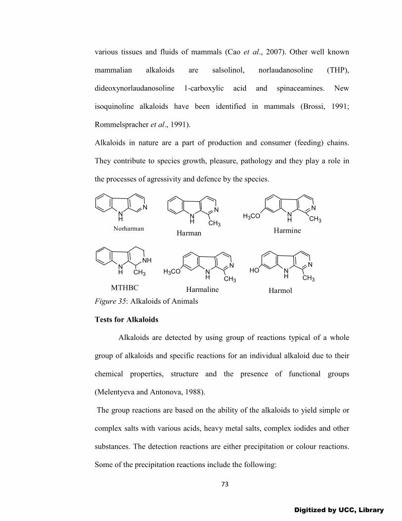
73
various tissues and fluids of mammals (Cao et al., 2007). Other well known
mammalian alkaloids are salsolinol, norlaudanosoline (THP),
dideoxynorlaudanosoline 1-carboxylic acid and spinaceamines. New
isoquinoline alkaloids have been identified in mammals (Brossi, 1991;
Rommelspracher et al., 1991).
Alkaloids in nature are a part of production and consumer (feeding) chains.
They contribute to species growth, pleasure, pathology and they play a role in
the processes of agressivity and defence by the species.
NH
N
Norharman
NH
N
Harman
NH
N
CH3H3CO
Harmine
NH
NH
CH3
MTHBC
CH3
NH
N
CH3
H3CO
Harmaline
NH
N
CH3
HO
Harmol Figure 35: Alkaloids of Animals
Tests for Alkaloids
Alkaloids are detected by using group of reactions typical of a whole
group of alkaloids and specific reactions for an individual alkaloid due to their
chemical properties, structure and the presence of functional groups
(Melentyeva and Antonova, 1988).
The group reactions are based on the ability of the alkaloids to yield simple or
complex salts with various acids, heavy metal salts, complex iodides and other
substances. The detection reactions are either precipitation or colour reactions.
Some of the precipitation reactions include the following:
Digitized by UCC, Library

74
1. A solution of iodine in potassium iodide (Bouchardat’s, Wagner’s or Lugol’s
reagent)
This reagent gives a brown precipitate with acidified aqueous solutions of
alkaloid salts. These reagents only differ in the concentration of the iodine and
potassium iodide.
2. A solution of mercury iodide in potassium iodide (Mayer’s reagent)
With most acidified or neutral alkaloid solutions, it yields white or slightly
yellowish precipitates. This reagent precipitates almost all the alkaloids except
caffeine and colchicine.
3. A solution of bismuth iodide in potassium iodide (Dragendorff’s reagent)
The reagent gives orange-red or reddish-brown amorphous and barely
crystalline precipitates with solutions of alkaloid sulphates and chlorides.
Dragendorff reagent was developed for detecting alkaloids, heterocyclic
nitrogen compounds and quaternary amines (Wagner et al., 1984). At least six
different Dragendorff reagents are known each containing potassium iodide.
4. Phosphomolybdic acid (Sonnenschein’s reagent)
This reagent is one of the most sensitive for alkaloids. It gives yellowish
amorphous precipitates that change to blue and green colour with time due to
the reduction of molybdic acid.
5. Phosphotungstic acid (Scheibler’s reagent)
This reagent forms amorphous white precipitates with almost all the alkaloids.
6. Tannic acid solution
Digitized by UCC, Library

75
This reagent contains a freshly prepared 10% aqueous tannic acid solution with
a 10% alcohol solution. The reagent forms white or yellow precipitates with
alkaloid salts in a neutral and weakly acidic medium.
7. 1% aqueous picric acid solution (Hager’s reagent)
The solution precipitates picrates with almost all the alkaloids except caffeine,
colchicine, coniine, morphine and theobromine. However, caffeine, a purine
derivative, does not precipitate like most alkaloids. It is usually detected by
mixing with a very small amount of potassium chlorate and a drop of
hydrochloric acid, evaporating to dryness and exposing the residue to ammonia
vapour. A purple colour is produced with caffeine and other purine derivatives
(Murexide test).
In addition to precipitation reactions, colour reactions can be used to test
for alkaloids. Colour reactions are based on the chemical reaction of water
removal, or on the oxidation of the alkaloids, or their condensation with
aldehydes. All these reactions proceed in the presence of concentrated sulphuric
acid absorbing water and are based on the features of the chemical structure of
the alkaloids and their functional groups. The most common reagents for these
coloured reactions are pure concentrated sulphuric acid, concentrated nitric acid,
and a mixture of these acids (Erdman’s reagent), a mixture of concentrated
sulphuric acid and molybdenum trioxide (Froehde’s reagent) and a mixture of
formaldehyde and concentrated sulphuric acid (Marchi’s reagent). For some
alkaloids, these reactions can be specific, while for others they can fail to be
characteristic. For example, a reaction with Marchi’s reagent is specific for
Digitized by UCC, Library

76
morphine, codeine and papaverine, while this reaction is not specific for other
alkaloids (Melentyeva and Antonova, 1988). Care must be taken in the
application of these alkaloidal tests, as the reagents also give precipitates with
proteins. So one can use acidic water-alkaline-extraction method to remove the
proteins and test for alkaloids.
Extraction and Isolation of Alkaloids
Extraction methods vary with the scale and purpose of the operation, and
with the raw material. Alkaloids are mostly alkaline and exist in organic salts
form as citrate, oxalate, tartrate, succinate, etc. Few exist in inorganic salt form,
such as berberine or morphine (as morphine sulphate) and in free form such as
amide alkaloids. Alkaloids in the free or salt form can be extracted with
inorganic acidic water in order to replace organic acids with inorganic acid salt
and increase its solubility. Both the free and salt alkaloids are soluble in alcohol
and so heated alcohol under reflux extraction or ultrasonic alcohol extraction
can be used. Most of the free alkaloids are lipophilic and can be extracted with
organic solvents such as chloroform, benzene, ether, etc. Most alkaloids
obtained by extraction are mixtures according to the class of alkaloids, basicity,
solubility differences and the functional groups present. The following methods
can be used in alkaloid extraction.
Acidic-water Extraction
This method is used to extract alkaloids which exist in the salt form
where the organic acid salt is replaced with inorganic acid salt, thereby
increasing the solubility of water. The method usually uses 0.1%-1% sulphuric
Digitized by UCC, Library

77
acid, hydrochloric acid, acetic acid or tartaric acid solution, by dipping,
maceration, percolation and sometimes refluxing (if the sample is less starchy)
extraction (Yubin et al., 2014). The method is relatively simple; however, there
is the wastage of solvents, difficulty in solvent recovery and has more water-
soluble impurities. The alkaloids can be purified using cationic exchange resin.
Aqueous-alcohol Extraction
Both free and salt alkaloids are soluble in alcohol and alcohol reflux,
cold maceration, percolation, etc can be used in extracting them. With this
method, different alkaline salts can be obtained and in addition water-soluble
impurities are less. However, more fat-soluble impurities are extracted. Total
alkaloids can be obtained by recovering the alcohol, adding dilute acidified
water, basifying and extracting with suitable lipophilic organic solvent.
Organic Solvent Extraction
Most free alkaloids are lipophilic and chloroform, benzene, ether and
methylene chloride can be used to extract them either by impregnating,
refluxing or continuous refluxing extraction. To make the alkaloids free and
also increase the solvent penetrating the plant tissue, a small amount of alkaline
wetting is recommended (Yubin et al., 2014).
With this method water-soluble impurities are less and the fat-soluble impurities
can be removed by acidic extraction. In addition, volatile alkaloids such as
ephedrine can
be obtained by steam distillation while sublimated alkaloids such as caffeine can
be extracted using sublimation method.
Digitized by UCC, Library

78
Beta-carboline Alkaloids
Beta-carboline alkaloids are a large group of natural and synthetic indole
alkaloids with different degrees of aromaticity. Some of these alkaloids are
widely distributed in nature, including various plants, foodstuffs, marine
creatures, insects, mammalians as well as human tissues and body fluids (Cao et
al., 2007). These compounds are of great interest due to their diverse biological
activities. Particularly, these compounds have been shown to intercalate into
DNA, to inhibit CDK, topisomerase, and monoamine oxidase, and to interact
with benzodiazepine receptors and 5-hydroxy serotonin receptors. These
chemicals also show a broad spectrum of pharmacological properties including
sedative, anxiolytic, hypnotic, anticonvulsant, antitumor, antiviral, antiparasitic
as well as antimicrobial activities (Cao et al., 2007). The prevalence of β-
carboline alkaloids is associated with the ease of forming the β-carboline core
from tryptamine in the intramolecular Mannich reaction. Simple (non-
isoprenoid) β-carboline derivatives include harmine, harmaline, harmane and a
slightly more complex structure of canthin-6-one.
Nomenclature of Beta-carboline Alkaloids
The beta-carboline alkaloids are a large group of natural and synthetic
indole alkaloids that possess a common tricyclic pyrido [3.4-b] indole ring
structure (Cao et al., 2007). These compounds are classified according to the
saturation of their nitrogen-containing six-membered ring. Unsaturated
members are named as fully aromatic β-carbolines (βCs), whereas the partially
or completely saturated ones are known as dihydro-β-carbolines (DHβCs) and
Digitized by UCC, Library
79
tetrahydro-β-carbolines (THβCs), respectively. These tricyclic compounds
usually contain several substituents both in the pyrido ring and/or the indole
ring. The photophysical properties of β-carboline alkaloids are strongly affected
by the presence of two different nitrogen atoms in the tricyclic system, the
pyridinic and the pyrrolic nitrogens. The pyridinic nitrogen is more basic than
the pyrrolic one, while its basicity increases upon excitation (Carmona et al.,
2000) and is affected by the substituents presence in the structure (Hidalgo et
al., 1990). Depending upon pH and solvent, β-carbolines can exist in four forms
(Varela et al., 2001): the cation, the neutral form, a zwitterion (or an alternative
quinine-type canonical form), and an anion.
Distribution of Beta-carboline Alkaloids
The plants that are rich in β-Carboline alkaloids include harmal
(Peganum harmala) which contains harmane, harmine and harmaline and the
Calabar bean (Physosstigma venenosum) containing physosstigma. Peganum
harmala is medicinal plant which is used traditionally as an emmenagogue and
abortifacient in the Middle East and North Africa (Mahmoudian et al., 2002).
The extracts of Peganum harmala have been traditionally used for hundreds of
years to treat the alimentary tract cancers and malaria in Northwest China (Chen
et al., 2005).
The Indian tribes in the south-western Amazon basin use plants containing β-
Carboline alkaloids as hallucinogenic drinks “ayahuasca” or snuffs. From the
past decades, numerous simple and complex β-carboline alkaloids containing
saturated or unsaturated tricyclic ring systems have been isolated from various
Digitized by UCC, Library

80
plants as the major bioactive constituents. Reports up to 2003 on the isolation
and characterization of simple β-carboline alkaloids including harman and
norharman have been documented (Pfau and Skog, 2004). Increasing evidence
shows that β-carboline alkaloids and related derivatives widely occur in nature,
especially in various tissues and body fluids of humans. And human beings are
sufficiently exposed to various β-carboline alkaloids, which are both present in
plants used for the preparation of hallucinogenic drinks and medicinal drugs,
and in tobacco smoke and well-cooked food (Cao et al., 2007). Additionally, it
has been found that humans can endogenously form various β-carboline
alkaloids, such as norharman and harman.
There have been many reports of the presence of simple and complex β-
carboline alkaloids in extracts from the leaves, barks and roots of a variety of
plants.
Additionally, numerous simple or complex β-carboline alkaloids have
been isolated and characterized from various marine invertebrates including
hydroids Aglao-phenia, bryozoans -Cribricellina,Caten-icella (Prinsep et al.,
1991; Harwood et al., 2003), soft corals Lignopsis, tunicates- Eudistoma,
Didemnum, Lissoclinum, Ritterella, Pseudodis-toma (Schuup et al., 2003) and
various sponges. Marine ascidians belonging to the genus Eudistoma (family
Polycitoridae) are another rich source of biologically active β-carboline
derivatives. Examples of such β-carboline alkaloids include eudistomins A-T
(Rinehart et al., 1984; Kobayashi et al., 1984), eudistomidins A-F (Kobayashi et
al., 1986, 1990), eudis-talbins A and B (Buckholtz et al., 1980), eudistomin U
Digitized by UCC, Library

81
and isoeudistomin U (Badre, et al., 1994), eudistomin V (Davis, et al., 1998)
and two new trypargine derivatives (Cao, et al., 2007).
It has been established that the simple β-carboline alkaloids, such as tetrahydro-
β-carboline-3-carboxylic acid and 1-methyl-tetrahydro-β-carboline-3-carboxylic
acid, are easily formed from tryptophan or tryptamine and formaldehyde or
pyruvate or acetate precursors by Pictet-Splengler reaction in foods and
berverages. Quite recently, it had been proven that various tetrahydro-β-
carboline and β-carboline alkaloids in variable but appreciable levels are present
in foods, alcoholic and non-alcoholic beverages, and fruit and fruit-derived
products.
The presence of β-carboline and its analogues in many ingested foodstuffs
strongly proved that diet is an important exogenous source of these compounds
in mammals and humans. The ingestion of these compounds could be partially
responsible for their further endogenous presence in various mammals' tissues,
organs and physiological fluids besides certain endogenous formation by
putative biosynthesis pathway (Myers, 1989; Herraiz, et al., 1993).
Since the isolation and characterization of endogenous pinoline (6-methoxy-
tetrahydro-β-carboline) from an extract of pineal gland tissue by Farrel and
Mclsaac, many researchers have focused on the detection and identification of
β-carboline alkaloids in mammals (Cao et al., 2007). Present reports confirm the
presence of numerous β-carboline alkaloids - norharman, harman, harmine, β-
CCE, harmaline, harmalan and several different tetra-hydro-β-carboline in
various tissues and fluids of a variety of mammals.
Digitized by UCC, Library

82
Biosynthesis of Beta-carboline Alkaloids
In the formation of simple β-carboline alkaloids, such as tetrahydro-β-
carboline-3-carboxylic acid, 1-methyl-1-tetrahydro-β-carboline-3-carboxylic
acid, harmine and harmaline, pyruvic acid acts as the keto acid precursor in the
Pictet-Splengler reaction in foods and beverages involving the use of tryptophan
or tryptamine. It is a cyclisation reaction involving indoleamines and
acetaldehyde to give simple tetrahydro-β-carboline alkaloids. Oxidation of
these simple β-carboline alkaloids gives the β-carbolines.
NH
NH2
R2R1
CH1
R1
O
NH
N
R2R1
CH
R1
NH
N
R2R1
CH
R1
NH
R1 NH
R2
R1 NH
R1 N
R2
R1
H
O R1= H, CH3
R2 = H, COOH, COOC2H5
R2 = H, OH
Figure 36: Biosynthesis of simple beta-carboline alkaloids
Synthesis of Beta-carboline Alkaloids
N-alkylated tryptamines have complex psychoactive properties. Routes
for their synthesis from the Internet websites involve the thermolytic
decarboxylation of tryptophan to tryptamine as a precursor to these compounds.
High boiling solvents and ketone catalysts are employed to facilitate the
decarboxylation process. However, there may be the formation of tetrahydro-β-
carboline (THBC) derivatives which may result from reaction with both the
solvent and the ketone catalysts (Brandt et al., 2006). This underlines the
problems associated with illicitly manufactured drugs and precursors that may
Digitized by UCC, Library

83
contain significant levels of impurities of which nothing is known of their
toxicities. The possible interaction of the contaminants and the principal product
in the human body may affect the efficacy of the drug and may put the user at
mortal risk.
Tryptophan (1) (Trp) and its analogues are readily available and are used as
starting materials for the synthesis of the corresponding tryptamine (2)
precursor via thermal decarboxylation. The chemically based conversion of Trp
is by far the simplest way to the synthesis of tryptamine and is done by
refluxing in a high boiling solvent with some modifications in achieving
success. For example, Hashimoto et al., (1986) used cyclohexanol as solvent
and observed an increased reaction times and a higher yield of amine product
with 2-cyclohexen-1-one as impurity. Other researchers used diphenylmethane
and diphenyl ether. Alternatively, there is also a two-step catalytic
decarboxylation by reacting tryptophan with copper acetate or zinc acetate with
the formation of metal chelate compounds that are then decarboxylated to
produce tryptamine hydrochloride, with indole as a by-product.
Other used L-tryptophan in refluxing tetralin with a catalytic amount of various
carbonyl compounds. This method has been modified where Trp was
decarboxylated in cyclohexanol: one method used tetralin that contains its
peroxide, another used tetralone followed by tetralin. A quantitative
decarboxylation of Trp in acetophenone at 1300C, using organic peroxides as
catalysts has also been reported (Brandt et al., 2006). A study of various
hydroxy- and methoxy-aromatic ketones as the decarboxylation media
Digitized by UCC, Library

84
concluded that of tryptophan and other α-amino acids proceed via the formation
of stable Schiff base intermediates-imines (Brandt et al., 2006). Some of these
intermediates after acidic or basic hydrolysis undergo transamination to a
degree depending on the ketone used with yields of 60-100 % tryptamine. An
interesting approach uses carvone (5-isoprenyl-2-mehtyl-cyclohex-2-enone) in
spearmint (Mentha spicata) oil as the ketone catalyst and either xylene or white
spirit as the refluxing solvent. It has been suggested that dill (Anethum
graveolens), caraway (Carum carvi) which contains carvone or pennyroyal
(Mentha pulegium) which contains D-pulegone, (5R)-methyl-2-isopropylidene-
cyclohexanone) essential oils could also employed as catalysts. Oil of turpentine
(the steam-volatile oil from rosin, exudates of pine trees) can also be used as
solvent. What is of synthetic interest is the range of side products that may be
present as trace constituents in the final products which may act as indicators to
the synthetic route. This problem has been rectified (Brandt et al., 2006) by the
analytical characterization of the synthetic route to tryptamine via
decarboxylation of Trp in the presence of ketone catalysts, with an emphasis on
the identification of possible by-products. It is a two-stage synthesis from
tryptophan to tryptamine and its subsequent methylation to N,N-
dimethyltryptamine using methyl iodide and benzyltriethylammonium
chloride/NaOH phase transfer catalyst (the so called Breadth of Hope
Synthesis).
According to this method, decarboxylation of tryptophan was achieved
by the suspension of tryptophan in a high boiling-point solvent under a nitrogen
Digitized by UCC, Library
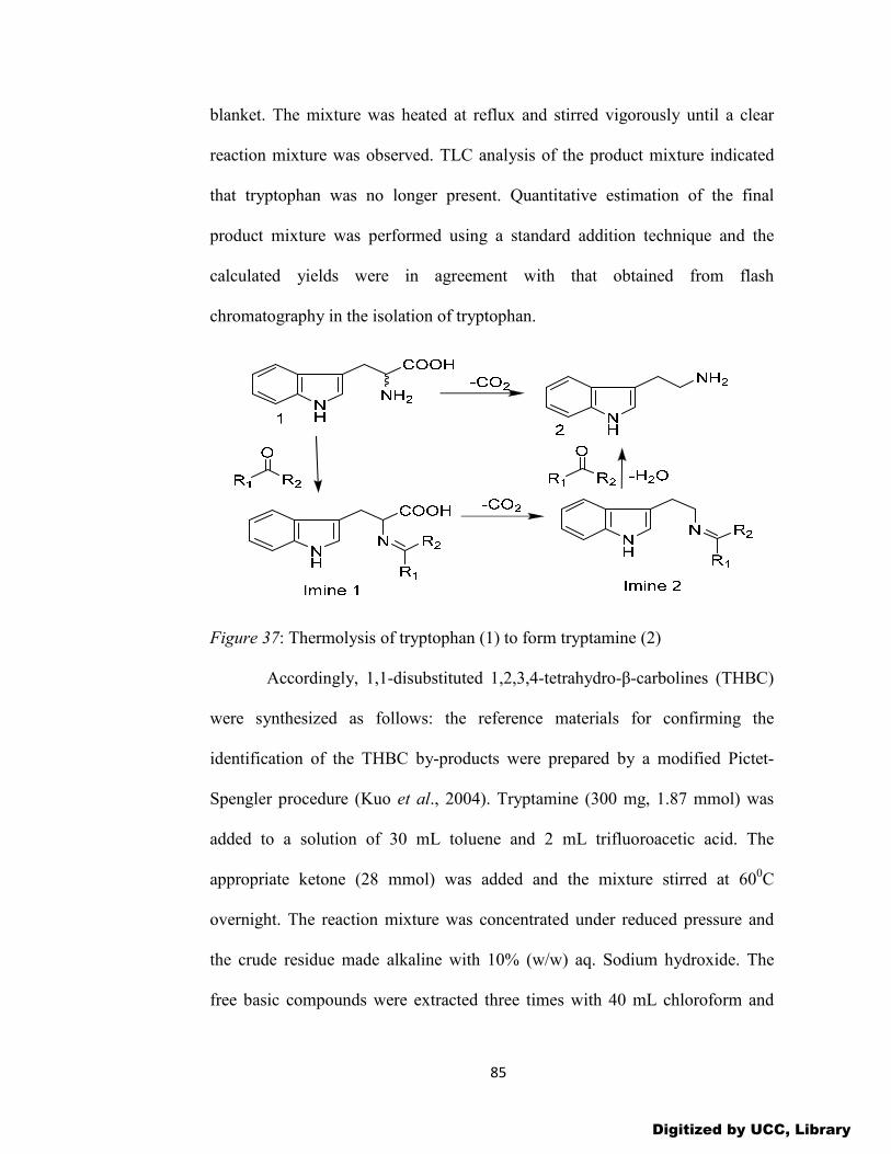
85
blanket. The mixture was heated at reflux and stirred vigorously until a clear
reaction mixture was observed. TLC analysis of the product mixture indicated
that tryptophan was no longer present. Quantitative estimation of the final
product mixture was performed using a standard addition technique and the
calculated yields were in agreement with that obtained from flash
chromatography in the isolation of tryptophan.
Figure 37: Thermolysis of tryptophan (1) to form tryptamine (2)
Accordingly, 1,1-disubstituted 1,2,3,4-tetrahydro-β-carbolines (THBC)
were synthesized as follows: the reference materials for confirming the
identification of the THBC by-products were prepared by a modified Pictet-
Spengler procedure (Kuo et al., 2004). Tryptamine (300 mg, 1.87 mmol) was
added to a solution of 30 mL toluene and 2 mL trifluoroacetic acid. The
appropriate ketone (28 mmol) was added and the mixture stirred at 600C
overnight. The reaction mixture was concentrated under reduced pressure and
the crude residue made alkaline with 10% (w/w) aq. Sodium hydroxide. The
free basic compounds were extracted three times with 40 mL chloroform and
Digitized by UCC, Library

86
washed twice with water. The chloroform layer was evaporated under reduced
pressure and subjected to flash chromatography using chloroform-methanol-
ammonia (0.88 s.g.) 9:1:0.1 as eluent. The corresponding THBCs were isolated
as oils and dried under vacuum over P2O5 where some of the products
solidified. THBC derivatives 6 and 7 were synthesized simultaneously using
pulegone as the ketone catalyst with heating at 600C for 3 days.
Figure 38: By-products of the thermolysis of tryptophan to form tryptamine
The 1,1-disubstituted-tetrahydro-β-carbolines 3-8 were identified as the
major by-products during the decarboxylation particularly when cyclohexanol
was used as the solvent. N-Benzyllidene-tryptamine was formed during
decarboxylation in diphenylmethane, possibly in the presence of benzaldehyde
contamination of the solvent.
Pharmacological Uses of Beta-carboline Alkaloids
Many researchers have focused on the effects of β-Carboline alkaloids
on the central nervous system (CNS), such as their affinity with benzodiazepine
Digitized by UCC, Library

87
receptors (BZRs), 5-HT2A and 5-HT2C (Cao et al., 2007). However, recent
attention has been shifted to their potent antitumor, antiviral, antimicrobial and
antiparasitic activities. The individual β-carboline alkaloids have been shown to
bind to different targets leading to various pharmacological activities. Both
harmine and harmaline have been shown to be hallucinogenic in humans.
Harmine has been shown to be inactive after oral (up to 960 mg) and
subcutaneous (up to 70 mg) administration, but induced some subjective effects
at 35-45 mg (Scoltin et al., 1970) and hallucinogenic effects at 150-200 mg via
intravenous administration (Naranjo et al., 1967).
Also, harmaline produced subjective effects in humans at a dose which is half of
what is required for harmine and its hallucinogenic effect was above 1 mg/Kg.
It has been observed that these hallucinogens produce their psychoactive
effects, at least in part, via interaction with 5-HT2 serotonin receptors in the
brain. It has been debated as to whether β-carboline alkaloids elicit
hallucinogenic actions in a manner consistent with classical hallucinogens
because many previous investigations demonstrated the modest interaction of β-
carboline alkaloids with 5-HT receptors. It is possible that the 6-methoxyl
moiety contributes to the hallucinogenic effects of these compounds. What is
more, the higher saturation in the tricyclic rings makes higher hallucinogenic
effects.
It is worth noting that harman and related β-carboline alkaloids play a
role in the process of substance abuse and dependence. The benzodiazepine
receptors of the mammalian central nervous system are able to mediate the
Digitized by UCC, Library

88
anxiolytic, anticonvulsant, sedative/hypotic action and myorelaxant of diazepam
(Cao, et al., 2007). During the past two decades, a wide variety of non-benzo-
diazepine molecules have been found to bind with high affinity to the
benzodiazepine receptors especially β-carboline alkaloids. Many of these com-
pounds have now been found to be benzodiazepine receptor inverse agonist or
antagonist (Cao, et al., 2007). For instance, 3-(ethoxy-carbonyl)-β-carboline (β-
CCE) and 3-(methoxycarbonyl)-β-carboline (β-CCM) were inverse agonists in
many animal behaviour models. The alkaloid also improved performance in
various learning and memory tests in animals when given prior to training (Cao
et al., 2007). The same alkaloid is able to exert stress-like effects including the
inhibition of locomotor exploration in post-weanling rats.
In contrast, pinoline showed no affinity for the benzodiazepine receptors
and had no convulsive activity. Rather, it demonstrated an anticonvulsive,
anxiogenic and antidepressant effects in some animal models. Hence, the
mechanism of action of pinoline is attributable to its neuropharmacological
effect and not its interaction with benzodiazepine receptors.
β-carboline alkaloids have also demonstrated promising antitumor activities
during the last decades. Ishida et al., (1999) reported that harmine and β-
carboline analogues exhibited significant activities against several human tumor
cell lines including three drug-resistant KB sublines with various resistance
mechanisms, and a-(4-nitrobenzylidine)-harmine had a broad cytotoxicity
spectrum against 1A9, KB, SaOS-2.A549, SK-MEL-2, U-87-MG and MCF-7
cells with ED50 values ranging from 0.3 to 1.2μg/mL. Structure activity
Digitized by UCC, Library

89
relationship analysis suggest that (1) introducing alkoxy substituents at C-7
leads to enhanced cytotoxic activities, (2) the length of C-7 alkoxy chain affects
both cytotoxicity and cell line specificity, (3) N9-alkylated β-carboline
derivatives exhibit strong cytotoxic effect, (4) C-6 brominated β-carboline
derivatives show selective cytotoxic activities, (5) N2-alkylated β-carboline
derivatives display specific cytotoxic activities and that (6) the 3,4-dihydro-p-
carboline derivatives are inactive. It has been reported that 3-substituted β-
carboline derivatives showed cytotoxic activities against human tumor cell lines
including HL-60, KB, Hela and BGC (Cao et al., 2007). Bis-3,4-dihydro-β-
carbolines and bis-β-carbolines have been synthesized and have been found to
be cytotoxic to L-1210 cells with micro-molar IC50, (Cao et al, 2007).
Numerous β-carboline derivatives with substituents at different positions have
been synthesized and evaluated for their antitumor activities in vitro and in vivo
(Cao et al., 2004, 2005). Most of the synthesized compounds showed
significant cytotoxic activities in vitro against a panel of human tumor cell lines
including non-small cell lung carcinoma (PLA-801), liver carcinoma (HepG2
and Bel-7402), gastric carcinoma (BGC-823), cervical carcinoma (HeLa) and
colon carcinoma (Lovo). Structure activity relationship analysis indicates that
(1) the β-carboline structure is an important basis for the design and synthesis of
new antitumor drugs, (2) appropriate substituents at position-1. 3 and 9 of β-
carboline ring might play a crucial role in determining their enhanced antitumor
activities, (3) the antitumor potencies of β-carboline derivatives are enhanced by
the introduction of benzyl substituent into the position-2, (4) the acute toxicity
Digitized by UCC, Library

90
of β-carboline derivatives reduced dramatically by the introduction of an
appropriate substituent into the position-3 and 9 and (5) the β-carboline
derivatives have the potential to be used as antitumor drug leads.
Moreover, β-carboline amino acid ester conjugates also exhibit potent cytotoxic
activities against human tumor cell lines including cervical carcinoma (Hela),
human breast cancer (MCF-7) and liver carcinoma (HepG2). The Lys/Arg
conjugates have the highest activities against human cervical carcinoma cells.
Many marine species contain β-carboline alkaloids with potent
antitumor properties. For instance, eudistomin exhibited potent cytotoxic
activities in vitro against murine P-388 cells with IC50 value of 0.01μg/mL and
the antitumor assay in vivo gave a T/C of 137% at 100 mg/kg, and a further
antitumor activity in vivo against L1210, A549 and HCT-8 cell lines. It has been
reported that 6-hydroxymanz-amine A and 3,4-dihydromanzamine A were cyto-
toxic against L1210 (IC50 1.5 and 4.8 μg/mL. respectively) and KB cells (IC50
2.5 and 0.61μg/mL, respectively in vitro. Accordingly, manzamine A, 8-
hydroxymanzamine A and 8-methoxymanzamine A showed significant
cytotoxicities against KB (IC50 0.05, 0.30 and 0.33 μg/mL respectively and
Lovo (IC50 0.15 0.26 and 0.1 μg/mL. respectively) cell lines. However, only
manzamine A exhibited cytotoxicity in the P-388 assay with 1C50 0.07 μg/mL
(Ichiba et al., 1994). A new manzamine dimer,-neo-Kauluamine exhibited
cytotoxicity with an IC50 1.0 μg/mL against human lung and colon carcinoma
cells (El Sayed et al., 2001), while Kauluamine was inactive in anticancer as-
says (Ohtani et al., 1995). Simple β-carboline alkaloids isolated from marine
Digitized by UCC, Library

91
bryozoan Cribricellina cribraria, differed markedly in their degree of biological
activity in the P-388 cytotoxicity assay (Prinsep et al., 1991). Also, l-Vinyl-8-
hydroxy-β-carboline had IC5o value of 100 g/mL against P-388, whereas other
1-alky substituted derivatives such as harman and 1-ethyl-p-carboline were
weakly cytotoxic. These results suggested that the vinyl group might be
important for P-388 cytotoxicity.
Apart from harmine and harman, the cantin-6-one alkaloids isolated
from Eurycoma longifolia exhibit cytotoxic activities against a panel of human
cancer cell types including breast, colon, fibrosarcoma, lung, melanoma,
KB.KB-V1 and murine lymphocytic leukaemia P-388 (Li et al., 1993). The β-
carboline alkaloids are also potent antiviral agents. Rinehart et al., (1984) have
reported of the antiviral activities of eudistomins C, E, K and L against herpes
simplex virus-1 (HSV-1), in vitro, were in the range in the range of 25-250
ng/12.5 mm disc. The eudistomins D, H, I, N and Q were also found to exhibit
modest activities against HSV-1 (Kobayashi et al., 1984). Also, high activities
for eudistomin K sulfoxide and the indole unsubstituted derivative eudistomin K
against both HSV-1 and polio vaccine type-1 virus have been reported (Lake et
al., 1988, 1989). The alkaloids of the bryozoan Cribricellina cribraria, also
displayed modest antiviral activities against HSV-1 and poliovirus grown on the
BSC cell line (Prinsep et al., 1991). Harman and its derivatives inhibit HIV
replication in H9 lymphocyte cells, and 9-n-butyl-harmine showed potent
activities with EC50 and therapeutic index values of 0.037 μM and 210 re-
spectively (Ishida et al., 2001).
Digitized by UCC, Library

92
From the structure activity relationship, significant antiviral activity is depended
upon both natural stereochemistry at both C (1) and C (13b) and the presence of
the C (1) -NH2 substituent. Recently, manzamine A,8-hydroxymanzamine A
and 6-deoxy-manzamine X were also found to possess anti-HIV activities
against human peripheral blood mononuclear (PBM) cells with median effective
concentrations (EC50) 0.59, 4.2 and 1.6 μM respectively (Cao et al., 2007).
Recent reports indicate β-carboline alkaloids have potent antimicrobial
activities. The eudistomins H, I, O and P exhibited modest antimicrobial
activities against Saccharomyces cerevisiae and the eudistomins D, I, N, O, P
and Q showed moderate activities against Bacillus subtilis, a gram-positive
bacterium. In another studies, alkaloids from the bryozoan Cribricellina
cribraria are active against two Gram-negative bacteria, Pseudomonas
aeruginosa and Escherichia coli), A gram-positive bacterium (Bacillus subtilis)
and three fungi-Candida albicans, Trichophyton mentagrophytes and
Cladisporum resinae (Prinsep et al., 1991).. During the last decades, the
antiparasitic activities of β-carbolines have attracted increasing attention.
Harmaline exhibited significant antiparasitic activities against Leishmania
mexicana amazonensis both in vitro and in vivo (Evans et al., 1987) and also
showed antileishmanial activity toward the intracellular amastigote form of
Leishmania. Recently, a series of 1-amino substituted β-carbolines were
synthesized and screened against the parasites T. cruzi (Tulahuen C4 strain), P.
falciparum (Kl strain), L. donovani (MHOM-ET-67/L84 strain) and T.b.
rhodesiense (STIB 900 strain) by the World Health Organization (WHO)
Digitized by UCC, Library

93
(Boursereau et al., 2004), all compounds were observed to exhibit significant
antiparasitic activities.
The structure activity relationships studies showed that the presence of a
carbomethoxy at position-3 and an aryl substituent at position-1 in β-carboline
nucleus effectively enhanced the antifilarial activities particularly against A.
viteae. Manzamine A and its hydroxy derivatives, (-)-8-hydroxymanzamine A,
were found to be active against the asexual erythrocytic stages of Plasmodium
beighei. Interestingly, three 50 µM/kg i. p. dose of ent-8-hydroxymanzamine A
were found to be curative and totally cleared the parasite, and two oral doses
(100μM/kg) provided a remarkable reduction of parasitemia.
The antimalarial activities of manzamines against malaria parasite
Plasmodium falciparum (Rao et al., 2003; Winkler et al., 2006) and Leishmania
donovani (Rao et al., 2003), the causative agent for visceral leishmaniasis have
been reported. Moreover, 3-carboline derivatives isolated from Eurycoma
longifolia were found to be effective antimalarial against three Plasmodium
falciparum clones, W2, D6 and TM91C235 (Kuo et al., 2003). There have been
few publications on the antithrombotic activities of β-carboline derivatives.
Tang et al., (1999, 2001) first reported that perlolyrine and its analogues
exhibited potent anti-aggregation activities in vitro and antithrombotic activities
in vivo. Conclusively the proposed biosynthesis pathways of those "endogenous
alkaloids'" in human body fluids and tissues have attracted much concern
because of their possible influence on the central nervous function. However, it
Digitized by UCC, Library
94
has been debated whether substantial amounts of them are derived from diet or
physiologically (Salmela et al., 1993).
Invariably, the β-carbolines have extensive biochemical activities and
multiple pharmacological effects. Individual compounds might selectively interact
with specific targets so as to lead to a variety of pharmacological actions in vitro
and in vivo. Therefore, the β-carboline alkaloids might be a particularly promising
lead compounds for discovering and developing novel clinical drugs. However, it
is also worthy to note that certain β-carbolines are very dangerous. Harman and
norharman are comutagens or precursors of mutagens; TaClo, TaBro and N-
methylated β-carboline derivatives are potent endogenous neurotoxins; and N-
nitroso derivatives of β-carboline and APNH derivatives are endogenous
mutagens and carcinogens. Moreover, humans are continuously exposed to
endogenous and exogenous β-carboline alkaloids. Therefore, further studies in
vivo with respect to possible actions on human health are urgently required.
INFLAMMATION
Inflammation is the body’s response to disturbed homeostasis caused by
infection, injury or trauma resulting in systemic and local effects. An
inflammatory reaction serves to establish a physical barrier against the spread of
infection and to promote healing of any damaged tissue (Hansson, 2005). It is a
protective response that involves immune cells, blood vessels and molecular
mediators purposely to eliminate the initial cause of injury, clear out worn
necrotic cells and tissues damaged from the original damaged and also to initiate
tissue repair. There are other instances where immune responses are mounted
Digitized by UCC, Library

95
inappropriately due to exposure to ultraviolet light, chemicals, innocuous foreign
particles (pollen) or even tissues of the body itself (auto immunity).
In the absence of inflammation, wounds and infections would never heal
and progressive destruction of the tissue would compromise the survival of the
organism. However, inflammation which runs unchecked can also lead to a host
of diseases, such as hay fever, atherosclerosis, and rheumatoid arthritis. An
inflammatory reaction may be triggered by infection (invasion and multiplication
within tissues by various bacteria, fungi, viruses and protozoa, which in many
instances, cause damage by release of toxins that directly destroy host cells),
trauma, thermal injury, chemical injury, and immunologically mediated injury. It
is characterized by excessive heat, swelling, pain, and redness. It is a common
factor in arthritic diseases or osteoarthritis. Inflammation can be categorized into
two folds, that is, acute and chronic inflammation. Acute inflammation is the
rapid response to an injurious agent that serves to deliver mediators of host
defence leukocytes and plasma proteins to the site of injury. Acute inflammation
has five cardinal signs: dolor (pain), calor (heat), rubor (redness), tumor
(swelling) and functionalaesa (loss of function). The redness and heat are due to
the increased blood flow to the affected area, swelling is due to the accumulation
of fluid, pain is due to the release of chemicals that stimulate nerve endings and
loss of function is due to a combination of factors. These signs are evident when
acute inflammation occurs on the surface of the body where as in internal organs
several of these signs are not present. Pain only occurs when there are sensitive
nerve endings at the inflamed area. For example, in the acute inflammation of the
Digitized by UCC, Library

96
lung (pneumonia), pain will only be felt if the inflammation affects the parietal
pleurae since the pain-nerve endings are located there. The characteristic heat of
inflammation occurs when there is entry of large amount of blood to the inflamed
area at body core temperature onto the normally cooler area.
It has three major components: vasodilation, vascular leakage, edema and
leukocyte emigration (mostly polymorphonuclear cells). When a host encounters
an injurious agent, such as an infectious microbe or dead cells, phagocytes that
reside in all tissues try to get rid of these agents. At the same time, phagocytes
and other host cells react to the presence of the foreign or abnormal substance by
liberating cytokines, lipid messengers, and the various other mediators of
inflammation. Some of these mediators act on endothelial cells in the vicinity and
promote the efflux of plasma and the recruitment of circulating leukocytes to the
site where the offending agent is located. The recruited leukocytes are activated
by the injurious agent and by locally produced mediators, and the activated
leukocytes try to remove the offending agent by phagocytosis (Amponsah, 2012).
As the injurious agent is eliminated and anti-inflammatory mechanisms become
active, the process subsides and the host returns to a normal state of health. The
acute inflammatory response is enhanced by chemical mediators such as kinin
system, vasoactive amines, arachidonic metabolites, complementary cascade and
coagulation cascade. If the injurious agent cannot be quickly eliminated, the result
may be chronic inflammation. Chronic inflammation is a pathological condition
characterised by recurrent active inflammation, tissue destruction, and attempts at
repair. It is not characterised by the classic signs of acute inflammation listed
Digitized by UCC, Library

97
above (Amponsah, 2012). The immune response is enhanced as a result of
lymphocytes, plasma cells and macrophages. Phagocytosis in chronic
inflammation is of two types namely; immune and non-immune phagocytosis.
This is because it is dependent on the inciting agent (antigenic or non-antigenic).
Necrosis occurs afterward and is followed by repair of damaged tissues through
new blood cell formation, fibroblastic proliferation and collagen deposition
(fibrosis). Chronic inflammation, also known as low level inflammation has been
implicated in a host of degenerative diseases such as heart disease, cancer,
chronic lower respiratory disease, stroke, Alzheimer’s disease, diabetes and
nephrit which contributes considerably to mortality (Amponsah, 2012). Chronic
inflammation can be triggered by cellular stress and dysfunction such as that
caused by excessive consumption of calories, elevated blood sugar levels and
oxidative stress. It is now clear that the destructive capacity of chronic
inflammation is unprecedented among physiological processes (Amponsah,
2012). Recent research has identified age-associated aberration of mitochondrial
function as a principal activator of chronic inflammation. Specifically
mitochondrial dysfunction brings about chronic inflammation firstly through the
accumulation of free radicals which induces mitochondrial membrane
permeability. Secondary, molecular components normally contained within the
mitochondria leaks into the cytoplasm. Thirdly, cytoplasmic pattern recognition
receptors (cPRRs) which detects and initiates an immune response against
intracellular pathogens, recognizes the leaked mitochondrial molecules as
potential threats. Finally, upon detection of the potential threats, cPRRs’s form a
Digitized by UCC, Library

98
complex called the inflammasone that activates the inflammatory cytokine
interleukin-1β, which then recruits components of the immune system to destroy
the “infected” cell. There are other inducers of chronic inflammation such as
circulating sugars which end up forming advanced glycated end products with
lipids and proteins. Also, pro-inflammatory instigators such as uric acid crystals,
oxidized lipoproteins, homocysteine etc. together promote a perpetual low level
chronic inflammatory state called para-inflammation (Amponsah, 2012).
Inflammatory Pathway
The acute inflammatory response occurs in three distinct phases. The
first phase is caused by an increased vascular permeability resulting in
exudation of fluids from the blood into the interstitial space; the second phase
involves the infiltrations of leukocytes from the blood into the tissue while the
third phase involves granuloma formation and tissue repair (Amponsah, 2012).
Mediators of inflammation originate either from plasma (e.g. complement
proteins kinins) or from cells. The production of active mediators is triggered by
microbial products or by host proteins (kinins) and coagulation systems that are
themselves activated by microbes and damaged tissues. Generally the mediators
of inflammation (figure 38) are histamine, prostaglandins (PGs), leukotrienes
(LTB4), nitric oxide (NO), platelet-activation factor (PAF), bradykinin,
serotonin, lipoxins, cytokines, and growth factors (Armah et al., 2015)
Digitized by UCC, Library

99
Figure 39: Pathways for the generation of the various mediators of inflammation. Experimental Models of Inflammation
Paw oedema, sponge implantation and air pouch granulomas are among
the models that are used in inflammation studies. These models employ a
variety of agents like formalin, Freunds adjuvant, carrageenan, monosodium
urate crystals and zymosan (Singh, 2000). Others include vasoactive agents (e.g.
platelet activating factor and histamine), weakened bacteria such as E. coli,
chemotactic factors (e.g. N-formyl-norleucyl-phennylalanine), injection of
polymorphonuclear leucocyte, leucotriene B4 and arachidonic acid in acetone
(Issekutz and Issekutz , 1989). Injecting these agents into various parts of the
body may induce acute inflammatory response.
Models of Acute Inflammation
Acute inflammatory response can be assessed by monitoring reactions
such as foot volume increase produced by oedema (e.g. in the rat’s paw),
Digitized by UCC, Library

100
presence of plasma markers in the skin, measurement of inflammatory
mediators in plasma exudates, local rise in the temperature of the skin,
hyperaemia (an increase in vascular permeability), monocyte infiltration,
polymorphonuclear leucocyte and lymphocyte accumulation (Issekutz and
Issekutz, 1989). Hyperaemia and the emigration of leucocytes are the basic
manifestations of the acute inflammatory reaction (Issekutz, 1981). Among the
lot, the most acceptable preliminary screening test for anti-rheumatic activity is
the carrageenan - induced acute footpad oedema in laboratory animals. This
model has been widely used to screen new anti-inflammatory drugs (Singh,
2000) and has been used in this current investigation with very excellent result.
Carrageenan-induced Paw Edema
This model is based on the principle of release of various inflammatory
mediators by carrageenan. The carrageenan-induced edema model in rodents is
based on the principle of release of various inflammatory mediators by
carrageenan and is the most accepted in vitro experimental model for anti-
rheumatic activities in laboratory animals (Singh et al., 2000).
Oedema formation due to carrageenan in the rat paw is a biphasic event. The
initial phase is attributed to the release of histamine and serotonin. The second
phase of oedema is due to the release of prostaglandins, protease and lysosome
(Amponsah, 2012). Subcutaneous injection of carrageenan into the rat paw
produces inflammation resulting from plasma extravasation, increased tissue
water and plasma protein exudation along with neutrophil extravasation, all due
to the metabolism of arachidonic acid (Chatpaliwar et al., 2002). The first phase
Digitized by UCC, Library

101
begins immediately after injection of carrageenan and diminishes in two hours.
The second phase begins at the end of the first phase and remains through the
third hour up to five hours.
Animals (rats/chicks) are divided into groups of about five each (n=5) prior
to the day of experiment. The control group receives vehicle orally, while other
groups receive test and standard drugs. The left paw is marked with ink at the
level of lateral malleolus; basal paw volume is measured by volume displacement
method using Plethysmometer, by immersing the paw till the level of lateral
malleolus. The animals are then given drug treatment. One hour after dosing (pre-
emptive), the rats are challenged by a subcutaneous injection of 0.1mL of 1%
solution of carrageenan into the sub-plantar side of the left hind paw. The paw
volume is measured again at 1, 2, 3, 4 and 5 hours after the challenge. The
increase in paw volume is calculated as percentage compared to the basal volume.
The difference of average values between treated animals and control group is
calculated for each time interval and evaluated statistically. The percent Inhibition
is then calculated (Armah, et al., 2015).
OXIDATIVE STRESS
Metabolic processes in the body generate highly reactive species, known
as free radicals, which injure cellular molecules. Free radicals are highly
reactive atomic or molecular species that contain one or more unpaired electrons
in their outermost atomic or molecular orbital and are capable of free existence
(Sen et al., 2010). Free radicals react quickly with the nearest stable molecule to
capture the electron they need to gain stability. The ‘injured’ molecule loses its
Digitized by UCC, Library
102
electron, becoming a free radical itself. They can damage vital cellular
components like nucleic acids, cell membranes and mitochondria, resulting in
subsequent cell death. As all aerobic organisms utilize oxygen during cellular
respiration and normal metabolism, the generation of free radicals by
biochemical cellular reactions and from the mitochondrial electron transport
chain is inevitable (Abdillahi, et al., 2011). The free radicals include reactive
oxygen and nitrogen species such as superoxide (O2.¯), hydroxyl (OH.)-, peroxyl
(ROO-), peroxinitrite (ONOO¯), and nitric oxide (NO·) radicals. All these are
produced through oxidative processes within the mammalian body (Abdel-
Hameed, 2009). They may also be generated through environmental pollutants
such as cigarette smoke, automobile exhaust fumes, radiation, air pollution and
pesticides (Sen et al., 2010). To protect the cells and organ systems of the body
against reactive oxygen and nitrogen species, humans have evolved a highly
sophisticated and complex antioxidant protection system, that functions
interactively and synergistically to neutralize free radicals. These antioxidants
are capable of stabilizing or deactivating, free radicals before they attack cells
(Almeida, et al., 2011). Antioxidant enzymes such as superoxide dismutase,
catalase, and glutathione peroxidase destroy toxic peroxides. In addition to
antioxidant enzymes, non-enzymatic molecules play important roles in
antioxidant defence systems. These non- enzymatic molecules are of an
exogenous nature and are obtained from foods. They include α-tocopherol, β-
carotene, and ascorbic acid, and such micronutrient elements as zinc and
selenium (Aremu, et al., 2011). Normally, there is a balance between free
Digitized by UCC, Library

103
radical generation and scavenging (Aremu, et al., 2011). Oxidative stress results
from an imbalance between excessive generation of oxidant compounds and
insufficient anti-oxidant defence mechanisms (Aremu, et al., 2011). When the
natural antioxidant mammalian mechanism becomes inadequate, the excess of
free radicals can damage both the structure and function of cell membranes in a
chain reaction leading to degenerative diseases and conditions such as
Alzheimer’s disease, cataracts, acute liver toxicity, arteriosclerosis, nephritis,
diabetes mellitus, rheumatism and DNA damage which can lead to
carcinogenesis (Aremu, et al., 2011).
ANTIOXIDANTS
All cells in eukaryotic organisms contain powerful antioxidant enzymes.
Endogenous antioxidants made in the body are believed to be more potent in
preventing free radical damage than exogenous antioxidants. The major classes
of endogenous antioxidant enzymes are the superoxide dismutases, catalases
and glutathione peroxidases (Almeida, et al., 2011), α-lipoic acid and coenzyme
Q10. In addition, there are numerous specialized antioxidant enzymes reacting
with and, in general, detoxifying oxidant compounds.
Superoxide dismutases are present in almost all aerobic cells and in extracellular
fluids (Aremu, et al., 2011). Superoxide dismutase enzymes contain metal ion
cofactors that, depending on the isozyme, can be copper, zinc, manganese or
iron. They catalyse the breakdown of the superoxide anion into oxygen and
hydrogen peroxide as shown in figure 39. Catalases, on the other hand, are
Digitized by UCC, Library

104
enzymes that catalyse the conversion of hydrogen peroxide to water and
oxygen, using either an iron or manganese cofactor (Sen et al, 2010).
Figure 40: Pathway for the detoxification of reactive oxygen species by superoxide dismutase, catalase and peroxidases.
Determination of Antioxidant Properties
The antioxidant activities of putative antioxidants have been attributed to
various mechanisms, among which are prevention of chain initiation, binding of
transition metal ion catalysts, decomposition of peroxides, prevention of continued
hydrogen abstraction and radical scavenging.
Several methods have been used to assess antioxidant activity of compounds,
extracts and nutritional supplements. These include the DPPH radical
scavenging, lipid peroxidation, reducing power and total antioxidant capacity
assays. Because different reactive oxygen species have different reaction
mechanisms, attempting to evaluate antioxidant activity using one assay in order
to claim ‘‘total antioxidant activity’’ is oversimplified and inappropriate.
Therefore in this study, the DPPH free radical scavenging activity, total
phenolic activity and the total antioxidant activity assays were used to assess the
antioxidant activity of the extracts and isolates.
Digitized by UCC, Library

105
Total Antioxidant Capacity
The total antioxidant capacity refers to a full spectrum of antioxidant
activity against various reactive oxygen/nitrogen radicals. The major advantage
of this test is that it measures the antioxidant capacity of all antioxidants in a
biological sample or extract and not just the antioxidant capacity of a single
compound. Major antioxidant capacity assays can be roughly divided into two
categories:
(1) hydrogen atom transfer (HAT) reaction based assays and
(2) single electron transfer (ET) reaction based assays (Amponsah, 2012).
These two mechanisms yield identical results, but they differ in terms of
kinetics and the potential for side reactions to occur.
HAT-based procedures measure the classical ability of an antioxidant to quench
free radicals by hydrogen donation (Amponsah, 2012);
X + AH → XH + A , where (AH = any H donor). HAT- based assays include
inhibition of induced low-density lipoprotein autoxidation, oxygen radical
absorbance capacity, total radical trapping antioxidant parameter, and crocin
bleaching assays. HAT reactions are solvent and pH independent and usually
are quite rapid; typically they are completed in seconds to minutes. A
disadvantage of the procedure, however, is that the presence of reducing agents,
such as metals, can lead to high apparent reactivity.
ET-based methods detect the ability of a potential antioxidant to transfer one
electron to reduce a species. They measure the capacity of an antioxidant to
reduce an oxidant, which changes colour when reduced. The degree of colour
Digitized by UCC, Library

106
change is correlated with the sample’s antioxidant concentration (Amponsah,
2012). ET-based assays include the total phenols assay with Folin-Ciocalteau
reagent, Trolox equivalence antioxidant capacity, ferric ion reducing antioxidant
power, total antioxidant potential assay using a Cu (II) complex as an oxidant,
phosphomolybdenum method and DPPH. ET reactions are usually slow and can
require long times to reach completion, so antioxidant calculations are based on
percent decrease in product rather than on kinetics. Trace compounds and
metals also interfere with ET methods and can account for high variability and
poor reproducibility of results (Amponsah, 2012).
DPPH Radical Scavenging Activity
The antioxidant ability of a sample can be estimated by determining the
hydrogen donating ability of the sample in the presence of 2,2-diphenyl-1-picryl-
hydrazyl or 1,1-diphenyl-2-picrylhydrazyl (DPPH) radical at 517 nm.
The DPPH assay is a valid and simplest assay to evaluate scavenging activity of
antioxidant, since the radical compound is stable and does not have to be
generated as in other radical scavenging assays (Muller, et al., 2011).
DPPH assay method is based on the reduction of purple methanolic DPPH to a
yellow coloured diphenyl picrylhydrazine and the remaining DPPH which
shows a maximum absorption at 517 nm is measured (Muller, et al., 2011). The
decrease in absorbance of DPPH at its absorption maxima of 517 nm is
proportional to concentration of free radical scavenger added to DPPH reagent
solution. Decrease in the DPPH solution absorbance indicates an increase of the
Digitized by UCC, Library
107
DPPH radical scavenging activity. The DPPH radical scavenging activity is
calculated according to the following equation:
% DPPH radical scavenging activity = 1 - [Asample /Acontrol] 100
Where Asample and Acontrol are absorbances of sample and control
The concentration of sample required to scavenge 50% of DPPH is expressed as
IC50 (Muller, et al., 2011).
Total Antioxidant Activity by Phosphomolybdenum Method
It is a spectroscopic method for the quantitative determination of
antioxidant activity, through the formation of phosphomolybdenum complex as
described by Lallianrawna et al., (2013). The assay is based on the reduction of
molybdenum, Mo (VI) to Mo (V), by the extract and subsequent formation of a
green phosphate/Mo (V) complex at acidic pH which is measured at 695 nm.
Total Phenolic Activity by Folin-ciocalteau Method
The antioxidant activities of most plants have been ascribed to their
phenolic constituents (Khomsug et al., 2010). In this study, the phenolic
constituent of the extracts were determined using the method described by
Lallianrawna et al., (2013). This method depends on the reduction of Folin-
Ciocalteau reagent by phenols to a mixture of blue oxides which have a
maximal absorption in the region of 760 nm. The reaction equation is as
follows:
Folin: Mo+6 (yellow) + ѐ (from antioxidant) → Mo+5 (blue)
Digitized by UCC, Library

108
Where the oxidizing reagent is a molybdophosphotungstic heteropolyacid and
comprised of 3H2O·P2O5·13WO3·5 MoO3·10H2O, in which the hypothesized
active centre is Mo+6.
The method is simple and sensitive, and can be useful in characterizing and
standardizing botanical samples. However, the reaction is slow and occurs at
acidic pH.
Digitized by UCC, Library

109
CHAPTER THREE
MATERIALS AND METHODS
Chemicals
All organic solvents used for the research were of analytical grade and
obtained from BDH Laboratory Supplies (Merck Ltd., Lutterworth, UK). The
standard reference drug, Diclofenac, was purchased from Troge (Hamburg,
Germany) while all other chemicals were obtained from Sigma-Aldrich Company
Ltd, (Poole, Dorset, UK).
General Experimental Procedures
1H and 13C NMR were obtained on a JEOL 500 MHz spectrometer
instrument. Chemical shifts were reported in δ (ppm) using the solvent (CDCl3
or methanol-D4), standard and coupling constants (J) were measured in hertz
(Hz). The high resolution (Q-ToF) mass spectroscopy instrument, SYNAPTG2-
Si#UGA333 (Thermo Fisher Scientific, UK), with an electrospray ionization
probe was used for accurate mass measurement over the full mass range of m/z
50-2000. Nano-electrospray analyses were performed in positive ionization
mode by using NanoMate to deliver samples diluted into MeOH+10%
NH4OAc. The temperature was set at 2000C, sheath gas flow of 2 units and
capillary (ionizing) voltage at 1.4 kV. Column chromatography was performed
with aluminum oxide neutral gel (grade II, 70-230 mesh) and TLC with silica
gel F254. Alkaloid detection was performed using Dragendorff’s reagent,
Mayer’s reagent and 3% Ce (NH4)2SO4 in 85% H3PO4.
Digitized by UCC, Library
110
Melting points were determined using electrochemical melting point-9100
apparatus.
Collection and Authentication of Plant Sample
The root bark of Anthostema aubryanum (Euphorbiaceae) was harvested
from Adukrom in the Nzema East Metropolis in the Western region of Ghana.
The plant was identified by Mr. Agyarkwa of the Department of Botany, School
of Biological Sciences, College of Agriculture and Natural Sciences, University
of Cape Coast, Cape Coast where a voucher specimen with reference number
(HBS/Antho/2014/R2895) has been deposited in the herbarium.
Processing of Plant Material
The root bark of A. aubryanum was air dried for three weeks. The dried
material (1200 g) was coarsely milled and packed into brown paper bags and kept
at the laboratory until required for use.
Phytochemical Screening of Crude Plant Extract
The root bark of A. aubryanum was screened for phytochemical
constituents as per the procedures given by Harborne (1998) with modifications
by Wanyama et al., (2011).
25 g of the plant sample was first defatted with petroleum ether (40/60) solvent in
a Soxhlet apparatus for 3 hrs. The ether extract was concentrated to 50 mL.
Analysis of the extract for various liposoluble chemical constituents were carried
out as described below.
Digitized by UCC, Library

111
Tests on the ether extract
Test for terpenoids
Ten (10 mL) of the ether extract was evaporated to dryness. The residue
was dissolved in acetic anhydride (0.5 mL) and then in 0.5mL of chloroform.
The solution was transferred to a dry test tube and conc. Sulphuric acid was
added to the bottom of the test tube by means of a dropping pipette. A
brownish-red or violet ring was formed and the supernatant layer turned green
indicating the presence of terpenoids.
Test for carotenoids
Ten (10 mL) of the ether extract was evaporated to dryness after which 2-
3 drops of concentrated sulphuric acid in chloroform were added. No intense
blue colour developed showing the absence of carotenoids in the plant extract.
Test for fatty acids
Ten (10 mL) of ether extract was exhaustively extracted with aqueous
sodium hydroxide solution. The aqueous alkaline layer was then acidified with
conc. HCl (pH= 3-4), thereby liberating the fatty acids from their alkaline salts.
The acid solution was then shaken several times with small portions of
petroleum ether in a separating funnel to extract the fatty acids. The ether layer
was then evaporated to dryness. An oily residue was observed which showed
the presence of fatty acids.
Test for flavonoid aglycones
Three (3 mL) of the ether extract was evaporated to dryness. The residue
was dissolved in 1-2 mL of methanol. A piece of magnesium ribbon was then
Digitized by UCC, Library

112
added to the solution followed by 4-5 drops of concentrated HCl. A pink or
magenta-red colour developed within 3 min indicating the presence of flavonoid
aglycones.
Test for anthraquinone aglycones (emodols)
To three (3 mL) of the ether extract in a test tube was added 1 mL of 10%
sodium hydroxide solution. A red colour was formed showing the presence of
anthraquinone aglycones.
Test for coumarins
Three (3 mL) of the ether extract was evaporated to dryness. The residue
was then dissolved in 2 mL of hot water and the solution allowed to cool to
room temperature. The filtrate was divided into equal parts, one of which served
as a reference. The other portion of the solution was made alkaline by adding
0.5 mL of 10% ammonia solution. An intense fluorescent colour was observed
under UV light indicated the presence of coumarins and their derivatives.
Tests on the alcohol extract
The mack obtained after extracting the root bark with ether was dried and
extracted three times with 95% ethanol. The alcohol extract was concentrated
under reduced pressure to 50 mL. The extract was screened for phenolic
compounds according to their physicochemical properties as described below.
Test for tannins
One (1 mL) of the alcohol extract was diluted with 2 mL of distilled water to
which 2-3 drops of iron (III) were added. A blue-black colour showed the
presence of catechol tannins.
Digitized by UCC, Library

113
Test for reducing sugar
One (1 mL) of the alcohol extract was diluted with (1-2mL) of distilled
water. One (1 mL) of Fehling solutions (I and II) were added to the solution and
the mixture was then heated. A brick-red precipitate was formed indicating the
presence of reducing sugars.
Test for alkaloids
Fifty (50 mL) of the extract was transferred to a capsule and evaporated on
a water bath. Ten (10 mL) of dilute HCl (10%) was added to the residue. The
solution was basified by adding aqueous ammonia (10%) to a pH of 8-9 and then
extracted with chloroform. The chloroform extract was evaporated to dryness and
the residue dissolved in HCl (20 mL, 2%) and the solution divided into two
portions. One portion was kept as a reference.
Precipitation reaction test for alkaloids
These tests were carried out by using Mayer’s, Dragendorff’s, Wagner’s,
Hager’s and Tannic acid reagents on the second portion of the test solution
which was also divided into five portions. 2 -3 drops of the alkaloid test
reagents were added to the test solutions.
The alkaloids formed coloured precipitates with the test reagents; orange-red
(Dragendorff’s), slightly yellowish (Mayer’s), brown (Wagner), yellow
(Hager’s) and white (tannic acid) which indicated the presence of alkaloids.
Colour reaction test for alkaloids
These tests were also carried out as with the precipitation tests but by using
the Froehde’s, Marchi’s and the Molisch’s test reagents. The alkaloids also
Digitized by UCC, Library

114
formed different colours for the colour reaction test; blue-black (Froehde’s
reagent), black-green (Marchi’s reagent) and pale yellow (Molisch’s reagent),
indicating the presence of alkaloids.
Tests on the hydrolysed alcohol and aqueous extracts
To the ethanol extract (25 mL) was added HCl (15 mL, 10%) and the
mixture heated under reflux for 10 minutes. During the hydrolysis of the
glycosides, the solution became opalescent due to the formation of aglycones as
a precipitate. The mixture was cooled and extracted three times with ether (10
mL) using a separating funnel. The ether extract (35 mL) were combined and
dried over anhydrous magnesium sulphate.
Test for anthracyanoside glycosides
The ether extract (5 mL) was evaporated to dryness. The residue was
then dissolved in methanol (2 mL, 50%) by heating and then magnesium ribbon
added followed by 5-6 drops of conc. HCl. There was a red solution which
turned blue in alkaline medium indicating the presence of anthracyanosides.
Test for polyuronide glucosides
The plant sample after the extraction with ether and alcohol was dried. It
was then extracted with warm distilled water for 20 minutes. The solution was
filtered and concentrated to 50 mL. Two (2 mL) of this aqueous extract was
added dropwise to a test tube containing 10 mL of methanol. No violet or blue
colour precipitate was observed showing the absence of polyuronide glucosides.
Digitized by UCC, Library

115
Test for glucosides
The aqueous extract (2 mL) was transferred into a petri dish and was
evaporated to dryness. 2-3 drops of concentrated sulphuric acid was added and
allowed to stand for 5 minutes. 3-4 drops of methanol saturated with thymol
(Molisch’s reagent) was then added. The absence of a red colour meant the
absence of glucosides.
Test for saponins
Two (2 mL) of the diluted aqueous extract (1:1) with distilled water was
shaken in a test tube for 20 minutes. The appearance of foam that lasted for
more than 20 min indicated the presence of saponins.
Test for anthraquinone glycosides
To the alcohol extract (25 mL) was added 15 mL of 10% dilute
hydrochloric acid and the mixture heated under reflux for 10 minutes. The
solution became opalescent due to the formation of aglycones as precipitates
during the hydrolysis of the glycosides. The mixture was cooled and then
extracted three times with ether (10 mL) using a separating funnel. The ether
extract (30 mL) was then dried over anhydrous magnesium sulphate. 4 mL of
the extract was concentrated to 2 mL ammonia solution was then added with
shaking. A red colour was observed indicating the presence of aglycones in
oxidized form.
Test for flavonoid glycosides
The ether extract (5 mL) from the hydrolysed alcohol extract was
evaporated to dryness. The residue was dissolved in 2 mL of 50% methanol by
Digitized by UCC, Library

116
heating and then added a small piece of magnesium ribbon followed by the
addition of 5-6 drops of concentrated hydrochloric acid. A red solution was
formed indicating the presence of flavonoid glycosides.
Test for cyanogenic glycosides
Fresh plant material (1.0 g) was cut into pieces and placed in a test tube
with 3.0 mL of distilled water and 6 drops of chloroform, followed by briefly
crushing the material with a glass rod. The test tube was stoppered with a cork
containing a strip of picrate-impregnated paper hanging down from the stopper
and incubated at ambient temperature for 2 h. The assay was performed in
triplicate. A colour change of the picrate-impregnated paper from yellow to
brown-red indicated the release of hydrogen cyanide and hence cyanogenic
glycosides.
Picrate paper preparation
Strips of filter paper (5.0 X 1.5cm) were soaked in an aqueous solution of
0.05M picric acid previously neutralized with sodium bicarbonate, and filtered.
The impregnated paper was left to dry at ambient temperature.
Extraction of Plant Material
The dried and powdered root bark of Anthostema aubryanum (1.20 Kg)
was moistened with NH3 (aq) (25%) and extracted by Soxhlet in 70% MeOH (2x
2.5 L) for 48 h. The combined extracts were concentrated under reduced
pressure to afford a brownish crude extract (32.20 g). The crude extract (31.20
g) was dissolved in 5% acetic acid, refrigerated for 24 h and filtered. The clear
acidic solution was extracted with Hexane (3x200 mL). The Hexane layer was
Digitized by UCC, Library
117
discarded and the aqueous phase basified with 10% NH3 (aq) (25%), extracted
with CH2Cl2 (3x150 mL). This organic layer was dried using MgSO4 and
evaporated under reduced pressure to dryness, light brownish crude (0.680 g,
yield= 0.1%) were obtained. The screening of this extract using Dragendorff’s
reagent, Mayer’s reagent and 3% Ce (NH4)2SO4 in 85% H3PO revealed the
presence of alkaloids. The two extracts were kept in desiccators in the
laboratory until needed.
Anti-inflammatory Assay of Extract
Experimental animals
Sprague Dawley rats were obtained from Noguchi Memorial Institute for
Medical Research, Accra, Ghana, and were housed in stainless steel cages (30 ×
47 × 20 cm) at a population density of 5 rats per cage. Food (Cheletin diet, from
GAFCO Tema, Ghana) and water were available ad libitum through 1-qt gravity-
fed feeders and waterers. The room temperature was maintained regularly
(25±2°C) with humidity of 30-60%, and overhead incandescent illumination was
maintained on 12-hour light-dark cycle. Daily maintenance was conducted during
the first quarter of the light cycle. Wood shavings were used as bedding for the
animals. Group sample size of 5 was used throughout the study.
Carrageenan-induced edema in rats
To evaluate folkloric claims, the effects of extracts and isolated compounds
from the root bark of A. aubryanum was studied using acute in vivo carrageenan-
induced hind paw oedema model of inflammation in rats (Kumar et al., 2014).
Carrageenan (10 µl of a 2% suspension in saline) was injected subplantar into the
Digitized by UCC, Library
118
right footpads of the rats. The foot volume was measured before injection and at
hourly intervals for 5 hours after injection by water displacement
plethysmography as described by Fereidoni et al., (2000) using an electronic Von
Frey plethysmometer (Model 2888, IITC life science inc. Ca 91367 Canada). The
oedema component of inflammation was quantified by measuring the difference
in foot volume before carrageenan injection and at the various time points.
Anti-inflammatory Assay of Crude Methanolic Extract
The experiment was aimed at investigating the effect of the extract and
standard drug (diclofenac) on edema 1 hour after carrageenan challenge and
continuing up to 5 hours. The drug was given through the intraperitoneal (i.p)
route and the extracts by the oral route. The test animals received the extract (30,
100 and 300 mg/kg, p.o.), diclofenac (10, 30 and 100 mg /kg, i.p.) whereas the
control animals received only the vehicle (2 mL/Kg normal saline). The foot
volumes were individually normalized as percentage of change from their values
at time zero and then averaged for each treatment group. The total inflammation
during the entire observation period for each treatment was also calculated in
arbitrary unit as the area under the curve (AUC) and compared with the untreated
group (Mireku et al., 2014). The doses for the hydro-alcoholic extracts were
prepared by dissolving a known weight of the extract in 2 % tragacanth mucilage.
All experimental protocols were in compliance with the National Institute of
Health guidelines for the care and use of laboratory animals and were approved
by the Department of Biomedical and Forensic Science, College of Agriculture
and Natural Sciences, University of Cape Coast Ethics Committee.
Digitized by UCC, Library

119
Anti-inflammatory Assay of Crude Alkaloid Extract
The crude alkaloid extract was tested for anti-inflammatory activities using
the method stated above.
Antioxidant Assay of Extracts
Total phenolic content assay
The total phenolic content (TPC) of crude methanol extract was
determined using the modified Folin-Ciocalteau method (Lallianrawna, et al.,
2013). In this method, I mL of the extract solution (1.0 mg/mL) in distilled water
was introduced into a test tube followed by 1 mL of Folin-Ciocalteau reagent and
I mL of 2.0% sodium carbonate. The content of the test tube was mixed
thoroughly and the reaction mixture was allowed to stand for 2 h with shaking at
250C in an incubator. The mixture was then centrifuged at 3000 rpm for 10
minutes before measuring the absorbance of the resulting complexes at 760 nm
using UV-VIS spectrophotometer (Cecil CE 7200 spectrophotometer, Cecil
instrument limited, Milton Technical Centre, England). Quantification of total
phenolic was based on a vitamin E standard curve generated by preparing 0-100
μg L-1of vitamin E. The TPC were expressed as milligrams of vitamin E
equivalents (VEE)/g extract.
Total antioxidant capacity assay
The assay is based on the reduction of molybdenum, Mo +6 to Mo +5, by
the extracts and subsequent formation of a green phosphate-molybdate (Mo +5)
complex at acidic pH (Lallianrawna, et al., 2013). Test tubes containing l mL
each of the extracts (0.25-2 mg/mL) and 3 mL of the reagent solution (0.6 M
Digitized by UCC, Library
120
sulphuric acid, 28 mM disodium phosphate and 4 mM ammonium molybdate)
were incubated at 95 oC for 90 minutes. After the mixture had cooled to room
temperature, the absorbance of the solution was measured at 695 nm. Four
concentrations of Vitamin E (0.025, 0.05, 0.1 and 0.2 mg/mL) was used to
construct a calibration curve. A blank solution was prepared by adding every
other solution but without extract or standard drug. The antioxidant capacity
was expressed as mg of Vitamin E equivalent (VEE)/g of extract. This
procedure was used for all the extracts and the isolates.
In vitro qualitative DPPH test
The qualitative test for antioxidant activity was performed using the rapid
DPPH radical scavenging assay (Muller, et al., 2011). 10 µl of the crude
methanolic extract was applied on silica gel plates 60 F254 (Merck, 0.25 mm
thick) and allowed to dry completely. The plate was then sprayed with a solution
of 2% DPPH in methanol. A pale yellow to white spot over a purple background
indicated a radical scavenging activity of the particular extract/isolate.
Quantitative Antioxidant Assays of Extracts
For the DPPH assay, the antioxidant activity of the crude methanol
extract was assessed in terms of the hydrogen donating or radical scavenging
abilities of the extract using the stable 2,2-diphenyl-1-picrylhydrazyl (DPPH)
radical method of Muller et al., (2011). Aliquots of the crude extract (0.25-2.0
mg/mL) and vitamin E (standard) (0.04-1.28 mg/mL) were mixed with 100 mM
Tris-HCl buffer (800 μL, pH= 7.4). Then 1 mL of freshly prepared 500 µM
DPPH in methanol was added to the mixture and allowed to stand for 30 min at
Digitized by UCC, Library

121
room temperature in the dark. The mixture was shaken vigorously and the
absorbance was measured at 517 nm with a spectrophotometer, (Cecil CE 7200
spectrophotometer, Cecil instrument Ltd, All samples were analyzed in triplicate.
Pure methanol was used as a blank. The actual decrease in absorption induced by
the test sample was compared with the positive control, vitamin E. The amount of
remaining DPPH against the sample concentration was plotted to obtain the
amount of antioxidant (μg) necessary to decrease free radicals by 50% (IC50). A
smaller IC 50 value corresponds to a higher antioxidant activity (Muller, et al.,
2011).
Statistical Analysis of Data
The raw scores for right foot volumes were individually normalized as
percentage of change from their values at time zero then averaged for each
treatment group. Total foot volume for each treatment was calculated in
arbitrary unit as the area under the curve (AUC). To determine the percentage
inhibition for each treatment, the following equation was used.
100AUC
AUCAUCedemaoofinhibition%
control
treatmentcontrol
Differences in AUCs were analyzed by one way analysis of variance followed by
Student-Newman-Keuls’ post hoc t test. Doses and concentrations responsible
for 50 % of the maximal effect (EC50 and IC50) for each drug/extract were
determined using an iterative computer least squares method, with the following
nonlinear regression (three-parameter logistic) equation.
Y= a+(b-a)
�1+10(LogEC50-X)�
Digitized by UCC, Library

122
Where, X is the logarithm of dose and Y is the response. Y starts at a (the
bottom) and goes to b (the top) with a sigmoid shape. The fitted midpoints
(ED50/IC50 values) of the curves were compared statistically using F test
(Armah, et al., 2015). Graph Pad Prism for Windows version 5.0 (Graph Pad
Software, San Diego, CA, USA) was used for all statistical analyses. P < 0.05
was considered statistically significant (Amponsah, 2012).
Fractionation of Alkaloid Extract
All doses of the dichloromethane/ alkaloid extract administered through
the same (oral) route displayed either comparable or better anti-inflammatory
activity as the standard drug- diclofenac.
Therefore the alkaloid extract was fractionated using column and preparative
thin layer chromatography coupled with spectroscopic analysis to isolate and
characterize the major anti-inflammatory constituents present in the root bark of
Anthostema aubryanum (Baill).
Chromatographic materials
One type of stationary phase material was used for the column
chromatographic technique: Aluminum oxide neutral gel (70-230) mesh (ASTM,
Merck Germany). Aluminum pre-coated silica gel plates 60 F254 (0.25 mm thick)
were used for the analytical thin layer chromatography (TLC).
Detection for analytical thin layer chromatography
The zones on TLC plates corresponding to separated compounds were
detected under UV light 254 nm and 365 nm and also by spraying with
Digitized by UCC, Library

123
Dragendorff’s and Ehrlich’s reagents followed by heating at 105 °C for 5-10
minutes.
Column Chromatography
The wet method was used in packing the column with aluminum oxide
neutral gel (70-230 mesh). A column with a diameter of 0.20 cm and height 3.20
cm was filled to one-third with dichloromethane and the aluminum oxide neutral
gel was gently packed on the glass column. The extract was dissolved in a
minimum amount of solvent and adsorbed onto a quantity of aluminum oxide
neutral gel. It was then allowed to dry completely and then placed on top of the
already packed column. The mobile phase (solvent or mixture of solvents) was
then placed on top of the packed column to separate the extract into different
fractions and the eluent collected into glass beakers.
Preparative-layer chromatography
The method of Waksmundzka-Hajnos et al (2006) was used.
Chromatography was performed on 20 cm x 20 cm glass plates precoated with
0.25 mm layers of aluminum oxide neutral gel 60 F254 (Merck). Samples were
applied by the use of a Desaga (Heidelberg, Germany) AS 30 automatic
applicator or were applied to the edge of the layer by use of capillary tubes.
Plates were developed face-down to a distance of 10 cm, in a horizontal Teflon
DS chamber (Chromdes, Lublin, Poland) after conditioning for 15 min with
dichloromethane. After development, the mobile phase was evaporated to dryness
and layers were scraped into glass beakers.
Digitized by UCC, Library
124
Development of thin layer chromatogram
The technique of thin layer chromatography-TLC (Bobbit, 1964) was
used to resolve the crude extract into its components, develop the best solvent
system for isolation and to check the purity of the isolated compounds. It was a
qualitative measure involving the use of solvents of different polarities and ratios
to obtain a suitable solvent system. The TLC plates were of dimensions 5 cm x 20
cm and precoated with silica gel 60 F254 with 0.2 mm layer thickness (Merck).
The plates were activated by heating them in the oven at 1100C for about 5
minutes before being used.
The one way ascending technique was used. The chamber was developed
for at least an hour before developing the plates to ensure homogeneity of the
atmosphere (to achieve equilibrium between the gaseous phase and the liquid
phase). Samples of mixtures/extracts to be analysed by TLC were dissolved in an
organic solvent and were applied on the TLC plates as spots with the aid of
capillary tubes at one end of the plate in a straight line about 2 cm above the edge
and 1.5 cm away from the margins. The spots were dried and the plates placed
inside a chromatographic tank containing the mobile phase. The mobile phase ran
along the TLC plate in an ascending manner due to capillary action, carrying with
it the components of the extract. When the solvent reached a reasonable height the
operation was stopped and the solvent front marked. After development, the
plates were air-dried for about 5 minutes. The separated compounds were
identified by observing them under ultra violet light for fluorescence; spots were
also developed in iodine tank followed by spraying with Dragendorff’s reagent.
Digitized by UCC, Library

125
Isolation of Compounds from the Crude Alkaloid Extract
Column chromatographic separation of the crude alkaloid extract
Neutral aluminum oxide gel (40g, 70-230 mesh ASTM) was wet packed
into a glass column (3.2cm × 0.2 cm). The crude alkaloid extract (0.68 g) was
dissolved in a minimum amount of methanol and mixed with 5 g of alumina gel,
allowed to dry to attain the same consistency as the alumina gel that was used,
and spread gently on top of the packed column. A wad of glass wool was placed
on top of the packed column in order not to disturb the surface of the packing.
The elution was done with a mixture of CH2Cl2-EtOAc then EtOAc-MeOH and
MeOH following a gradient of polarity.
Elution with a mixture of CH2Cl2 - EtOAc (50:25 v/v) led to fraction I which
was colourless (190 mg). Elution with CH2Cl2 - EtOAc (50:50 v/v) gave a
brownish fraction II (230 mg). The fraction III was obtained with a mixture of
EtOAc – MeOH (50:25 v/v) to MeOH (100%). This fraction was light yellow
(250 mg). Each fraction collected was tested for alkaloids by the use of
Dragendorff’s reagent and confirmed by using Ehrlich’s reagent. The fraction I
was purified by preparative TLC on aluminum oxide neutral gel and
crystallized in EtOAc to give 160 mg of compound M1 (Rf 0.7 in toluene-
EtOAc 50:50 v/v) which was a yellowish needle-like crystals. Fraction II was
purified by preparative TLC on aluminum oxide gel neutral. The elution with
toluene-EtOAc (75:25 v/v) gave sub-fractions II1 and II2 which were
respectively crystallized in absolute ethanol and yielded the compounds: M2 (80
mg, Rf 0.40 in toluene-EtOAc 50:50 v/v) and M3 (90 mg, Rf 0.50 in toluene-
Digitized by UCC, Library
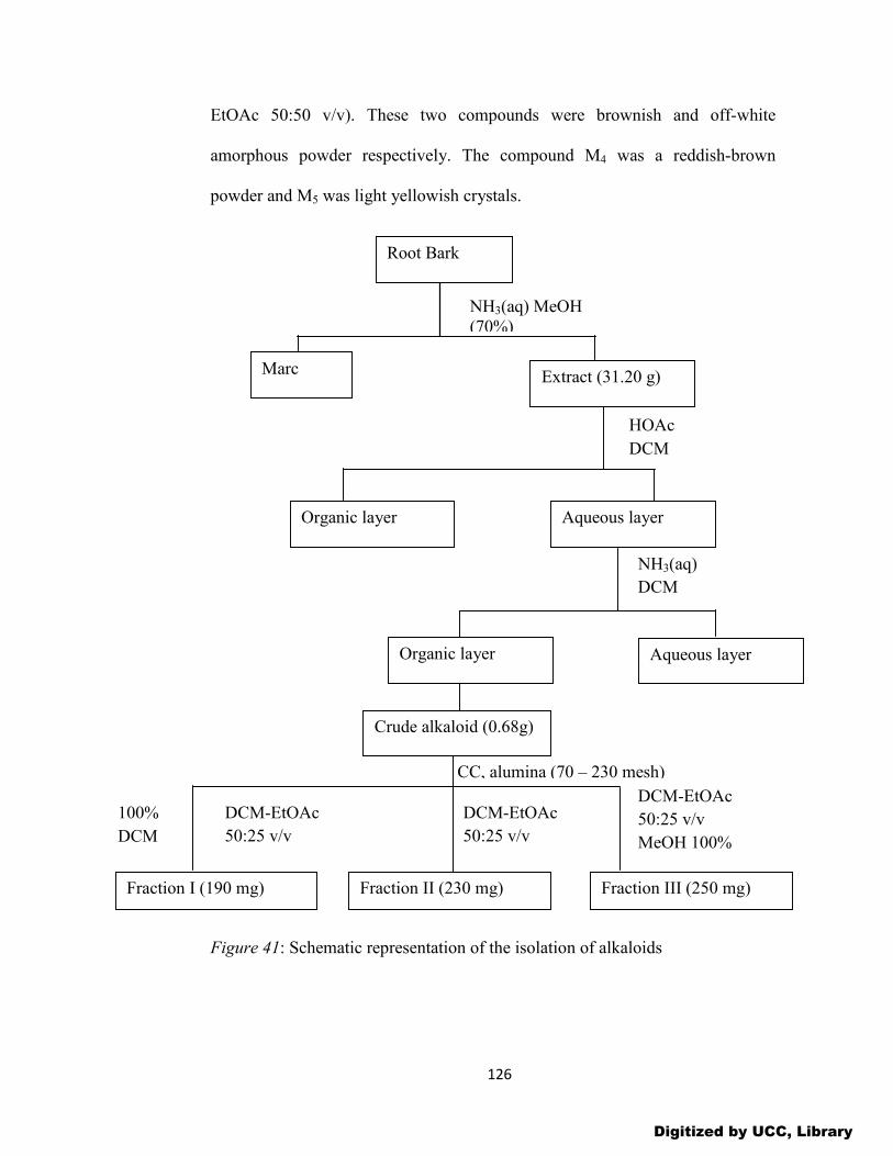
126
EtOAc 50:50 v/v). These two compounds were brownish and off-white
amorphous powder respectively. The compound M4 was a reddish-brown
powder and M5 was light yellowish crystals.
Figure 41: Schematic representation of the isolation of alkaloids
Fraction I (190 mg) Fraction II (230 mg) Fraction III (250 mg)
100%
DCM
DCM-EtOAc
50:25 v/v
DCM-EtOAc
50:25 v/v
DCM-EtOAc
50:25 v/v
MeOH 100%
CC, alumina (70 – 230 mesh)
NH3(aq)
DCM
Organic layer
Crude alkaloid (0.68g)
Marc Extract (31.20 g)
Organic layer Aqueous layer
Root Bark
NH3(aq) MeOH (70%)
HOAc
DCM
Aqueous layer
Digitized by UCC, Library

127
Figure 42: TLC analysis of crude alkaloid extract
Further purification of fraction III by preparative TLC on aluminum
oxide gel neutral with toluene-EtOAc (75:25 v/v) gave sub-fractions IIIa and
IIIb. These sub-fractions were crystallized in absolute ethanol to give
compounds: M4 (70 mg, Rf 0.45 in toluene-EtOAc 50:50 v/v) and M5 (120 mg,
Rf 0.60 in toluene-EtOAc 50:50 v/v) respectively.
M1
M5
Digitized by UCC, Library
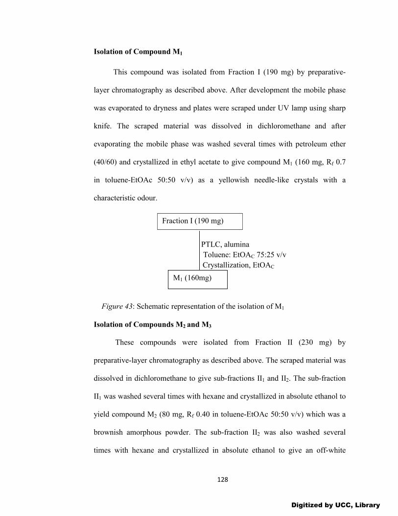
128
Isolation of Compound M1
This compound was isolated from Fraction I (190 mg) by preparative-
layer chromatography as described above. After development the mobile phase
was evaporated to dryness and plates were scraped under UV lamp using sharp
knife. The scraped material was dissolved in dichloromethane and after
evaporating the mobile phase was washed several times with petroleum ether
(40/60) and crystallized in ethyl acetate to give compound M1 (160 mg, Rf 0.7
in toluene-EtOAc 50:50 v/v) as a yellowish needle-like crystals with a
characteristic odour.
PTLC, alumina
Toluene: EtOAC 75:25 v/v
Crystallization, EtOAC
Figure 43: Schematic representation of the isolation of M1
Isolation of Compounds M2 and M3
These compounds were isolated from Fraction II (230 mg) by
preparative-layer chromatography as described above. The scraped material was
dissolved in dichloromethane to give sub-fractions II1 and II2. The sub-fraction
II1 was washed several times with hexane and crystallized in absolute ethanol to
yield compound M2 (80 mg, Rf 0.40 in toluene-EtOAc 50:50 v/v) which was a
brownish amorphous powder. The sub-fraction II2 was also washed several
times with hexane and crystallized in absolute ethanol to give an off-white
Fraction I (190 mg)
M1 (160mg)
Digitized by UCC, Library
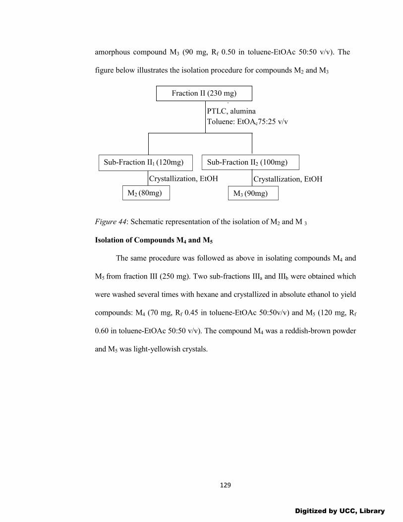
129
amorphous compound M3 (90 mg, Rf 0.50 in toluene-EtOAc 50:50 v/v). The
figure below illustrates the isolation procedure for compounds M2 and M3
Figure 44: Schematic representation of the isolation of M2 and M 3
Isolation of Compounds M4 and M5
The same procedure was followed as above in isolating compounds M4 and
M5 from fraction III (250 mg). Two sub-fractions IIIa and IIIb were obtained which
were washed several times with hexane and crystallized in absolute ethanol to yield
compounds: M4 (70 mg, Rf 0.45 in toluene-EtOAc 50:50v/v) and M5 (120 mg, Rf
0.60 in toluene-EtOAc 50:50 v/v). The compound M4 was a reddish-brown powder
and M5 was light-yellowish crystals.
Sub-Fraction II1 (120mg) Sub-Fraction II2 (100mg)
M3 (90mg)
Fraction II (230 mg)
M2 (80mg)
Crystallization, EtOH Crystallization, EtOH
PTLC, alumina
Toluene: EtOAc75:25 v/v
Digitized by UCC, Library

130
Figure 45: Schematic representation of the isolation of M4 and M5
After the extraction and the isolation process, only compounds M1 and M5
were found to be pure enough, based on their TLC analysis, for spectroscopic
analyses. They were therefore sent to Greenwich University, UK for spectral
analyses.
Anti-inflammatory Activity of Isolated Compounds
The isolated compounds M1 and M5 were pure and were therefore tested
for their anti-inflammatory potential using the method described above. However,
the doses for the isolated compounds and the standard drug- diclofenac were 3, 10
and 30 mg/Kg body weight.
In vitro DPPH radical scavenging activity of isolated compounds
The free radical scavenging activity of the isolated compounds M1 and M5
was determined using the method stated above.
Sub -fraction IIIa (100 mg Sub -fraction IIIb (140
M5 (120 mg)
)
M4 (70 mg)
Fraction III (250
mg)
Crystallization EtOH
PTLC, alumina
Toluene: EtOAC 75: 25
v/v
Crystallization EtOH
Digitized by UCC, Library
131
CHAPTER FOUR
RESULTS AND DISCUSSION
INTRODUCTION
The preliminary phytochemical analyses have revealed that the crude
extract of Anthostema aubryanum is characterized by the presence of alkaloids,
terpenoids, flavonoids, coumarins, anthraquinones, fatty acids, reducing sugars,
cyanogenic glycosides, tannins and saponins. Carotenoids and glucosides were
not detected or were absent. The presence mainly of alkaloids, flavonoids,
steroids and terpenoids may largely contribute to the observed pharmacological
activity because more chemicals belonging to these phytochemicals present in
other medicinal plants had previously been reported to exhibit such
pharmacological activity (Agnihotri, et al., 2010). It has been established that
flavonoids are the major anti-inflammatory agents. Some of them act as
phospholipase inhibitors and some have been demonstrated as TNF-α inhibitors
in different inflammatory conditions (Agnihotri, et al., 2010). Flavonoids inhibit
human neutrophil elastase (HNE) and the matrix metalloproteinases (MMP-2).
Biochemical investigations have also shown that flavonoids can inhibit both
cyclooxygenase and lipoxygenase pathways of the arachidonic metabolism
depending upon their chemical structures (Agnihotri et al., 2010).
Digitized by UCC, Library

132
Table 1: Phytochemical Analysis of A. aubryanum
Constituents
Observation
Alkaloids +
Terpenoids +
Flavonoid aglycones +
Coumarins +
Anthraquinone aglycones +
Fatty acids +
Reducing sugars +
Tannins +
Anthraquinone glycosides +
Flavonoid glycosides +
Saponins +
Carotenoids -
Glucosides -
Cyanogenic glycosides +
(+) = Present, (-) = Absent Source: Laboratory data (2015)
Quercetin is a bioflavonoid compound that blocks the release of
histamine and other anti-inflammatory enzymes. Although human studies with
arthritic patients are lacking at this time, anecdotal evidence is strong for this
application, as is experimental research investigation. There are no well-known
side effects or drug-nutrient interactions for quercetin (Agnihotri, et al., 2010).
Alkaloids in asserted skeletal type based on pyridine ring system have been
Digitized by UCC, Library
133
presented with striking anti-inflammatory activity, e.g. Berberine from Berberis
is a traditional remedy against rheumatism (Agnihotri, et al., 2010).
Terpenoids significantly inhibit the development of chronic joint swelling. In
Western medicine, the treatment often involves topical application of
corticosteroids which are symptomatically effective but have inherent
disadvantages. Terpenoids may affect different mechanism relevant to
inflammations arising in response to varied etiological factors (Changa et al.,
2008). Phytol, the aliphatic diterpene found in F. thonningii has anti-
inflammatory effects and has been reported as a potential therapeutic agent for
the treatment of rheumatoid arthritis and possibly other chronic inflammatory
diseases such as asthma (Dangarembizi et al., 2013). Hence the use of A.
aubryanum against inflammation, wounds and infectious diseases may be
rationalized by the presence of these compounds in the plant.
This is the first phytochemical report on the constituents of Anthostema
aubryanum.
Characterization and Identification of Isolated Compounds
Comprehensive chromatographic analysis coupled with spectroscopic
study has led to the isolation, characterization and identification of two of the
major anti-inflammatory alkaloids as 5-methoxy-canthin-6-one [1] and canthin-
6-one [2].
Identification of M1 as 5-methoxy-canthin-6-one [1] M1 was obtained as yellow needles with a characteristic odour and bright
yellow-green fluorescence at 360 nm. The bright yellow-green UV fluorescence
Digitized by UCC, Library

134
at 360 nm suggested it to be a canthinone alkaloid and this was confirmed by
phytochemical analysis (Zapesochnaya et al., 1991).
It gave a positive test with Dragendorff’s reagent on analysis by TLC suggesting
it to be an alkaloid. It was soluble in chloroform, m.p 223-2340C; (nm, log ε):
EtOH- 269 sh. (4.31), 277(4.41), 297 sh. (3.93), 308 sh.(3.90), 355 sh.(4.01),
376(4.09).
Elemental analysis: Found: C, 72.03; H, 3.92; N, 11.08. C15H10N2O2 requires C.
71.99; H, 4.03, N, 11.19 %; δH (500MHz, MeOD, J/Hz) 8.02 (1H,, H-1, J=5.0),
8.68 (1H,d,H-2, J=5.0), 7.27 (1H,s, H-4, J=10), 8.24 (1H,d, H-8, J=7.7), 7.59
(1H,t, H-9, J=7.7), 7.73 (1H,t, H-10, J=7.7), 8.58 (1H,d, H-11, J=7.7), 4.06 (s,
3H). HR-MS (m/z) 251.0898 [M+H] - (calc. for C15H10N2O2) (Appendix 1C).
It has a DBE of 12, indicating an ABCD aromatic system (O’Donnell &
Gibbons, 2007)
N
N
O
A
B C
D
The 1H and 13C-NMR data (table 2, Appendix 1A-B) were similar to those of
compound 2; however, ring D was seen to be a single substituted aromatic
system and a deshielded methoxy singlet was present at δ 4.06 in the 1H-NMR
spectrum. In addition to the methoxy group, the 1H –NMR for M1 displayed
seven signals in the aromatic region (δ7.27-8.68). Two proton doublets at δH
8.02 and 8.68 (J=5.0Hz) are characteristic of pyridine protons and assignable to
Digitized by UCC, Library

135
H-1 and H-2 respectively, and a pair of doublets occurring at δH 8.24 and δH
8.58 (J=7.7Hz) are typical of indole protons and are assignable to H-8 and H-11
respectively. The pair of triplets at δH 7.59 and δH 7.73 (J=7.7) further confirmed
the presence of indole protons and are assignable to H-9 and H-10 respectively.
Further ortho coupled aromatic system was evident: a cis double-bond
positioned next to aromatic nitrogen at δH 8.68 and δH 8.02 (J=5.0). In addition
to seven methine carbons, the 13C-NMR spectrum revealed the presence of
seven aromatic quaternary carbons (δ 124.29-146.78) and one deshielded signal
consistent with a carbonyl carbon (δ158.40). Ring A is an aromatic system by
correlations. The triplet at δ 7.59 (H-9; J=7.7Hz) correlated to the triplet at δ
7.73 (H-10; J=7.7 Hz) which in turn coupled to the doublet at δ 8.58 (H-11;
J=7.7 Hz).
Ring C was consistent with ortho coupled pair of hydrogens (H-1, δ 8.02 and H-
2, δ 8.68 J=5.0 Hz) positioned on a pyridine ring. H-1 is correlated to the
quaternary carbon C-15 (δ 131.33), completing the assignment of ring B, while
H-2 is also correlated to the C-14 quaternary (δ 131.89). The correlation
between H-1 and H-11 concluded the β-carboline skeleton of the canthin-6-one
structure.
The resonance at δ152.0 could be unambiguously assigned to C-5 by irradiating
the 5-methoxyl protons at δ 4.06. The complex multiplet of C-5 can be
converted to a clean doublet due to the coupling to the H-4 proton. The doublets
of C-14 at δ 131.89 and C-15 at δ 131.33 could each be analyzed in terms of
three-bond coupling of 8.02 Hz. The assignment can be confirmed by irradiating
Digitized by UCC, Library
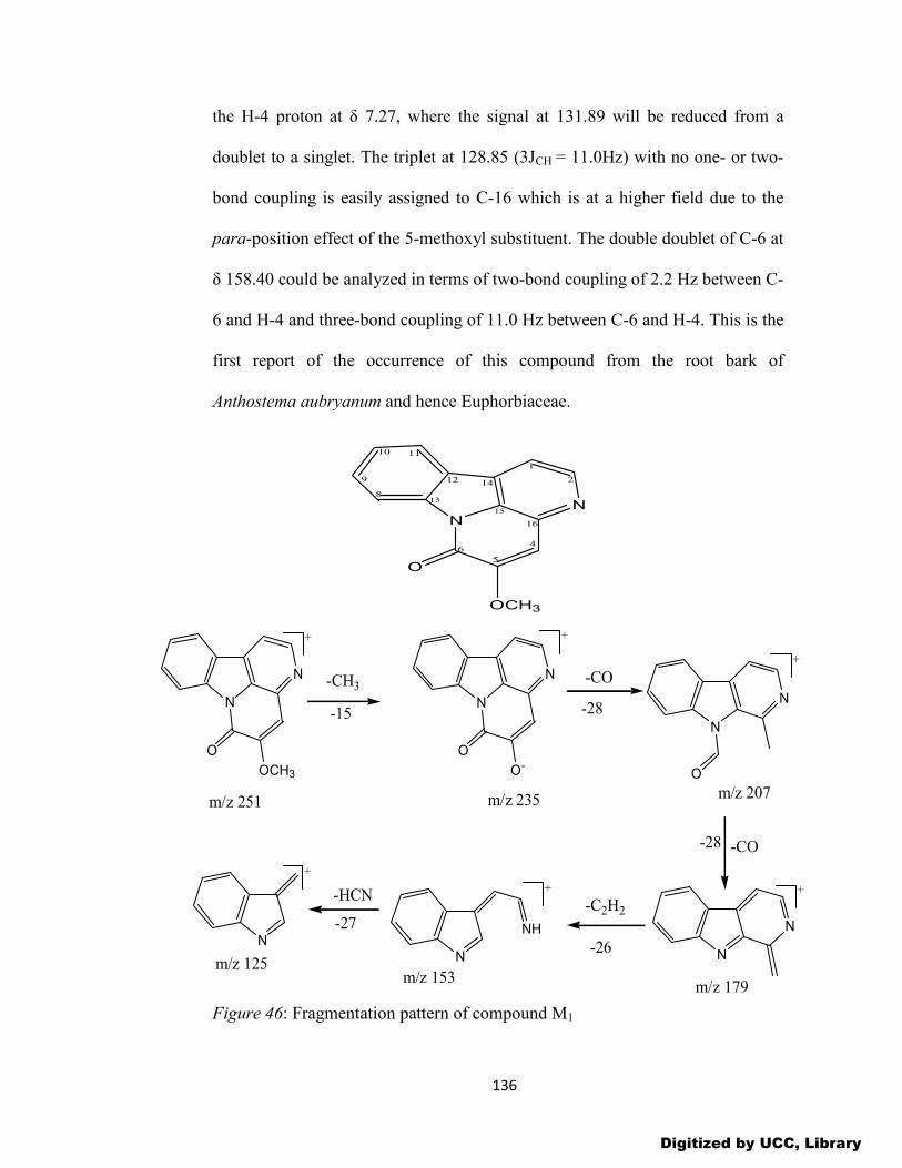
136
the H-4 proton at δ 7.27, where the signal at 131.89 will be reduced from a
doublet to a singlet. The triplet at 128.85 (3JCH = 11.0Hz) with no one- or two-
bond coupling is easily assigned to C-16 which is at a higher field due to the
para-position effect of the 5-methoxyl substituent. The double doublet of C-6 at
δ 158.40 could be analyzed in terms of two-bond coupling of 2.2 Hz between C-
6 and H-4 and three-bond coupling of 11.0 Hz between C-6 and H-4. This is the
first report of the occurrence of this compound from the root bark of
Anthostema aubryanum and hence Euphorbiaceae.
NN
O
OCH3
1
2
4
5
6
8
9
10 11
12
13
14
15
16
Figure 46: Fragmentation pattern of compound M1
N
N
O
OCH3
N
N
O
O-
N
N
O
N
N
-CH3
m/z 251 m/z 235
-15
-CO
-28
m/z 207
m/z 179
-CO-28
N
NH
m/z 153
-C2H2
-26N
m/z 125
-HCN
-27
+ +
+
++
+
Digitized by UCC, Library
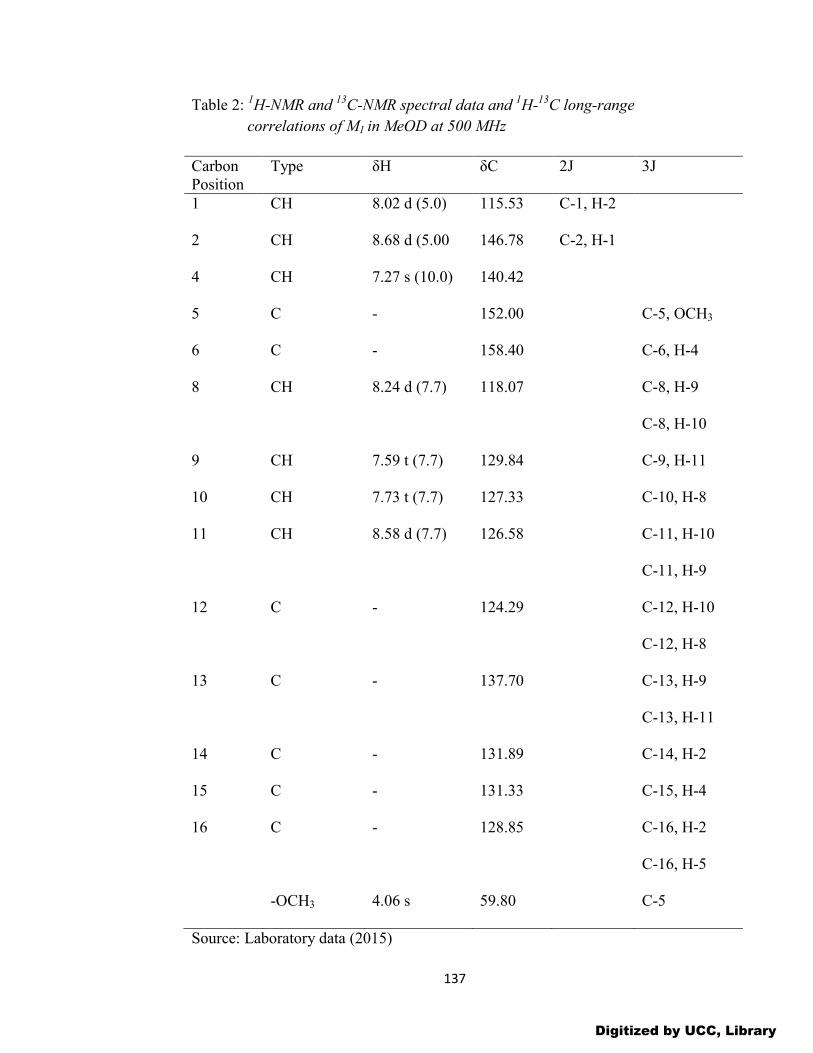
137
Table 2: 1H-NMR and 13C-NMR spectral data and 1H-13C long-range
correlations of M1 in MeOD at 500 MHz
Carbon Position
Type δH δC 2J 3J
1 CH 8.02 d (5.0) 115.53 C-1, H-2
2 CH 8.68 d (5.00 146.78 C-2, H-1
4 CH 7.27 s (10.0) 140.42
5 C - 152.00 C-5, OCH3
6 C - 158.40 C-6, H-4
8 CH 8.24 d (7.7) 118.07 C-8, H-9
C-8, H-10
9 CH 7.59 t (7.7) 129.84 C-9, H-11
10 CH 7.73 t (7.7) 127.33 C-10, H-8
11 CH 8.58 d (7.7) 126.58 C-11, H-10
C-11, H-9
12 C - 124.29 C-12, H-10
C-12, H-8
13 C - 137.70 C-13, H-9
C-13, H-11
14 C - 131.89 C-14, H-2
15 C - 131.33 C-15, H-4
16 C - 128.85 C-16, H-2
C-16, H-5
-OCH3 4.06 s 59.80 C-5
Source: Laboratory data (2015)
Digitized by UCC, Library

138
Identification of M5 as canthin-6-one [2]
M5 was obtained as light yellow needles with a light blue fluorescence at
360 nm. It gave a positive test with Dragendorff’s reagent on analysis by TLC. It
was soluble in chloroform; m.p 156-1570C, (Lit. 155-156, Zapesochnaya, et al.,
1991); (nm, log ε): EtOH-251(4.10), 259(4.12), 268(4.07), 300(3.92), 347(3.94),
362(4.15), 380(4.13).
Elemental analysis: Found: C, 76.32; H, 3.63; N 12.78. C14H8N2O requires C,
76.35; H, 3.66; N, 12.72%; δH (500MHz, MeOD, J/Hz) 8.0 (1H,d, H-
1,J=5.0Hz), 8.72 (1H,d, H-2, J=5.0), 8.11 (1H,d, H-4, J=10.0), 6.93 (1H,d,
J=10.0) 8.47 (1H,d, H-8, J=10.0), 7.68 (1H,t, H-9, J=8.5), 7.52 (1H,t, H-10,
J=8.5), 8.18 (1H,d, H-11,J=8.5). HR-MS (m/z) 221.0755 [M+H]- (calc. for
C14H8N2O)
It has a DBE of 12 which completes an ABCD aromatic ring system.
The spectral data (1H-NMR, 13C-NMR, table 4, appendix 2A-B) of compound 2
revealed that this compound was canthin-6-one, previously isolated from
Ailanthus altissima (Simaroubaceae) by Koike and Ohmoto (1985). The NMR
spectra (table 4; appendix 2A-B) show that it is unsubstituted as shown by the
characteristic doublets of the H-4 and H-5 protons with the constant J= 10 Hz,
and also by the doublets of a pair of vicinal protons (H-1 and H-2). In addition
to the signals of these protons of rings C and D (H-1, H-2, H-4 and H-5), its
spectra also contain the signals of four aromatic protons (H-8, H-9, H-10 and H-
11) which are characteristic of an unsubstituted canthinone. In the long-range
selective proton decoupling, irradiation of either H-1 proton at δ 8.0 or the H-4
Digitized by UCC, Library

139
proton at 8.1 reduced the triplet signal at δ 133.21 to a doublet, revealing three-
bond couplings among C-15, H-1 and H-4. Irradiation of either the H-2 proton
at δ 8.7 or the H-5 at δ 6.9 reduced the triplet signal at δ 136.92 to a doublet,
revealing three-bond couplings among C-16, H-2 and H-5. At the same time,
irradiation of the H-1 or H-2 proton reduced the double doublet at signal δ
132.09 to a doublet, revealing two-bond coupling between C-14 and H-1 of 3.7
Hz and three-bond coupling between C-14 and H-2 of 8.1 Hz. Irradiation of the
H-4 or H-5 proton reduced the double doublet signal at δ 160.96 to a doublet,
showing two-bond coupling between C-6 and H-4 of 2.2 Hz and three-bond
coupling between C-6 and H-5 of 11.0 Hz. On the basis of the above evidence
and comparison with the published data, the structure of M5 was established as
canthin-6-one. Canthin-6-one alkaloids occur plentifully in many plants of
Simaroubaceae and Rutaceae (Koike and Ohmoto, 1985). However, to the best
of our knowledge, this is the first report of its occurrence in A. aubryanum and
hence Euphorbiaceae.
Figure 47: The structure of compound M5
NN
O
1
2
4
5
6
8
9
10 11
12
13
14
15
16
Digitized by UCC, Library

140
Table 3: 13C-NMR chemical shifts (ppm) of canthin-6-one and compound M5
Carbon position
*Canthin-6-one Compound M5
1 115.37 117.85
2 144.84 146.88
4 138.57 140.69
5 127.98 127.11
6 158.21 160.96
8 116.29 118.13
9 129.84 129.92
10 124.69 124.31
11 121.61 120.00
12 123.33 125.65
13 138.24 140.29
14 128.99 133.21
15 130.91 132.13
16 135.23 136.92
Source: Laboratory data (2015), *(Koike and Ohmoto, 1985)
Digitized by UCC, Library
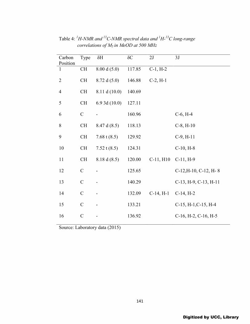
141
Table 4: 1H-NMR and 13C-NMR spectral data and 1H-13C long-range
correlations of M5 in MeOD at 500 MHz
Carbon Position
Type δH δC 2J 3J
1 CH 8.00 d (5.0) 117.85 C-1, H-2
2 CH 8.72 d (5.0) 146.88 C-2, H-1
4 CH 8.11 d (10.0) 140.69
5 CH 6.9 3d (10.0) 127.11
6 C - 160.96 C-6, H-4
8 CH 8.47 d (8.5) 118.13 C-8, H-10
9 CH 7.68 t (8.5) 129.92 C-9, H-11
10 CH 7.52 t (8.5) 124.31 C-10, H-8
11 CH 8.18 d (8.5) 120.00 C-11, H10 C-11, H-9
12 C - 125.65 C-12,H-10, C-12, H- 8
13 C - 140.29 C-13, H-9, C-13, H-11
14 C - 132.09 C-14, H-1 C-14, H-2
15 C - 133.21 C-15, H-1,C-15, H-4
16 C - 136.92 C-16, H-2, C-16, H-5
Source: Laboratory data (2015)
Digitized by UCC, Library

142
BIOASSAYS
Anti-inflammatory activity of root bark extract
The stem and root bark of Anthostema aubryanum are routinely employed
in traditional medicine to treat a variety of disease conditions including
inflammatory pain, wounds, boil and edema. Many compounds with numerous
pharmacological activities have been isolated from Euphorbiaceae but little is
known about the pharmacology of the root bark of Anthostema aubryanum.
In our experimental conditions, we first used a positive control diclofenac which
showed a time-dependent anti-inflammatory effect at all hours (figure 48). The
AUC calculation showed that the three tested doses (10, 30 and 100 mg/Kg
BDW) of diclofenac suppressed the carrageenan-induced edema under the
experimental condition by 36.16 ± 2.4, 48.94 ± 2.2 and 59.20 ± 2.6 respectively.
From figure 46, it can be seen that oral administration of the methanolic extract of
the root bark of A. aubryanum similarly suppressed the carrageenan-induced
inflammation in a dose-and time-dependent manner. The extract was given orally
to the rats at 30 mg/kg, 100 mg/kg and 300 mg/kg (weight of concentrated
solution), 1 hour before induction of oedema with carrageenan. Diclofenac (10-
100 mg/kg, i.p) was used as reference drug. Induction of acute inflammation in
control rats resulted in a prominent increase in paw thickness, which began 1 hour
after intraplantar injection of carrageenan and reached a peak of inflammation
after 2 hours (figure 48) and slowly declined for the next 3 hours. The extract
caused significant (P < 0.000 1) dose-dependent inhibition of the carrageenan -
induced inflammation in the six weeks old rats, the effect of which began 2 hours
after carrageenan injection (figure 48). Diclofenac (10-100 mg /kg, i.p) showed
Digitized by UCC, Library
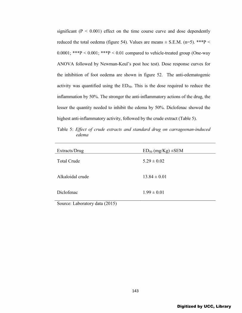
143
significant (P < 0.001) effect on the time course curve and dose dependently
reduced the total oedema (figure 54). Values are means ± S.E.M. (n=5). ***P <
0.0001; ***P < 0.001; ***P < 0.01 compared to vehicle-treated group (One-way
ANOVA followed by Newman-Keul’s post hoc test). Dose response curves for
the inhibition of foot oedema are shown in figure 52. The anti-edematogenic
activity was quantified using the ED50. This is the dose required to reduce the
inflammation by 50%. The stronger the anti-inflammatory actions of the drug, the
lesser the quantity needed to inhibit the edema by 50%. Diclofenac showed the
highest anti-inflammatory activity, followed by the crude extract (Table 5).
Table 5: Effect of crude extracts and standard drug on carrageenan-induced edema
Extracts/Drug ED50 (mg/Kg) ±SEM
Total Crude 5.29 ± 0.02
Alkaloidal crude 13.84 ± 0.01
Diclofenac 1.99 ± 0.01
Source: Laboratory data (2015)
Digitized by UCC, Library

144
Figure 48: Time-course edema development following carrageenan injection
into rat paws and dose (mg/Kg)-dependent anti-inflammatory
effect of the standard positive control, diclofenac.
Digitized by UCC, Library
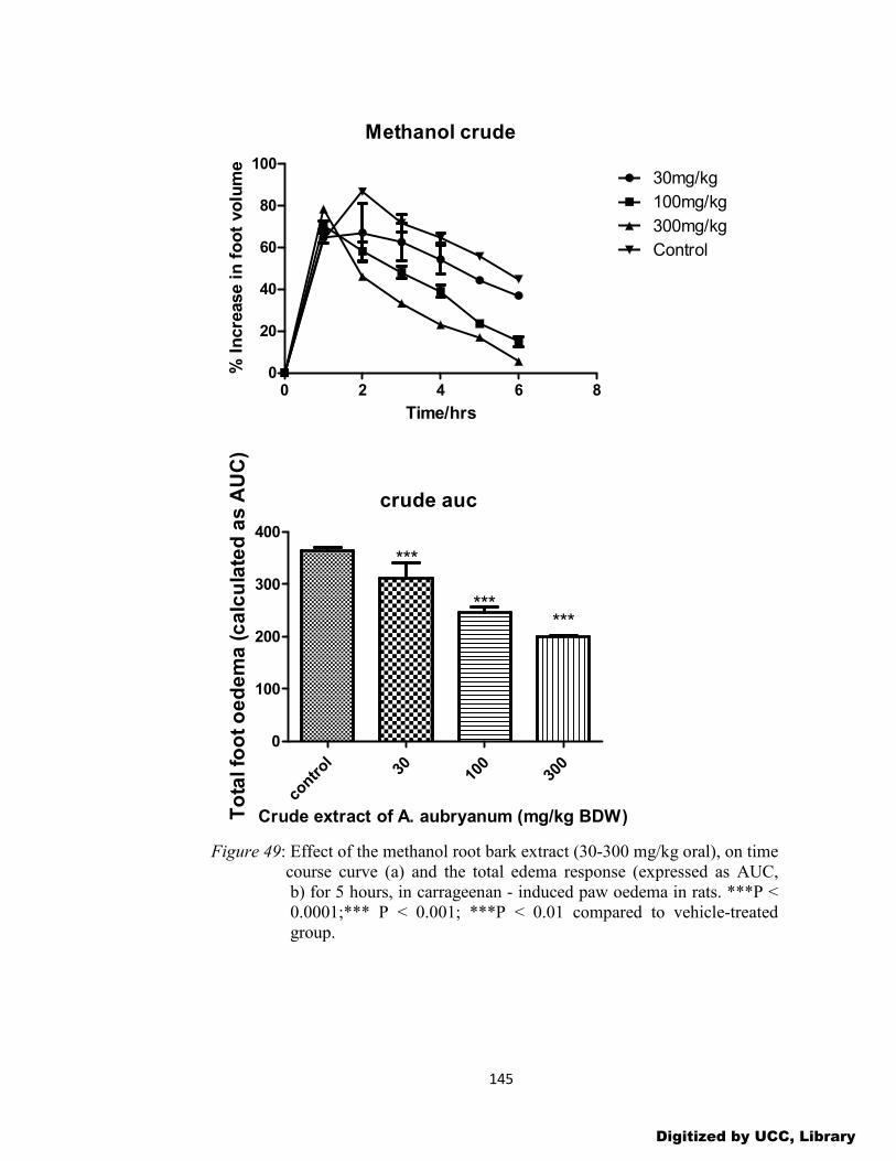
145
Methanol crude
0 2 4 6 80
20
40
60
80
10030mg/kg
100mg/kg
300mg/kg
Control
Time/hrs
% I
ncre
ase i
n f
oo
t vo
lum
e
crude auc
contr
ol30 10
030
0
0
100
200
300
400
******
***
Crude extract of A. aubryanum (mg/kg BDW)To
tal fo
ot
oe
de
ma
(c
alc
ula
ted
as
AU
C)
Figure 49: Effect of the methanol root bark extract (30-300 mg/kg oral), on time course curve (a) and the total edema response (expressed as AUC, b) for 5 hours, in carrageenan - induced paw oedema in rats. ***P < 0.0001;*** P < 0.001; ***P < 0.01 compared to vehicle-treated group.
Digitized by UCC, Library

146
Figure 50: Effect of the alkaloidal extract (30-300 mg/kg oral), on time course curve (a) and the total oedema response (expressed as AUC, b) for 5 hours, in carrageenan - induced paw oedema in rats.***P < 0.0001; *** P < 0.001;***P < 0.01 compared to vehicle-treated group.
0 2 4 6 80
20
40
60
80
10030mg/kg
100mg/kg
300mg/kg
Control
Time/hrs
% I
ncre
ase i
n f
oo
t vo
lum
e
Digitized by UCC, Library
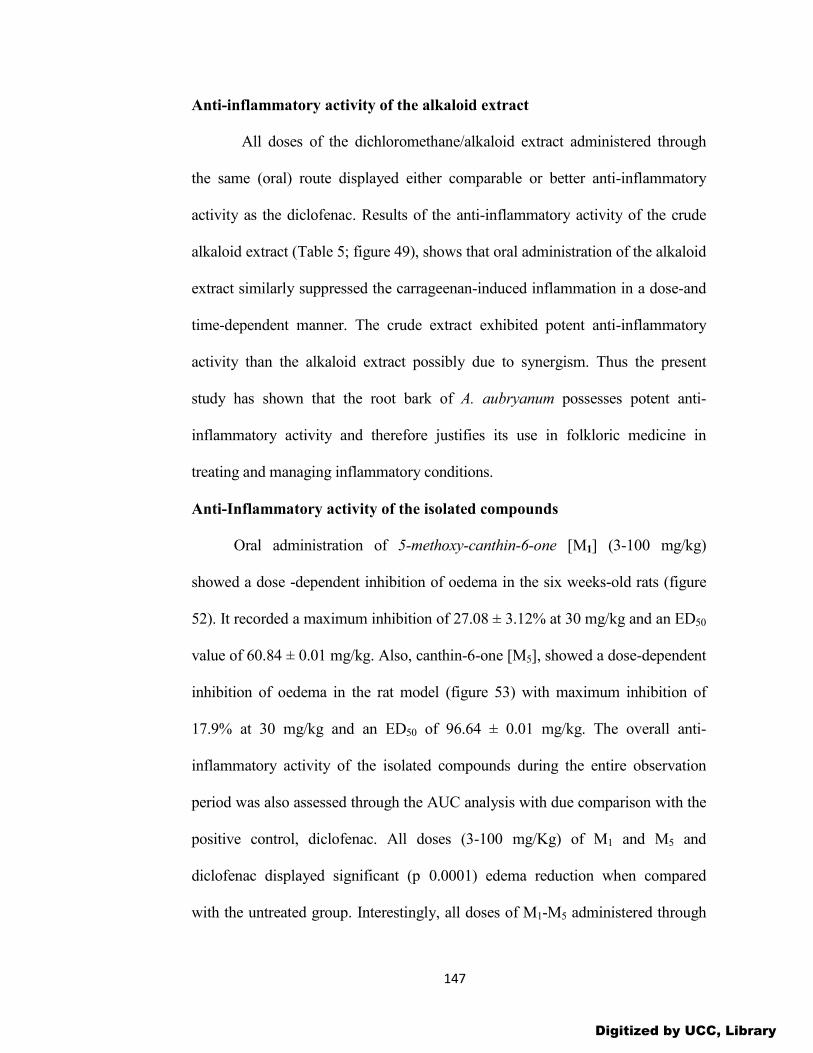
147
Anti-inflammatory activity of the alkaloid extract
All doses of the dichloromethane/alkaloid extract administered through
the same (oral) route displayed either comparable or better anti-inflammatory
activity as the diclofenac. Results of the anti-inflammatory activity of the crude
alkaloid extract (Table 5; figure 49), shows that oral administration of the alkaloid
extract similarly suppressed the carrageenan-induced inflammation in a dose-and
time-dependent manner. The crude extract exhibited potent anti-inflammatory
activity than the alkaloid extract possibly due to synergism. Thus the present
study has shown that the root bark of A. aubryanum possesses potent anti-
inflammatory activity and therefore justifies its use in folkloric medicine in
treating and managing inflammatory conditions.
Anti-Inflammatory activity of the isolated compounds
Oral administration of 5-methoxy-canthin-6-one [M1] (3-100 mg/kg)
showed a dose -dependent inhibition of oedema in the six weeks-old rats (figure
52). It recorded a maximum inhibition of 27.08 ± 3.12% at 30 mg/kg and an ED50
value of 60.84 ± 0.01 mg/kg. Also, canthin-6-one [M5], showed a dose-dependent
inhibition of oedema in the rat model (figure 53) with maximum inhibition of
17.9% at 30 mg/kg and an ED50 of 96.64 ± 0.01 mg/kg. The overall anti-
inflammatory activity of the isolated compounds during the entire observation
period was also assessed through the AUC analysis with due comparison with the
positive control, diclofenac. All doses (3-100 mg/Kg) of M1 and M5 and
diclofenac displayed significant (p 0.0001) edema reduction when compared
with the untreated group. Interestingly, all doses of M1-M5 administered through
Digitized by UCC, Library

148
the same (oral) route displayed either comparable or better anti-inflammatory
activity as diclofenac. The 5-methoxycanthin-6-one [M1] exhibited a higher anti-
inflammatory activity than its unsubstituted analogue [M5] (Table 8). The
observed activity of M1 is due to the presence of the methoxy group which makes
it less polar/lipophobic or more lipophilic to be able to cross the membranes or
the blood brain barriers. While the presence of other minor constituents with a
similar pharmacological effect cannot be ruled out, the isolated compounds as
major constituents of the root bark of A. aubryanum are likely to play a major role
for the reported medicinal uses of the plant. The dose response curves (figure 53),
show the highest activity for diclofenac, crude extract, alkaloid extract, 5-
methoxycanthin-6-one and canthin-6-one. This is shown by the sigmoid nature of
the curves, the more sigmoid the curve, the higher the activity.
Table 6: Effect of M1 and M5 on carrageenan-induced edema
Alkaloid/Drug ED50 mg/Kg ± SEM
5-methoxy-canthin-6-one 60.84 ± 0.01
Canthin-6-one 96.64 ± 0.01
Diclofenac 1.99 ± 0.01
Source: Laboratory data (2015)
Digitized by UCC, Library
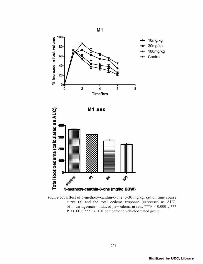
149
M1
0 2 4 6 80
20
40
60
80
10010mg/kg
30mg/kg
100mg/kg
Control
Time/hrs
% I
ncre
ase i
n f
oo
t vo
lum
e
Figure 51: Effect of 5-methoxy-canthin-6-one (3-30 mg/kg; i.p) on time course curve (a) and the total oedema response (expressed as AUC, b) in carrageenan - induced paw edema in rats. ***P < 0.0001; *** P < 0.001; ***P < 0.01 compared to vehicle-treated group.
Digitized by UCC, Library

150
M5
0 2 4 6 80
20
40
60
80
10010kg/mg
30kg/mg
100kg/mg
Control
Time/hrs
% I
ncre
ase i
n f
oo
t vo
lum
e
Figure 52: Effect of canthin-6-one (3-30 mg/kg; i.p) on time course curve (a) and the total oedema response (expressed as AUC, b) in carrageenan – induced paw edema in rats. ***P < 0.0001, ***P < 0.001, ***P < 0.01 compared to vehicle-treated group
Digitized by UCC, Library

151
Figure 53: Dose response curves for standard drug, extracts and isolated compounds on carrageenan - induced foot edema in rats.
Antioxidant Activity of Extracts
Antioxidant activity of crude extracts and isolated compounds
The qualitative DPPH test showed the two extracts and the isolated
compounds bleaching the purple DPPH radical, thus giving pale spots over a
purple background. This indicates that they contain some antioxidant constituents.
The DPPH assay is a valid and simplest assay to evaluate scavenging activity of
antioxidant, since the radical compound is stable and does not have to be
generated as in other radical scavenging assays. Antioxidants scavenge the DPPH
radical by donating a proton. Different authors use different initial radical
concentrations and different reaction times. The extract showed a concentration
dependent DPPH radical scavenging activity. The decrease in the absorbance of
DPPH was due to phytoconstituents in the plant extracts acting as antioxidants by
hydrogen donation.
Digitized by UCC, Library

152
Quantitative antioxidant assay of extracts
Three methods were used to determine quantitatively the antioxidant
activity of both the crude and the alkaloid extracts. They are the total phenolic
content, total antioxidant capacity and DPPH radical scavenging assays.
Total phenolic content
The total phenolic content of the extracts was determined using the Folin-
ciocalteau reagent and vitamin E as standard. The total phenolic content was
expressed as mg of vitamin E equivalents (VEE) per g of extract. Table 7 shows
the total phenolic contents of the crude methanolic (CE) and alkaloid (AC)
extracts. The crude methanolic extract had the highest phenolic content.
Table 7: Total phenolic content of root extracts
Extracts (1.5mg/mL) Mean (mg VEE/g) ± SEM
CE 74.53±0.00
AE 59.54±0.00
Source: Laboratory work (2015)
0 20 40 600.0
0.2
0.4
0.6
0.8
r2 = 0.9632
conc.
Ab
sorb
ance
(n
m)
Figure 54: Absorbance against concentration of vitamin E used in the calibration curve.
Digitized by UCC, Library
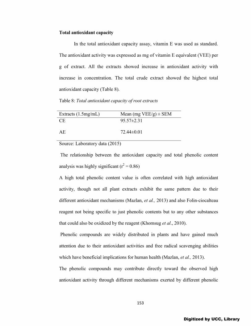
153
Total antioxidant capacity In the total antioxidant capacity assay, vitamin E was used as standard.
The antioxidant activity was expressed as mg of vitamin E equivalent (VEE) per
g of extract. All the extracts showed increase in antioxidant activity with
increase in concentration. The total crude extract showed the highest total
antioxidant capacity (Table 8).
Table 8: Total antioxidant capacity of root extracts
Extracts (1.5mg/mL) Mean (mg VEE/g) ± SEM
CE 95.57±2.31
AE 72.44±0.01
Source: Laboratory data (2015)
The relationship between the antioxidant capacity and total phenolic content
analysis was highly significant (r2 = 0.86)
A high total phenolic content value is often correlated with high antioxidant
activity, though not all plant extracts exhibit the same pattern due to their
different antioxidant mechanisms (Mazlan, et al., 2013) and also Folin-ciocalteau
reagent not being specific to just phenolic contents but to any other substances
that could also be oxidized by the reagent (Khomsug et al., 2010).
Phenolic compounds are widely distributed in plants and have gained much
attention due to their antioxidant activities and free radical scavenging abilities
which have beneficial implications for human health (Mazlan, et al., 2013).
The phenolic compounds may contribute directly toward the observed high
antioxidant activity through different mechanisms exerted by different phenolic
Digitized by UCC, Library

154
compounds or through synergistic effects with other non phenolic compounds
(Mazlan, et al., 2013).
It has been established that compounds with high antioxidant activities may
also contribute toward the inhibition of tyrosinase, cholinesterase (AChE) and
nitric oxide (NO) production in cells. Inflammatory conditions may enhance the
production of reactive oxygen/nitrogen species (ROS/NOS), which leads to
oxidative stress that can damage important organic substrates. Antioxidants can
scavenge free radicals and protect organisms from ROS/NOS-induced damage,
leading to a reduction in inflammation (Abdillahi et al., 2011; Almeida et al.,
2011). Antioxidants can also prevent major degenerative diseases and aging and
might have protective effects toward Alzheimer’s disease (Aremu et al., 2011).
The inhibition of cholinesterase is suggested to be quite useful in the treatment of
Alzheimer’s disease and other diseases including senile dementia, ataxia and
Parkinson’s disease. Alzheimer’s disease is the result of a deficiency in the
cholinergic system due to the rapid hydrolysis of acetylcholine. Hence, nerve
impulse transmission is terminated at the cholinergic synapses. By suppressing
cholinesterase, cholinergic neurotransmission can be restored (Mazlan, et al.,
2013). Tacrine is one of the synthetic drugs used for treating the symptoms of
cognitive dysfunction or memory loss associated with Alzheimer’s disease.
However, adverse effects have been reported for these synthetic drugs, including
gastrointestinal disturbances and suppression of bioavailability. Oxidative –
related processes coupled with tyrosinase activity can also trigger melanogenesis,
which causes skin pigmentation (Abdillahi et al., 2011). There are no reports of
Digitized by UCC, Library
155
the cholinesterase inhibition properties of any Anthostema species. However,
Anthostema species are expected to have cholinesterase (AChE) inhibition
properties because it has been reported that plants belonging to the Euphorbiaceae
family have AChE inhibitory potential (Mazlan, et al., 2013).
Thus, the high levels of antioxidant activity found in the plant extract may also
result in a higher inhibition of tyrosinase and cholinesterase activities as well as
nitric oxide production. Antioxidant activity of plant extract is not limited to
phenolic compounds. Activity may also be due to the presence of other
antioxidant secondary metabolites, such as flavonoids, volatile oils, carotenoids
and vitamins. Flavonoids are good antioxidants which scavenge and reduce free
radical formation (Grassi et al., 2010). The C-glucosylflavonoids (orientin,
vitexin and isovitexin) which have been isolated from many medicinal plants such
as Ficus thonningii, pigeon pea, linseed oil and in rooibos tea possess antioxidant
properties (Dangarembizi, et al., 2013). Orientin possesses free radical
scavenging activity based on its ene-diol functionality i.e. its dihydroxy
substituents in the B ring and the double bond characteristic of the C ring
(Dangarembizi, et al., 2013). Vitexin and isovitexin also possess antioxidant
though to a lesser extent than orientin due to the lack of OH substituent. In
addition to flavonoid, stilbenes also exhibit antioxidant activity. Resveratrol and
its methylated derivatives, trans-3,3’, 5,5-tetrahydroxy-4-methoxystilbene,
possess antioxidative effects against oxidative stress induced by reactive nitrogen
species and reactive oxygen species (Dangarembizi, et al., 2013). Resveratrol and
its derivatives have also been shown to reduce peroxynitrite which is one of the
Digitized by UCC, Library

156
most potent reactive nitrogen species (Olas et al., 2008). High levels of
peroxynitrite are generated in inflammatory based disease conditions
(Dangarembizi, et al., 2013). There is also the possibility of synergistic
interactions between flavonoids and stilbenes.
DPPH radical scavenging activity of extracts of A. aubryanum
The results of the free radical scavenging potential of the total and alkaloid
extracts of A. aubryanum using DPPH free radical scavenging method are shown
in the table below. The reference drug, vitamin E (0.003-0.03 mg/mL) and the
extracts (0.5-1.5 mg/mL) exhibited concentration-dependent free radical
scavenging activity (table 9). The concentration that provided 50% radical
scavenging (IC50) of the crude extract was determined as 8.84±0.02 compared to
the vitamin E standard of 8.61±0.00.
The order of decreasing activity (as defined by IC50 in mg/mL) was found to be:
vitamin E total crude alkaloid crude. The results indicate that the root bark
of A. aubryanum possess potent antioxidant activity.
Table 9: DPPH scavenging activity of extracts of A. aubryanum root bark
Extracts IC50 (μg/mL) ± SEM
Total Crude 8.84 ± 0.01
Alkaloidal crude 23.12 ±0.01
Vitamin E 8.61 ± 0.01
Source: Laboratory data (2015)
Many medicinal plants possessing antioxidant activities have been
shown to possess protective effects on the erythrocyte membrane from
Digitized by UCC, Library

157
acetaminophen-induced membrane peroxidation (Ahur et al., 2010). The
antihaemolytic and haematinic potential of medicinal plants is possibly due to
its antagonistic activity against the depletion of glutathione and hence
prevention of inflammation. Thus the present study has shown that the root bark
of A. aubryanum possess significant antioxidant properties and may contribute
to the retardation of the inflammatory process. This is because inflammatory
tissue injuries are mediated by reactive oxygen metabolites from phagocytic
leukocytes (e.g. neutrophils, monocytes, macrophages and eosinophils) that
invade the tissues and cause injury to essential cellular components (Amponsah,
2012).
Antioxidant Activity of Isolated Compounds
Quantitative DPPH radical scavenging test
5-methoxycanthin-6-one [1] canthin-6-one [2] showed various degrees
of antioxidant properties, with 5-methoxycanthin-6-one being the most active
(Table 10). Vitamin E (VE) was used as the standard antioxidant drug. The
order of decreasing activity as indicated by the IC50 is VE > 5-methoxycanthin-
6-one > canthin-6-one. From the concentration response curves for the standard
drug, extracts and isolated compounds (figure 53), the more sigmoid the curve,
the higher the activity. The standard drug and the crude extract show the highest
activity followed by the alkaloid extract, 5-methoxycanthin-6-one and canthin-
6-one. These results are further confirmed by the percent inhibition curve
(figure 56) and the DPPH absorption spectra (figure 57).
Digitized by UCC, Library
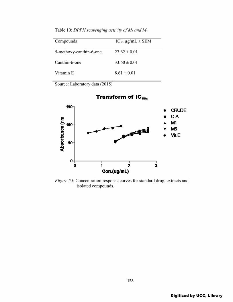
158
Table 10: DPPH scavenging activity of M1 and M5
Compounds IC50 µg/mL ± SEM
5-methoxy-canthin-6-one 27.62 ± 0.01
Canthin-6-one 33.60 ± 0.01
Vitamin E 8.61 ± 0.01
Source: Laboratory data (2015)
Figure 55: Concentration response curves for standard drug, extracts and isolated compounds.
Digitized by UCC, Library

159
Figure 56: Plot of percent inhibition against concentration of extracts and isolated compounds.
Figure 57: DPPH absorption spectra of extracts and isolated compounds.
Compounds that have scavenging activities toward free radicals have
been found to be beneficial in inflammatory diseases. The antioxidant activity
0
20
40
60
80
100
120
0 100 200 300 400 500 600
% in
hib
itio
n
Concentration(µg/ml)
M1 CA M5 CRUDE VIT E
Digitized by UCC, Library
160
of the root bark reported in this study support its traditional use for wound
healing. This is because in acute and chronic wounds, oxidants cause cell
damage and thus inhibits wound healing (Thang et al., 2001). The
administration of antioxidants or free radical scavengers is reportedly helpful,
notably to limit the delayed sequel of thermal trauma and to enhance the healing
process (Thang et al., 2001).
Digitized by UCC, Library

161
CHAPTER FIVE
SUMMARY, CONCLUSIONS AND RECOMMENDATIONS
INTRODUCTION
The present study was aimed at investigating the root bark of A.
aubryanum (Baill., family, Euphorbiaceae) for phytochemical constituents and
pharmacological activity using the acute carrageenan-induced foot edema model
in six weeks old rats and to isolate the compounds which may be responsible for
this activity. Also since free radicals and reactive oxygen species are implicated
in inflammatory diseases, the antioxidant potential of extracts and isolated
compounds were investigated in in vitro experimental models.
Summary
In African folk medicine, the stem and root bark of Anthostema
aubryanum (Baill) are used as an effective remedy against several inflammatory
ailments including rheumatism and renal inflammations.
The preliminary phytochemical analyses have revealed that methanolic extract
of Anthostema aubryanum is characterized by the presence of alkaloids,
steroids, flavonoids, coumarins, fatty acids, reducing sugars, cyanogenic
glycosides, tannins, anthraquinones and saponins. Carotenoids and glucosides
were not detected. These classes of compounds are known to have established
biochemical activities and multiple pharmacological effects and hence the use
of this plant in ethnomedicine may be rationalized by the presence of these
compounds in this plant.
Digitized by UCC, Library
162
Oral administration of both the total crude and crude alkaloid extracts of
A. aubryanum resulted in suppression of the carrageenan-induced inflammation
in a dose- and time-dependent manner and thus, presumably, inhibited the
synthesis and release of prostaglandins as well as kinins responsible for the
inflammation. A low ED50, indicating high anti-inflammatory activity, was
recorded for the total methanol root bark extract (ED50 5.294 ± 0.02 mg/kg
BDW). The crude alkaloid extract of the root bark also exhibited dose-
dependent reduction in foot volume but with comparatively lower activities than
the total methanol extract (ED50 = 13.84 ± 0.01 mg/kg BDW) due to synergism.
The antioxidant activity of Anthostema aubryanum (Baill) was evaluated
by the DPPH assay. The concentration that provided 50% radical scavenging
(IC50) was determined as 8.84±0.01 which was equivalent to the vitamin E
standard of 8.61±0.0. Two alkaloids, 5-methoxy-canthin-6-one [1] and canthin-
6-one [2] were isolated from the root bark. The time course study clearly shows
that all the two major compounds isolated from A. aubryanum displayed anti-
inflammatory activity in a dose dependent manner with ED50 values of 60.84 ±
0.01 and 96.64 ± 0.01mg/kg body weight respectively.
All the isolated compounds showed concentration-dependent DPPH
scavenging effect with respective IC50 values of 27.62 ± 0.01 and 33.60 ±
0.01µg/mL. The anti-inflammatory and antioxidant activities of the compounds
were much lower than those of their respective extract from which they were
isolated due to synergism with other secondary metabolites.
Digitized by UCC, Library

163
Conclusions
The pharmacodynamic basis supporting the use of A. aubryanum
extracts in ethnomedicinal systems has been established and pharmacological
studies have demonstrated the anti-inflammatory and antioxidant effects of the
plant extracts and isolated compounds. The remarkable therapeutic effects
exhibited by A. aubryanum are a result of array of phytochemicals presents in
the plant. The antioxidant potency of the crude extract was found to be equal to
that of vitamin E.
Comprehensive chromatographic analysis coupled with spectroscopic
study on the root bark have resulted in the identification of two major alkaloids
[1-2] that displayed anti-inflammatory and antioxidant activities comparable with
the positive controls diclofenac and vitamin E respectively. The 5-
methoxycanthin-6-one alkaloid, however, exhibited higher activities than the
canthin-6-one alkaloid due to the presence of the methoxy group which makes it
less polar/lipophobic or more lipophilic and is able to cross the membranes or the
blood brain barrier to elicit the observed pharmacological activity. Although the
synergistic effects of other minor constituents with similar pharmacological
effects are possible, canthin-6-one and 5-methoxycanthin-6-one as major
constituents of the root bark of A. aubryanum are likely to play major role in the
reported ethnomedicinal uses of the plant. The canthin-6-one alkaloid [2] has
been isolated from Simaroubaceae and Rutaceae (Koike and Ohmoto, 1985;
Cebrian-Torrejon et al., 2011).
Digitized by UCC, Library

164
The canthin-6-one alkaloid has been shown to inhibit cytotoxic activities against
a panel of human cancer cell types including breast, colon, fibrosarcoma, lung,
melanoma, KB, KB-V1 and murine lymphocytic leukaemia P-388 (Cao, et al.,
2007). Moreover, 1-methoxy-canthin-6-one inhibited the growth of a panel of
human tumor cell lines, including epiderimoid carcinoma of the nasopharynx
(KB), lung carcinoma (A-549), ileocecal carcinoma (HCT-8), renal cancer (CAK-
1), breast cancer (MCF-7) and melanoma (SK-MEL-2), with IC50 value in the
range of 2.5-20 μg/mL. Also, canthin-6-one and 1-methoxycanthin-6-one
exhibited aspirin, indomethacin, phenylbutazone and reserpine induced gastric
and duodenal antiulcer (10 mg/Kg) activity in rats’ model.
Canthin-6-one exhibited a broad spectrum of antifungal activity against
Aspergillus fumigatus, A. niger, A. terreus, Candida albicans, C. tropicalis, C.
glabrata, Cryptococcus neoformans, Geotrichum candidum, Saccharomyces
cerevisiae, Trichosporon beigelii, T. cutaneum and T. mentagrophytes var.
interdigitale with minimum inhibitory concentration values between 5.30 and
46μmol/L (Thouvenel et al., 2003).
Canthin-6-one also possesses a broad spectrum of leishmanicidal activity.
Canthin-6-one exhibited a strong trypanocidal activity in vivo in the mouse model
of acute and chronic infection and due to its very low toxicity, it is possible that
long-term oral treatment with this natural product could prove advantageous
compared to the current chemotherapy of Chagas disease.
It has also been reported that canthin-6-one exhibited antiplasmodial activity with
IC50 on chloroquine/mefloquine resistant and sensitive strains of Plasmodium
Digitized by UCC, Library
165
farciparum of 2.0-5.3 and 5.1-10.4μg/mL respectively (Cebrian-Torrejon et al.,
2011).
The remarkable anti-inflammatory activity of the isolated alkaloids supports the
assertion that alkaloids in asserted skeletal type based on pyridine ring system
possess striking anti-inflammatory activity (Agnihotri, et al., 2010).
The multiple pharmacological effects of these β-carboline alkaloids go to prove
that individual compounds might selectively interact with specific targets so as to
lead to a variety of pharmacological actions in vitro and in vivo. Thus various
substituents at different positions of β-carboline ring system might play a crucial
role in determining their multiple pharmacological functions (Cao et al., 2007).
Therefore, the β-carboline alkaloids might be a particularly promising lead
compounds for discovering and developing novel clinical drugs.
In view of the present findings and above mentioned numerous pharmacological
and biochemical activities of these compounds, the ethnomedicinal uses of the
Anthostema aubryanum for inflammatory conditions, wound healing, pain
suppression and as antimicrobial agent appears to be justified. To the best of our
knowledge, this is the first report on the isolation of this group of alkaloids from
Anthostema aubryanum (Baill) and the family Euphorbiaceae as well as their
pharmacological activity.
Recommendations
From the research results obtained, it is recommended that further
elucidation of the molecular mechanisms underlying the activity of these
chemicals is also critical to evaluate the possibility of using the extracts for future
Digitized by UCC, Library

166
drug development. Research could also target the effect of A. aubryanum on the
nervous system, the endocrine system as well as its interaction with the immune
system in fighting diseases.
The investigations of the anti-inflammatory activity of the canthinone alkaloids
should be continued to determine the in vivo activities and to evaluate their
toxicity.
Also structural modifications of both alkaloids, to obtain a more potent anti-
inflammatory and antioxidant compounds, should be considered in future
collaborative research.
Toxicity studies of the root bark extract and on newly isolated alkaloids should be
considered in future work. This is due to the fact that certain β-carboline alkaloids
are very dangerous. For instance, harman and norharman are comutagens or
precursors of mutagens; 1-trichloromethyl-1,2,3,4-tetrahydro-β-carboline (Taclo)
and its analogue Tabro, and N-methylated β-carboline derivatives are potent
endogenous neurotoxins; and N-nitroso derivatives of β-carboline and
aminophenylnorharman (APNH) derivatives are also endogenous mutagens and
carcinogens (Cao, et al. 2007). Interestingly, humans are continuously exposed to
endogenous and exogenous β-carboline alkaloids. There is therefore the need to
study their biological and pharmacological activities to reduce their potential risk
and to develop new drugs. Moreover, further studies in vivo with respect to
possible actions on human health are urgently required.
Considering the pharmacological activities shown in the present study, the root
bark should be investigated for wound healing activity in future research. A
Digitized by UCC, Library

167
topical formulation could be made for both deep and superficial wounds after the
toxicity profile of the extract has been established.
Suggestions for Further Research
Beta-carboline alkaloids are of great interest due to their diverse biological
activities. Particularly, these compounds have been shown to intercalate into
DNA, to inhibit CDK, topisomerase and monoamine oxidase, and to interact with
benzodiazepine receptors and 5-hydroxy serotonin receptors. Therefore, further
research should consider the biochemical activities of the isolated alkaloids.
Also, further research should consider the biochemical and pharmacological
activities of flavonoids and terpenoids present in the plant and to isolate and
characterize the compounds responsible for these activities. These
phytoconstituents have potent biochemical and pharmacological activities.
Last but not the least, combine therapy has been used for centuries in Africa
ethnomedicine, therefore, work on the biochemical and pharmacological activities
of the crude extract should be consider in further research and standardize it to
augment or replace the currently available therapeutics which have several
adverse effects.
Digitized by UCC, Library

168
REFERENCES
Abbiw, T. (1990). Study of Tropical Shrubs and Plants. Journal of Biogeography,
591-602.
Abdillahi, H.S., Finnie, J.F., & Van Staden, J. (2011). Anti-inflammatory, antioxidant,
anti-tyrosinase and phenolic contents of four Podocarpus species used in
traditional medicine in South Africa. Journal of Ethnopharmacology, 136 (3):
496-503.
Abe, I., Takahashi, Y., Morita, H., & Noguchi, H. (2001). Benzalacetone synthase: A
novel polyketide synthase that plays a crucial role in the biosynthesis of
phenylbutanones in Rheum palmatum. Eur. J. Biochem. 268:335-9.
Abdel-Hameed, E.S. (2009). Total phenolic contact and free radical scavenging
activity of certain Egyptian Ficus species leaf samples. Food chemistry,
144:1271-1277.
Abreu, P.M., Martins, E.S., Kayser, O., Bindseil, K.U., & Siens, K. (1999).
Antimicrobial, antitumor and antileishmania screening of medicinal plants
from Guinea-Bissau. Phytomedicine, 6: 187-192.
Abou-Donia, A.H., Amer, M.E, Darwish, F.A., Kassem, F.F., Hammoda, H.M.,
Abdel-Kader, M.S., Zhou, B.N., & Kingston, D.G.I (2002). Two new
alkaloids of the crinane series from Pancratium sickenbergi. Planta Medica,
68:379-381.
Agnihotri, S, Wakode, S. and Agnihotri, A. (2010). An overview of an anti-
inflammatory properties and chemo-profiles of plants used in traditional
medicine. Indian Journal of Natural Products and Resources, 3: 150-167.
Ahur, V.M., Madubunyi, I., Adinkola, A.Y. and Udem, S.C. (2010): The effect of ethyl
acetate extract of Ficus thonningii (Blume) leaves on erythrocyte osmotic
Digitized by UCC, Library

169
fragility and haematological parameters in acetaminophen-treated rats.
Com.Clin. Pathol, 10: 1107-1112.
Al-Douri, N. A. (2000). A Survey of medicinal plants and their traditional uses in
Iraq. Pharmaceutical Biology, 38:74-79.
Almeida, I.M.C., Barreira, J.C.M., Oliveira, B.P.P., & Ferreira, I.C. (2011). Dietary
antioxidant supplements: benefits of their combined use. Food and Chemical
Toxicology, 49 (120): 3232-3237.
Amaral, A. C. F., & Barnes. R. A (1997). Alkaloids of Croton celtidifolius. Planta
Med. 63: 485-490.
Amponsah, I.K. (2012). Chemical constituents, anti-inflammatory, antioxidant and
Antimicrobial activities of the stem bark and leaves of Ficus exasperate
(Vahl). (Kwame Nkrumah University of Science and Technology, Kumasi).
Retrieved from https://ir.knust.edu.gh/xmlui/handle/12345678914002.pdf.
Aniszewski, T. (1994). The biological basis of quinolizidine alkaloids. Sciences of
Legumes, 1:1-24.
Aniszewski, T. (1995). Grain legumes, evaluation and genetic resource (2nd ed.).
Cambridge University Press.
Aniszewski, T. (2005). Elements of Applied Botany. Socrates / Erasmus Programme.
Warsaw University, [In Polish]. Joensuu; Joensuu University Press.
Aniszewski, T. (2007). Alkaloids-Secrets of life, alkaloid chemistry, biological
significance, applications and ecological roles (1st ed.). Amsterdam, Elsevier.
Armah, F.A., Annan, K., Mensah, A.Y., Amponsah, I.K., Tocher, D.A., &
Habtemariam, S. (2015). Erythroivorensin: A novel anti-inflammatory
diterpene from the root-bark of Erythrophleum ivorense (A Chev.).
Fitoterapia, 105: 37-42.
Digitized by UCC, Library
170
Araujo-Junior, V. T., da Silva, M. S., da Cunha, E. V. L., Agra, M. D., da Silva, R.
N., Barbosa, J. M., & Braz-Filho, R. (2004). Alkaloids and diterpenes from
Croton moritibensis. Pharm. Biol. 42: 62-67.
Aremu, A.O., Amoo, S.O., Ndhlala, A.R., Finnie, J.F., & Van Staden, J. (2011).
Antioxidant activity, acetylcholinesterase inhibition, iridoid content and
mutagenic evaluation of Leucosidea sericea. Food and Chemical Toxicology,
49 (5): 1122-1128.
Attioua, B. K., Harisolo, R., Boti, J. B., Adiko, V.A. Tonziba, F. Z., & Djakoure L. A.
(2012). Isolation and identification of alkaloids from Croton lobatus. Internal
Journal of Pharmaceutical Sciences Review and Research, 13(2): 1-4.
Athikomkulchai, S., Prawat, H., Thasana, N., Ruangrungsi, N., & Ruchirawat,
S.(2006).COX-1.COX-2 inhibitors and antifungal agents from Croton
hutchinsonianus. Chem. Pharm. Bull. 54: 262-4.
Badre, A., Boulanger, A., Abou-Mansour, E., Banaigs, B., Combaut, G. &
Francisco, C. (1994). Eudistomin U and Isoeudistomin U, New
Alkaloids from the Caribbean Ascidian Lissoclinum fragile. J. Nat.
Prod. 57: 528-533.
Bandara, B. M. R.; Wimalasiri, W. R. & Bandara, K. A. N. P. (1987). Isolation
and insecticidal activity of (-)-hardwickiic acid from Croton aromaticus.
Plants Med. 6: 575-578.
Bannister, J., Banister, W., & Rotilio, G. (1981). Aspects of the structure, function and
application of superoxide dismutase. CRC critical Reviews in Biochemistry
and Molecular biology, 22 (2): 111-180.
Bastida, J., Salles, M., Codina, C., Viladomat, F., & Luz, J.L. (1996). Alkaloids from
Behriatenuiflora Greene. Planta Medica, 64:679-680.
Digitized by UCC, Library

171
Barbosa, P. R., Fascio, M., Martins, D., & Roque, N. F. (2004). Benzoyl-
methylpolyols from Croton species (Euphorbiaceae). Arkivoc, 6: 95-
102.
Barnes, R. A. & Soeiro, O. M, (1981). The alkaloids of Croton salutaris.
Phytochemistry, 20: 543-44.
Barku, V.Y.A (2015). Antioxidant and wound healing properties of some
selected plants from Kpando traditional area. Isolation of flavonoids
from Anogeissus leicarpus (DC) and Perr (Combretaceae). (University
of Cape Coast). Retrieved from
https://erl.ucc.edu.gh/jspui/bitstream/123456789/2594/BARKU%202015.pdf.
Berkada, B (1978). Preliminary report on warfarin for the treatment of herpes simplex.
J. Irish Coll. Phys. Surg. 22: 56-62.
Bhat, S. V., Nagasampagi, B. A., & Sivakumar, M. (2007). Chemistry of Natural
Products (3rd ed.). New Delhi: Narosa publishing house.
Blundell, M. (1987). Collins guide to the wild flowers of East Africa. London: Collins
Publishers.
Bobbit, J. (1964). Thin Layer Chromatography. Reinhold, New York.
Boursereau, Y., & Coldham, I (2004). Synthesis and biological studies of 1-
amino betacarbolines, Bioorg. Med. Chem. 14: 5841-4.
Bracca, A., Bader, A., Siciliano, T., Morelli, I., & Tommasi, N. D. (2003). New
pyrrolizidine alkaloids and glycosides from Anchusa strigosa. Planta Medica,
69: 835-841.
Brandt, S.D. Mansell, D., Freeman, S., Fleet, I.A., & Alders, J.F. (2006). Analytical
characterization of the routes by thermolytic decarboxylation from tryptophan
Digitized by UCC, Library

172
to tryptamine using ketone catalysts, resulting in tetrahydro-β-carboline
formation. Journal of Pharmaceutical and Biomedical Analysis, 41: 872-882
Bringmann, G., Feineis, D., Friedrich, H., & Hille, A. (1991). Endogenous alkaloids
in man-synthesis analytics, in vivo identification, and medicinal importance.
Planta Medica, 57: 573-584.
Brossi, A. (1991). Mammalian-alkaloids: Conversion of tetrahydroisoquinoline-
carboxylic acids derived from Dopamine. Planta Medica, 57: 593-5100.
Bruno, E. (2012). Research in Clinical Phytopharmacology to Develop Health Care in
Developing Countries: State of the art and perspectives. Phytopharmacology,
12: 1-57.
Buckholtz N . S (1980). Neurobiology of tetrahydro-beta-carbolines. Life Sci.,
27: 893-897.
Camacho, M. R. (2000). Oxoaporphine alkaloids and quinones from Stephania dinklage
and evaluation of their antiprotozoal activities. Planta Medica, 66: 478-486.
Cao, R., Peng, W., Wang, Z., & Xu, A. (2007). β-Carboline Alkaloids: Biochemical and
Pharmacological Functions, Current Medicinal Chemistry, 14: 479-500.
Catalan, C. A. N.; Heluani, C. S.; Kotowicz, C; Gedris, T, E. & Herz, V. (2003).
A linear sesquiterpene, two squalene derivatives and two peptide
derivatives from Croton hieronymi. Phytochemistry, 64: 625-9.
Cebrian-Torrejon, G. Spellman, K., Karine, L., & Erwan, P. (2011). The
antiplasmodium effects of a traditional South American remedy: Zanthoxylum
Chiloperone Var. angustifolium against Chloroquine resistant and chloroquine
sensitive strains of Plasmodium farciparum, Revista Brasileira De
Farmacognosia, 21 (4): 652-61.
Digitized by UCC, Library

173
Cebrian–Torrejon, G. Kablan, L., Ferreira, M.E., Dela Cruz, D.K., Domenech, A, and
De Bilbao, N.V. (2015). Harvesting Canthinones: Identification of the optimal
seasonal point of harvest of Zanthoxylum chiloperone leaves as a source of 5-
methoxycanthin-6-one, Natural Product Research, 29 (21): 1-5.
Changa, C., Wena, Z., Wangc, S. and Duha C. (2008). New anti-inflammatory steroids
from clavularia viridis. Formosan Soft Coral, Steroids, 73: 562-567.
Charris, J., Dominguez, J., De La Rosa, C., & Caro, C. (2000). (-)-Amuronine from the
leaves of Croton havens L. (Euphorbiaceae), Biochem. Syst. Ecol. 28: 795-98,
Chatpaliwar, V.A. Johrapukar, A.A., Wanjari M.M., Chakraborty, R.R., & Kharkar,
V.T. (2002). Anti-inflammatory activity of Martynia diandra glox. Indian
Drugs, 39: 543-545.
Chen, J.J., Chang, Y.L., Teng, C.M., & Chen, I.S. (2000). A New Aristolactam
Alkaloid and Anti-Platelet Aggregation Constituents from Piper taiwanense.
Planta Med, 66: 251-256.
Chen, K. S., Chang, Y. L., Teng, C. M., Chen, C. F., & Wu, Y. C. (2000).
Furoquinolines with antiplatelet aggregation activity from leaves of Melicope
confuse. Planta Medica, 66: 80-81.
Chen, Y. W., Li, D. G., Wu, J. X., Chen, Y. W., & Lu, H. M. (2005). Tetrandrine
inhibits activation of rat hepatic stellate cells stimulated by transforming
growth factor- beta in vitro via up-regulation of Smad 7. Journal of
Ethnopharmacology, 100: 299-305.
Cheng, D., & Roder, E (1986). Pyrrolizidine alkaloids of Emelia sonchifolia. Planta
Medica, 24: 484-486.
Digitized by UCC, Library
174
Chi, Y., Jong, H., Sonk, Charg, H., Kang, S. & Kim H. (2001). Effects of naturally
occurring prenylated flavonoids on enzymes metabolizing arachidonic acid:
cyclooxygenases and lipooxygenases. Biochemical Pharmal, 62: 1185-1191.
Clark, V.C., Raxworthy, C.S., Rakotomalala, V., Sierwald, P., & Fisher, B.L. (2005).
Convergent evolution of chemical defence in poison frogs and arthropod prey
between Madagascar and the Neotropics. Proceedings from the National
Academy of Sciences of the United States of America, 102: 11617-11622.
Contreras-Guzman, E.S., & Strong, F.C. (1982). Determination of tocopherols (vitamin
E) by reduction of cupric ion. Journal of the Association of Official Analytical
Chemists, 65: 1215-1222.
Costa, T.O., Morales, K. A., Brito, J.P., Gordo, M., Pinto A.C., & Bloch, C. Jr. (2005).
Occurrence of bufetonin in the Osteocephalus genus (Anura: Hylidae).
Toxicon, 46: 371-375.
Cowan, M. M. (1999). Plant products as antimicrobial agents. Clinical Microbiology
reviews, 52: 564-582.
Critchfield, J. W., Butera, S. T., & Folks, T.M. (1996). Inhibition of HIV activation in
latently infected cells by flavonoid compounds. AIDS Res. Hum. Retroviruses,
12: 39-42.
Daly, J.W., Spande, T.F., & Garrafo, H.M. (2005). Alkaloids from Amphibian skin: A
tabulation of over eight-hundred compounds. Journal of Natural Products,
68:1556-1575.
Dalsgaard, P.W. Blunt, J.W., Munro, M.H.G., Frisvald, J.C., & Christophersen, C.
(2005). Communesin G and H, new alkaloids from the Psychotolerant fungus
Penicillium rivulum. Journal of Natural Products, 68: 258-261.
Digitized by UCC, Library

175
Dangarembizi, R., Kennedy, H., Erlwanger, Moyo, D., & Chivandi, E. (2013).
Phytochemistry, pharmacology and ethnomedicinal uses of Ficus thonningii
(Blume Moraceae): A review. Afr. J. Tradit. Complementary. Altern. Med. 12:
203-212.
Davis, R.H. (1991). Cyanogens. In D’Mello, J.P.F., Duffus, C.M., & Duffus, J.H. (Eds),
toxic substances in crop plants (pp. 152-171). Cambridge, England: The Royal
Society of Chemistry Publications.
De Heluani, C. S., Catalan, C. A., N., Hernandez, L. R., Burgueno-Tapia, E., &
Joseph-Nathan, P (2000). Three new diterpenoids based on the novel
sarcopetalane skeleton from Croton sarcopetalus. J. Nat. Prod. 63: 222-
225.
Deodhar, S.D., Sethi, R. & Srimal, R.C. (1980). Preliminary Studies on anti-rheumatic
activity of curcumin. Indian J. Med. Res. 71: 632-634.
Dewick, P. M. (2002). Medicinal Natural Products. A biosynthetic approach. (2nd ed.).
Chickester-New York: John Wiley and Sons Ltd.
Duka, T. (1991). In New Concepts in Anxiety. In Briley, M., & File, S. (Eds),
controlling anxiety (pp. 440-457). CRC Press: Boca Raton, FL.
Dupont, C., Couillerot, E., Gillet, R., Caron, C., Zeches-Hanrot, M., Rion, R. F.,
&Trentesanx, C. (2005). The Benzophenanthridine alkaloid fagaronine,
induces erythroleukemic cell differentiation by gene activation. Planta
Medica, 71: 489-494.
Eisenreich, W. J., Hofner, G., & Bracher, F. (2003). Alkaloids from Croton
flavens L and their affinities to GABAA-Receptors. Nat. Prod. Res. 17: 437-
441.
Digitized by UCC, Library

176
El Sayed, K.A., Kelly, M., Kara, U. A. K., Ang, K. K. H., Katsuyama, 1., Dunbar,
D. C., Khan, A. A., & Hamann, M. T. (2001). New manzamine alkaloids
with potent activity against infectious diseases. J. Am. Chem. Soc, 123:
1804-1808.
Endress, M. E., Sennblad, B., Nilsson, S., Civeyrel, L., Chase M. W., Huysmans, S.,
Grafsrom, E., & Bremer, B. (1996). A phylogenetic analysis of Apocynaceae
Str. and some related taxa in Gentianales: A multidisciplinary approach.
Opera Botanical Belgica, 7: 59-102.
Erdemgil, F. Z., Telezhenetskaya, M. V., Baser, K. H. C., & Kirimer, N. (2000).
Alkaloids of Thalictrum orientale growing in Turkey. Chemistry of Natural
Compounds, 3(2): 223-224.
Evans, A. T., & Croft, S. L (1987). Antileishmanial activity of harmaline and other
tryptamine derivatives. Phytother. Res., 1: 25-27.
Evidente, A., Cicala, M.R., Randazzo, G., Riccio, R., Calabrese, G., Liso, R., &Arrigori,
O. (1983). Lycorine structure-activity relationships. Phytochemistry, 22: 2193-
2196.
Farsam, H., Yassa, N., Sarkhail, P. & Shafiee, A. (2000). New Pyrrolizidine Alkaloids
from Heliotropium crassifolium. Planta Med., 66: 389-394.
Fereidoni, M., Ahmadiani, A. & Samnanian, S. (2000). An accurate and simple method
for measurement of paw oedema. Journal of Pharmacology and Toxicological
Methods, 43:11-14.
Frederlich, M., De Pauw, M.C., & Llabres, G. (2004). New antimalarial and cytotoxic
sungucine derivatives from Strychnos icaja roots. Planta Medica, 66: 262-269.
Forgo, P., & Hohmann, J. (2005). Leucoverine and acetylcoverine alkaloids from
Leucojum verunum. Journal of Natural Product, 68: 1588-1591.
Digitized by UCC, Library
177
Gallimore, W.A., Kelly, M., & Scheuer, P.J. (2005). Alkaloids from the Sponge
Monanchora unguifera. Journal of Natural products, 68: 139-1399.
Geissman, T.A. (1963). Flavonoid compounds, tannins, lignins and related compounds.
In M. Florkin and E. H Stotz (Ed.), Pyrrole pigments, isoprenoid compounds
and phenolic plant constituents, volume 9 (pp. 348-359). Elsevier, New York,
N. Y.
Giang, P. M.; Son, P. T; Hamada, Y., & Otsuka, H (2005). Cytotoxic diterpenoids
from Vietnamese medicinal plant Croton tonkinensis Gagnep. Chem.
Pharm. Bull. 53: 296-300.
Gonzalez-Vazquez, R., Diaz, B. K., Aguilar, M. 1., Diego, N., & Lotina-
Hennsen, B. (2006). Pachypodol from Croton ciliatoglanduliferus Ort as
water-splitting enzyme inhibitor on thylakoids, J Agric, Food Chem. 54:
1217-21.
Goren, A. C., & Yue, S. M. (2005). Hasubanan type alkaloids from Stephania longa.
Journal of Natural Products, 68: 1201-1207.
Govaerts, R., Frodin, D.G. & Radcliff-Smith, A. (2000). World checklist and
bibliography of Euphorbiaceae (and Pandaceae). Kew Publishing, Royal
Botanic Gardens, Kew.
Grassi, D., Desideri, D., & Ferri, C. (2010). Flavonoids antioxidants against
atherosclerosis. Nutrients, 2: 889-902.
Guerrero, M. F., Puebla, P., Carron, R., Martin, M. L., & Roman, L. S. (2002).
Assessment of the antihypertensive and vasodilator effects of ethanolic
extracts of some Colombian medicinal plants. J. Pharm. Pharmacol.54:
1373-1378.
Digitized by UCC, Library

178
Guo, Z.B., & Fu J. G. (2005). Progress of cardiovascular Pharmacologic study on
berbamine. [Abstract in English]. Chinese journal of Integrated Traditional
and Western Medicine, 25:765-768.
Hamburger, M., & Hostettman, K. (1991). Bioactivity in plants, the link between
phytochemistry and medicine. Phytochemistry, 30 (12): 3864-3874.
Hampel, C. A., & Hawley, G. G. (1976). Glossary of Chemical terms. New York:
Nostrand Reinhold Publishing Company.
Hansson, G.K. (2005). Inflammation, atherosclerosis and coronary artery disease. New
England Journal of Medicine, 23: 1685-1695.
Hao, H., & Qicheng, F. (1986). Chemical study on alkaloids from Corgalis bulleyana.
Planta Medica, 12: 193-198.
Harborne J.B. (2008). Phytochemical Methods: A guide to modern techniques of plants
analysis. Chapman and Hall, London.
Haslam, E. (1996). National polyphenols (vegetable tannins) as drugs possible modes of
action. J. Nat. Prod. 59: 205-215.
Hawthorne, W. & Jongkind, C. (2006). Forest: A guide to the forest trees, shrubs and
lianes from Senegal to Ghana. Kew Publishing, Royal Botanic Gardens, Kew.
He, J., Lion, U., Sattler, I., Gollmick, F.A., Grabley, S, Cai, J., Meiners, M., Schunke,
H., Schaumann, K., Deschert Y., & Krohn, M. (2005). Diastereomeric
quinolinone alkaloids from the marine-derived fungus Penicillium janczewskii.
Journal of Natural products, 68:1397-1399.
Hegnauer, K. (1989). Chemotaxonomie der pflanzerr (8th ed.). Basel, Birkhauser-Verlag
Publishers.
Hernandez. J., & Delgado, G. (1992). Terpenoids from aerial parts of Croton draco.
Fitoterapia. 63: 377-378.
Digitized by UCC, Library
179
Hidalgo, J., Balon, M., Carmona, C., Munoz, M., Pappalardo, Rafael R., Marcos, E.,
& Sanchez, J. (1990). AM1 Study of a (β-carboline set). Part II: pyrrole-N
deprotonated species. Phytochemistry, 35: 1025-1032.
Hoelzel S. C. S. M., Vieira, E. R., Giacomelli, S. R. Dalcol M, Zanatta, N., & Morel,
A. F. (2005). An unusual quinolinone alkaloid from Waltheria douradinha.
Phytochemistry, 66: 1163-1167.
Hostettman, K. (2005). Methylpyrole tropane alkaloids from the bark of Erythroxylum
vacciniifolium. Journal of National Products, 68: 1153-1158.
Houghton, P.J. (1995). The role of plants in traditional medicine and current therapy.
Journal of Alternative and Complementary Medicine, 1:131-143.
Hoult, J. R, S., & Paya, M. (1996). Pharmacological biochemical actions of simple
coumarins, natural products with therapeutic potential. Gen. Pharmacology,
27: 713-722.
Ichiba, I., Corgiat, J. M., Scheuer, P. J., & Borges, M. K (1994). 8-Hydroxymanzamine
A, a beta-carbolines alkaloid from a sponge, Pachypellina sp. Journal of
Natural Products, 57: 168-170.
Ishida, J., Wang, H.K., Bastow, K.F. Hu C.Q., & Lee K. H. (1999). Antitumor agents
201.Cytotoxicity of harmine and ß-carboline analogues. Bioorg. Med. Chem.
Lett. 9: 3319-24.
Ishida, J., Wang, H. K., Oyama, M., Cosentino, M. L., Hu, C. Q., & Lee, K. H.
(2001). Anti-AIDS agents, 46. Anti-HIV activity of harman, an anti-HIV
principle from Symplocos setchuensis, and its derivatives. J. Nat. Prod., 64:
958-962.
Issekutz, C.A., & Issekutz, T.B. (1989). Quantization of blood cell accumulation and
vascular responses in inflammatory reactions. J. Nat. Prod, 6 (4): 235-238.
Digitized by UCC, Library

180
Issekutz, A.C. (1981). Vascular responses during neutrophic acute inflammation, their
relationship to in vivo neutrophil emigration. Laboratory investigation, 45 (5):
435-441.
Itokawa, H., Ichihara, Y., Mochizuki, M., Enomori, T., Morita, H., Shirota, 0.,
Inamatsu, M., & Takeya, K. (1991). A cytotoxic substance from Sangre de
Grado. Chem. Pharm. Bull. 39: 1041-2.
Jakubke, H. D. Jeschkeit, H., & Eagleson, M. (1994). Concise Encyclopaedia
Chemistry. Berlin, New York, Walter de Gruyter.
Joubert, E., & Ferreira, D. (1996). Antioxidants of rooibos tea, a possible exploration
for its health promoting properties. The South African Journal of Food Science
and Nutrition, 8: 79-83.
Judd, W. S., Campbell, C. S., Kellogg, E. A., & Stevens, P. C. (1999). Plant
Systematics. A phylogenetic approach. Flora Europaea, volumes, 1-5.
Cambridge University press.
Jussieu, A. H. L. (1824). De Euphorbiaceanum Generibus Medicisque earumdem
Viribus tentamen. Tabulis aeneis 18 illustratum 56.
Keating, G. J., & O’ Kennedy, R. (1997). The Chemistry and Occurrence of Coumarins.
In R. O’ Kennedy & R. D. Thornes (Ed.), Coumarins: biology, applications
and mode of action. (pp. 348-355) John Wiley and Sons. Inc. New York.
Keawpradub, N., Eno-Amooquaye, E., Burke, P. S., & Houghton, P. S. (1999).
Cytotoxic activity of indole alkaloid from Alstonia macrophylla. Planta
Medica, 65: 311-315.
Khomsug, P., Thongjaroenbuangam, W., Pakdeenarong, N., Suttajit, M., &
Chantiratikul, P. (2010). Antioxidative activities and phenolic content of
Digitized by UCC, Library

181
extracts from Okra (Abelmoschus esculentus L.). Research Journal of
Biological Sciences, 5 (4): 310-313.
Kobayashi, J., Harbour, G .C., Gilmore J., & Reinhart, K.L., Jr (1984). Eudistomins A,
D, G, H, I, J, M, N, 0, P, and Q, bromo, hydroxy, pyrrolyl and iminoazepino
β-carbolines from the antiviral Caribbean tunicate Eudistoma olivaceum. J.
Am. Chem. Soc. 106: 1526-28.
Kobayashi, J., Nakamura, H., Ohizumi, Y., & Hirata, Y. (1986). A novel
antagonist of serotonergic receptors, hymenidin, isolated from the Okinawan
marine sponge Hymeniacidon sp. Tetrahedron Lett. 27: 1191-7.
Kobayashi, J., Chong. J., Ohta. T., Nozoe. S., Ohizumi, Y., & Sasaki, T. (1990).
Eudi s tomidins B, C, and D: Novel antileukemic alkaloids from the
Okinawan marine tunicate Eudistoma glaucus. J. Org. Chem.55: 3666-
3670.
Kobayashi, J., Tsuda, M., & Rawasaki, N. (1994). 6-Hydroxymanzamine A and
3,4-dihydromanzamine A, new alkaloids from the Okinawan marine sponge
Amphimedon Sp. J. Nat. Prod., 57: 1737-1745.
Kobayashi, M., Chen, Y.J., Aoki, S., In, Y., Ishida. T., & Kitagawa, I. (1995).
Four new β-carboline alkaloids isolated from two Okinawan marine sponges
of Xestospongia sp. and Haliclona sp. Tetrahedron, 51: 3727-3743.
Koike, K. & Ohmoto, T. (1985). Carbon- 13 Nuclear Magnetic Resonance study of
Canthin-6-one Alkaloids. Chem. Pharm. Bull. 33 (12): 5219-5244.
Kostova, I., Iossifova, T., Rostan, L., Vogler, B., Kraus, W., & Navas, H. (1999).
Chemica l and biological studies on Croton panamensis latex (Dragon's
Blood). Pharm. Pharmacol. Lett. 9: 34-42.
Digitized by UCC, Library

182
Krebs, H. C., & Ramiarantsoa, H (1996). Clerodane diterpenes and other
constituents of Croton hovarum. Phytochemistry, 41: 561-567.
Krebs, H. C., & Ramiarantsoa, H (1997). Clerodane diterpenes of Croton hovarum.
Phytochemistry, 45: 379-386.
Kuo, P. C., Shi, L.S., Damu, A. G., Su. C. -R., Huang, C. H., Ke, C-H., Wu, J.
B., Lin, A. J., Bastow, K. F., Lee, K. F., & Wu, T. S. (2003). Cytotoxic
and antimalarial beta-carboline alkaloids from the roots of Eurycoma
longifolia. J. Nat. Prod., 66: 1324-1331.
Lake, R. J., Blunt, J. W., & Munro. M. H. G. (1989). Eudistomins from the New
Zealand ascidian Ritterella sigillinoides. Aust. J. Chem. 42: 1201-1206.
Lake, R.J., Brennan, M.M., Blunt, J.W., Munro, M.H.G., & Pannell, L.K. (1988).
Eudistomin K sulfoxide -an antiviral sulfoxide from the New Zealand ascidian
Ritterella sigillinoides. Tetrahedron Lett. 29: 2255-2256.
Lallianrawna, S., Muthukumaran, R., Raite, V., Gurusubramanian, K., & Kumar, S.N.
(2013). Determination of total phenolic content, total flavonoid content and
total antioxidant capacity of Ageratina adenophora. Science Vision, 148-156.
Lansiaux, A., Bailly, C., Houssier, C., Colson, P., Pauw-Gillet, M, C, de Frederich et
al., (2002). Apoptosis of H-60 Leukaemia cells induced by the bisindole
alkaloids sungucine and isosungucine from Strychnos icaja. Planta Medica,
35: 73-74.
Lewis, J.R. (2000). Amaryllidaceae, muscarine, imidazole, oxazole, thiazole and
peptide alkaloids and other miscellaneous alkaloids. Natural Product Reports,
8 (17): 52-54.
Digitized by UCC, Library
183
Li G.Y., Li, B.G. Yin, H.Y., Liu, G.Y., & Zhang, G.L. (2005). Sesterterpenoids,
terretonins A-D and an alkaloid, Asterrelenin, from Aspergillus terreus.
Journal of Natural Products, 68:1243-1246.
Li, H.Y., Koike K., & Ohmoto T (1993). New alkaloids, picrasidines W, X and Y, from
Picrasma quassioides and X-ray crystallographic analysis of picrasidine Q.
Chem Pharm Bull, 41:1807-1811.
Lin, C.R., Amaya, F., Barret, L., Wang, H., Takada, J., Samad, T.A., & Woolf, C.J.
(2006). Prostaglandin E2 receptor EP4 contributes to inflammatory pain
hypersensitivity. Journal of Pharmacology and Experimental Therapeutics.
319:1096-1103.
Ma, W.G., Fukushi, Y., & Tahara, S. (1999). Fungitoxic alkaloids from Hokkaido
corydalis species. Fitoterapia, 70: 258-261.
Macabeo, A. P., Krohn, K., Gehle, D., Read, R. W., Brophy, J. S., Cordell, G. A. et
al. (2005). Indole alkaloids from the leaves of Philippine Alstonia scholaris.
Phytochemistry, 66: 1158-1162.
Maciel, M. A. M., Pinto, A. G., Arruda, A. C., Pamplona, S. G. S. R., Vanderlinde, F.
A., Lapa, A. J., Echevarria, A., Grynberg, N. E., Colus, I. M. S., Farias, R. A.
F., Costa, A. M. L., & Rao, V. S. N. (2000). Ethnopharmacology,
phytochemistry and pharmacology; a successful combination in the study of
Croton cajucara, J. Ethnopharmacol. 70: 41-55.
Magalhaes, J. C., Criddle, D. N., Tavares, R. A., Melo, E, M., Mota, T. L., & Leal-
Cardoso, J. H. (1998). Intestinal myorelaxant and anti-spasmodic effects of
the essential oil of Croton nepetaefolius, and its constituents cineole, methyl-
eugenol and terpineol. Phytother. Res. 12: 172-77.
Digitized by UCC, Library

184
Mahmoudian, M., Jalilpour, H., & Salehian, P. (2002). Toxicity of Peganum harmala:
Review and a case report. Iranian, J. Pharmacol. Ther.1: 1-4.
Mazlan, N.A., Mediani, Ahmed, Abas, F., Ahmad, S., Shaari, K., Khamis, S., & Lajis,
N. H. (2013). Antioxidant, antityrosinase, anticholinesterase and nitric oxide
inhibition activities of three Malaysian Macaranga species. The Scientific
World Journal, 45: 312-320.
Mebs, D., Pogoda, W., Maneyro, R., & Kwet, A. (2005). Studies on the poisonous
skin secretion of individual red bellied toads, Melanophryniscus montevidensis
(Anura, Bufonidae) from Uruguay. Toxicon, 46: 641-650.
Melentyeva, G., & Antonova, L (1988). Pharmaceutical Chemistry. (2nd ed). Mir
Publishers, Moscow.
Miller, J.R. (2003). Graph Pad in Prism version 4.0 step-by step examples. San Diego,
CA, Graph Pad Software Inc.
Milo, B., Riseo, F., Vila, R., Iglesias, J., & Canigueral, S. C. (2002).
Characterizat ion of a fucoarabinogalactan, the main polysaccharide from
the gum exudate of Croton urucurana. J. Nat. Prod. 65: 1143-6.
Minh, P.T. H.., Ngoc, P.1.1., Quang, D. N., Hashimoto, T., Takaoka, S., &
Asakawa, Y. (2003). A novel ent-kaurane diterpenoid from the Croton
tonkinensis Gagnep. Chem. Pharm. Bull. 51: 590-595.
Mireku, E.A, Mensah A.Y., Mensah M.L.K., & Habtemariam, S. (2014a). Anti-
inflammatory properties of the stem- bark of Anopyxis kalineanaand its major
constituent, methyl angolensate. Phytother, Res, 28: 1855-1860.
Mireku, E.A., Mensah, A.Y., Mensah, M.L.K., Ekuadzi, E & Dickson, R.A. (2014b).
Antimicrobial and Antioxidant Activities of the Stem Bark of Cussonia
bancoensis. Journal of Medical and Biomedical Sciences, 3 (2): 7-13.
Digitized by UCC, Library

185
Muller, L., Frohlich, K., & Bohm, V. (2011). Comparative antioxidant of carotenoids
measured by Ferric reducing antioxidant power (FRAP), ABTS bleaching
assay (α TEAC), DPPH assay and peroxyl radical scavenging assay. Food
Chemistry, 129 (1): 139-148.
Murillo, R. M., Jakupovic, J., Rivera. J., & Castro, V. H. (2001). Diterpenes and
other constituents from Croton draco. Rev. Biol. Trop.49: 259-64.
Murray, M. (1995). The Healing Power of Herbs. Prima Publishing, Rocklin, CA.
Muganza, M.D., Fruth, B.I., Nzunzu Lami, J., Mesia, G.K., Kambu,O.K., & Tona, G.L.
(2012). In vitro antiprotozoal and cytotoxic activity of 33 ethnopharmacologically
selected medicinal plants from Democratic Republic of Congo. J.
Ethnopharmacol, 141: 301-308.
Muqishima, T., Tsuda, M., Kasai, Y., Ishiyama, H., Fukushi, E., & Kawabata, J.,
(2005). Absolute stereochemistry of citrinadas A and B from marine-derived
fungus. The Journal of Organic Chemistry, 70: 9430-9435.
Myers, R.D., (1989). Isoquinolines, beta-carbolines and alcohol drinking: involvement
of opioid and dopaminergic mechanisms. Experienta, 45: 436-443.
Nakaoji, K., Nayeshiro, H., Tanahashi, T., Su, Y., & Nagakura, N. (1997).
Bisbenzylisoquinoline alkaloids from Stephania cepharantha and their effects
on proliferation of cultured cells from the Murine Hair apparatus. Planta
Medica, 63: 425-428.
Namba, T., Morita, O., Huang, S. L., Goshima, K., Hattori, M., & Kakiuchi, N. (1988).
Studies on cardio-active crude drugs. Effects of coumarins on cultured
myocardial cells. Planta Med. 54: 277-282.
Naranjo, C. (1967). In Ethnopharmacologic search for Psychoactive Drug. US
Government Printing Office, Washington DC.
Digitized by UCC, Library

186
Nazrullaev, S.S., Bessonova, I. A., & Akhmedkhodzhaeva, K. S. (2001). Estrogenic
activity as a function of Chemical structure in Haplophyllum quinoline
alkaloids. Chemistry of Natural Compounds, 37 (6): 551-555.
Ngadjui, B.T., Abegaz. B. M.., Keumedjio, F., Folefoc, G. N., & Kapche, G.W.F.
(2002). Diterpenoids from the stem bark of Croton zambesicus.
Phytochemistry, 60: 345-348.
Njoronge, G. N., & Bussman, R. W. (2006). Herbal usage and informant consensus in
ethnoveterinary management of cattle diseases among Kikuyus (Central
Kenya). J. Ethnopharmacol, 108: 332-339.
Njoronge, G.N. & Kibunga J. W. (2007). Herbal medicine acceptance, sources and
utilization for diarrhoea management in a cosmopolitan urban (Thika, Kenya).
Afr. J. Ecol. 45: 65-70.
O’Donnell, G., & Gibbons, S. (2007). Antibacterial activity of two canthin-6-one
alkaloids from Allium neapolitanum. Phytotherapy research, 213-218.
Ohtani, I. 1., Ichiba, T., Isobe, M., Kelley-Borges, M., & Scheuer, P. J. (1995). New
manzamine alkaloids with potent activity against infectious diseases. J. Am.
Chem, Soc. 117: 10743-10746.
Olas, B., Wachowicz, B., Nowak, P., Stochmal, A., Oleszek, W., Glowacki, I., & Bald,
E. (2008). Comparative studies of the antioxidant effects of a naturally
occurring resveratrol analogue trans 3,3,5,5–tetrahydroxy-4-methoxystilbene
and resveratrol against oxidation and nitration of biomolecules in blood
platelets. Cell Biol Toxicol. 24: 331-340.
Orallo, F, (2004). Acute cardiovascular effects of (+) - nantenine, an alkaloid isolated
from platycapnos spicata, in anaesthetised normotensive rats. Planta Medica,
70: 117-126.
Digitized by UCC, Library

187
Otshudi, A. L., Vercruysse A., & Foriers A. (2000). Contribution to the ethnobotanical,
phytochemical and pharmacological studies of traditionally used medicinal
plants in the treatment of dysentery and diarrhoea in Lomela area, Democratic
Republic of Congo (DRC). Journal of Ethnopharmacology, 71: 411-423.
Otshudi, A.L., Apers, S., Pieters, L., Claeys, M., Pannecouque, C., Clerg, E. de, Van
Zeebroeck, A., Lauwers, S., Frederich, M., & Foriers, A. (2005). Biologically
active bisbenzylisoquinoline alkaloids from the root bark of Epinetrum
villosum. Journal of Ethnopharmacology, 102: 89-94.
Palmeira, S. F., Conserva, L. M., & Silveira, E. R. (2005). Two clerodanes and
flavonoids from Croton brasiliensis. J. Braz. Chem. Soc. 16: 1420-24.
Pan, L., Zhang, X. F., Deng, Y., Wang, H., & Deng L. S (2010). Chemical constituents
investigation of Daphne tangutica. Fitoterapia, 81: 38-41.
Pelser, P. B., Vos de, H., Theuring, C., Beuerle, T., Vrieling, K., & Hartmann, T.
(2005). Frequent gain and loss of pyrrolizidine alkaloids in the evolution of
Senecio section Jacobaea (Asteraceae). Phytochemistry, 66: 1285-1295.
Perret, S., Whitfield, P. J., Sanderson, L., & Bartlett, A. (1995). The plant molluscicide
Milletia Thoningii (Leguminoseae) as a topical antischistosomal agents. J.
Ethnopharmacol. 47: 49-54.
Peters, L. Wright, A.D. Kehraus, S., Gundisch, D., Tilotta, M.C., & Koning G.M.
(2004a). Prenylated indole alkaloids from Flustra foliacea with subtype
specific binding on NAChRs. Planta Medica, 70: 883-886.
Peters, L., Wright, A.D., Krick, A., & Koning G.M. (2004b). Variation of brominated
indoles and terpenoids within single and different colours of the marine
bryozoan Flustra foliacea. Journal of chemical Ecology, 30: 1165-1181.
Digitized by UCC, Library

188
Pfau, W. & Skog, K (2004). Exposure to beta-carbolines norharman and harman.
Phytochemistry, l3 (802) 115-26.
Phipps, S.M., Sharaf, M.H., & Butterweck, V. (2007). Assessing antioxidant activity in
Botanicals and other dietary supplements. Pharmacopeia Forum, 33 (4):
810-814.
Potterat, O., Puder, C., Bolek, W., Wagner, K., Ke, C., Ye, Y., & Gillardan, F. (2005).
Clauzine Z, new carbazole alkaloids from Clausena excavata with inhibitory
activity on CDK5. Die Pharmacia, 60: 637-639.
Poulton, J. E. (1989). Toxic compounds in plant foodstuffs: cyanogens. In JE Kinsella &
WG Soucie (Eds), food protein (pp. 381-401). The American Oil Chemists
society, Campaign, IL: Irwin
Poulton J E. (1990). Cyanogenesis in Plants. Plant Physiol, 94: 401-405.
Pieters, L., De Bruyne, T., Van Pod, B., Vingerhoets, R., Totte, J. & Van Den
Benghe, D. (1995). In vivo wound healing activity of Dragon's Blood (Croton
spp.), a traditional South American drug, and its constituents. Phytomedicine,
2: 17-21.
Prinsep, M. R., Blunt, J. W., & Munro, M. H. G. (1991). New cytotoxic beta-carboline
alkaloids from the marine bryozoan, Cribricellina cribraria. J. Nat. Prod, 54:
1068-76.
Przybylak, J.K., Ciesiolka, D., Wysocka, W., Garcia- Lopez, P. M. Ruiz -Lopez, M. A.
Wysocki, W., & Gulewicz, K. (2005). Alkaloid profiles of Mexican wild lupin
and an effect of alkaloid preparation from Lupinus exaltatus seeds on growth
and yield of paprika (Capsicum annuum L.). Industrial Crops and Products,
21 (1): 1-7.
Digitized by UCC, Library

189
Puebla, P., Correa, S. X., Guerrero, R., & Feliciano, A. S. (2005). Phenylbutanoid
derivatives from Croton schiedeanus. Biochem. Syst. Ecol. 33: 849-4.
Purseglove, J.W. (1979). Tropical crops. Dicotyledons. London: Longman.
Rakotoson, J. H., Fabre, N., Jacquemond, I., Hannedouche, S. Fourasie, I., & Moulis, C.
(1998). Alkaloids from Galipea officinalis. Planta Medica, 64: 762-763.
Ramawat, K. G., Doss, S., & Mathura, M. (2009). The chemical diversity of bioactive
molecules and therapeutic potential of medicinal plants. In K. G. Ramawat
(Eds.), Herbal drugs ethnomedicine to modern medicine, (pp. 7-31). Springer -
Verlag Berlin Heidelberg.
Rao, L. V., Santarsiero, B. D., Mesecar, A. D., Schinazi, R. F., Tekwani, B. I., &
Hamann, M. T. (2003). New manzamine alkaloids with activity against
infectious and tropical parasitic diseases from an Indonesian sponge. J. Nat.
Prod. 66: 823-828.
Rates, S.M.K., (2001). Plants as source of drugs. Toxicon, 39: 603-613.
Rinehart. K. L., Jr., Kobayashi, J., Harbour, G. C., Hughes, R. G., Jr., Mizask, S. A., &
Scahill, T. A. (1984). Eudistomins C E K and L, potent antiviral compounds
containing a novel oxathiazepine ring from the Caribbean tunicate Eudistoma
olivaceum, J. Am Chem. Soc. 106: 1524-26.
Risco, E., Ghia, P., Vila. R., Iglesias. 1., Alvarez, E., & Canigueral, S. (2003).
Immunomodulatory activity and chemical characterisation of sangre de drago
(dragon's blood) from Croton lechleri. Planta Med. 69: 785-792.
Rizk, A. F. K., & El- Missiry M. M. (1987). In naturally occurring phorbol esters. In F.
J. Evans (Ed). Diterpenes of Euphorbiaceae. (pp. 107-138). Boston, MA:
McGraw-Hill.
Digitized by UCC, Library

190
Roengsumran, S., Musikul, K., Petsom, A., Vilaivan, T., Sangvanich, P., Pompakakul,
S., Puthong, S., Chaichantipyuth, C., .Jaiboon. N., & Chaichit, N. (2002).
Croblongifolin, a new anticancer clerodane from Croton oblongifolius. Planta
Med. 68: 274-7.
Rommelspracher, H., May, T., & Susilo, R. (1991a). β-carbolines and
tetrahydroisoquinolines: Detection and function in mammals. Planta Medica,
57: 585-592.
Rommelspacher, H., Schmiditd, L. G., & May, T. (1991b). Plasma norharman
(beta-carboline) levels are elevated in chronic alcoholics. Alcohol Clin Exp.
Res. 15: 553-558.
Roth, A., Kuballa, B., Bounthanh, C., Cabalion P., Sevenet, T., Beck, J.P., & Anton, R.
(1986). Cytotoxic activity of polyindole alkaloids of Psychotria forsteriana
(Rubiaceae). Planta Medica, 32: 450-453.
Said, O., Khalil, K., Fulder, S., & Azaizeh, H. (2002). Ethnopharmacological Survey of
medicinal herbs in Israel, the Golan Heights and the West Bank region.
Journal of Ethnopharmacology, 83: 251-256.
Salmela, K. S., Roine, R. P., Koivisto, T., Hook-Nikanne, J. Kosunen.T.U., &
Salaspuro, M. (1993). Characteristics of Helicobacter pylori alcohol
dehydrogenase. Gastroenterology, 105: 325-30.
Salatino, F., & Negri G. (2007). Traditional uses, Chemistry and Pharmacology of
Croton Species (Euphorbiaceae). J. Braz. Chem. Soc, 18 (1): 11-33.
Scalbert, A. (1991). Antimicrobial properties of tannins. Phytochemistry, 30: 3875-
3883.
Digitized by UCC, Library

191
Schuup, P., Poehner, T., Edrada, R., Ebel, R., Berg, A., Wray, V., & Proksch, P.
(2003). Eudistomins W and X, two new beta-carbolines from the Micronesian
tunicate Eudistoma sp. Tetrahedron, 5 (9): 29-32.
Scoltin, T. A., Di Stefano, V., & Au, W. Y. W. J. (1970). Human pharmacology of
ayahuasca. Pharmacol. Exp. Ther. 173: 26-32.
Schwarz, A., Felippe, E. C., Bernardi, M. M., & Spinosa, H. S. (2005). Impaired
female sexual behaviour of rat offspring exposed to Solanum lycocarpum
unripe fruits during gestation and lactation: Lack of hormonal and fertility
alterations. Pharmacology, Biochemistry, and Behaviour, 81: 928-934.
Seigler, D.S. (1994). Annals of the Missouri Botanical Garden. New York, Port
Chester : Melbourne.
Sen, S., Chakraborty, R., Sridhar, C., Reddy, Y.S.R., & Biplab D. (2010). Free
radicals, antioxidants and Phytomedicines: Current status and future Prospect.
International Journal of Pharmaceutical sciences Review and Research, 3 (1):
91-100.
Sheen, W. S., Tsai, I L., Teng, C. M., Ko, F. N., & Chen I. S. (1996).
Indolopyridoquinazoline alkaloids with antiplatelet aggregation activity from
Zanthoxylum integrifolium. Planta Medica, 62: 175-176.
Singh, E., Sharma, E.S., Pareck, A., Dwivedi, J., Yadav, S., Sharma S. (2011).
Phytochemistry, Traditional uses and cancer chemopreventive activity of
Amla (Phyllantus emblica): The Sustainer. S. App. Pharm, Sci. 6: 179-183.
Singh, H., Kumar, S., Dewan, S., & Kumar, V.L. (2000). Inflammation induced by
latex of Calotropis procera- a new model to evaluate anti-inflammatory drugs.
Journal of Pharmacological and Toxicological Methods, 43: 219-224.
Digitized by UCC, Library

192
Soto-Hernandez, M., & Jackson, A. H. (1993). Studies of alkaloids in foliage of
Erythrinia berteroana and E. poeppigiana: detection of β-erythroidine in
goats’ milk. Phytochemical Analysis, 4: 97-99.
Staerk, D., Lemmich, E., Christensen, J., Kharazmi, A., Olsen, C. E., & Jaroszewski, J.
W. (2000). Leishmanicidal, antiplasmodial and cytotoxic activity of indole
alkaloids from Corynanthe pachyceras. Planta Medica, 66: 531-536.
Struwe, L., Albert, V. A., & Bremen, B. (1994). Cladistics and family level
classification of the Gentianales. Cladistics, 10: 175-206.
Stuart. K. L., & Graham, L (1973). Alkaloid biosynthesis in Croton linearis.
Phytochemistry, 25: 71-75.
Suarez, A. I., Suleyma, B., Delle Monache, F., Compagnone, R. S., & Arvelo. F.
(2004). Three new Glutarimide Alkaloids from Croton cuneatus. Nat. Prod.
Res. 18: 421-426.
Suladze, T.S., & Vachnadze, V.Y. (2002). Alkaloids of Veratrum lobelianum growing
in Georgia. Chemistry of Natural Compounds, 38 (5): 470-478.
Sultankhodzaev, M.N. Tashkhodzhaev, B., Averkiev, B.B., & Antipin, M., Yu, K.
(2002). Secokaraconitine, a new diterpene alkaloid from Aconitum
karacolicum. Chemistry of Natural Compounds, 38 (1): 78-82.
Takada, W., Sakata, T., Shimano, S., Enami, Y., Mori, N., Nishida, T., & Kuvahara, Y.
(2005). Scheloribatidmites as the source of pumiliotixins in dendrobatid frogs.
Journal of Chemical Ecology, 31: 2403-2415.
Tan, C. H., Wang, B. D., Jiang, S. H., & Zhu D. Y. (2002). New lycopodium
alkaloids from Huperzia serrata. Planta Medica, 68: 186-188.
Digitized by UCC, Library

193
Tandon, V., Yadav, A.K., & Das, B. (2011). Phytochemicals as cure of worm infections
in traditional medicine systems, emerging trends in Zoology, Narendra
Publishing House.
Thang, P.H., Patrick, S., Teik, L.S. & Yung, C.S. (2001). Antioxidant effects of the
extracts from the leaves of Chromolaena odorata on human dermal fibroblast
and epidermal Keratinocytes against hydrogen peroxide and hypoxanthine-
xanthine oxidase induced damage, Burns, 27: 319-327.
Thanikaimani, G. (1986). Evolution of Menispermaceae. Canadian Journal of Botany,
64: 3130-3133.
Thastrup, O., Knudsen, J. B., Lemmich, J., & Winther, K. (1985). Inhibition of human
platelet aggregation by dihydropyrano – and dihydrofurano coumarins, a new
class of cAMP phosphodiesterase inhibitors. Biochem. Pharmacol, 34: 2137-
2140.
Thongtan, J., Kittakoop, P., Ruangrungsi, N., Saenboonrueng, J., & Thebtaranonth. Y
(2003). New antimycobacterial and antimalarial 8,9-secokaurane diterpenes
from Croton kongensis. J. Nat. Prod. 66: 868-70.
Thouvenel, C., Gantier, J.C., Duret, P., Fourneau, C., Hocquemiller, R. (2003).
Antifungal Compounds from Zanthoxylum chiloperone Var. angustifolium.
Phytotherapy Research, 17: 678-680.
Tsacheva, I., Rostan, J., Iossifova, T., Vogler, B., Odjakova, M., Navas, H., Kostova, I.,
Kojoharova, M., & Kraus, W. (2004). Complement inhibiting properties of
dragon's blood from Croton draco. Z. Naturforsch, C: J. Biosci., 59 (78): 528-
32.
Tsuchiya, H. M., Sato, T., Miyazaki, S., Fujiwura, S., Tanigaki, M., Ohyama, M.,
Tanaka, T.Z., & Linuma, M. (1996) Comparative Study on the antibacterial
Digitized by UCC, Library

194
activity of phytochemical flavanones against methicillin–resistant staphylococcus
aureus. J. Ethnopharmacol, 50: 27-34.
Tulyaganov, T. S., Nazarov, O. M., Levkovich, M. G., & Abdullaev, N. D. (2001a).
Alkaloids of the Nitraria genus. Komavine and acetylkomavine. Chemistry of
Natural Compounds, 37 (1): 61-62.
Tulyaganov, T. S., & Allaberdiev, F. K. (2001b). Alkaloids of Nitraria sibirica.
Dihydroschoberine and nitrabirine N-Oxide. Chemistry of Natural Compounds,
37(6) 556-558.
Unver, N., Gozler, T., Walch, N., Gozler, B and Hesse, M (1999). Two novel
dinitrogenous alkaloids from Galanthus plicatus subsp. Byzantinus
(Amaryllidaceae). Phytochemistry, 50: 1255-1261.
Unver, N., Kaya, G.I., Werner, C., Verpoorte, R., & Gozler, B. (2003). Galanthindole:
A new indole alkaloid from Galanthus plicatus sp. Byzantinus (Amaryllidaceae).
Planta Medica, 69: 869-871.
Vani, T., Rajani, M., Sarker, S., & Shishoo, C.J. (1997). Antioxidant properties of the
Ayurvedic formulation triphala and its constituent. International Journal of
Pharmacognosy, 35: 313-317.
Vaisberg, A. J., Milla, M., Planas, M. D., Cordova, .1. L., Deagusti, E. R., Ferreyra,
R., Mustiga, M. D., Carlin, L., & Hammond, G. B. (1989). Taspine is the
cicatrizant principle in Sangre de Grado extracted from Croton lechleri.
Planta Med. 55: 140-145.
Varchi, G., Battaglia, A., Samori, C., Baldelli, E., Danieli, B., Fontana, G., Guerrini,
A., & Bombardelli, E. (2005). Synthesis of deserpine from reserpine. Journal
of Natural Products, 68: 1629-1631.
Digitized by UCC, Library
195
Varela, A. P.; Burrows, H. D.; Douglas, P., & Miguel, M. da G..I (2001). Electronic
spectroscopy of the P-carboline derivatives nitronorharmanes, nitroharmanes,
nitroharmines and chloroharmines in homogeneous media and in solid matrix,
Photochem. Photobiol., 146: 29-35.
Vavreckova, C., Gawlik, I., & Muller, K. (1996a). Benzophenanthridine alkaloids of
Chelidonium majus. II Potent inhibitory action against the growth of human
Keratinocytes. Planta Medica, 62: 491-494.
Vavreckova, C., Gawlik, I., & Muller, K. (1996b). Benzophenanthridine alkaloids of
Chelidonium majus. I. Inhibition of 5-and 12-lipoxygenase by a non-redox
mechanism. Planta Medica, 62: 397-401.
Volken, M.C. (1999). Biological and phytochemical investigations of Euphorbiaceae
from Papua New Guinea. (Swiss Federal Institute of Technology, Zurich).
Retrieved from https://e-collection.library.ethz.ch/eserv/eth:23324/eth-23324-
02.pdf.
Vongchareonsathit, A., & De-Eknamkul, W (1998): Rapid TLC-densitometric analysis
of plaunotol from Croton sublyratus leaves. Planta Med. 64: 279-80.
Wagner, H. Bladt, S., & Zyainski, E.M. (1984). Plant drug analysis. A Thin Layer
Chromatography Atlas. Berlin-Heidelberg, Myer Publishers.
Waksmundzka-Hajnos, M., Wawrzynowicz, T., Hajnos, M.L., & Jozwiak (2006).
Preparative Layer Chromatography. Chromatographic Science Series, vol.95,
Taylor and Francis, New York.
Walt, J.M., & Breyer-Brandwijk.M.G., (1962). Medicinal and poisonous plants of
Southern and Eastern Africa. E. and S. Livingstone: Edinburgh.
Wanyama, P.A.G., Kiremire, B.T., Murumu, J.E.S., & Kamoga, O. (2011). Textile
Dyeing and Phytochemical characterization of crude plant extracts derived
Digitized by UCC, Library

196
from selected dye-yielding plants in Uganda. Internal journal of Natural
Products Research, 1 (2): 26-31.
Weniger, B., Italiano, L. Beck, J.P., Bastida, J., Bergonon, S., Codina, C., Lobstein, A.,
& Anton, R. (1995). Cytotoxic activity of Amaryllidaceae alkaloids. Planta
Medica, 61: 77-79.
Wu, S.J., Tsai, J.Y., Chang, S.P., Lin, D.L., Wang, S.S., Huang, S.N. and Ng, L.T.
(2006). Supercritical Carbon dioxide extract exhibits enhanced antioxidant and
anti-inflammatory activities of Physalis peruviana. Journal of Ethnopharmacology,
108: 407-413.
Wungsintaweekul, J., & De-Eknamkul, W (2005). Biosynthesis of plaunotol in Croton
stellatopilosus proceeds via the deoxyxylulose phosphate pathway. Tetrahedron
Lett. 46 (21): 25-28.
Yubin, J.I., Miao, Y., Bing, W., & Yao, Z. (2014).The extraction, separation and
purification of alkaloids in the natural medicine. Journal of Chemical and
Pharmaceutical Research, 6 (1): 338-345.
Zapesochnaya, G.G., Pervykh, L.N., & Kurkin, V.A. (1991). A study of the herb Aerva
lanata. Chemistry of Natural Compounds, 27 (3): 336-340.
Digitized by UCC, Library
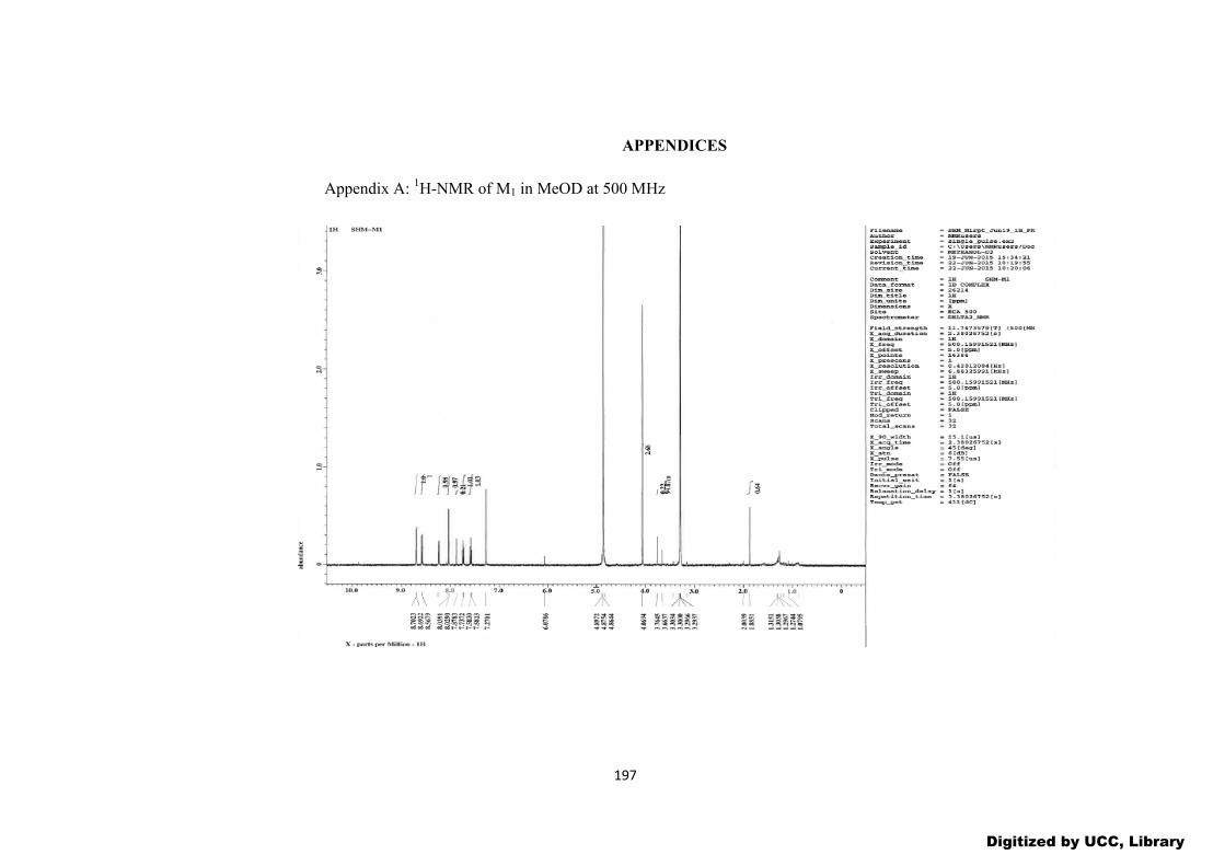
197
APPENDICES
Appendix A: 1H-NMR of M1 in MeOD at 500 MHz
Digitized by UCC, Library

198
Appendix B: Integrated 1H-NMR of M1 in MeOD at 500 MHz
Digitized by UCC, Library

199
Appendix C: 13C-NMR of M1 in MeOD at 500 MHz
Digitized by UCC, Library

200
Appendix D: Expanded 13C-NMR of M1 in MeOD at 500 MHz
Digitized by UCC, Library

201
Appendix E: Mass spectrum of M1
Digitized by UCC, Library
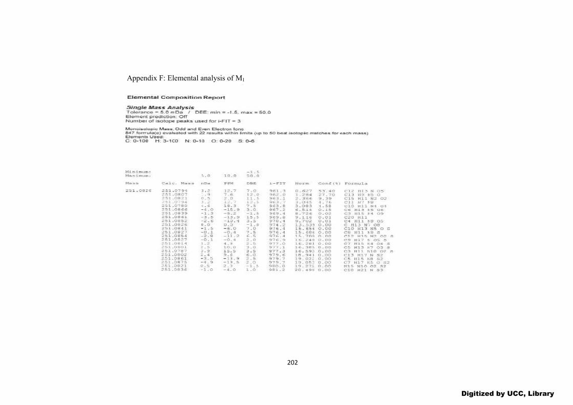
202
Appendix F: Elemental analysis of M1
Digitized by UCC, Library

203
Appendix G: 1H-NMR of M5 in MeOD at 500 MHz
Digitized by UCC, Library

204
Appendix H: Integrated 1H-NMR of M5 in MeOD at 500 MHz
Digitized by UCC, Library
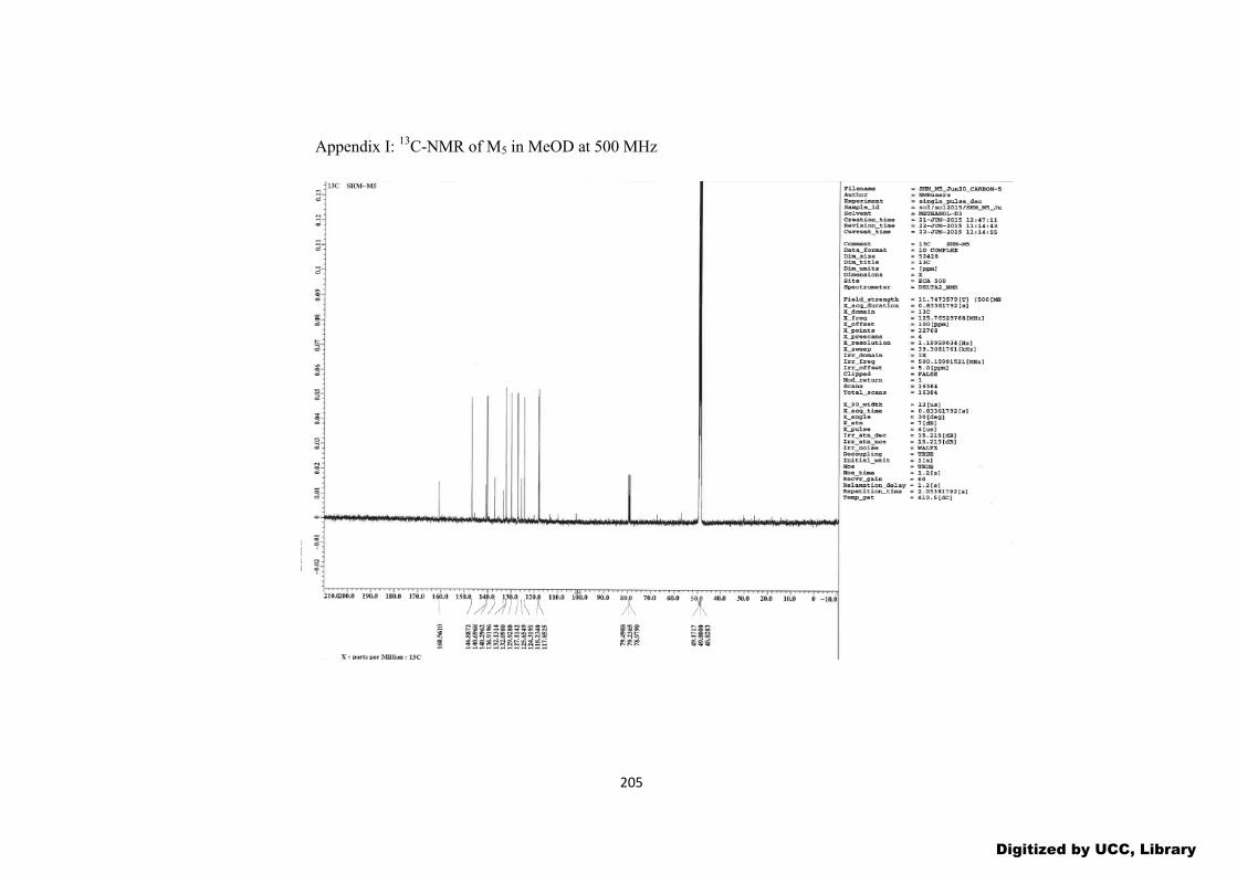
205
Appendix I: 13C-NMR of M5 in MeOD at 500 MHz
Digitized by UCC, Library
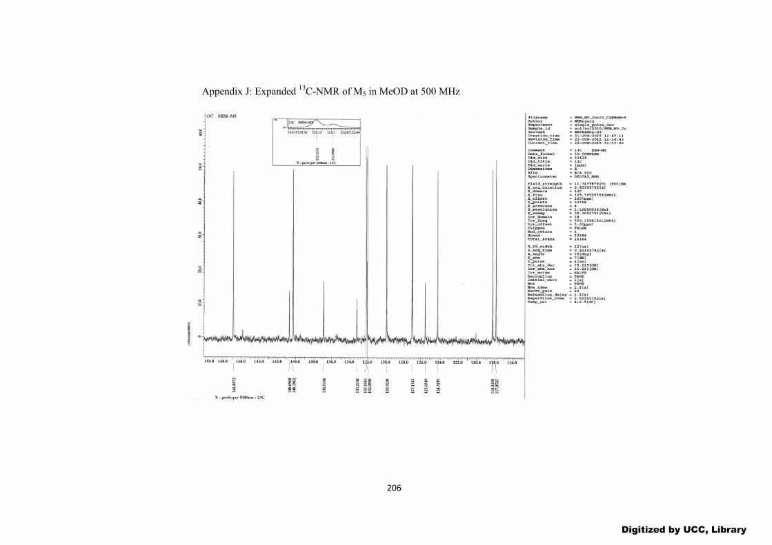
206
Appendix J: Expanded 13C-NMR of M5 in MeOD at 500 MHz
Digitized by UCC, Library
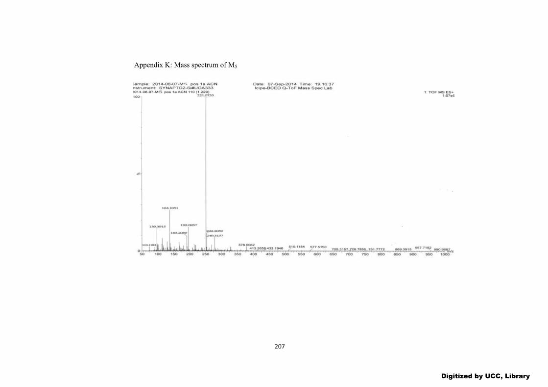
207
Appendix K: Mass spectrum of M5
Digitized by UCC, Library