Embed Size (px)
Citation preview

University of Groningen
Genetics of human cardiovascular traitsVerweij, Niek
IMPORTANT NOTE: You are advised to consult the publisher's version (publisher's PDF) if you wish to cite fromit. Please check the document version below.
Document VersionPublisher's PDF, also known as Version of record
Publication date:2015
Link to publication in University of Groningen/UMCG research database
Citation for published version (APA):Verweij, N. (2015). Genetics of human cardiovascular traits. [Groningen]: University of Groningen.
CopyrightOther than for strictly personal use, it is not permitted to download or to forward/distribute the text or part of it without the consent of theauthor(s) and/or copyright holder(s), unless the work is under an open content license (like Creative Commons).
Take-down policyIf you believe that this document breaches copyright please contact us providing details, and we will remove access to the work immediatelyand investigate your claim.
Downloaded from the University of Groningen/UMCG research database (Pure): http://www.rug.nl/research/portal. For technical reasons thenumber of authors shown on this cover page is limited to 10 maximum.
Download date: 20-02-2019

1 3 71 3 7
f i f t y - t w o
G e n e t i c
l o c i
i n f l u e n c i n G
H u m a n
e l e c t r i c a l l y
a c t i v e
m y o c a r d i a l
m a s s
6.

1 3 9
Pim
van
der
Har
st*,
jess
ica v
an s
ette
n*, n
iek
verw
eij*,
Geo
rg v
ogle
r*,
lude
fra
nke*
, mat
thew
mau
rano
*, X
inch
en w
ang*
, ire
ne m
ateo
lea
ch*,
mar
k ei
jgel
shei
m, n
ona
soto
odeh
nia,
car
olin
e H
ayw
ard,
ros
sella
sor
ice,
oso
rio m
eire
lles,
leo-
Pekk
a ly
ytik
äine
n, ozr
en P
olaš
ek, t
oshi
ko t
anak
a, d
an
ark
in, s
heila
uliv
i, st
ella
tro
mpe
t, m
artin
a mül
ler-n
uras
yid,
alb
ert v
. sm
ith, m
arcu
s dör
r, k
athl
een
f. k
err,
jare
d w
. mag
nani
, fab
iola
del
Gre
co
m.,
wei
hua
zha
ng, i
lja m
. nol
te, c
laud
ia t
. silv
a, s
ando
sh P
adm
anab
han,
vin
iciu
s tr
agan
te, t
õnu
esko
, Gon
calo
r. a
beca
sis, k
arl a
nder
sen,
Ph
il Ba
rnet
t, jo
sh c
. Bis,
rol
f Bod
mer
, Bre
ndan
m. B
uckl
ey, H
arry
cam
pbel
l, m
egan
v. c
anno
n, a
ravi
nda
cha
krav
arti,
lin
y. c
hen,
ale
ssan
dro
del
itala
, ric
hard
B. d
ever
eux,
Pie
ter
a. d
oeve
ndan
s, fr
anci
sco
s. d
omin
gues
, ann
a f.
dom
inic
zak,
lui
gi f
erru
cci,
ian
ford
, chr
istia
n G
iege
r, ta
mar
a B. H
arris
, eric
Hau
gen,
den
a G. H
erna
ndez
, Han
s l. H
illeg
e, a
lber
t Hof
man
, ann
amar
ia io
rio, m
ika k
ähön
en, i
vana
kol
cic,
ishm
inde
r k.
koo
ner,
jasp
al s
. koo
ner,
jan
a. k
ors,
edw
ard
G. l
akat
ta, k
aspe
r lag
e, l
enor
e j. l
aune
r, d
anie
l lev
y, a
licia
lun
dby,
Pete
r mac
farla
ne, d
alit
may
, Th
omas
mei
tinge
r, a
ndre
s met
spal
u, s
tefa
nia
nap
po, s
ilvia
nai
tza,
sha
ne n
eph,
ale
x s.
nor
d, t
ersa
nut
ile, P
eter
m. o
kin,
jesp
er v
. olse
n, B
en
a. o
ostr
a, G
ina
Pelo
so, j
osef
m. P
enni
nger
, len
a. P
enna
cchi
o, s
iegr
ied
Perz
, ann
ette
Pet
ers,
arn
e Pf
eufe
r, m
aria
Gra
zia
Pilia
, Pet
er P
. Pra
mst
alle
r, Br
am P
. Prin
s, o
lli t
. rai
taka
ri, s
oum
ya r
aych
audh
uri,
ken
m. r
ice,
jero
me
i. r
otte
r, d
avid
sch
less
inge
r, c
arst
en o
. sch
mid
t, jo
banp
reet
seh
mi,
Her
man
H.w
. sill
jé, G
ianf
ranc
o si
nagr
a, m
oritz
f. s
inne
r, k
amil
slow
ikow
ski,
elsa
yed
z. s
olim
an, t
imot
hy d
. spe
ctor
, wilc
o sp
ierin
g, jo
hn a
. st
amat
oyan
nopo
ulos
, r
onal
d P.
sto
lk, k
onst
antin
str
auch
, sia
n-ts
ung
tan,
kiri
ll v.
tar
asov
, Bos
co t
rinh,
and
re G
. uitt
erlin
den,
mal
ou v
an d
en
Boog
aard
, cor
nelia
m. v
an d
uijn
, wie
k H
. van
Gils
t, jo
rma
s. v
iikar
i, Pe
ter v
issch
er, v
eron
ique
vita
rt, u
we
völ
ker,
mel
anie
wal
denb
erge
r, c
hris-
tian
X. w
eich
enbe
rger
, Har
m j.
wes
tra,
cisc
a w
ijmen
ga, B
ruce
H. w
olffe
nbut
tel,
jian
yang
, Pat
ricia
B. m
unro
e, H
arol
d sn
iede
r, a
lan
f. w
right
, ig
or r
udan
, lau
rie a
. Boy
er, f
olke
rt w
. ass
elbe
rgs,
dirk
j. v
an v
eldh
uise
n, B
runo
H.c
h. s
ticke
r, Br
uce m
. Psa
ty, m
arin
a ciu
llo, s
eren
a san
na, t
erho
le
htim
äki,
jam
es f
. wils
on, s
tefa
nia
Band
inel
li, a
lvar
o a
lons
o, P
aolo
Gas
parin
i, j.
wou
ter j
ukem
a, s
tefa
n k
ääb,
vilm
undu
r Gud
naso
n, s
teph
an B
. fe
lix, s
usan
r. H
eckb
ert,
rud
olf a
. de B
oer,
chr
istop
her n
ewto
n-c
heh,
and
rew
a. H
icks
, joh
n c
. cha
mbe
rs*,
yald
a jam
shid
i*, a
xel v
isel*,
vin
cent
m
. chr
istoff
els*
, aar
on is
aacs
*, n
ilesh
j sa
man
i* a
nd P
aul i
.w. d
e Ba
kker
, **
Thes
e au
thor
s con
trib
uted
equ
ally
to th
is w
ork.
a B s t r a c t
myocardial depolarisation is a key de-terminant of cardiac muscle contraction and is reflected by the amplitude and duration of the Qrs complex on the electrocardiogram (ecG). increased amplitude and prolonged duration of the Qrs are associated with increased electrically active myocardial mass and greater risk of heart failure and mortal-ity. we carried out a well-powered me-ta-analysis of genome-wide association studies of 4 Qrs traits in up to 73,518 individuals of european ancestry, fol-lowed by extensive biological and func-tional assessment. Here we report the identification of 52 genomic loci, of which 32 are novel, reliably associated with one or more Qrs phenotypes at P<1×10⁻⁸, that mapped to established and putatively novel regulators of left
ventricular mass. we observed enrich-ment in regions of open chromatin, histone modifications, and transcrip-tion factor binding in the human heart suggesting that they represent regions of the genome that are actively tran-scribed. we further highlighted 65 can-didate genes at the identified loci that are preferentially expressed in cardiac tissue and enriched for cardiac abnor-malities in Drosophila melanogaster and Mus musculus. we validated the reg-ulatory function of a novel variant in the SCN5A/SCN10A locus in vitro and in vivo. taken together, our findings provide new insights into the genetic mechanisms and biological pathways controlling electrically active myocar-dial mass and identify novel therapeu-tic targets.

1 4 0 1 4 1
m a i n
The role of the heart is to provide ad-equate circulation of blood to meet the body’s requirements of oxygen and nutrients. cyclical depolarisation of the cardiac ventricular muscle causes contraction and results in blood flow. The Qrs complex on the ecG is the most common measurement of cardi-ac depolarisation. increased amplitude and duration of the Qrs complex1-3 are common alterations of myocardi-al depolarisation and are indicators of increased electrical activity of the left ventricular mass. alterations in the amplitude and duration of the Qrs complex are associated with clinical and preclinical cardiovascular disease such as cardiac hypertrophy and heart failure, and predict cardiovascular mor-tality4,5. identification of specific genes influencing the Qrs complex may thus lead to advances in the prevention of cardiovascular disease and death. two previous genome-wide association stud-ies (Gwass) have identified 22 genetic loci associated with Qrs duration6,7. to further refine our understanding of the genetic factors influencing the Qrs complex, we carried out a large scale Gwas and replication study of 4 related and clinically applied Qrs traits: the sokolow-lyon8,9, cornell9-11 and 12-lead-voltage duration products (12-leadsum)9,11, and Qrs duration9 (supplementary note). multiple ecG markers of increased left ventricular
mass were examined because the sensi-tivity of any one of these markers alone is relatively limited and because their performance can vary with sex, eth-nicity and body characteristics.12 our study design is summarized in figure s1. findings of variants convincingly associated with the Qrs complex traits (P<1×10⁻⁸) were then examined in other ethnicities and studied for possible pro-tein disrupting variants and co-locali-sation with regulatory dna elements. furthermore, we prioritised candidate genes by studying their cardiac gene-ex-pression profiles and the consequenc-es of gene knock-downs in Drosophila melanogaster and Mus musculus. Briefly, we combined Gwas summary data from 24 studies with up to 2,766,983 autosomal snPs. These studies together comprise 60,255 in-dividuals of european ancestry ascer-tained in north america and europe, with a maximum sample size of 54,993 for sokolow-lyon, 58,862 for cornell, 48,632 for 12-leadsum, and 60,255 for Qrs duration. characteristics of participants, genotyping arrays and imputation are summarized in supple-mentary tables 1 and 2. for each snP, evidence of association was combined across studies using an inverse-variance fixed-effect meta-analysis carried out by two independent meta-analysis groups. we performed replication testing for 35 loci showing suggestive association
(1×10⁻⁸<P<5×10⁻⁷) in 12,838 individu-als using a combination of in silico data and direct genotyping (supplementary tables 1, 2, and supplementary note). The threshold for genome-wide signifi-cance was set at P<1×10⁻⁸, allowing for a conservative Bonferroni correction for both the ~106 effective independent snPs associations tested13,14, and the 4 inter-related Qrs phenotypes (supple-mentary note). across the genome, 52 inde-pendent loci, 32 of which are novel, reached genome-wide significance for association with one or more Qrs phe-notypes (figure 1, figure s2, and table s3). for descriptive and downstream purposes, we defined a single ‘sentinel’ snP for each locus with the lowest P-value against any of the four pheno-types; regional association plots for the 52 loci are shown in figure s3. full lists of the sentinel snPs and the snPs as-sociated with any phenotype at P<10-6 are in supplementary tables 3 and 4. among the 52 known and novel loci, 32 were associated with one Qrs pheno-type, and 20 with two or more pheno-types (figure s4). The total number of locus-phenotype associations at P<10-8 was 79 (72 snPs), of which 59 are novel (table s5). all previously known Qrs duration loci showed evidence for as-sociation (P<10-6, table s6). among the 32 novel loci, 8 demonstrated ge-nome-wide significant association with sokolow-lyon, 9 with cornell, 20 with 12-leadsum, and 9 with Qrs duration. collectively, the total variance explained by the 52 sentinel snPs for the Qrs
traits was between 2.7% (sokolow-ly-on) and 5.0% (Qrs-duration) (table s7). in addition, we found evidence for 16 snPs at 5 of the 52 loci that were not in ld with the corresponding senti-nel snP but were associated with Qrs phenotypes at P<10-8 in conditional analyses15, suggesting independent as-sociations at these loci (table s8 and supplementary note). among the 52 loci identified, 7 have been associated previously with Pr interval (reflecting atrial activity and av node function), 3 with Qt duration (ventricular repo-larisation) and 1 with heart rate (sinus node function) (table s6), indicating at least some overlap in the genetic deter-minants of different cardiac measures. our primary Gwas and repli-cation was based on individuals of eu-ropean ancestry. to assess whether our findings are also relevant in non-eu-ropean individuals, we evaluated the directional consistency of the associ-ations in >3,600 african americans and >4,600 indian asians (figure s1, table s9, and supplementary note). in the african american sample, 35 of 51 available locus-phenotype associations had the same direction of effect as seen in the european sample (P=3.19×10⁻³, binomial test) and in the indian asian sample, 22 of 29 available locus-pheno-type associations showed the same di-rection of effect (P=2.91×10⁻³). Thus, in concordance with observations for other traits16, a large proportion of common variants identified in this study likely contribute to cosmopolitan genetic ar-chitecture of Qrs traits.

1 4 2 1 4 3
region snp maf pheno p
1p36.12 rs2849028 0.26 12ls 1.69e-091p32.3 rs17391905 0.04 dur 1.07e-111p31.3 rs2207790 0.47 dur 6.71e-191p13.1 rs12039739 0.29 dur 6.51e-091q22 rs2274317 0.32 12ls 1.82e-11
1q23.3 rs12036340 0.24 12ls 1.49e-091q32.1 rs10920184 0.38 cor 5.01e-091q32.1 rs4288653 0.23 12ls 3.50e-102p23.3 rs6710065 0.42 cor 4.12e-092p22.2 rs3770770 0.20 dur 4.95e-112q31.2 rs3816849 0.46 12ls 5.77e-153p22.2 rs6801957 0.42 dur 6.90e-403p21.1 rs4687718 0.13 dur 6.70e-103p14.1 rs2242285 0.42 dur 5.65e-093p14.1 rs13314892 0.23 12ls 8.93e-093q27.2 rs10937226 0.35 12ls 6.82e-164p15.31 rs1344852 0.16 dur 1.21e-095q33.2 rs13185595 0.37 cor 2.10e-246p21.31 rs1321311 0.27 dur 1.03e-376p21.1 rs1015150 0.45 sok 1.28e-09
6q22.31 rs11153730 0.50 dur 7.44e-297p14.3 rs1419856 0.16 dur 6.67e-187p12.3 rs6968945 0.44 dur 5.14e-097q31.2 rs11773845 0.41 dur 7.50e-10
8q24.13 rs4367519 0.04 sok 4.15e-118q24.13 rs10105974 0.36 12ls 6.25e-1110q21.1 rs1733724 0.26 cor 1.75e-1410q21.3 rs12414364 0.21 12ls 1.22e-1010q21.3 rs10509289 0.11 12ls 9.16e-1110q22.2 rs7099599 0.15 12ls 5.51e-1310q25.2 rs7918405 0.26 dur 1.05e-1411p11.2 rs2269434 0.32 cor 7.38e-1011q12.2 rs174577 0.34 dur 4.28e-11
12q13.13 rs736825 0.36 cor 7.20e-1112q13.3 rs2926743 0.27 12ls 3.74e-26
12q24.21 rs7132327 0.27 12ls 1.27e-1713q14.13 rs1408224 0.31 12ls 3.60e-1013q22.1 rs728926 0.38 dur 5.60e-1114q24.2 rs12880291 0.26 dur 4.41e-1415q25.3 rs7183401 0.44 12ls 1.10e-2615q26.3 rs8038015 0.38 12ls 4.93e-2116q23.3 rs6565060 0.08 12ls 6.30e-1317q11.2 rs7211246 0.46 12ls 6.01e-09
17q21.31 rs242562 0.38 12ls 1.57e-1417q24.2 rs9912468 0.44 sok 3.11e-1218q12.1 rs617759 0.33 12ls 5.63e-1018q12.2 rs879568 0.33 dur 8.45e-0918q12.3 rs10853525 0.42 dur 1.41e-1420p12.3 rs3929778 0.20 cor 6.42e-0920q11.22 rs2025096 0.21 cor 4.51e-1121q21.1 rs7283707 0.13 12ls 3.76e-0921q21.3 rs13047360 0.18 dur 4.02e-10
sentinel- log 10 (p value)
candidate genes
znf436n,c1orf213n
cdkn2cn
nfian
casQ2ng
mef2dng
olfml2Bn
tnnt2ng
PlekHa6n
dPysl5n
strnn
ttnn,g
scn10ane
tktn
lriG1n,slc25a26n
mitfng
senP2nce
slit2n
Hand1ng
cdkn1an,e
tfeBn,g
slc35f1n,Plng,ceP85lc
tBX20n,g
tns3n
cav1ng
klHl38nc,fBXo32g
mtss1n
dkk1ng
ctnna3ng
ctnna3ng
sec24cn,synPo2lc
vti1an
myBPc3ng,maddn,nr1H3e,acP2c
fads2n
HoXc4n,HoXc6n,HoXc5n
nacanc
tBX3ng
lrcH1n
klf12n
siPa1l1n
alPk3n
iGf1rn
cdH13ng
nsrP1n,efcaB5c
maPtn
Prkcan
maPre2n
fHod3n,c
setBP1n
BmP2n
Gssn,edem2ec,myH7Bc
usP25n
adamts5n
fig1
0.0
2.0
4.0
6.0
8.1
0 10 20 30 40 50
1 4 4 1 4 5
# of
mat
ched
rand
om re
plic
ates
, Hap
map
% snPs in dHss of human fetal heart
fig.2a
figure 1overlay manhattan plot showing the results for the ge-nome-wide associations with Qrs traits amongst europe-ans. snPs reaching genome-wide significance (P<1x10⁻⁸) are coloured dark grey (novel loci) or light grey (previously reported loci).
52 Gwa sentinal snPs
P=7.7x10⁻¹²

Poise
d en
hanc
ers
0
0.5
1
1.5
2
2.5
3
3.5
0 1 2 3 4 5 6 7 8 9 10
0
0.5
1
1.5
2
2.5
3
3.5
0.001 0.01 0.1 1 10 100 1000 1000 0
0
0.5
1
1.5
2
2.5
3
3.5
0 1 2 3 4 5 6 7 8 9 10
Repressive marks Activation marks
1 4 6 1 4 7
average human fetal heart (n=12)average other human tissue (n=337)
95% c.i
enric
hmen
t (fo
ld)
enric
hmen
t (fo
ld)
z=4.91 P=2.30e-06
distance treshold (kb), log scale
Gwas -log10(p) value treshold
enrichment (fold)
act
ive
enha
ncer
s
enric
hmen
t (fo
ld)
Gwas -log10(p) value treshold
fig.2b
fig.2b fig.2d
fig.2c
5.54
6.42
6.73
5.48
6.62
2.97
3.59
2.06
0 2 4 6 8
0.02
1.6E-4
1.5E-3
1.8E-11
2.2E-8
8.3E-12
6.7E-11
1.5E-8
ESC
MES
CP
CM
ESC
MES
CP
CM
H3k27acH3k4me3H3k4me1
H3k36me3H3k27me3
H3k9me3

adul
tfe
tal
3 .2 3
0 .5 0
2 .6 9
2 .2 8
2 .0 4
4 .3 6
4 .9 5
4 .6 7
1 .7 5
4 .9 6
4 .4 0
0 1 2 3 4 5 6
1 .E -2 0
0.3
2 .E -1 6
1 .E -1 0
4 .E -1 7
4 .E -2 3
3 .E -6
2 .E -1 6
0.07
1 .E -2 0
1 .E -8
SRF
Nkx2-5
TBX5
GATA4
TBX3
Nkx2-5
p300
p300
RNAP2
P value
MEF2
GATA4
1 4 8 1 4 9
Hl1
cel
l lin
e(m
ouse
), H
e a
etal
. Pro
c n
atl a
cad
sci u
sa 2
011
mou
se h
eart
(adu
lt), v
an d
enBo
ogaa
rd e
t al.
jc
lin in
vest
201
2
Hum
an h
eart
,m
ay d
et a
l. n
atG
enet
201
1
enrichment (fold)
figure 2.aThe 52 sentinel snPs are significantly enriched in dHss of the human foetal heart compared to the matched random
distribution of Hapmap snPs. b
furthermore, we detect higher enrichment of Qrs-trait snPs in dHss of human foetal heart (n=12) compared to all other tissues and cells (n=337) across the full range of P-val-
ues. c
The impact of physical distance between snPs that meet ge-nome wide significance (P<1×10⁻⁸) on enrichment of foetal heart relative to all other tissues at dHs. The enrichment is strongest at the snP’s location and decreases after 100bp
from the snP sites.d
snPs associated with Qrs traits are enriched for the activa-tion histone modifications H3k27ac, H3k4me3, H3k4me1 and H3k36me3 in human left ventricle. The repressive mark H3k27me3 is not enriched while H3k9me3 is significantly reduced, suggesting that Qrs-trait loci are predominantly
expressed in the left ventricle.e
enrichment of the 52 loci for histone modifications during cardiomyocyte differentiation (mouse). enhancers are anno-tated by H3k4me1 peaks at least +/- 1kb away from an an-notated tss and designated as active or poised based on the
presence (active) or absence (poised) of H3k27ac. f
snPs (P<1×10⁻⁸) were also significantly enriched for vari-ous factors in the human heart, mouse heart and the Hl-1
cell-line.
fig.2f

1 5 0 1 5 1
a regulatory role for qrs associa-tions
to better capture common sequence variants at the 52 loci, we queried the 1000 Genomes Project dataset17, and identified 41 non-synonymous snPs in 17 genes that are in high ld (r2>0.8) with 12 of our sentinel snPs (table s10). although this is not a significant enrichment compared to the expecta-tion under the null hypothesis (P=0.08, supplementary note), these non-syn-onymous sites represent an initial set of candidate variants that may have an effect on the Qrs phenotypes through changes in protein structure and func-tion. further sequencing studies will be essential to achieve a more complete as-sessment of potentially causal variants. approximately 80% of the lead signals from the combined traits were not in strong ld with known non-synony-mous protein-coding variants, consis-tent with most of the strongest signals reported here resulting from variation in non-coding functional sequence. to assess the importance of gene expression regulatory elements in Qrs complex genetic architecture, we tested the loci identified in this study for enrichment of deoxyribonuclease i (dnase i) hypersensitive sites (dHss), which are experimentally determined markers of regulatory activity and whose cell-type specific patterns encode early developmental fate decisions18. cardiac-specific gene expression pro-grams can be regulated by the binding of key developmental transcription fac-tors (tfs) such as tbx5 and nkx2-5.
when considering 349 diverse cell lines, cultured primary cells and foe-tal tissues 19 mapped by the encode project20 and the niH roadmap epig-enomics Program21, 42 (81%) of our 52 sentinel snPs lie in dHss. we found that 22 (42%) of our 52 loci overlapped dHss collected from human foetal heart tissue sampled days 96-147 after conception, representing a ~3.5-fold enrichment compared to permuta-tion testing with 52 randomly selected snPs matched to our sentinel snPs (P=7.7×10⁻¹², figure 2a, supplemen-tary note). This suggests a regulatory role specific to cardiac tissue. when we considered snPs below genome-wide significance, we observed continued enrichment in dHs that diminished at the most liberal P-value thresholds (figure 2B). The enrichment of ge-nome-wide significant snPs (P<1×10⁻⁸) in dHss was strongest within the first 100 bp around the sentinel variants (figure 2c), suggesting a specific con-centration of Qrs-associated variation in functional regulatory dna. cardiac-specific regulatory patterns are encoded during develop-ment by sequence-specific factors such as tBX5 and nkX2-5, which recruit activating and repressive factors to im-pose characteristic covalent modifica-tions of histone proteins at regulatory sites22,23. we investigated the enrich-ment of identified variants in regions of covalently modified histones in hu-man cardiac tissue mapped by the niH roadmap epigenomics Program (sup-plementary note and table s11).21,24
we observed a strong enrichment for histone marks associated with active enhancers, promoters and transcription (H3k4me1, H3k4me3, H3k27ac, and H3k36me3) which increased at more stringent Gwas P-value thresholds; by contrast no enrichment was observed for transcriptionally repressive histone marks (H3k9me3 and H3k27me3) (figure 2d). Given the potential for genetic variants to influence the Qrs complex through alterations in cardiac development, we also investigated the chromatin landscapes of mouse embry-onic stem cells (esc) differentiated in culture into mesoderm (md), cardiac precursor (cP), and cardiomyocytes (cm) 23. we observed that the enrich-ment of significant genomic regions for activating histone marks matched the degree of differentiation towards cardiomyocytes (figure 2e). together, these data suggest that trait-associated snPs are enriched in regulatory ele-ments and may alter the recruitment of sequence-specific tfs, leading to pathogenic transcriptional dysregula-tion19. to test this hypothesis, we surveyed our genome-wide significant snPs in dHss for perturbation of tf recognition sequences, because since these sites can point directly to binding events (supplementary note). of the 22 sentinel snPs in foetal heart dHss, 11 are predicted to alter tf recognition sequences (table s12). when consider-ing all significant snPs as well as those in high ld (r2>0.8), 402 snPs in the colocalising dHss perturb transcrip-
tion recognition sequences, including those of important cardiac and muscle developmental regulators like tBX, Gata-4, and mef2. we intersect-ed our Gwas results with chiP-seq profiling of mouse and human cardiac tissue25-27 and observed enrichment in enhancers marked by p300, sites bound by rna Polymerase ii (rnaP2), and the transcription factors nkX2-5, Gata-4, tBX3, tBX5, and srf (figure 2f). nine of our 52 loci both have overlapping foetal heart dHss and chiP-seq validated tf sites. snPs overlapping tf binding sites were 5.65 fold enriched within dHss (P=9.0×10-
10) but not outside dHss (P=0.20).in vitro and in vivo validation of a func-tional cardiac enhancer to confirm the presence of cardiac functional enhancers in our identified regions in vivo by direct ex-perimentation, we examined whether enhancer candidate sequences present in these intervals are sufficient to drive reporter gene expression in the heart in transgenic mouse assays (supplementa-ry note). focusing on regions up to 200 kb away from the sentinel snPs, we validated several candidate regions pre-dicted by chiP-seq as described above as reproducible in vivo heart enhancers. four examples of newly identified en-hancers with reproducible in vivo cardi-ac activity located in the vicinity of lead snPs are shown in figure 3a, addi-tional examples of previously described enhancers near lead snPs are provided in figure s5 24,26. recently, rs6801957 (figure 1) in the SCN5A/SCN10A locus

fig.3a
100kbrs6781009
SCN5A SCN10AEXOGACVR2B
Scn5a Prom .
Cornell
Sokolow
Leadsum
TBX3
Tbx5
p300
QRS duration
ChI
P4C
GW
ASC
EU L
OD
2kb50kb
Contact
10.10.010.001
20th %ile
80th %ileMedian
Window coverage(median)
Main trend
1 5 2 1 5 3
was reported to affect gene expression of another regulatory element near SC-N5A/SCN10A through the alteration of a t-box factor binding sequence 27. our conditional analysis (table s8) revealed that rs6781009 is an additional novel independent signal at this locus. This variant is located in a mouse and hu-man heart dHs region highly occupied by activating chromatin marks and tfs (tbx3, tbx5 and P300) in heart tissue (figure 3B), suggesting that this vari-ant represents a potential additional cardiac regulatory element near SC-N5A. Given that enhancers are thought to function by physically contacting promoters through dna looping, we performed 4c-seq analysis oe human ventricular tissue and found an interac-tion between the Scn5a promoter and ths region around rs6781009 (figure 3B). evaluation of the in vivo activity pattern of thn regulatory element with a lacz reporter vector showed specific expression in the interventricular sep-tum for the major allele, which is ab-sent for the minor allele (figure 3c), resembling the endogenous expression pattern of both Scn5a and Scn10a27,28. furthermore, functional analysis using a luciferase reporter assay in H10 cells29 showed high constitutive activity of the human enhancer element containing the major allele for rs6781009, which was reduced when the minor allele was introduced into a large enhancer fragment or the enhancer core element (figure 3d). collectively, our results confirm the presence of in vivo heart enhancers in genome regions associated
with Qrs traits, and provide a starting point to identify and explore the mech-anism of potentially causative variants outside protein-coding sequences.
identification of candidate genes
across the 52 loci, 974 annotated genes are located within 1 mb of a sentinel snP. among these genes, we prior-itized potential candidates using an established complementary strategy (supplementary note) 30,31; we chose (i.) Genes nearest to the sentinel snP, and any other genes within 10kb (56 genes; figure 1); (ii.) Genes contain-ing a non-synonymous snP in high ld (r2>0.8) with the sentinel snP (11 genes; table s10); (iii.) Genes with cis-eQtl associated with sentinel snP in peripheral blood mononucleated cells (8 genes; table s13), and (iv.) Grail literature analysis32 (16 genes table s14) with top 3 keywords retrieved from Grail describing the observed functional connections being ‘cardiac’, ‘muscle’, and ‘heart’. in total, this strat-egy identified 65 candidate genes at the 52 loci (figure 1). Pathway analysis confirmed that the list of 65 candidate genes is strongly enriched for genes known to be involved in cardiac hypertrophy, car-diovascular disease and developmental disorder (P=1×10−39; supplementary note)). The top 3 functional annota-tions for the 65 candidate genes were ‘development of cardiac muscle’ (7 can-didate genes, P=1×10⁻⁹), ‘morphogenesis of cardiac muscle’ (6 candidate genes, P=3×10⁻⁹) and ‘heart disease’ (19 candi-
fig.3b
hs2177chr3:38594857-38597991 scn5a
hs2188c h r 6:118963898-118966469ceP85l,Pln
hs2266chr3:38547891-38551036eXoG
hs2271chr1:26292440-26297330
0
1
2
3
4
5
6
Major Minor Major Minor
Enhancer
(1.5kb)
Enhancer core
(0.6kb)
rs6781009
T>C
*
rv
ra
lv
la
5/6ivs
rv
ra
lv
la
0/3*ivs
1 5 4 1 5 5
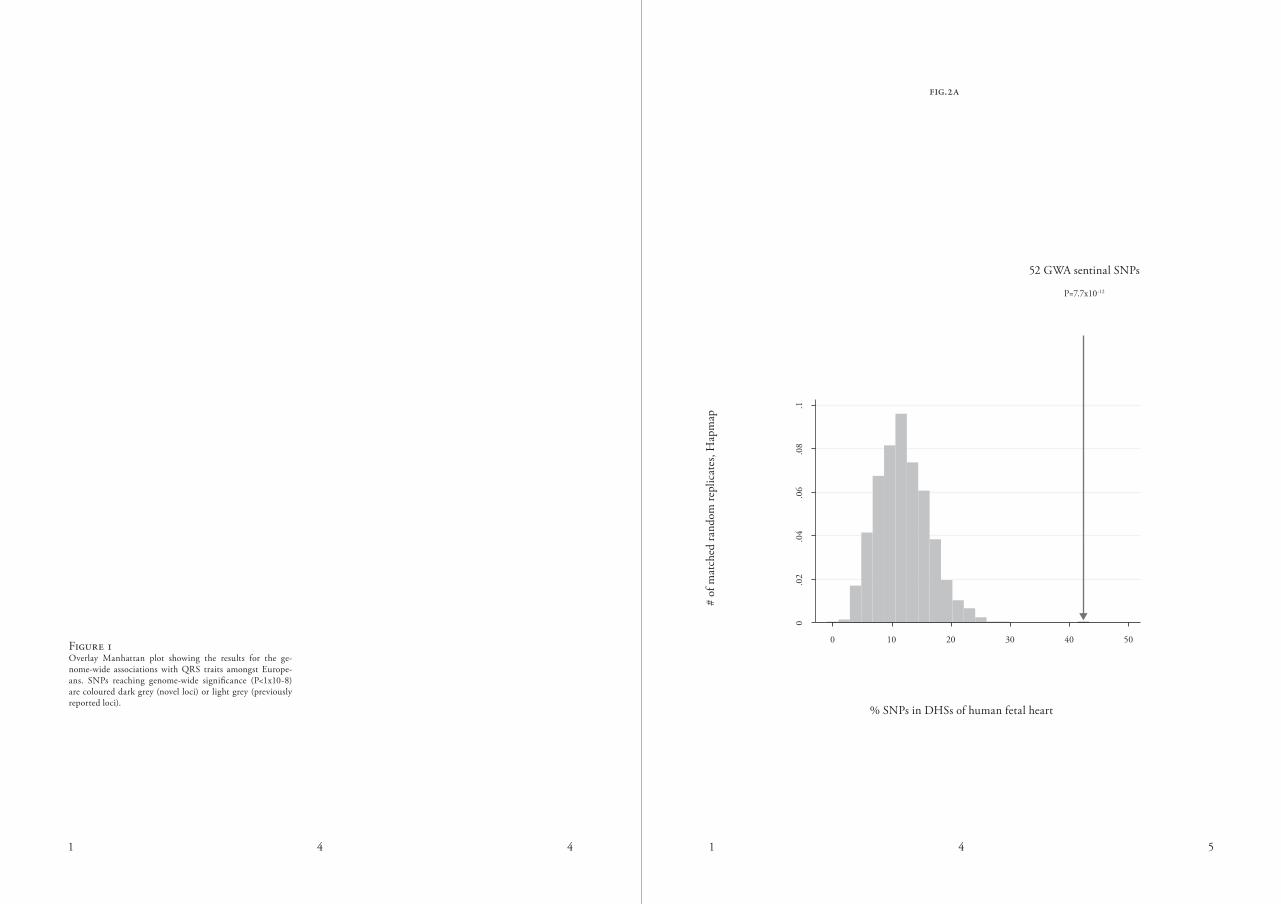
figure 3.ain vivo activity of exemplar human cardiac enhancers in em-bryonic transgenic mice stained for lacz enhancer reporter
activity (dark blue). b
Position of the regulatory element containing rs6781009 plotted over the Gwas signals (-log(P) on the SCN5A-SC-N10A locus. The regulatory element is bound by tbx3, tbx5, and P300 (lower black traces) in mouse, and the contact profile of the scn5a promoter obtained by 4c-seq in human ventricular tissue revealed an interaction between this regulatory element and the scn5a promoter (upper black trace and contact profile). normalized contact intensities (gray dots) and their running median trends (black line) are depicted for the scn5a promoter viewpoint. medians are computed for 4 kb windows and the grey band displays the 20–80% percentiles for these windows. Below the profile, statistical enrichment across differently scaled window sizes (from 2 kb (top row) to 50 kb (bottom)) is depicted of the observed number of sequenced ligation products over the expected total coverage of captured products, with the latter being estimated based on a probabilistic background model. local changes in colour codes indicate regions statistically
enriched for captured sequences.c
luciferase assay performed in H10 cells showing a high con-stitutive activity for the enhancer core element (0.6kb) con-taining the major allele for rs6781009, which is reduced for the minor allele in both a large enhancer construct (1.5kb),
as well as in the core enhancer element (0.6kb) *P<0.01 d
dorsal views of hearts containing the human regulatory ele-ment with the major vs minor allele for rs6781009 in a lacz reporter vector, showing specific expression of the enhancer in the interventricular septum (ivs, black bars) for the major allele, which is absent for the minor allele *P<0,05. ra, right
atrium; la, left atrium; rv, right ventricle; lv, left ventricle.
m a j o r m i n o r
fig.3c
fig.3d

Heart Atria
P = 2.8 x 10⁻⁶
CandidateGenes
OtherGenes
Heart
P = 4.1 x 10⁻⁶
CandidateGenes
OtherGenes
Heart Ven-tricles
P = 1.2 x 10⁻⁵
CandidateGenes
OtherGenes
Muscles
P = 1.8 x 10⁻⁵
CandidateGenes
OtherGenes
-2
0
2
4
6
8
10
12
P = 0.088
CandidateGenes
OtherGenes
P = 0.007
CandidateGenes
OtherGenes
-2
0
2
4
Embryonic Stem Cell
Myocyte Embryonic Stem Cell
P = 0.157
CandidateGenes
OtherGenes
Cardio-myocyte
Progenitor
P = 8.7 x 10 -8
CandidateGenes
OtherGenes
Cardio-myocyte
1 5 6 1 5 7
fig.4a
rel
ativ
e ge
ne e
xpre
ssio
n
rel
ativ
e ge
ne e
xpre
ssio
n
CT
NN
A3
P
LN
M
ITF
M
YH
7B
M
YB
PC
3
SY
NP
O2
L
SL
C3
5F
1
CD
H1
3
NF
IA
OL
FM
L2
B
NA
CA
F
BX
O3
2
FH
OD
3
ED
EM
2
AD
AM
TS
5
CA
SQ
2
CA
V1
C
DK
N1
A
TT
N
TN
NT
2
SE
C2
4C
M
EF
2D
U
SP
25
M
AP
RE
2
PR
KC
A
AL
PK
3
TB
X2
0
IGF
1R
L
RC
H1
S
LIT
2
BM
P2
N
R1
H3
V
TI1
A
SE
TB
P1
A
CP
2
ST
RN
P
LE
KH
A6
S
IPA
1L
1
DK
K1
D
PY
SL
5
HA
ND
1
FA
DS
2
TB
X3
L
RIG
1
MA
DD
S
EN
P2
K
LF
12
T
KT
M
AP
T
TN
S3
G
SS
M
TS
S1
1 5 8 1 5 9
embryonic stem cell
myocyte embryonic stem cell
cardiomyocyte Progenitor
cardiomyocyte
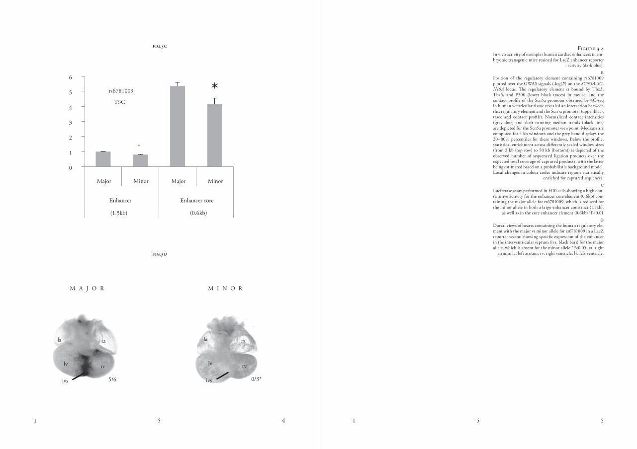
figure 4.awithin heart and muscle tissue microarray-based data the candidate genes are significantly more highly expressed as
compared to non-candidate genes. B
The 54 candidate genes are highly expressed in rna-seq data of cardiomyocytes, compared to non-candidate genes.
c unsupervised hierarchical clustering of rna-seq based expression data of 54 candidate genes in 4 different car-diomyocyte (precursors) reveals that most of the genes are
abundantly expressed in cardiomyocytes.
fig.4b
Gene expression
low High

1 6 0 1 6 1
date genes, P=8×10⁻⁹) (supplementary tables 15 and 16). current knowledge on gene function for all 65 candidates is summarized in table s17. a systematic search in the online mendelian inher-itance in man (omim) catalogue re-vealed that several candidate genes are known to cause familial cardiomyopa-thies (TNNT2, TTN, PLN, MYBPC3) or cardiac arrhythmias (CASQ2) in humans (table s17). we also identified genes that are associated with atrial sep-tal defects (TBX20) and more complex syndromes involving cardiac abnormal-ities such as the schinzel-Giedion mid-face retraction syndrome (SETBP1)33 and the ulnar-mammary syndrome (TBX3)34.
insights from gene exPression Profil-ing and model organisms
we explored gene expression profiles of our candidate genes in data derived from 37,427 affymetrix u133 Plus 2.0 arrays across 40 annotated tis-sues (supplementary note). of our 65 candidate genes we could reliably as-sign a probe for 63 genes; these tran-scripts were on average expressed at higher levels in cardiac-derived sam-ples compared to other transcripts in the same sample (P=4.1×10⁻⁶ for heart tissue; wilcoxon test; figure 4a). fur-ther, expression of these transcripts was expressed at higher levels in cardiac-de-rived samples compared to other tissues (P=0.006 after Bonferroni correction; figure s6). to further investigate the potential role of these candidate genes in cardiac development, we assessed
temporal gene expression patterns during in vitro differentiation of mouse embryonic stem cells (esc) via meso-derm (md) and cardiac precursor (cP) cells to cardiomyocytes (cm). eight percent of genes are mainly expressed during the esc stage, 19% during md stage, 8% in the cP stage and 62% in the cardiomyocyte stage (fig-ure 4B). compared to other genes, the candidate genes are high expressed in cardiomyocytes (P=4.7×10⁻⁸; wilcoxon test; figure 4c). These results suggest that our candidate gene set is enriched for genes relevant to cardiac biology, and include a number of genes differ-entially expressed in cardiac tissue and increasingly expressed during cardiac development. next, we analysed data from model organisms to explore the func-tion of the selected candidate genes (supplementary note). from cardi-ac tissue-specific rnai knockdown data collected in D. melanogaster35, we found that the 65 candidate genes were 2.3-fold enriched for stress-in-duced premature cardiac death (9 genes, P=5.4×10⁻³; figure s7a). four of these genes had been studied pre-viously in Drosophila and shown to have cardiac abnormalities in Drosoph-ila (Mhc/myH7B36, Slit/slit237, EcR/nr1H38, Hand/Hand139). Perform-ing heart-specific rnai knockdown with the cardiac Hand4.2-Gal4 driver line (supplementary note), we re-test-ed EcR, which has multiple homolo-gous genes in mammals, and Hand as well as the remaining four genes where
fig.5

1 6 2 1 6 3
the cardiac phenotype was not known (we did not test titin, since it is a well-known cardiomyopathy gene40). adult hearts of EcR/nr1H, NACα/naca, Hand/Hand1 and cka/strn rnai showed severe cardiac defects (figure 5). knockdown of Hand/Hand1 and Cka/strn both had a reduced cardi-ac heart rate (figure s8). while Hand/Hand1 knockdown hearts appeared structurally normal, we observed se-verely disorganized and misoriented myofibrillar arrangements within the cardiomyocytes in Cka/strn rnai hearts (figure 5), which caused a re-duction in diastolic diameters and frac-tional shortening (suppl. figure s7c). naca/naca mutants had the most severe phenotype with a complete loss of cardiac tissue beginning at eclosion, while the hearts of naca mutant lar-vae were still intact, indicating a critical role for naca during cardiomyocyte remodelling. rnai-mediated knock-down of CG4743/slc25a26 and Fhos/fHod3 did not reveal cardiac pheno-types. in addition, from the mouse Genome informatics database, knock-out models were annotated for 44 of the 65 orthologous genes, of which 18 (41%) revealed a cardiac phenotype (table s17). This represents a 5.4-fold enrichment compared to randomly matched sets of 65 genes (P=4.8×10⁻¹⁶; figure s7B). This further demonstrates the relevance of these genes for cardiac function.
for some loci, there are addi-tional candidate genes. a notable exam-ple is the 11p11.2 locus, which harbours multiple candidate genes (figure 1), in-cluding MYBPC3, ACP2, MADD, and NR1H3. MYBPC3 deficiency is well established to cause hypertrophic and dilated cardiomyopathies in both hu-man and mouse models and provides a plausible candidate gene (table s17). in addition to MYBPC3, eQtl and his-tone modification data also suggests a potential role for NR1H3 (figure s9). decreased expression of NR1H3 was associated with higher Qrs voltages. However, NR1H3 deficient mice do not spontaneously develop a cardiac hyper-trophic phenotype (mGi: 1352462). to study the potential cardiac effects of NR1H3 overexpression, we created a transgenic mouse with cardio-specif-ic overexpression of NR1H3 under the control of the Myh6 promoter. while the wild-type mouse was sensitive to perturbations (such as transverse aortic constriction and angiotensin 2 infu-sion) that provoke cardiac hypertro-phy41, these effects were absent in the transgenic mouse. This observation is in line with protective effects due to treatment with t0901317, a synthetic NR1H3 agonist, in mice challenged with aortic constriction42. These data highlight the importance of systematic approaches to identify causal genes be-yond identification of a first recognis-able candidate.
figure 5.cardiac defects upon heart-specific rnai knockdown in drosophila . (a) wild-type dorsal heart tube stained with the f-actin stain phalloidin. magnified region (right) is high-lighted. arrowheads point to ostia (inflow tracks), arrow shows the circumferential orientation of myofibrils. (b) cka/striatin rnai induces myofibrillar disarrangement. myofi-brils are oriented in a disorganized, mainly anterior-posteri-or orientation with gaps in between (arrow). (c) knockdown of naca/naca causes severe cardiac tissue disintegration. adult cardiomyocyte tissue may be complete absent (aster-isk), while some heart-associated longitudinal muscles are still present (arrowheads). at larval stages the heart is much less affected, suggesting a maturation or remodeling defect. (d) knockdown of ecr/nr1H blocks cardiac remodelling and causes myofibrillar disarray (arrow). ventral longitudi-nal muscles are also abnormal (arrowhead).

1 6 4 1 6 5
c o n c l u s i o n s
we report a meta-analysis of Gwas in 73,518 individuals for 4 quantitative Qrs phenotypes and identify 52 inde-pendent genetic loci influencing these traits with 79 locus-phenotype associ-ations; the majority of these discoveries are novel. our analyses prioritized a set of candidate genes that potentially im-pact cardiac function, which we expect to facilitate in-depth studies towards identifying definitive mechanisms. our loci co-localised with open chromatin, histone modification, and tf binding sites specifically in cardiac tissue, and provide examples of in vivo functional enhancers within the identified loci. we also provide direct evidence that
rs6781009, located within a predicted enhancer, interacts with the promoter of SCN5A to modify expression lev-els. in parallel, we have prioritized a set of 65 genes based on differential gene expression in the human heart, making them promising candidates for mediating effects on Qrs phenotypes. further functional support is obtained from genetic overlap with inherited mendelian disorders and from model organism studies. we anticipate that in-depth functional studies will now detail mechanisms affecting cardiac function and advance understanding of cardiac hypertrophy and heart failure.
m e t H o d s
a summary of the methods can be found in supplementary information and includes detailed information on: study populations; genotyping meth-ods and quality control; electrocardio-
graphic measurements; genome-wide association and meta-analysis methods; gene prioritisation strategies; experi-mental data sets and analytical meth-ods; in vivo and in vitro experiments.
l i t e r a t u r e

1 6 6 1 6 7
1. levy, d. et al. determinants of sen-sitivity and specificity of electrocardiographic criteria for left ventricular hypertrophy. Circu-lation 81, 815-20 (1990).
2. devereux, r.B., koren, m.j., de sim-one, G., okin, P.m. & kligfield, P. methods for detection of left ventricular hypertrophy: application to hypertensive heart disease. Eur Heart J 14 suppl d, 8-15 (1993).
3. okin, P.m. et al. time-voltage Qrs area of the 12-lead electrocardiogram: detection of left ventricular hypertrophy. Hypertension 31, 937-42 (1998).
4. kannel, w.B., Gordon, t. & offutt, d. left ventricular hypertrophy by electrocar-diogram. Prevalence, incidence, and mortality in the framingham study. Ann Intern Med 71, 89-105 (1969).
5. verdecchia, P. et al. Prognostic value of a new electrocardiographic method for diag-nosis of left ventricular hypertrophy in essen-tial hypertension. J Am Coll Cardiol 31, 383-90 (1998).
6. sotoodehnia, n. et al. common vari-ants in 22 loci are associated with Qrs dura-tion and cardiac ventricular conduction. Nat Genet 42, 1068-76 (2010).
7. Holm, H. et al. several common variants modulate heart rate, Pr interval and Qrs duration. Nat Genet 42, 117-22 (2010).
8. sokolow, m. & lyon, t.P. The ven-tricular complex in left ventricular hypertrophy as obtained by unipolar precordial and limb
leads. Am Heart J 37, 161-86 (1949).
9. carlsson, m.B. et al. left ventricular mass by 12-lead electrocardiogram in healthy subjects: comparison to cardiac magnetic res-onance imaging. J Electrocardiol 39, 67-72 (2006).
10. casale, P.n. et al. electrocardio-graphic detection of left ventricular hypertro-phy: development and prospective validation of improved criteria. J Am Coll Cardiol 6, 572-80 (1985).
11. molloy, t.j., okin, P.m., devereux, r.B. & kligfield, P. electrocardiographic de-tection of left ventricular hypertrophy by the simple Qrs voltage-duration product. J Am Coll Cardiol 20, 1180-6 (1992).
12. Hancock, e.w. et al. aHa/accf/Hrs recommendations for the standardization and interpretation of the electrocardiogram: part v: electrocardiogram changes associated with cardiac chamber hypertrophy: a scientif-ic statement from the american Heart asso-ciation electrocardiography and arrhythmias committee, council on clinical cardiology; the american college of cardiology founda-tion; and the Heart rhythm society: endorsed by the international society for computerized electrocardiology. Circulation 119, e251-61 (2009).
13. Genome-wide association study of 14,000 cases of seven common diseases and 3,000 shared controls. Nature 447, 661-78 (2007).
14. Pe’er, i., yelensky, r., altshuler, d.
& daly, m.j. estimation of the multiple testing burden for genomewide association studies of nearly all common variants. Genet Epidemiol 32, 381-5 (2008).
15. yang, j. et al. conditional and joint multiple-snP analysis of Gwas summary statistics identifies additional variants influenc-ing complex traits. Nat Genet 44, 369-75, s1-3 (2012).
16. teslovich, t.m. et al. Biological, clinical and population relevance of 95 loci for blood lipids. Nature 466, 707-13 (2010).
17. abecasis, G.r. et al. a map of hu-man genome variation from population-scale sequencing. Nature 467, 1061-73 (2010).
18. stergachis, a.B. et al. epigenetic memory of developmental fate and time encod-ed in human regulatory dna landscapes. Cell 154, 888-903 (2013).
19. maurano, m.t. et al. systematic lo-calization of common disease-associated vari-ation in regulatory dna. Science 337, 1190-5 (2012).
20. Thurman, r.e. et al. The accessible chromatin landscape of the human genome. Nature 489, 75-82 (2012).
21. Bernstein, B.e. et al. The niH road-map epigenomics mapping consortium. Nat Biotechnol 28, 1045-8 (2010).
22. miller, s.a., mohn, s.e. & wein-mann, a.s. jmjd3 and utX play a demethy-lase-independent role in chromatin remodeling
to regulate t-box family member-dependent gene expression. Mol Cell 40, 594-605 (2010).
23. wamstad, j.a. et al. dynamic and coordinated epigenetic regulation of develop-mental transitions in the cardiac lineage. Cell 151, 206-20 (2012).24. dunham, i. et al. an integrated en-cyclopedia of dna elements in the human ge-nome. Nature 489, 57-74 (2012).
25. He, a., kong, s.w., ma, Q. & Pu, w.t. co-occupancy by multiple cardiac tran-scription factors identifies transcriptional en-hancers active in heart. Proc Natl Acad Sci U S A 108, 5632-7 (2011).
26. may, d. et al. large-scale discovery of enhancers from human heart tissue. Nat Genet 44, 89-93 (2012).
27. van den Boogaard, m. et al. Genetic variation in t-box binding element functionally affects scn5a/scn10a enhancer. J Clin Invest 122, 2519-30 (2012).
28. remme, c.a. et al. The cardiac sodi-um channel displays differential distribution in the conduction system and transmural hetero-geneity in the murine ventricular myocardium. Basic Res Cardiol 104, 511-22 (2009).
29. jahn, l. et al. conditional differenti-ation of heart- and smooth muscle-derived cells transformed by a temperature-sensitive mutant of sv40 t antigen. J Cell Sci 109 ( Pt 2), 397-407 (1996).
30. Gieger, c. et al. new gene functions in megakaryopoiesis and platelet formation.

1 6 8 1 6 9
Nature 480, 201-8 (2011).
31. van der Harst, P. et al. seventy-five genetic loci influencing the human red blood cell. Nature 492, 369-75 (2012).
32. raychaudhuri, s. et al. identify-ing relationships among genomic disease re-gions: predicting genes at pathogenic snP associations and rare deletions. PLoS Genet 5, e1000534 (2009).
33. Hoischen, a. et al. de novo muta-tions of setBP1 cause schinzel-Giedion syn-drome. Nat Genet 42, 483-5 (2010).
34. linden, H., williams, r., king, j., Blair, e. & kini, u. ulnar mammary syn-drome and tBX3: expanding the phenotype. Am J Med Genet A 149a, 2809-12 (2009).
35. neely, G.G. et al. a global in vivo drosophila rnai screen identifies not3 as a conserved regulator of heart function. Cell 141, 142-53 (2010).
36. melkani, G.c., Bodmer, r., ocorr, k. & Bernstein, s.i. The unc-45 chaperone is critical for establishing myosin-based myo-fibrillar organization and cardiac contractility in the drosophila heart model. PLoS One 6, e22579 (2011).
37. Qian, l., liu, j. & Bodmer, r. slit and robo control cardiac cell polarity and mor-phogenesis. Curr Biol 15, 2271-8 (2005).
38. monier, B., astier, m., semeriva, m. & Perrin, l. steroid-dependent modification of Hox function drives myocyte reprogramming
in the drosophila heart. Development 132, 5283-93 (2005).
39. Han, z., yi, P., li, X. & olson, e.n. Hand, an evolutionarily conserved bHlH tran-scription factor required for drosophila cardio-genesis and hematopoiesis. Development 133, 1175-82 (2006).
40. Herman, d.s. et al. truncations of titin causing dilated cardiomyopathy. N Engl J Med 366, 619-28 (2012).
41. cannon, m.v. et al. cardiac lXrα overexpression protects against pathological hy-pertrophy and dysfunction by enhancing glu-cose uptake and utilization. (2014).
42. kuipers, i. et al. activation of liver X receptors with t0901317 attenuates cardiac hy-pertrophy in vivo. Eur J Heart Fail 12, 1042-50 (2010).


![A new functional flow equation for Einstein-Cartan …arXiv:1410.7003v1 [hep-th] 26 Oct 2014 MITP/14-061 A new functional flow equation for Einstein-Cartan quantum gravity U. Harst](https://img.pdfslide.net/doc/110x75/5e9756efe2744c16fc4955f5/a-new-functional-iow-equation-for-einstein-cartan-arxiv14107003v1-hep-th-26.jpg)