Embed Size (px)
Citation preview
University of Groningen
LactococcinsVenema, K; Kok, Jan; Venema, Gerhardus
Published in:International Dairy Journal
DOI:10.1016/0958-6946(95)00033-X
IMPORTANT NOTE: You are advised to consult the publisher's version (publisher's PDF) if you wish to cite fromit. Please check the document version below.
Document VersionPublisher's PDF, also known as Version of record
Publication date:1995
Link to publication in University of Groningen/UMCG research database
Citation for published version (APA):Venema, K., Kok, J., & Venema, G. (1995). Lactococcins: Mode of action, immunity and secretion.International Dairy Journal, 5(8), 815-832. DOI: 10.1016/0958-6946(95)00033-X
CopyrightOther than for strictly personal use, it is not permitted to download or to forward/distribute the text or part of it without the consent of theauthor(s) and/or copyright holder(s), unless the work is under an open content license (like Creative Commons).
Take-down policyIf you believe that this document breaches copyright please contact us providing details, and we will remove access to the work immediatelyand investigate your claim.
Downloaded from the University of Groningen/UMCG research database (Pure): http://www.rug.nl/research/portal. For technical reasons thenumber of authors shown on this cover page is limited to 10 maximum.
Download date: 30-05-2018
Inr. Dairy Journal 5 (1995) 815-832
0 1995 Else&r Science Limited
Printed in Ireland. All rights reserved
0958-6946/95/$9.50 + .OO
0958-6946(95)00033-X
Lactococcins: Mode of Action, Immunity and Secretion
K. Venema, G. Venema,* & J. Kok
Department of Genetics, Groningen Biomolecular Sciences and Biotechnology Institute, University of Groningen, Kerklaan 30, 9751 NN Haren, The Netherlands
ABSTRACT
Lactococcus lactis subsp. cremoris 9B4 produces three small (around SkDa), heat- stable, non-lanthionine containing, membrane active bacteriocins. Amino acid uptake experiments andproton motiveforce measurements have indicated that these peptides most probably form pores in the cytoplasmic membrane of sensitive cells. For bacteriocin activity a membrane protein (receptor) is necessary. The bacter- iocin producing strain protects itself against the deleterious action of the bacteriocin by producing an immunity protein expressed from the same operon. The immunity protein works by blocking the receptor, thereby preventing the bacteriocin to form pores.
The producer cell most probably secretes these peptides by a set-independent transport machinery, homologous to the E. cob haemolysin secretion system. Two membrane-located proteins (LcnC and LcnD) are necessary for extracellular, active bacteriocin. By PhoAILacZ fusion studies the topology of LcnD has been determined. The protein contains one transmembrane a-helix near its N-terminus. The N-terminus is located inside, the C-terminus is on the outside of the cell. LcnC most probab1.v contains six transmembrane helices. The present model suggests that both membrane proteins are necessary for export of the bacteriocins, forming a dedicated transport-machinery of the ATP-binding cassette family.
INTRODUCTION
The past several years have seen an explosion of research activity on bacteriocins produced by lactic acid bacteria (LAB) (De Vuyst, 1993; Hoover & Steenson, 1993; James et al., 1992; Jung & Sahl, 1991; Klaenhammer, 1988, 1993; Kolter & Moreno, 1992; Ray et al., 1992; Nettles & Barefoot, 1993). As this field continues
*Author to whom all correspondence should be addressed.
815
816 Koen Venema et al.
to expand, our knowledge about LAB and their bacteriocins is increasing rapidly, owing to detailed studies of the peptides, their mechanism of action, immunity-, processing and secretion systems, and the genes involved in these processes. Here we focus on lactococcins, peptide bacteriocins produced by L. lactis. For data on other (similar) bacteriocins the reader is referred to the excellent reviews that appeared recently (Klaenhammer, 1993; Nettles & Barefoot, 1993; Kok et al., 1993).
Bacteriocins, as defined by Tagg et al. (1976), are proteinaceous compounds that kill closely related bacteria. This definition is true for the majority of bacteriocins investigated, but gradually it has become evident that certain bacteriocins may also elicit bactericidal activity against bacterial species that are not closely related to the bacteriocin producer. Moreover, there are examples of bacteriocinogenic complexes of a protein and another heterogeneous moiety that is required for activity and may include lipid or carbohydrate (Schved et al., 1993; Jimenez-Diaz et al., 1993; Lewus et al., 1992; Upreti & Hinsdill, 1973; Upreti & Hinsdill, 1975).
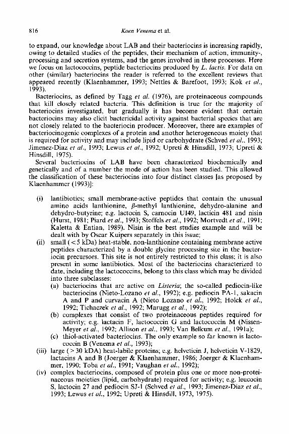
Several bacteriocins of LAB have been characterized biochemically and genetically and of a number the mode of action has been studied. This allowed the classification of these bacteriocins into four distinct classes [as proposed by Klaenhammer (1993)]:
(i) lantibiotics; small membrane-active peptides that contain the unusual amino acids lanthionine, P-methyl lanthionine, dehydro-alanine and dehydro-butyrine; e.g. lactocin S, carnocin U149, lacticin 481 and nisin (Hurst, 1981; Piard et al., 1993; Stoffels et al., 1992; Mortvedt et al., 1991; Kaletta & Entian, 1989). Nisin is the best studies example and will be dealt with by Oscar Kuipers separately in this issue;
(ii) small (< 5 kDa) heat-stable, non-lanthionine containing membrane active peptides characterized by a double glycine processing site in the bacter- iocin precursors. This site is not entirely restricted to this class; it is also present in some lantibiotics. Most of the bacteriocins characterized to date, including the lactococcins, belong to this class which may be divided into three subclasses: (a) bacteriocins that are active on Listeria; the so-called pediocin-like
bacteriocins (Nieto-Lozano et al., 1992); e.g. pediocin PA-l, sakacin A and P and curvacin A (Nieto Lozano et al., 1992; Holck et al., 1992; Tichaczek et al., 1992; Marugg et al., 1992);
(b) complexes that consist of two proteinaceous peptides required for activity; e.g. lactacin F, lactococcin G and lactococcin M (Nissen- Meyer et al., 1992; Allison et al., 1993; Van Belkum et al., 1991a);
(c) thiol-activated bacteriocins. The only example so far known is lacto- coccin B (Venema et al., 1993);
(iii) large ( > 30 LDA) heat-labile proteins; e.g. helveticin J, helveticin V-1829, lactacins A and B (Joerger & Klaenhammer, 1986; Joerger & Klaenham- mer, 1990; Toba et al., 1991; Vaughan et al., 1992);
(iv) complex bacteriocins, composed of protein plus one or more non-protei- naceous moieties (lipid, carbohydrate) required for activity; e.g. leucocin S, lactocin 27 and pediocin SJ-1 (Schved et al., 1993; Jimenez-Diaz et al., 1993; Lewus et al., 1992; Upreti & Hinsdill, 1973, 1975).
Lactococcins, mode of action, immunity, secretion 817
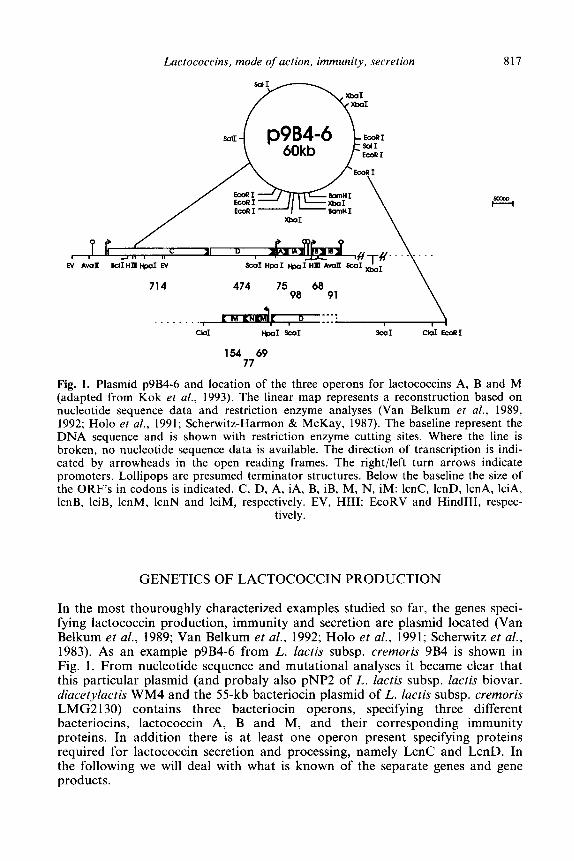
BclIHl Hpd EV SC01 HpaI HpaI HI Avon:
714
Cd HpaI SW1 SW1 C&I ECORI
154 69 77
Fig. 1. Plasmid p9B4-6 and location of the three operons for lactococcins A, B and M (adapted from Kok et al., 1993). The linear map represents a reconstruction based on nucleotide sequence data and restriction enzyme analyses (Van Belkum et al., 1989, 1992; Ho10 et al., 1991; Scherwitz-Harmon & McKay, 1987). The baseline represent the DNA sequence and is shown with restriction enzyme cutting sites. Where the line is broken, no nucleotide sequence data is available. The direction of transcription is indi- cated by arrowheads in the open reading frames. The right/left turn arrows indicate promoters. Lollipops are presumed terminator structures. Below the baseline the size of the ORF’s in codons is indicated. C, D, A, iA, B, iB, M, N, iM: lcnC, IcnD, lcnA, lciA, IcnB, lciB, lcnM, 1cnN and IciM, respectively. EV, HIII: EcoRV and HindIII, respec-
tively.
GENETICS OF LACTOCOCCIN PRODUCTION
In the most thouroughly characterized examples studied so far, the genes speci- fying lactococcin production, immunity and secretion are plasmid located (Van Belkum et al., 1989; Van Belkum et al., 1992; Ho10 et al., 1991; Scherwitz et al., 1983). As an example p9B4-6 from L. lactis subsp. cremoris 9B4 is shown in Fig. 1. From nucleotide sequence and mutational analyses it became clear that this particular plasmid (and probaly also pNP2 of L. Zuctis subsp. lactis biovar. diacetylactis WM4 and the 55-kb bacteriocin plasmid of L. lactis subsp. cremoris LMG2 130) contains three bacteriocin operons, specifying three different bacteriocins, lactococcin A, B and M, and their corresponding immunity proteins. In addition there is at least one operon present specifying proteins required for lactococcin secretion and processing, namely LcnC and LcnD. In the following we will deal with what is known of the separate genes and gene products.
818 Koen Venema et al.
LACTOCOCCIN STRUCTURAL AND IMMUNITY GENES
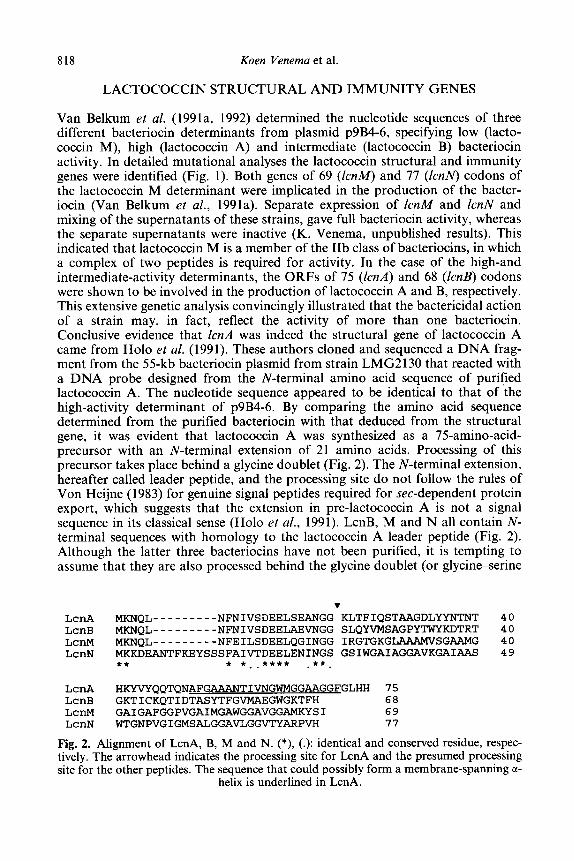
Van Belkum et al. (1991a, 1992) determined the nucleotide sequences of three different bacteriocin determinants from plasmid p9B4-6, specifying low (lacto- coccin M), high (lactococcin A) and intermediate (lactococcin B) bacteriocin activity. In detailed mutational analyses the lactococcin structural and immunity genes were identified (Fig. 1). Both genes of 69 (IcnM) and 77 (ZcnN) codons of the lactococcin M determinant were implicated in the production of the bacter- iocin (Van Belkum et al., 1991a). Separate expression of 1cnM and ZcnN and mixing of the supernatants of these strains, gave full bacteriocin activity, whereas the separate supernatants were inactive (K. Venema, unpublished results). This indicated that lactococcin M is a member of the IIb class of bacteriocins, in which a complex of two peptides is required for activity. In the case of the high-and intermediate-activity determinants, the ORFs of 75 (ZcnA) and 68 (Zcnl3) codons were shown to be involved in the production of lactococcin A and B, respectively. This extensive genetic analysis convincingly illustrated that the bactericidal action of a strain may, in fact, reflect the activity of more than one bacteriocin. Conclusive evidence that ZcnA was indeed the structural gene of lactococcin A came from Ho10 et al. (1991). These authors cloned and sequenced a DNA frag- ment from the 55-kb bacteriocin plasmid from strain LMG2130 that reacted with a DNA probe designed from the N-terminal amino acid sequence of purified lactococcin A. The nucleotide sequence appeared to be identical to that of the high-activity determinant of p9B4-6. By comparing the amino acid sequence determined from the purified bacteriocin with that deduced from the structural gene, it was evident that lactococcin A was synthesized as a 75-amino-acid- precursor with an N-terminal extension of 21 amino acids. Processing of this precursor takes place behind a glycine doublet (Fig. 2). The N-terminal extension, hereafter called leader peptide, and the processing site do not follow the rules of Von Heijne (1983) for genuine signal peptides required for set-dependent protein export, which suggests that the extension in pre-lactococcin A is not a signal sequence in its classical sense (Ho10 et al., 1991). LcnB, M and N all contain N- terminal sequences with homology to the lactococcin A leader peptide (Fig. 2). Although the latter three bacteriocins have not been purified, it is tempting to assume that they are also processed behind the glycine doublet (or glycineserine
. LcnA MKNQL---------NFNIVSDEELSEANGG KLTFIQSTAAGDLYYNTNT 40
LcnB MKNQL---------NFNIVSDEELAEVNGG SLQYVMSAGPYTWYKDTRT 40
LCnM MKNQL---------NFEILSDEELQGINGG IRGTGKGLAAAMVSGAAMG 40 LcnN MKKDEANTFKEYSSSFAIVTDEELENINGS GSIWGAIAGGAVKGAIAAS 49
** * **.**** .**.
LcrlA HKYVYQQTQNAFGWTIVNGWMGGAAGGFGLHH 75 LcnB GKTICKQTIDTASYTFGVMAEGWGKTFH 68 LcnM GAIGAFGGPVGAIMGAWGGAVGGAMKYSI 69 LcnN WTGNPVGIGMSALGGAVLGGVTYARPVH 77
Fig. 2. Alignment of LcnA, B, M and N. (*), (.): identical and conserved residue, respec-
tively. The arrowhead indicates the processing site for LcnA and the presumed processing site for the other peptides. The sequence that could possibly form a membrane-spanning a-
helix is underlined in LcnA.
Lactococcins, mode of action, immunity, secretion 819
sequence in the case of LcnN). The glycine processing site is found in numerous class II peptide bacteriocins of LAB, and even in some lantibiotics and colicin V, an E. coli bacteriocin (Hastings et al., 1991; Marugg et al., 1992; Fremaux et al., 1993; Quadri et al., 1994; Tichaczek et al., 1992; Havarstein et al., 1994, Hynes et al., 1993).
Van Belkum et al. (1991a, 1992) made deletion or frameshift mutations in either of the three larger genes, expected to be the immunity genes [the genes of 154, 98 and 91 codons for the lactococcin M, A and B determinants, respectively, (Fig. l)] without disturbing the corresponding bacteriocin structural genes. Unexpectedly, L. lactis transformants carrying the various constructs proved viable, although the colonies were initially small and grew poorly. After serial transfers in fresh medium the cells gradually started to grow more rapidly. If lactococcin [50% (vol/vol) of a supernatant of a lactococcin producing strain] was added to the plates on which the initial transformants were to be selected, no transformants were obtained. Also, when the presumed immunity genes were cloned separately from the bacteriocin genes under the control of a promoter, they provided immunity to the transformed lactococcal strain. These results indicated that the presumed immunity genes indeed specify bacteriocin immunity, and also that cells can overcome the lethal action of lactococcin at a stage when little bacteriocin is produced. Several possibilities leading to tolerance towards lactococcin can be envisaged at this point, one of which, in the case of lactococcin A, is the loss or mutational alteration of its receptor (see below). The number of transformants obtained with a plasmid carrying IcnA and a mutated immunity gene is the same as that obtained with a plasmid carrying functional 1cnA and 1ciA (K. Venema, unpublished results). Apparently, the absence of a functional immunity gene does not interfer with the viability of these transformants. It is unlikely that each of the viable transformants would carry a mutation in the lactococcin receptor. A more likely explanation is that the viability of these transformants is due to a non-genetic change in membrane-lipid composition, such that the lactococcin cannot form pores in the altered membranes. Because of the frequent occurrence of the acquisition of tolerance towards lactococcin, we envisage this change in lipid composition to be an reversible adaptation rather than a mutation. Here we will restrict the term immunity to lactococcin-insensi- tivity caused by the lci product. We propose to denote the reversible acquisition of insensitivity to bacteriocins as tolerance, whereas the term resistance should be reserved for permanent acquisition of insensitivity to bacteriocins.
LcnC AND LcnD ARE ESSENTIAL FOR LACTOCOCCIN PRODUCTION
Nucleotide sequence analysis of a 5.2-kb DNA fragment derived from plasmid pNP2 of L. lactis subsp. lactis biovar. diacetylactis WM4 revealed that this plasmid also specifies lactococcin A (Stoddard et al., 1992). Moreover, it was shown that two additional genes were required for bacteriocin activity. These genes, desig- nated 1cnC and ZcnD, are located in an operon immediately upstream of the lacto- coccin A structural and immunity genes (Fig. 1). Another copy of these genes is probably also present upstream of the lactococcin M operon (Kok et al., 1993). Tn5 insertions in either 1cnC or lcnD eliminated lactococcin production without
820 Koen Venema et al.
negating immunity (Stoddard et al., 1992). A promoter immediately upstream of 1cnA drives expression of both lactococcin A activity and immunity (Van Belkum et al. 1991a). Therefore, loss of lcnC or 1cnD functions were reasoned to impact the maturation and/or secretion processes (Stoddard et al., 1992). Comparisons of LcnC and LcnD with protein database sequences revealed that these proteins share significant similarities to Gram-negative proteins implicated in signal sequence- independent secretion pathways. Stoddard et al. (1992) showed that LcnC is a member of the HlyB-like family of ATP-binding cassette (ABC) transporters (Higgins, 1993; Fath & Kolter, 1993). The similarity is most pronounced in the C- terminal stretch of 200 amino acids of LcnC, which contains the ATP-binding cassette. Protein structure-predicting computer programs indicate six hydrophobic regions in the N-terminus of LcnC which could promote binding at the cytoplasmic membrane, a situation similar to HlyB (Gentschev & Goebel, 1992). Hydro- phobicity analysis of LcnD indicated that it is largely hydrophilic, with the excep- tion of the N-terminal 43 amino acids. This region in LcnD might be a transmembrane domain, as was shown for HlyD (see also below) (Schtilein et al., 1992). LcnD does not show similarity with HlyD, however. These results strongly suggested that LcnC and LcnD are required for secretion of the lactococcins via a system dedicated to bacteriocin export (Stoddard et al., 1992; Kok et al., 1993). Similar dedicated secretion systems have now been proposed for other LAB bacteriocins (Marugg et al., 1992; Kuipers et al., 1993).
It appears that L. lack strains IL1403 carries genes for LcnC and LcnD on its chromosome, explaining why plasmids carrying only the bacteriocin structural and immunity genes still produced active bacteriocin (Van Belkum et al., 1991a, 1992; Ho10 et al., 1991). Using a DNA probe encompassing 1cnC and lcnD, in Southern hybridizations, a signal with the chromosome of IL1403 was obtained (Kok et al., 1993). When probed with the various bacteriocin structural and immunity genes, no chromosomal signal was observed (K. Venema, unpublished data). Also, PCR products encompassing almost the entire operon were obtained using primers designed from the plasmid encoded 1cnC an ZcnD genes (K. Venema, unpublised results), indicating that the chromosomally located IcnC and fcnD of IL1403 are highly homologous to their plasmid encoded counterparts. Part of the chromosomally encoded IcnC was cloned and sequenced. Several nucleotide substitutions were found compared to the plasmid encoded ZcnC (K. Venema, unpublished data), but none of these led to amino acid changes in the translation product. The reduced production of lactococcin in IL1403 carry- ing a plasmid with only the bacteriocin operon, as compared to the wild type strain carrying all of the essential genes on a plasmid, can be explained by assuming that the lower copy number of the chromosomally located genes (most probably one) results in a lower secretion efficiency. This conclusion was corro- borated by introducing lcnC and IcnD, originating from p9B4, on a plasmid in IL1403. This led to a drastically increased production of lactococcin A.
TOPOLOGY OF LcnD IN THE LACTOCOCCAL CYTOPLASMIC MEMBRANE
The results presented so far indicate that LcnC and LcnD might form a dedicated secretion machinery for lactococcins. However, no evidence has been presented to
Lactococcins, mode of action, immunity, secretion 821
justify such a conclusion. To characterize the putative secretion machinery, Franke et al. (1995) used fusions with the E. coli reporter proteins LacZ and PhoA to determine the topology of LcnC and LcnD in the cytoplasmic membrane. Since the studies with LcnC are still in progress, only the data for LcnD will be discussed here.
Fusions of LacZ, a cytoplasmic protein, to sequences that are normally located in the cytoplasm result in an active fi-galactosidase. Fusions between LacZ and parts of a protein that are normally extracellular will result in highly reduced /?-galatosidase activities. For PhoA, a periplasmic enzyme, the reverse is true (Manoil, 1990; Muller ef al., 1993; Boyd et al., 1993).
Hydrophobicity analysis of LcnD indicates that the stretch of amino acids from residue 22 to 38 might form a transmembrane a-helix. The rest of the protein is rather hydrophilic. Taking this computer prediction as a guide, Franke et al. (1995) constructed four LacZ and four PhoA fusions in front of, in, and behind the predicted transmembrane helix and at the complete C-terminal end of LcnD. The analysis showed that, indeed, the N-terminus of LcnD was in the cytoplasm, the stretch of amino acids between residue 22 to 38 crossed the membrane, and the remainder of the protein was located at the outside of the cell.
PROCESSING OF BACTERIOCINS; INVOLVEMENT OF THE ABC-TRANSPORTERS IN PROCESSING
As discussed above, class II bacteriocins are synthesized as prebacteriocins, containing a leader peptide that is cleaved behind a glycine doublet before or during transport. Until recently, it was unclear whether the presumed leader peptidase activity was also encoded by the bacteriocin operon, or whether it was a more general peptidase present elsewhere on the chromosome. The first indica- tion that a protein encoded by the bacteriocin operon was involved in this process came from studies by Venema et al. (1995, submitted) on the pediocin operon.
As for lactococcins, production of active pediocin requires, in addition to the structural gene, pedal, two additional genes (pedC and pedD) that show homology to the proteins involved in set-independent secretion of polypeptides (Marugg et al., 1992). PedC has only structural homology to LcnD, whereas PedD has considerable amino acid sequence homology to LcnC (49% identity with an over- all homology of 67%). The homology of the latter two proteins is not restricted to the ABC-box, but extends over the entire polypeptide. This is also the case with several other ABC-transporters involved in secretion of bacteriocins that contain the double glycine motif in the leader peptide. Notably, a region in the N-terminal 190 amino acids is conserved in these proteins (Venema et al., 1995).
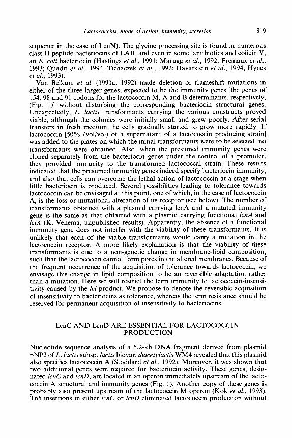
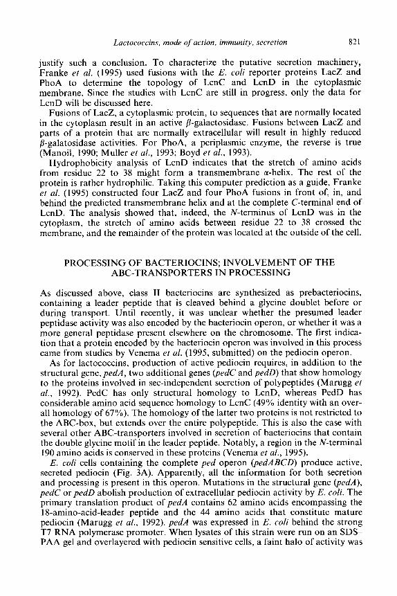
E. coli cells containing the complete ped operon (pedABCD) produce active, secreted pediocin (Fig. 3A). Apparently, all the information for both secretion and processing is present in this operon. Mutations in the structural gene @edA), pedC or pedD abolish production of extracellular pediocin activity by E. coli. The primary translation product of pedA contains 62 amino acids encompassing the 18-amino-acid-leader peptide and the 44 amino acids that constitute mature pediocin (Marugg et al., 1992). pedA was expressed in E. coli behind the strong T7 RNA polymerase promoter. When lysates of this strain were run on an SDS- PAA gel and overlayered with pediocin sensitive cells, a faint halo of activity was
822 Koen Venema et al.
ext. ped int. preped int. ped
A c3 pedA spedD+ - -
B e pedA
+
D G) pedD - + + pedA
Fig. 3. The effect of pedC and pedD on pediocin secretion and processing. Ext. ped, int. preped, int. ped: extracellular pediocin, intracellular prepediocin and intracellular pediocin activity, respectively. (+), (-): activity present and absent, respectively. The presence of the various genes (pedA, pedC, pedD) in the E. coli clones (A, B, C, D) is indicated by the
arrows. For details, see text.
observed, corresponding to a peptide with a lower electrophoretic mobility than that of mature pediocin produced by E. coli containing the complete ped operon. Apparently, the primary translation product of pedA is biologically active albeit at a very reduced level. No extracellular activity was found in the strain carrying pedA alone (Fig. 3B). To determine which of the genes in the operon was responsible for pediocin secretion and/or processing, pedC and pedD were expressed separately or together in the strain producing prepediocin from the T7 promoter. When both pedC and pedD were present, active extracellular pediocin was produced. However, when only pedC (Fig. 3C) or pedD (Fig. 3D) were present, no extracellular activity was found. Lysates of the strain carrying pedA and pedC contained prepediocin activity only. However, lysates of the strain carrying pedA and pedD showed, in addition to prepediocin activity, an activity on the gel that was indistinguishable from that of mature pediocin (Fig. 3D). These results conclusively show that both PedC and PedD are involved in secre- tion of pediocin. In addition, we demonstrate that PedD is capable of processing the pediocin precursor. Since mature pediocin was found intracellularly in the strain containing only pedA and pedD, we postulate that the conserved 190- amino-acid-domain in the N-terminus of the protein, which is shown to be intra- cellular by topology studies (E. Emond, pers. commun.), is involved in processing of the leader peptide. The results also show that bacteriocin processing can be uncoupled from secretion.
We predict that, similar to PedD, LcnC will be the processing enzyme for the lactococcins. Since cloning of IcnC in E. coli is lethal we have not yet been able to verify this prediction.
MODE OF ACTION OF LACTOCOCCIN A AND B
Lactococcins A and B specifically inhibit the growth of lactococci. Both are small cationic, hydrophobic peptides that structurally resemble several peptide anti-
Lactococcins, mode of action, immunity, secretion 823
biotics permeabilizing membranes (Gao et al., 1991; Galvez et al., 1991; Kordel & Sahl, 1986; Kordel et al., 1988; Schaller et al., 1989). A possible target for their action could thus be the cytoplasmic membrane of sensitive cells. Van Belkum et al. (1991b) investigated this possibility in detail for lactococcin A. The mode of action of purified lactococcin A was studied on whole cells and membrane vesi- cles of sensitive and immune lactococcal strains, and on liposomes obtained from lactococcal phospholipids. Venema et al. (1993) performed similar studies with partially purified lactococcin B on whole cells. At lactococcin concentrations that did not affect immune cells, both bacteriocins rapidly dissipated the membrane potential (and in the case of lactococcin B also the pH-gradient across the membrane) of glucose-energized sensitive cells. A non-metabolizable alanine analogue (AIB, 2-cl-amino isobutyric acid) that is taken up in symport with a proton [and of which the uptake is thus driven by the proton motive force (pmf) (Konings et al., 19891 was rapidly lost from sensitive whole cells treated with lactococcin (Van Belkum et al. 1991b; Venema et al., 1993). To show that efflux was a direct consequence of permeability changes in the cytoplasmic membrane, and not caused by the absence of a pmf, uptake of L-glutamate was studied. Glutamate uptake by L. lactis is driven by a phosphate bond-linked unidirec- tional process (Poolman et al., 1987) and dissipation of the pmf should, therefore, not lead to efflux of accumulated glutamate. However, addition of either lacto- coccin to sensitive cells that had accumulated glutamate resulted in the immediate efflux of the amino acid, even when the pmf in the cells was dissipated prior to the addition of the bacteriocin (Van Belkum et al., 1991b; Venema et al. 1993). These results indicated that both lactococcins are able to form pores in the membrane in a voltage-independent manner.
Lactococcin A inhibited leucine uptake in cytoplasmic membrane vesicles from sensitive lactococcal cells, but not from vesicles derived from Bacillus subtilis, Clostridium acetobutyricum or E. coli membranes (Van Belkum et al., 199lb). Liposomes derived from lactococcal phospholipids were not affected by lacto- coccin A. From these data, and the observation that lactococcin A specifically inhibits lactococcal strains, Van Belkum et al. (199 1 b) concluded that lactococcin A forms pores in the cytoplasmic membrane of sensitive cells using a Lacto- coccus-specific receptor protein (Van Belkum et al., 1991b).
A 21-amino acid sequence between residues Ala30 and PheSO in lactococcin A (Fig. 2) could possibly form a membrane-spanning cc-helix (Kok et al., 1993). Lactococcin A could be anchored to the cytoplasmic membrane of sensitive cells by this hypothetical transmembrane helical segment. A large number of pore-forming toxins are known to create channels through a “barrel stave” mechanism (Ojcius & Young, 1991; Sansom et al., 1991). Pore formation requires the molecules to aggregate like barrel staves surrounding a central water-filled pore. It is assumed that lactococcin A and lactococcin B form pores by this mechanism, although in the case of lactococcin B the method of Kyte & Doolittle (1982) does not predict a (clearcut) membrane spanning helix. The size of the pore would be dictated by the number of molecules involved in pore formation. An indication that different pore sizes can exist was derived from the observation that low concentrations of lactococcin B allow leakage of protons and ions, whereas for leakage of glutamate 150 times more bacteriocin was needed (Venema et al., 1993). Apparently, at low concentrations a multi- peptide complex of lactococcin B molecules forms a pore which is too narrow
824 Koen Venema et al.
0 2 4 6 8 10 12 14
time (In mln)
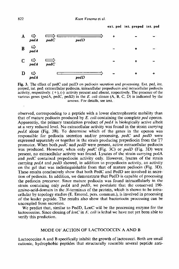
Fig. 4. Effect of LcnB on the membrane potential of glucose-energized lactococcal cells (adapted from Venema et al., 1993). A: no addition, + : addition of non-DTT-treated LcnB, 0: addition of DTT-treated LcnB, 0: addition of HgC&treated LcnB, 0: addition
of HgClz-treated LcnB, subsequently reduced with DTT.
for the passage of amino acids, but allows the passage of protons and other ions.
A prerequisite for lactococcin B activity is that its only cysteine residue is in the reduced state (Cys24, Venema et al., 1993). Partially purified lactococcin B was almost inactive on whole cells (Fig. 4, curve +). Only after addition of small amounts of dithiothreitol (DTT) was lactococcin B capable of dissipating the membrane potential (Fig. 4, 0). Reduction of Cys24 by DTT was counteracted by HgC12 (Fig. 4, q ), while HgClz-oxidized lactococcin B could be reactivated by DTT (Fig. 4, curve 0).
Cys24 of lactococcin B was replaced via site directed mutagenesis by all other possible 19 amino acids. Surprisingly, 16 of the 19 mutants were active, and even more so than the wild type bacteriocin. The inactive mutants were the ones in which the cysteine had been replaced by a positively charged amino acid (Cys24His, Cys24Lys and Cys24Arg; K. Venema, unpublished data). Apparently, Cys24 is not essential for activity of lactococcin B. When Cys24 is present, oxidation leads to inactivation of lactococcin B. In this regard lacto- coccin B resembles a group of thiol-activated toxins in which the reduced state of a cysteine residue appears to be essential for the generation of functional lesions in toxin-treated membranes (Boulnois et al., 1991). Also in these mole- cules the cysteine can be replaced without loss of activity. The reason for inac- tivation of lactococcin B by oxidation of Cys24 is unclear, but since all mutants carrying a positively charged amino acid at position 24 are inactive, for the
Lactococcins, mode of action, immunity, secretion 825
bacteriocin to be active a negative or no charge at this specific site is apparently essential.
MODE OF ACTION OF THE IMMUNITY PROTEIN LCIA
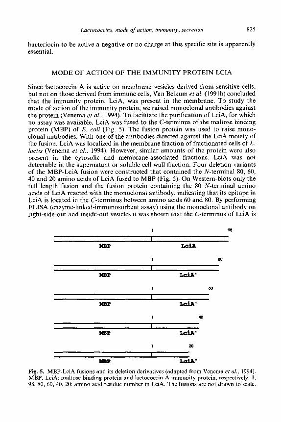
Since lactococcin A is active on membrane vesicles derived from sensitive cells, but not on those derived from immune cells, Van Belkum et al. (1991b) concluded that the immunity protein, LciA, was present in the membrane. To study the mode of action of the immunity protein, we raised monoclonal antibodies against the protein (Venema et al., 1994). To facilitate the purification of LciA, for which no assay was available, LciA was fused to the C-terminus of the maltose binding protein (MBP) of E. coli (Fig. 5). The fusion protein was used to raise mono- clonal antibodies. With one of the antibodies directed against the LciA moiety of the fusion, LciA was localized in the membrane fraction of fractionated cells of L. lactis (Venema et al,, 1994). However, similar amounts of the protein were also present in the cytosolic and membrane-associated fractions. LciA was not detectable in the supernatant or soluble cell wall fraction. Four deletion variants of the MBP-LciA fusion were constructed that contained the N-terminal 80, 60, 40 and 20 amino acids of LciA fused to MBP (Fig. 5): On Western-blots only the full length fusion and the fusion protein containing the 80 N-terminal amino acids of LciA reacted with the monoclonal antibody, indicating that its epitope in LciA is located in the C-terminus between amino acids 60 and 80. By performing ELISA (enzyme-linked-immunosorbent assay) using the monoclonal antibody on right-side-out and inside-out vesicles it was shown that the C-terminus of LciA is
1 98
WBP LciA’
1 40
MBP LciA’
1 20
llBP L&A’
Fig. 5. MBP-LciA fusions and its deletion derivatives (adapted from Venema et al., 1994). MBP, LciA: maltose binding protein and lactococcin A immunity protein, respectively. 1, 98. 80, 60, 40, 20: amino acid residue number in LciA. The fusions are not drawn to scale.
826 Keen Venema et al.
located at the outside of the cytoplasmic membrane of an LciA-producing strain (Venema et al., 1994). Right-side-out membrane vesicles derived from a strain producing both lactococcin A and the immunity protein did not react with the antibody. The same was observed when lactococcin A was added externally to right-side-out vesicles derived from a strain producing only LciA. Apparently, the bacteriocin shielded the epitope in LciA from reacting with the monoclonal anti- body. Right-side-out membrane vesicles of immune as well as sensitive cells were treated with proteinase K and subsequently used in leucine uptake experiments. Although LciA is cleaved by proteinase K (Venema et al., 1994) immune vesicles incubated with the proteinase were not affected by the bacteriocin. However, also proteinase K-treated sensitive vesicles became insensitive to the bacteriocin. These data suggested that, in addition to digesting LciA, proteinase K had also digested the bacteriocin receptor, previously inferred by van Belkum et al. (1991b) on the basis of the lactococcin A mode of action studies, rendering the membrane vesicles insensitive to the action of lactococcin A.
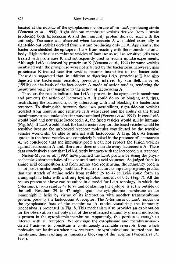
Thus far, the results indicate that LciA is present in the cytoplasmic membrane and prevents the action of lactococcin A. It could do so by binding and, thus, neutralizing the bacteriocin, or by interacting with and blocking the bacteriocin receptor. To distinguish between these two possibilities, right-side-out vesicles isolated from immune and sensitive cells were fused and the ability of the fused membranes to accumulate leucine was examined (Venema et al. 1994). In case LciA would bind and neutralize lactococcin A, the fused vesicles would still be immune (Fig. 6A). If LciA would block the bacteriocin receptor, the fused vesicles would be sensitive because the unblocked receptor molecules contributed by the sensitive vesicles would still be able to interact with lactococcin A (Fig. 6B). As leucine uptake in the fused vesicles was completely blocked in the presence of lactococcin A, we concluded that the immunity protein can not protect the fusion vesicles against lactococcin A and, therefore, does not titrate away lactococcin A. These data conclusively show that LciA directly interacts with the lactococcin A receptor.
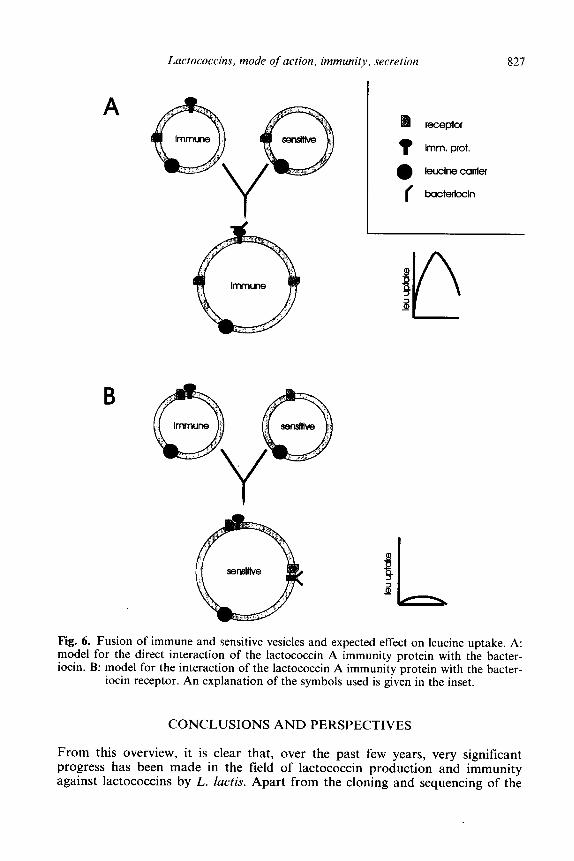
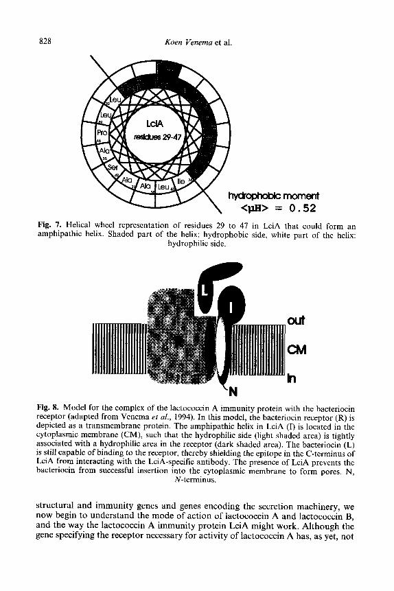
Nissen-Meyer et al. (1993) have purified the LciA protein by using the physi- cochemical characteristics of its deduced amino acid sequence. As judged from its amino acid composition and from amino acid sequencing, the immunity protein is not post-translationally modified. Protein structure computer programs predict that the stretch of amino acids from residue 29 to 47 in LciA could form an cr-amphiphilic helix with a strong hydrophobic moment of 0.52 (Fig. 7). All the results presented above can be united in a model for LciA topology, in which the C-terminus, from residue 48 to 98 and containing the epitope, is at the outside of the cell. Residues 29 to 47 might span the cytoplasmic membrane as an cl-amphiphilic helix by virtue of its interaction with another transmembrane protein, possibly the lactococcin A receptor. The N-terminus of LciA resides on the cytoplasmic face of the membrane. A model visualizing the immunity mechanism is presented in Fig. 8. This mechanism also provides an explanation for the observation that only part of the synthesized immunity protein molecules is present in the cytoplasmic membrane. Apparently, this portion is enough to interact with all receptors. We envisage the cytoplasmic and membrane-asso- ciated fractions to constitute a continuously available reservoir from which molecules can be drawn when new receptors are synthesized and inserted into the membrane, thus resulting in immediate blocking of the receptor (Venema et al., 1994).
Lactococcins, mode of action, immunity, secretion 827
A receptor
t lrnm. prot.
a leuclne carrier
I bacterlccin
Fig. 6. Fusion of immune and sensitive vesicles and expected effect on leucine uptake. A: model for the direct interaction of the lactococcin A immunity protein with the bacter- iocin. B: model for the interaction of the lactococcin A immunity protein with the bacter-
iocin receptor. An explanation of the symbols used is given in the inset.
CONCLUSIONS AND PERSPECTIVES
From this overview, it is clear that, over the past few years, very significant progress has been made in the field of lactococcin production and immunity against lactococcins by L. luctis. Apart from the cloning and sequencing of the
828 Koen Venema et al
moment 0.52
in LciA that could form an side, white part of the helix:
Fig. 7. Helical wheel representation of residues 29 to 47 amphipathic helix. Shaded part of the helix: hydrophobic
hydrophilic side.
h
Fig. 8. Model for the complex of the lactococcin A immunity protein with the bacteriocin receptor (adapted from Venema et al., 1994). In this model, the bacteriocin receptor (R) is depicted as a transmembrane protein. The amphipathic helix in LciA (I) is located in the cytoplasmic membrane (CM), such that the hydrophilic side (light shaded area) is tightly associated with a hydrophilic area in the receptor (dark shaded area). The bacteriocin (L) is still capable of binding to the receptor, thereby shielding the epitope in the C-terminus of LciA from interacting with the LciA-specific antibody. The presence of LciA prevents the bacteriocin from successful insertion into the cytoplasmic membrane to form pores. N,
N-terminus.
structural and immunity genes and genes encoding the secretion machinery, we now begin to understand the mode of action of lactococcin A and lactococcin B, and the way the lactococcin A immunity protein LciA might work. Although the gene specifying the receptor necessary for activity of lactococcin A has, as yet, not
Lactococcins, mode of action, immunity, secretion 829
been identified, the presumed receptor is likely to be the site of action of the immunity protein. In analogy to the system for pediocin secretion, it is likely that LcnC and LcnD are involved in processing and secretion of the lactococcins. In the coming years we expect that the receptor will be identified and that molecular details concerning the mode of action combined with structure-function studies of the lactococcins will allow the construction of bacteriocins with enhanced or altered activities and broader specificities.
ACKNOWLEDGEMENTS
The contribution of colleagues who provided prepublication information and fruitful discussions is gratefully acknowledged. K. Venema was supported by the EC BRIDGE T-project on LAB. J. Kok is the recipient of a fellowship of the Royal Netherlands Academy of Arts and Sciences (KNAW).
REFERENCES
Allison, G., Fremaux, C., Ahn. C. & Klaenhammer, T. R. (1993). Expansion of bacteriocin activity and host range upon complementation of two peptides encoded within the lactacin F operon. J. Bacterial., 176, 223541.
Boulnois, G. J., Paton, J. C., Mitchell, T. J. & Andrew, P. W. (1991). Structure and function of pneumolysin, the multifunctional, thiol-activated toxin of Streptococcus pneumoniae. Mol. Microbial., 5, 2611-16.
Boyd, D., Traxler, B. & Beckwith, J. (1993). Analysis of the topology of a membrane protein by using a minimum number of alkaline phosphatase fusions. J. Bacterial.. 175, 553-56.
De Vuyst, L. (1993) Bacteriocins of Lactic Acid Bacteria: Microbiology, Genetics and Applications. Elsevier Applied Science, Barking.
Fath, M. J. & Kolter, R. (1993) ABC transporters: bacterial exporters. Microbiof. Rev., 57, 995-1017.
Fremaux. C., Ahn, C. & Klaenhammer, T. R. (1993). Molecular analysis of the lactacin F operon. Appl. Environ. Microbial., 59, 390615.
Galvez, A., Maqueda, M., Martinez-Bueno, M. & Valdivia, E. (1991). Permeation of bacterial cells, permeation of cytoplasmic and artificial membrane vesicles, and channel formation in lipid bilayers by peptide antibiotic AS-48. J. Bacterial., 173, 886-92.
Gao. F. H., Abee, T. & Konings, W. N. (1991). The mechanism of action of the peptide antibiotic nisin in liposomes and cytochrome C oxidase proteoliposomes. Appl. Environ. Microbial., 57, 216470.
Gentschev, I. & Goebel, W. (1992). Topological and functional studies on HlyB of Escherichia coli. Mol. Gen. Genet., 232, 4048.
Hastings, J. W., Sailer, M., Johnson, K., Roy, K. L., Vederas, J. C. & Stiles, M. E. (1991). Characterization of leucocin A-UAL 187 and cloning of the bacteriocin gene from Leuconostoc gelidum. J. Bacterial., 174, 568692.
Havarstein, L. S., Holo, H. & Nes, I. F. (1994). The leader peptide of colicin V shares consensus sequences with leader peptides that are common among peptide bacteriocins produced by Gram-positive bacteria. Microbiology, 140, 2383-89.
Holck, A., Axelsson. L., Birkeland, S.-E., Aukrust, T. & Blom, H. (1992). Purification and amino acid sequence of sakacin A, a bacteriocin from Lactobacillus sake Lb706. J. Gen. Microbial., 138, 2715-20.
830 Koen Venema et al.
Holo, H., Nilssen, 0. and Nes, I. F. (1991). Lactococcin A, a new bacteriocin from Lactococcus lactis subsp. cremoris: isolation and characterization of the protein and its gene. J. Bacterial., 173, 3879-87.
Hoover, D. & Steenson, L. (1993). Bacteriocins of Lactic Acid Bacteria. Academic Press, New York, NY, U.S.A.
Hurst, A. (1981). Nisin. Adv. Appl. Microbial., 27, 85-127. Hynes, W., Ferreti, J. J. & Tagg J. R. (1993). Cloning the gene encoding streptococcin A-
FF22, a novel lantibiotic produced by Streptococcus pyrogenes, and determination of its nucleotide sequence. Appl. Environ. Microbial., 59, 1969-7 1.
James, R., Lazdunski, C. & Pattus, F. (1992). Bacteriocins, Microcins and Lantibiotics. Springer-Verlag, Berlin, Heidelberg.
Jimenez-Diaz, R., Rios-Sanchez, R. M., Desmazeaud, M., Ruiz-Barba, J. L. & Piard, J.-C. (1993). Plantaricins S and T, two new bacteriocins produced by Lactobacilluis plantarum LCPOlO isolated from a green olive fermentation. Appl. Environ. Microbial., 59, 1416 24.
Joerger, M. C. & Klaenhammer, T. R. (1986). Characterization and purification of helveticin J and evidence for a chromosomally determined bacteriocin produced by Lactobacillus helveticus 481. J. Bacterial.. 167, 43946.
Joerger, M. C. & Klaenhammer, T. R. (1990). Cloning, expression and nucleotide sequence of the Lactobacillus helveticus 481 gene encoding the bacteriocin helveticin J. J, Bacterial., 171, 633947.
Jung, G. & Sahl, H.-G. (1991). Nisin and Novel Lantibiotics. ESCOM, Leiden. Kaletta, C. & Entian, K.-D. (1989). Nisin, a peptide antibiotic, cloning and sequencing of
the nisA gene and posttranslational processing of its peptide product. J. Bacterial., 171, 1597401.
Klaenhammer, T. R. (1988). Bacteriocins of lactic acid bacteria. Biochimie, 70, 33749. Klaenhammer, T. R. (1993). Genetics of bacteriocins produced by lactic acid bacteria.
FEMS Microbial. Rev., 12, 39-86. Kok, J., Holo, H., Van Belkum, M. J., Haandrikman, A. J. & Nes, I. F. (1993). In
Bacteriocins of Lactic Acid Bacteria, eds, Hoover, D. and Steenson, L., Academic Press, New York, NY, U.S.A. pp 121-50.
Kolter, R. & Moreno, F. (1992) Genetics of ribosomally synthesized peptide antibiotics. Annu. Rev. Microbial., 46, 141-63.
Konings, W. N., Poolman, B. & Driessen, A. J. M. (1989). Bioenergetics and solute transport in lactococci. CRC Critical Rev. Microbial., 16, 419-76.
Kordel, M., Benz, R. & Sahl, H.-G. (1988). Mode of action of the staphylococcin-like peptide Pep% voltage-dependent depolarization of bacterial and artificial membranes. J. Bacterial., 170, 8488.
Kordel, M. & Sahl, H.-G. (1986). Susceptibility of bacterial, encraiotic and artificial membranes to the disruptive action of cationic peptides PEP5 and nisin. FEMS Microbial. Lett., 34, 139-44.
Kuipers, 0. P., Beerthuyzen, M. M., Siezen, R. J. & De Vos, W. M. (1993). Characterization of the nisin gene cluster nisABTCIPR of: requirement of expression of nisA and nisi genes for producer immunity. Eur. J. Biochem., 216, 281-91.
Kyte, J. & Doolittle, R. F. (1982). A simple model for displaying the hydrophatic character of a protein. J. Mol. Biol., 157, 105-32.
Lewus, C. B., Sun. S. & Montville, T. J. (1992). Production of an amylase-sensitive bacteriocin by an atypical Leuconostoc paramesenteroides strain. Appl. Environ. Microbial., 58, 143-49.
Manoil, C. (1990). Analysis of’ protein localization by use of gene fusions with complementary properties. J. Bacterial., 172, 103542.
Marugg, J. D., Gonzalez, C. F., Kunka, BS., Ledeboer, A. M., Pucci, M. J., Toonen, M. Y., Walker, S. A., Zoetmulder, L. C. M. & Vandenbergh, P. A. (1992). Cloning, expression, and nucleotide sequence of genes involved in production of pediocin PA- 1, a
Lactococcins, mode of action, immunity, secretion 831
bacteriocin from Pediococcus acidilactici PACI .O. Appl. Environ. Microbial., 58, 236& 67.
Mortvedt, C. I., Nissen-Meyer, J., Sletten, K. & Nes, I. F. (1991). Purification and amino acid sequence of lactocin S, a bacteriocin produced by Lactobacillus sake L45. Appl. Environ. Microbial., 5’7, 1829934.
Muller, M. M., Vianney, A., Lazzaroni, J.-C., Webster, R. E. & Porttalier, R. (1993). Membrane topology of the Escherichia coli TolR protein required for cell envelope integrity. J. Bacterial.. 175, 6059961.
Nettles, C. G. & Barefoot, S. F. (1993). Biochemical and genetic characteristics of bacteriocins of food-associated lactic acid bacteria. J. Food Protection, 56, 338- 56.
Nieto Lozano, J. C., Nissen-Meyer, J., Sletten, K., Pelaz, C. & Nes, I. F. (1992). Purification and amino acid sequence of a bacteriocin produced by Pediococcus acidilactici. J. Gen. Microbial., 138, 1985-90.
Nissen-Meyer, J., Havarstein. L. S., Holo, H., Sletten, K. & Nes, I. F. (1993). Association of lactococcin A immunity factor with the cell membrane: purification and characterization of the immunity factor. J. Gen. Microbial., 139, 1503, 1509.
Nissen-Meyer, J., Holo, H., Havarstein, L. S., Sletten, K. & Nes, I. F. (1992). A novel lactococcal bacteriocin whose activity depends on the complementary action of two peptides. J. Bacterial., 174, 568692.
Ojcius, D. M. & Young, J. D.-E. (1991). Cytolytic pore-forming proteins and peptides: is there a common structural motif. J. Trends in Biochem Sci., 16, 225-29.
Piard, J.-C., Kuipers, 0. P., Rollema, H. S., Desmazeaud, M. J. & De Vos, W. M. (1993). Structure, organization, and expression of the let gene for lacticin 48 1, a novel Iantibiotic produced by Lactococcus lactis. J. Biol. Chem., 268, 1636168.
Poolman, B., Smid, E. J. & Konings, W. N. (1987). kinetics of a phosphate-bond driven glutamate-glutamine transport system in Streptococcus lactis and Streptococcus cremoris. J. Bacterial., 169, 1460-68.
Quadri, L. E. N., Sailer, M, Roy, K. L., Vederas, J. C. & Stiles, M. E. (1994). Chemical and genetic characterization of bacteriocins produced by Carnobacterium piscicola LVl7B. J. Biol. Chem., 269, 12204-11.
Sansom, M. S. P., Kerr, I. D. & Mellor, I. R. (1991). Ion channels formed by amphipathic helical peptides; a molecular modelling study. Eur. Biophys. J., 20, 229940.
Schaller, F., Benz, R. & Sahl, H.-G. (1989). The peptide antibiotic subtilin acts by formation of voltage-dependent multi-state pores in bacterial and artificial membranes. Eur. J. Biochem., 182, 182-86.
Scherwitz, K. M., Baldwin, K. A. & McKay, L. L. (1983). Plasmid litrkage of a bacteriocin- like substance in Streptococcus Iactis subsp. diacetylactis strain WM4: transferability to Streptococcus lactis. Appl. Environ. Microbial., 45, 150612.
Scherwitz-Harmon, K. M. & McKay, L. L. (1987). Restriction enzyme analysis of lactose and bacteriocin plasmids from Streptococcus lactis subsp. diacetylactis strain WM4 and cloning of BclI fragments coding for bacteriocin production. Appt. Environ. Microbial., 53, 1171-74.
Schtilein, R., Gentschev, I., Mollenkopf, H.-J. & Goebel, W. (1992). A topological model for the haemolysin translocator protein HlyD. Mol. Gen. Genet., 234, 155-63.
Schved, F. Lazazar, A., Henis, Y &’ Juven, B. J. (1993). Purification, partial characterization and plasmid linkabe od pedicoin SJ-I, a bacteriocin produced by Pediococcus acidilactici. J. Appl. Bacterial., 74, 67-77.
Stoddard, G. W., Petzel, J. P., Van Belkum, M. J., Kok, J. & McKay, L. L. (1992). Molecular analyses of the lactococcin A gene cluster from Lactococcus lactis subsp. lactis biovar. diacetylaqtis WM4. Appt. Environ. Microbial., 58, 1952261.
Stoffels, G., N&en-Meyer, J., Gudmundsdottir, A., Sletten, K., Holo, H. & Nes, I.F. (1992). Purification and characterization of a new bacteriocin isolated from a Carnobacterium sp. Appi. Environ. Microbial., 58, 1417-22.
832 Koen Venema et al.
Tagg, J. R., Dajani, A. S. AZ Wannamaker, L. W. (1976). Bacteriocins of Gram-positive bacteria. Microbial. Rev., 40, 722-56.
Tichaczek, P. S., Nissen-Meyer, J., Nes, I. F., Vogel, R. F. & Hammes, W. P. (1992). Characterization of the bacteriocins curvacin A from Lactobacillus curvatus LTH 1174 and sakacin P from Lactobacillus sake LTH673. Syst. Appt. Microbial., 15, 461X68.
Toba, Y., Yoshioka, E. & Itoh, T. (1991). Lacticin, a bacteriocin produced by Lactobaciflus delbrueckii subsp. lactis. Lett. Appl. Microbial., 12, 43345.
Upreti, G. C. & Hinsdill, R. D. (1973). Isolation and characterization of a bacteriocin from a homofennentative Lactobacillus. Antimicrob. Agents Chemother., 4, 487-94.
Upreti, G. C. & Hinsdill, R. D. (1975). Production and mode of action of lactocin 27, a bacteriocin from a homofermentative Lactobacillus. Antimicrob. Agents Chemother., 7, 13945.
Van Belkum, M. J., Hayema, B. J., Geis, A., Kok, J. & Venema, G. (1989). Cloning of two bacteriocin genes from a lactococcal bacteriocin plasmid. Appt. Environ. Microbial., 55, 1187-91.
Van Belkum, M. J., Hayema, B. J., Jeeninga, R. E., Kok, J & Venema, G. (1991a). Organization and nucleotide sequence of two lactococcal bacteriocin operons. Appl. Environ. Microbial.. 57, 492498.
Van Belkum, M. J., Kok, J. & Venema, G. (1992). Cloning, sequencing, and expression in Escherichia coli of lcnB, a third bacteriocin determinant from the lactococal bacteriocin plasmid p9B4-6. Appl. Environ. Microbial., 58, 572-77.
Van Belkum, M. J. Kok, J., Venema, G., Holo, H., Nes, I. F., Konings, W. N. & Abee, T. (1991b). The bacteriocin lactococcin A specifically increases the permeability of lactococcal cytoplasmic membranes in a voltage-independent, protein-mediated manner. J. Bacterial., 173, 793441.
Vaughan, E. E., Daly, C. & Fitzgerald, G. F. (1992). Identification and characterization of helveticin V-1829, a bacteriocin produced by Lactobacillus helveticus 1829. J. Appl. Bacterial., 73, 299-308.
Venema, K., Abee, T., Haandrikman, A. J., Leenhouts, K. J., Kok, J., Konings, W. N. & Venema, G. (1993). Mode of action of lactococcin B, a thiol-activated bacteriocin from Lactococcus lactis. Appl. Environ. Microbial., 59, 104148.
Venema, K., Haverkort, R. E., Abee, T., Haandrikman, A. J., Leenhouts, K. J., De Leij, L., Venema, G. & Kok, J. (1994). Mode of action of LciA, the lactococcin A immunity protein. Mol Microbial., 14 521-32.
Venema, K., Kok, J., Marngg, J. D., Toonen, M. Y., Ledeboer, A. M., Venema, G. & Chikindas, M. L. (1995). Functional analysis of the pediocin operon of Pediococcus acidilactici PAC1.O: PedB is the immunity protein and PedD is the processing enzyme. (submitted.)
Von Heijne, G. (1983). Pattern of amino acids near signal-sequence cleavage sites. Eur. J. Biochem., 133, 17-21.


![Pressure-Induced Polymerization of 24NHBn( Dehydro [24] annulenes )](https://img.pdfslide.net/doc/110x75/56816938550346895de09bf8/pressure-induced-polymerization-of-24nhbn-dehydro-24-annulenes-.jpg)


![Pressure-Induced Polymerization of Dehydro [24] annulenes Derivative](https://img.pdfslide.net/doc/110x75/56816938550346895de09bff/pressure-induced-polymerization-of-dehydro-24-annulenes-derivative.jpg)