Embed Size (px)
Citation preview
University of Groningen
Midgut carcinoids; surgical aspects, biogenic amines and vascular effectsVries, Harry de
IMPORTANT NOTE: You are advised to consult the publisher's version (publisher's PDF) if you wish to cite fromit. Please check the document version below.
Document VersionPublisher's PDF, also known as Version of record
Publication date:2006
Link to publication in University of Groningen/UMCG research database
Citation for published version (APA):Vries, H. D. (2006). Midgut carcinoids; surgical aspects, biogenic amines and vascular effects. Eburon.
CopyrightOther than for strictly personal use, it is not permitted to download or to forward/distribute the text or part of it without the consent of theauthor(s) and/or copyright holder(s), unless the work is under an open content license (like Creative Commons).
The publication may also be distributed here under the terms of Article 25fa of the Dutch Copyright Act, indicated by the “Taverne” license.More information can be found on the University of Groningen website: https://www.rug.nl/library/open-access/self-archiving-pure/taverne-amendment.
Take-down policyIf you believe that this document breaches copyright please contact us providing details, and we will remove access to the work immediatelyand investigate your claim.
Downloaded from the University of Groningen/UMCG research database (Pure): http://www.rug.nl/research/portal. For technical reasons thenumber of authors shown on this cover page is limited to 10 maximum.
Download date: 22-03-2022
Baroreflex sensitivity
89
Chapter 6a
DIMINISHED BAROREFLEX SENSITIVITY IN CARCINOID PATIENTS WITHOUT SIGNS OF EARLY ATHEROSCLEROSIS OR ENDOTHELIAL
DYSFUNCTION. H. de Vries1 A.J. Smit4 P.H.B. Willemse2 J.A Gietema2 I.P. Kema3 A.N.A. van der Horst2 E.G.E. de Vries2
Departments of 1Surgery, 2Medical Oncology, 3Pathology and Laboratory Medicine and 4Vascular Medicine, University of Groningen and Univer-sity Medical Center Groningen, Groningen, The Netherlands Submitted
Chapter 6a
90
Abstract Serotonin or other vasoactive amines produced by a carcinoid tumor are thought to be responsible for the characteristic changes in the mesenteric vessels known as vascular elastosis. The aim of this study was to evaluate the structural and dynamic properties of the vessel wall of carcinoid patients outside the mesentery as compared to healthy controls. In 17 carcinoid patients with elevated platelet serotonin level and 21 healthy age and sex matched volunteers the intima-media complex thickness (IMT) of the common carotid artery as a marker of early atherosclerosis, and flow-mediated dilation (FMD) of the brachial artery to assess endothelial function were determined. Baroreflex sensitivity (BRS) as function of autonomic modulation and regulation of vessel tone was measured using transfer function analysis of the Finapres® signal. No differences were found in IMT or FMD between the two groups, suggesting no structural or functional alterations in the brachial and carotid artery in carcinoid patients. The BRS, however, was lower in the carcinoid group (1.5 ± 0.3 msec/mmHg) versus controls (2.1 ± 0.5 msec/mmHg, p<0.0001) indicating an overbearing sympathetic system. In conclusion, the degree of BRS reduction may indicate an increased risk for cardiac events.
Baroreflex sensitivity
91
Introduction A carcinoid tumor usually is a relatively slowly growing tumor originating from enterochromaffin cells. Especially the midgut carcinoids are able to produce various biogenic amines, among which serotonin and its me-tabolite 5-hydroxyindoleacetic acid (5-HIAA). Serotonin is thought to be responsible for the characteristic vascular changes in the mesentery known as vascular elastosis, which is pathognomonic for carcinoid disease. It consists of fibrosis of media and adventitia of mesenteric vessels causing narrowing of the lumen.1,2-4 Abdominal angina is a known complication in advanced stage carcinoid disease.5 The abdominal pain can be reduced with sublingual medication e.g. nitroglycerin, indicating a dynamic component in the mesenteric ischemia which might be due to endothelial dysfunction.6 Elevated serotonin levels will eventually lead to carcinoid heart disease. This disorder is usually located within the right heart, consisting of fibrous depositions in the endocardium and the valves leading to valve insufficiency, cardiac failure and cardiac death.7 Measurement of the intima media complex thickness (IMT) of carotid or femoral vessels has in recent years become a generally accepted method to assess structural abnormalities in larger arteries and to detect early atherosclerosis. Besides structural changes in the vessel wall several reports indicate functional changes due to circulating biogenic amines, presumably serotonin.8-10 Since the original article of Celermajer and Deanfield in 1993, flow-mediated dilation of the brachial artery has become an important method to assess endothelial function.11,12 Several articles have shown impaired flow-mediated dilation in smokers, patients with hypertension or diabetes or some of the other conditions associated with increased cardiovascular risk.13 Vascular function includes not only endothelial function, but also autonomic modulation and regulation of vessel tone. These functional properties reflecting cardiac autonomic nervous balance can be assessed by measuring the baroreflex sensitivity (BRS). The primary purpose of the arterial baroreflex is to keep blood pressure close to a particular set point over a relatively short period of time via a negative feedback system, counteracting transient changes in blood pressure. A sudden fall in blood pressure will trigger an autonomic response in the baroreceptors situated mainly in the aortic arch and carotid artery in order to increase heart rate and cardiac contractility with the purpose to regain blood pressure. An impaired BRS is an independ-ent risk factor for sudden death after myocardial infarction.14,15 In hyper-tensive humans and animals, the baroreflex control of heart rate is diminished.16 Besides the pathognomonic morphological changes in mesenteric vessel wall, there are no data on endothelial dynamic and
Chapter 6a
92
autonomic function or on structural disorders of the peripheral vessel wall in carcinoid disease even when such functional and structural vascu-lar changes may have prognostic cardiovascular relevance. In this study these aspects were studied in carcinoid patients and healthy volunteers. Patients and methods From September 1999 to January 2000, all consecutive midgut carcinoid patients with platelet serotonin levels of 3 times the upper reference limit and higher during prior visits, visiting our outpatient clinic were invited to participate in this study. Patient’s history was obtained regarding diabetes, cardiovascular disease and hypertension. From August 2003 to February 2004, controls matched for sex and within the age range with the patient group were recruited. In none of the controls manifest car-diovascular or renal disease, hypertension or diabetes was present. The study was approved by the Medical Ethical Committee. All patients and volunteers gave informed consent. Carotid Intima-Media Thickness (IMT) measurements IMT measurements were performed using a Pie Medical Scanner 200 device with a linear array transducer of 7.5 MHz. The IMT was measured at the posterior wall of the left common carotid artery approximately 1 cm proximal to the bulbus at 3 different positions. A B-mode image was obtained of the carotid artery after which a M-line was positioned per-pendicular to the posterior wall, showing an intima-media complex. The radio frequency signals and the electrocardiogram were stored on hard disk for 3 periods of 4 sec and IMT was calculated for the total 4-sec periods. The recorded files were processed using the wall thickness calculation section of the Wall Track System 2.0 software (Pie Medical, Maastricht, The Netherlands). The mean of the 3 measurements was used to calculate IMT. Technicians blinded for patient characteristics performed all off-line analyses. The intra-observer variability in our laboratory is 0.051 mm, or 8.1% of the mean IMT, and is independent of wall thickness (R2=0.13, non-significant) Flow-mediated dilation (FMD) procedure The method is based on ultrasonography of the brachial artery to assess endothelium-dependent and -independent function. The measurement system consisted of an ultrasound scanner (Scanner 200, Pie Medical), and a personal computer with a high-speed data acquisition board, frequency sample 21.5 MHz. Dedicated software (Wall Track System 2.0, Pie Medical) was used to measure and analyze changes in brachial artery
Baroreflex sensitivity
93
vessel diameter. Using a 7.5 MHz transducer the brachial artery was visualized. A two-dimensional longitudinal B-mode image of the brachial artery was obtained. The radiofrequency (RF) signals from the M-mode output were relayed to the wall tracking system and stored digitally. Using the RF signal the anterior and posterior vessel wall are identified and marked. Vessel wall movements are tracked using off-line analysis. This enabled measurement of end-diastolic diameter for each beat. Measurements were conducted in supine position at a constant room temperature. A custom built holder was used to stabilize the probe during the measurements. Procedures were as follows: (1) two brachial artery baseline diameter measurements (2) arterial occlusion by inflation of a pneumatic tourniquet placed around the forearm distal to the seg-ment of artery scanned (3) deflation of the tourniquet after 4 min, result-ing in an increased blood flow to the distal part of the forearm inducing an endothelium-dependent vasodilatation (4) measurement of the bra-chial artery diameter after deflation continuously for 6 cycles of 22 sec (5) measurement of the brachial artery 2.5 and 5 min after giving nitro-glycerin 0.4 mg sublingually, resulting in endothelium-independent vasodilatation. For each measurement consisting of 22 sec data acquisi-tion the average end-diastolic diameter of these 22 sec was used. Intra- and inter-observer variability of this system is 2.5% and 5.0%, respec-tively. The off-line data analysts were blinded for the clinical parameters of the subjects. FMD was calculated as the percent maximal increase in arterial diameter during hyperemia compared to the average of 2 baseline diameters, and nitroglycerin induced dilation was calculated the same way for the maximal post-nitroglycerin diameter. BRS measurements BRS was investigated using Finapres® equipment (Ohmeda TM2300, Inglewood, Co, USA). Patients were asked not to drink coffee or smoke prior to the investigations. A Finapres cuff was applied to the midpha-lanx of the third finger for continuous beat-to-beat blood pressure measurement. In short, the BRS was determined by the transfer function technique using the CARSPAN program (IEC ProGamma, Groningen, the Neth-erlands), as described previously.17,18 This program allows discrete Fou-rier transformation of non-equidistant samples of blood pressure and RR interval series. After correction for artifacts and checks for stationarity, BRS is defined as the mean modulus between spectral values of systolic blood pressure variability and heart rate variability in the low frequency
Chapter 6a
94
(LF, 0.07-0.15 Hz) power spectrum band with at least 0.3 coherence, expressed in ms/mmHg. Statistics Statistical analysis of the data of the two groups was performed using the independent sample t-test. BRS values are given as natural logarithms. Only p-values <0.05 were considered significant. Results Eighteen consecutive midgut carcinoid patients meeting the inclusion criteria were invited to participate. One patient refused, 17 gave in-formed consent, 10 males and 7 females. Median age was 60 (range 51-73) years. The median platelet serotonin at time of the investigation was 24 nmol/109 platelets (range 17-39) and the median urinary 5-HIAA excretion 23 mmol/mmol creatinine (range 8-324). The median reported time of symptoms of disease prior to the investigation was 10 years (range 3-18). None of the patients had a history of diabetes, cardiovascu-lar disease or hypertension prior to the onset of the carcinoid disease. The control group consisted of 21 healthy volunteers, median age 58 (range 43-73) years and were sex matched. Common carotid Intima Media Complex The mean IMT of the far wall of the left carotid artery in carcinoid patients was similar to that of control namely 0.70 mm ± 0.11 and 0.75 mm ± 0.17 respectively (p=ns). Flow mediated and nitroglycerin-induced dilation of brachial artery For this parameter there were also no differences between the carcinoid patients and the controls (Table 1). The mean diameter of the brachial artery was 4.9 mm ± 1.2 in carcinoid patients compared to controls 5.1 mm ± 0.8 (p=ns). After occlusion the mean diameter was 5.1 mm ± 1.1 and 5.3 mm ± 0.7 respectively. The mean percentage dilation after occlusion was 4.7% ± 7.5 and 6.9% ± 8.7 respectively (p=ns). Compared to the non-endothelial dependent dilation (i.e. after sublingual nitroglyc-erin), there was no difference between the groups either. In carcinoid patients the dilation after occlusion was 95.3% ± 5.9 of the dilation after nitroglycerin administration, in controls 92.2% ± 6.7 (p=ns).
Baroreflex sensitivity
95
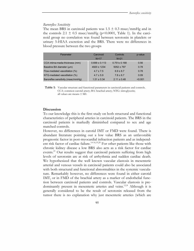
Baroreflex Sensitivity The mean BRS in carcinoid patients was 1.5 ± 0.3 msec/mmHg and in the controls 2.1 ± 0.5 msec/mmHg (p<0.0001, Table 1). In the carci-noid group no correlation was found between serotonin in platelets or urinary 5-HIAA excretion and the BRS. There were no differences in blood pressure between the two groups
Parameter Carcinoid Controls p-value
N=17 N=21
CCA intima-media thickness (mm) 0.699 ± 0.113 0.751± 0.168 0.56
Baseline BA diameter (µm) 4920 ± 1234 5052 ± 797 0.78Flow-mediated vasodilation (%) 4.7 ± 7.5 6.9 ± 8.7 0.78
NTG-mediated vasodilation (%) 4.7 ± 5.9 7.8 ± 6.7 0.09
Baroreflex sensitivity (msec/mmHg) 1.51 ± 0.34 2.11 ± 0.48 <0.001 Discussion To our knowledge this is the first study on both structural and functional characteristics of peripheral arteries in carcinoid patients. The BRS in the carcinoid patients is markedly diminished compared to sex and age matched controls. However, no differences in carotid IMT or FMD were found. There is abundant literature pointing out a low value BRS as an unfavorable prognostic factor in post-myocardial infraction patients and as independ-ent risk factor of cardiac failure.14-16,19,20 For other patients like those with chronic kidney disease a low BRS also acts as a risk factor for cardiac events.21 Our results suggest that carcinoid patients suffering from high levels of serotonin are at risk of arrhythmia and sudden cardiac death. We hypothesized that the well known vascular elastosis in mesenteric arterial and venous vessels in carcinoid patients could also be associated with both structural and functional abnormalities in the systemic vascula-ture. Remarkably however, no differences were found in either carotid IMT, or in FMD of the brachial artery as a marker of endothelial func-tion between carcinoid patients and controls. Vascular elastosis is pre-dominantly present in mesenteric arteries and veins.1,2,4 Although it is generally considered to be the result of serotonin released from the tumor there is no explanation why just mesenteric arteries (which are
Table 1: Vascular structure and functional parameters in carcinoid patients and controls. CCA: common carotid artery; BA: brachial artery; NTG: nitroglycerin; all values are means ± SD.
Chapter 6a
96
efferent to the tumor) and not extra-mesenteric arteries are involved. Several authors suggested local factors inducing local vascular fibro-sis.22,23 In 2004 Modlin et al surveyed the literature over the last 40 years covering the incidence, diagnosis, therapy and biological basis for carci-noid-associated fibrosis. They conclude that the mechanism of fibrosis is still poorly understood and there are no means by which this complica-tion can be predicted or monitored.24 The present study in carcinoid patients with high circulating serotonin levels tends to support the theory that vascular elastosis in patients with a midgut carcinoid is a local, rather than a systemic problem. Increasingly data emerge that heart failure due to carcinoid heart disease is not merely a result of fibrotic plaques in the right side of the heart but also the left side, pericardial effusions and cardiac metastases.25 Pre-sumably loss of vasomotor control in carcinoid disease is caused by alterations in pre- and postsynaptic receptor configuration resulting in an impaired cardiac autonomic nervous function. Future investigation elucidating the cause of the BRS impairment in carcinoid patients should focus on autonomic dysregulation, rather than on structural morphologic changes in the vessel wall.26,27
Baroreflex sensitivity
97
References 1. Bircher J, Bartholomew LG, Cain JC, Adson MA. Syndrome of intestinal arterial
insufficiency ("abdominal angina"). Arch Intern Med 1966; 117:632-638. 2. Palvio DH, Kristensen ES, Falk E. Intestinal ischemia due to vascular elastosis
caused by metastasizing carcinoid tumor of Meckel's diverticulum. Dis Colon Rec-tum 1985; 28:746-748.
3. Anthony PP. Gangrene of the small intestine a complication of argentaffin carcinoma. Br J Surg 1970; 57:118-122.
4. Qizilbash AH. Carcinoid tumors, vascular elastosis, and ischemic disease of the small intestine. Dis Colon Rectum 1977; 20:554-560.
5. de Vries H, Wijffels RTM, Willemse PHB, Verschueren RCJ, Kema IP, Karren-beld A, Prins TR, de Vries EGE. Abdominal angina in patients with a midgut carcinoid, a sign of severe pathology. World J Surg 2005; 29:1139-1142.
6. Brada SJ, Wijffels RTM, Kahraman T, de Vries EGE. Sublingual nitrate pro-vides cause for fear of food in a carcinoid patient. Ann Oncol 1997; 8:1053-1054.
7. Modlin IM, Kidd M, Latich I, Zikusoka MN, Shapiro MD. Current status of gastrointestinal carcinoids. Gastroenterology 2005; 128:1717-1751.
8. Houston DS, Vanhoutte PM. Serotonin and the vascular system. Role in health and disease, and implications for therapy. Drugs 1986; 31:149-163.
9. Kucuk O, Noskin G, Petersen K, Ezdinli E, Rollins D, Singh S, Sarpel S. Lower extremity vasospasm associated with ischemic neuropathy, dermal fibrosis, and digital gangrene in a patient with carcinoid syndrome. Cancer 1988; 62:1026-1029.
10. Petersen KG, Seemann WR, Plagwitz R, Kerp L. Evidence for coronary spasm during flushing in the carcinoid syndrome. Clin Cardiol 1984; 7:445-448.
11. Celermajer DS, Sorensen K, Ryalls M, Robinson J, Thomas O, Leonard JV, Deanfield JE. Impaired endothelial function occurs in the systemic arteries of children with homozygous homocystinuria but not in their heterozygous par-ents. J Am Coll Cardiol 1993; 22:854-858.
12. Bellien J, Joannides R, Iacob M, Eltchaninoff H, Thuillez C. Role of endothe-lium-derived nitric oxide in sustained flow-dependent dilatation of human pe-ripheral conduit arteries. Arch Mal Coeur Vaiss 2003; 96:738-741.
13. Celermajer DS, Sorensen KE, Georgakopoulos D, Bull C, Thomas O, Robinson J, Deanfield JE. Cigarette smoking is associated with dose-related and potentially reversible impairment of endothelium-dependent dilation in healthy young adults. Circulation 1993; 88:2149-2155.
14. Barron HV, Viskin S. Autonomic markers and prediction of cardiac death after myocardial infarction. Lancet 1998; 351:461-462.
15. La-Rovere MT, Bigger-JT J, Marcus FI, Mortara A, Schwartz PJ. Baroreflex sensitivity and heart-rate variability in prediction of total cardiac mortality after myocardial infarction. ATRAMI (Autonomic Tone and Reflexes After Myocar-dial Infarction) Investigators. Lancet 1998; 351:478-484.
16. Landolina M, Mantica M, Pessano P, Manfredini R, Foresti A, Schwartz PJ, De Ferrari GM. Impaired baroreflex sensitivity is correlated with hemodynamic de-terioration of sustained ventricular tachycardia. J Am Coll Cardiol 1997; 29:568-575.
17. Robbe HW, Mulder LJM, Ruddel H, Veldman JBP, Langewitz WA, Mulder G. Assessment of baroreflex sensitivity by means of spectral analysis. Hypertension 1987; 10:538-543.
Chapter 6a
98
18. Van Steenis HG, Tulen JFM, Mulder LJM. Heart rate variability spectra based on nonequidistant sampling: the spectrum of counts and the instantaneous heart rate spectrum. Med Eng Phys 1994; 16:35-36.
19. Lengyel C, Torok T, Varkonyi T, Kempler P, Rudas L. Baroreflex sensitivity and heart-rate variability in insulin-dependent diabetics with polyneuropathy. Lancet 1998; 351:1436-1437.
20. Schachinger V, Britten MB, Zeiher AM. Prognostic impact of coronary vasodila-tor dysfunction on adverse long-term outcome of coronary heart disease. Circula-tion 2000; 101:1899-1906.
21. Bavanandan S, Ajayi S, Fentum B, Paul SK, Carr SJ, Robinson TG. Cardiac baroreceptor sensitivity: a prognostic marker in predialysis chronic kidney dis-ease patients? Kidney Int 2005; 67:1019-1027.
22. La Rosa S, Chiaravalli AM, Capella C, Uccella S, Sessa F. Immunohistochemical localization of acidic fibroblast growth factor in normal human enterochromaf-fin cells and related gastrointestinal tumours. Virchows Arch 1997; 430:117-124.
23. Zhang PJ, Furth EE, Cai X, Goldblum JR, Pasha TL, Min KW. The role of beta-catenin, TGF beta 3, NGF2, FGF2, IGFR2, and BMP4 in the pathogenesis of mesenteric sclerosis and angiopathy in midgut carcinoids. Hum Pathol 2004; 35:670-674.
24. Modlin IM, Shapiro MD, Kidd M. Carcinoid tumors and fibrosis: an association with no explanation. Am J Gastroenterol 2004; 99:2466-2478.
25. Pellikka PA, Tajik AJ, Khandheria BK, Seward JB, Callahan JA, Pitot HC, Kvols LK. Carcinoid heart disease. Clinical and echocardiographic spectrum in 74 pa-tients. Circulation 1993; 87:1188-1196.
26. Hoffmann J, Grimm W, Menz V, Wied M, Sprenger A, Arnold R, Maisch B. Prognostic value of heart rate variability analysis in patients with carcinoid syn-drome. Digestion 2001; 63:35-42.
27. Hoffmann J, Grimm W, Menz V, Wied M, Funck R, Arnold R, Maisch B. Heart rate variability in carcinoid heart disease. Am J Cardiol 1999; 83:128-131.
Serotonin response in carcinoid vascular rings
99
Chapter 6b LOSS OF PRE-SYNAPTIC SEROTONIN VASOCONSTRICTOR CONTROL IN ISOLATED POPLITEAL ARTERY PREPARATIONS FROM A PATIENT WITH
MIDGUT CARCINOID H. de Vries1 A. van Buiten2 P.H.B. Willemse4 E.G.E. de Vries4 H. Buikema2 S.A. Nelemans3
Departments of 1Surgery, 2Clinical Pharmacology, 4Medical Oncology, University of Groningen and University Medical Center Groningen, Groningen, 3Department of Molecular Pharmacology, University of Groningen, Groningen, The Netherlands
Chapter 6b
100
Abstract Metastatic carcinoid disease can be associated with vasomotor instability causing flushes and hypotension or hypertension. In addition morpho-logical changes in the vessel wall (i.e. vascular elastosis) with thickening of the intima-media complex resulting in reduced compliance can occur. Pharmacological studies on isolated arteries from carcinoid patients have not yet been performed. Therefore this study investigates functional alterations in serotonin response in isolated popliteal artery preparations of a carcinoid patient. The effect of pre-synaptic inhibition on serotonin induced contraction and the involvement of serotonin-2A (5-HT2A) receptors in these responses as well as the role of contractile pros-taglandins was analyzed. Methods: In a metastatic carcinoid patient a below knee amputation was performed because of necrosis due to ischemia. The popliteal arteries were dissected and conserved for in vitro isolated vascular ring perfusion tests. In an organ bath tests were performed using increasing concentra-tions of serotonin (30 nmol L-1 - 30 µmol L-1). Several serotonin induced contraction cycles were performed in the presence of pre- and post-synaptic blockers (ondansetron, ketanserin, indomethacin). The same tests were performed on popliteal vascular rings from a healthy control who underwent a posttraumatic upper leg amputation. Results: A tenfold higher serotonin concentration was needed to start contraction in carcinoid rings compared to control rings. Contractions in carcinoid rings were predominately mediated by 5-HT2A receptors, whereas in normal popliteal artery there were also other functional serotonin receptors besides the 5-HT2A subtype. In the normal popliteal artery serotonin vasoconstrictor control is supported by a pre-synaptic rescue mechanism, becoming active in case of blockade of smooth muscle 5-HT2A receptor by ketanserin. This mechanism might be acti-vated by serotonin stimulation and is probably mediated via contractile cyclooxygenase-derived prostanoids. Such a rescue mechanism by pre-synaptic activation appeared to be absent in carcinoid artery rings. Conclusion: In vitro midgut carcinoid vascular rings are characterized by loss of pre-synaptic serotonin vasomotor control and non-responsive-ness to low serotonin concentrations.
Serotonin response in carcinoid vascular rings
101
Introduction A midgut carcinoid is a neuroendocrine tumor that can produce several metabolic active substances, including serotonin, prostaglandins (PGEs) and catecholamines.1,2 Vasomotor instability in patients with metastatic carcinoid disease is associated with hot flushes, hypotension or hyperten-sion. Morphological changes can occur in the vessel wall (i.e. vascular elastosis) with thickening of the intima-media complex, resulting in reduced compliance of the vessel. This is observed, predominantly in mesenteric vessels, in the proximity of a carcinoid tumor mass. A well known distant effect of serotonin is carcinoid fibrotic heart disease.3,4 Carcinoid disease is often associated with high serum levels of serotonin and the symptoms mentioned are at least partly considered to be due to serotonin.3,5 Elevated circulating serotonin levels in carcinoid patients might cause the display of impaired responses in the vessel wall. Phar-macologically consistent with long-term exposure to high serotonin levels, a downregulation of serotonin (5-HT)2A receptors has been re-ported in platelets. Platelets form the serotonin reservoir of patients with carcinoid tumors.6 Apart from direct effects of serotonin on vascular tone also an indirect effect via so-called contractile PGEs might be involved.7 Highly increased plasma levels of contractile PGEs, particu-larly PGE2, have been observed in carcinoid patients.8-10 So far, pharmacological studies on isolated arteries from carcinoid patients are not available. Normally serotonin causes at the vascular level constriction via activation of 5-HT2A receptors present on the smooth muscle cells and/or activation of the 5-HT3 receptor subtype present on post-ganglionic sympathetic neurons, representing an intrinsic transmit-ter-gated ion channel.11 Dynamic responses to serotonin receptor stimu-lation within the vascular wall can be elegantly demonstrated using isolated vascular ring perfusions in vitro.12,13 The role of vascular sero-tonin receptors in carcinoid disease could be elucidated by analyzing the pre-synaptic inhibition on serotonin-induced contraction as well as the involvement of 5-HT2A receptors in these responses and the role of contractile PGEs. Therefore we evaluated in the present study the functional alterations in serotonin responses in isolated popliteal artery preparations of a carci-noid patient and a normal popliteal artery.
Chapter 6b
102
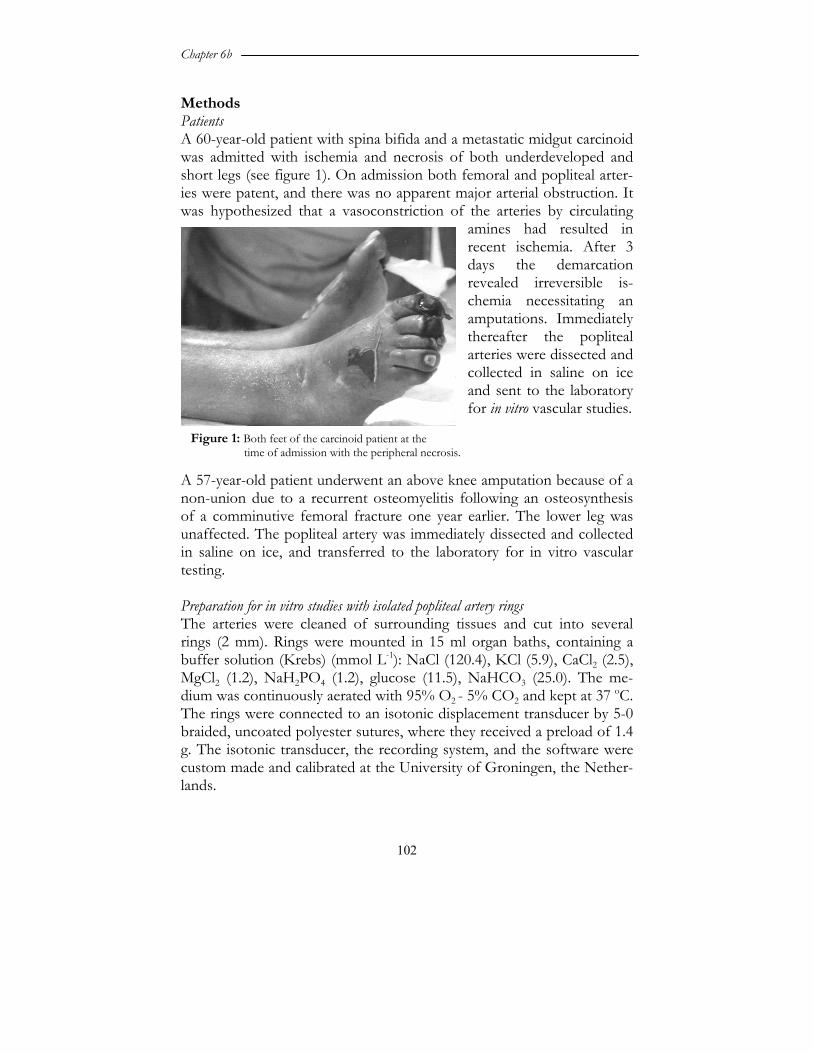
Methods Patients A 60-year-old patient with spina bifida and a metastatic midgut carcinoid was admitted with ischemia and necrosis of both underdeveloped and short legs (see figure 1). On admission both femoral and popliteal arter-ies were patent, and there was no apparent major arterial obstruction. It was hypothesized that a vasoconstriction of the arteries by circulating
amines had resulted in recent ischemia. After 3 days the demarcation revealed irreversible is-chemia necessitating an amputations. Immediately thereafter the popliteal arteries were dissected and collected in saline on ice and sent to the laboratory for in vitro vascular studies.
A 57-year-old patient underwent an above knee amputation because of a non-union due to a recurrent osteomyelitis following an osteosynthesis of a comminutive femoral fracture one year earlier. The lower leg was unaffected. The popliteal artery was immediately dissected and collected in saline on ice, and transferred to the laboratory for in vitro vascular testing. Preparation for in vitro studies with isolated popliteal artery rings The arteries were cleaned of surrounding tissues and cut into several rings (2 mm). Rings were mounted in 15 ml organ baths, containing a buffer solution (Krebs) (mmol L-1): NaCl (120.4), KCl (5.9), CaCl2 (2.5), MgCl2 (1.2), NaH2PO4 (1.2), glucose (11.5), NaHCO3 (25.0). The me-dium was continuously aerated with 95% O2 - 5% CO2 and kept at 37 oC. The rings were connected to an isotonic displacement transducer by 5-0 braided, uncoated polyester sutures, where they received a preload of 1.4 g. The isotonic transducer, the recording system, and the software were custom made and calibrated at the University of Groningen, the Nether-lands.
Figure 1: Both feet of the carcinoid patient at the time of admission with the peripheral necrosis.
Serotonin response in carcinoid vascular rings
103
Artery rings were allowed to equilibrate for 60 min during which regular washing periods were performed. Rings were primed and checked for viability by repeated stimulation (three times) with 60 mmol L-1 KCl and intermediate washing and stabilization periods. The third response to KCl was referred to as the 100% referral maximum contraction ampli-tude for each ring and all other contractile responses were expressed as a percentage of this response to further reduce inter-ring variability. Experimental protocol for contractile responses to serotonin Parallel rings were simultaneously studied during three consecutive series of measurements for contractile responses to increasing concentrations serotonin (30 nmol L-1 - 30 µmol L-1 bath-concentrations) in each series, and in the selective absence and presence of various compounds known to interfere with receptor signal transduction processes. In the first series of measurements, parallel rings were studied for serotonin responses under control conditions and under conditions of pre-synaptic inhibi-tion. In the second and third series of measurement, the additional involvement of serotonin receptors and cyclooxygenase-derived PGEs was studied. To create these conditions, we used vehicle as a control, 10 µmol L-1 ketanserin to inhibit 5-HT2A receptors14, 10 µmol L-1 indomethacin to inhibit cyclooxygenase-derived PGEs7, and 1 µmol L-1 ondansetron to obtain serotonin receptor mediated pre-synaptic inhibition.15 To obtain general pre-synaptic inhibition general a combination of ondansetron with 1 µmol L-1 tetrodotoxin (an inhibitor of voltage sensitive Na+ channels16 and 300 µmol L-1 suramin (a blocker of pre-synaptic P2-purinergic receptors) was used.17 Rings were pre-incubated with the appropriate inhibitors for at least 30 min before stimulation with sero-tonin. Observations of serotonin contractility under certain conditions were obtained from 3-6 rings for each condition. Thickening of the intima media complex causing a diminished compli-ance of the vessel wall, a well-known symptom in carcinoid patients, may interfere with contraction amplitudes. To correct for this and to reduce inter-ring variability, we expressed receptor-mediated vasoconstrictor responses to serotonin as percentage of maximal constriction with KCl.
Chapter 6b
104
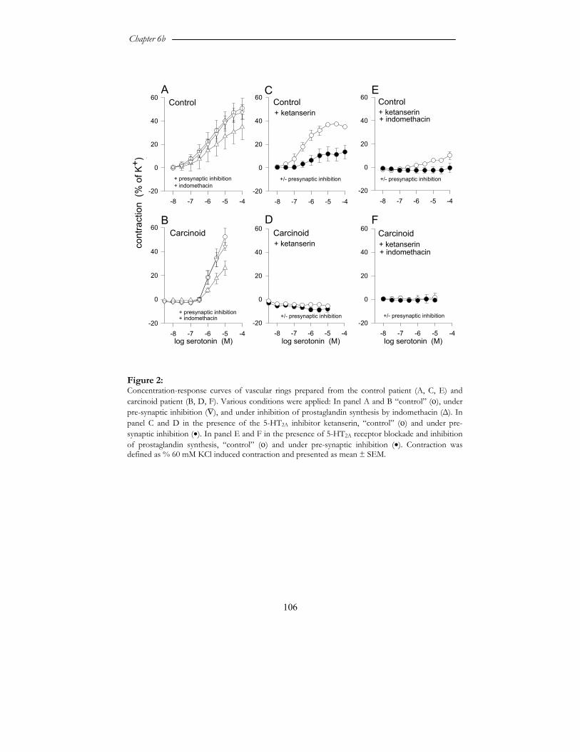
Drugs used The following chemicals and drugs were used: serotonin, indomethacin, ketanserin, suramin and tetrodotoxin (all from Sigma-Aldrich Chemie, Zwijndrecht, The Nederlands) ondansetron (Glaxo Smith Kline BV, Zeist, The Netherlands). Stock solutions were prepared of indomethacin (10 mmol L-1, in ethanol), ketanserin (10 mmol L-1, in H2O), tetrodotoxin (1 mmol L-1, in H2O), suramin (300 mmol L-1, H2O), and ondansetron (1 mmol L-1, H2O), stored at -20oC and diluted in Krebs solution to reach final concentrations. Serotonin was prepared in H2O (100 mmol L-1), and further dilutions were performed directly in Krebs solution. Compounds for the Krebs buffer solution were all obtained from Merck (Darmstadt, Germany). Calculations and statistical analysis All responses of individual rings to serotonin were expressed as a per-centage of the maximum contraction-response to 60 mmol L-1 KCl, and used for calculations and graphic representations. Maximal effect (Emax) and the effective concentration producing 50% of the maximal effect, expressed as negative logarithm (pEC50), were obtained from the indi-vidual concentration-response curves. Observations of serotonin con-tractility under a specific condition were obtained from 3-6 rings. Com-parison between percentual responses was made using Student’s t-test. Data are expressed as mean ± standard error of the mean (SEM). P-values <0.05 were considered significant. Results Receptor-independent contraction induced by a high concentration of KCl (60 mM), resulting in maximal contraction in popliteal artery rings was 40% lower in rings from the carcinoid patient (593 ±73 µm versus 950 ± 68 displacement, p<0.001). Serotonin induced, concentration-dependent contractions in popliteal artery rings were obtained from the control patient and the carcinoid patient (Figure 2A, B). Maximal con-traction (Emax) was similar; in carcinoid rings: 51.8 ± 8.7 % and in control rings 52.2 ± 7.5 % of the maximal contraction as induced by 60 mM KCl. Also pEC50 values were not different (carcinoid rings: 5.9 ± 0.1; control rings 5.8 ± 0.1), however the threshold of contraction was at a much higher serotonin concentration for carcinoid-derived rings 1 μM than 0.1 μM for control rings. In the presence of pre-synaptic inhibitors

Serotonin response in carcinoid vascular rings
105
similar results were obtained. Treatment with ondansetron alone, to block pre-synaptic serotonin receptors, was as ineffective as when given in combination with other inhibitors of post-ganglionic synaptic trans-mission, tetrodotoxin and suramin (data not shown). Indomethacin as inhibitor of cyclooxygenase-derived prostanoids synthesis, only slightly changed contraction in carcinoid rings (Emax: 26.2 ± 5.9 %; pEC50: 5.7 ± 0.1, Figure 2A, p=n.s.) and control rings (Emax: 34.6 ± 10.7 %; pEC50: 5.8 ± 0.1). In contrast, the 5-HT2A receptor blocker ketanserin totally abol-ished contractions in carcinoid vascular rings with an Emax: -5.6 ± 0.1% and after presynaptic inhibition -8.3 ± 2.6%; p<0.05. In control rings ketanserin decreased contractions to 37.0 ± 0.4 % with pEC50: 6.5 ± 0.1 (p<0.05) and in the presence of presynaptic inhibition even further (Emax: 13.4 ± 5.6%; pEC50: 6.0 ± 0.1; p<0.05). The combination of ketanserin and indomethacin in control rings slightly decreased contraction in the absence and presence of presynaptic inhibi-tors to Emax: 10.0 ± 3.2% and -0.5 ± 3.9%, respectively (Figure 2E), but totally blocked contractions in carcinoid rings.
Chapter 6b
106
log serotonin (M)-8 -7 -6 -5 -4
-20
0
20
40
60Carcinoid+ ketanserin
+ presynaptic inhibition
-8 -7 -6 -5 -4
cont
ract
ion
(% o
f K+ )
-20
0
20
40
60Control
+ ketanserin
A
B
-8 -7 -6 -5 -4-20
0
20
40
60 Control+ ketanserin+ indomethacin
-8 -7 -6 -5 -4-20
0
20
40
60 Control
+ indomethacin
C
D
log serotonin (M)-8 -7 -6 -5 -4
-20
0
20
40
60Carcinoid
E
+ indomethacin
log serotonin (M)-8 -7 -6 -5 -4
-20
0
20
40
60 Carcinoid+ ketanserin+ indomethacin
F
+/- presynaptic inhibition+/- presynaptic inhibition
+/- presynaptic inhibition +/- presynaptic inhibition
+ presynaptic inhibition
Figure 2: Concentration-response curves of vascular rings prepared from the control patient (A, C, E) and carcinoid patient (B, D, F). Various conditions were applied: In panel A and B “control” (ο), under pre-synaptic inhibition (∇), and under inhibition of prostaglandin synthesis by indomethacin (Δ). In panel C and D in the presence of the 5-HT2A inhibitor ketanserin, “control” (ο) and under pre-synaptic inhibition (•). In panel E and F in the presence of 5-HT2A receptor blockade and inhibition of prostaglandin synthesis, “control” (ο) and under pre-synaptic inhibition (•). Contraction was defined as % 60 mM KCl induced contraction and presented as mean ± SEM.
Serotonin response in carcinoid vascular rings
107
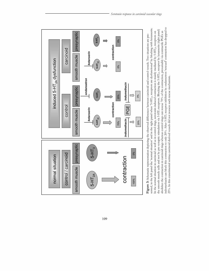
Discussion This is the first study, on pharmacologically characterized vascular function of isolated arteries obtained from a patient with midgut carci-noid. Some striking differences became apparent in these arteries com-pared to arteries from a control patient. Carcinoid-derived popliteal artery rings were less responsive to the receptor-independent contractile agent KCl than control popliteal artery rings (0.6-fold). This decrease in contractility is probably due to structural changes in the vessel wall as observed in carcinoid patients.4,18 Interestingly, a similar reduction of contractility due to structural changes in the vessel wall has been re-ported for popliteal artery rings obtained from patients with peripheral occlusive arteriosclerosis.19 When normalized for the maximal KCl contraction, receptor-induced contraction to serotonin appeared to be very similar for carcinoid rings and control. The presence of pre-synaptic inhibitors did not affect the responses to serotonin. This suggests that modulation of muscle contrac-tion by activation of post-ganglionic neurons is not influenced by pre-synaptic factors. However, when the rings were incubated with the 5-HT2A blocker ketanserin, contractions in carcinoid rings were totally blocked while control rings were still able to contract (figure 2A versus 2B). This contraction must be mediated via contractile PGEs and mainly involves pre-synaptic activation via pre-synaptic 5-HT3 receptors in view of the results obtained with ondansetron and the PGE blocker indo-methacin experiments (figure 2 C and E). This rescue mechanism is absent in carcinoid vascular rings suggesting loss of pre-synaptic control in these rings. A scheme depicting the hypothesized explanations of these findings is shown in figure 3. The mechanism by which this occurs remains to be solved. It could resemble the observations in tracheal ring preparations of the guinea-pig. In this previous study, a chain of events leads from receptor activation to production of contractile PGEs and subsequent smooth muscle contraction via prostanoid receptors.20 PGE synthesis and secretion was induced by activation of Ca2+-dependent cytosolic phospholipase-A2, generating arachidonic acid, the substrate for cyclooxygenase.7 As mentioned, serotonin receptor activation results in Ca2+ influx, which might be well capable of starting similar events in the arteries tested. Functional downregulation of the pre-synaptic sero-tonin receptor subtype in carcinoid rings, due to long-term exposure to high serotonin concentrations in vivo, might be responsible for the
Chapter 6b
108
disappearance of the rescue mechanism in carcinoid patients. A general neuropathy as cause is less likely, since the effect observed is already detectable by solely blocking serotonin receptors by ondansetron. We studied the baroreflex sensitivity in carcinoid patients with elevated platelet serotonin and showed a reduced baroreflex sensitivity compared to sex and age matched controls.21 Reason for this may be the loss of the fine-tuning of the sympathetic presynaptic control. A ten-fold high serotonin was required to induce contractions in carci-noid vascular rings compared to control rings (Figure 2B). This is consis-tent with downregulation of 5-HT2A receptors as observed in platelets of patients with carcinoid tumors.6 In conclusion, this is the first report to demonstrate that in the popliteal artery of a carcinoid patient the constriction of an isolated vascular ring was predominately mediated by 5-HT2A receptors, whereas in normal popliteal artery there are also other functional serotonin receptors. Our results suggest, that in normal popliteal artery serotonin vasomotor control is supported by a pre-synaptic rescue mechanism, becoming apparent after blocking of the smooth muscle 5-HT2A receptor. This mechanism might be activated by serotonin stimulation and can be blocked by indomethacin indicating a role for contractile cyclooxy-genase-derived prostanoids. Such a rescue mechanism seems to be absent in carcinoid arteries.
Serotonin response in carcinoid vascular rings
109
Figu
re 3
: Sch
emat
ic re
pres
enta
tion
depi
ctin
g th
e ob
serv
ed d
iffer
ence
s bet
wee
n ca
rcin
oid
and
cont
rol v
esse
ls. T
wo
situa
tions
are
pre
-se
nted
, in
the
left
pane
l the
‘nor
mal
situa
tion’
and
in th
e rig
ht p
anel
whe
n ‘5
-HT 2
A re
cept
ors a
re d
ysfu
nctio
nal’
by b
lock
ing
with
ket
anse
rin.
In th
e no
rmal
situa
tion
in c
arci
noid
as w
ell a
s in
cont
rol r
ings
, ser
oton
in-in
duce
d co
ntra
ctio
n is
main
ly m
ediat
ed b
y 5-
HT 2
A re
cept
ors o
n th
e sm
ooth
mus
cle c
ells
and
not b
y pr
e-sy
napt
ic st
imul
atio
n vi
a 5-
HT 3
rece
ptor
s. Bl
ocki
ng th
e 5-
HT 2
A re
cept
or b
y ke
tans
erin
(rig
ht p
anel)
ab
olish
es th
e co
ntra
ctio
n in
car
cino
id ri
ngs w
here
as c
ontro
l rin
gs c
an re
scue
70%
of t
he c
ontra
ctio
n, p
resu
mab
ly vi
a co
ntra
ctile
PG
Es a
s in
dom
etha
cin
can
bloc
k th
is co
ntra
ctio
n to
onl
y 20
%. A
fter 5
-HT 3
rece
ptor
inhi
bitio
n by
ond
anse
tron
‘resc
ued’
con
tract
ion
has d
ropp
ed to
25
%. I
n th
is ex
perim
enta
l set
ting
carc
inoi
d de
rived
ves
sels
did
not p
osse
ss su
ch re
scue
mec
hani
sm.
norm
al s
ituat
ion
indu
ced
5-H
T 2A
dysf
unct
ion
cont
rol
carc
inoi
d
5-H
T 2A
cont
ract
ion
70%
5-H
T 3
cont
ract
ion
cont
rol /
car
cino
id
5-H
T 2A
5-H
T 3
cont
ract
ion
smoo
th m
uscl
epr
esyn
aptic
smoo
th m
uscl
epr
esyn
aptic
ketanserin
25%on
dansetron
PGE
indomethacin
0%0%
20%
0%
smoo
th m
uscl
epr
esyn
aptic
smoo
th m
uscl
epr
esyn
aptic
smoo
th m
uscl
epr
esyn
aptic
100%
0%
5-H
T 2A
ketanserin
5-H
T 3
indomethacin
Chapter 6b
110
References 1. Feldman JM. Increased dopamine production in patients with carcinoid tumors.
Metabolism 1985; 34:255-260. 2. Kema IP, de Vries EGE, Slooff MJ, Biesma B, Muskiet FA. Serotonin, cate-
cholamines, histamine, and their metabolites in urine, platelets, and tumor tissue of patients with carcinoid tumors. Clin Chem 1994; 40:86-95.
3. Simula DV, Edwards WD, Tazelaar HD, Connolly HM, Schaff HV. Surgical pathology of carcinoid heart disease: a study of 139 valves from 75 patients spanning 20 years. Mayo Clin Proc 2002; 77:139-147.
4. Anthony PP, Drury RA. Elastic vascular sclerosis of mesenteric blood vessels in argentaffin carcinoma. J Clin Pathol 1970; 23:110-118.
5. Zuetenhorst JM, Bonfrer JM, Korse CM, Bakker R, van Tinteren H, Taal BG. Carcinoid heart disease: the role of urinary 5-hydroxyindoleacetic acid excretion and plasma levels of atrial natriuretic peptide, transforming growth factor-beta and fibroblast growth factor. Cancer 2003; 97:1609-1615.
6. Spigset O, Kristoffersson A, Mjorndal T. Platelet serotonin 5-HT2A receptor binding in patients with carcinoid tumor. Scand J Clin Lab Invest 2004; 64:3-8.
7. Schaafsma D, Gosens R, Bos IS, Meurs H, Zaagsma J, Nelemans SA. Role of contractile prostaglandins and Rho-kinase in growth factor-induced airway smooth muscle contraction. Respir Res 2005; 6:85.
8. Boyd EJ, Hulks G, Thomas JS, McColl KE. Hypertrophic gastritis associated with increased gastric mucosal prostaglandin E2 concentrations in a patient with the carcinoid syndrome. Gut 1988; 29:1270-1276.
9. Jaffe BM. Prostaglandins and serotonin: nonpeptide diarrheogenic hormones. World J Surg 1979; 3:565-578.
10. Jaffe BM, Behrman HR, Parker CW. Radioimmunoassay measurement of prostaglandins E, A, and F in human plasma. J Clin Invest 1973; 52:398-405.
11. Martin GR. 5-Hydoxytryptamine receptors. The IUPHAR compendium of receptor characterization and classification. IUPHAR Media, London; 1998:167-185.
12. Van Melle JP, Buikema H, Van den Berg MP, Van Buiten A, Van Veldhuisen DJ, Boonstra PW, Van Gilst WH. Sertraline causes strong coronary vasodilation: possible relevance for cardioprotection by selective serotonin reuptake inhibi-tors. Cardiovasc Drugs Ther 2004; 18:441-447.
13. Buikema H, Grandjean JG. Potentiation of alpha-adrenoceptor-mediated vasoconstriction by sumatriptan. Lancet 1993; 342:1121.
14. Thollon C, Bidouard JP, Cambarrat C, Delescluse I, Villeneuve N, Vanhoutte PM, Vilaine JP. Alteration of endothelium-dependent hyperpolarizations in por-cine coronary arteries with regenerated endothelium. Circ Res 1999; 84:371-377.
15. Hope AG, Peters JA, Brown AM, Lambert JJ, Blackburn TP. Characterization of a human 5-hydroxytryptamine-3 receptor type A (h5-HT3R-AS) subunit stably expressed in HEK 293 cells. Br J Pharmacol 1996; 118:1237-1245.
16. Molleman A, Nelemans SA, Van den AJ, Duin M, Den Hertog A. Voltage-dependent sodium and potassium, but no calcium conductances in DDT1 MF-2 smooth muscle cells. Pflugers Arch 1991; 417:479-484.
Serotonin response in carcinoid vascular rings
111
17. Henning RH, Rowan EG, Braga MF, Nelemans SA, Harvey AL. The prejunc-tional inhibitory effect of suramin on neuromuscular transmission in vitro. Eur J Pharmacol 1996; 301:91-97.
18. Qizilbash AH. Carcinoid tumors, vascular elastosis, and ischemic disease of the small intestine. Dis Colon Rectum 1977; 20:554-560.
19. Ortega A, Avellanal M, Espana G, Flores A, Aleixandre A. Effect of nitroglycer-ine in popliteal preparations from patients with peripheral occlusive arteriopathy precontracted with KCl or 5-hydroxytryptamine. Clin Exp Pharmacol Physiol 2003; 30:528-531.
20. Schaafsma D, Gosens R, Bos IS, Meurs H, Zaagsma J, Nelemans SA. Role of contractile prostaglandins and Rho-kinase in growth factor-induced airway smooth muscle contraction. Respir Res 2005; 6:85.
21. de Vries H, Smit AJ, Willemse PHB, Gietema JA, Kema IP, de Vries EGE. No signs of early atherosclerosis or endothelial dysfunction, but disturbed baroreflex sensitivity in carcinoid patients. submitted 2006.

Chapter 6b
112