Embed Size (px)
Citation preview
University of Groningen
Therapeutic immunization strategies against cervical cancerBungener, Laura
IMPORTANT NOTE: You are advised to consult the publisher's version (publisher's PDF) if you wish to cite fromit. Please check the document version below.
Document VersionPublisher's PDF, also known as Version of record
Publication date:2004
Link to publication in University of Groningen/UMCG research database
Citation for published version (APA):Bungener, L. B. (2004). Therapeutic immunization strategies against cervical cancer: induction of cell-mediated immunity in murine models s.n.
CopyrightOther than for strictly personal use, it is not permitted to download or to forward/distribute the text or part of it without the consent of theauthor(s) and/or copyright holder(s), unless the work is under an open content license (like Creative Commons).
Take-down policyIf you believe that this document breaches copyright please contact us providing details, and we will remove access to the work immediatelyand investigate your claim.
Downloaded from the University of Groningen/UMCG research database (Pure): http://www.rug.nl/research/portal. For technical reasons thenumber of authors shown on this cover page is limited to 10 maximum.
Download date: 08-06-2018

Chapter 2
Genetic immunization against cervical carcinoma: induction of cytotoxic T lymphocyte activity with a recombinant alphavirus vector expressing human papillomavirus type 16 E6 and E7
Toos Daemen, Frens Pries, Laura Bungener, Marian Kraak, Joke Regts and
Jan Wilschut
Gene Therapy 7(21):1859-66.
38 Chapter 2
Abstract Infection of genital epithelial cells with human papillomavirus (HPV) types 16 and 18 is closely associated with the development of cervical carcinoma. The transforming potential of these high-risk HPV depends on the expression of the E6 and E7 early viral gene products. Since the expression of E6 and E7 is selectively maintained in premalignant and malignant cervical lesions these proteins are attractive candidates for immunotherapeutic and prophylactic strategies. This report describes the construction, characterization and the in vivo immunotherapeutic potential of recombinant Semliki Forest virus (SFV) expressing the HPV16 E6 and E7 proteins (SFVE6E7). Western blot analysis and immunofluorescence staining demonstrated expression of E6 and E7 in BHK cells infected with SFVE6E7. Immunization of mice with SFVE6E7 resulted in an efficient in vivo priming of HPV-specific CTL activity. The induced CTL lysed murine tumor cells transformed with the HPV16 genome and EL-4 cells loaded with an immunodominant class I-binding HPV E7 peptide. CTL could reproducibly be induced by immunization with three injections of as few as 105 infectious units of SFVE6E7. Protection from tumor challenge was studied using the tumor cell line TC-1. Immunization with 5x10⁶ SFVE6E7 particles protected 40% of the mice from tumor challenge. These results indicate that E6E7 expression by the efficient and safe recombinant SFV system represents a promising strategy for immunotherapy or immunoprophylaxis of cervical carcinoma.
Induction of CTL and anti tumor responses by rSFVE6E7 39
Introduction From molecular, clinical and epidemiological studies it is evident that the high-risk human papillomaviruses HPV16 and HPV18 are linked to the development of precursor lesions of cervical cancer and invasive cervical carcinoma [Zur Hausen 1991]. The HPV genome encodes 7 early (E) nonstructural regulatory proteins, and two late (L) structural proteins. Integration of the viral DNA in the genome of the host cell, which is an essential step in HPV16- or HPV18-induced development of cervical carcinoma, results in a loss of E1- or E2-mediated transcriptional control. As a consequence the transformed cells overexpress the E6 and E7 proteins, initiating the malignant transformation process [Münger 1992, Werness 1990, Pei 1996, Jones 1996, Gulliver 1997]. Specific cell-mediated immunity is believed to play an essential role in the control of HPV infections and cervical carcinoma. This assumption is based on observations showing: (i) that HPV-induced lesions regress spontaneously in the majority of individuals, and (ii) that immunodeficient patients develop significantly more HPV-related proliferative lesions in skin and anogenital tissue than immunocompetent individuals [Porreco 1975, Johnson 1992]. In several animal models it has been demonstrated that the HPV E6 and E7 proteins, constitutively expressed in HPV-transformed cells, can act as targets for CTL-mediated tumor cell killing and stimulation of tumor-specific CTL activity [Feltkamp 1993, Feltkamp 1995, Jochmus 1997, de Bruijn 1998].
Induction of an antigen-specific CTL response requires intracellular processing of the target antigen and presentation of antigenic peptides by MHC class I molecules. This can be achieved efficiently with recombinant viral vectors. In the present study, our approach is to use the Semliki Forest virus (SFV) expression system. SFV belongs to the genus Alphavirus of the family of the Togaviridae [Strauss 1994]. Alphaviruses consist of a nucleocapsid with one copy of a single-stranded RNA molecule surrounded by an envelope containing spike proteins. Alphavirus RNA has a positive polarity enabling the genomic RNA to initiate an infection when introduced into the cytoplasm of a cell. In addition, the RNA is self-replicating, since it encodes its own replicase, and replication results in high-level expression of the viral proteins in host cells. A full-length cDNA copy of the viral genome has been cloned in a bacterial plasmid including a prokaryotic DNA-dependent RNA polymerase such that viral RNA can be transcribed in vitro. These RNA transcripts are fully infectious, i.e. introduction into cells suffices to initiate replication and a full infection cycle, resulting in virus formation [Liljeström 1991a]. Liljeström and coworkers developed a vector system that allows for efficient expression of foreign coding sequences as part of the SFV RNA replicon [Liljeström 1991a, Liljeström 1991b, Berglund 1993]. A high biosafety level is obtained by separating the replicase and structural genes of the viral genome. Thus, recombinant
40 Chapter 2
virus particles can be produced that infect cells only once. In addition, the SFV helper (containing the structural genes) was mutated in the gene encoding one of the spike proteins [Liljeström 1991b]. In effect, such virus particles cannot infect cells unless they are activated with exogenous protease.
In this article we describe the construction of recombinant SFV encoding HPV16 E6 and E7 and the cellular immune response in mice induced by these recombinant SFVE6E7 particles. Materials and methods Cell lines Baby hamster kidney cells (BHK) were obtained from the American Type Culture Collection (CCL-10). The cells were grown in GMEM (Life Technologies, Paisley, UK) containing 5% fetal calf serum (PAA Laboratories, Linz, Austria). C3 cells, 13-2 cells and TC-1 cells were kindly provided by Dr. C Melief and Dr. R Offringa (Leiden University, The Netherlands). The C3 cell line was derived from C57BL/6 (H-2b) embryonic cells transfected with a plasmid containing the complete HPV16 genome. The 13-2 cell line was generated from C57Bl/6 (H-2b) embryonic cells transfected with the E1 region of adenovirus type 5 in which the adenoviral E1A epitope SGPSNTPPEI is replaced by a HPV16 E7 CTL epitope, aa 49-57 (RAHYNIVTF) (R Offringa, personal communication). The TC-1 cell line was generated from C57Bl/6 primary lung epithelial cells infected with a retroviral vector expressing HPV16 E6E7 plus a retrovirus expressing activated c-Ha-ras [Lin 1996]. EL-4 cells were kindly provided by Dr. L Leserman (Centre d�Immunologie de Marseille-Luminy, France). C3, 13-2, TC-1 and EL-4 cells were grown in Iscove�s medium (IMDM, Life Technologies) supplemented with 10% fetal calf serum. Both media contained penicillin and streptomycin (Life Technologies; 100 U/ml and 100 mg/ml, respectively). Mice Specific pathogen-free female C57Bl/6 mice (Harlan CPB, Zeist, The Netherlands) were between 6 and 10 weeks of age at the start of the immunization protocols. The protocol for the animal experiments described in this paper was approved by the Animal Experimentation Ethical Committee of the University of Groningen. Peptide The HPV16 H-2Db binding E7 peptide RAHYNIVTF (residue 49-57) was synthesized and purified by Dr. JW Drijfhout (Academic Hospital Leiden, The Netherlands). The peptide was analyzed by reverse phase HPLC and found to be over 90% pure.

Induction of CTL and anti tumor responses by rSFVE6E7 41
Cloning of HPV16 E6 and E7 in pSFV3 pSFVHelper 2 [Berglund 1993] and pSFV3 [Liljeström 1991b] were kindly provided by Dr. P Liljeström (Karolinska Institute, Stockholm, Sweden). The HPV16 E6 and E7 genes were obtained from the plasmid pRSV-HPV16E6E7 [Smits 1990], which was kindly provided by Dr. J ter Schegget (Free University, Amsterdam, The Netherlands). In this plasmid the HPV16 E6 and E7 genes are present in tandem, with a stop codon after the E6 gene. Amplification of the E6E7 tandem gene was done by PCR using the following primers, written in 5� to 3� direction: GACGGATCCAAAGAGAACTCCAATG (E6 forward) and GAGAATTCGGATCCGCCATGGTAGATTAT (E7 reverse). The PCR product was digested with BamHI and cloned into the BamHI site of pGEM7Zf+. After sequence confirmation, the E6E7 fragment was cloned into the unique BamHI site of pSFV3, producing pSFV3E6E7. Production and purification of recombinant SFV particles pSFV3LacZ [Liljeström 1991b] containing the β-galactosidase sequence was a kind gift from Dr. P Liljeström. The pSFV3E6E7, pSFV3LacZ and the pSFVHelper 2 plasmids were isolated using the Qiagen midi plasmid purification kit and linearized by digestion with SpeI (Life Technologies). RNA was synthesized from the linearized DNA by in vitro transcription using SP6 RNA polymerase (Amersham Pharmacia Biotech, Piscataway, NJ, USA). Capping analogue was obtained from Life Technologies. Fifteen mg SFV3E6E7 or SFV3LacZ and 7.5 mg SFVHelper 2 RNA were admixed and cotransfected into 8x10⁶ BHK cells in 0.8 ml GMEM by electroporation using the Biorad Gene Pulser II (two pulses of 850 V/25 mF; Biorad, Hercules, CA, USA). After pulsing, the cells were suspended in 10 ml GMEM and cultured for 36 h at 37°C and 5% CO2. The medium, containing the SFVE6E7 or SFVLacZ particles was centrifuged twice in a JA 20 rotor (Beckman, St Paul, MN, USA) at 1800 r.p.m. (i.e. 40000 g at rmax) to remove cells and cellular debris.
The SFV particles were purified on a discontinuous sucrose density gradient (2 ml of a 15% sucrose solution (w/v) and 1 ml of a 50% sucrose solution (w/v) in TNE buffer (50 mm TrisCl, 100 mm NaCl, 1 mm EDTA, pH 7.4)). Virus was collected from the interface. Sucrose was removed from the virus solution by overnight dialysis against TNE buffer. The virus suspension was concentrated approximately 10-fold (Centricon 30 filter; Millipore, Bedford, MA, USA), quickly frozen in N2 and stored in aliquots at -80°C.
Before use, SFV particles were incubated with 1/20 volume of α-chymotrypsin (10 mg/ml; Sigma Chemical, St Louis, MO, USA) for 30 min at room temperature to cleave the mutated spike proteins. Subsequently, α-chymotrypsin was inactivated by the addition of 0.5 volume of aprotinin (2 mg/ml; Sigma Chemical).
42 Chapter 2
Titer determination of SFV particles Recombinant SFV particles were titrated by serial dilution on monolayers of BHK cells. After infection and overnight incubation the cells were fixed for 10 min in 10% acetone and stained using a polyclonal rabbit anti-replicase (nsP3) antibody (a kind gift from Dr. T Ahola, Biocentre Viiki, Helsinki, Finland) as primary antibody and FITC-labeled goat-anti-rabbit IgG as a secondary antibody (Southern Biotech. Ass. Birmingham, AL, USA). Positive cells were counted and the titer was determined after correcting for the dilution factor and the dilution caused by the activation and the volume of particles added. Analysis of E6 and E7 expression by Western blotting BHK cells were infected with SFVE6E7 particles or as a control with SFVLacZ particles. After overnight incubation, the cells were harvested and lysed in lysis buffer (50 mm TrisCl, 5 mm EDTA, 150 mm NaCl, 0.5% Triton X-100, pH 7.4). Cell-free extracts were analyzed by SDS-PAGE. The proteins were blotted on to PVDF membrane (Immobilon-P; Millipore Corp, Bedford, MA, USA) and E6 and E7 were detected with a polyclonal rabbit anti-HPV16 E6 antibody (a kind gift from Dr. I Jochmus, Deutsches Krebsforschungszentrum, Heidelberg, Germany) and a monoclonal mouse anti-HPV16 E7 antibody (Zymed, South San Francisco, CA, USA), respectively. After incubation with alkaline phosphatase-linked secondary antibodies, the blots were stained with nitroblue tetrazolium and 5-bromo-4-chloro-3-indolylphosphate (Sigma Chemical). Indirect immunofluorescence analysis of E6 and E7 in SFVE6E7-infected cells In an 8-well culture chamber slide (Life Technologies) a monolayer of BHK cells was infected with SFVE6E7. Fixation of the cells and staining was done as described for the immunofluorescence with anti-replicase, except for the used antibodies. As primary antibodies, anti-HPV16 E6 or anti-HPV16 E7, as mentioned above, were used. The secondary antibodies were FITC-labeled anti-rabbit IgG and anti-mouse IgG, respectively (Southern Biotech. Ass.). Immunizations Mice were immunized subcutaneously (s.c.) and intraperitoneally (i.p.) and boosted twice with a 2-week interval, with 104 to 5x10⁶ SFVE6E7 particles. As negative controls, mice were injected with equal doses of SFVLacZ particles or PBS. CTL assay Seven to 21 days after the last booster immunization, spleen cells were isolated and cocultured with irradiated (100 Gy) C3 cells in a ratio of 25:1, in 25 cm2 culture flasks, placed upright. After 1 and 2 weeks in culture, cells were harvested and restimulated
Induction of CTL and anti tumor responses by rSFVE6E7 43
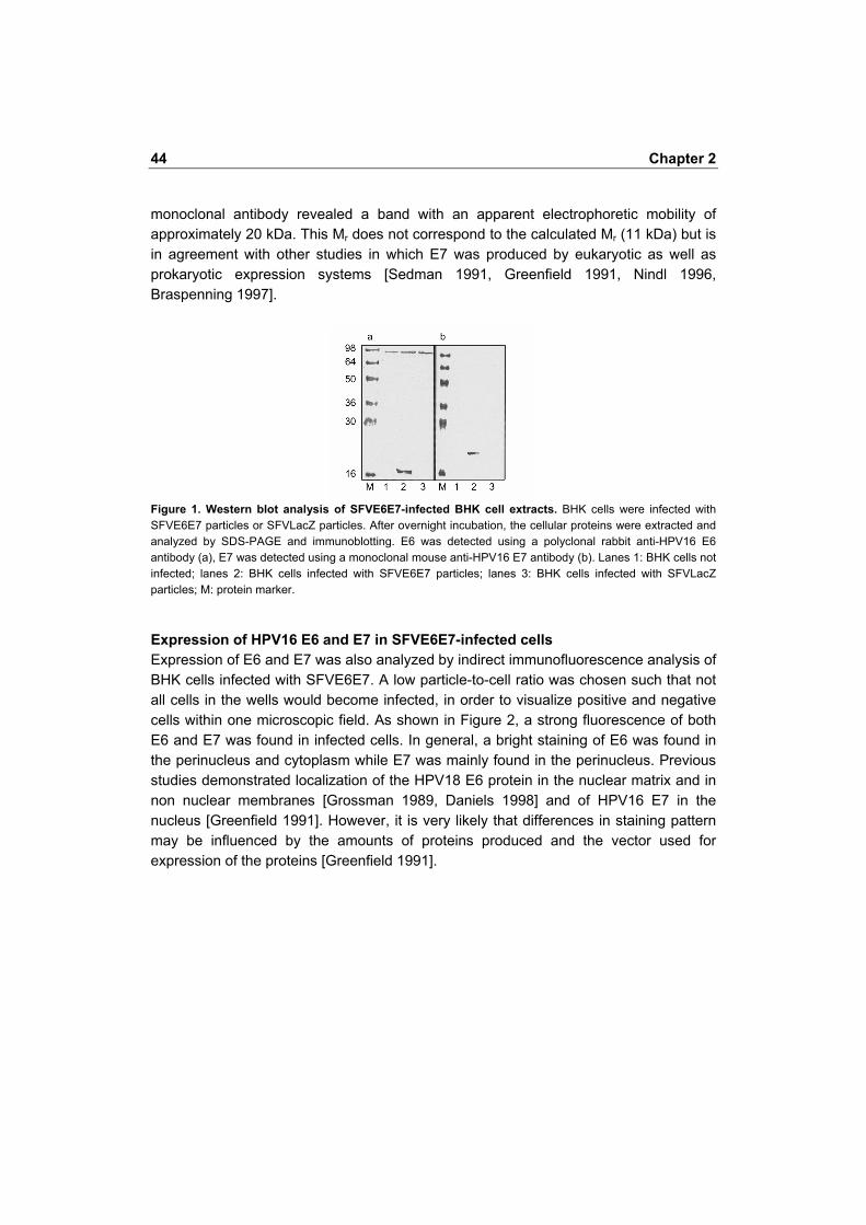
with irradiated naive spleen cells (30 Gy) and irradiated C3 cells in a ratio of 2:5:0.1 in 24-well plates in the presence of 4 IU of recombinant hIL2/ml (Strathmann Biotech, Hamburg, Germany). Five days after the first and/or second restimulation, cells were harvested and a CTL assay was performed by a standard 4 h 51Cr release assay in triplicate determinations. Target cells were labeled for 1 h with 3.7 MBq 51Cr/106 cells in 100 µl medium (51Cr was from Amersham, London, UK). EL-4 target cells were loaded with the HPV16 E7 49�57 (RAHYNIVTF) peptide by a 1 h incubation of the cells in the presence of 15 µg/ml of peptide in 100 µl of culture medium before labeling the cells with 51Cr. The mean percentage of specific 51Cr release of triplicate wells was calculated according to the formula: % specific release = [(experimental release - spontaneous release)/(maximal release - spontaneous release)] c.p.m. x 100. The spontaneous 51Cr release was always <15%. The standard errors of the means of the triplicate determinations were <10% of the value of the mean. Tumor challenge experiments Mice were immunized and boosted as described above with 106 to 5x10⁶ SFVE6E7 particles, SFVLacZ particles or PBS. One week after the last booster immunization the mice were challenged s.c. with 2x10⁴ TC-1 cells suspended in 0.2 ml Hank�s buffered salt solution (Life Technologies). Tumor measurements were always done by the same skilled technician. At a tumor volume of approximately 1000 mm3, the mice were killed. Results Production and titer determination of SFV particles Recombinant SFV particles were produced in BHK cells by electroporation of recombinant and Helper 2 RNA into these cells. After 24 h the medium containing the virus was removed from the cells and the virus particles were purified. Titers were determined by immunofluorescence using an antibody against SFV-nsP3 (replicase). This antibody was chosen because replicase is present in all cells infected with recombinant SFV. Thus, titers of different recombinant SFVs can be determined, independent of the inserted foreign gene(s). Typically, titers of unpurified virus were 109�1010 infectious units/ml. After purification titers were between 1010 and 1011 infectious units/ml. Western blot analysis of E6 and E7 expression In order to verify that SFVE6E7 induced expression of the recombinant E6 and E7 proteins, BHK cells were infected with SFVE6E7 or SFVLacZ serving as negative control. In Figure 1, Western blots of cell lysates probed with anti-HPV16 E6 (panel a) or anti-HPV16 E7 (panel b) are shown. Staining with the anti-E6 polyclonal antibody revealed a band with a Mr of approximately 17 kDa. Staining with the anti-E7
44 Chapter 2
monoclonal antibody revealed a band with an apparent electrophoretic mobility of approximately 20 kDa. This Mr does not correspond to the calculated Mr (11 kDa) but is in agreement with other studies in which E7 was produced by eukaryotic as well as prokaryotic expression systems [Sedman 1991, Greenfield 1991, Nindl 1996, Braspenning 1997].
Figure 1. Western blot analysis of SFVE6E7-infected BHK cell extracts. BHK cells were infected with SFVE6E7 particles or SFVLacZ particles. After overnight incubation, the cellular proteins were extracted and analyzed by SDS-PAGE and immunoblotting. E6 was detected using a polyclonal rabbit anti-HPV16 E6 antibody (a), E7 was detected using a monoclonal mouse anti-HPV16 E7 antibody (b). Lanes 1: BHK cells not infected; lanes 2: BHK cells infected with SFVE6E7 particles; lanes 3: BHK cells infected with SFVLacZ particles; M: protein marker. Expression of HPV16 E6 and E7 in SFVE6E7-infected cells Expression of E6 and E7 was also analyzed by indirect immunofluorescence analysis of BHK cells infected with SFVE6E7. A low particle-to-cell ratio was chosen such that not all cells in the wells would become infected, in order to visualize positive and negative cells within one microscopic field. As shown in Figure 2, a strong fluorescence of both E6 and E7 was found in infected cells. In general, a bright staining of E6 was found in the perinucleus and cytoplasm while E7 was mainly found in the perinucleus. Previous studies demonstrated localization of the HPV18 E6 protein in the nuclear matrix and in non nuclear membranes [Grossman 1989, Daniels 1998] and of HPV16 E7 in the nucleus [Greenfield 1991]. However, it is very likely that differences in staining pattern may be influenced by the amounts of proteins produced and the vector used for expression of the proteins [Greenfield 1991].
Induction of CTL and anti tumor responses by rSFVE6E7 45
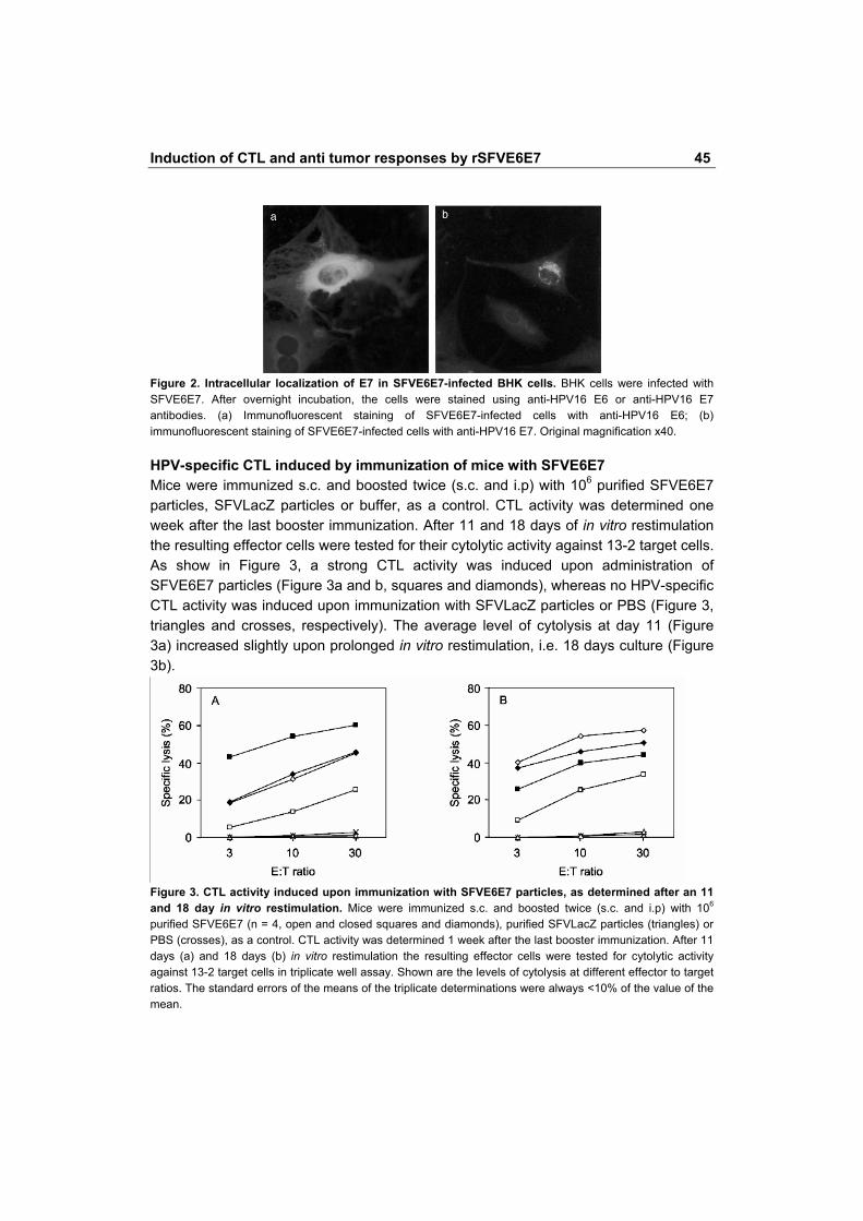
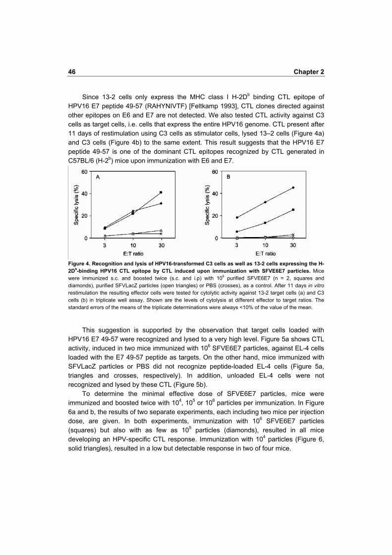
Figure 2. Intracellular localization of E7 in SFVE6E7-infected BHK cells. BHK cells were infected with SFVE6E7. After overnight incubation, the cells were stained using anti-HPV16 E6 or anti-HPV16 E7 antibodies. (a) Immunofluorescent staining of SFVE6E7-infected cells with anti-HPV16 E6; (b) immunofluorescent staining of SFVE6E7-infected cells with anti-HPV16 E7. Original magnification x40. HPV-specific CTL induced by immunization of mice with SFVE6E7 Mice were immunized s.c. and boosted twice (s.c. and i.p) with 106 purified SFVE6E7 particles, SFVLacZ particles or buffer, as a control. CTL activity was determined one week after the last booster immunization. After 11 and 18 days of in vitro restimulation the resulting effector cells were tested for their cytolytic activity against 13-2 target cells. As show in Figure 3, a strong CTL activity was induced upon administration of SFVE6E7 particles (Figure 3a and b, squares and diamonds), whereas no HPV-specific CTL activity was induced upon immunization with SFVLacZ particles or PBS (Figure 3, triangles and crosses, respectively). The average level of cytolysis at day 11 (Figure 3a) increased slightly upon prolonged in vitro restimulation, i.e. 18 days culture (Figure 3b).
Figure 3. CTL activity induced upon immunization with SFVE6E7 particles, as determined after an 11 and 18 day in vitro restimulation. Mice were immunized s.c. and boosted twice (s.c. and i.p) with 106 purified SFVE6E7 (n = 4, open and closed squares and diamonds), purified SFVLacZ particles (triangles) or PBS (crosses), as a control. CTL activity was determined 1 week after the last booster immunization. After 11 days (a) and 18 days (b) in vitro restimulation the resulting effector cells were tested for cytolytic activity against 13-2 target cells in triplicate well assay. Shown are the levels of cytolysis at different effector to target ratios. The standard errors of the means of the triplicate determinations were always <10% of the value of the mean.
46 Chapter 2
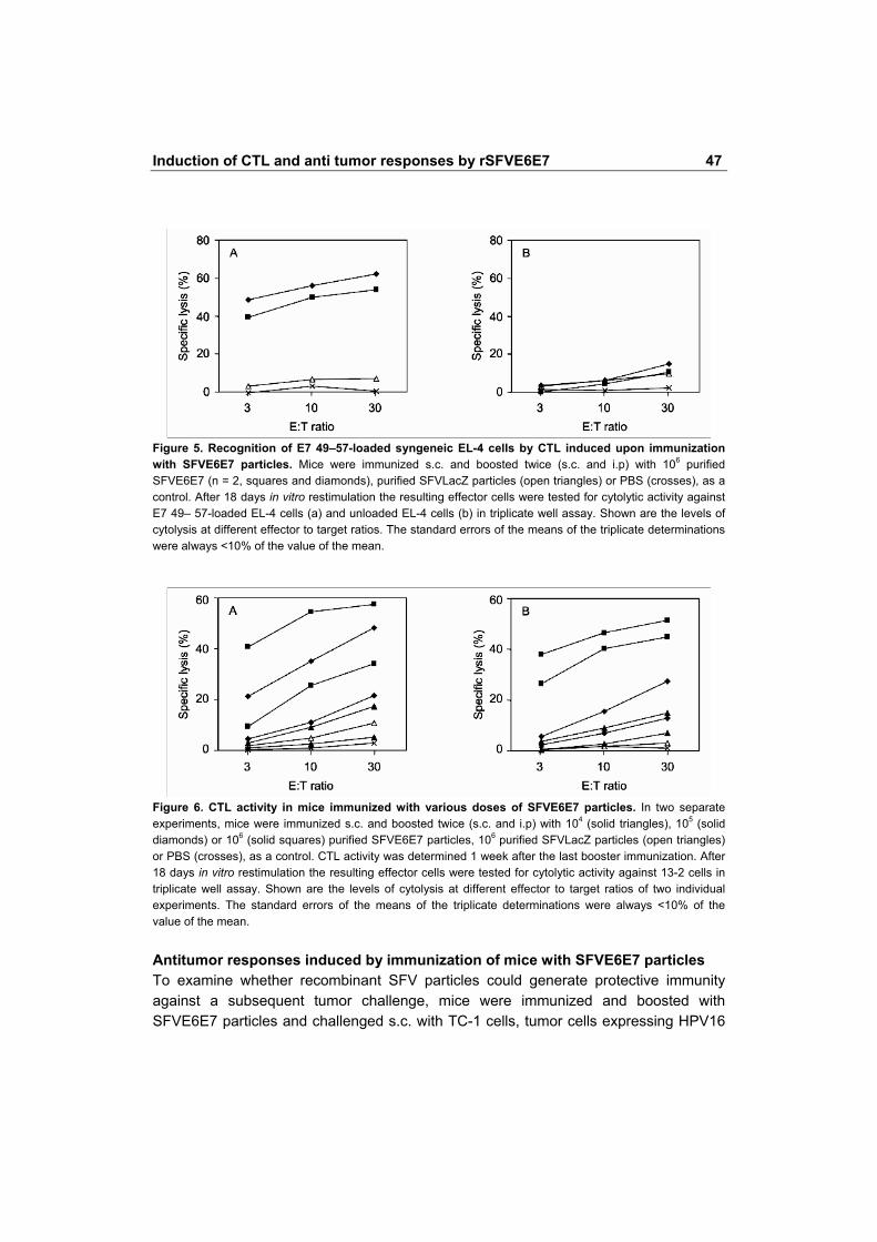
Since 13-2 cells only express the MHC class I H-2Db binding CTL epitope of HPV16 E7 peptide 49-57 (RAHYNIVTF) [Feltkamp 1993], CTL clones directed against other epitopes on E6 and E7 are not detected. We also tested CTL activity against C3 cells as target cells, i.e. cells that express the entire HPV16 genome. CTL present after 11 days of restimulation using C3 cells as stimulator cells, lysed 13�2 cells (Figure 4a) and C3 cells (Figure 4b) to the same extent. This result suggests that the HPV16 E7 peptide 49-57 is one of the dominant CTL epitopes recognized by CTL generated in C57BL/6 (H-2b) mice upon immunization with E6 and E7.
Figure 4. Recognition and lysis of HPV16-transformed C3 cells as well as 13-2 cells expressing the H-2Db-binding HPV16 CTL epitope by CTL induced upon immunization with SFVE6E7 particles. Mice were immunized s.c. and boosted twice (s.c. and i.p) with 106 purified SFVE6E7 (n = 2, squares and diamonds), purified SFVLacZ particles (open triangles) or PBS (crosses), as a control. After 11 days in vitro restimulation the resulting effector cells were tested for cytolytic activity against 13-2 target cells (a) and C3 cells (b) in triplicate well assay. Shown are the levels of cytolysis at different effector to target ratios. The standard errors of the means of the triplicate determinations were always <10% of the value of the mean.
This suggestion is supported by the observation that target cells loaded with
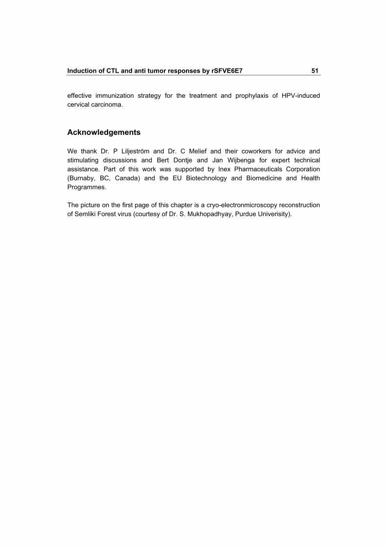
HPV16 E7 49-57 were recognized and lysed to a very high level. Figure 5a shows CTL activity, induced in two mice immunized with 106 SFVE6E7 particles, against EL-4 cells loaded with the E7 49-57 peptide as targets. On the other hand, mice immunized with SFVLacZ particles or PBS did not recognize peptide-loaded EL-4 cells (Figure 5a, triangles and crosses, respectively). In addition, unloaded EL-4 cells were not recognized and lysed by these CTL (Figure 5b).
To determine the minimal effective dose of SFVE6E7 particles, mice were immunized and boosted twice with 104, 105 or 106 particles per immunization. In Figure 6a and b, the results of two separate experiments, each including two mice per injection dose, are given. In both experiments, immunization with 106 SFVE6E7 particles (squares) but also with as few as 105 particles (diamonds), resulted in all mice developing an HPV-specific CTL response. Immunization with 104 particles (Figure 6, solid triangles), resulted in a low but detectable response in two of four mice.
Induction of CTL and anti tumor responses by rSFVE6E7 47
Figure 5. Recognition of E7 49�57-loaded syngeneic EL-4 cells by CTL induced upon immunization with SFVE6E7 particles. Mice were immunized s.c. and boosted twice (s.c. and i.p) with 106 purified SFVE6E7 (n = 2, squares and diamonds), purified SFVLacZ particles (open triangles) or PBS (crosses), as a control. After 18 days in vitro restimulation the resulting effector cells were tested for cytolytic activity against E7 49� 57-loaded EL-4 cells (a) and unloaded EL-4 cells (b) in triplicate well assay. Shown are the levels of cytolysis at different effector to target ratios. The standard errors of the means of the triplicate determinations were always <10% of the value of the mean.
Figure 6. CTL activity in mice immunized with various doses of SFVE6E7 particles. In two separate experiments, mice were immunized s.c. and boosted twice (s.c. and i.p) with 104 (solid triangles), 105 (solid diamonds) or 106 (solid squares) purified SFVE6E7 particles, 106 purified SFVLacZ particles (open triangles) or PBS (crosses), as a control. CTL activity was determined 1 week after the last booster immunization. After 18 days in vitro restimulation the resulting effector cells were tested for cytolytic activity against 13-2 cells in triplicate well assay. Shown are the levels of cytolysis at different effector to target ratios of two individual experiments. The standard errors of the means of the triplicate determinations were always <10% of the value of the mean. Antitumor responses induced by immunization of mice with SFVE6E7 particles To examine whether recombinant SFV particles could generate protective immunity against a subsequent tumor challenge, mice were immunized and boosted with SFVE6E7 particles and challenged s.c. with TC-1 cells, tumor cells expressing HPV16

48 Chapter 2
E6E7. Tumor inoculation studies performed before initiating these immunization studies revealed that s.c. inoculation of 2x10⁴ TC-1 cells reproducibly induced tumors within 2 to 4 weeks after inoculation in all mice tested (n = 15). Figures 7 and 8 show the combined results of two separate immunization studies. Control mice, injected with PBS (n = 10) or SFVLacZ particles (n = 10) developed palpable tumors within 2 to 4 weeks after tumor cell inoculation (Figure 7a and b, respectively; Figure 8, open circles and open squares, respectively). Immunization with 106 SFVE6E7 particles (n = 10) resulted in a delay in tumor onset in 50% of the mice as compared with control mice, with one of 10 mice not developing a tumor (Figure 7c; Figure 8, diamonds). Upon immunization with a five-fold higher dose of SFVE6E7 particles two of five mice did not develop a tumor (Figure 7d; Figure 8, closed squares).
Figure 7. Growth of TC-1 tumor cells in SFVE6E7-immunized mice. Mice were immunized s.c. and boosted twice (s.c. and i.p.) with PBS (a; n = 10), 5x10⁶ SFVLacZ particles (b; n = 10), 106 SFVE6E7 particles (c; n = 10) or 5x10⁶ SFVE6E7 particles (d; n = 5). Tumor growth was monitored twice weekly. Each line represents the tumor volume of a separate mouse.
Figure 8. Growth of TC-1 tumor cells in SFVE6E7 immunized mice. Mice were immunized s.c. and boosted twice (s.c. and i.p.) with PBS (n = 10; open circles), 5x10⁶ SFVLacZ particles (n = 10; open squares), 106 SFVE6E7 particles (n = 10; solid diamonds) or 5x10⁶ SFVE6E7 particles (n = 5; solid squares). Tumor growth was monitored twice weekly. Shown are the percentages of mice with non-palpable tumors.

Induction of CTL and anti tumor responses by rSFVE6E7 49
Discussion This report describes the construction, characterization and cellular immunotherapeutic potential of recombinant SFV particles encoding the early proteins E6 and E7 of HPV16. The ultimate aim of our studies is to develop an effective immunization strategy for the treatment and/or prevention of HPV-induced cervical carcinoma.
Immunization of mice with SFV particles encoding HPV16 E6 and E7 resulted in a HPV-specific CTL response. Three injections of as few as 104 SFV particles sufficed for the induction of a CTL response in 50% of the mice, while three immunizations with 105 SFV particles induced a HPV-specific CTL response in all mice tested. Increasing the dose to 106 SFV particles per injection resulted in a reproducible CTL response with a high level of specific tumor cell lysis. In vitro blocking experiments with antibodies against CD4 and CD8 revealed that the lytic activity was due to CD8+ T cells; no inhibition was found with anti-CD4 antibodies (not shown). Tumor challenge experiments demonstrated that immunization with 106 SFVE6E7 particles resulted in a delay in tumor onset while one of 10 mice did not develop a tumor. Upon immunization with a five-fold higher dose of SFVE6E7 particles 40% of the mice did not develop a tumor.
In the last few years a number of peptide/protein-based or genetic immunization strategies have been described for the induction of HPV-specific CTL activity [Feltkamp 1995, Jochmus 1997, de Bruijn 1998]. Major drawbacks associated with a peptide-based approach include the problem of MHC polymorphism and the risk of inducing T cell tolerance rather than T cell activation. Due to the induction of specific T cell tolerance, vaccination with a tumor-specific peptide has been shown to result in an enhanced outgrowth of the tumor [Toes 1996]. Immunization with larger proteins would overcome these problems, but requires efficient antigen delivery systems and/or safe adjuvants for efficient immune priming. Several groups have described the induction of HPV-specific CTL responses in mice upon immunization with recombinant vaccinia virus expressing HPV E6 or E7 [Boursnell 1996, Lin 1996] or with syngeneic cells retrovirally transfected with the HPV E6 gene [Chen 1992] In a phase I/II trial involving eight patients with late stage cervical cancer, vaccination with recombinant vaccinia virus expressing HPV18 E6 and E7 induced HPV-specific CTL in one of three evaluable patients [Borysiewicz 1996]. Potential drawbacks associated with the use of viral vector systems are immune responses against viral proteins in pre-immune patients (vaccinia virus) or integration of recombinant genes into the host cell genome (retrovirus). Especially, when the recombinant virus encodes oncoproteins such as HPV E6 or E7, the risk of integration into the host cell genome is a point of major concern.
We have chosen the SFV expression system, which apart from its infection efficiency and high biosafety, would appear to be especially suited to safely induce cellular immune responses against oncoproteins such as HPV16 E6 and E7. First, SFV

50 Chapter 2
is an RNA virus replicating in the cell cytosol; therefore, there is no risk of integration of the E6 and E7 genes in the cellular genome. Moreover, SFV infection is cytolytic by apoptosis [Strausss 1994, Glasgow 1997]. Therefore, no genetic information of E6 and E7 will persist for more than 1 week after injection. In addition, no other vector proteins are produced, other than small amounts of viral replicase. Berglund et al. [Berglund 1999] demonstrated that immune responses against the vector itself did not inhibit boost responses by subsequent immunizations with the same vector.
Recognition by the immune system of virally infected cells or tumor cells occurs via virus- or tumor-specific antigenic peptides presented in the context of MHC class I molecules. Infection of cells with recombinant SFV particles results in the production of the recombinant protein within the cytoplasm permitting presentation of the recombinant protein via the conventional MHC class I presentation route. However, for the induction of tumor or virus-specific CTL, antigen presentation has to be accompanied by costimulatory signaling. Costimulatory molecules are confined to professional antigen-presenting cells (APC). Therefore, the CTL response induced upon immunization with SFVE6E7 particles may occur through infection of APC in vivo. Alternatively, APC may take up residues of other cells that have been infected in a process of cross-priming. The uptake of debris from infected cells by APC is expected to be very efficient since an SFV infection induces apoptotic cell death [Strausss 1994, Glasgow 1997]. Upon uptake of infected-cell material (exogenous antigen) the recombinant protein will be processed and presented by MHC class II molecules thereby activating CD4+ T helper cells. In addition, dendritic cells and macrophages are able to present exogenous antigen in the context of MHC class I molecules for presentation to and activation of CD8+ T cells [Rock 1996]. Thus, both arms of the cellular immune system, essential for the induction of an optimal immune response will be activated upon administration of recombinant SFV particles, thereby eliciting a potent CTL response. Moreover, SFV immunization will introduce both class I and class II antigenic epitopes into one and the same the APC which has recently been demonstrated to be required for a full activation of APC [Bennet 1998, Lanzavecchia 1998, Ridge 1998, Schoenberger 1998]. As demonstrated by Zhou et al. [Zhou 1995] immunization of mice with SFV particles encoding for the nucleoprotein of influenza virus not only induces influenza-specific CTL activity, but also a nucleoprotein-specific antibody response. This observation supports the hypothesis of cross-priming and indirect presentation of antigenic peptides.
In conclusion, we have demonstrated that immunization of mice with recombinant SFVE6E7 particles induces a potent CTL response against HPV-transformed tumor cells. This promising result, combined with studies showing the high efficacy of the SFV system for priming the immune system of mice as well as primates, [Berglund 1999, Zhou 1995, Berglund 1997] and the recent development of the extremely safe two-helper system [Smerdou 1999] provide the essential steps towards the design of an
Induction of CTL and anti tumor responses by rSFVE6E7 51
effective immunization strategy for the treatment and prophylaxis of HPV-induced cervical carcinoma. Acknowledgements We thank Dr. P Liljeström and Dr. C Melief and their coworkers for advice and stimulating discussions and Bert Dontje and Jan Wijbenga for expert technical assistance. Part of this work was supported by Inex Pharmaceuticals Corporation (Burnaby, BC, Canada) and the EU Biotechnology and Biomedicine and Health Programmes. The picture on the first page of this chapter is a cryo-electronmicroscopy reconstruction of Semliki Forest virus (courtesy of Dr. S. Mukhopadhyay, Purdue Univerisity).
52 Chapter 2
References 1. Bennett SR, Carbone FR, Karamalis F, Flavell RA, Miller JF, Heath WR. Help for cytotoxic-
T-cell responses is mediated by CD40 signalling. Nature. 1998;393(6684):478-80. 2. Berglund P, Sjoberg M, Garoff H, Atkins GJ, Sheahan BJ, Liljeström P. Semliki Forest virus
expression system: production of conditionally infectious recombinant particles. Biotechnology (N Y). 1993;11(8):916-20.
3. Berglund P, Quesada-Rolander M, Putkonen P, Biberfeld G, Thorstensson R, Liljeström P. Outcome of immunization of cynomolgus monkeys with recombinant Semliki Forest virus encoding human immunodeficiency virus type 1 envelope protein and challenge with a high dose of SHIV-4 virus. AIDS Res Hum Retroviruses. 1997;13(17):1487-95.
4. Berglund P, Fleeton MN, Smerdou C, Liljestrom P. Immunization with recombinant Semliki Forest virus induces protection against influenza challenge in mice. Vaccine. 1999;17(5):497-507.
5. Borysiewicz LK, Fiander A, Nimako M, Man S, Wilkinson GW, Westmoreland D, Evans AS, Adams M, Stacey SN, Boursnell ME, Rutherford E, Hickling JK, Inglis SC. A recombinant vaccinia virus encoding human papillomavirus types 16 and 18, E6 and E7 proteins as immunotherapy for cervical cancer. Lancet. 1996;347(9014):1523-7.
6. Boursnell ME, Rutherford E, Hickling JK, Rollinson EA, Munro AJ, Rolley N, McLean CS, Borysiewicz LK, Vousden K, Inglis SC. Construction and characterisation of a recombinant vaccinia virus expressing human papillomavirus proteins for immunotherapy of cervical cancer. Vaccine. 1996;14(16):1485-94.
7. Braspenning J, Manetti R, Zumbach K, Meschede W, Gissmann L, Tommasino M. A general purification protocol for E7 proteins from "high- and low-risk" human papillomavirus types expressed in the yeast Schizosaccharomyces pombe. Protein Expr Purif. 1997;10(2):192-201.
8. Chen L, Mizuno MT, Singhal MC, Hu SL, Galloway DA, Hellstrom I, Hellstrom KE. Induction of cytotoxic T lymphocytes specific for a syngeneic tumor expressing the E6 oncoprotein of human papillomavirus type 16. J Immunol. 1992;148(8):2617-21.
9. Daniels PR, Sanders CM, Maitland NJ. Characterization of the interactions of human papillomavirus type 16 E6 with p53 and E6-associated protein in insect and human cells. J Gen Virol. 1998;79 ( Pt 3):489-99.
10. De Bruijn ML, Schuurhuis DH, Vierboom MP, Vermeulen H, de Cock KA, Ooms ME, Ressing ME, Toebes M, Franken KL, Drijfhout JW, Ottenhoff TH, Offringa R, Melief CJ. Immunization with human papillomavirus type 16 (HPV16) oncoprotein-loaded dendritic cells as well as protein in adjuvant induces MHC class I-restricted protection to HPV16-induced tumor cells. Cancer Res. 1998;58(4):724-31.
11. Feltkamp MC, Smits HL, Vierboom MP, Minnaar RP, de Jongh BM, Drijfhout JW, ter Schegget J, Melief CJ, Kast WM. Vaccination with cytotoxic T lymphocyte epitope-containing peptide protects against a tumor induced by human papillomavirus type 16-transformed cells. Eur J Immunol. 1993;23(9):2242-9.
12. Feltkamp MC, Vreugdenhil GR, Vierboom MP, Ras E, van der Burg SH, ter Schegget J, Melief CJ, Kast WM. Cytotoxic T lymphocytes raised against a subdominant epitope offered as a synthetic peptide eradicate human papillomavirus type 16-induced tumors. Eur J Immunol. 1995;25(9):2638-42.
13. Glasgow GM, McGee MM, Sheahan BJ, Atkins GJ. Death mechanisms in cultured cells infected by Semliki Forest virus. J Gen Virol. 1997;78 ( Pt 7):1559-63.
14. Greenfield I, Nickerson J, Penman S, Stanley M. Human papillomavirus 16 E7 protein is associated with the nuclear matrix. Proc Natl Acad Sci. 1991;88(24):11217-21.
15. Grossman SR, Mora R, Laimins LA. Intracellular localization and DNA-binding properties of human papillomavirus type 18 E6 protein expressed with a baculovirus vector. J Virol. 1989;63(1):366-74.
Induction of CTL and anti tumor responses by rSFVE6E7 53
16. Gulliver GA, Herber RL, Liem A, Lambert PF. Both conserved region 1 (CR1) and CR2 of the human papillomavirus type 16 E7 oncogene are required for induction of epidermal hyperplasia and tumor formation in transgenic mice. J Virol. 1997;71(8):5905-14.
17. Jochmus I, Osen W, Altmann A, Buck G, Hofmann B, Schneider A, Gissmann L, Rammensee HG. Specificity of human cytotoxic T lymphocytes induced by a human papillomavirus type 16 E7-derived peptide. J Gen Virol. 1997;78 ( Pt 7):1689-95.
18. Johnson JC, Burnett AF, Willet GD, Young MA, Doniger J. High frequency of latent and clinical human papillomavirus cervical infections in immunocompromised human immunodeficiency virus-infected women. Obstet Gynecol. 1992;79(3):321-7.
19. Jones DL, Münger K. Interactions of human papillomavirus E7 protein with cell cycle regulators. Semin Cancer Biol 1996;7:327�37.
20. Lanzavecchia A. Immunology. Licence to kill. Nature. 1998;393(6684):413-4. 21. Liljeström P, Lusa S, Huylebroeck D, Garoff H. In vitro mutagenesis of a full-length cDNA
clone of Semliki Forest virus: the small 6,000-molecular-weight membrane protein modulates virus release. J Virol. 1991a;65(8):4107-13.
22. Liljeström P, Garoff H. A new generation of animal cell expression vectors based on the Semliki Forest virus replicon. Biotechnology (N Y). 1991b;9(12):1356-61.
23. Lin KY, Guarnieri FG, Staveley-O'Carroll KF, Levitsky HI, August JT, Pardoll DM, Wu TC. Treatment of established tumors with a novel vaccine that enhances major histocompatibility class II presentation of tumor antigen. Cancer Res. 1996;56(1):21-6.
24. Munger K, Scheffner M, Huibregtse JM, Howley PM. Interactions of HPV E6 and E7 oncoproteins with tumour suppressor gene products. Cancer Surv. 1992;12:197-217.
25. Nindl I, Gissmann L, Fisher SG, Bribiesca LB, Berumen J, Muller M. The E7 protein of human papillomavirus (HPV) type 16 expressed by recombinant vaccinia virus can be used for detection of antibodies in sera from cervical cancer patients. J Virol Methods. 1996;62(1):81-5.
26. Pei XF. The human papillomavirus E6/E7 genes induce discordant changes in the expression of cell growth regulatory proteins. Carcinogenesis. 1996;17(7):1395-401.
27. Porreco R, Penn I, Droegemueller W, Greer B, Makowski E. Gynecologic malignancies in immunosuppressed organ homograft recipients. Obstet Gynecol. 1975;45(4):359-64.
28. Ridge JP, Di Rosa F, Matzinger P. A conditioned dendritic cell can be a temporal bridge between a CD4+ T-helper and a T-killer cell. Nature. 1998;393(6684):474-8.
29. Rock KL. A new foreign policy: MHC class I molecules monitor the outside world. Immunol Today. 1996;17(3):131-7.
30. Schoenberger SP, Toes RE, van der Voort EI, Offringa R, Melief CJ. T-cell help for cytotoxic T lymphocytes is mediated by CD40-CD40L interactions. Nature. 1998;393(6684):480-3.
31. Sedman SA, Barbosa MS, Vass WC, Hubbert NL, Haas JA, Lowy DR, Schiller JT. The full-length E6 protein of human papillomavirus type 16 has transforming and trans-activating activities and cooperates with E7 to immortalize keratinocytes in culture. J Virol. 1991;65(9):4860-6.
32. Smerdou C, Liljeström P, Two-helper RNA system for production of recombinant Semliki Forest virus particles. J Virol. 1999;73(2):1092-8.
33. Smits PH, Smits HL, Jebbink MF, ter Schegget J. The short arm of chromosome 11 likely is involved in the regulation of the human papillomavirus type 16 early enhancer-promoter and in the suppression of the transforming activity of the viral DNA. Virology. 1990;176(1):158-65.
34. Strauss JH, Strauss EG. The alphaviruses: gene expression, replication, and evolution. Microbiol Rev. 1994;58(3):491-562.
35. Toes RE, van der Voort EI, Schoenberger SP, Drijfhout JW, van Bloois L, Storm G, Kast WM, Offringa R, Melief CJ. Enhancement of tumor outgrowth through CTL tolerization after peptide vaccination is avoided by peptide presentation on dendritic cells. J Immunol. 1998;160(9):4449-56.

54 Chapter 2
36. Werness BA, Levine AJ, Howley PM. Association of human papillomavirus types 16 and 18 E6 proteins with p53. Science. 1990;248(4951):76-9.
37. Zhou X, Berglund P, Zhao H, Liljeström P, Jondal M. Generation of cytotoxic and humoral immune responses by nonreplicative recombinant Semliki Forest virus. Proc Natl Acad Sci. 1995;92(7):3009-13.
38. zur Hausen H. Viruses in human cancers. Science. 1991;254(5035):1167-73.