Embed Size (px)
Citation preview
Lea AlhilaliGillian Lieberman, MD
Unraveling Testicular Torsion
Lea Alhilali, Harvard Medical School Year IIIGillian Lieberman, MD
October 2002
Hosp Med 2002 Aug; 63 (8): 459-9
2
Lea AlhilaliGillian Lieberman, MD
Normal Testicular Anatomy
http://www.aafp.org/afp/990215ap/817.html
3
Lea AlhilaliGillian Lieberman, MD
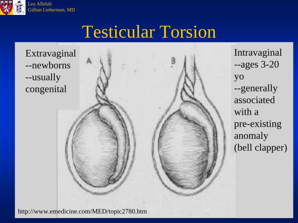
Testicular TorsionExtravaginal--newborns--usually congenital
Intravaginal--ages 3-20 yo--generally associatedwith a pre-existing anomaly (bell clapper)
http://www.emedicine.com/MED/topic2780.htm
4
Lea AlhilaliGillian Lieberman, MD
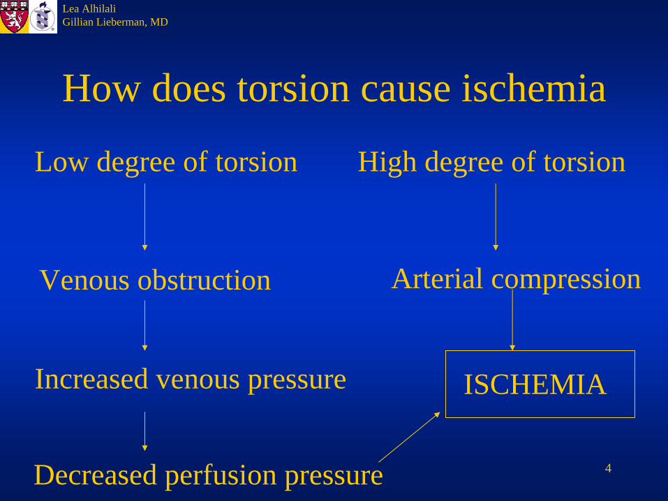
How does torsion cause ischemia
Low degree of torsion High degree of torsion
Venous obstruction
Increased venous pressure
Decreased perfusion pressure
ISCHEMIA
Arterial compression
5
Lea AlhilaliGillian Lieberman, MD
The Acute Scrotum
• Scrotal Trauma• Scrotal Inflammation:
Epidydimitis/orchitis• Ischemia: torsion of the
testicular/epididymal appendages, testicular torsion, traumatic infarction, postherniorrhaphy/strangulated hernia
• Rare: Schonlein-Henoch purpura, neoplasm, varicocele, idiopathic
YOU MUST R/O TORSION!www.ew.com
6
Lea AlhilaliGillian Lieberman, MD
Torsion on my mind…• Short duration of sx• Negative urinalysis• PE: diffuse tenderness, negative Cremaster• Age: usually between 3-20 yrs, with 65%
between 12 and 18 yrsArce et al, Ped Rad 2002 Jul; 32 (7): 485-91
However—don’t forget it in adults, it has been reported in ages up to 62 yrs, with lower salvage ratesCummings et al, J Urol 2002 May; 167 (5): 2109-10
• Common! (1 in 4000 <25yrs)Wu et al, Clin Nuc Med 2002 Jul; 27(7): 490-3
7
Lea AlhilaliGillian Lieberman, MD
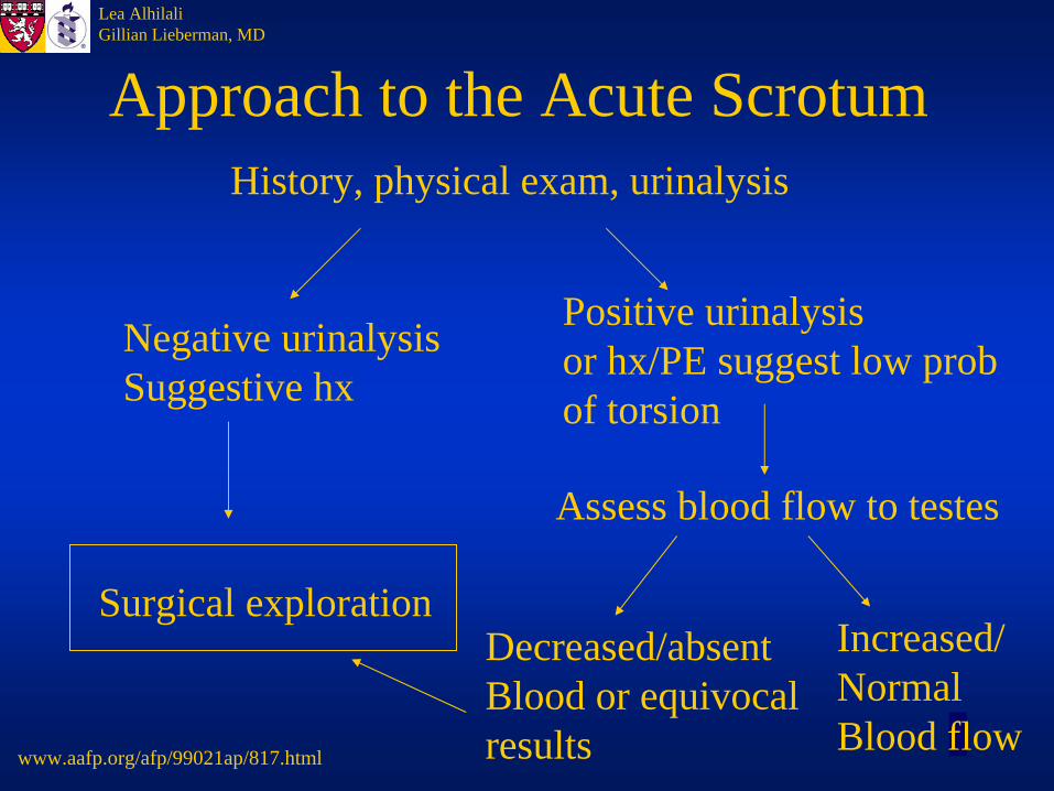
Approach to the Acute Scrotum History, physical exam, urinalysis
Negative urinalysisSuggestive hx
Surgical exploration
Positive urinalysisor hx/PE suggest low probof torsion
Assess blood flow to testes
Decreased/absentBlood or equivocalresults
Increased/Normal Blood flow
www.aafp.org/afp/99021ap/817.html
8
Lea AlhilaliGillian Lieberman, MD
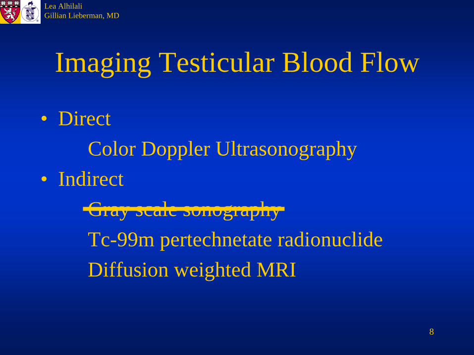
Imaging Testicular Blood Flow
• DirectColor Doppler Ultrasonography
• IndirectGray scale sonographyTc-99m pertechnetate radionuclideDiffusion weighted MRI
9
Lea AlhilaliGillian Lieberman, MD
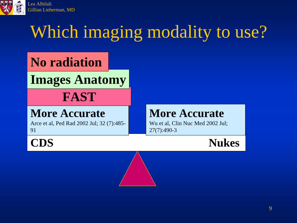
Which imaging modality to use?
CDS Nukes
More AccurateWu et al, Clin Nuc Med 2002 Jul; 27(7):490-3
More Accurate Arce et al, Ped Rad 2002 Jul; 32 (7):485- 91
FASTImages AnatomyNo radiation
10
Lea AlhilaliGillian Lieberman, MD
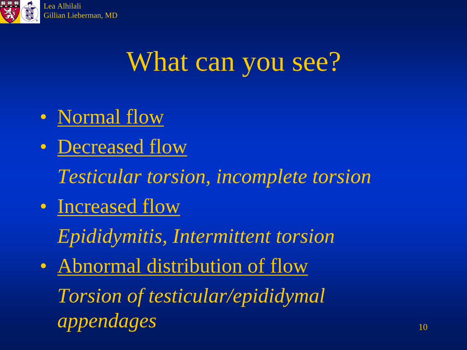
What can you see?
• Normal flow• Decreased flow
Testicular torsion, incomplete torsion• Increased flow
Epididymitis, Intermittent torsion• Abnormal distribution of flow
Torsion of testicular/epididymal appendages
11
Lea AlhilaliGillian Lieberman, MD
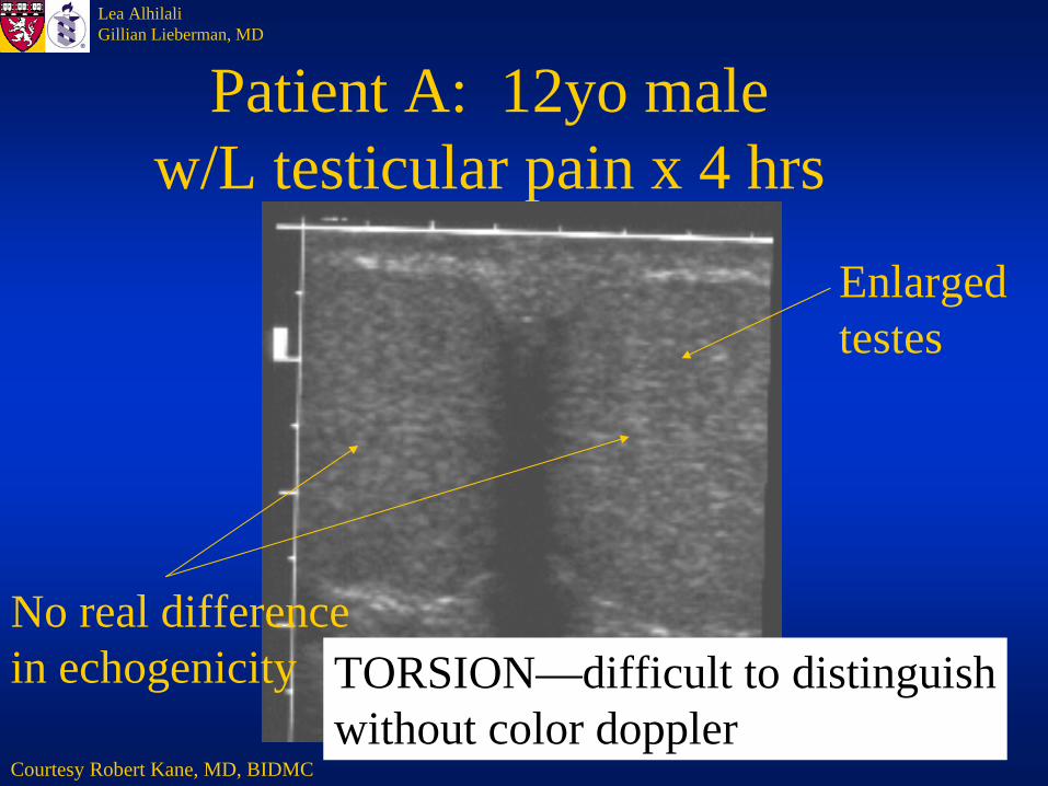
Patient A: 12yo male w/L testicular pain x 4 hrs
Courtesy Robert Kane, MD, BIDMC
TORSION—difficult to distinguishwithout color doppler
No real difference in echogenicity
Enlargedtestes
12
Lea AlhilaliGillian Lieberman, MD
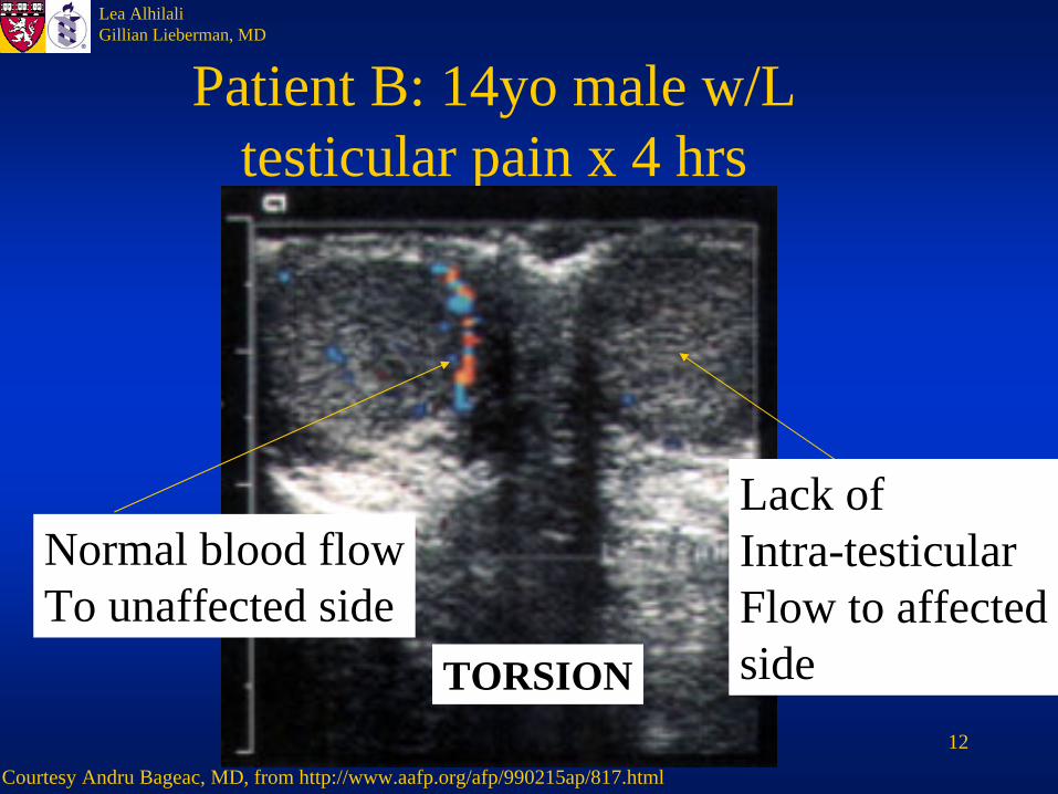
Patient B: 14yo male w/L testicular pain x 4 hrs
TORSION
Courtesy Andru Bageac, MD, from http://www.aafp.org/afp/990215ap/817.html
Normal blood flowTo unaffected side
Lack of Intra-testicularFlow to affectedside
13
Lea AlhilaliGillian Lieberman, MD
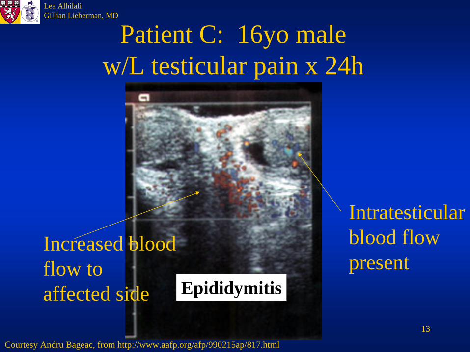
Patient C: 16yo male w/L testicular pain x 24h
Epididymitis
Courtesy Andru Bageac, from http://www.aafp.org/afp/990215ap/817.html
Increased bloodflow to affected side
Intratesticularblood flowpresent
14
Lea AlhilaliGillian Lieberman, MD
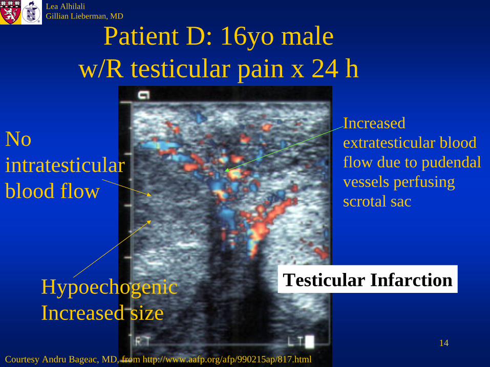
Patient D: 16yo male w/R testicular pain x 24 h
Testicular Infarction
Courtesy Andru Bageac, MD, from http://www.aafp.org/afp/990215ap/817.html
HypoechogenicIncreased size
No intratesticularblood flow
Increased extratesticular blood flow due to pudendalvessels perfusingscrotal sac
15
Lea AlhilaliGillian Lieberman, MD
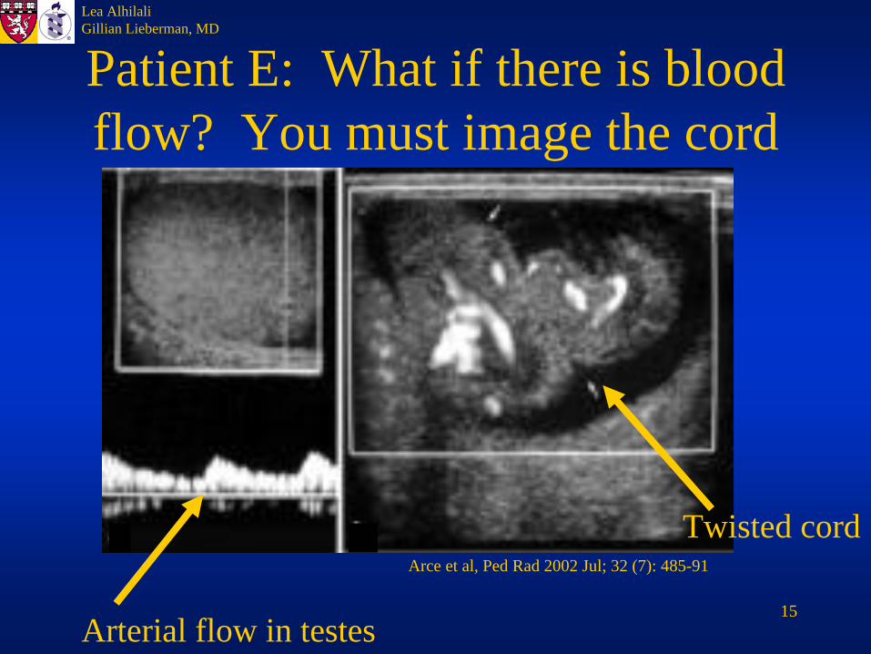
Patient E: What if there is blood flow? You must image the cord
Arterial flow in testes
Twisted cordArce et al, Ped Rad 2002 Jul; 32 (7): 485-91
16
Lea AlhilaliGillian Lieberman, MD
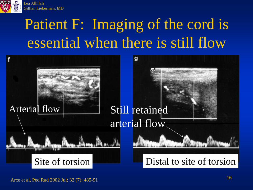
Patient F: Imaging of the cord is essential when there is still flow
Arce et al, Ped Rad 2002 Jul; 32 (7): 485-91
Site of torsion Distal to site of torsion
Arterial flow Still retainedarterial flow
17
Lea AlhilaliGillian Lieberman, MD
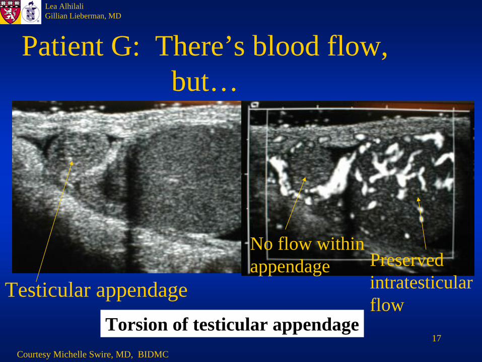
Patient G: There’s blood flow, but…
Torsion of testicular appendageCourtesy Michelle Swire, MD, BIDMC
Testicular appendagePreserved intratesticularflow
No flow withinappendage
18
Lea AlhilaliGillian Lieberman, MD
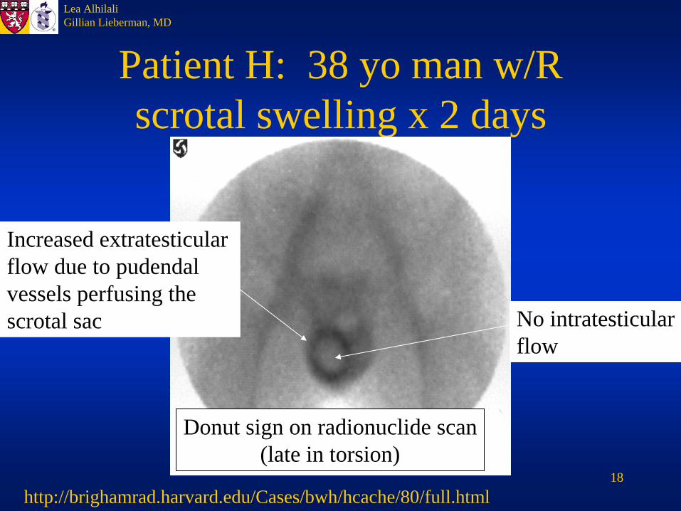
Patient H: 38 yo man w/R scrotal swelling x 2 days
http://brighamrad.harvard.edu/Cases/bwh/hcache/80/full.html
Donut sign on radionuclide scan(late in torsion)
No intratesticularflow
Increased extratesticularflow due to pudendalvessels perfusing the scrotal sac
19
Lea AlhilaliGillian Lieberman, MD
The role of MRI in torsion
• DWI imaging detects tissue ISCHEMIA by measuring changes in cellular water content and water diffusion
• Using ischemia instead of perfusion as criteria means detection at early phases (where arterial perfusion still present) and intermittent torsion
20
Lea AlhilaliGillian Lieberman, MD
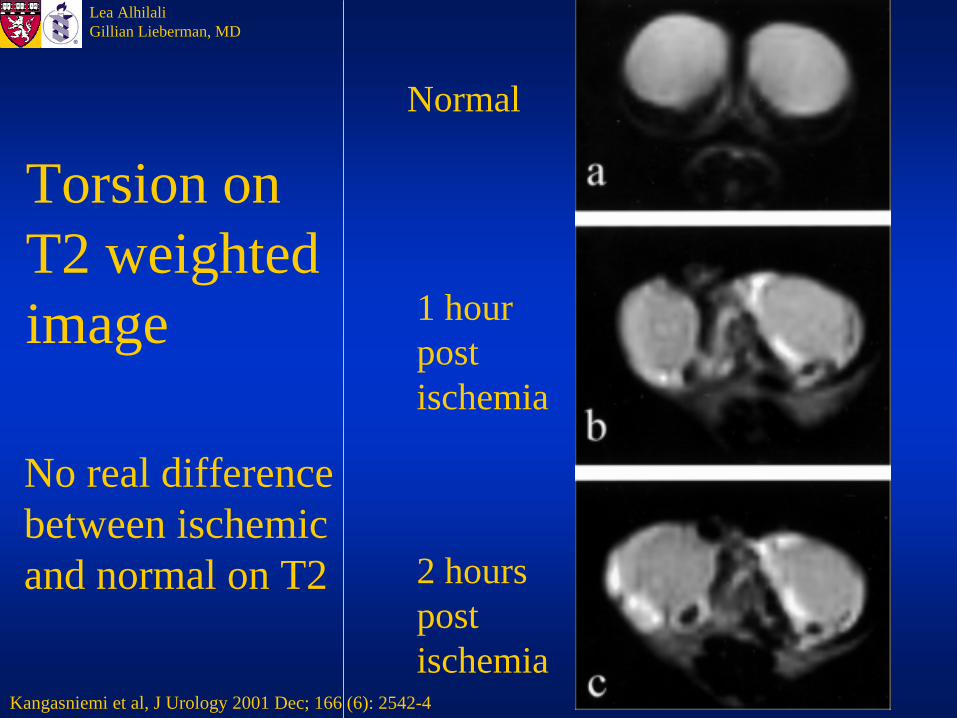
Torsion on T2 weighted image
Normal
1 hourpost ischemia
2 hourspostischemia
Kangasniemi et al, J Urology 2001 Dec; 166 (6): 2542-4
No real differencebetween ischemicand normal on T2
21
Lea AlhilaliGillian Lieberman, MD
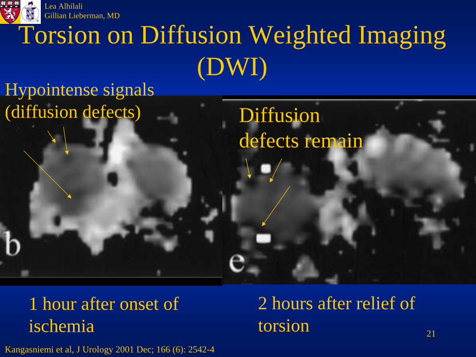
Torsion on Diffusion Weighted Imaging (DWI)
1 hour after onset of ischemia
2 hours after relief of torsion
Kangasniemi et al, J Urology 2001 Dec; 166 (6): 2542-4
Hypointense signals (diffusion defects) Diffusion
defects remain
22
Lea AlhilaliGillian Lieberman, MD
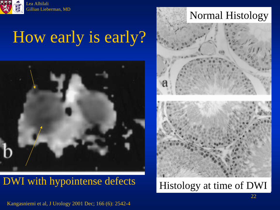
How early is early?Normal Histology
Histology at time of DWIDWI with hypointense defects
Kangasniemi et al, J Urology 2001 Dec; 166 (6): 2542-4
23
Lea AlhilaliGillian Lieberman, MD
Conclusions
• CDS is the modality of choice for imaging the acute scrotum to differentiate torsion from other etiologies
• CDS examination is not complete without imaging of the spermatic cord or considering intermittent torsion
• DWI imaging offers a chance to detect torsion at its earliest stages as well as intermittent torsion
24
Lea AlhilaliGillian Lieberman, MD
References• Arce JD, Cortes M, Vargas JC. Sonographic diagnosis of acute spermatic cord torsion. Ped Rad
2002; 32 (7): 485-91./SLIDE #• Bree RL, Hoang DT. Scrotal Ultrasound in The Radiologic Clinics of North America: Advances
in Uroradiology II. Dunnick, NR ed., WB Saunders, 1996.• Cummings JM, Boullier JA, Sekhon D, Bose K. Adult Testicular Torsion. J Urol 2002; 167 (5):
2109-10.• Kangasniemi M, Kaipia A, Joensuu, R. Diffusion Weighted Mangetic Resonance Imaging of
Rat Testes: A Method for Early Detection of Ischemia. J Urol 2001; 11 (12): 2589-92.• Pavlica P, Barozzi L. Imaging of the acute scrotum. Eur Radiol. 2001;11(2):220-8
• Shergill IS, Foley CL, Arya M, Bott SR, Mundy AR. Testicular torsion unravelled. Hosp Med. 2002 Aug;63(8):456-9
• The Swollen or Painful Scrotum. Harvard Men’s Health Watch 2002; 6 (7): 4-7.• Wu HC, Sun SS, Kao A, Chuang FJ, Lin CC, Lee CC. Comparsion of Radionuclide Imaging
and Ultrasonography in the Differentiation of Acute Testicular Torsion and Inflammatory Testicular Disease. Clin Nuc Med 2002; 27 (7): 490-3.
25
Lea AlhilaliGillian Lieberman, MD
Acknowledgements
• Michelle Swire, MD• Andru Bageac, MD• Robert Kane, MD• Ilse Castro-Aragon, MD• Larry Barbaras and Cara Lyn D’amour
our Webmasters• Gillian Lieberman, MD• Pamela Lepkowski