Embed Size (px)
Citation preview
reviewofophthalmology.com
July 2018
rreviewofophthalmology.com
JJulyyyy 20022 1888
UPDATE ON AUTOMATED DISEASE DETECTION P. 15 • REVIEWING THE EYE LITERATURE P. 46
HOW TO TACKLE LOW ASTIGMATISM P. 48 • ONE PHYSICIAN’S VIEW ON CO-MANAGEMENT P. 52
HOW TO MANAGE CRANIOSYNOSTOSIS P. 58 • WILLS RESIDENT CASE REPORT P. 63Review
of Oph
thalm
ology Vol. X
XV
, No. 7 • Ju
ly 2018 • Pearls for P
lacing S
ulcu
s IOL
s • Refractive S
urgery in
Pediatric C
ases • Non
-femtosecon
d Meth
ods for Capsu
lotomies
Expert surgeons share their
insights on novel techniques
and technology.
Pearls for Putting an IOL in the Sulcus P. 22
Indications for Refractive Surgery in Children P. 34
Non-femtosecond Methods for Creating Capsulotomies P. 40
001_rp0618_fc.indd 1001_rp0618_fc.indd 1 6/22/18 3:54 PM6/22/18 3:54 PM
The next evolution of the LENSX® Laser is here.
© 2018 Novartis 6/18 US-LSX-18-E-1334
Go beyond your best procedure.
RP0718 Alcon Lensx.indd 1 6/13/18 11:25 AM
The future is in your
hands. One tap, many
possibilities.
Experience the digital edition on your handheld device. Use your smart device to
scan the code below or visit:
®
Innovative products to enhance your practice
Download a QR scanner app. Launch app and hold your mobile device over the code to view
www.reviewofophthalmology.com/publications/archive.
www.reviewofophthalmology.com/publications/archive
25TH ANNUAL
Product GuideOPHTHALMICOPHTHALMIC
003_rp0618_fractionals.indd 1 5/29/18 11:45 AM
LenSx® Laser Important Product Information for Cataract and Corneal Flap
Treatments:Caution: United States Federal Law restricts this device to sale and use by or on the order of a physician or licensed eye
care practitioner.
Indication:Cataract Surgery Indication: The LenSx® Laser is indicated for use in patients undergoing cataract surgery for
removal of the crystalline lens. Intended uses in cataract surgery include anterior capsulotomy, phacofragmentation,
and the creation of single plane and multi-plane arc cuts/incisions in the cornea, each of which may be performed either
individually or consecutively during the same procedure.
Corneal Flap Indication: The LenSx® Laser is indicated for use in the creation of a corneal flap in patients undergoing
LASIK surgery or other treatment requiring initial lamellar resection of the cornea.
Restrictions:• Patients must be able to lie flat and motionless in a supine position.
• Patient must be able to understand and give an informed consent.
• Patients must be able to tolerate local or topical anesthesia.
• Patients with elevated IOP should use topical steroids only under close
medical supervision.
Contraindications:Cataract Surgery Contraindications:
• Corneal disease that precludes applanation of the cornea or transmission of laser light at 1030 nm wavelength
• Descemetocele with impending corneal rupture
• Presence of blood or other material in the anterior chamber
• Poorly dilating pupil, such that the iris is not peripheral to the intended diameter for the capsulotomy
• Conditions which would cause inadequate clearance between the intended capsulotomy depth and the
endothelium (applicable to capsulotomy only)
• Previous corneal incisions that might provide a potential space into which the gas produced by the procedure can
escape
• Corneal thickness requirements that are beyond the range of the system
• Corneal opacity that would interfere with the laser beam
• Hypotony, glaucoma* or the presence of a corneal implant
• Residual, recurrent, active ocular or eyelid disease, including any corneal abnormality (for example, recurrent
corneal erosion, severe basement membrane disease)
• History of lens or zonular instability
• Any contraindication to cataract or keratoplasty
• This device is not intended for use in pediatric surgery.
* Glaucoma is not a contraindication when these procedures are performed using the LenSx® Laser SoftFit™ Patient
Interface Accessory
Corneal Flap Contraindications:• Corneal lesions
• Corneal edema
• Hypotony
• Glaucoma
• Existing corneal implant
• Keratoconus
• This device is not intended for use in pediatric surgery.
Warnings:The LenSx® Laser System should only be operated by a physician trained in its use.
The LenSx® Laser delivery system employs one sterile disposable Patient Interface consisting of an applanation lens
and suction ring. The Patient Interface is intended for single use only. The disposables used in conjunction with ALCON®
instrument products constitute a complete surgical system. Use of disposables other than those manufactured by Alcon
may affect system performance and create potential hazards.
The physician should base patient selection criteria on professional experience, published literature, and educational
courses. Adult patients should be scheduled to undergo cataract extraction.
Precautions:• Do not use cell phones or pagers of any kind in the same room as the LenSx® Laser.
• Discard used Patient Interfaces as medical waste.
Complications:Cataract Surgery AEs/Complications:
• Capsulotomy, phacofragmentation, or cut or incision decentration
• Incomplete or interrupted capsulotomy, fragmentation, or corneal incision procedure
• Capsular tear
• Corneal abrasion or defect
• Pain
• Infection
• Bleeding
• Damage to intraocular structures
• Anterior chamber fluid leakage, anterior chamber collapse
• Elevated pressure to the eye
Corneal Flap AEs/Complications:• Corneal edema
• Corneal pain
• Epithelial in-growth
• Epithelial defect
• Infection
• Flap decentration
• Incomplete flap creation
• Flap tearing or incomplete lift-off
• Free cap
Attention:Refer to the LenSx® Laser Operator’s Manual for a complete listing of indications, warnings and precautions.
© 2018 Novartis 5/18 US-LSX-18-E-1334
003_rp0718_fractionals.indd 3003_rp0718_fractionals.indd 3 6/22/18 3:59 PM6/22/18 3:59 PM

NEWSRE
VIE
W
4 | Review of Ophthalmology | July 2018
Volume XXV • No. 7 • July 2018
The U.S. Food and Drug Administration recently approved the CustomFlex Artifi cial Iris (HumanOptics/Clinical Research Consultants) for the treat-ment of vision and cosmetic problems arising from congenital, surgical or traumatic aniridia in adults and children. As its name indicates, the CustomFlex is a thin, fl exible artifi cial iris made of medical-grade silicone that’s customized to the patient for size and iris color.
In practice, the provider takes a photo of the unaffected eye. (In the case of bilateral aniridia, the patient chooses the eye’s appearance.) The manufacturer hand-paints a replica of the index iris to create the Custom-Flex, which can be rolled to enter the eye through a small incision, and then smoothed fl at after insertion. This may mean a less-traumatic procedure for aniridic eyes—which often have co-morbid conditions—compared to rigid aniridia devices that may require larg-er incisions. The surgeon can trephine the CustomFlex to fi t into the sulcus if it’s too large; he or she can also cut it to cover smaller segmental iris defects.
The artifi cial iris can be useful in counteracting the psychological dis-tress arising from the aniridic eye’s ap-pearance, as well as the accompanying light sensitivity and glare. The Cus-tomFlex was granted Breakthrough Device Designation to expedite the regulatory path of the implant. Trial in-vestigator Michael E. Snyder, MD, on the Board of Directors at Cincinnati Eye Institute and a volunteer faculty member at the University of Cincin-
nati, says, “Once the study data was submitted, the approval came quickly with the FDA’s new Breakthrough Devices pathway. This unique product can be life-changing for patients.” The CustomFlex met the following criteria to qualify for this designation: the de-vice must provide more effective treat-ment or diagnosis of a life-threatening or irreversibly debilitating disease or condition; the device must also be ei-ther a breakthrough technology, have no approved or cleared alternatives, or its availability must be in the best in-terest of patients.
Dr. Snyder and other investigators at sites around the country have been offering patients access to the device since 2013 through a nonrandom-ized interventional study comprising a PMA study cohort, a continued-ac-cess cohort and a compassionate-use cohort. “In addition to reduction of photic symptoms, the custom-made device is crafted to match a picture taken from an unaffected eye, so that improvement in cosmesis is also a happy outcome of implantation,” says Dr. Snyder. Outcomes included pa-tients’ self-reports of decreased light and glare sensitivity, improved health-related quality of life and satisfaction with cosmetic appearance of the oper-ated eye. More than 70 percent of 389 patients reported signifi cantly less light and glare sensitivity postoperatively as well as improved health-related qual-ity of life after surgery. Ninety-four percent of the CustomFlex patients reported satisfaction with the appear-ance of the prosthetic iris.
Adverse events arising from the de-vice or the surgery in the study were rare, but included: the device shift-ing or dislocating in the eye; strands of fi ber from the device in the eye; increased IOP; iritis; synechiae; and a need for second procedure to reposi-tion, remove or replace the prosthesis. Surgical complications reported in-cluded increased IOP, blood leakage in the eye, cystoid macular edema, iri-tis, retinal detachment and secondary surgery.
Kevin M. Miller, MD, professor of clinical ophthalmology, David Geffen School of Medicine at UCLA, says that although he and fellow investigators are thrilled by the FDA approval, the CustomFlex still needs to surmount some regulatory and insurance hur-dles. “All of the investigators involved in the HumanOptics clinical trial are elated with the recent FDA approval of the CustomFlex artifi cial iris device. The approval represents the culmina-tion of years of collaborative effort by many individuals. FDA labeling is-sues will have to be resolved before the product can be sold commercially in the United States. Thereafter, in-surance coverage issues will have to be tackled and ophthalmologists who wish to implant the device will have to be trained. However, FDA approval is a major step in the forward direc-tion. It’s a huge win for patients with iris defects. The CustomFlex device is not only safe and effective, it’s also cosmetically beautiful and it gives pa-tients another option for dealing with a debilitating problem.”
CustomFlex Artificial Iris Finally Approved by FDA
004_rp0718_news.indd 4004_rp0718_news.indd 4 6/22/18 3:42 PM6/22/18 3:42 PM

INDICATIONS AND USAGE
OMIDRIA® (phenylephrine and ketorolac intraocular solution) 1% / 0.3% is added to ophthalmic irrigating solution used during cataract surgery or intraocular lens replacement and is indicated for maintaining pupil size by preventing intraoperative miosis and reducing postoperative ocular pain.
IMPORTANT SAFETY INFORMATION
OMIDRIA must be added to irrigating solution prior to intraocular use.
OMIDRIA is contraindicated in patients with a known hypersensitivity to any of its ingredients.
Systemic exposure of phenylephrine may cause elevations in blood pressure.
Use OMIDRIA with caution in individuals who have previously exhibited sensitivities to acetylsalicylic acid, phenylacetic acid derivatives, and other nonsteroidal anti-inflammatory drugs (NSAIDs), or have a past medical history of asthma.
The most commonly reported adverse reactions at ≥2% are eye irritation, posterior capsule opacification, increased intraocular pressure, and anterior chamber inflammation.
Please see the Full Prescribing Information for OMIDRIA at www.omidria.com/prescribinginformation.
You are encouraged to report Suspected Adverse Reactions to the FDA. Visit www.fda.gov/medwatch, or call 1-800-FDA-1088.Reference: 1. OMIDRIA [package insert]. Seattle, WA: Omeros Corporation; 2017.
Visit www.omidria.com
OMEROS®, the OMEROS logo®, OMIDRIA®, the OMIDRIA logo®, and OMIDRIAssure® are registered trademarks of Omeros Corporation.© Omeros Corporation 2018, all rights reserved. 2018-013
THE POWER OF PREEMPTION
OMIDRIA® is the first and only FDA-approved drug that provides continuous intracameral delivery
of NSAID and mydriatic/anti-miotic therapy during cataract surgery1
• Beginning October 1, 2018, OMIDRIA use in cataract and lens replacement surgery for patients with Medicare Part B coverage will be separately reimbursed for an additional 2 years
Coming October 1, 2018—Reinstatement of separate payment under Medicare Part B extends the benefit of OMIDRIA to more patients.
• Centers for Medicare & Medicaid Services (CMS) reimbursement will be managed under the same procedures that were in effect through 2017
• Omeros continues to support access to OMIDRIA through the OMIDRIAssure® Patient Assistance Program
Omeros does not guarantee reimbursement by any third-party payer. To be eligible for the
“Equal Access” Patient Assistance Program, patients must be enrolled in OMIDRIAssure prior to
surgery. For any patient for whom your facility received a free vial through the “Equal Access”
Patient Assistance Program, the patient’s insurance carrier(s) should not be billed for OMIDRIA.
OMIDRIAssure program services are subject to change without notice.
RP0618_Omeros.indd 1 5/18/18 10:08 AM

6 | Review of Ophthalmology | July 2018
EditorialBoardR
EV
IEW
CONTRIBUTORS
ADVISORY BOARD
REVIEW OF OPHTHALMOLOGY (ISSN 1081-0226; USPS No. 0012-345) is published monthly, 12 times per year by Jobson Medical Informa-tion. 440 Ninth Avenue, 14th Floor, New York, N.Y. 10001. Periodicals postage paid at New York, NY and additional mailing offi ces. Postmaster: Send address changes to Review of Ophthalmology, PO Box 71, Congers, NY 10929-0071. Subscription Prices: US One Year $63.00, US Two Year $112.00, Canada One Year $99.00, Canada Two Year $181.00, Int’l One Year $158.00, Int’l Two Year $274.00. For subscription information call (877) 529-1746 (USA only); outside USA, call (845-267-3065. Or email us at [email protected]. Canada Post: Publications Mail Agreement #40612608. Canada Returns to be sent to Bleuchip International, P.O. Box 25542, London, ON N6C 6B2.
BUSINESS OFFICES
11 CAMPUS BOULEVARD, SUITE 100
NEWTOWN SQUARE, PA 19073
SUBSCRIPTION INQUIRIES (877) 529-1746
(USA ONLY); OUTSIDE USA, CALL (847) 763-9630
BUSINESS STAFF
PUBLISHER
JAMES HENNE
(610) 492-1017 [email protected]
REGIONAL SALES MANAGER
MICHELE BARRETT
(610) 492-1014 [email protected]
REGIONAL SALES MANAGER
MICHAEL HOSTER
(610) 492-1028 [email protected]
CLASSIFIED ADVERTISING
(888)-498-1460
VICE PRESIDENT OF OPERATIONS
CASEY FOSTER
(610) 492-1007 [email protected]
PRODUCTION MANAGER
SCOTT TOBIN
(610) 492-1011 [email protected]
SUBSCRIPTIONS
$63 A YEAR, $99 (U.S.) IN CANADA,
$158 (U.S.) IN ALL OTHER COUNTRIES.
SUBSCRIPTIONS E-MAIL:
CIRCULATION
PO BOX 71, CONGERS, NY 10920-0071
(877) 529-1746
OUTSIDE USA: (845) 267-3065
SENIOR CIRCULATION MANAGER
HAMILTON MAHER
(212) 219-7870 [email protected]
CEO, INFORMATION GROUP SERVICES
MARC FERRARA
SENIOR VICE PRESIDENT, OPERATIONS
JEFF LEVITZ
VICE PRESIDENT, HUMAN RESOURCES
TAMMY GARCIA
VICE PRESIDENT, CREATIVE SERVICES & PRODUCTION
MONICA TETTAMANZI
CORPORATE PRODUCTION DIRECTOR
JOHN ANTHONY CAGGIANO
VICE PRESIDENT, CIRCULATION
EMELDA BAREA
440 Ninth Avenue, 14th Floor
New York, N.Y. 10001
PENNY A. ASBELL, MD, NEW YORK CITY
WILLIAM I. BOND, MD, PEKIN, ILL.
ALAN N. CARLSON, MD, DURHAM, N.C.
Y. RALPH CHU, MD, EDINA, MINN.
ADAM J. COHEN, MD, DOWNERS GROVE, ILL.
UDAY DEVGAN, MD, FACS, LOS ANGELES
ERIC DONNENFELD, MD, ROCKVILLE CENTRE, N.Y.
DANIEL S. DURRIE, MD, KANSAS CITY, MO.
ROBERT EPSTEIN, MD, MCHENRY, ILL.
ROBERT D. FECHTNER, MD, NEWARK, N.J.
WILLIAM J. FISHKIND, MD, TUCSON, ARIZ.
JAMES P. GILLS, MD, TARPON SPRINGS, FLA.
HARRY GRABOW, MD, SARASOTA, FLA.
DOUGLAS K. GRAYSON, MD, NEW YORK CITY
THOMAS S. HARBIN, MD, MBA, ATLANTA
DAVID R. HARDTEN, MD, MINNEAPOLIS
KENNETH J. HOFFER, MD, SANTA MONICA, CALIF.
JACK T. HOLLADAY, MD, MSEE, HOUSTON
JOHN D. HUNKELER, MD, KANSAS CITY, MO.
THOMAS JOHN, MD, TINLEY PARK, ILL.
ROBERT M. KERSHNER, MD, MS, FACS, BOSTON
GUY M. KEZIRIAN, MD, PARADISE VALLEY, ARIZ.
TERRY KIM, MD, DURHAM, N.C.
TOMMY KORN, MD, SAN DIEGO
DAVID A. LEE, MD, HOUSTON
FRANCIS S. MAH, MD, PITTSBURGH
NICK MAMALIS, MD, SALT LAKE CITY
WILLIAM G. MARTIN, MD, OREGON, OHIO
MIKE S. MCFARLAND, MD, PINE BLUFF, ARK.
JEFFREY B. MORRIS, MD, MPH, ENCINITAS, CALIF.
MARLENE R. MOSTER, MD, PHILADELPHIA
ROBERT J. NOECKER, MD, FAIRFIELD, CONN.
ROBERT OSHER, MD, CINCINNATI
MARK PACKER, MD, BOULDER, CO.
STEPHEN PASCUCCI, MD, BONITA SPRINGS, FLA.
PAUL PENDER, MD, BEDFORD, N.H.
CHRISTOPHER J. RAPUANO, MD, PHILADELPHIA
AUGUST READER III, MD, SAN FRANCISCO
TONY REALINI, MD, MORGANTOWN, W.V.
KENNETH J. ROSENTHAL, MD, GREAT NECK, N.Y.
ERIC ROTHCHILD, MD, DELRAY BEACH, FLA.
SHERI ROWEN, MD, BALTIMORE
JAMES J. SALZ, MD, LOS ANGELES
INGRID U. SCOTT, MD, MPH, HERSHEY, PA.
JOEL SCHUMAN, MD, PITTSBURGH
GAURAV SHAH, MD, ST. LOUIS
DAVID R. STAGER JR., MD, DALLAS
KARL STONECIPHER, MD, GREENSBORO, N.C.
JAMES C. TSAI, MD, NEW YORK CITY
VANCE THOMPSON, MD, SIOUX FALLS, S.D.
FARRELL C. TYSON, MD, CAPE CORAL, FLA.
R. BRUCE WALLACE III, MD, ALEXANDRIA, LA.
ROBERT G. WILEY, MD, CLEVELAND
FRANK WEINSTOCK, MD, CANTON, OHIO
CHIEF MEDICAL EDITOR
Mark H. Blecher, MD
CONTACT LENSES
Penny Asbell, MD
CORNEA / ANTERIOR SEGMENT
Thomas John, MD
GLAUCOMA MANAGEMENT
Peter Netland, MD, PHDKuldev Singh, MD
MASTERS OF SURGERY
Taliva D. Martin, MDSara J. Haug, MD, PhD
MEDICARE Q & A
Paul M. Larson, MBA
PEDIATRIC PATIENT
Wendy Huang, MD
PLASTIC POINTERS
Ann P. Murchison, MD, MPH
REFRACTIVE SURGERY
Arturo S. Chayet, MD
RETINAL INSIDER
Carl Regillo, MD, FACSEmmett T. Cunningham Jr., MD, PHD, MPH
TECHNOLOGY UPDATE
Steven T. Charles, MDMichael Colvard, MD
WILLS RESIDENT CASE SERIES
Thomas Jenkins, MD
004_rp0718_news.indd 6004_rp0718_news.indd 6 6/22/18 3:42 PM6/22/18 3:42 PM

ED I T O R I A L STA F F
Editor in Chief
Walter C. Bethke(610) 492-1024
Senior Editor
Christopher Kent(814) 861-5559
Senior Associate Editor
Kristine Brennan(610) 492-1008
Associate Editor
Liam Jordan(610) 492-1025
Chief Medical Editor
Mark H. Blecher, MD
Art Director
Jared Araujo(610) 492-1032
Senior Graphic Designer
Matt Egger(610) 492-1029
International coordinator, Japan
Mitz [email protected]
Business Offi ces
11 Campus Boulevard, Suite 100Newtown Square, PA 19073
(610) 492-1000 Fax: (610) 492-1039
Subscription inquiries:
United States — (877) 529-1746Outside U.S. — (845) 267-3065
E-mail:
[email protected]: www.reviewofophthalmology.com
®
“I particularly like the I-Ring because
tear the sphincter.”
— Eric Donnenfeld, MD
I-Ring® Pupil Expander
A safe and effective solution for intraoperative small pupil expansion• Gentle on the iris and other intraocular tissue
• Iris quickly returns to natural shape post-surgery
•
• Easy insertion
Call your local sales rep or customer service at 1-866-906-8080For clinical information, visit iring.netFor information on our complete line products, visit bvimedical.com
BVI, BVI Logo and all other trademarks (unless noted otherwise) are property of Beaver-Visitec International (“BVI”) © 2018 BVI
004_rp0718_news.indd 7004_rp0718_news.indd 7 6/22/18 3:42 PM6/22/18 3:42 PM

†To help reduce the risk of progression for patients with moderate-to-advanced age-related macular degeneration.
* These statements have not been evaluated by the Food and Drug Administration.This product is not intended to diagnose, treat, cure or prevent any disease.
Now, with new PreserVision® Chewables, it’s even easier for patients to get all the benefi ts of the clinically proven AREDS 2 Formula*:■ Contains the exact levels of nutrients recommended by the National Eye Institute1,2†
■ Ideal for patients who have diffi culty swallowing pills■ Great-tasting mixed berry fl avor
Same proven nutrient formula, now in a convenient chewablechewablechewable
Because it’s their vision. PreserVisionwww.preservision.com
PreserVision is a trademark of Bausch & Lomb Incorporated or its affi liates. AREDS and AREDS2, studies conducted by the National Eye Institute (NEI), are registered trademarks of the United States Department of Health and Human Services (HHS). ©2018 Bausch & Lomb Incorporated. PVC.0018.USA.18
Welcome a new addition to the PreserVision® family
References: 1. Yong JJ, Scott IU, Greenberg PB. Ophthalmology. 2015;122(3):595-599. 2. Age-Related Eye Disease Study 2 Research Group. JAMA. 2013;309(19):2005-2015.
See better. Live better.
Recommend NEW PreserVision AREDS 2 Formula Chewables today!
RP0618_BL Preservision.indd 1 5/11/18 11:07 AM

July 2018 | reviewofophthalmology.com | 9
July 2018 • Volume XXV No. 7 | reviewofophthalmology.com
Cover FocusPlacing the IOL in the Sulcus:
When, Why & How
Christopher Kent, Senior EditorWhen placing a lens in the sulcus, it pays to be prepared—and to know what not to do.
Refractive Surgery in Pediatric Patients
Asha Balakrishnan, MDA discussion of when and how to perform refractive surgery in children.
Tools for Femto-free Capsulotomy
Kristine Brennan, Senior Associate EditorNew tech that may improve your capsulotomies without breaking the bank.
22 |
34 |
40 |
34
009_rp0718_toc.indd 9009_rp0718_toc.indd 9 6/22/18 4:01 PM6/22/18 4:01 PM
10 | Review of Ophthalmology | July 2018
Departments15
48
58
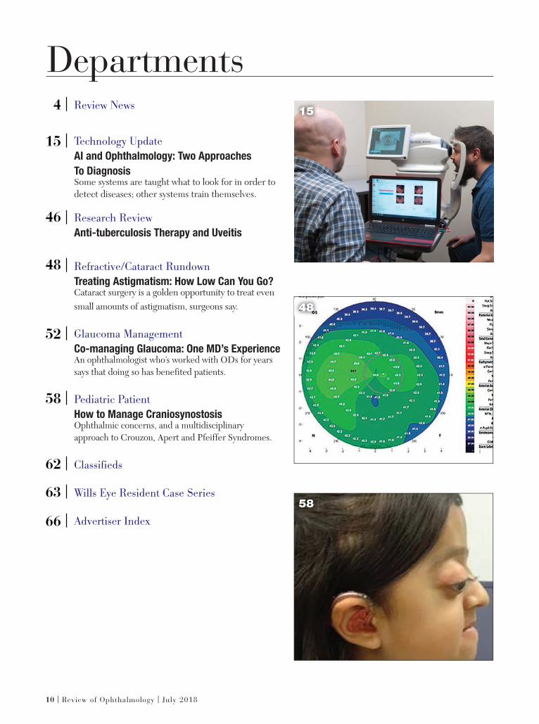
Review News
Technology UpdateAI and Ophthalmology: Two Approaches
To DiagnosisSome systems are taught what to look for in order to detect diseases; other systems train themselves.
Research Review Anti-tuberculosis Therapy and Uveitis
Refractive/Cataract RundownTreating Astigmatism: How Low Can You Go?Cataract surgery is a golden opportunity to treat even small amounts of astigmatism, surgeons say.
Glaucoma ManagementCo-managing Glaucoma: One MD’s ExperienceAn ophthalmologist who’s worked with ODs for years says that doing so has benefited patients.
Pediatric PatientHow to Manage CraniosynostosisOphthalmic concerns, and a multidisciplinary approach to Crouzon, Apert and Pfeiffer Syndromes.
Classifieds
Wills Eye Resident Case Series
Advertiser Index
4 |
15 |
46 |
48 |
52 |
58 |
62 |
63 |
66 |
009_rp0718_toc.indd 10009_rp0718_toc.indd 10 6/22/18 4:01 PM6/22/18 4:01 PM

The Keeler 3 Trade In Program
The Power of 3. Purchase any 3 Keeler Slit Lamps and trade in 3 of your old Slit Lamps and we’ll send you a 4th Keeler Slit Lamp absolutely free of charge.
Keeler Instruments, Inc. • 3222 Phoenixville Pike, bldg. 50 • Malvern, PA 19355Tel: (800) 523-5620 • Fax: (610) 353-7814 • email: [email protected]
Offer valid until September 30, 2018.
Lamp
KSL-H KSL-H-D
KSL-H-DKSL-Z
(also in Digital Ready) (full Digital)
(full Digital)(also in Digital Ready)
Contact Keeler or one of our authorized dealers for more information.
Buy 3 // Trade 3 // Get 1 Free
RP0618_Keeler Slit.indd 1 5/17/18 3:24 PM

Indications and UsageBromSite® (bromfenac ophthalmic solution) 0.075% is a nonsteroidal anti-infl ammatory drug (NSAID) indicated for the treatment of postoperative infl ammation and prevention of ocular pain in patients undergoing cataract surgery.
Recommended Dosing One drop of BromSite® should be applied to the affected eye twice daily (morning and evening) 1 day prior to surgery, the day of surgery, and 14 days postsurgery.
Important Safety Information• Slow or Delayed Healing: All topical nonsteroidal
anti-infl ammatory drugs (NSAIDs), including BromSite®, may slow or delay healing. Topical corticosteroids are also known to slow or delay healing. Concomitant use of topical NSAIDs and topical steroids may increase the potential for healing problems.
• Potential for Cross-Sensitivity: There is the potential for cross-sensitivity to acetylsalicylic acid, phenylacetic acid derivatives, and other NSAIDs, including BromSite®.
Therefore, caution should be used when treating individuals who have previously exhibited sensitivities to these drugs.
• Increased Bleeding Time of Ocular Tissue: With some NSAIDs, including BromSite®, there exists the potential for increased bleeding time due to interference with platelet aggregation. There have been reports that ocularly applied NSAIDs may cause increased bleeding of ocular tissues (including hyphemas) in conjunction with ocular surgery. It is recommended that BromSite® be used with caution in patients with known bleeding tendencies or who are receiving other medications which may prolong bleeding time.
• Keratitis and Corneal Effects: Use of topical NSAIDs may result in keratitis. In some susceptible patients, continued use of topical NSAIDs may result in epithelial breakdown, corneal thinning, corneal erosion, corneal ulceration or corneal perforation. Patients with evidence
RP0318_Sun.indd 2RP0318_Sun.indd 2 2/22/18 10:28 AM2/22/18 10:28 AM

The FIRST and ONLY NSAID indicated to prevent ocular pain in cataract surgery patients1
FOR YOUR CATARACT SURGERY PATIENTS
A DROP OF PREVENTION
DELIVERY SYSTEMFormulated with
NSAID=nonsteroidal anti-infl ammatory drug.
of corneal epithelial breakdown should immediately discontinue use of topical NSAIDs, including BromSite®, and should be closely monitored for corneal health. Patients with complicated ocular surgeries, corneal denervation, corneal epithelial defects, diabetes mellitus, ocular surface diseases (e.g., dry eye syndrome), rheumatoid arthritis, or repeat ocular surgeries within a short period of time may be at increased risk for corneal adverse events which may become sight threatening. Topical NSAIDs should be used with caution in these patients. Post-marketing experience with topical NSAIDs also suggests that use more than 24 hours prior to surgery or use beyond 14 days postsurgery may increase patient risk for the occurrence and severity of corneal adverse events.
• Contact Lens Wear: BromSite® should not be administered while wearing contact lenses. The preservative in BromSite®, benzalkonium chloride, may be absorbed by soft contact lenses.
• Adverse Reactions: The most commonly reported adverse reactions in 1% to 8% of patients were anterior chamber infl ammation, headache, vitreous fl oaters, iritis, eye pain, and ocular hypertension.
Please see brief summary of Full Prescribing Information on the adjacent page.
Sun Ophthalmics is a division of Sun Pharmaceutical Industries, Inc. © 2017 Sun Pharmaceutical Industries, Inc. All rights reserved.BromSite and DuraSite are registered trademarks of Sun Pharma Global FZE.SUN-OPH-BRO-219 03/2017
References: 1. BromSite® [package insert]. Cranbury, NJ: Sun Pharmaceutical Industries, Inc.; 2016. 2. Hosseini K, Hutcheson J, Bowman L. Aqueous humor concentration of bromfenac 0.09% (Bromday™) compared with bromfenac in DuraSite® 0.075% (BromSite™) in cataract patients undergoing phacoemulsifi cation after 3 days dosing. Poster presented at: ARVO Annual Meeting; May 5-9, 2013; Seattle, Washington. 3. ClinicalTrials.gov. Aqueous humor concentration of InSite Vision (ISV) 303 (bromfenac in DuraSite) to Bromday once daily (QD) prior to cataract surgery. https://clinicaltrials.gov/ct2/show/results/NCT01387464?sect=X70156&term=insite+vision&rank=1. Accessed March 2, 2017. 4. Si EC, Bowman LM, Hosseini K. Pharmacokinetic comparisons of bromfenac in DuraSite and Xibrom. J Ocul Pharmacol Ther. 2011;27(1):61-66. 5. Bowman LM, Si E, Pang J, et al. Development of a topical polymeric mucoadhesive ocular delivery system for azithromycin. J Ocul Pharmacol Ther. 2009;25(2):133-139.
Defend against ocular pain and combat postoperative infl ammation with the penetrating power of BromSite® formulated with DuraSite®1
• DuraSite® increases ocular surface retention time, resulting in increased bromfenac absorption2-5
• Provides 24-hour coverage with BID dosing1
• Available in 5 mL bottle
Visit bromsite.com to fi nd out more.
RP0318_Sun.indd 3RP0318_Sun.indd 3 2/22/18 10:28 AM2/22/18 10:28 AM

USE IN SPECIFIC POPULATIONS Pregnancy
Risk Summary There are no adequate and well-controlled studies in pregnant women to inform any drug associated risks. Treatment of pregnant rats and rabbits with oral bromfenac did not produce teratogenic effects at clinically relevant doses.
Clinical Considerations Because of the known effects of prostaglandin biosynthesis-inhibiting drugs on the fetal cardiovascular system (closure of ductus arteriosus), the use of BromSite® during late pregnancy should be avoided.
Data Animal Data Treatment of rats with bromfenac at oral doses up to 0.9 mg/kg/day (195 times a unilateral daily human ophthalmic dose on a mg/m2 basis, assuming 100% absorbed) and rabbits at oral doses up to 7.5 mg/kg/day (3243 times a unilateral daily dose on a mg/m2 basis) produced no structural teratogenicity in reproduction studies. However, embryo-fetal lethality, neonatal mortality and reduced postnatal growth were produced in rats at 0.9 mg/kg/day, and embryo-fetal lethality was produced in rabbits at 7.5 mg/kg/day. Because animal reproduction studies are not always predictive of human response, this drug should be used during pregnancy only if the potential benefit justifies the potential risk to the fetus.
Lactation
There are no data on the presence of bromfenac in human milk, the effects on the breastfed infant, or the effects on milk production; however, systemic exposure to bromfenac from ocular administration is low. The developmental and health benefits of breastfeeding should be considered along with the mother’s clinical need for bromfenac and any potential adverse effects on the breast-fed child from bromfenac or from the underlying maternal condition.
Pediatric Use
Safety and efficacy in pediatric patients below the age of 18 years have not been established.
Geriatric Use
There is no evidence that the efficacy or safety profiles for BromSite® differ in patients 65 years of age and older compared to younger adult patients.
NONCLINICAL TOXICOLOGY Carcinogenesis, Mutagenesis and Impairment of Fertility
Long-term carcinogenicity studies in rats and mice given oral doses of bromfenac up to 0.6 mg/kg/day (129 times a unilateral daily dose assuming 100% absorbed, on a mg/m2 basis) and 5 mg/kg/day (540 times a unilateral daily dose on a mg/m2 basis), respectively revealed no significant increases in tumor incidence.
Bromfenac did not show mutagenic potential in various mutagenicity studies, including the bacterial reverse mutation, chromosomal aberration, and micronucleus tests.
Bromfenac did not impair fertility when administered orally to male and female rats at doses up to 0.9 mg/kg/day and 0.3 mg/kg/day, respectively (195 and 65 times a unilateral daily dose, respectively, on a mg/m2 basis).
Rx Only
Distributed by: Sun Pharmaceutical Industries, Inc. Cranbury, NJ 08512
INDICATIONS AND USAGE BromSite® (bromfenac ophthalmic solution) 0.075% is indicated for the treatment of postoperative inflammation and prevention of ocular pain in patients undergoing cataract surgery.
CONTRAINDICATIONS None
WARNINGS AND PRECAUTIONS Slow or Delayed Healing
All topical nonsteroidal anti-inflammatory drugs (NSAIDs), including BromSite® (bromfenac ophthalmic solution) 0.075%, may slow or delay healing. Topical corticosteroids are also known to slow or delay healing. Concomitant use of topical NSAIDs and topical steroids may increase the potential for healing problems.
Potential for Cross-Sensitivity
There is the potential for cross-sensitivity to acetylsalicylic acid, phenylacetic acid derivatives, and other NSAIDs, including BromSite® (bromfenac ophthalmic solution) 0.075%. Therefore, caution should be used when treating individuals who have previously exhibited sensitivities to these drugs.
Increased Bleeding Time of Ocular Tissue
With some NSAIDs, including BromSite® (bromfenac ophthalmic solution) 0.075%, there exists the potential for increased bleeding time due to interference with platelet aggregation. There have been reports that ocularly applied NSAIDs may cause increased bleeding of ocular tissues (including hyphemas) in conjunction with ocular surgery.
It is recommended that BromSite® be used with caution in patients with known bleeding tendencies or who are receiving other medications which may prolong bleeding time.
Keratitis and Corneal Reactions
Use of topical NSAIDs may result in keratitis. In some susceptible patients, continued use of topical NSAIDs may result in epithelial breakdown, corneal thinning, corneal erosion, corneal ulceration or corneal perforation. These events may be sight threatening. Patients with evidence of corneal epithelial breakdown should immediately discontinue use of topical NSAIDs, including BromSite® (bromfenac ophthalmic solution) 0.075%, and should be closely monitored for corneal health.
Post-marketing experience with topical NSAIDs suggests that patients with complicated ocular surgeries, corneal denervation, corneal epithelial defects, diabetes mellitus, ocular surface diseases (e.g., dry eye syndrome), rheumatoid arthritis, or repeat ocular surgeries within a short period of time may be at increased risk for corneal adverse events which may become sight threatening. Topical NSAIDs should be used with caution in these patients.
Post-marketing experience with topical NSAIDs also suggests that use more than 24 hours prior to surgery or use beyond 14 days postsurgery may increase patient risk for the occurrence and severity of corneal adverse events.
Contact Lens Wear
BromSite® should not be administered while wearing contact lenses. The preservative in BromSite®, benzalkonium chloride, may be absorbed by soft contact lenses.
ADVERSE REACTIONSThe following serious adverse reactions are described elsewhere in the Brief Summary:• Slow or Delayed Healing • Potential for Cross-Sensitivity • Increased Bleeding Time of Ocular Tissue• Keratitis and Corneal Reactions • Contact Lens Wear
Clinical Trial Experience
Because clinical trials are conducted under widely varying conditions, adverse reaction rates observed in the clinical trials of a drug cannot be directly compared to rates in the clinical trials of another drug and may not reflect the rates observed in clinical practice.
The most commonly reported adverse reactions in 1–8% of patients were: anterior chamber inflammation, headache, vitreous floaters, iritis, eye pain and ocular hypertension.
BromSite® (bromfenac ophthalmic solution) 0.075%
Brief Summary
BromSite is a registered trademark of Sun Pharma Global FZE. SUN-OPH-BRO-017-1 03/2017
RP0318_Sun PI.indd 1RP0318_Sun PI.indd 1 2/22/18 10:26 AM2/22/18 10:26 AM
Technology Update R
EV
IEW
July 2018 | reviewofophthalmology.com| 15This article has no commercial sponsorship.
Edited by Michael Colvard, MD, and Steven Charles, MD
It’s become clear that computer-ized image analysis can be a power-
ful tool for helping to diagnose some diseases, including diabetic retinopa-thy and some types of macular de-generation. But one question that remains to be answered is how the ar-tifi cial intelligence should learn what to look for. One approach is to teach the software to analyze and quantify specifi c known signs of the disease, much as a human specialist would do. The other approach is to allow the system to determine on its own how to identify healthy vs. diseased eyes by showing it a large number of samples of each. Both approaches have shown signifi cant promise.
The First Approved System
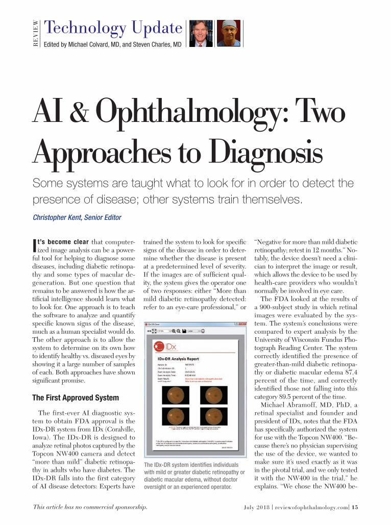
The first-ever AI diagnostic sys-tem to obtain FDA approval is the IDx-DR system from IDx (Coralville, Iowa). The IDx-DR is designed to analyze retinal photos captured by the Topcon NW400 camera and detect “more than mild” diabetic retinopa-thy in adults who have diabetes. The IDx-DR falls into the first category of AI disease detectors: Experts have
trained the system to look for specifi c signs of the disease in order to deter-mine whether the disease is present at a predetermined level of severity. If the images are of suffi cient qual-ity, the system gives the operator one of two responses: either “More than mild diabetic retinopathy detected: refer to an eye-care professional,” or
“Negative for more than mild diabetic retinopathy; retest in 12 months.” No-tably, the device doesn’t need a clini-cian to interpret the image or result, which allows the device to be used by health-care providers who wouldn’t normally be involved in eye care.
The FDA looked at the results of a 900-subject study in which retinal images were evaluated by the sys-tem. The system’s conclusions were compared to expert analysis by the University of Wisconsin Fundus Pho-tograph Reading Center. The system correctly identified the presence of greater-than-mild diabetic retinopa-thy or diabetic macular edema 87.4 percent of the time, and correctly identifi ed those not falling into this category 89.5 percent of the time.
Michael Abramoff, MD, PhD, a retinal specialist and founder and president of IDx, notes that the FDA has specifi cally authorized the system for use with the Topcon NW400. “Be-cause there’s no physician supervising the use of the device, we wanted to make sure it’s used exactly as it was in the pivotal trial, and we only tested it with the NW400 in the trial,” he explains. “We chose the NW400 be-
Christopher Kent, Senior Editor
Some systems are taught what to look for in order to detect the presence of disease; other systems train themselves.
AI & Ophthalmology: Two Approaches to Diagnosis
The IDx-DR system identifi es individuals
with mild or greater diabetic retinopathy or
diabetic macular edema, without doctor
oversight or an experienced operator.
015_rp0718_tech.indd 15015_rp0718_tech.indd 15 6/22/18 4:03 PM6/22/18 4:03 PM

TechnologyUpdate R
EV
IEW
16 | Review of Ophthalmology | July 2018
cause of its ease of use for operators who have never used a retinal camera. Because guidance is provided by the AI, the operator only needs to have a high-school graduate level of edu-cation. In fact, the operators in the clinical trial were asked if they’d ever used a retinal camera; if they had, they couldn’t be part of the trial. Cur-rently, we’re about to start additional studies using other cameras.
“The data showed that 96 percent of patients successfully received dis-ease-level evaluation,” he continues. “That means that the system was able to say yes or no regarding the pres-ence of diabetic retinopathy 96 per-cent of the time. Furthermore, fewer than a quarter of the subjects needed dilation. The reason we were able to achieve these numbers is that the de-vice actually incorporates two AI sys-tems—one makes the diagnosis, while the other helps the inexperienced op-erator take high-quality images of the correct part of the retina. It tells the operator that he missed an area, or that one part is out of focus, and he needs to retake the picture.”
Asked why the device exclusively
detects “more-than-mild” disease, Dr. Abramoff says this was chosen based on the American Academy of Oph-thalmology’s Preferred Practice Pat-terns for Diabetic Retinopathy. “The idea was to catch the patients who can’t wait 12 months to be seen,” he says. “The Preferred Practice Pattern says those with more-than-moderate disease or macular edema need to be seen by an ophthalmologist sooner than that. With a different system you could detect those who have a tiny number of microaneurysms—pa-tients who could wait 12 months to be seen—but we chose to detect just those individuals who need to be seen more urgently.”
Dr. Abramoff says that several con-cerns guided the development of the system. One key concern was creating a system that could make a clinical decision by itself, with no physician oversight. “We care about maintain-ing both health care affordability and high quality, and this was the best way to do that,” he says. “However, auton-omous AI requires much stricter per-formance standards than something that simply helps a specialist like me
to make a clinical decision. In those situations, I’m responsible. Here, I’m relying on the IDx-DR output.
“A second issue was being able to produce disease-level output for the vast majority of patients,” he contin-ues. “Otherwise, you wouldn’t be able to use the system without specialist oversight. Fortunately, we were able to achieve that. A third issue was that the system had to be as unbiased as possible in terms of race, ethnicity, sex and age. Removing potential bias im-pacts both how you design the AI and also how you validate it. Our detec-tors identify biomarkers, regardless of these factors.”
Making It Explainable
Dr. Abramoff says another impor-tant issue is one that separates this technology from other “machine-learning” systems. “We wanted our system to be explainable,” he says. “One reason for this is psychological. It’s very important to be able to explain how the system reaches a conclusion, even when it makes a mistake. For centuries doctors have known to look for hemorrhages and exudates and microaneurysms and other lesions, because if the eye has those lesions, the patient has diabetic retinopathy. So we decided to build detectors for each of these lesion types, and then combine those outputs into a single disease output.
“Using this approach makes the sys-tem explainable,” he continues. “We can validate each of these detectors independently, and we were able to show the FDA how the system works. We can point to an image and say the device detected hemorrhages and ex-udates here, and therefore it reported the presence of disease. And if the de-tector doesn’t work properly in some situation, we’ll know why it didn’t.
“Some other groups use the ‘black box’ approach,” he notes. “That typi-cally involves a single convolutional
The IDx-DR was able to successfully produce a disease evaluation for 96 percent of
subjects in a clinical trial, with fewer than a quarter of them needing dilation. The artifi cial
intelligence built into the device helps ensure that the operator gets high-quality images.
015_rp0718_tech.indd 16015_rp0718_tech.indd 16 6/22/18 4:03 PM6/22/18 4:03 PM
www.compulinkadvantage.com/onetab 800.456.4522
EHR PM RCM ASC OPTICAL ANALYTICS PATIENT ENGAGEMENT TELEHEALTH AUDIT PROTECTION
Request a Demo Today
“ As a physician leader at Georgia Eye Partners, one
of the premier ophthalmology practices in America,
Compulink helps me focus on our patients and not on
the administrative processes of healthcare. ”
Parul Khator, MD, Georgia Eye Partners
Ophthalmology’s All-in-One EHR Solution
AI-Driven EHR Designed For Speed
Supports ALL Ophthalmology Sub-Specialties
Chart From A Single Screen
Ophthalmology’s Leader for 33 Years!
RP0618_Compulink.indd 1RP0618_Compulink.indd 1 5/14/18 9:51 AM5/14/18 9:51 AM

TechnologyUpdate R
EV
IEW
18 | Review of Ophthalmology | July 2018
neural network. They say, ‘Here are a bunch of images; here’s the disease output you ought to have for each image.’ They don’t know how the sys-tem makes its decision. It could be because in the image the disc is in a certain location, and in the training data, people with disease mostly had the disc in that location. In some cases these systems have factored in part of the image that’s outside of the reti-na, even including the square border around the image of the retina, e.g., the mask. Proponents of this approach don’t care what part of the image was used to make the decision, as long as it makes the right decision. That’s not explainable, because they can’t tell you how it works.
“If I give clinicians an image with a bunch of hemorrhages,” he continues, “they’ll say, ‘This is likely diabetic reti-nopathy.’ If I start taking those hem-orrhages away, eventually they’ll say, ‘There’s no disease here.’ Biomarker AI systems like ours work similarly. On the other hand, using the ‘black box’ approach, you can change less than 0.1 percent of the image, so it looks exactly the same to any human clinician—and the system may flip its diagnosis. Those algorithms are highly sensitive to specifi c alterations of pixels, and not necessarily sensitive to hemorrhages and microaneurysms. That’s a concern because we don’t know what these systems are looking for, or if they fail, why they did. We call it catastrophic failure, because it’s very unexpected. For example, an image might look very abnormal, full of disease, but the system says it’s normal.”
Machine, Teach Thyself
Google is one of a number of com-panies developing a diagnostic system using the other machine-learning ap-proach. “Many different companies are working on different diseases such as glaucoma and macular degenera-
tion where there’s a very large global need for screening,” says Peter A. Karth, MD, a vitreoretinal specialist at Oregon Eye Consultants and a phy-sician consultant to Google. “Google continues to be the research leader in this space. Google’s goal is to create a system that will set the gold stan-dard for accuracy with the ability to parse out all levels of diabetic reti-nopathy—including diabetic macular edema—with a very economical cost structure. They’re getting closer to a system that will be really effective when deployed.”
Dr. Karth says the approval of IDx-DR is an important milestone. “Of course, unlike Google’s system, IDx-DR is a feature-recognition-based sys-tem that teaches the machine to look for certain things, such as bad blood vessels, microaneurysms and hemor-rhages,” he notes. “Most of the other companies, including Google, are us-ing systems based on machine learn-ing. In machine learning you don’t teach the machine anything; it learns on its own.”
Asked whether he’s concerned that doctors won’t know what the system is
basing its diagnosis on, Dr. Karth says he’s not worried. “The system is defi -nitely picking up on things that we’re not, but I don’t think it’s necessary to know what those things are in order to have a robust algorithm,” he says. “It’s possible that surgeons could benefi t from learning what those factors are, and there are teams trying to parse that out, but the results aren’t ready for release yet.”
So which approach (machine learn-ing vs. feature recognition) is likely to be most effective? “There are good arguments on both sides,” notes Dr. Karth. “The IDx people have good data supporting their approach. But with all of the research and people working on using machine learning to allow in-depth grading of diabetic ret-inopathy and diagnose multiple dis-eases, I believe this will be the wave of the future. At this point, of course, it’s impossible to say whether one tech-nology or the other will become the gold standard.”
Asked about the nature of the evi-dence supporting these technologies, Dr. Karth acknowledges that much of the evidence comes from retrospec-
Rather than teaching the AI system what to look for, systems using “deep learning” are
exposed to many labeled images, allowing the system to gradually determine by itself
what image characteristics relate to that label. Data received at the “input layer” are
processed by a system of weighted connections developed based on the system’s past
experience, leading to a conclusion presented at the “output layer.” This process can be
used to identify the presence of disease based on image characteristics.
015_rp0718_tech.indd 18015_rp0718_tech.indd 18 6/22/18 4:03 PM6/22/18 4:03 PM
NEW!
REDNESS RELIEVER EYE DROPSBRIMONIDINE TARTRATE OPHTHALMIC SOLUTION 0.025%
F R O M T H E E Y E C A R E E X P E R T S A T
A NEW REDNESS RELIEVER LIKE NO OTHER:
Images representative of clinical trials results individual results may vary.
redness in clinical trials
lasts for up to 8 hours
Learn more at lumifydrops.com/professional
LUMIFY redness reliever eye drops are available at national retailers in 2.5mL and 7.5mL sizes.
Introduce your patients to a new kind of redness reliever.
Before After
RP0718_BL Lumify.indd 1 6/14/18 9:33 AM
TechnologyUpdate R
EV
IEW
20 | Review of Ophthalmology | July 2018
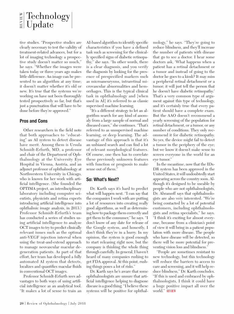
tive studies. “Prospective studies are clearly necessary to test the validity of treatment-related advances, but for a lot of imaging technology a prospec-tive study doesn’t matter so much,” he says. “Whether the images were taken today or three years ago makes little difference. An image can be pre-sented to an algorithm at any time; it doesn’t matter whether it’s old or new. It’s true that the systems we’re working on have not been thoroughly tested prospectively so far, but that’s just a punctuation that will have to be done before they’re approved.”
Pros and Cons
Other researchers in the fi eld note that both approaches to “educat-ing” an AI system to detect disease have merit. Among them is Ursula Schmidt-Erfurth, MD, a professor and chair of the Department of Oph-thalmology at the University Eye Hospital in Vienna, Austria, and an adjunct professor of ophthalmology at Northwestern University in Chicago, who is known for her work with arti-fi cial intelligence. (She founded the OPTIMA project, an interdisciplinary laboratory including computer sci-entists, physicists and retina experts introducing artifi cial intelligence into ophthalmic image analysis, in 2013.) Professor Schmidt-Erfurth’s team has conducted a series of studies us-ing artificial intelligence to analyze OCT images to try to predict clinically relevant issues such as the optimal anti-VEGF injection interval when using the treat-and-extend approach to manage neovascular macular de-generation patients. As part of that effort, her team has developed a fully automated AI system that detects, localizes and quantifi es macular fl uids in conventional OCT images.
Professor Schmidt-Erfurth sees ad-vantages to both ways of using artifi -cial intelligence as an analytical tool. “It makes a lot of sense to train an
AI-based algorithm to identify specifi c characteristics if you have a defi ned task such as screening for the clinical-ly specifi ed signs of diabetic retinopa-thy,” she says. “In other words, there is a clear diagnosis, and you verify the diagnosis by looking for the pres-ence of prespecified markers such as microaneurysms, intraretinal mi-crovascular abnormalities and hem-orrhages. This is the typical clinical task in ophthalmology and [when used in AI] it’s referred to as classic supervised machine learning.
“It’s a different strategy to let an al-gorithm search for any kind of anom-aly from a large sample of normal and diseased cases,” she continues. “That’s referred to as unsupervised machine learning, or deep learning. The ad-vantage of this approach is that it’s an unbiased search and can fi nd a lot of relevant morphological features. Of course, one then has to correlate these previously unknown features with function or prognosis to make sense out of them.”
So: What’s Next?
Dr. Karth says it’s hard to predict what will happen next. “I can say that the companies I work with are putting a lot of resources into creating really good algorithms, as well as determin-ing how to package them correctly and get them to the consumer,” he says. “I don’t know of any date for release of the Google system, and honestly, I don’t think they’re in a hurry. In my opinion, the system is good enough to start releasing right now, but the company is thinking the whole thing through carefully. In general, I haven’t heard of many companies rushing to get FDA approval. At this point, rush-ing things poses a lot of risks.”
Dr. Karth says he’s aware that some ophthalmologists are unsure that arti-fi cial intelligence helping to diagnose disease is a good thing. “I believe these systems will be positive for ophthal-
mology,” he says. “They’re going to reduce blindness, and they’ll increase the number of patients with disease that go to see a doctor. I hear some doctors ask, ‘What happens when a patient has a retinal detachment or a tumor and instead of going to the doctor he goes to a kiosk? It may miss a peripheral retinal detachment or a tumor; it will just tell the person that he doesn’t have diabetic retinopathy.’ That’s a very common type of argu-ment against this type of technology, and it’s certainly true that every pa-tient should have a complete exam. But the AAO doesn’t recommend a yearly screening of the population for retinal detachment, or a tumor, or any number of conditions. They only rec-ommend it for diabetic retinopathy. So yes, the device might fail to detect a tumor in the periphery of the eye; but we know it doesn’t make sense to screen everyone in the world for an eye tumor.”
In the meantime, now that the IDx-DR system has been approved in the United States, it will undoubtedly start appearing across the country soon. Al-though it’s designed to be useable by people who are not ophthalmologists, Dr. Abramoff says that ophthalmolo-gists are also very interested. “We’re being contacted by a lot of potential customers, including ophthalmolo-gists and retina specialists,” he says. “I think it’s exciting for almost every-one, because from a clinician’s point of view it will bring in a patient popu-lation with more disease. The people who have disease will be detected, so there will be more potential for pre-venting vision loss and blindness.”
“People are sometimes resistant to new technology, but this technology will reduce the barriers to access to care and screening, and it will help re-duce blindness,” Dr. Karth concludes. “If this is used and embraced by oph-thalmologists, I think it could have a huge positive impact all over the world.”
015_rp0718_tech.indd 20015_rp0718_tech.indd 20 6/22/18 4:04 PM6/22/18 4:04 PM

STRIVE TO SEE THE BEST RESULTS POSSIBLERegular fixed-interval dosing of long-term treatment with anti-VEGFs has been shown to provide better gains and maintenance of vision, compared to PRN or treat-and-extend dosing regimens, in some patients with Wet AMD and DME.1-6
Ask your Regeneron representative what we can do to help ensure patients come in for their scheduled treatment.
anti-VEGF = anti–vascular endothelial growth factor; AMD = Age-related Macular Degeneration; DME = Diabetic Macular Edema.
References: 1. Heier JS et al. Ophthalmology. 2016;123(11):2376-2385. 2. Rosenfeld PJ et al. N Engl J Med. 2006;355(14):1419-1431. 3. Ho AC et al. Ophthalmology. 2014;121(11):2181-2192. 4. Nguyen QD et al. Ophthalmology. 2012;119(4):789-801. 5. Kaiser PK et al. Ophthalmol Retina. 2017;1(4):304-313. 6. Peden MC et al. Ophthalmology. 2015;122(4):803-808.
© 2018, Regeneron Pharmaceuticals, Inc. All rights reserved.777 Old Saw Mill River Road, Tarrytown, NY 10591 01/2018 US-OPH-13911
RP0318_Regeneron.indd 1 2/13/18 2:56 PM

22 | Review of Ophthalmology | July 2018
RE
VIE
W
This article has no commercial sponsorship.
Cover Focus
The IOL in the Sulcus: When, Why & HowChristopher Kent, Senior Editor
P lacing an IOL in the sulcus instead of the capsular bag is rarely a surgeon’s preference,
but sometimes intraoperative circum-stances require it. In that situation, choosing the right lens and placing it properly can make the difference between a perfectly good outcome that will last for years and a potential future problem.
Here, experienced surgeons offer detailed advice on what to do, how to do it, and why it works.
When to Choose the Sulcus
“Putting the lens into the sulcus isn’t a first choice,” notes Uday Devgan, MD, FACS, FRCS, chief of ophthal-mology at Olive View UCLA Medical Center and associate clinical profes-sor at the UCLA School of Medicine. “The ideal is to put the lens in the cap-sular bag so that the natural bag that held the human lens will now hold the man-made lens. That’s what we do in 99 percent of eyes. It puts the IOL in the best, most stable, least risky and most physiologic position. We typi-cally only place an IOL in the sulcus when there’s a defect in the posterior capsule that would prevent us from placing the IOL inside the bag.
“Nevertheless, sulcus placement is more common than you might think,”
he says. “In the United States, the average surgeon does 250 cataracts in a year, and somewhere between 1 and 5 percent of cataract cases result in a complication that would cause the surgeon to place the lens in the sulcus. Generally, sulcus placement happens in eyes that have a posterior capsule defect, which is the most common reason, or because the eye has loose zonules.”
Use of sulcus placement in the lat-ter situation, however, is subject to some debate. “Some people have said if there’s zonular dehiscence or severe pseudoexfoliation, you might be bet-ter off putting a lens in the sulcus, possibly with a capsular tension ring underneath,” notes Douglas K. Gray-son, MD, medical director and chief of glaucoma and cataract surgery at Omni Eye Services in New York and New Jersey, and assistant professor of ophthalmology at the New York Ear and Ear Infi rmary of Mt. Sinai and the Hackensack Meridian School of Med-icine. “The reasoning is that putting a lens in the bag may stress the zonules and cause them to break, resulting in the lens being likely to decenter at a higher rate in the future. I haven’t generally put the lens in the sulcus in that situation, because I feel that if I have enough zonules to get the lens into the bag in the fi rst place it may
When you need to
place a lens in the
sulcus, it pays to
be prepared—and
to know what not
to do.
Cataract/Refractive Surgery
022_rp0718_f1.indd 22022_rp0718_f1.indd 22 6/22/18 4:12 PM6/22/18 4:12 PM
be more stable that way, pushing out the zonules. However, I can see some merit to the other point of view, espe-cially in cases of really severe pseudo-exfoliation or zonular compromise due to trauma.”
Jorge L. Alio, MD, PhD, a professor and chairman of the department of ophthalmology at Miguel Hernandez University in Alicante, Spain, says he considers placing a lens in the sulcus when he encounters an unstable cap-sular bag. “If I have a subluxation, for example, I prefer the sulcus-placed lens, even though I also use a capsular tension ring to fi x the capsular bag in place,” he says. “My experience is that even with the capsular tension ring, sometimes you’ll have a lens disloca-tion over time. I also prefer to use a sulcus-placed lens when piggybacking to deal with a refractive surprise, or in secondary implantations when I have to explant an IOL—for instance, following opacifi cation. Of course, in these cases I’d rather put the lens into the capsular bag, if this is possible.”
“Some surgeons place a sulcus IOL in eyes with uveitis to avoid iris-cap-sule synechiae, but I disagree with that reasoning,” Dr. Devgan adds. “In that situation I just perform a large capsulorhexis.”
Picking the Right Lens
“Placing the lens in the sulcus is a great option when the posterior cap-sule can’t support an intraocular lens in the bag and the anterior capsule is intact,” says Thomas A. Oetting, MD, MS, a professor of clinical oph-thalmology at the University of Iowa. “The problem in the United States, at least, is that we no longer have a per-fect sulcus IOL. We only have three-piece IOLs with relatively short hap-tic lengths. As such, the only way to ensure long-term stability with sulcus haptic placement is to have an intact anterior capsule that allows capture of the optic into the bag.”
Dr. Oetting points out that none of the FDA-approved in-the-bag IOLs can be counted on to remain stable in the sulcus. “The main reason for this is that the haptics aren’t long enough,” he explains. “What you want is a haptic length of about 14 mm or more. The longest haptic length we have in the U.S. is 13 mm. If you simply place an IOL with a 13-mm haptic length in the sulcus of a large eye without capturing the optic, it can decenter and tilt.
“Pavlina S. Kemp, MD, from our group, conducted a study1 which showed that when the eyes are bigger, you can’t count on the Alcon MA50—which has a 13-mm haptic length—to stay centered long-term,” he notes. “That means that if you place an IOL in the sulcus, unless it’s a small eye, you should capture the optic with the anterior capsule. If you can’t do optic capture, the IOL may decenter over time. In the United States we used to have access to the STAAR AQ2010, that was well-suited for sulcus place-ment because it had a 14-mm haptic and a large optic, but that lens is no
longer available to us. As a result, if you’re not able to capture the optic, you may be forced to place a lens in the sulcus knowing that it may decen-ter over time.”
To make the best of this situation, Dr. Oetting notes that you need to pick a three-piece lens with long, thin haptics.2 “You want the longest hap-tics you can get,” he says. “Also, you want a lens with a big optic, so that if it decenters a little bit it will be less of an issue.”
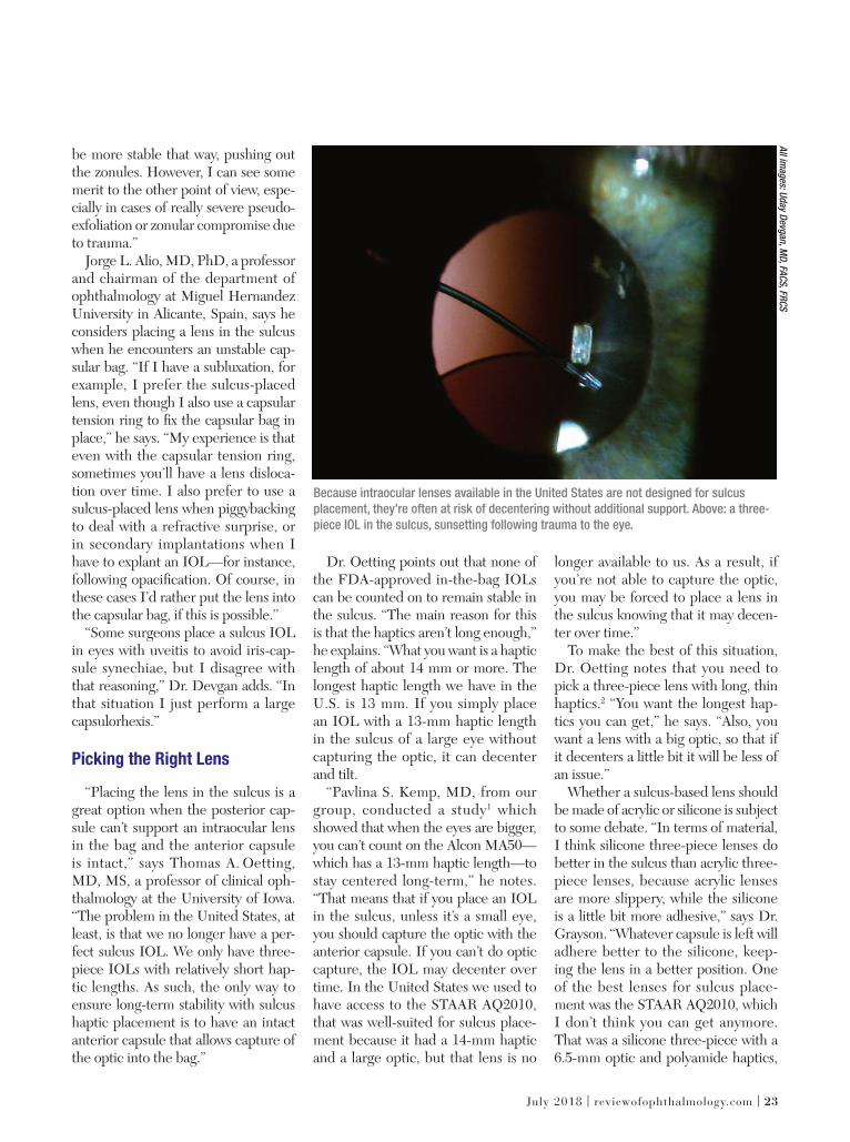
Whether a sulcus-based lens should be made of acrylic or silicone is subject to some debate. “In terms of material, I think silicone three-piece lenses do better in the sulcus than acrylic three-piece lenses, because acrylic lenses are more slippery, while the silicone is a little bit more adhesive,” says Dr. Grayson. “Whatever capsule is left will adhere better to the silicone, keep-ing the lens in a better position. One of the best lenses for sulcus place-ment was the STAAR AQ2010, which I don’t think you can get anymore. That was a silicone three-piece with a 6.5-mm optic and polyamide haptics,
Because intraocular lenses available in the United States are not designed for sulcus
placement, they’re often at risk of decentering without additional support. Above: a three-
piece IOL in the sulcus, sunsetting following trauma to the eye.
All im
ag
es: U
day D
evg
an
, MD
, FAC
S, F
RC
S
July 2018 | reviewofophthalmology.com | 23
022_rp0718_f1.indd 23022_rp0718_f1.indd 23 6/22/18 4:13 PM6/22/18 4:13 PM

24 | Review of Ophthalmology | July 2018
CoverFocus R
EV
IEW
Cataract/Refractive Surgery
which were very stiff. The next-best lens is the Tecnis Z9002 silicone three-piece. It has rounded, not square, edg-es, so you’re not going to get as much chafi ng.”
Dr. Oetting, however, notes some reasons to choose a lens made of acryl-ic rather than silicone. “One of the issues you have to keep in mind is that when we’re placing these lenses in the sulcus, there’s often a posterior capsule injury,” he says. “Those pa-tients are at increased risk of need-ing a vitrectomy in the future, and that may involve silicone oil. Silicone lenses can have problems with air-fl uid exchanges and silicone oil; lenses made of acrylic won’t have a problem in that situation.
“The IOL I tend to use, which is a decent lens for the sulcus, is the Alcon MA50,” he adds. “It’s a three-piece lens with a 6.5-mm acrylic optic, which makes it suitable for capturing in slightly large anterior capsuloto-mies. However, this still isn’t a perfect choice, because the haptic length isn’t long enough.”
One-piece IOLs in the Sulcus
Dr. Devgan says that three-piece lenses work better in the sulcus than most one-piece lenses. “When you talk about single-piece lenses, you’re primarily talking about the single-piece acrylic lenses,” he notes. “You must never place a single-piece acrylic IOL in the sulcus. That’s an absolute rule—the single-piece acrylic IOLs are meant solely for in-the-bag place-ment.
“There are three main problems with them in the sulcus,” he explains. “First, the haptics of a single-piece acrylic lens are as thick as the lens itself. A thick haptic can chronically rub the back side of the iris, causing the iris to lose pigment, leading to in-fl ammation and microbleeding in the eye and causing UGH syndrome—uveitis, glaucoma and hyphema. The
lens will also tend to decenter; then the surgeon will have to explant and exchange the lens. In comparison, the haptics of a three-piece lens are much thinner.
“For example, I saw one patient whose surgeon had placed a single-piece acrylic in the sulcus,” he says. (See picture, p. 26) “The thick haptic had been chronically rubbing raw the back side of the iris. It scraped the back of the iris so much that the red refl ex, the iris transillumination defect, was lit-erally the outline of the haptic. This is not what you want! So you should never put a single-piece acrylic lens in the sulcus.
“A second issue is that the lens of a single-piece IOL is planar,” he con-tinues. “If you look at its side profi le, it’s totally fl at. In contrast, if you look at a three-piece lens from the side, the lens is slightly posteriorly vault-ed. That means the optic is set back a little, so it doesn’t scrape the back of the iris.
“The third issue,” he adds, “is that the single-piece acrylic IOLs may be smaller in total length than the three-
piece IOLs, and their haptics don’t have suffi cient rigidity. As such, they don’t fi t well in the sulcus. They tend to sunset.”
Dr. Devgan notes, however, that a single-piece lens made of PMMA can sometimes work in the sulcus. “The PMMA single-piece lenses are rigid and nonfoldable,” he explains. “Even though they’re single-piece, they have very thin arms, and they’re OK for sul-cus placement. Not the best, but OK. However, those lenses are rarely used in the United States—they represent far less than 1 percent of the market. That’s because being rigid and non-foldable, they require a 6- or 7-mm incision to put them inside the eye. Today, we make 2.5-mm incisions—or smaller—all day long.”
Dr. Grayson notes one situation in which he’d consider putting a one-piece IOL in the sulcus. “If your sur-gical plan was to put in a Symfony or a Symfony toric and you break the posterior capsule, you don’t have too many options,” he says. “You either try to place the lens in the sulcus with op-tic capture through the capsulorhexis,
One way to provide additional support for an IOL placed in the sulcus is to suture the
haptics to the iris or sclera. Above: The eye shown on the previous page, with the IOL optic
recentered and the haptics sutured to the iris at 12 and 6 o’clock.
022_rp0718_f1.indd 24022_rp0718_f1.indd 24 6/22/18 4:13 PM6/22/18 4:13 PM

www.rickbayfoundation.org
The Rick Bay Foundationfor Excellence in Eyecare Education
Scholarships are awarded to advance the education
of students in both Optometry and Ophthalmology,
and are chosen by their school based on qualities that
embody Rick’s commitment to the profession, including
integrity, compassion, partnership and dedication to the
greater good.
Support the Education of Future Healthcare & Eyecare Professionals
About RickRick Bay served as the publisher of The Review Group for more than 20 years.
To those who worked for him, he was a leader whose essence was based in a fi erce and
boundless loyalty.
To those in the industry and the professions he served, he will be remembered for his unique array of skills and for his dedication to exceeding the expectations of his customers, making many of them fast friends.
Interested in being a partner with us?Visit www.rickbayfoundation.org
(Contributions are tax-deductible in accordance with section 170 of the Internal Revenue Code.)
(The Rick Bay Foundation for Excellence in Eyecare Education is a nonprofi t, tax-exempt organization under section 501(c)(3) of the Internal Revenue Code.)
2015_rickbay_housead.indd 12015_rickbay_housead.indd 1 7/5/17 3:00 PM7/5/17 3:00 PM

26 | Review of Ophthalmology | July 2018
or you have to abandon that lens and put in a monofocal. Alternatively, you could put in a multifocal three-piece of a different design, such as the Alcon ReSTOR with the 4.0 add, or the Tec-nis with the 4.0 add. If there was an astigmatic component, you’d have to deal with it later using PRK.
“The problem with changing to a different lens,” he notes, “is that the patient has paid $3,500 or $4,000 ahead of time and is expecting to get a certain result. It’s hard to just say, ‘Sor-ry, we couldn’t do it.’ It’s even worse if you’ve done one eye with the Symfony toric, and you’re on the second eye.
“Of course, without optic capture, the one-piece lenses are too small and too thick to place in the sulcus, and they move too much,” he says. “For that reason I wouldn’t put a one-piece in the sulcus unless I had anterior cap-sular capture, so the optic is in the bag, below the capsular edge. The hap-tics of a one-piece lens are thick, but the tension from the lens capture will keep them away from the iris. They won’t be sitting fl at; they’ll be angled slightly downward. So there’s a very
good chance that you’ll be able to do this, especially with a capsulorhexis made by a femtosecond laser, which I always use when implanting multi-focals, premium lenses and torics. In addition, capturing the lens works out well—especially for torics—because they don’t move.”
The Lens-capture Option
Dr. Devgan says that having the haptics in the sulcus with the optic captured back through the capsu-lorhexis is probably the best position for long-term IOL stability. “Doing that is like putting a manhole cover on a manhole,” he says. “The lens be-comes a really good barrier, so you won’t get vitreous prolapse, and it’s really solid. It will never move. Fur-thermore, because you’re pushing the optic farther back, close to where it would have been if it were placed in-side the bag, the issue of refractive shift is minimized. You don’t need to adjust the lens power.”
“Using IOL capture when putting a lens in the sulcus is by far the best way
to do it, and femtosecond lasers have made that a lot easier because you get a perfectly symmetrical capsulotomy of a consistent size,” notes Dr. Gray-son. “If you create a 5- or 4.8-mm cap-sulotomy with a femtosecond laser—I use 4.8 mm—you can then capture the optic. Using this approach you can put a three-piece lens in the sulcus; in fact, you can even put a one-piece lens in the sulcus. I’ve put Symfony torics in the sulcus using IOL capture after femtosecond capsulorhexis, and it’s worked out fi ne.”
Dr. Oetting agrees that when you need to place a three-piece IOL in the sulcus, the best approach is to use optic capture. “That’s clearly the best choice if you can’t place the IOL in the bag,” he says. “In fact, it may even be better than simply placing the IOL in the bag in cases where you’re worried about progressive zonular weakness, such as pseudoexfoliation and retinitis pigmentosa. By placing the haptics in the sulcus and the optic in the bag you nearly eliminate the possibility of phimosis. The anterior capsule can only contract so much because the lens is keeping it open. Because of that, you tend to get less zonular dam-age and more long-term stabilization. The haptics in the sulcus provide ad-ditional centration and support, inde-pendent of the zonules. I really think it’s a nice technique to have in your bag of tricks.”
Dr. Oetting also notes that zonu-lar dehiscence can sometimes be a reason to consider placing the IOL haptics in the sulcus. “For example,” he says, “if you had mild zonular weak-ness and were worried about phimosis and long-term centration of the lens, it would be a good option to place a three-piece lens with the haptics in the sulcus and the optic in the bag. That can give you really nice centra-tion both short and long term.
“However,” he adds, “you do have other options in that situation, includ-ing placing a capsular tension ring and
CoverFocus R
EV
IEW
Placing a single-piece IOL in the sulcus is never recommended. Above: a decentered single-
piece acrylic multifocal IOL has decentered and is now causing damage to the back of the
iris, as demonstrated by the transillumination defect in the shape of the IOL haptic.
Cataract/Refractive Surgery
022_rp0718_f1.indd 26022_rp0718_f1.indd 26 6/22/18 4:13 PM6/22/18 4:13 PM

E-NEWS YOU CAN USE
Have you been receiving and reading custom e-blasts from Review of Ophthalmology?
If not, you’re missing out on valuable information!
You’re a busy practitioner and not surprisingly, your e-mail inbox is often full. Fortunately, when you scan through the sender list, determining which messages to delete and which to save or read, you can feel confi dent knowing that e-blasts from Review of Ophthalmology, a Jobson Medical Information LLC publication, contain the most current and comprehensive information available in the fi eld to keep you on the cutting edge.
Review of Ophthalmology’s online stable of products includes editorial newsletters and pro-motional information about new products, treatments and surgical techniques, as well as alerts on continuing education courses for ophthalmologists.
Our FREE weekly e-newsletter, Review of Ophthalmology Online, brings you the latest in ophthalmic research, as well as industry news. In an eff ort to keep eyecare professionals informed, this resource is waiting in your inbox every Monday morning.
Retina Online, our free monthly e-newsletter, is for retina specialists and generalophthalmologists interested in enhancing their knowledge on the topics of retina and related disease diagnosis and treatment, as well as the latest in surgical procedures.
EVERY MONDAY
Your time is valuable — and so is your practice. These e-products are the most eff ective way for you to receive updates on breaking news and research — all just a click away.DON’T MISS OUT!
Unfamiliar with our products? Visit www.reviewofophthalmology.com and check out our newsletter archives.Go to www.jobson.com/globalEmail/default.aspx to sign up for the e-newsletters that interest you.
2015_RPonline_house.indd 12015_RPonline_house.indd 1 12/10/14 2:38 PM12/10/14 2:38 PM

28 | Review of Ophthalmology | July 2018
then putting a standard lens in the bag, or placing an Ahmed segment in the area of weakness, and then putting the lens in the bag. So placing the lens in the sulcus is just one of several op-tions when you’re dealing with weak zonules.”
Dr. Oetting points out another cap-ture-based option that’s useful when you’re faced with a very young patient or someone who won’t be able to sit still for a possible future YAG capsu-lotomy, a technique he learned from Lisa Arbisser, MD. “In addition to creating an anterior capsule opening, you can also create a posterior capsule opening that’s round and continuous,” he explains. “Then you place the hap-tics in the sulcus and push the optic all the way back, so it’s captured by both the anterior and posterior capsule. Doing this is a two-for-one, because it creates a very stable lens confi gura-tion that dramatically reduces the pos-sibility of lens decentration and also prevents posterior capsular opacity. Dr. Arbisser calls this ‘bicapsular cap-ture,’ and while the indication doesn’t come up very often, the technique can be very useful.” Dr. Oetting adds that when using this technique an IOL can often be placed with no vitreous pro-lapse, despite the posterior capsular opening. “If vitreous prolapse does occur,” he says, “you’ll have to perform an anterior vitrectomy to limit vitreous traction and the risk of retinal detach-ment.”
Dr. Oetting says that he’s aware of some options for stabilizing a lens in the sulcus (when the optic isn’t cap-tured) that he hasn’t tried. “Some peo-ple talk about bending the haptics to change the angle at which the haptic comes off of the lens, effectively mak-ing them longer,” he says. “Another option is to suture the lens to the iris. You could also try using the Yamane technique, or the Agarwal glued-IOL technique with a three-piece IOL. And it’s always an option to place an anterior chamber lens. But just plac-
ing the haptics into the sulcus in a large eye [without using any of these techniques] is inviting decentration.”
Getting the Lens In
As with any surgery, using the right technique matters. Surgeons offer these suggestions to minimize the like-lihood of an undesirable outcome:
• Enlarge your incision slight-ly. “Most three-piece IOL inserters require a bigger incision,” notes Dr. Oetting. “Focus on making it easy to get the injector in. Don’t try to force it or push it hard through a too-small in-cision. If you push hard, you may lose some viscoelastic, and the next thing you know, vitreous is coming forward.”
• Use a dispersive viscoelastic. “When you fi nd you have an issue with the posterior capsule, you want to be smart about your choice of viscoelas-tic,” says Dr. Oetting. “I usually switch over to a dispersive viscous OVD like Alcon’s Viscoat. Use the OVD to cre-ate space between the iris and the anterior capsule to better allow the leading haptic into the sulcus. Ideally you want to open the sulcus for 360 degrees, although that can be hard to do.
“The reason a dispersive OVD is ideal is that in cases like this you’re very likely to leave a little bit of visco-elastic material behind,” he explains. “You’ll get fewer pressure spikes if the OVD you leave behind is dispersive rather than cohesive. A dispersive vis-coelastic also coats and sticks to tissue better.”
• Insert the lens carefully. “My incision size is about 2.75 mm, as we need to introduce the tip of the injec-tor into the anterior chamber,” says Dr. Alio, noting that he prefers to use the Alcon MA60 or MN60 lenses. “I deliver the distal haptic into the sulcus and place the trailing haptic over the iris. Then with a Lester hook I care-fully dial the second haptic into the sulcus. Finally, I use a Sinskey hook to check for stability and see that the lens is properly placed and stable.”
“When you’re putting a lens in the sulcus, make sure the haptic is on top of the capsular shelf,” says Dr. Gray-son. “You can skim the haptic along the bottom edge of the iris. Then, once you launch the lens, you’re usu-ally left with one haptic outside the eye. You want to carefully rotate that in, skimming the back surface of the iris to make sure that the haptic stays
CoverFocus R
EV
IEW
A single-piece acrylic IOL loose in the sulcus with one of the haptic coming above the iris.
Cataract/Refractive Surgery
022_rp0718_f1.indd 28022_rp0718_f1.indd 28 6/22/18 4:13 PM6/22/18 4:13 PM

NuLids
™
transformational
dry eye therapy
Transforms your patients’ lives
Finally… A doctor-directed at-home treatment for dry eye disease that is safe, effective and easy to use.
A multi-center study1 showed:• 50% decrease in dry eye symptoms• 65% improvement in TBUT • 80% increase in meibomian glands yielding liquid secretions.
For more information visit www.NuSightMedical.com or call us at 833-468-5437
1 Schanzlin, Olkowski, Coble, Gross. NuLids II Study, April 2018
Doctor prescribed, at-home dry eye relief
in the sulcus. You don’t want to have one haptic end up stuffed into capsular bag remnants, and the other in the sul-cus; then you’ll have uneven torsional forces which can cause the lens to decenter.”
“After you think the lens is in position, use your chopper to lift up the iris,” Dr. Devgan adds. “You can directly visualize whether the haptic is in the sulcus. It should be in front of the anterior capsular bag.”
• Place the haptics where the lens will get the most support. “If you have an area where there’s weakness, such as a defect in the posterior capsule and/or weak zonules, you don’t want to put the sulcus lens haptics where the weakness is,” says Dr. Devgan. “So, if the weak zonules are at 12:00, I put the lens horizontally at 3:00 and 9:00.” (He adds that if the anterior capsule rim is intact and no zonular weakness is present, the orientation of the lens doesn’t really matter.)
• Consider using a capsular tension ring. Dr. Grayson notes that in terms of allowing the lens to be placed in the bag, a capsular tension ring can’t always save the day. “Once a bag has been compromised, I don’t think you want to put a capsular tension ring in there,” he says. “It’s not going to go well. It will end up dragging the capsule and ripping more zonules. It can also be challenging just to get the CTR into the bag if the bag is compromised.”
Dr. Grayson says in certain rare circumstances, he might place a capsular tension ring in the sulcus with the lens on top of it. “That might be a more stable situation than in-the-bag placement,” he says. “Normally we use a capsular tension ring when we have zonular compromise with an intact bag. But if you don’t have an intact bag, or if you have an intact bag with bad pseudoexfoliation and you don’t want to put the lens in the bag, putting the ring in the bag isn’t going to help. Putting a capsular tension ring in the sulcus is not a common thing to do, and I’ve only done it once or twice, but it may provide more of a shelf to keep the haptics in the sulcus.”
• Avoid sulcus placement if the eye isn’t stable. “If things don’t look stable, you shouldn’t put the lens in the sulcus,” says Dr. Grayson. “It’s important to make sure the eye has enough of an anterior capsular shelf to allow sulcus placement. If half of the anterior capsular shelf is gone, and your posterior capsule is gone, the sulcus may not be a stable location. This is a situation that requires a judgment call. If the eye is only missing 90 degrees of the anterior capsular shelf, odds are that the lens will be stable enough in the sul-cus. It might even be possible to place a capsular tension ring in the sulcus and place the lens on top of it. But if you have 180 degrees of support missing, even with a capsular tension ring, anything in there could take a dive.”
Should You Suture?
Dr. Devgan says he doesn’t usually use suture fi xation un-
022_rp0718_f1.indd 29022_rp0718_f1.indd 29 6/22/18 4:13 PM6/22/18 4:13 PM
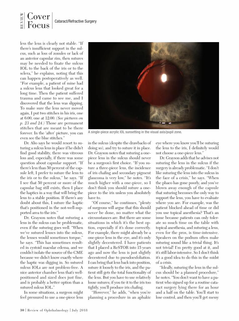
30 | Review of Ophthalmology | July 2018
less the lens is clearly not stable. “If there’s insuffi cient support in the sul-cus, such as loss of zonules or lack of an anterior capsular rim, then sutures may be needed to fixate the sulcus IOL to the back of the iris or to the sclera,” he explains, noting that this can happen postoperatively as well. “For example, a patient of mine had a sulcus lens that looked great for a long time. Then the patient suffered trauma and came to see me, and I discovered that the lens was slipping. To make sure the lens never moved again, I put two stitches in his iris, one at 6:00, one at 12:00. (See pictures on p. 23 and 24.) Those are permanent stitches that are meant to be there forever. In the ‘after’ picture, you can even see the blue stitches.”
Dr. Alio says he would resort to su-turing a sulcus lens in place if he didn’t fi nd good stability, there was vitreous loss and, especially, if there was some question about capsular support. “If there’s less than 50 percent of the cap-sule left, I prefer to suture the lens to the iris or to the sulcus,” he says. “If I see that 50 percent or more of the capsular bag still exists, then I place the haptics in a way that will bring the lens to a stable position. If there’s any doubt about this, I suture the haptic that’s positioned in the not-well-sup-ported area to the iris.”
Dr. Grayson notes that suturing a lens in the sulcus can be problematic, even if the suturing goes well. “When we’ve sutured lenses into the sulcus, the lenses would sometimes torque,” he says. “This has sometimes result-ed in cystoid macular edema, and we couldn’t isolate the source of the CME because we didn’t know exactly where the haptic was digging in. So sutured sulcus IOLs are not problem-free. A nice anterior chamber lens that’s well-positioned and sized does just fine, and is probably a better option than a sutured sulcus IOL.”
In some situations, a surgeon might feel pressured to use a one-piece lens
in the sulcus (despite the drawbacks of doing so), and try to suture it in place. Dr. Grayson notes that suturing a one-piece lens in the sulcus should never be a surgeon’s fi rst choice. “If you su-ture a three-piece lens, the incidence of iris chafi ng and secondary pigment glaucoma is very low,” he notes. “It’s much higher with a one-piece, so I don’t think you should suture a one-piece to the iris unless you absolutely have to.
“Of course,” he continues, “plenty of surgeons will argue that this should never be done, no matter what the circumstances are. But there are some situations in which it’s the best op-tion, especially if it’s done correctly. For example, there might already be a one-piece lens in the eye, and it’s only slightly decentered. I have patients that I placed a ReSTOR into 15 years ago and now the lens is just slightly decentered due to pseudoexfoliation. I can bring that lens back into position, suture it loosely to the iris, and the pa-tient still gets the total functionality of the lens. But you have to use relatively loose sutures; if you tie it to the iris too tightly, you’ll produce iris chafi ng.
“However,” he adds, “when you’re planning a procedure in an aphakic
eye where you know you’ll be suturing the lens to the iris, I defi nitely would not choose a one-piece lens.”
Dr. Grayson adds that he advises not suturing the lens in the sulcus if the surgery is already problematic. “I don’t like suturing the lens into the sulcus in the face of a crisis,” he says. “When the phaco has gone poorly, and you’ve blown away enough of the capsule that suturing becomes the only way to support the lens, you have to evaluate where you are. For example, was the patient blocked ahead of time or did you use topical anesthesia? That’s an issue because patients can only toler-ate so much time on the table with topical anesthesia, and suturing a lens, even for the pros, is time-intensive. Speakers on the podium often make suturing sound like a trivial thing. It’s not trivial! I’m pretty good at it, and it’s still labor-intensive. So I don’t think it’s a good idea to do this in the midst of a crisis.
“Ideally, suturing the lens in the sul-cus should be a planned procedure,” he notes. “You don’t want to have a pa-tient who signed up for a routine cata-ract surgery lying there for an hour and a half on the table. You’ll start to lose control, and then you’ll get messy
CoverFocus R
EV
IEW
A single-piece acrylic IOL sunsetting in the visual axis/pupil zone.
Cataract/Refractive Surgery
022_rp0718_f1.indd 30022_rp0718_f1.indd 30 6/22/18 4:14 PM6/22/18 4:14 PM

NuLids
™
transformational
dry eye therapy
Transforms your practice
The NuLids System helps reduce patient out-of-pocket expenses while creating an additional revenue stream for the practice – all without impacting overhead costs or disrupting other in-office treatments.
A multi-center study1 showed 95% were satisfied or very satisfied with the treatment.
For more information visit www.NuSightMedical.com or call us at 833-468-5437
1 Schanzlin, Olkowski, Coble, Gross. NuLids II Study, April 2018
Doctor prescribed, at-home dry eye relief
results. If you can’t put a lens in the sulcus without suturing it, then your best options are to either leave the patient apha-kic for the time being, or put in an anterior chamber lens.
“Nobody should be afraid of just leaving the eye without a lens,” he adds. “You can come back another day and put in a sulcus- or iris-fi xated IOL. If the surgery up to that point has been diffi cult, leaving the eye alone and coming back another day gives the cornea a chance to clear. If it doesn’t clear and the patient ends up needing a DSEK, at least that can be a nice, planned procedure with a sutured lens. Surgeons shouldn’t feel this intense pressure to implant a lens in a crisis situation. Those are the cases that end up doing the worst.
“And remember,” he adds, “there’s nothing wrong with placing a good anterior chamber lens, as long as the lens fi ts and it’s well-positioned.”
Adjusting the Lens Power
Moving the lens forward inside the eye—and possibly changing the type of lens—may necessitate adjusting the re-fractive power of the lens. Surgeons have different preferred approaches to deciding how much refractive adjustment is necessary.
“If you can’t use lens capture, you have to go a solid half-diopter lower,” says Dr. Grayson. “You also have to take into consideration the A-constant, because most of the one-piece lenses are aspheric and have a relatively high A-constant, in contrast to most of the three-piece lenses, which are plano convex, giving them a lower A-constant. That means those lenses are already a half-diopter lower.
“For example,” he continues, “if you planned to use a one-piece 20-D lens, but you’re going to the sulcus and switching to a three-piece, you’d probably go a half-diopter lower to compensate for the different position, but you’d have to go another half-diopter down because of the A-constant. In ef-fect, you’re going down a full diopter. If you’re able to do an IOL capture, then you could probably just go down a half-diopter and use the 19.5-D three-piece. You have to make that calculation in your head on the fl y, unless you write it out ahead of time, which would be ideal.” (Dr. Grayson adds that if you can capture the lens in the anterior capsule, you may not need to change the refractive power at all.)
Dr. Devgan has created a simple system that tells him how much he’ll need to adjust the lens power if he fi nds that the lens needs to be placed in the sulcus instead of the bag; he calls it the rule of nines. “The fi rst step is to make sure your A-constant is accounted for,” he says. “All lenses are slightly different, even if they’re marked the same power. A 20-D lens, for example, may not be the same as a 20-D lens from a different manufacturer. The A-constant allows us to com-pensate for factors such as lens geometry, the lens material and how much posterior vault the lens has. It’s a number you
022_rp0718_f1.indd 31022_rp0718_f1.indd 31 6/22/18 4:14 PM6/22/18 4:14 PM
32 | Review of Ophthalmology | July 2018
need when you do your lens-power calculation, and it’s specifi c to every type of lens.
“For example, if a surgeon typically uses a single-piece acrylic IOL where the A-constant is 119.2 when the lens is in the bag, he or she must begin by calculating the power for the three-piece IOL that will be placed in the sulcus, where the A-constant is 118.7 when the lens is in the bag,” he says. “That means dropping the IOL power by 0.5 diopters, which is the difference between 119.2 and 118.7. Of course, if you just use the same three-piece lens for every patient, then this is a moot point.
“Once that’s accounted for, you can use the rule of nines,” he continues. “That will tell you how much you need to lower the lens power. In a nutshell, if the lens power is between 0 and 9 D, you don’t need to change the power when you move it forward. If it’s between 9.5 and 18 D, you subtract 0.5 D from the power. If it’s between 18.5 and 27 D, you subtract 1 D. If it’s above 27 D, subtract 1.5 D. And so forth.
“For example,” he continues, “let’s say a patient is so myopic that the lens power is zero. That means the lens is plano; it has no power at all. In this case, it doesn’t matter where you put the lens. This is basically true as long as the lens power is below 9 D; moving it doesn’t cause much of a change in its power. On the other hand, a 30-D lens is powerful; moving it just a little will change the power a lot. So you have to subtract 1.5 D from the power if you move it forward from the bag to the sulcus.”
Dr. Devgan notes that the rule of nines is an estimation. “Of course you can calculate the exact amount of change for the lens you’re dealing with if you want to, but it comes out to be the same, because lenses only come in half-diopter steps,” he says. “A 12-D and 18-D lens are not the same, but since the lenses only come
in half steps, the amount of change needed when you move it forward into the sulcus is about a half-diopter less for both lenses.”
Strategies for Success
Surgeons offer these general sug-gestions to help ensure that you won’t be caught off-guard when the need to place a lens in the sulcus suddenly arises:
• Have an appropriate lens avail-able. Dr. Oetting says he believes you shouldn’t start surgery unless you have a lens for the sulcus at hand. “This is one situation that surgeons are like-ly to encounter from time to time,” he says. “You’re working at a surgery center and all they have for you to work with is a single-piece lens. If you run into trouble, it forces you to think about placing a single-piece lens in the sulcus—a bad idea—or putting the single-piece lens into a compromised bag. Either way you’re setting yourself up for potential trouble.
“I think no matter where you’re op-erating, you have to have an anterior chamber IOL and a sulcus lens avail-able,” he continues. “Is it likely that you’ll need to place a lens in the sul-cus? No. Could you stop and bring the patient to another OR, or have them come back another day to put a lens in? Yes. But to me it seems like the better thing to do is be prepared and have a consignment of sulcus lenses and a consignment of anterior cham-ber lenses wherever you operate.”
• Practice using the three-piece injector that goes with your pre-ferred sulcus IOL ahead of time. “All of the three-piece injectors are slightly different,” Dr. Oetting points out. “So, if you’re going to use a par-ticular sulcus lens, the time to practice using that injector is not when you’re in the middle of a horrible case. Prac-tice using that injector when the seas are calm.
“I suggest placing your choice of
three-piece lens in a few standard cas-es,” he continues. “That will help you get used to putting it in. You can go over this with your company rep a few times. Another option is to practice on a Phillips artifi cial eye; you can prac-tice making an anterior capsulotomy and placing lenses in the sulcus or the bag. The point is to be accustomed to using the sulcus lens you’re going to choose. When you’re facing a diffi cult case, you should already know how to do what you need to do.”
Dr. Grayson points out that many surgeons today have become accus-tomed to using one-piece lenses and their injectors. “When they suddenly have to use a three-piece injector, it’s awkward,” he says. “For one thing, it’s loaded differently, so if your tech-nician isn’t familiar with loading a three-piece, the haptics could end up damaged. Second, when you’re intro-ducing it into the eye, the stiff haptics have a different feel. You have to make sure the haptics don’t scrape against the cornea, and you need to make sure you don’t introduce the lens with the haptic directly into the posterior cavity where it could potentially sink.
“Injecting a three-piece lens re-quires a certain amount of fi nesse,” he continues. “Any surgeon who’s been in practice for 25 years knows about this, because we didn’t have one-piece lenses for the longest time. But the younger surgeons are not accustomed to it. I see this when I do resident grand rounds. When they have to put in a three-piece, it’s a project because they’re not as familiar with it. Ideally, residents should be learning to im-plant both types of lenses.”
• Tell the patient what hap-pened. “The reason this is a good idea is that eventually the different lens location will be noticed,” says Dr. Oet-ting. “Telling the patient right away is much better than saying nothing and having them be surprised later. I’ve had many patients come to me saying their doctor never told them that there
CoverFocus R
EV
IEW
Cataract/Refractive Surgery
022_rp0718_f1.indd 32022_rp0718_f1.indd 32 6/22/18 4:14 PM6/22/18 4:14 PM
VisiPlug® –a visibly better plug
to treat dry eye!
Lacrimedics’ VisiPlug is not like “other” punctum plugs. We have no reported “pop-outs” since VisiPlug’s introduction in 2003… and it’s distributed in over forty countries!
FDA approved for the treatment of the Dry Eye components of varying Ocular Surface Diseases (OSD), after surgery to prevent complications due to Dry Eye Disease, and to enhance the efficacy of topical medications.
1Dramatization. Not a real patient. ©2017 Lacrimedics, Inc.
(800) 367-8327E-mail: [email protected]
www.lacrimedics.com
VisiPlugis the only one for me… No pop-out!
was an issue when the lens was implanted, and they’re very disappointed in the surgeon. For that reason, I fi nd it’s better to go through the discomfort of telling the patient that the surgery wasn’t perfect.
“Every patient understands that there can be issues dur-ing surgery,” he continues. “They just expect your expertise to carry them through so they don’t get into trouble. So, I tell the patient that we had a problem with the posterior capsule (‘It’s thinner than a red blood cell’) so I placed a dif-ferent kind of lens. I tell these patients that they’ll likely do very well, but that I might watch them a little more closely because the IOL is in a different position inside the eye. I assure them they’ll have good vision with the new lens, and I reassure them again at the one-month visit.”
• Don’t co-manage these patients. “I don’t think this is a good situation in which to turn the patient over to somebody else,” says Dr. Oetting. “You should personally follow these patients closely. Make sure there’s no unusual infl ammation and no increased intraocular pressure. Make sure the lens is staying centered. It’s not that big a deal, but it’s not a standard situation, so you should watch it a little more closely.”
• Take advantage of educational resources. Dr. Devgan says he recently launched a new website called cataractcoach.com. “It’s totally noncommercial,” he says. “It simply coaches the viewer through diffi cult cataract surgery cases. I’ve been putting up new cases every day, recorded in HD with a full voiceover. One of them, for example, discuss-es dealing with a ruptured posterior capsule. It shows how to get the lens into the sulcus and make sure it’s secure. Another shows how to capture the optic of a three-piece lens in the sulcus back through the capsulorhexis.” Dr. Devgan says he’s planning to post a couple of new surgical videos every week.
• Be prepared—this will happen sooner or later. “An unexpected problem requiring IOL placement in the sulcus is the kind of thing that gets a surgeon’s heart racing,” notes Dr. Oetting. “Some surgical challenges happen with advance warning, so you can brush up on a technique before you use it, but sulcus IOL placement often comes up right in the middle of a routine case. This is something you simply have to be prepared for ahead of time. You may go for two years without encountering it, but then it happens.”
Drs. Oetting, Grayson and Devgan report no fi nancial ties to any product discussed. Dr. Alio is a consultant for Akko-lens, Hanita Lenses, Oculentis and Zeiss.
1. Kemp PS, Oetting TA. Stability and safety of MA50 intraocular lens placed in the sulcus. Eye (Lond) 2015;29:11:1438-41.2. Chang DF, Masket S, Miller KM, et al. Complications of sulcus placement of single-piece acrylic intraocular lenses: Recommendations for backup IOL implantation following posterior capsule rupture. ASCRS Cataract Clinical Committee. J Cataract Refract Surg 2009;35:8:1445-58.
022_rp0718_f1.indd 33022_rp0718_f1.indd 33 6/22/18 4:14 PM6/22/18 4:14 PM

34 | Review of Ophthalmology | July 2018
RE
VIE
W
This article has no commercial sponsorship.
Cover Focus Cataract/Refractive Surgery
Refractive Surgery in Pediatric PatientsAsha Balakrishnan, MD, Encino, Calif.
When compared to adult re-fractive surgery, the fi eld of pediatric refractive surgery
has remained relatively limited, with slower growth. Indeed, refractive sur-gery has a limited scope in the pedi-atric population; however, in certain cases, it can be a crucial treatment for high refractive error and anisometro-pia. Addressing these conditions in young children is critical to healthy visual maturation. High refractive er-rors that go uncorrected are almost certain to have a signifi cant negative impact on a child’s intellectual and so-cial development. When conservative methods such as glasses and contact lenses fail, surgical correction of re-fractive error is the most reasonable solution. In this article, I’ll describe the best applications for refractive sur-gery in this unique population.
Why Refractive Surgery?
Glasses, contact lenses and occlu-sion therapy can fail in children for a number of reasons. Developmental delay in conditions such as autism, ce-rebral palsy and Down syndrome can make patient compliance with these methods of correction extremely diffi -cult. Many of these children don’t like the sensation of glasses touching their face. In cases of high ametropia, spec-
tacles can be diffi cult to wear due to prismatic aberration, reduced fi eld of vision through the lenses and cosmetic appearance. Contact lens placement is challenging in all children, whether developmentally appropriate or not. Even when children seemingly com-ply with glasses, there’s a good chance that they may be favoring their good eye without their parents’ knowledge. Finally, the effi cacy of occlusion ther-apy is highly dependent on the pa-tient’s and family’s compliance with the regimen. Regardless of the treatment modality, in all cases of conservative management there’s a small window of time for optimal visual development, so consistency and adherence to thera-py is essential for success.
The main indications for refractive surgery in the pediatric population are anisometropic amblyopia and bilat-eral high ametropia. There have also been some studies of the role of re-fractive surgery for high accommoda-tive esotropia. Elimination or drastic reduction of anisometropia equalizes the visual input of the two eyes and prevents favoring one eye over the other. Similarly, correction of the high refractive error improves the quality of visual input to both eyes. As stated previously, spectacles for high myo-pia and hyperopia can reduce image quality, which can compromise visual
When and how to
perform refractive
surgery in children.
034_rp0718_f2.indd 34034_rp0718_f2.indd 34 6/22/18 4:17 PM6/22/18 4:17 PM
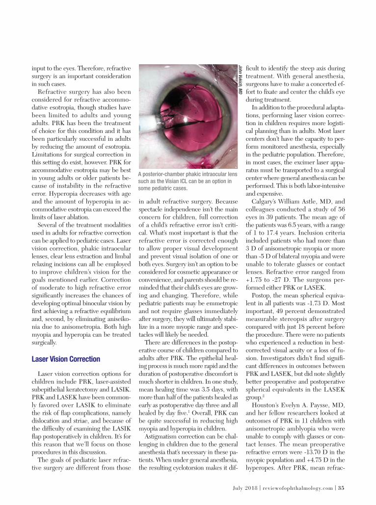
July 2018 | reviewofophthalmology.com | 35
input to the eyes. Therefore, refractive surgery is an important consideration in such cases.
Refractive surgery has also been considered for refractive accommo-dative esotropia, though studies have been limited to adults and young adults. PRK has been the treatment of choice for this condition and it has been particularly successful in adults by reducing the amount of esotropia. Limitations for surgical correction in this setting do exist, however. PRK for accommodative esotropia may be best in young adults or older patients be-cause of instability in the refractive error. Hyperopia decreases with age and the amount of hyperopia in ac-commodative esotropia can exceed the limits of laser ablation.
Several of the treatment modalities used in adults for refractive correction can be applied to pediatric cases. Laser vision correction, phakic intraocular lenses, clear lens extraction and limbal relaxing incisions can all be employed to improve children’s vision for the goals mentioned earlier. Correction of moderate to high refractive error signifi cantly increases the chances of developing optimal binocular vision by fi rst achieving a refractive equilibrium and, second, by eliminating aniseiko-nia due to anisometropia. Both high myopia and hyperopia can be treated surgically.
Laser Vision Correction
Laser vision correction options for children include PRK, laser-assisted subepithelial keratectomy and LASIK. PRK and LASEK have been common-ly favored over LASIK to eliminate the risk of fl ap complications, namely dislocation and striae, and because of the diffi culty of examining the LASIK fl ap postoperatively in children. It’s for this reason that we’ll focus on those procedures in this discussion.
The goals of pediatric laser refrac-tive surgery are different from those
in adult refractive surgery. Because spectacle independence isn’t the main concern for children, full correction of a child’s refractive error isn’t criti-cal. What’s most important is that the refractive error is corrected enough to allow proper visual development and prevent visual isolation of one or both eyes. Surgery isn’t an option to be considered for cosmetic appearance or convenience, and parents should be re-minded that their child’s eyes are grow-ing and changing. Therefore, while pediatric patients may be emmetropic and not require glasses immediately after surgery, they will ultimately stabi-lize in a more myopic range and spec-tacles will likely be needed.
There are differences in the postop-erative course of children compared to adults after PRK. The epithelial heal-ing process is much more rapid and the duration of postoperative discomfort is much shorter in children. In one study, mean healing time was 3.5 days, with more than half of the patients healed as early as postoperative day three and all healed by day fi ve.1 Overall, PRK can be quite successful in reducing high myopia and hyperopia in children.
Astigmatism correction can be chal-lenging in children due to the general anesthesia that’s necessary in these pa-tients. When under general anesthesia, the resulting cyclotorsion makes it dif-
fi cult to identify the steep axis during treatment. With general anesthesia, surgeons have to make a concerted ef-fort to fi xate and center the child’s eye during treatment.
In addition to the procedural adapta-tions, performing laser vision correc-tion in children requires more logisti-cal planning than in adults. Most laser centers don’t have the capacity to per-form monitored anesthesia, especially in the pediatric population. Therefore, in most cases, the excimer laser appa-ratus must be transported to a surgical center where general anesthesia can be performed. This is both labor-intensive and expensive.
Calgary’s William Astle, MD, and colleagues conducted a study of 56 eyes in 39 patients. The mean age of the patients was 6.5 years, with a range of 1 to 17.4 years. Inclusion criteria included patients who had more than 3 D of anisometropic myopia or more than -5 D of bilateral myopia and were unable to tolerate glasses or contact lenses. Refractive error ranged from +1.75 to -27 D. The surgeons per-formed either PRK or LASEK.
Postop, the mean spherical equiva-lent in all patients was -1.73 D. Most important, 49 percent demonstrated measurable stereopsis after surgery compared with just 18 percent before the procedure. There were no patients who experienced a reduction in best-corrected visual acuity or a loss of fu-sion. Investigators didn’t fi nd signifi -cant differences in outcomes between PRK and LASEK, but did note slightly better preoperative and postoperative spherical equivalents in the LASEK group.2
Houston’s Evelyn A. Paysse, MD, and her fellow researchers looked at outcomes of PRK in 11 children with anisometropic amblyopia who were unable to comply with glasses or con-tact lenses. The mean preoperative refractive errors were -13.70 D in the myopic population and +4.75 D in the hyperopes. After PRK, mean refrac-
A posterior-chamber phakic intraocular lens
such as the Visian ICL can be an option in
some pediatric cases.
Joh
n V
ukich
, MD
034_rp0718_f2.indd 35034_rp0718_f2.indd 35 6/22/18 4:17 PM6/22/18 4:17 PM

OCTOBER 12-13, 2018
CSE GCSE GLAUCOMALAUCOMA F FELLOWSELLOWS
SAVE THE DATE
Fort Worth, Texas
GLAUCOMA FELLOWSCONTINUING SPECIALIZED EDUCATION
For More Information & to Register:
www.revophth.com/2018cseglaucomaEmail: [email protected] or call Denette Holmes 866-627-0714
Dear Fellowship Program Director and Coordinator,
We would like to invite you to review the upcoming 2018 Glaucoma Fellowship Program and Wet Lab in Fort Worth at the
Renaissance Worthington hotel. The program offers a unique educational opportunity for fellows by providing the chance to meet
and exchange ideas with some of the most respected thought leaders in glaucoma. The Glaucoma Fellows Program and Wet
Lab is designed to provide your fellows with a state-of-the-art didactic and wet lab experience. The program also serves as an
opportunity for your fellows to network with fellows from other programs.
After reviewing the material, it is our hope that you will select and encourage your fellows to attend this educational activity which
is CME accredited to ensure fair balance.
Sincerely,
Postgraduate Healthcare Education
Endorsed by
Review of Ophthalmology®
Supported by an unrestricted independent medical educational grant from
Alcon
Jointly provided by
Participants in this course must be enrolled in a glaucoma fellowship program at the time of the course.There is no registration fee for these activities. Air, ground transportation in Dallas Fort Worth Airport, hotel accommodations and
modest meals will be provided through an educational scholarship for qualifi ed participants.
Satisfactory Completion - Learners must pass a post-test and complete an evaluation form to receive a certifi cate of completion. Your chosen sessions must be attended in their entirety. Partial credit of individual sessions is not available. If you are seeking continuing education credit for a specialty not listed below, it is your responsibility to contact your licensing/certifi cation board to
determine course eligibility for your licensing/certifi cation requirement.
Physicians - In support of improving patient care, this activity has been planned and implemented by Amedco LLC and Postgraduate Healthcare Education. Amedco LLC is jointly accredited by the Accreditation Council for Continuing Medical Education (ACCME), the Accreditation Council for Pharmacy Education (ACPE), and the American Nurses Credentialing Center (ANCC), to provide continuing education for the healthcare team
Credit Designation Statement - Amedco designates this live activity for AMA PRA Category 1 CreditsTM. Physicians should claim only the credit commensurate with the extent of their participation in the activity.
JOINTLY ACCREDITED PROVIDERTM
INTERPROFESSIONAL CONTINUING EDUCATION
Course Director:Kuldev Singh, MD
Stanford, CA
Wet Lab Directors:
Douglas J. Rhee, MDCleveland, OH
Ronald Fellman, MDDallas, TX
Faculty:
Lee Alward, MD
Donald Budenz, MD
Joseph Caprioli, MD
Steven Gedde, MD
Dale Heuer, MD
Michele Lim, MD
Shan Lin, MD
Marlene Moster, MD
Louis Pasquale, MD
Pradeep Ramulu, MD
Joel Schuman, MD
Arthur Sit, MD
George Spaeth, MD
Josh Stein, MD

July 2018 | reviewofophthalmology.com | 37
CoverFocus R
EV
IEW
Cataract/Refractive Surgery
tive errors improved to -3.55 D in the myopes and +1.41 D in the hyperopes. Additionally, cycloplegic refraction confi rmed that the two eyes of each patient were within 3 D of each other postoperatively. Uncorrected and best-corrected visual acuity improved by at least two lines in two-thirds of the patients.3
The primary risk after surface abla-tion in children is the development of corneal haze and ectasia. Noncompli-ance with a longer duration of postop-erative steroid use is the principal risk factor for the development of corneal haze. In the Astle study, patients who underwent LASEK instead of PRK didn’t develop haze. The investigators favored LASEK over PRK both for this reason and because LASEK has less postoperative discomfort.2
Parents of children who are candi-dates for laser refractive surgery also should be counseled regarding the risk of keratoconus. Most pediatric patients won’t exhibit any subclinical or overt signs of keratoconus at such a young age. Therefore, at-risk candidates can’t be identifi ed in the same way that adult refractive patients can. It’s important for parents to understand this risk and the possibility of accelerating or uncov-ering a corneal ectatic disorder down the road.
While there are drawbacks to la-ser refractive surgery in children, surface ablation remains a strong op-tion for correcting refractive error in this young population. Even though LASEK has dropped in popularity for adult patients, it can still have a solid role in children given the absence of flap complications, enhanced post-operative comfort and healing, and a lower risk of haze formation.
Phakic IOL Implantation
Phakic IOLs can be considered for children with high refractive errors and amblyopia. There are three variet-ies of these types of lenses: iris-fi xated;
sulcus-supported; and angle-support-ed. Both iris-fixated and posterior chamber lenses have shown promise in correcting refractive error, provid-ing binocular fusion and preventing amblyopia. Iris-fi xated lenses, such as the Artisan/Verisyse, have been con-sidered the lens of choice based on several studies. The fi rst iris claw lens was implanted in a pediatric patient in 1997.
• Phakic IOL advantages. Phakic IOLs offer several benefi ts over other refractive surgery alternatives. They can correct a wider range of refrac-tive error than laser surgery and their outcomes are more predictable. Also, there’s no risk of corneal haze, corneal thinning or future corneal ectasia with these IOLs. Phakic lenses are much less risky than refractive lens exchange, which can lead to glaucoma and reti-nal detachment. Posterior capsule fi brosis is not a concern with phakic intraocular lenses, and postop visual recovery and rehabilitation is much faster. Additionally, phakic IOLs can be removed relatively easily compared to IOLs placed after clear lens extrac-tion. The logistical obstacles that come with pediatric laser refractive surgery are also avoided.
• Phakic IOL disadvantages. The disadvantages of phakic intraocular lenses include risks of endothelial cell loss, IOL dislocation, pigment disper-sion, cataract formation and shallow-ing of the anterior chamber. Poste-rior chamber phakic lenses can cause anterior subcapsular cataracts if they are not adequately spaced from the natural lens. Complications associated with phakic intraocular lenses in adults have not been observed in children; however, this is likely secondary to a smaller number of cases performed and the need for longer-term follow-up. A caveat with iris-fi xated lenses is that they shouldn’t be placed in ante-rior chambers that are smaller than 3.2 mm, to permit safe insertion and minimize the risk of long-term endo-
thelial cell loss.4 • Data on phakic lenses. In 2011,
Jorge Alio, MD, of Alicante, Spain, and his colleagues conducted a small retro-spective study of 10 eyes in 10 children over fi ve years. Nine had placement of an iris-fi xated pIOL and one had a posterior chamber pIOL. At the time of implantation, patients ranged from 5 to 15 years old. All the patients with iris-fi xated pIOLs had visual improve-ment of more than three lines of log-MAR visual acuity. The patient with a posterior chamber pIOL had one line of improvement. Endothelial cell count in all patients remained over 2,000 cells/cm2.5
Lawrence Tychsen, MD, of St. Louis Children’s Hospital at the Washington University School of Med-icine, studied 20 eyes after anterior chamber phakic IOL implantation to correct high myopia or hyperopia in the setting of neurobehavioral dis-orders. In the study, myopia ranged from -10 D to -22.75 D, and hyperopia ranged from +10.25 to +10.75 D. The patients’ ages ran from 4 to 17 years. Eighty-six percent of eyes were cor-rected to within 1 D of emmetropia. The remaining eyes were within 2 D of plano. Mean uncorrected visual acu-ity improved from 20/3,400 to 20/57. The residual visual acuity defi cits were secondary to comorbid ocular condi-tions, including amblyopia, nystagmus, albinism and regressed retinopathy of prematurity.6
Andrea Ryan and her co-workers at Children’s University Hospital in Dub-lin, Ireland, looked at the results of 11 eyes in six children who couldn’t toler-ate contact lens or spectacle correction due to neurobehavioral disorders and who underwent implantation of the foldable Artifl ex iris-fi xated pIOL (not available in the United States). Refrac-tive indications included high bilateral myopia, anisometropia and myopic astigmatism. Mean spherical equiva-lent improved from -14.6 D to -2.4 D. Mean logMAR vision improved from
034_rp0718_f2.indd 37034_rp0718_f2.indd 37 6/22/18 4:17 PM6/22/18 4:17 PM
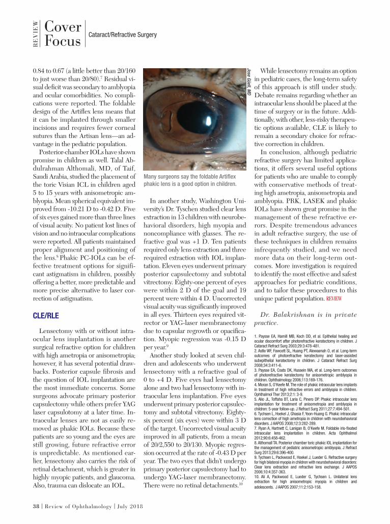
RE
VIE
W
38 | Review of Ophthalmology | July 2018
CoverFocus Cataract/Refractive Surgery
0.84 to 0.67 (a little better than 20/160 to just worse than 20/80).7 Residual vi-sual defi cit was secondary to amblyopia and ocular comorbidities. No compli-cations were reported. The foldable design of the Artifl ex lens means that it can be implanted through smaller incisions and requires fewer corneal sutures than the Artisan lens—an ad-vantage in the pediatric population.
Posterior-chamber IOLs have shown promise in children as well. Talal Ab-dulrahman Althomali, MD, of Taif, Saudi Arabia, studied the placement of the toric Visian ICL in children aged 5 to 15 years with anisometropic am-blyopia. Mean spherical equivalent im-proved from -10.21 D to -0.42 D. Five of six eyes gained more than three lines of visual acuity. No patient lost lines of vision and no intraocular complications were reported. All patients maintained proper alignment and positioning of the lens.8 Phakic PC-IOLs can be ef-fective treatment options for signifi-cant astigmatism in children, possibly offering a better, more predictable and more precise alternative to laser cor-rection of astigmatism.
CLE/RLE
Lensectomy with or without intra-ocular lens implantation is another surgical refractive option for children with high ametropia or anisometropia; however, it has several potential draw-backs. Posterior capsule fibrosis and the question of IOL implantation are the most immediate concerns. Some surgeons advocate primary posterior capsulectomy while others prefer YAG laser capsulotomy at a later time. In-traocular lenses are not as easily re-moved as phakic IOLs. Because these patients are so young and the eyes are still growing, future refractive error is unpredictable. As mentioned ear-lier, lensectomy also carries the risk of retinal detachment, which is greater in highly myopic patients, and glaucoma. Also, trauma can dislocate an IOL.
In another study, Washington Uni-versity’s Dr. Tyschen studied clear lens extraction in 13 children with neurobe-havioral disorders, high myopia and noncompliance with glasses. The re-fractive goal was +1 D. Ten patients required only lens extraction and three required extraction with IOL implan-tation. Eleven eyes underwent primary posterior capsulectomy and subtotal vitrectomy. Eighty-one percent of eyes were within 2 D of the goal and 19 percent were within 4 D. Uncorrected visual acuity was signifi cantly improved in all eyes. Thirteen eyes required vit-rector or YAG-laser membranectomy due to capsular regrowth or opacifi ca-tion. Myopic regression was -0.15 D per year.9
Another study looked at seven chil-dren and adolescents who underwent lensectomy with a refractive goal of 0 to +4 D. Five eyes had lensectomy alone and two had lensectomy with in-traocular lens implantation. Five eyes underwent primary posterior capsulec-tomy and subtotal vitrectomy. Eighty-six percent (six eyes) were within 3 D of the target. Uncorrected visual acuity improved in all patients, from a mean of 20/2,550 to 20/130. Myopic regres-sion occurred at the rate of -0.43 D per year. The two eyes that didn’t undergo primary posterior capsulectomy had to undergo YAG-laser membranectomy. There were no retinal detachments.10
While lensectomy remains an option in pediatric cases, the long-term safety of this approach is still under study. Debate remains regarding whether an intraocular lens should be placed at the time of surgery or in the future. Addi-tionally, with other, less-risky therapeu-tic options available, CLE is likely to remain a secondary choice for refrac-tive correction in children.
In conclusion, although pediatric refractive surgery has limited applica-tions, it offers several useful options for patients who are unable to comply with conservative methods of treat-ing high ametropia, anisometropia and amblyopia. PRK, LASEK and phakic IOLs have shown great promise in the management of these refractive er-rors. Despite tremendous advances in adult refractive surgery, the use of these techniques in children remains infrequently studied, and we need more data on their long-term out-comes. More investigation is required to identify the most effective and safest approaches for pediatric conditions, and to tailor these procedures to this unique patient population.
Dr. Balakrishnan is in private practice.
1. Paysse EA, Hamill MB, Koch DD, et al. Epithelial healing and ocular discomfort after photorefractive keratectomy in children. J Cataract Refract Surg 2003;29:3:478-481.2. Astle WF, Fawcett SL, Huang PT, Alewaenah O, et al. Long-term outcomes of photorefractive keratectomy and laser-assisted subepithelial keratectomy in children. J Cataract Refract Surg 2008;34:3:411-6.3. Paysse EA, Coats DK, Hussein MA, et al. Long-term outcomes of photorefractive keratectomy for anisometropic amblyopia in children. Ophthalmology 2006;113:169-176.4. Moran S, O’Keefe M. The role of phakic intraocular lens implants in treatment of high refractive errors and amblyopia in children. Ophthalmol Ther 2013;2:1: 3-9. 5. Alio JL, Toffaha BT, Laria C, Pinero DP. Phakic intraocular lens implantation for treatment of anisometropia and amblyopia in children: 5-year follow-up. J Refract Surg 2011;27:7:494-501.6. Tychsen L, Hoekel J, Ghasia F, Yoon-Huang G. Phakic intraocular lens correction of high ametropia in children with neurobehavioral disorders. J AAPOS 2008;12:3:282-289.7. Ryan A, Hartnett C, Lanigan B, O’Keefe M. Foldable iris-fi xated intraocular lens implantation in children. Acta Ophthalmol 2012;90:6:458-462.8. Althomall TA. Posterior chamber toric phakic IOL implantation for the management of pediatric anisometropic amblyopia. J Refract Surg 2013;29:6:396-400.9. Tychsen L, Packwood E, Hoekel J, Lueder G. Refractive surgery for high bilateral myopia in children with neurobehavioral disorders: Clear lens extraction and refractive lens exchange. J AAPOS 2006;10:4:357-363.10. Ali A, Packwood E, Lueder G, Tychsen L. Unilateral lens extraction for high anisometropic myopia in children and adolescents. J AAPOS 2007;11:2:153-158.
Many surgeons say the foldable Artifl ex
phakic lens is a good option in children.
Jose
Gü
ell, M
D
034_rp0718_f2.indd 38034_rp0718_f2.indd 38 6/22/18 4:18 PM6/22/18 4:18 PM

Left your Review of Ophthalmology
magazine at the offi ce? No problem!
Read Review on the go
from any mobile device!
Simply go to www.reviewofophthalmology.com and click on the digimag link to get your current issue.
2015 Digimag hous ad_RP.indd 12015 Digimag hous ad_RP.indd 1 6/21/18 3:34 PM6/21/18 3:34 PM

40 | Review of Ophthalmology | July 2018
Cover Focus RE
VIE
W
This article has no commercial sponsorship.
Cataract/Refractive Surgery
Tools for Femto-free CapsulotomyKristine Brennan, Senior Associate Editor
A perfectly round, continuous curvilinear capsulorhexis sets the tone for the rest of a cata-
ract surgery case, and is arguably the hardest part to learn. The feel of the capsule during cutting has been lik-ened to easily-torn cellophane,1 so it’s understandably hard to consistently create a perfectly round rhexis that will cover the lens for 360 degrees. It’s well worthwhile though, because such a circle will support your IOL and keep it well positioned. The ad-vent of femtosecond-created capsu-lotomy contributed to the creation of predictably sized, quick and reproduc-ible capsulotomies, but at a signifi cant extra cost not reimbursed by insurers. Here, surgeons discuss two devices currently available in the United States and two emerging technologies that aid in creating perfect capsulotomies without a femtosecond laser.
Zepto
Vance Thompson, MD, of Thomp-son Vision in Sioux Falls, South Dakota, remains a femtosecond la-ser enthusiast, but finds that Zepto (Mynosys; Fremont, California) is a valued addition to his cataract-surgery armamentarium. “It’s become main-stream in my practice; it’s not just a research tool anymore,” he says. “I’m
a big fan of femto. I have a femto. But for surgeons who may not want to in-vest in femto, but like the idea of an automated capsulotomy, I think Zepto is a great answer.”
The Zepto, FDA approved in 2017 and commercially available, consists of a small console that provides energy to a disposable handpiece equipped with a fl exible nitinol ring. The Zepto ring folds to go through a small incision, typically 2.4 mm, then springs back into circular shape inside the eye. To ensure 360-degree contact with the anterior capsule, the Zepto employs a clear silicone suction cup over the membrane to gently draw it towards the ring. A quick 0.4-msec pulse of thermoelectric energy instantly cre-ates a round capsulotomy about 5.2 mm in diameter. The surgeon then retracts the cutting element and with-draws it through the wound and the surgery proceeds.
Dr. Thompson likes the Zepto for both premium IOLs and diffi cult cas-es, such as intumescent milky cata-racts. “In people with corneal scar-ring, whether it’s from RK or previous trauma, it can be diffi cult to use femto, and the Zepto can be very helpful. In small pupils you can’t do femto and it’s a challenge to do manual, but you can fi t the Zepto technology inside a small pupil and get a nice capsuloto-
New tech that
may improve your
capsulotomies
without breaking
the bank.
040_rp0718_f3.indd 40040_rp0718_f3.indd 40 6/22/18 4:32 PM6/22/18 4:32 PM
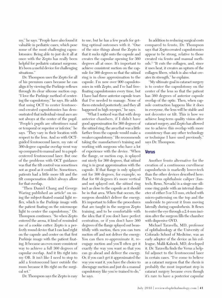
July 2018 | reviewofophthalmology.com | 41
my,” he says. “People have also found it valuable in pediatric cases, which pose some of the most challenging capsu-lotomies: Being able to just do it all at once with the Zepto has really been helpful for pediatric cataract surgeons. It’s been a useful device for challenging situations.”
Dr. Thompson uses the Zepto for all of his premium cases because he can align it by viewing the Purkinje refl exes through its clear silicone suction cup. “I love the Purkinje method of center-ing the capsulotomy,” he says. He adds that using OCT to center femtosec-ond-created capsulotomies has dem-onstrated that individual visual axes are not always at the center of the pupil. “People’s pupils are oftentimes nasal or temporal or superior or inferior,” he says. “They vary in their location with respect to the lens. And so with OCT-guided femtosecond lasers, my rate of 360-degree capsular overlap went way up compared to manual and to a pupil-centered femtosecond laser. But one of the problems with OCT guidance was that the tilt control was sometimes not as good as it could be. Sometimes, patients had a little more tilt and the tilt compensation didn’t always allow for that overlap.
“Then Daniel Chang and George Waring published an article2 on us-ing the subject-fi xated coaxial light re-fl ex, which is the Purkinje image with the patient fi xating on the microscope light to center the capsulotomy,” Dr. Thompson continues. “So when Zepto entered the arena, it kind of reminded me of a cookie cutter: Zepto is a per-fectly round device that I can land right on the capsule and center on that fi rst Purkinje image with the patient fi xat-ing. It became an even more consistent way to achieve a full 360 degrees of capsular overlap. And it fi ts right into my OR. It isn’t like I need to stop to add a femtosecond laser outside the room because it fi ts right on the surgi-cal set.”
Dr. Thompson says the Zepto is easy
to use, but he has a few pearls for get-ting optimal outcomes with it. “One of the nice things about the Zepto is that it sucks right onto the capsule and creates the capsular opening for 360 degrees all at once. It’s important to achieve consistent suction on the cap-sule for 360 degrees so that the nitinol ring is in close approximation to the capsule. I’m now over 900 capsuloto-mies in with Zepto, and I’ve had free-fl oating capsulotomies every time, but I have had three anterior capsule tears that I’ve needed to manage. None of them extended posteriorly; and they all happened in high myopes,” he says. “What I noticed was that with deep anterior chambers, if I didn’t have good approximation for 360 degrees of the nitinol ring, the area that was a little farther from the capsule would make a weaker capsulotomy.” He recommends taking the manufacturer’s training and working with surgeons who have a lot of experience with the device. “When the fl ange, or suction cup, is splayed out nicely for 360 degrees, that nitinol ring is in great approximation with the capsule. If that fl ange is only splayed out for 300 degrees, for example, so that for 60 degrees it’s more vertical and not splayed out, the nitinol ring isn’t as close to the capsule as it should be in that area. When that occurs, the surgeon shouldn’t deliver the energy. It’s important to follow the procedures that are taught in the surgeon Zepto training, and to be comfortable with the idea that if you don’t have perfect centration, or if you don’t have 360 degrees of the fl ange splayed out beau-tifully with suction, then you can turn suction off and not deliver the energy: You can then re-approximate it, re-engage suction and you’ll often get it exactly the way you want so that you can go ahead and deliver the energy. Or, if you can’t get it approximated the way you want it, you have the choice to disengage suction and just do a manual capsulotomy like you’re trained to do,” he says.
In addition to reducing surgical costs compared to femto, Dr. Thompson says that Zepto-created capsulotomies appear to be strong, relative to those created via femto and manual meth-ods.3 “It cuts the collagen, and, since it uses heat, it creates an upturn of the collagen fi bers, which is also what cre-ates its strength,” he explains.
“My ultimate goal in cataract surgery is to center the capsulotomy on the center of the lens so that the patient has 360 degrees of anterior capsule overlap of the optic. Then, when cap-sule contraction happens like it does in everyone, the lens will be stable and not decenter or tilt. This is how we achieve long-term quality vision after cataract surgery, and Zepto has allowed me to achieve this overlap with more consistency than any other technology or technique I have used previously,” says Dr. Thompson.
Verus
Another femto alternative for the creation of a continuous curvilinear capsulorhexis is markedly lower-tech than the other devices described here: The Verus ophthalmic caliper (Ian-tech; Reno, Nevada) is a singe-use sili-cone ring guide with an internal diam-eter of either 5 or 5.5 mm, etched with micro-patterning on the top and the underside to prevent it from moving laterally during capsulorhexis. It fl exes to enter the eye through a 2.4-mm inci-sion after the surgeon fi lls the chamber with dispersive OVD.
Michael Taravella, MD, professor of ophthalmology at the University of Colorado School of Medicine, was an early adopter of the device as his col-league, Malik Kahook, MD, developed it. Dr. Taravella fi nds the Verus a help-ful adjunct to the femtosecond laser in certain cases. “I’ve come to believe as a cataract surgeon that the rhexis is probably the most important step in cataract surgery because even though it’s rare to have a posterior capsular
040_rp0718_f3.indd 41040_rp0718_f3.indd 41 6/22/18 4:32 PM6/22/18 4:32 PM
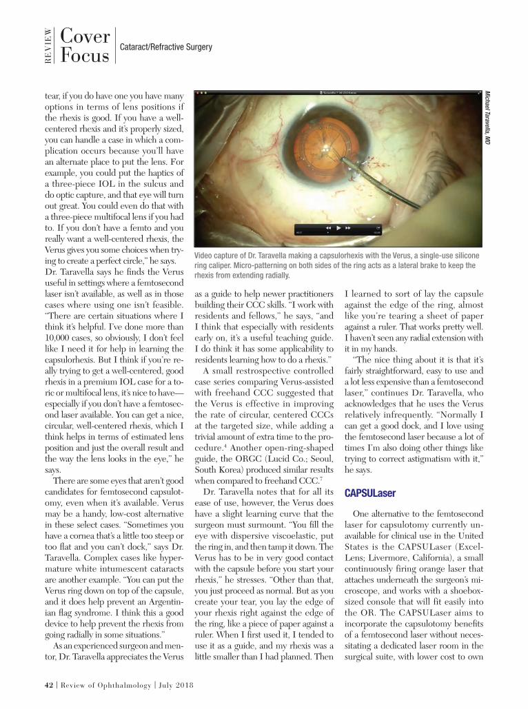
RE
VIE
W
42 | Review of Ophthalmology | July 2018
Cataract/Refractive SurgeryCoverFocus
tear, if you do have one you have many options in terms of lens positions if the rhexis is good. If you have a well-centered rhexis and it’s properly sized, you can handle a case in which a com-plication occurs because you’ll have an alternate place to put the lens. For example, you could put the haptics of a three-piece IOL in the sulcus and do optic capture, and that eye will turn out great. You could even do that with a three-piece multifocal lens if you had to. If you don’t have a femto and you really want a well-centered rhexis, the Verus gives you some choices when try-ing to create a perfect circle,” he says. Dr. Taravella says he fi nds the Verus useful in settings where a femtosecond laser isn’t available, as well as in those cases where using one isn’t feasible. “There are certain situations where I think it’s helpful. I’ve done more than 10,000 cases, so obviously, I don’t feel like I need it for help in learning the capsulorhexis. But I think if you’re re-ally trying to get a well-centered, good rhexis in a premium IOL case for a to-ric or multifocal lens, it’s nice to have—especially if you don’t have a femtosec-ond laser available. You can get a nice, circular, well-centered rhexis, which I think helps in terms of estimated lens position and just the overall result and the way the lens looks in the eye,” he says.
There are some eyes that aren’t good candidates for femtosecond capsulot-omy, even when it’s available. Verus may be a handy, low-cost alternative in these select cases. “Sometimes you have a cornea that’s a little too steep or too fl at and you can’t dock,” says Dr. Taravella. Complex cases like hyper-mature white intumescent cataracts are another example. “You can put the Verus ring down on top of the capsule, and it does help prevent an Argentin-ian fl ag syndrome. I think this a good device to help prevent the rhexis from going radially in some situations.”
As an experienced surgeon and men-tor, Dr. Taravella appreciates the Verus
as a guide to help newer practitioners building their CCC skills. “I work with residents and fellows,” he says, “and I think that especially with residents early on, it’s a useful teaching guide. I do think it has some applicability to residents learning how to do a rhexis.”
A small restrospective controlled case series comparing Verus-assisted with freehand CCC suggested that the Verus is effective in improving the rate of circular, centered CCCs at the targeted size, while adding a trivial amount of extra time to the pro-cedure.4 Another open-ring-shaped guide, the ORGC (Lucid Co.; Seoul, South Korea) produced similar results when compared to freehand CCC.7
Dr. Taravella notes that for all its ease of use, however, the Verus does have a slight learning curve that the surgeon must surmount. “You fi ll the eye with dispersive viscoelastic, put the ring in, and then tamp it down. The Verus has to be in very good contact with the capsule before you start your rhexis,” he stresses. “Other than that, you just proceed as normal. But as you create your tear, you lay the edge of your rhexis right against the edge of the ring, like a piece of paper against a ruler. When I fi rst used it, I tended to use it as a guide, and my rhexis was a little smaller than I had planned. Then
I learned to sort of lay the capsule against the edge of the ring, almost like you’re tearing a sheet of paper against a ruler. That works pretty well. I haven’t seen any radial extension with it in my hands.
“The nice thing about it is that it’s fairly straightforward, easy to use and a lot less expensive than a femtosecond laser,” continues Dr. Taravella, who acknowledges that he uses the Verus relatively infrequently. “Normally I can get a good dock, and I love using the femtosecond laser because a lot of times I’m also doing other things like trying to correct astigmatism with it,” he says.
CAPSULaser
One alternative to the femtosecond laser for capsulotomy currently un-available for clinical use in the United States is the CAPSULaser (Excel-Lens; Livermore, California), a small continuously firing orange laser that attaches underneath the surgeon’s mi-croscope, and works with a shoebox-sized console that will fit easily into the OR. The CAPSULaser aims to incorporate the capsulotomy benefi ts of a femtosecond laser without neces-sitating a dedicated laser room in the surgical suite, with lower cost to own
Video capture of Dr. Taravella making a capsulorhexis with the Verus, a single-use silicone
ring caliper. Micro-patterning on both sides of the ring acts as a lateral brake to keep the
rhexis from extending radially.
Mich
ael Ta
rave
lla, M
D
040_rp0718_f3.indd 42040_rp0718_f3.indd 42 6/22/18 4:33 PM6/22/18 4:33 PM

A P U B L I C A T I O N B Y
Retina Specialist focuses on the latest advances in the diagnosis, medical management and surgical treatment of diseases of the retina, along with practical, real-world advice from leading clinicians and other experts on managing the successful retina practice.
The new addition to the Review family serves the entire Retina Specialist marketplace. Look for your issues in 2018:
» MARCH » JUNE» SEPTEMBER» NOVEMBER
For inquiries contact [email protected]
Advertising opportunitiesJIM HENNE • PUBLISHER • 610-492-1017 • [email protected] MICHELE BARRETT • 215-519-1414 • [email protected]
MICHAEL HOSTER • 610-492-1028 • [email protected]
www.retina-specialist.com
2017_retinaspecialist_housead.indd 12017_retinaspecialist_housead.indd 1 1/16/18 4:30 PM1/16/18 4:30 PM

RE
VIE
W
44 | Review of Ophthalmology | July 2018
CoverFocus Cataract/Refractive Surgery
and operate. Unlike some other femto alterna-
tives, the CAPSULaser isn’t pulsed; it fi res continuously to cut in a circular pattern, creating a tag-free capsulot-omy in one second, according to the manufacturer. For the capsule to ab-sorb the CAPSULaser’s cutting energy, the surgeon must stain it with trypan blue, and then wash it out. (Excel-Lens offers proprietary blue intraocular dye and cohesive and dispersive OVDs, which are CE-certifi ed.) The patient fixates on a beam of light while the surgeon places a handheld contact lens over the eye to aim the beam, which is controlled by a foot pedal. The device can cut a capsulorhexis in diameters ranging from 4.5 to 6 mm in 0.1-mm increments. The CAPSULaser’s ener-gy transforms the collagen of the rhexis edge, making it smooth and elastic, ac-cording to the manufacturer.
Richard Packard, MD, FRCS, FRCOphth, presented clinical trial data at the recent American-European Congress of Ophthalmic Surgery’s Eu-ropean Summer Symposium in Berlin. In 125 patients randomized to CAP-SULaser or manual capsulorhexis, Dr. Packard reported that the CAPSULa-ser produced greater circularity and centration and achieved a rhexis size within 0.1 mm of target more often, as well as a greater rate of 360-degree capsulotomy coverage and 100 percent free-fl oating capsulotomies.6
Aperture CTC
“Manual capsulorhexis has been the gold standard for a long time now, but it’s still the most diffi cult step of the cataract procedure. Even in the best hands, there can still be heart-stopping moments,” says Mark Packer, MD, of Mark Packer MD Consulting in Boul-der, Colorado, and chief medical of-fi cer of International Biomedical De-vices in Mt. Pleasant, South Carolina. “One of my colleagues from London, Brian Little, and I published a paper7
several years ago about how to save some of these situations where the capsulorhexis goes astray. That actu-ally became one of my most popular publications, somewhat to my surprise. But it shows that people are really in search of a better way to do this that has a higher success rate. Published studies show complication rates from 2 to 5 percent for manual capsulorhexis. So the idea is to achieve a more consis-tent, more reproducible and safer way of opening the anterior capsule.”
Dr. Packer’s company is attempting to answer that need with the Aperture CTC (“continuous thermal capsuloto-my”), which consists of a console that provides constant energy to a single-use, stainless steel ring-shaped cut-ting element that retracts and re-opens to fit through a small incision and then contact the capsule 360 degrees around without suction. The dispos-able ring, integrated within a dispos-able handpiece that the manufacturer says feels similar to a phaco handpiece, comes in sizes ranging from 4.5 to 6 mm, in 0.5-mm increments. Thermal energy quickly cuts a free-fl oating cap-sulotomy; the surgeon easily retracts the loop with the cap and removes it through the entry wound for disposal.
“Essentially, it is a stainless steel cir-
cle that is stretched to a narrow ellipse that allows entry through what is cur-rently a 2.4-mm incision, but further refi nements may decrease that a little bit,” Dr. Packer explains. “Once inside the eye, it re-forms into a full circle. Then, by depressing the foot pedal, a very short pulse of electricity is sent through the device in milliseconds. Again, time span is one of the things we’re still toying with a little bit. As the thermoelectric current passes through the circle, the heating essentially sur-passes the melting temperature of col-lagen, creating an instantaneous com-plete capsulotomy. Once that’s done, the circle is converted again into a very narrow ellipse that can be withdrawn from the eye, taking with it the central cap. The eye is then ready for surgery for the extraction of the lens.”
Dr. Packer thinks that the Aper-ture CTC is easy for surgeons to learn and safe for their patients. “I think it’s extremely easy,” he says. “It’s very intuitive. The user interface is very simple. There are a bunch of safety checks that are built into the circuitry so that you can’t really mess up. If anything is not connected properly, the device will let you know, and it won’t fi re.” He adds that the dispos-able handpiece is suitable for right- or
The Aperture CTC is a small red-orange laser that fi res continuously instead of in pulses,
controlled by a foot pedal when the fl exible, disposable stainless-steel cutting element is
in proper contact with the capsule.
Mark
Pack
er, M
D
040_rp0718_f3.indd 44040_rp0718_f3.indd 44 6/22/18 4:33 PM6/22/18 4:33 PM

left-handed surgeons and just a little bit lighter than a phaco handpiece.
“We have an alignment mark in the center of the circle, which can be aligned with the Purkinje light refl exes, so if the surgeon wants to center on the optical axis, a line from the first Purkinje to the fourth Purkinje—or even the third Purkinje—that can be done,” he says. Unlike the Zepto, Ap-erture CTC doesn’t incorporate suc-tion into making contact between the ring and the capsule. “We don’t need any vacuum: Basically, you just place the stainless-steel ring on the capsule,” he says. “It just has to be in 360-degree contact, and minimal downward pres-sure is needed.”
Although the manufacturer empha-sizes the Aperture CTC’s potential niche as a less costly, easy-to-use femto alternative, Dr. Packer doesn’t see the emerging device—which will be in hu-man trials later this year—as a compet-itor with femto. “As everyone knows, femtosecond lasers are very expensive and their use is therefore limited to the premium channel of refractive cataract surgery,” he says. “The cost is often a click fee of several hundred dollars, and so the only way that works is if the patient is paying for the advantages that accrue with femto, such as better astigmatism correction with toric axis guidance built into the laser, for exam-ple. Plus, the laser also performs other steps besides the capsulotomy. But it’s fundamentally a premium-channel device, and Aperture CTC is not. Al-though I can’t currently tell you exactly what the price point will be, our goal is to make it very affordable for routine cataract surgery so as not to add much cost to the procedure, and to perhaps even save some money by reducing the complication rate. So we’re not really competing with femto. I think femto will always have a role for refractive cases; we’re looking at this as a device for precision capsulotomy in routine, everyday cataract surgery.”
Dr. Packer says that he believes that
one possible advantage the Aperture CTC may enjoy over manual capsu-lotomy is that the denatured collagen edges roll over on themselves for add-ed smoothness and strength, a feature thought to be shared by the Zepto. “With the Zepto device, they noted that their capsulotomy was actually stronger than a manual rhexis, presum-ably because of the fact that when the capsulotomy is constructed, the edge of the capsule actually rolls over itself. We have seen that same constellation of fi ndings,” he says. “The cut edge is actually facing away from the open-ing because it’s rolled back on itself. I believe that occurs because you’re heating the cut edge to the point where it cuts; but beyond that, it’s also that a little bit of heat is causing a tightening of the lens capsule.” Dr. Packer likens the tightening effect on the rhexis edge to what occurs in laser facial resurfac-ing. “You can see the collagen in the facial tissue tighten up when heat is applied. It constricts, and that’s what also strengthens the capsulotomy,” he says.
Dr. Packer is the chief medical offi cer of International Biomedical Devices. Dr. Thompson is a researcher and con-sultant for Mynosys, and served as an FDA investigator for the Zepto trials. Dr. Taravella is consultant for Johnson and Johnson Vision-AMO for the Visx, and is a proctor for both the Visx and IntraLase lasers.
1. Mohammadpour M, Erfanian R, Karimi N. Capsulorhexis: Pearls and pitfalls. Saudi J of Ophthalmol. 2012;26:1:33-40. 2. Chang DH, Waring GO. The subject-fi xated coaxially sighted corneal light refl ex: A clinical marker for centration of refractive treatments and devices. American J Ophthalmol 2014;158:5:863-74.3. Thompson VM, Berdahl JP, Salono JM, Chang DF. Comparison of manual, femtosecond laser, and precision pulse capsulotomy edge tear strength in paired human cadaver eyes. Ophthalmol 2016:123:265-74.4. Kahook MY, Cionni RJ, Taravella MJ, Ang RE, Waite AN, Solomon JD, Uy HS. Continuous curvilinear capsulorhexis performed with the VERUS ophthalmic caliper. J Refract Surg 2016; 32:10:654-8.5. Lee JH, Yong EL, Joo C. Clinical results of the open ring PMMA guider assisted capsulorrhexis in cataract surgery. BMC Ophthalmol 2018;18:1:116.6. CAPSULaser. Latest CAPSULaser data presented at AECOS. https://www.CAPSULaser.com/latest-ce-data-for-CAPSULaser-at-aecos. Accessed 13 June 2018.7. Little BC, Smith JH, Packer M. Little capsulorhexis tear-out rescue. J Cat Refract Surg 2006;32:9:1420-22.
COMBOCHAIRS & STANDS
Aff ordable, space-saving chair & stand solutions.
Optimise space and functionality.
Small footprint 41.2” x 34.2”
040_rp0718_f3.indd 45040_rp0718_f3.indd 45 6/22/18 4:33 PM6/22/18 4:33 PM

Research Review R
EV
IEW
46 | Review of Ophthalmology | July 2018 This article has no commercial sponsorship.
Anti-tuberculosis Therapy and UveitisIn a retrospective cohort study,
researchers sought to explore the clinical features of patients with uveitis associated with latent tu-berculosis. They also examined the effect of anti-TB treatment on uveitis outcomes.
The researchers looked at 199 eyes of 129 patients diagnosed with uveitis associated with latent TB (89 patients received anti-TB treatment, while 40 did not). They were evaluated for recurrence of disease following treatment. In-formation was gathered retrospec-tively regarding clinical outcome, vision and treatment. Outcome measures included best-corrected visual acuity and rate of disease recurrence.
The researchers treated the uve-itis with local and systemic anti-in-flammatory and immunosuppres-sive therapy in all patients. The mean change in BCVA following treatment was 4.5 ±1.4 letters over the follow-up period, with no dif-ference between eyes of patients receiving ATT and those who did not. However, 68 eyes (34.9 per-cent) had a recurrence of uveitis (0.64 ±0.08 recurrences per year), with eyes of patients receiving ATT less likely to develop a recur-rence compared to those not re-ceiving ATT (29.5 percent vs. 48.2
percent). Eyes treated with ATT recurred at an estimated median of 120 months, compared with 51 months in eyes with no treatment (p=0.005).
Based on these results, the re-searchers concluded that treat-ment with ATT halved the risk of uveitis recurrence and delayed the onset of the first recurrence in eyes with uveitis associated with latent TB.
Am J Ophthalmol 2018;190:164-170.Tomkins-Netzer O, Leong BCS, Zhang X, et al.
Outcomes of Cataract Surgery in nAMD Patients
Researchers from Australia evalu-ated the outcomes and predic-
tive factors of visual acuity change after cataract surgery in patients who were being treated for neo-vascular age-related macular de-generation.
In this retrospective, matched case-control study, researchers studied eyes undergoing cataract surgery that had been tracked since they first started treatment for nAMD. These eyes were com-pared with a cohort of unoper-ated phakic eyes being treated for nAMD matched for treatment duration before cataract surgery, baseline VA, age and length of fol-
low-up.The study included 124 patients
who had cataract surgery and 372 matched controls. The mean VA gained was 10.6 letters (range: 7.8 to 13.2; p<0.001) 12 months af-ter surgery; 26 percent had gained ≥3 lines, and 1.6 percent had lost ≥3 lines of VA. Visual acuity 12 months after surgery was higher in eyes that had cataract extrac-tion compared with controls (65.8 ±17.1 vs. 61.3 ±20.8 letters, re-spectively, p=0.018). In the study group, the proportion of visits where the choroidal neovascular lesion was graded active and the mean number of injections were similar before and after surgery (p=0.506 and p=0.316, respective-ly), whereas both decreased in the control group, suggesting that sur-gery modestly increased the level of activity of the CNV lesion.
Mean VA prior to surgery was lower in eyes that gained ≥15 letters compared with eyes that gained zero to 14 letters (40.2 ±21.4 vs. 62.1 ±15.1, p<0.001). Pa-tients undergoing cataract surgery within the first six months of anti-VEGF therapy were more likely to lose rather than gain vision (20.8 percent lost vision vs. 12.8 per-cent and 4.4 percent gaining ≥15 or zero to 14 letters, respectively,
046_rp0718_rr.indd 46046_rp0718_rr.indd 46 6/22/18 4:49 PM6/22/18 4:49 PM
p=0.023). Receiving an injection at least two weeks before surgery, age and the CNV lesion type had no discernible association with VA outcomes.
The researchers found evidence of a modest effect of cataract sur-gery on CNV lesion activity in eyes being treated for nAMD. Despite this, visual outcomes were favor-able. Cataract surgery within six months of starting treatment for nAMD should be avoided if pos-sible, the researchers say.
Am J Ophthalmol 2018;190:50-57.Daien V, Nguyen V, Morlet N, et al.
Medical Professional Liability Claims
Researchers from the Duke Uni-versity Medical Center, Dur-
ham, North Carolina, and the Physician Insurers Association of America, Rockville, Maryland, conducted a retrospective analy-sis of medical professional liabil-ity claims recorded by the PIAA’S Data Sharing Project over a 10-year period to examine the char-acteristics of these claims against ophthalmologists in the United States.
The authors compared ophthal-mology and all health care special-ties for physician demographics, prevalence and costs associated with closed claims and resolution of claims. They also compared the most prevalent chief medical fac-tor, presenting medical condition, operative procedure, outcomes and resolution of ophthalmology claims between two periods: 2006 to 2010 and 2011 to 2015.
The data showed that:• only 24 percent of c losed
claims against ophthalmologists resulted in payment;
• two-thirds were dropped, withdrawn or dismissed;
• ninety percent of claims that received a verdict were favorable toward the ophthalmologist;
• cataract and cornea surgeries were the most prevalent and most costly operative procedures, ac-counting for half of all claims and $47,641,376 and $32,570,148 in total paid indemnity, respectively;
• average indemnity was higher for corneal procedures ($304,476) than v i treoret inal procedures ($270,141) or oculoplastic proce-dures on the eyelid ($222,471) or orbit and eyeball ($183,467);
• the prevalence and cost of claims related to endophthalmitis declined from the 2006 to 2010 pe-riod (n=38/1,160 [3.3 percent]; av-erage indemnity, $516,875) to the 2011 to 2015 period (n=26/1,165 [2.2 percent]; average indemnity, $247,083); and
• the average indemnity paid ($280 ,227 v s . $335 ,578) and amount spent on legal defense ($41,450 vs. $46,391) was slight-ly lower among ophthalmologists compared with all health-care spe-cialties, respectively.
Based on these results, the re-searchers concluded that ophthal-mology has a relatively low num-ber of malpractice claims reported compared with other health-care specialties and shows less spend-ing on average indemnity and de-fense.
Further studies are needed to in-vestigate the reasons for the high-er prevalence of claims related to cataract and corneal surgeries and the higher average indemnity paid for corneal procedures relative to vitreoretinal or oculoplastic pro-cedures.
Ophthalmology 2018;125:631-641Thompson AC, Parikh PD, Lad EM
(Continued on page 61)
.
SLITLAMPS
EXCEPTIONAL OPTICS
S4OPTIK’s converging binoculars
European craftsmanship and engineering provide
Vertical and compact
H-Series
Z-SeriesSLIT LAMPS
www.s4optik.com l 888.224.6012Sensible equipment. Well made, well priced.
For today’s modern office.
046_rp0718_rr.indd 47046_rp0718_rr.indd 47 6/22/18 4:52 PM6/22/18 4:52 PM
48 | Review of Ophthalmology | July 2018 This article has no commercial sponsorship.
Edited by Arturo Chayet, MD
Refractive/CataractRundown R
EV
IEW
Low-level astigmatism (0.25 to 1.25 D)is not an uncommon fi nd-
ing in eyes presenting for cataract sur-gery, and it represents a treatment opportunity.1 Correcting for nearly distortion-free vision may make pa-tients even happier with their new IOLs. Here, experienced refractive cataract surgeons explain the impor-tance of considering the role even slight astigmatism plays in visual out-comes, and share techniques for de-creasing it as much as possible.
How Much is Treatable?
Robin Vann, MD, assistant profes-sor of ophthalmology at Duke Univer-sity and medical director of Duke Eye Center’s operating rooms, says that surgeons who ignore small amounts of astigmatism are missing out an a rela-tively easy avenue to patient satisfac-tion. “A lot of cataract surgeons seem to have this concept regarding astig-matism treatment, that they’ll be very selective about who they’ll start with. They’ll say things like, ‘I’m not going to offer this treatment to anyone with un-der two diopters of astigmatism.’ But I believe that’s really fl awed thinking because the vast majority of American
patients have low amounts of astigma-tism; the majority has between one and two diopters. You’re essentially exclud-ing an awful lot of patients if you hold off until people present with higher amounts of astigmatism,” he says.
“I generally try to get patients to have less than half a diopter of astigmatism if I can,” Dr. Vann explains. “Assuming they’re not myopic or hyperopic with spectacles or after surgery, most pa-tients can tolerate up to a half-diopter of astigmatism, and in some cases 0.75, before they can’t read the 20/20 line. So that would be acceptable for most patients. While I’d love to make astig-matism zero in every case, we don’t yet have a tunable lens implant in our hands to make that goal realistic.” Dr. Vann notes that the RxSight lens, FDA approved in November 2017, is not yet commercially available.
Limbal Relaxing Incisions
Limbal relaxing incisions are a safe and straightforward astigmatism treat-ment option during cataract surgery.2
Complications such as infections are rare, although potentially serious.3,4
Both monofocal implantation with LRIs and toric lens implantation for
low astigmatism can signifi cantly im-prove mean uncorrected visual acuity, although torics may produce slightly better visual outcomes.5 Tal Raviv, MD, FACS, associate clinical profes-sor of ophthalmology at the New York Eye and Ear Infi rmary of Mount Sinai Icahn School of Medicine at Mount Sinai, and the founder and medical di-rector of the Eye Center of New York, thinks torics are the best option. “We’ve learned more about astigmatism plan-ning in the past half-decade than ever before. We know that toric IOLs are more accurate than LRIs, so we should use torics whenever possible,” he says.
“In my practice there are three rea-sons for patients to get LRIs instead of torics,” says Dr. Vann. “Number one is if their astigmatism isn’t great enough to warrant a toric lens implant. I have the Barrett Toric Calculator printed out as part of my biometry calculations for formulas: If it’s telling me not to put in the lowest-powered toric—a T2 [Alcon ReSTOR, 1 D of cylinder correction], for example, and the fi eld is just blank, then the patient doesn’t qualify for a toric. I’ll think about doing LRIs to re-duce their astigmatism when they’re greater than a half diopter but not high enough to warrant a toric.
Surgeons say that even a little bit of astigmatism is worth treating, and cataract surgery presents a golden opportunity.
Treating Astigmatism: How Low Can You Go?
Kristine Brennan, Senior Associate Editor
048_rp0718_rcr.indd 48048_rp0718_rcr.indd 48 6/22/18 4:55 PM6/22/18 4:55 PM

July 2018 | reviewofophthalmology.com | 49
“Another situation is where a patient unfortunately just can’t afford a toric,” Dr. Vann continues. “The third scenario is when the ocular surface of the eye makes it very diffi cult to get accurate readings. In those cases I may delay, or I may determine that I don’t want to be doing astigmatism management with torics in unusual eyes. Irregular astigmatism might be an example; or a patient with bad keratoconus or with dry eyes so severe that you can’t even do good biometry testing.” He also says that patients with irregularities to the contours of their eyes due to conditions such as Salzmann’s nodular degenera-tion and anterior basement membrane dystrophy may be poor candidates for toric lenses.
Although LRIs are simple and effec-tive, Dr. Vann says that some patients become disappointed if they can’t try a toric IOL for astigmatism treatment, in part because new patients in his practice get an overview of the exciting premium and toric IOL technologies available. “Unfortunately, they may get all pumped and excited about these technologies, and then I’ve got to tell them, ‘Sorry, but I can’t use them in you.’ So sometimes you have to gently bring patients back to reality.”
Toric Tips
For patients who are good toric-IOL candidates, Dr. Raviv urges surgeons to correctly factor the astigmatism that any type of incision will induce into lens selection. “We haven’t been properly calculating surgically induced astigmatism in the past. Instead, we’ve just entered numbers such as 0.5 D for 2.75-mm corneal incisions,” he ex-plains. “It turns out that the net vector induced astigmatism is quite randomly distributed, and best practice today is to calculate centroid surgically In-duced astigmatism using Warren Hill’s SIA Calculator, or to simply use 0.1 D, which likely covers most temporal inci-sions from 2.2 to 2.6 mm.”
Dr. Vann encourages patients with less than 2 D of astigmatism to con-sider torics if they can afford them. “I think that for treating astigmatism, the toric lens implant is a tried-and- true technology. There are now several publications demonstrating that toric lens implants tend to leave less residual astigmatism and provide better long-term vision. I think they work incred-ibly well,” he says.
“For astigmatism treatment at the time of cataract surgery, use toric IOLs whenever you can—roughly speaking, for with-the-rule astigmatism greater than 1.5 D and against-the-rule greater than 0.4 D,” recommends Dr. Raviv. He adds that it’s especially important to neutralize astigmatism to get good re-sults with presbyopic IOLs. “Because most cataract patients are older, with high prevalence of against-the-rule astigmatism, I fi nd that more than half of my presbyopia-correcting IOL pa-tients get the toric version.”
Dr. Vann notes that a good workup is the foundation for good results, and that if your preoperative workup and measurements are uniformly thorough and comprehensive, you needn’t vary your workup too much for toric-lens patients. “For a very long time, we were very selective in how we did our
preoperative testing, but in order to meet our volume demands while try-ing to offer all of our patients all of the services that they’re eligible for, we ultimately decided to simply do test-ing consistently across the board. So if somebody’s coming to me for a cataract evaluation, they’re going to get a stan-dard set of tests,” he explains. “If their tests on the fi rst pass aren’t up to snuff, I’m going to repeat the suspect testing; and if they have a lot of astigmatism, then that might include repeating not just the biometry, but also the topogra-phy. I do biometry and topography on just about everyone unless they have an incredibly small amount of astigma-tism on the biometry testing, which we do before topography.
“It’s important to use a methodology that’s going to give you a high degree of accuracy,” Dr. Vann continues. “I use the Verion image-guided system so that I’ll have the reference vessels of the eye to determine where zero and 180 degrees are. We actually did a study that we haven’t published, that found a high degree of correlation of the Verion axis with the axis of my Lenstar (Haag-Streit). I use the Lenstar values to help me determine where to orient the to-ric lens implant, and the Barrett Toric Calculator. So if Barrett tells me axis
Dr. Raviv has developed this visual aid to help surgeons remember keys to astigmatism
management with toric IOLs, such as taking into account the tendency of astigmatism
to drift from WTR to ATR at the rate of about 0.34 D per decade, and the importance of
posterior corneal astigmatism in lens selection.
Tal R
aviv
048_rp0718_rcr.indd 49048_rp0718_rcr.indd 49 6/22/18 4:55 PM6/22/18 4:55 PM
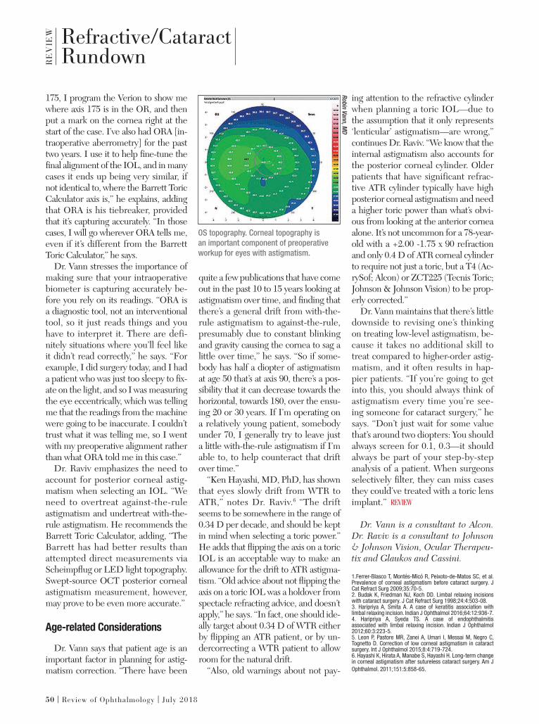
Refractive/CataractRundown R
EV
IEW
50 | Review of Ophthalmology | July 2018
175, I program the Verion to show me where axis 175 is in the OR, and then put a mark on the cornea right at the start of the case. I’ve also had ORA [in-traoperative aberrometry] for the past two years. I use it to help fi ne-tune the fi nal alignment of the IOL, and in many cases it ends up being very similar, if not identical to, where the Barrett Toric Calculator axis is,” he explains, adding that ORA is his tiebreaker, provided that it’s capturing accurately. “In those cases, I will go wherever ORA tells me, even if it’s different from the Barrett Toric Calculator,” he says.
Dr. Vann stresses the importance of making sure that your intraoperative biometer is capturing accurately be-fore you rely on its readings. “ORA is a diagnostic tool, not an interventional tool, so it just reads things and you have to interpret it. There are defi-nitely situations where you’ll feel like it didn’t read correctly,” he says. “For example, I did surgery today, and I had a patient who was just too sleepy to fi x-ate on the light, and so I was measuring the eye eccentrically, which was telling me that the readings from the machine were going to be inaccurate. I couldn’t trust what it was telling me, so I went with my preoperative alignment rather than what ORA told me in this case.”
Dr. Raviv emphasizes the need to account for posterior corneal astig-matism when selecting an IOL. “We need to overtreat against-the-rule astigmatism and undertreat with-the-rule astigmatism. He recommends the Barrett Toric Calculator, adding, “The Barrett has had better results than attempted direct measurements via Scheimpfl ug or LED light topography. Swept-source OCT posterior corneal astigmatism measurement, however, may prove to be even more accurate.”
Age-related Considerations
Dr. Vann says that patient age is an important factor in planning for astig-matism correction. “There have been
quite a few publications that have come out in the past 10 to 15 years looking at astigmatism over time, and fi nding that there’s a general drift from with-the-rule astigmatism to against-the-rule, presumably due to constant blinking and gravity causing the cornea to sag a little over time,” he says. “So if some-body has half a diopter of astigmatism at age 50 that’s at axis 90, there’s a pos-sibility that it can decrease towards the horizontal, towards 180, over the ensu-ing 20 or 30 years. If I’m operating on a relatively young patient, somebody under 70, I generally try to leave just a little with-the-rule astigmatism if I’m able to, to help counteract that drift over time.”
“Ken Hayashi, MD, PhD, has shown that eyes slowly drift from WTR to ATR,” notes Dr. Raviv.6 “The drift seems to be somewhere in the range of 0.34 D per decade, and should be kept in mind when selecting a toric power.” He adds that fl ipping the axis on a toric IOL is an acceptable way to make an allowance for the drift to ATR astigma-tism. “Old advice about not fl ipping the axis on a toric IOL was a holdover from spectacle refracting advice, and doesn’t apply,” he says. “In fact, one should ide-ally target about 0.34 D of WTR either by fl ipping an ATR patient, or by un-dercorrecting a WTR patient to allow room for the natural drift.
“Also, old warnings about not pay-
ing attention to the refractive cylinder when planning a toric IOL—due to the assumption that it only represents ‘lenticular’ astigmatism—are wrong,” continues Dr. Raviv. “We know that the internal astigmatism also accounts for the posterior corneal cylinder. Older patients that have significant refrac-tive ATR cylinder typically have high posterior corneal astigmatism and need a higher toric power than what’s obvi-ous from looking at the anterior cornea alone. It’s not uncommon for a 78-year-old with a +2.00 -1.75 x 90 refraction and only 0.4 D of ATR corneal cylinder to require not just a toric, but a T4 (Ac-rySof; Alcon) or ZCT225 (Tecnis Toric; Johnson & Johnson Vision) to be prop-erly corrected.”
Dr. Vann maintains that there’s little downside to revising one’s thinking on treating low-level astigmatism, be-cause it takes no additional skill to treat compared to higher-order astig-matism, and it often results in hap-pier patients. “If you’re going to get into this, you should always think of astigmatism every time you’re see-ing someone for cataract surgery,” he says. “Don’t just wait for some value that’s around two diopters: You should always screen for 0.1, 0.3—it should always be part of your step-by-step analysis of a patient. When surgeons selectively fi lter, they can miss cases they could’ve treated with a toric lens implant.”
Dr. Vann is a consultant to Alcon.
Dr. Raviv is a consultant to Johnson & Johnson Vision, Ocular Therapeu-tix and Glaukos and Cassini.
1.Ferrer-Blasco T, Montés-Micó R, Peixoto-de-Matos SC, et al. Prevalence of corneal astigmatism before cataract surgery. J Cat Refract Surg 2009;35:70-5.2. Budak K, Friedman NJ, Koch DD. Limbal relaxing incisions with cataract surgery. J Cat Refract Surg 1998;24:4:503-08.3. Haripriya A, Smita A. A case of keratitis association with limbal relaxing incision. Indian J Ophthalmol 2016;64:12:936-7.4. Haripriya A, Syeda TS. A case of endophthalmitis associated with limbal relaxing incision. Indian J Ophthalmol 2012;60:3:223-5.5. Leon P, Pastore MR, Zanei A, Umari I, Messai M, Negro C, Tognetto D. Correction of low corneal astigmatism in cataract surgery. Int J Ophthalmol 2015;8:4:719-724. 6. Hayashi K, Hirata A, Manabe S, Hayashi H. Long-term change in corneal astigmatism after sutureless cataract surgery. Am J Ophthalmol. 2011;151:5:858-65.
OS topography. Corneal topography is
an important component of preoperative
workup for eyes with astigmatism.
Rob
in V
an
n, M
D
048_rp0718_rcr.indd 50048_rp0718_rcr.indd 50 6/22/18 4:56 PM6/22/18 4:56 PM

CME Accredited Surgical Training Videos Now Available Online: www.MackoolOnlineCME.com
Welcome to the third year of Mackool Online CME! With the generous support of several ophthalmic companies, I am honored to have our viewers join me in the operating room as I demonstrate the technology and techniques that I have found to be most valuable, and that I hope are helpful to many of my colleagues. We continue to edit the videos only to either change camera perspective or to reduce down time – allowing you to observe every step of the procedure.
As before, one new surgical video will be released monthly, and physicians may earn CME credits or just observe
the case. New viewers are able to obtain additional CME credit by reviewing previous videos that are located in our archives.
I thank the many surgeons who have told us that they have found our CME program to be interesting and instructive; I appreciate your comments, suggestions and questions. Thanks again for joining us on Mackool Online CME.
Richard J. Mackool, MD
MackoolOnlineCME.com MONTHLY Video Series
Episode 31:“Anticipation and Prevention”
Surgical Video by:Richard J. Mackool, MD
MonthlyMonthly
MACKOOL ONLINE CMECCCCCMMMMMEEEEE SSSSSEEEEERRRRRIIIIIEEEEESSSSS | SURGICAL VIDEOS
Richard Mackool, MD, a world renowned anterior segment ophthalmic microsurgeon, has assembled a web-based video collection of surgicalcases that encompass both routine and challenging cases, demonstrating both familiar and potentially unfamiliar surgical techniques using a varietyof instrumentation and settings.
This educational activity aims to present a series of Dr. Mackool’s surgical videos, carefully selected to address the specifi c learning objectives of this activity, with the goal of making surgical training available as needed online for surgeons motivated to improve or expand their surgical repertoire.
Learning Objective:After completion of this educational activity, participants should be able to:
• Prepare both the patient and surgeon for possible intraoperative cough-related issues
• Demonstrate steps to overcome infusion misdirection syndrome• Discuss current concepts regarding intracameral antibiotic administration.
Video Overview:This is a routine case during which I discuss anticipation and prevention of problems
caused by patient coughing, infusion misdirection
syndrome, and fi nally the intracameral antibiotic
controversy.
Satisfactory Completion - Learners must pass a post-test and complete an evaluation form to receive a certifi cate of completion. Your chosen sessions must be attended in their entirety. Partial credit of individual sessions is not available. If you are seeking continuing education credit for a specialty not listed below, it is your responsibility to contact your licensing/certifi cation board to determine course eligibility for your licensing/certifi cation requirement.
Physicians - In support of improving patient care, this activity has been planned and implemented by Amedco LLC and Postgraduate Healthcare Education. Amedco LLC is jointly accredited by the Accreditation Council for Continuing Medical Education (ACCME), the Accreditation Council for Pharmacy Education (ACPE), and the American Nurses Credentialing Center (ANCC), to provide continuing education for the healthcare team
Credit Designation Statement - Amedco designates this live activity for a maximum of .25 AMA PRA Category 1 CreditsTM. Physicians should claim only the credit commensurate with the extent of their participation in the activity.
JOINTLY ACCREDITED PROVIDERTM
INTERPROFESSIONAL CONTINUING EDUCATION
Additionally Supported by:
GlaukosMST
Crestpoint ManagementCarl Zeiss Meditec
Endorsed by: Review of Ophthalmology®
Video and Web Production by:JR Snowdon, Inc
Supported by an unrestricted independentmedical educational grant from:
Alcon&
Jointly provided by:
To view CME videogo to:
www.MackoolOnlineCME.com

This article has no commercial sponsorship.
Glaucoma Management Edited by Kuldev Singh, MD, MPH, and Peter A. Netland, MD, PhDR
EV
IEW
52 | Review of Ophthalmology | July 2018
The subject of ophthalmologists co-managing patient care with optometrists has always been controversial. The official position of the American Academy of Ophthalmology, which the column editors (Drs. Netland and Singh) support, can be found online at aao.org/ethics-detail/guidelines-comanagement-postoperative-care. This article presents an alternative perspective.
Health care is an evolving fi eld. Human beings, however, almost
always dislike change, and we find it easy to argue for maintaining the status quo. So perhaps it’s not surprising that some evolutionary changes occurring in ophthalmology today are meeting with resistance. One change that’s been occurring—and meeting signifi cant resistance—is the growing prevalence of MDs co-managing glaucoma patients with optometrists.
Here, I’d like to discuss the reasons for this increase in co-management, evaluate the reservations that MDs sometimes express, and share my personal, positive experience working
with ODs. As I hope to demonstrate, this shift is happening for a number of good reasons, and it’s part of a larger trend in medicine in which basic care and monitoring of patients with well-controlled disease is gradually being transferred to, or co-managed with, other health-care professionals. In addition, I’ll share some evidence that a team-based approach can end up providing glaucoma patients with timely and complete care.
There are two main reasons that many MDs don’t want to refer to op-tometrists: the perception that ODs mismanage glaucoma patients, and fear that practice revenue will drop as MDs lose patients to “the com-petition.” My experience has been very different.
How Competent are ODs?
As with many beliefs, the idea that ODs may not be capable of managing glaucoma patients probably started from some poor outcomes in the past that were blamed on inadequate care received from an OD. This has resulted in many MDs making general assumptions about ODs that,
in my experience, are simply not true. A few points:
• It makes a difference how ex-tensively an OD has engaged in continuing education. Since the 1990s, glaucoma management guidelines have largely been influ-enced by the canon of national ran-domly controlled clinical trials, including the Early Manifest Glau-coma Trial, Ocular Hypertension Treatment Study, Advanced Glauco-ma Intervention Study and Collabor-ative Normal Tension Glaucoma Study. As a result, ODs have had to keep up-to-date by learning glaucoma management from publications and CE courses while in practice. (In fairness, we MDs have had a stronger foundation of medical and surgical management in medical school and residency, but we’ve benefi ted from CME courses in a similar way.)
Having received glaucoma surgical patient referrals almost equally from ODs and MDs, I’ve reviewed piles of charts from both. I personally have not witnessed a lower level of management quality from the ODs. I see good documentation with re-peated Humphrey visual fields and
An ophthalmologist who has worked with ODs for years says that doing so has benefi ted his patients and his practice.
Co-managing Glaucoma: One MD’s Experience
Rajen U. Desai, MD, East Brunswick, N.J.
052_rp0718_gm.indd 52052_rp0718_gm.indd 52 6/22/18 4:57 PM6/22/18 4:57 PM
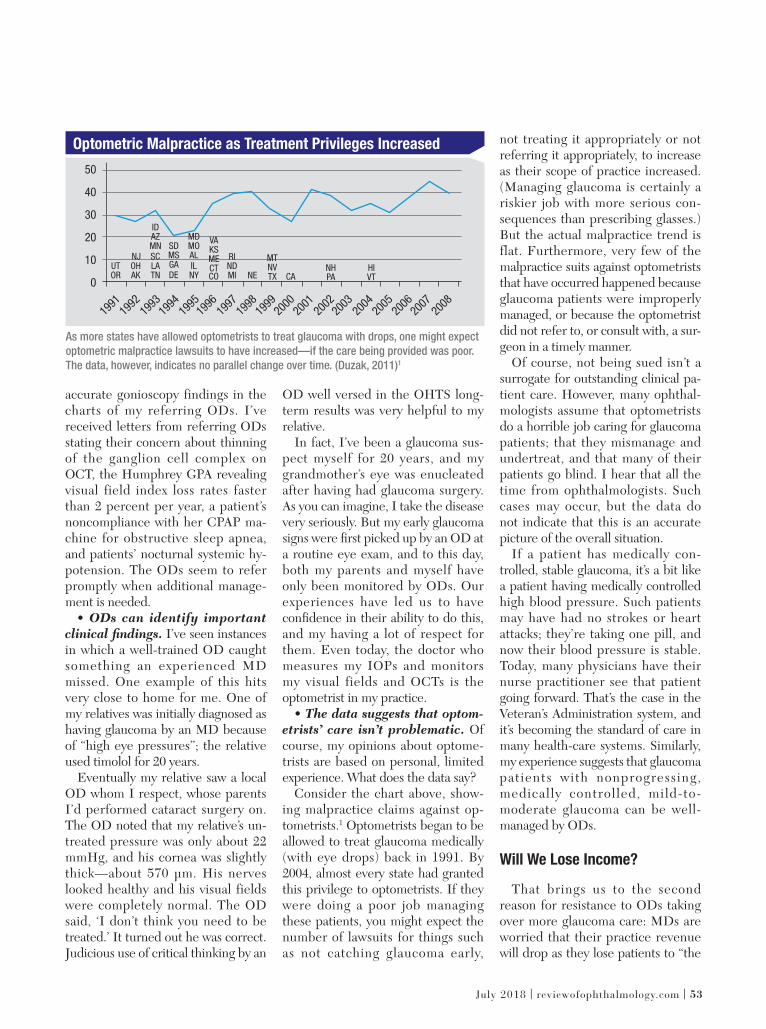
July 2018 | reviewofophthalmology.com | 53
accurate gonioscopy findings in the charts of my referring ODs. I’ve received letters from referring ODs stating their concern about thinning of the ganglion cell complex on OCT, the Humphrey GPA revealing visual field index loss rates faster than 2 percent per year, a patient’s noncompliance with her CPAP ma-chine for obstructive sleep apnea, and patients’ nocturnal systemic hy-potension. The ODs seem to refer promptly when additional manage-ment is needed.
• ODs can identify important clinical fi ndings. I’ve seen instances in which a well-trained OD caught something an experienced MD missed. One example of this hits very close to home for me. One of my relatives was initially diagnosed as having glaucoma by an MD because of “high eye pressures”; the relative used timolol for 20 years.
Eventually my relative saw a local OD whom I respect, whose parents I’d performed cataract surgery on. The OD noted that my relative’s un-treated pressure was only about 22 mmHg, and his cornea was slightly thick—about 570 µm. His nerves looked healthy and his visual fields were completely normal. The OD said, ‘I don’t think you need to be treated.’ It turned out he was correct. Judicious use of critical thinking by an
OD well versed in the OHTS long-term results was very helpful to my relative.
In fact, I’ve been a glaucoma sus-pect myself for 20 years, and my grandmother’s eye was enucleated after having had glaucoma surgery. As you can imagine, I take the disease very seriously. But my early glaucoma signs were fi rst picked up by an OD at a routine eye exam, and to this day, both my parents and myself have only been monitored by ODs. Our experiences have led us to have confi dence in their ability to do this, and my having a lot of respect for them. Even today, the doctor who measures my IOPs and monitors my visual fields and OCTs is the optometrist in my practice.
• The data suggests that optom-etrists’ care isn’t problematic. Of course, my opinions about optome-trists are based on personal, limited experience. What does the data say?
Consider the chart above, show-ing malpractice claims against op-tometrists.1 Optometrists began to be allowed to treat glaucoma medically (with eye drops) back in 1991. By 2004, almost every state had granted this privilege to optometrists. If they were doing a poor job managing these patients, you might expect the number of lawsuits for things such as not catching glaucoma early,
not treating it appropriately or not referring it appropriately, to increase as their scope of practice increased. (Managing glaucoma is certainly a riskier job with more serious con-sequences than prescribing glasses.) But the actual malpractice trend is flat. Furthermore, very few of the malpractice suits against optometrists that have occurred happened because glaucoma patients were improperly managed, or because the optometrist did not refer to, or consult with, a sur-geon in a timely manner.
Of course, not being sued isn’t a surrogate for outstanding clinical pa-tient care. However, many ophthal-mologists assume that optometrists do a horrible job caring for glaucoma patients; that they mismanage and undertreat, and that many of their patients go blind. I hear that all the time from ophthalmologists. Such cases may occur, but the data do not indicate that this is an accurate picture of the overall situation.
If a patient has medically con-trolled, stable glaucoma, it’s a bit like a patient having medically controlled high blood pressure. Such patients may have had no strokes or heart attacks; they’re taking one pill, and now their blood pressure is stable. Today, many physicians have their nurse practitioner see that patient going forward. That’s the case in the Veteran’s Administration system, and it’s becoming the standard of care in many health-care systems. Similarly, my experience suggests that glaucoma patients with nonprogressing, medically controlled, mild-to-moderate glaucoma can be well-managed by ODs.
Will We Lose Income?
That brings us to the second reason for resistance to ODs taking over more glaucoma care: MDs are worried that their practice revenue will drop as they lose patients to “the
Optometric Malpractice as Treatment Privileges Increased
50
40
30
20
10
0
1991
1992
1993
1994
1995
1996
1997
1998
1999
2000
2001
2002
2003
2004
2005
2006
2007
2008
UTOR
OHAK
LATN NY
IDAZMN
MDMOAL
VAKSMECT
RINDMI
MTNVTX
NHPA
HIVTCANE
SDMSGA
NJ SCIL
CODE
As more states have allowed optometrists to treat glaucoma with drops, one might expect
optometric malpractice lawsuits to have increased—if the care being provided was poor.
The data, however, indicates no parallel change over time. (Duzak, 2011)1
052_rp0718_gm.indd 53052_rp0718_gm.indd 53 6/22/18 4:57 PM6/22/18 4:57 PM

GlaucomaManagement R
EV
IEW
54 | Review of Ophthalmology | July 2018
competition.” There are a number of reasons to conclude that this is not going to happen.
• The number of glaucoma pa-tients is steadily increasing. This is a trend that will continue for many years to come. As the baby boomers age, demand for eye care—especially cataract surgery and glaucoma—will exceed the supply of newly graduated ophthalmologists. The reality is that with a given staff and supply of resources, there’s a limit to how many patients you can efficiently see in a day. Even if you hire more staff, you may not have more rooms. That’s why the typical MD can’t see more than 30 to 60 patients a day. One consequence of that is that new patients will in-creasingly have to wait months for an appointment.
At my previous practice in Brooklyn, the wait time to see me was often three months for a new appointment. That can be unnerving for the patient, and a wait may undercut high-quality health care. (Imagine a patient with severe glaucoma damage and IOPs in the high 30s who has to wait months for a consultation.) At the same time, trying to deal with this by overbooking would often lead to patients waiting nearly 90 minutes to see me, and my discussions with patients were often more rushed that I desired. I was concerned that my patient care was suffering.
I think most busy ophthalmologists are in a similar situation, and the patient crunch is only going to get worse as the baby boomers age.
• We’re detecting glaucoma earlier, increasing the patient load. Thanks to optical coherence tomography we’re learning to identify glaucoma sooner. This technology is allowing us to pick up on mild, preperimetric cases of glaucoma earlier than in the past. As a result, not only will the number of patients be increasing, the number of people with a glaucoma diagnosis is going to
increase exponentially as well.• The number of ophthalmolo-
gists is likely to remain the same or shrink. As the number of patients needing care increases, more and more ophthalmologists are retiring, and the influx of new MDs doesn’t appear to be sufficient to meet the increasing demand.
All of this adds up to a simple conclusion: The current situation isn’t going to be sustainable if we hope to be able to see patients within a clinically acceptable amount of time and have impactful chair time. In short, we’re not in danger of losing patients—quite the opposite: We’re almost certainly not going to be able to provide high-quality care to the exploding number of people with glaucoma.
• Patients are demanding faster access and better hours. This is true throughout medicine, and eye care is no exception. Baby boomers are getting more and more active in their retirement, and more and more people are being diagnosed with glaucoma early, before they’re 65, while they’re still working. Working people need to be seen at non-standard times, such as in the evening or on the weekend; they can’t afford to take much time off from work. Today, ODs are often providing that access to care. There are more ODs than MDs, which translates to better access to care in broader geographic areas. Patients tend to think of the
care as similar, so they go where the convenience is. Expanding your circle of care to include ODs can lead to happier patients for this reason alone.
• Doing more surgery instead of seeing so many well-controlled patients is not a financially det-rimental proposition. As far as losing revenue if ODs co-manage more of our patients, my experience has been the opposite. Over the past four years, I’ve changed my practice to judiciously refer out certain stable glaucoma patients (especially low-risk glaucoma suspects and ocular hypertensive patients) to ODs whom I trust, while keeping most other glaucoma patients who have uncontrolled IOPs, are rapid progressors, have moderate-to-severe disease, or who have low-teen target IOPs in my practice. I’m booking three times as many surgeries as before, and arguably doing more good for my community with my 30 patients per day than I used to do with 40, because I’m seeing more patients who truly need my expertise. My practice pattern of co-managing with ODs has helped, not hurt, my fi nances.
Test Case: The Mayo Clinic
The Mayo Clinic, which is often at the cutting edge of effi cient, quality health care, has an excellent team-based eye-care system. This system, diagrammed in the chart on the facing page, was analyzed and evaluated in a study that looked over the clinic’s handling of patients over a 25-year period.2
Here’s how their system works: Pa-tients who have been screened and may have glaucoma are always seen by a glaucoma specialist first. The glaucoma specialist then triages the patient into a high-risk, medium-risk or low-risk category. High-risk patients are seen every three to four months by the OD and every year by the specialist. Medium-risk patients
If we can make this new paradigm work to everyone’s benefi t, we’ll provide much better patient care.
052_rp0718_gm.indd 54052_rp0718_gm.indd 54 6/22/18 4:58 PM6/22/18 4:58 PM

July 2018 | reviewofophthalmology.com | 55
are seen every four to six months by the OD and every other year by the specialist. Low-risk patients are seen by the OD every six months and by the specialist every three years. The ODs handle most of the testing, including visual fields and OCTs. The MDs interpret the test results, deciding what is indicative of progression and what the target IOP for that patient should be. Finally, if the glaucoma is not controlled, the patient is sent back to the MD.
The question, of course, is whether
this model actually works. According to the Preferred Practice Patterns designed by the American Academy of Ophthalmology, every glaucoma patient who is being treated properly should have certain testing done. The study authors checked to see how many of these tasks were handled before and after this team-based model was implemented.
The results were impressive. (See chart on the following page.) Before this system was in place, some patients were seen by the glaucoma
specialist; some were just seen by the OD. (The doctor that was seen was chosen based primarily on insurance and convenience.) Once the Mayo Clinic set criteria and rules for referrals, more patients had target IOPs set; more patients had their angles checked with gonioscopy; more patients had an OCT done; and more patients had visual fi elds done. So using this team-based system, compliance with these basic, com-mon sense aspects of monitoring glau-coma was signifi cantly improved.
How Should We Co-manage?
Obviously the idea of ODs and MDs co-managing glaucoma patients isn’t new. But the controversy about this idea in some circles is refl ected by a range of rules issued by different states regarding how such co-management must be done—if it’s allowed at all.3
Some states, like Nevada and Georgia, dictate that all patients must be referred to an ophthalmologist for all further care once they’re found to have glaucoma. Other states, like Oregon, list specifi c criteria for such a referral, such as if the patient is using more than two drops or shows progression on visual fields. New Hampshire, New York and Florida all list specifi c rules about how and when referrals should take place. In Maryland, where the co-management rules are listed on the state website, an MD and OD must co-sign a detailed contract specifying how the doctors will work together. They have to agree to the number of times a year the patient will get his eye pressure checked, have a visual fi eld and have his optic nerve assessed; what the target IOP is; what drops the patient should be on; and how frequently each doctor will see the patient.
The main problem is, these rules are not based on clinical judgment; they’re based on lobbying efforts by
Mayo Clinic’s Team-based Glaucoma Management System2
Healthy patients
No ophthalmology examination
Confi rmation by a glaucoma specialist
Does the patient have glaucoma?
Does the patient have glaucoma?
No
No
Return to optometry for follow-up examinations
Glaucoma specialist (confi rm progression if
possible)
Ophthalmologist/optometrist based on
risk category
Target IOP established by glaucoma specialist. Recommended treatment stated.
Yes
Yes
All baseline information is obtained (examination with CCT, gonio, OCT,
AVF, photos of nerve)
Laser/surgery/postop (With copies of OCT/AVF from intermediate providers)
• Not at target IOP• AVF/OCT/optic nerve worse• Signifi cant cataract plus
glaucoma (combined surgery)• Intolerant of medications
Patients with suspected glaucoma
Screened by optometry and/or software and/or intermediate providers
Green risk category: Seen every 6 months by optometrist. Seen every
3 years by specialist
Yellow risk category: Seen every 4 to 6 months by optometrist. Seen by specialist every 2 years.
Red risk category: Seen every 3 or 4 months by
optometrist. Seen once a year by specialist.
052_rp0718_gm.indd 55052_rp0718_gm.indd 55 6/22/18 4:58 PM6/22/18 4:58 PM

GlaucomaManagement R
EV
IEW
56 | Review of Ophthalmology | July 2018
ODs and MDs. In short, lobbying and political pressure are guiding patient care.
Our Approach
I’ve settled on using a variation of Maryland’s system with my referring ODs; I create a co-management plan for each patient on a case-by-case basis. This works well, because the situation is different with each of my four groups of referring ODs. While the first group of ODs is happy to share patient management, a second group tells me they’d prefer to not be involved with managing the patient’s glaucoma at all, so I manage those patients exclusively. A third group of ODs says, “I know glaucoma and I’ve been trained to manage it, but I don’t have an OCT or visual fi eld machine, so I’d prefer to have you manage the patient.” The fourth group only refers me patients who need surgery, or pa-tients with severe disease.
Creating a plan for each patient works well. I enjoy collaborating and discussing the details of the patient’s case. The ODs like it, because they appreciate being treated as part-ners. We’re not following rules that lobbyists got the state board to agree to; we’re working together to do what’s best for the patient.
Often the OD and I discuss the options by phone, text or email. I give the referring OD my recommendation for a target IOP; when visual field testing should be done; what OCT testing should be done; and the guidelines for me being involved. I fi nd out what the OD wants in terms of managing the patient; does he or she just want to see the patient for pressure checks and have him come back to me every year, or just have me see the patient “as needed”? Eventually I have a good sense of how each OD prefers to work with me.
By the time the patient comes in, the OD has already sent me a packet of
information showing all of the testing going back years. I love this system because I can spend more time with the patient. I have time to examine the angle and visualize my MIGS options (should I perform a Kahook Dual Blade or an ab interno canaloplasty?) scrutinize the visual fields for the subtlest signs of progression, and have a great discussion with the patient about the options. My morale level is much higher now because the patients are done with the testing by the time they see me. Patient satisfaction is also higher. Now they’re in a good mood and receptive; we can just talk about solutions. And, I’m able to see many more surgical patients.
A Few Suggestions
If you’ve decided to proceed with co-managing your glaucoma patients, these strategies will help.
• Add MIGS to your surgical repertoire. Thanks to OCT, it’s be-come apparent that many individuals have glaucoma even with pressures only in the mid to upper teens. Partly for that reason, over the past 10 years it’s become more widely accepted that the presence of visual field defects indicates that your target pressure should be under 15
mmHg, especially in young patients, African-American patients and those with moderate-stage disease. (The ex-ceptions might include an unusually high IOP due to pigment dispersion, pseudoexfoliation, steroid response, angle closure or herpetic disease.) It’s very hard to achieve that goal consistently over a period of years with eye drops alone. For that reason, these patients may need surgery.
In the past, the only way to get a pressure under 15 was through tra-ditional glaucoma surgery—trabecu-lectomy or a tube shunt. But when it comes to co-management, many ODs have serious reservations about sending their patients out for very invasive glaucoma surgeries if the goal is only to get a few mmHg of pressure lowering. Now, however, the advent of minimally invasive glaucoma sur-gery has changed that equation. As suggested by Kuldev Singh, MD, and Anurag Shivastava, MD, our choice of target IOP should be decided in part by the side effects of the intervention.4 So if an intervention has very low side effects—one of the hallmarks of MIGS procedures—we can target a lower IOP with fewer concerns about side effects and complications.
Admittedly, it’s not always possible to get the pressure below 15 mmHg
-8.26
Trea
tmen
t Gro
up
Before & After Team-based Glaucoma Care at the Mayo Clinic
Characteristic measured Before team-based care (n=54)
After team-based care (n=75)
p-value
Compliance 19 (35.2) 57 (76.0) <0.001Corneal thickness 50 (92.6) 71 (94.7) 0.63IOP 54 (100) 75 (100) NACup/disk ratio 54 (100) 75 (100) NAVisual acuity 54 (100) 75 (100) NAIOP target 23 (42.6) 50 (66.7) 0.007Gonioscopy 36 (66.7) 70 (93.3) <0.001Fundus photos 24 (44.4) 55 (73.3) <0.001OCT 26 (48.1) 51 (68.0) 0.02Visual fi elds 44 (81.5) 68 (90.7) 0.13
Appropriate testing of glaucoma patients increased in every category that had room for
improvement after the team-based approach to managing glaucoma was instituted at the
Mayo Clinic. 2 (Numbers in parentheses are percentages.)
052_rp0718_gm.indd 56052_rp0718_gm.indd 56 6/22/18 4:58 PM6/22/18 4:58 PM

July 2018 | reviewofophthalmology.com | 57
with MIGS alone. However, I would argue that if MIGS can produce a stable IOP in the high teens on one medication, that’s better for the pa-tient than using three medications.
• Perform your own testing if the OD doesn’t have the right equipment. Obviously, the two main ways we monitor glaucoma are with optic nerve imaging such as OCT, and automatic visual fi elds. Ophthalmologists routinely use both technologies. But from 2001 to 2009, more and more ODs have been monitoring glaucoma using imaging alone rather than doing visual fi elds, according to a study done by Josh Stein, MD, chair of the American Glaucoma Society patient care com-mittee.5 That’s understandable, be-cause visual fi eld testing is cumber-some and annoying; no one enjoys doing a visual fi eld test. Furthermore, visual fi eld testing involves a lot more staffing and expense than doing an OCT, which is much quicker, more convenient for the patient and easier to interpret.
However, when you’re managing moderate to severe glaucoma, OCT imaging is not going to be as sensitive as a visual fi eld. Once a patient has visual field defects, you really need to do a visual field to help monitor progression. This stage of the disease simply can’t be monitored with OCT alone. At the practical level, this means that an ophthalmologist should be concerned about referring patients to an OD who doesn’t have an automated visual fi eld machine.
In fact, many ODs currently use an FDT Matrix to test patients. That’s a great screening test to pick up early glaucoma, but it was not designed to monitor glaucoma progression. When I encounter an OD who relies on this test, I explain that there are no clinical trials in which FDT was used to monitor glaucoma. Hence, we cannot provide the most ideal evidence-based care with this equipment. If
an OD doesn’t have a Humphrey or Octopus automated visual field testing instrument in the office, I don’t think that a patient with visual field defects can be managed well in that setting. That patient should undergo testing by the MD who does have the instrument.
The same, of course, is true for OCT and early disease. Many ODs who refer to me tell me that they know how to manage glaucoma but they don’t have an OCT, so they’re referring the patient to me. They understand that the data has shown that OCT detects preperimetric glaucoma fi ve to eight years before a defect appears on a visual fi eld. I appreciate that they know how to triage appropriately, and they’re putting the patient’s interests fi rst. I always assure them I’ll send the patient back to them for annual eye exams and glasses.
• Build trust by not duplicating the referring doctors’ services. My current practice is referral-only, so it’s important to us to build a high level of trust with those who refer patients to us. One way we do that is by not having an optical shop.
Most other doctors, whether MD or OD, do have an optical shop. Not surprisingly, they want their patients to come to them for both routine care and glasses. But if you want to earn patient referrals, that won’t help; it makes potential referring ODs hesi-tate because of the distinct possibility that the patient will never return to their practice. One reason our practice has had a good reputation for 20 years is that we don’t do that. We handle the medical/surgical job very well, but we don’t prescribe or sell glasses. We make sure the patient goes back to the referring doctor. Referring ODs know that we’ll let them take care of their patient’s routine eye-care needs.
The Wave of the Future
I defi ne co-management as a colla-
borative effort, in which MDs and ODs talk together about what’s best for the patient. This leads to better patient care, and I think it also leads to better doctor morale.
The reality is that medicine is changing. Nurse practitioners are taking over more primary care; cer-tified registered nurse anesthetists are taking over providing ambulatory anesthesia. Letting someone else manage the more basic glaucoma management tasks—especial ly now that MIGS is making surgical treatment safer and more interesting for the surgeon—is a similar evolu-tionary change. And the number of patients needing treatment is in the process of exploding.
Like it or not, co-management is the wave of the future. Glaucoma is too serious a disease—and there are too many patients—for us to let stereotypes and our egos affect our judgment. We need to work together, and if we can make this new paradigm work to everyone’s benefi t, we’ll pro-vide much better patient care.
Dr. Desai is a glaucoma specialist at the Witlin Center for Advanced Eye Care in East Brunswick. He received his MD from the Stanford School of Medicine and was a Heed glaucoma fellow at Northwestern University. He is a consultant for Gore and New World Medical.
Much of this material was originally presented in a session at the annual meeting of the American Glaucoma Society in March 2018.
1. Duzak RS, Duszak R. Malpractice payments by optometrists: An analysis of the national practitioner database over 18 years. Optometry 2011;82:1:32-7.2. Winkler NS, Khanna SS, Khanna CL, et al. Analysis of a physician-led, team-based care model for the treatment of glaucoma. J Glaucoma 2017;26:702-7.3. Chodnicki K, Jampel H, Saeedi O, et al. A systemic evaluation of state laws governing optometric glaucoma management in the US up to 2015. J Glaucoma 2018;27:3:233-238.4. Singh K, Shrivastava A. Early aggressive intraocular pressure lowering, target intraocular pressure, and a novel concept for glaucoma care. Surv Ophthalmol 2008;53:S33-8.5. Stein JD, Talwar N, Laverne AM, Nan B, Lichter PR. Trends in utilization of ancillary glaucoma tests for patients with OAG from 2001-9. Ophthalmol 2012;119:4:748-58.
052_rp0718_gm.indd 57052_rp0718_gm.indd 57 6/22/18 4:58 PM6/22/18 4:58 PM
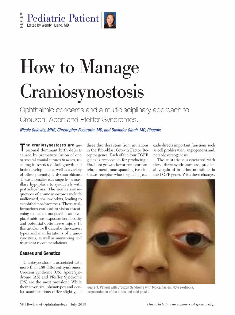
This article has no commercial sponsorship.
Edited by Wendy Huang, MDRE
VIE
W Pediatric Patient
58 | Review of Ophthalmology | July 2018
The craniosynostoses are au-tosomal dominant birth defects
caused by premature fusion of one or several cranial sutures in utero, re-sulting in restricted skull growth and brain development as well as a variety of other phenotypic dysmorphisms. These anomalies can range from max-illary hypoplasia to syndactyly with psittichorhina. The ocular conse-quences of craniosynostoses include malformed, shallow orbits, leading to exophthalmos/proptosis. These mal-formations can lead to vision-threat-ening sequelae from possible amblyo-pia, strabismus, exposure keratopathy and potential optic nerve injury. In this article, we’ll describe the causes, types and manifestations of cranio-synostosis, as well as monitoring and treatment recommendations.
Causes and Genetics
Craniosynostosis is associated with more than 180 different syndromes. Crouzon Syndrome (CS), Apert Syn-drome (AS) and Pfeiffer Syndrome (PS) are the most prevalent. While their severities, phenotypes and ocu-lar manifestations differ slightly, all
three disorders stem from mutations in the Fibroblast Growth Factor Re-ceptor genes. Each of the four FGFR genes is responsible for producing a fi broblast growth factor receptor pro-tein, a membrane-spanning tyrosine kinase receptor whose signaling cas-
cade directs important functions such as cell proliferation, angiogenesis and, notably, osteogenesis.
The mutations associated with these three syndromes are, predict-ably, gain-of-function mutations in the FGFR genes. With these changes,
How to ManageCraniosynostosis Ophthalmic concerns and a multidisciplinary approach to Crouzon, Apert and Pfeiffer Syndromes.Nicole Salevitz, MHS, Christopher Fecarotta, MD, and Davinder Singh, MD, Phoenix
Figure 1. Patient with Crouzon Syndrome with typical facies. Note exotropia,
excyclorotation of the orbits and mild ptosis.
058_rp0718_peds.indd 58058_rp0718_peds.indd 58 6/22/18 5:03 PM6/22/18 5:03 PM

July 2018 | reviewofophthalmology.com | 59
FGFR proteins become better ligand binders—hyperactive, or ligand-independent, receptors that result in an abnormally quick ossification of the cranial sutures. Although ge-netic analysis has made great strides in identifying the precise mutations responsible for each syndrome, espe-cially with AS, in which 98 percent of cases stem from one of two point mu-tations,1 the vast majority of FGFR-related craniosynostosis syndromes are examples of allelic heterogeneity. Thus, clinically the focus is often on phenotype instead of specifi c geno-type.
Clinical Presentations
The mutations are completely pen-etrant, meaning all carriers have traits, yet the carriers exhibit variable ex-pressivity. Therefore, clinical presen-tations can vary. Crouzon Syndrome is the most common craniosynosto-sis and is the mildest FGFR-related craniosynostosis phenotypically. The degree of facial deformity is often less severe and there is no involvement of the extremities. First described in 1912, the Crouzon disease triad consists of calvarial deformities, facial anomalies and proptosis,2 and patients rarely exhibit developmental delay.
Conversely, patients with Apert and Pfeiffer Syndromes can have mild to moderate intellectual disabilities and extremity involvement, in addition to the facial deformities exhibited in CS. AS patients often present with syn-dactyly in hands and feet, and PS pa-tients possess variable brachydactyly and broad, medially deviated thumbs and toes.
The wide range of clinical presen-tations of these syndromes is due to the variable nature of the cranial su-tures involved in each case. Although early fusion of the coronal suture is the most common manifestation, CS, AS and PS often exhibit complex or even pansynostosis, involving several
if not all sutures. Such fusion means growth can’t occur in a direction per-pendicular to the suture, thus limiting the skull’s development anteriorly and leading to the common anatomical characteristics shared by the three syndromes: hypertelorism; proptosis; and midface hypoplasia. (Figures 1 and 2)
Ocular Manifestations
The complications due to facial hypoplasia and shallow orbits are numerous and consistent between the different syndromes; however, severity will vary. Although there is some evidence that FGFR2 may have a direct role in ocular anterior cham-ber development,3 the ophthalmic se-quelae of craniosynostosis is truly due to defi cient anterior calvarial growth, midfacial hypoplasia and increased intracranial pressure.
The most common cause of de-creased visual acuity in these patients is proptosis-related strabismus leading to amblyopia.4 Shallow orbits increase the risk of exocyclorotated orbits and, therefore, aberrant insertion or even malformation of the extraocular mus-cles. This orbital abnormality often results in incomitant strabismus. Exo-tropia is the most common horizontal manifestation. The combination of shallow orbits, lack of inferior support due to midface hypoplasia and orbital fl oor recession results in overaction of the inferior oblique, leading to verti-cal misalignments. Collectively, this can manifest in V-pattern strabismus and, if untreated, eventual amblyopia.
However, amblyopia can be stra-bismus-independent and instead due to undiagnosed refractive errors and anisometropia. These patients exhibit higher rates of ametropia and astig-matism, contributing to a higher oc-
Figure 2. Profi le view highlighting shallow orbits with exophthalmos and proptosis,
psittichorhina, and maxillary hypoplasia.
058_rp0718_peds.indd 59058_rp0718_peds.indd 59 6/22/18 5:04 PM6/22/18 5:04 PM
PediatricPatient R
EV
IEW
60 | Review of Ophthalmology | July 2018
currence of refractive amblyopia.Another cause of amblyopia can be
related to lid ptosis observed alongside superior rectus hypoplasia and malfor-mation of the superior levator palpe-brae muscle. These patients may adopt a chin-up posture in an effort to com-pensate for ptosis. These abnormalities can cause deprivation amblyopia.
In addition, vision may be affected by optic nerve injury that can result from either severe exophthalmos caus-ing tension, increased intracranial pressure or a combination of the two. Shallow orbits (Figure 2) can also put the patient at risk for globe subluxation or herniation of the globe itself, con-tributing to optic nerve injury.
Lastly, corneal scarring from expo-sure keratopathy can be a major con-cern. Regular monitoring of the ocular surface for breakdown and dryness is required. This can occur with even small degrees of craniosynostosis-relat-ed proptosis. Whether it’s from struc-tural exposure, lid abnormalities or nasolacrimal dysfunction, the corneal epithelium can be at risk. Resulting scarring or surface irregularities can lead to refractive or deprivation am-blyopia with permanent vision loss.
Diagnosis
Craniosynostosis syndromes like CS, AS and PS may be detectable prena-tally by noting an aberrant skull shape and can be confirmed with a family history and genetic testing for FGFR mutations. Children may not present with proptosis immediately at birth, but the symptoms are progressive and will likely develop within the fi rst years of life. Thus, the role of the ophthal-mologist, initially, is to perform early, complete exams. Amblyopia should be monitored and it’s recommended that the patient undergo refraction with cycloplegic retinoscopy. Regular di-lated fundus exams are recommended to monitor for possible optic atrophy. If the patient is cooperative and able,
visual fi eld testing should be used to detect any loss of peripheral vision, and optical coherence tomography can visualize and monitor the optic nerves.
Treatment
There is no singular treatment to remedy all of the above symptoms, yet surgical release of the involved su-tures in the fi rst year of life can sig-nifi cantly improve brain development and reduce midface hypoplasia and proptosis. Increasing cranial volume and normalizing intracranial pressure are often the foremost goals of early treatment. Surgical treatment drasti-cally reduces the risk of optic atrophy due to increased intracranial pressure.
Regardless of concomitant manifes-tations of the syndrome, amblyopia, whether secondary to refractive error, deprivation, strabismus or a combi-nation, should be treated as early as possible after confi rmation via a com-plete eye exam. For refractive causes, proper correction should be given in the form of spectacles. If a difference in vision persists even with proper re-fractive correction, patching or atro-pinization of the dominant eye may be necessary. It’s critical to start early and work to maintain vision until major craniofacial surgeries are completed and any necessary strabismus surgeries are performed.
Ideally, strabismus surgery should be performed at the earliest possible time to facilitate the development of proper vision and binocularity; however, if sur-gery to advance the midface or adjust the orbit is anticipated, strabismus sur-gery should be delayed six months to a year,5,6 as such craniofacial surgery will alter the orbital volume and ability to achieve perfect alignment.
When strabismus surgery is per-formed, it can be complicated due to the wide range of anatomical anoma-lies. Surgery should be tailored to the specific misalignment with a goal of orthophoria and improved binocular-
ity. Again, particular attention must be paid to alignment in side gaze due to vertical muscle dysfunction.
Additionally, steps should be taken early to address any signs of exposure keratopathy. Treatment often con-sists of lubrication and taping shut the eyes at night. Associated ptosis can be protective and prevent corneal break-down despite signifi cant proptosis. If the proptosis does result in exposure keratopathy, temporary partial lateral tarsorrhaphy or orbital decompression may be necessary. The latter also has the added benefi t of further alleviating the possible risk of compressive optic nerve injury.
In conclusion, the ophthalmic con-cerns for a patient with craniosynos-tosis are extensive and variable, and early, full exams should be performed to catch and assess amblyopia and other symptoms as soon as possible. However, whether a child presents with Crouzon, Apert, Pfeiffer or an-other such syndrome, complete man-agement demands a multidisciplinary approach. The subspecialists involved should include ophthalmologists, cra-niofacial surgeons, geneticists, pediat-ric maxillofacial surgeons and, possibly, psychologists and therapists.
Ms. Salevitz is a medical student at the University of Arizona College of Medicine.
Dr. Fecarotta is an attending phy-sician and Dr. Singh is chief of the plastic surgery division at Phoenix Children’s Hospital.
1. Ferreira JC, Carter SM, Bernstein PS, et al. Second-trimester molecular prenatal diagnosis of sporadic Apert syndrome following suspicious ultrasound fi ndings. Ultrasound Obstet Gynecol 1999;14:426–30.2. Crouzon F. Dysostose cranio-faciale hereditaire. Bull Mem Soc Med Hop Paris 1912;33:545–555.3. K. Okajima, L.K. Robinson, M.A. Hart, D.N. Abuelo, L.S. Cowan, T. Hasegawa, et al. Ocular anterior chamber dysgenesis in craniosynostosis syndromes with a fi broblast growth factor receptor 2 mutation, Am J Med Genet 1999;85:160-170.4. Gray TL, Casey T, Selva D, et al. Ophthalmic sequelae of Crouzon syndrome. Ophthalmology 2005;112:1129–1134.5. G.R. Diamond, L. Whitaker. Ocular motility in craniofacial reconstruction Plast Reconstr Surg 1984;73:31-37.6. Morax S. Change in eye position after cranio-facial surgery. J Maxillofac Surg 1984;12:47-55.
058_rp0718_peds.indd 60058_rp0718_peds.indd 60 6/22/18 5:04 PM6/22/18 5:04 PM
ResearchReview R
EV
IEW
Primary Tube vs. Trabeculectomy Follow-up
In an effort to report one-year treat-ment outcomes in the Primary
Tube Versus Trabeculectomy study, researchers conducted a multicenter, randomized clinical trial of 242 eyes of 242 patients with medically un-controlled glaucoma and no previous incisional ocular surgery, including 125 in the tube group and 117 in the trabeculectomy group.
Patients were enrolled at 16 clini-cal centers and assigned randomly to treatment with a tube shunt (350-mm2 Baerveldt glaucoma implant) or trabeculectomy with mitomycin C. Primary outcome measures were in-traocular pressure, glaucoma medi-cal therapy, visual acuity, visual fi elds, surgical complications and failure de-
fi ned as IOP of more than 21 mmHg; IOP reduced by less than 20 percent from baseline; IOP of 5 mmHg or less; reoperation for glaucoma; or loss of light-perception vision.
The cumulative probability of fail-ure during the fi rst year of follow-up was 17.3 percent in the tube group and 7.9 percent in the trab-eculectomy group (p=0.01; hazard ratio, 2.59; 95 percent confidence interval, 1.20 to 5.60). Mean IOP was 13.8 ±4.1 mmHg in the tube group and 12.4 ±4.4 mmHg in the trabeculectomy group at one year (p=0.01). The number of glaucoma medications was 2.1 ±1.4 in the tube group and 0.9 ±1.4 in the trabecu-lectomy group (p<0.001). Postop-erative complications developed in 36 patients (29 percent) in the tube group and 48 patients (41 per-cent) in the trabeculectomy group
(p=0.06). Serious complications re-quiring reoperation or producing a loss of two Snellen lines or more occurred in one patient (1 percent) in the tube group and eight patients (7 percent) in the trabeculectomy group (p=0.03).
According to these researchers, trabeculectomy with MMC had a higher surgical success rate than tube shunt implantation after one year in the PTVT Study. Lower IOP with use of fewer glaucoma medications was achieved after trabeculectomy with MMC compared with tube shunt surgery during the fi rst year of follow-up. However, the frequency of serious complications producing vision loss or requiring reoperation was lower after tube shunt surgery than trabeculectomy with MMC.
Ophthalmology 2018;125:650-663Gedde SJ, Feuer WJ, Shi W, et al.
(Continued from page 47)
046_rp0718_rr.indd 61046_rp0718_rr.indd 61 6/22/18 4:50 PM6/22/18 4:50 PM

62 | Review of Ophthalmology | July 2018
REVIEW Classifi eds
Career Opportunities
Career Opportunities
CONTACT US TODAY888-498-1460
classifiedsopti
FIND MORE PRACTICES FOR SALE ON
Practice For Sale
OPHTHALMOLOGISTSDanbury, CT
with long standing Ophthalmolo-gist in Danbury, CT. High quality
equipment. $2,250 per month or
equipment- $1,750 per month.
203-545-3539 or email [email protected]
FOR CLASSIFIED ADVERTISING
CONTACT US TODAY:
Toll free: 888-498-1460
E-mail: [email protected]
Targeting Ophthalmologists?
CLASSIFIED ADVERTISING
WORKS
Contact us today for classified advertising:Toll free: 888-498-1460
E-mail: [email protected]
ROPH0718.indd 62 6/13/18 1:00 AM
RE
VIE
WResident Case SeriesWills EyeWills EyeWills EyeWills Eye Edited by Thomas Jenkins, MD
July 2018 | reviewofophthalmology.com | 63
Presentation
An 80-year-old female presented with a two-month history of binocular horizontal diplopia. Her symptoms started im-mediately following a motor vehicle accident. She lost consciousness at the time of the accident. Computed tomography of the head and orbits showed no acute intracranial or intraorbital abnormalities. After discharge from an outside hospital, she was seen by a local ophthalmologist and continued to complain of constant horizontal binocular diplopia. She was then referred for evaluation of possible extraocular muscle entrapment.
Medical History
Past medical history was signifi cant for psoriasis treated with ustekinumab. She denied any history of diabetes or hyper-tension. Past surgical history included an appendectomy in childhood and a remote history of unspecifi ed knee and elbow surgeries. Her family history was signifi cant for diabetes, hypertension, heart disease and thyroid disease. The patient de-nied tobacco and illicit drug use; she drank alcohol only socially.
Examination
The patient’s vital signs were within normal limits. Ophthalmologic examination demonstrated a visual acuity of 20/80 in the right eye, improving to 20/40 with pinhole, and 20/25 in the left eye with no improvement with pinhole. Pupils were equally round and briskly reactive to light, without relative afferent pupillary defect. Visual fi elds were full to confrontation bilaterally. Ishihara color plates were brisk and full (8/8) bilaterally. External examination showed deep superior sulci in both upper eyelids with symmetric lid position. Hertel exophthalmometry was symmetric. A right esotropia was present in primary gaze. Extraocular motility measurements revealed an incomitant strabismus (Figure 1). A mild right supraorbital hypesthesia was present and infraorbital nerve function was normal. Intraocular pressure by Goldmann tonometry was 9 mmHg on the right and 12 mmHg on the left.
Anterior segment slit lamp examination was unremarkable bilaterally. Dilated funduscopic examination of the right eye was normal. On the left, one intraretinal hemorrhage was noted nasally.
Forced duction and force generation testing were performed after instillation of viscous lidocaine. Forced duction testing of the right eye was normal. Force generation testing confi rmed a paretic right lateral rectus.
Phoebe Mellen, MD, and Jurij Bilyk, MD
Double vision after a car accident brings an elderly woman to Wills Eye Hospital for an evaluation.
What is your diagnosis? What further workup would you pursue? The diagnosis appears on p. 64.
063_rp0718_wills.indd 63063_rp0718_wills.indd 63 6/22/18 4:47 PM6/22/18 4:47 PM

RE
VIE
W
64 | Review of Ophthalmology | July 2018
Resident Case Series
Given the onset following trauma, normal orbital CT, and clinical findings, a diagnosis of traumatic CN VI palsy (abducens nerve palsy) was made. Observation with serial ex-
aminations for s p o n t a n e o u s recovery was recommended.
Workup, Diagnosis and Treatment
Horizontal diplopia of acute on-set has multiple etiologies, includ-ing paresis of the abducens nerve (e.g., ischemia from microvascular disease, ischemia from giant cell ar-teritis or increased intracranial pres-sure), dysfunction of the lateral rec-tus (e.g., laceration or contusion), medial rectus muscle restriction (e.g., medial orbital wall fracture, thyroid eye disease or orbital mass), and neuromuscular junction abnor-malities (e.g., myasthenia gravis), among others. In an 80-year-old woman, the two most dangerous etiologies for abducens nerve palsy are giant cell arteritis and increased intracranial pressure. In this case, the patient’s recent motor vehicle accident points to two probable causes: abducens nerve palsy from intracranial injury (with or without increased intracranial pressure) or medial rectus muscle restriction from a fracture of the lamina papy-
racea. The results of the forced duc-tion and force generation tests ruled out a restrictive strabismus.
Traumatic cranial neuropathies occur in up to 13 percent of all head traumas.1 Furthermore, motor ve-hicle accidents are the leading cause of intracranial injury that results in cranial neuropathies.1 In the acute setting of diplopia after a trauma, a thorough history detailing the mechanism of trauma and any co-existing injuries should be elicited. Characteristics of the diplopia, in-cluding direction—horizontal, verti-cal, oblique—and binocularity/mon-ocularity should be clarifi ed with the patient. A full ophthalmic examina-tion, including a dilated fundus ex-amination to rule out globe rupture, should be performed. In cases in which the etiology is unclear, forced duction and force generation testing are helpful in differentiating par-alytic from restrictive strabismus.
Review of head and orbital imaging studies often clarifi es the etiology of the diplopia.
Forced duction testing is a rela-tively simple diagnostic maneuver that checks for restriction (Figure 2A). It doesn’t require patient co-operation and can therefore be per-formed on patients who are coma-tose or heavily sedated in the setting of trauma. Viscous lidocaine is high-ly effective in providing conjunctival anesthesia. Two large-toothed (0.5-mm Castroviejo or Bishop Harmon) forceps are used to grasp the limbal conjunctiva fi rmly 90 degrees from the muscle of interest. It should be noted that grasping the muscle in-sertion is painful and runs the risk of a corneal abrasion if the forceps slip. The globe is then moved away from the nidus of suspected restriction. The globe will move smoothly away from a normal extraocular muscle, while an entrapped or enlarged
Discussion
Figure 1. External photograph demonstrating extraocular
motility in all fi elds of gaze. Note the decreased
abduction in right gaze.
063_rp0718_wills.indd 64063_rp0718_wills.indd 64 6/22/18 4:47 PM6/22/18 4:47 PM
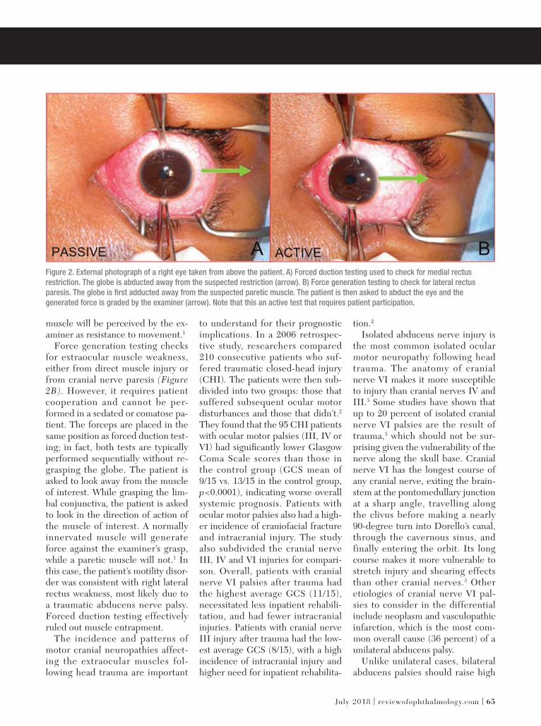
July 2018 | reviewofophthalmology.com | 65
muscle will be perceived by the ex-aminer as resistance to movement.1
Force generation testing checks for extraocular muscle weakness, either from direct muscle injury or from cranial nerve paresis (Figure 2B). However, it requires patient cooperation and cannot be per-formed in a sedated or comatose pa-tient. The forceps are placed in the same position as forced duction test-ing; in fact, both tests are typically performed sequentially without re-grasping the globe. The patient is asked to look away from the muscle of interest. While grasping the lim-bal conjunctiva, the patient is asked to look in the direction of action of the muscle of interest. A normally innervated muscle will generate force against the examiner’s grasp, while a paretic muscle will not.1 In this case, the patient’s motility disor-der was consistent with right lateral rectus weakness, most likely due to a traumatic abducens nerve palsy. Forced duction testing effectively ruled out muscle entrapment.
The incidence and patterns of motor cranial neuropathies affect-ing the extraocular muscles fol-lowing head trauma are important
to understand for their prognostic implications. In a 2006 retrospec-tive study, researchers compared 210 consecutive patients who suf-fered traumatic closed-head injury (CHI). The patients were then sub-divided into two groups: those that suffered subsequent ocular motor disturbances and those that didn’t.2
They found that the 95 CHI patients with ocular motor palsies (III, IV or VI) had signifi cantly lower Glasgow Coma Scale scores than those in the control group (GCS mean of 9/15 vs. 13/15 in the control group, p<0.0001), indicating worse overall systemic prognosis. Patients with ocular motor palsies also had a high-er incidence of craniofacial fracture and intracranial injury. The study also subdivided the cranial nerve III, IV and VI injuries for compari-son. Overall, patients with cranial nerve VI palsies after trauma had the highest average GCS (11/15), necessitated less inpatient rehabili-tation, and had fewer intracranial injuries. Patients with cranial nerve III injury after trauma had the low-est average GCS (8/15), with a high incidence of intracranial injury and higher need for inpatient rehabilita-
tion.2
Isolated abducens nerve injury is the most common isolated ocular motor neuropathy following head trauma. The anatomy of cranial nerve VI makes it more susceptible to injury than cranial nerves IV and III.3 Some studies have shown that up to 20 percent of isolated cranial nerve VI palsies are the result of trauma,3 which should not be sur-prising given the vulnerability of the nerve along the skull base. Cranial nerve VI has the longest course of any cranial nerve, exiting the brain-stem at the pontomedullary junction at a sharp angle, travelling along the clivus before making a nearly 90-degree turn into Dorello’s canal, through the cavernous sinus, and finally entering the orbit. Its long course makes it more vulnerable to stretch injury and shearing effects than other cranial nerves.3 Other etiologies of cranial nerve VI pal-sies to consider in the differential include neoplasm and vasculopathic infarction, which is the most com-mon overall cause (36 percent) of a unilateral abducens palsy.
Unlike unilateral cases, bilateral abducens palsies should raise high
Figure 2. External photograph of a right eye taken from above the patient. A) Forced duction testing used to check for medial rectus
restriction. The globe is abducted away from the suspected restriction (arrow). B) Force generation testing to check for lateral rectus
paresis. The globe is fi rst adducted away from the suspected paretic muscle. The patient is then asked to abduct the eye and the
generated force is graded by the examiner (arrow). Note that this an active test that requires patient participation.
063_rp0718_wills.indd 65063_rp0718_wills.indd 65 6/22/18 4:47 PM6/22/18 4:47 PM

RE
VIE
W
Resident Case Series
For advertising opportunities contact:
Michele Barrett (610) 492-1014 or [email protected]
James Henne (610) 492-1017 or [email protected]
Michael Hoster (610) 492-1028 or [email protected]
Alcon Laboratories 2-3
Phone (800) 451-3937
Fax (817) 551-4352
Bausch + Lomb 8, 19
Phone (800) 323-0000
Fax (813) 975-7762
Beaver-Visitec International, Inc. 7
Phone (866) 906-8080Fax (866) 906-4304
www.beaver-visitec.com
Compulink 17
Phone (800) 456-4522
www.compulinkadvantage.com
Keeler Instruments 11
Phone (800) 523-5620
Fax (610) 353-7814
Lacrimedics, Inc 33
Phone (800) 367-8327Fax (253) 964-2699 [email protected]
www.lacrimedics.com
NuSight Medical Operations 29, 31
Phone (833) 468-5437
www.NuSightMedical.com
Omeros 5
Phone (206) 676-5000
Fax (206) 676-5005
Regeneron Pharmaceuticals, Inc. 21
Phone (914) 847-7000
www.regeneron.com
S4OPTIK 45, 47
Phone (888) 224-6012
Shire Ophthalmics 67, 68
www.shire.com
Sun Ophthalmics 12-13, 14
SunIsOnTheRise.com
AdvertisingIndexR
EV
IEW
This advertiser index is published as a convenience and not as part of the advertising contract. Every care will be taken to index correctly. No allowance will be made for errors due to spelling, incorrect page number, or failure to insert.
66 | Review of Ophthalmology | July 2018
suspicion for brainstem lesions and intracranial hem-orrhage. Increased intracranial pressure must be ruled out expeditiously in any patient presenting with a posttraumatic abducens nerve palsy; an immediate examination of the optic nerve heads with a direct ophthalmoscope is warranted, and if disc swelling is identifi ed, emergency neuroimaging with CT must be performed.
Management of traumatic abducens nerve palsies is typically conservative, with serial observation over several months. In a study from 1998, the recovery rate of cranial nerve VI palsies after trauma was re-ported to be 73 percent, with a rate as high as 84 per-cent in unilateral cases.4 In a 2001 study, research-ers identified predictors of nonrecovery, such as bilaterality and a complete palsy at presentation with inability to abduct past the midline; this conferred the highest risk ratio of 9.11.5 In our case, the patient had a unilateral partial paresis, which connotes a relatively good prognosis for recovery. Strabismus surgery should be delayed for at least six months or longer following traumatic palsy to allow for sponta-neous recovery. Diplopia symptoms can be managed temporarily with occlusion of one eye or with Fresnel prisms; permanent prisms should be avoided in the acute phase due to their high cost.
In conclusion, diplopia after a motor vehicle ac-cident can be a symptom of many underlying etiolo-gies, the most dangerous of which is increased intra-cranial pressure. Critical examination techniques include measurement of the strabismus pattern as well as forced duction and force generation testing, which can help differentiate between a paretic and an entrapped extraocular muscle as the source of dip-lopia. Abducens nerve palsies are the most common traumatic ocular motor nerve palsy.
1. Murchison A, Bilyk J, Savino P. Traumatic Cranial Neuropathies. In: Black E, Nesi F, Cal-vano CJ, Glastone GJ, Levine MR. Smith and Nesi’s Ophthalmic Plastic and Reconstructive Surgery. 3rd Ed. Springer New York. 2012:165-199.2. Dhaliwal A, West AL, Trobe JD, Musch DC. Third, fourth, and sixth cranial nerve palsies following closed head injury. J Neuroophthalmol. 2006;26:4-10.3. Elder C, Hainline C, Galetta SL, Blacer LJ, Rucker JC. Isolated abducens nerve palsy: Update on evaluation and diagnosis. Curr Neurol and Neurosci Rep 2016; 16:1-7.4. Holmes JM, Droste PJ, Beck RW. The natural history of acute traumatic sixth nerve palsy or paresis. J AAPOS 1998;2:265-810.1007/s11910-016-0671-4.5. Holmes JM, Beck RW, Kip KE, Droste PJ, Leske DA, Pediatric Eye Disease Investigator Group. Predictors of nonrecovery in acute traumatic sixth nerve palsy and paresis. Ophthalmology 2001;108:1457-60.
063_rp0718_wills.indd 66063_rp0718_wills.indd 66 6/22/18 4:47 PM6/22/18 4:47 PM

BRIEF SUMMARY: Consult the Full Prescribing Information for complete product information.
INDICATIONS AND USAGE Xiidra®
DOSAGE AND ADMINISTRATION Instill one drop of Xiidra twice daily (approximately 12 hours
Contact lenses should be removed prior to the administration
administration.
CONTRAINDICATIONS Xiidra is contraindicated in patients with known hypersensitivity
formulation.
ADVERSE REACTIONS Clinical Trials Experience Because clinical studies are conducted under widely varying conditions, adverse reaction rates observed in clinical studies of a drug cannot be directly compared to rates in the clinical
approximately 12 months. The majority of the treated patients
dysgeusia and reduced visual acuity. Other adverse reactions
conjunctival hyperemia, eye irritation, headache, increased lacrimation, eye discharge, eye discomfort, eye pruritus and sinusitis. Postmarketing Experience
postapproval use of Xiidra. Because these reactions are reported voluntarily from a population of uncertain size, it is not always possible to reliably estimate their frequency or establish a causal relationship to drug exposure.
Rare cases of hypersensitivity, including anaphylactic reaction, bronchospasm, respiratory distress, pharyngeal edema, swollen tongue, and urticaria have been reported. Eye swelling and rash have been reported.
USE IN SPECIFIC POPULATIONS Pregnancy There are no available data on Xiidra use in pregnant women to
relevant systemic exposures. Intravenous administration of
an increased incidence of omphalocele at the lowest dose
the recommended human ophthalmic dose [RHOD], based on
risk of Xiidra use in humans during pregnancy is unclear.
Animal Data
an increase in mean preimplantation loss and an increased
the RHOD of Xiidra, based on AUC. No teratogenicity was
an increased incidence of omphalocele was observed at the
the rabbit.
Lactation
milk, the effects on the breastfed infant, or the effects on milk
ocular administration is low. The developmental and health
the mother’s clinical need for Xiidra and any potential adverse effects on the breastfed child from Xiidra.
Pediatric Use
years have not been established.
Geriatric Use No overall differences in safety or effectiveness have been observed between elderly and younger adult patients.
NONCLINICAL TOXICOLOGY Carcinogenesis, Mutagenesis, Impairment of Fertility Carcinogenesis: Animal studies have not been conducted
Mutagenesis: in vitro
in vivo mouse micronucleus assay. In an in vitro chromosomal aberration assay using mammalian cells (Chinese
concentration tested, without metabolic activation. Impairment of fertility:
fertility and reproductive performance in male and female treated rats.
Marks designated ®
Patented: please see
Rx Only
RP0718_Shire PI.indd 1RP0718_Shire PI.indd 1 6/18/18 10:33 AM6/18/18 10:33 AM
For additional safety information, see accompanying Brief Summary of Safety Information on the adjacent page and Full Prescribing Information on Xiidra-ECP.com.
©2018 Shire US Inc., Lexington, MA 02421. 1-800-828-2088. All rights reserved. SHIRE and the Shire Logo
Marks designated ®
SIGNS & SYMPTOMSHAVE MET THEIIR MATCH
Xiidra is the only treatment FDA-approved to treat both signs and symptoms of Dry Eye
Indication Xiidra®
Important Safety Information
AT 12 WEEKS
Proven to treat
AT 6 AND 12 WEEKS
Proven to treat
Check it out at Xiidra-ECP.com
Xiidra is the only prescription eye drop to show symptom relief from eye dryness in 2 out of 4 clinical trialsIN AS LIITTLE AS 2 WEEKS
RP0718_Shire.indd 1RP0718_Shire.indd 1 6/18/18 10:31 AM6/18/18 10:31 AM