Embed Size (px)
Citation preview
UPPER EXTREMITY
NEURO EXAM & COMMON
PATHOLOGIESEdward Babigumira, M.D.
Board Certified PM&R, Pain Medicine
Introduction
• A good physical exam is the cornerstone art of a physiatrist. “we should own it”
• The art of a good neurologic exam is comprehensive, meticulous and provides a unique perspective to disease process.
• Should usually narrow down your differential diagnosis, help with recommending ancillary studies.

Goals
• Learn to scrutinize pt for unique physical and key findings
• To master the art of the upper extremity and neck neurologic exam
• To define the disabilties and handicaps that emanate from the disease
• Identify common nerve entrapments and syndromes encountered in our practice
Nervous system history
• Common chief
complaints:
• neck pain,
• shoulder pain,
• numbness, tingling,
parasthesia.
• weakness,
Swelling,clumsiness,
nocturnal pain.
• Limited range of
motion
History of Presenting Complaint
• Site
• Onset( Acute vs Chronic)
• Frequency
• Duration
• Precipitating and relieving factors( walking, neck movement, or rest)
• History of trauma.
Review of Systems
Sphincter disorder: bowel/bladder incontinence
• Weight Loss.(malignancy)
• Seizures,tremor, fatigue
• Fevers/Chills/septic source(eg teeth, etc)
• Skin marks: rashes,café-au-lait, angiomata.
• Cardiac murmurs, cyanosis, resp insuff, pulse
irregularity.
Past Med History
• Diabetes,hypertension,stroke
• Spinal cord injury(spondylosis, myelopathy,
radiculopathy, stenosis)
• Infections:( polio,HIV, syphilis,TB, fungal,
parasitic, abscess.
• Vit B12 Deficiency
• Syringomyelia,Multiple sclerosis
• Auto immnune Dz.
(Rheumatoid,Sjogrens,Lupus)
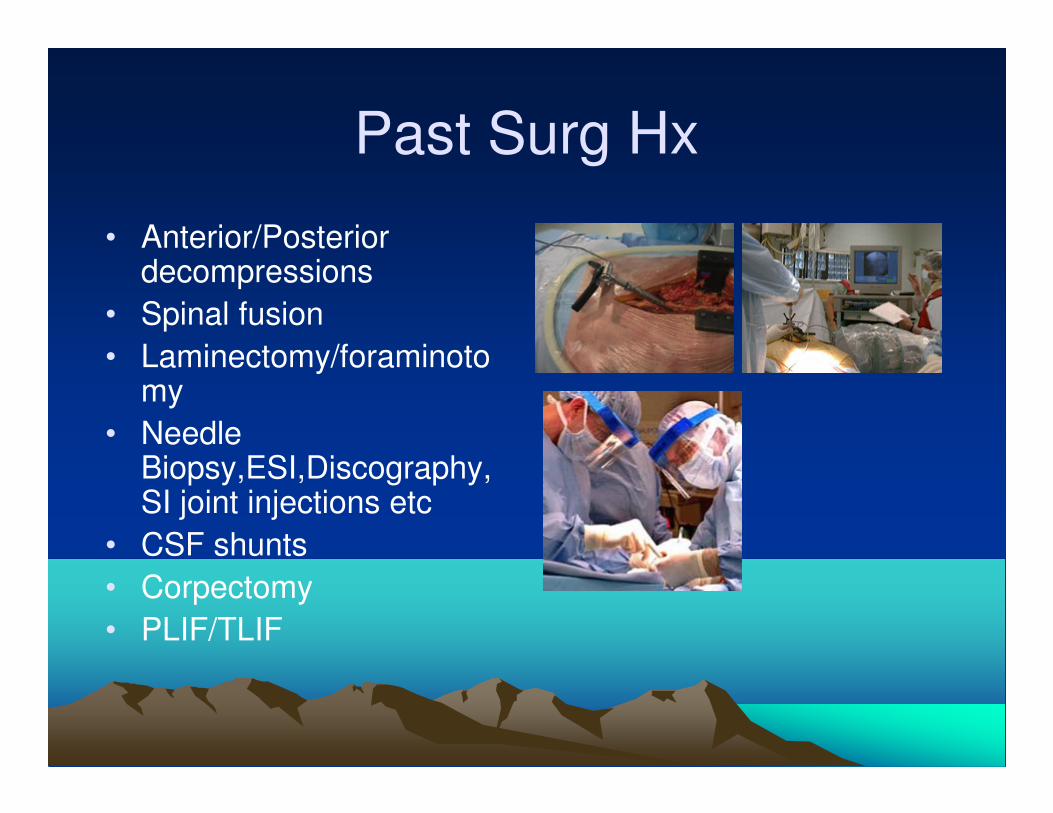
Past Surg Hx
• Anterior/Posterior decompressions
• Spinal fusion
• Laminectomy/foraminotomy
• Needle Biopsy,ESI,Discography,SI joint injections etc
• CSF shunts
• Corpectomy
• PLIF/TLIF
Social/Occupational History
• Alcohol consumption
• Smoking
• IVDA
• Employment, any work limitations, toxins, repetitive trauma.
• Pre-morbid function hx
• Exercise, lifestyle, etc
• Hx of litigation
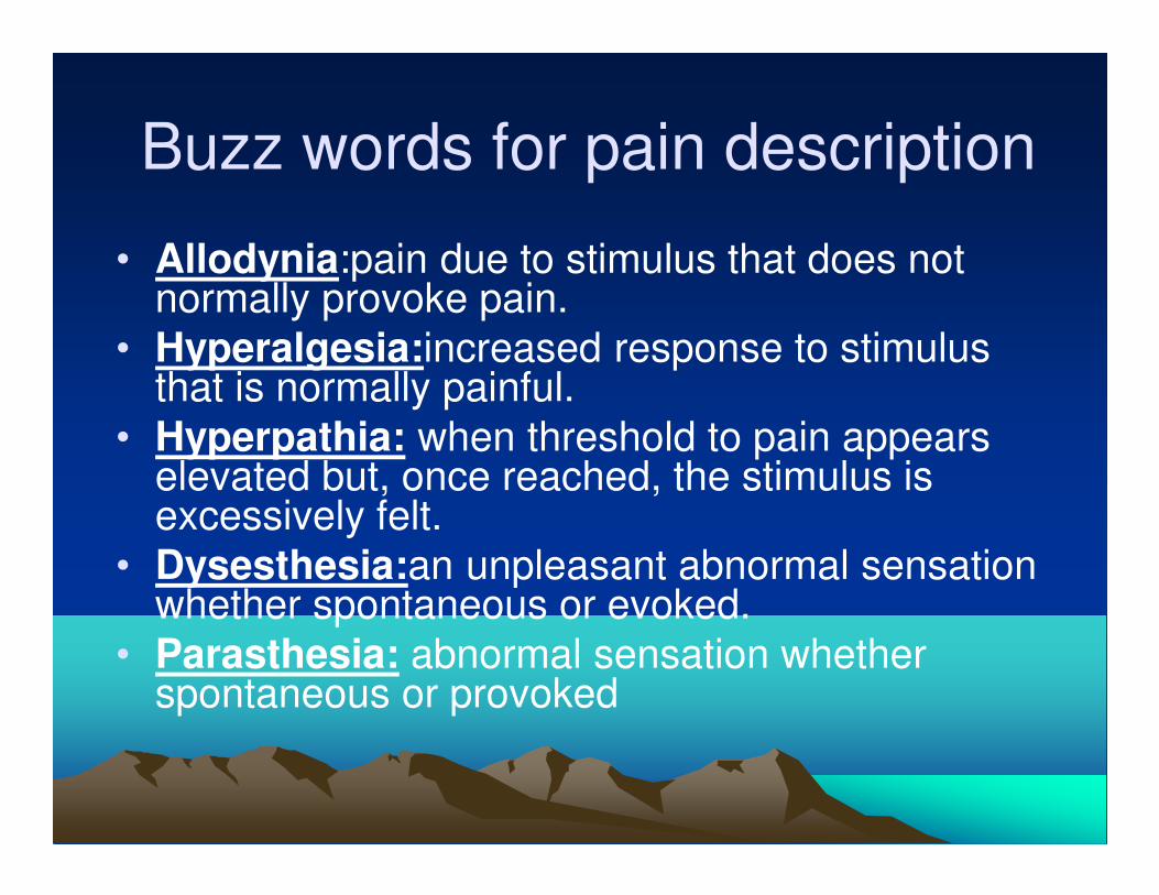
Buzz words for pain description
• Allodynia:pain due to stimulus that does not normally provoke pain.
• Hyperalgesia:increased response to stimulus that is normally painful.
• Hyperpathia: when threshold to pain appears elevated but, once reached, the stimulus is excessively felt.
• Dysesthesia:an unpleasant abnormal sensation whether spontaneous or evoked.
• Parasthesia: abnormal sensation whether spontaneous or provoked
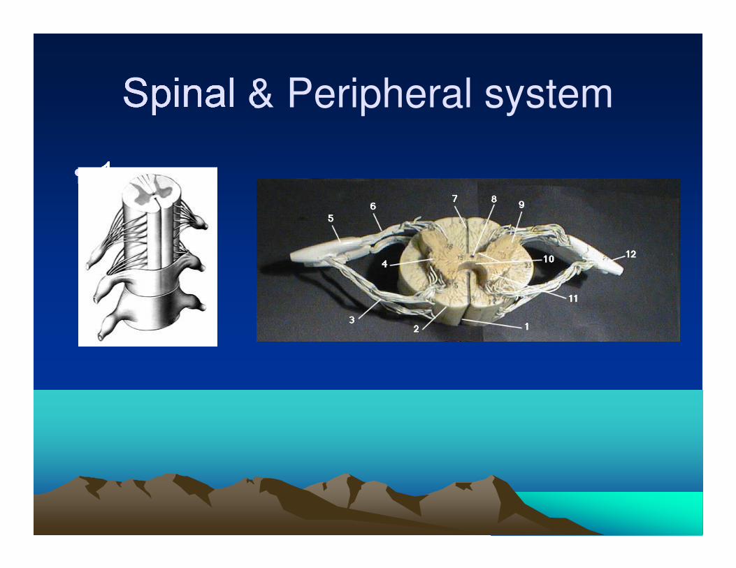
Spinal & Peripheral system
• 1
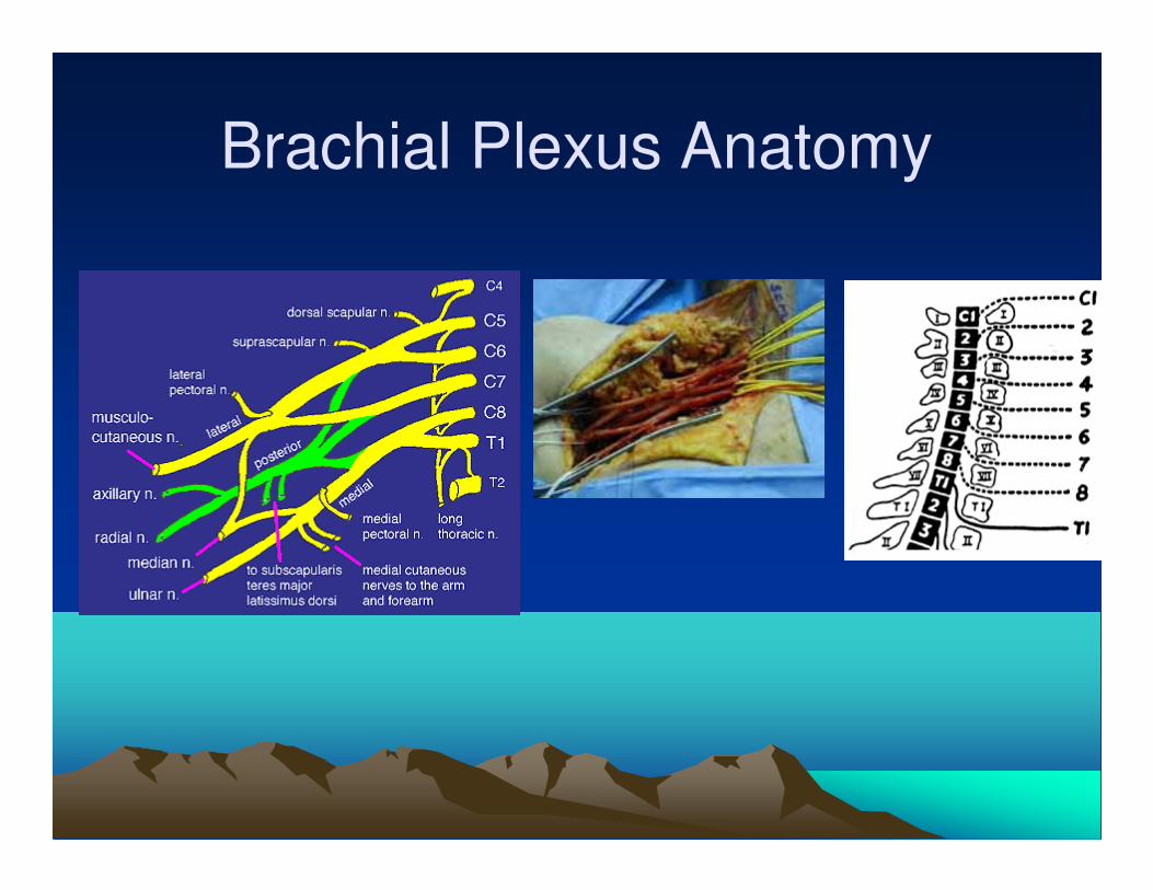
Brachial Plexus Anatomy
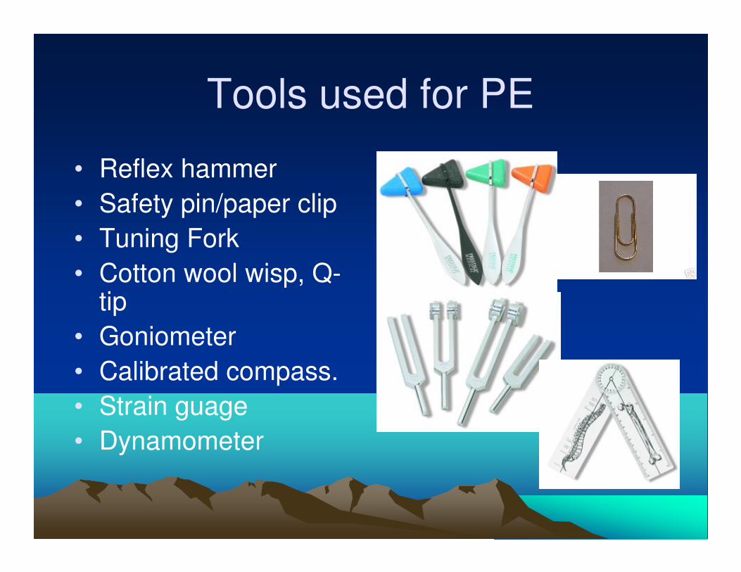
Tools used for PE
• Reflex hammer
• Safety pin/paper clip
• Tuning Fork
• Cotton wool wisp, Q-tip
• Goniometer
• Calibrated compass.
• Strain guage
• Dynamometer
Sensory exam
• Pain
• Light touch
• Temperature
• Joint position
• Vibration
• Two point Discrimination
• Sensory inattention
• Stereognosis
• Graphesthesia
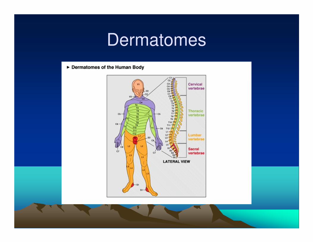
Dermatomes
• C5: shoulder
• C6: root of thumb
• C7:Middle digit
• C8: little digit
• T1: axilla.
• REMEMBER C7 EXTENDS DOWN MIDDLE FINGER.
Dermatomes
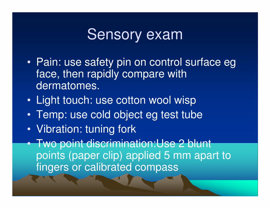
Sensory exam
• Pain: use safety pin on control surface eg face, then rapidly compare with dermatomes.
• Light touch: use cotton wool wisp
• Temp: use cold object eg test tube
• Vibration: tuning fork
• Two point discrimination:Use 2 blunt points (paper clip) applied 5 mm apart to fingers or calibrated compass
Sensory exam
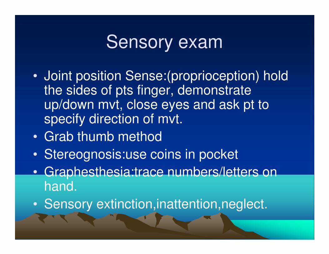
• Joint position Sense:(proprioception) hold the sides of pts finger, demonstrate up/down mvt, close eyes and ask pt to specify direction of mvt.
• Grab thumb method
• Stereognosis:use coins in pocket
• Graphesthesia:trace numbers/letters on hand.
• Sensory extinction,inattention,neglect.
Sensory exam
• Classification; with pin prick
• 0: no sensation at all
• 1: feels pressure, no sharp sensation
• 2: normal sensation
Motor exam
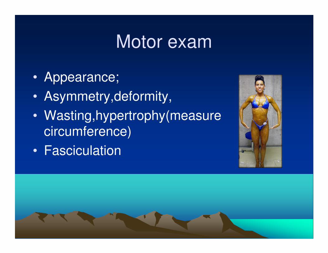
• Appearance;
• Asymmetry,deformity,
• Wasting,hypertrophy(measure circumference)
• Fasciculation
UMN Vs LMN lesion
• Upper motor neuron signs;
• Hypereflexia, spasticity increased tone, clonus,tremors,chorea,ballismus, athetosis dystonia, apraxia.
• Lower Motor Neuron signs;
• Hyporeflexia, flaccidity, atrophy and fasciculations.
Co-ordination
• Dysmetria; finger to nose, intention tremor
• Dysdiadochokinesia
• Arm bounce: excessive swinging
• Rebound phenomenon: flex elbow against resistance, sudden release may cause hand to strike face due to delay in triceps contraction.
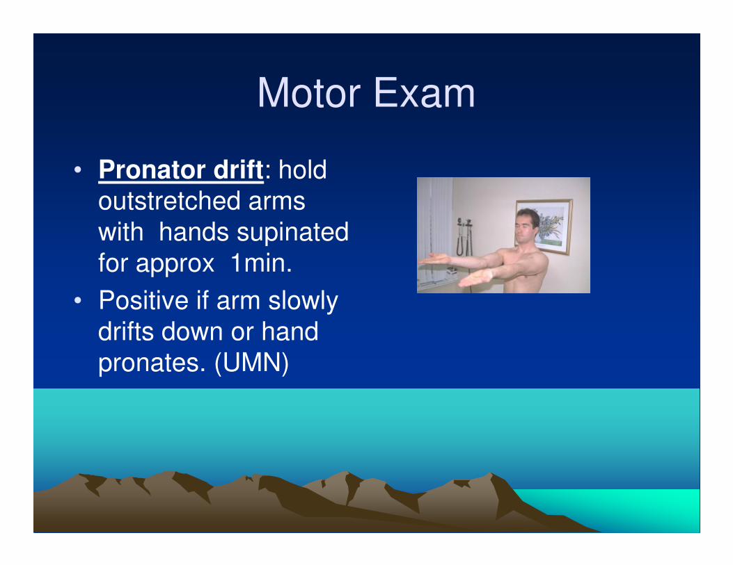
Motor Exam
• Pronator drift: hold
outstretched arms
with hands supinated
for approx 1min.
• Positive if arm slowly
drifts down or hand
pronates. (UMN)
Reflexes
• Biceps Jerk: C5,C6 musculocutaneous.nerve.
• Brachioradialis jerk: C5,C6
• Pronator teres jerk: C6,C7
• Supinator Jerk: C6,C7, radial nerve.
• Triceps jerk: C7, C8 radial nerve
• Hoffmann reflex :C7,C8.(Flick pts terminal
phalanx of third digit, suddenly stretching flexor
tendon on release,quick thumb flexion mvt
indicates UMN.)
Tone
• Assess by flexing and extending the elbow and wrist.
• Clasp knife: initial resistance to mvt, that is suddenly overcome.(UMN)
• Lead pipe: steady increase in resistance thru mvt.(extra pyramidal lesion)
• Cog wheel: Ratchet like increase in rigidity.(extrapyramidal lesion).

Ashworth Classification
• 0. No increase in muscle tone.
• 1. Slight increase in tone giving a “catch” when affected part is moved in flexion or extension.
• 1+. Slight increase in muscle tone, manifested by a catch, followed by minimal resistance throughout the reminder (less than half) of the range of motion.
• 2 More marked increase in muscle tone through most of the range of motion, but affected part(s) easily moved.
• 3. Considerable increase in tone; passive movement difficult.
• 4. Affected part is rigid in flexion or extension.
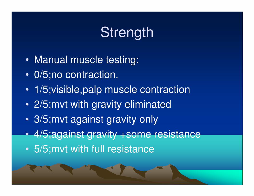
Strength
• Manual muscle testing:
• 0/5;no contraction.
• 1/5;visible,palp muscle contraction
• 2/5;mvt with gravity eliminated
• 3/5;mvt against gravity only
• 4/5;against gravity +some resistance
• 5/5;mvt with full resistance
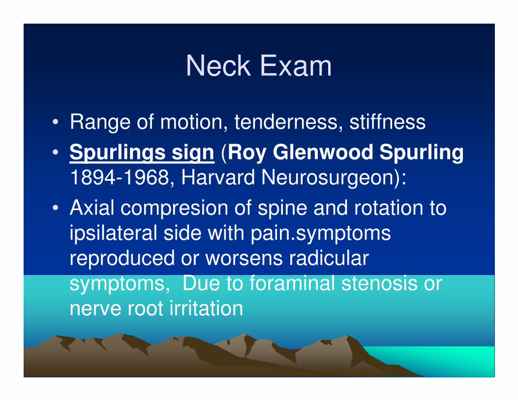
Neck Exam
• Range of motion, tenderness, stiffness
• Spurlings sign (Roy Glenwood Spurling1894-1968, Harvard Neurosurgeon):
• Axial compresion of spine and rotation to ipsilateral side with pain.symptoms reproduced or worsens radicular symptoms, Due to foraminal stenosis or nerve root irritation
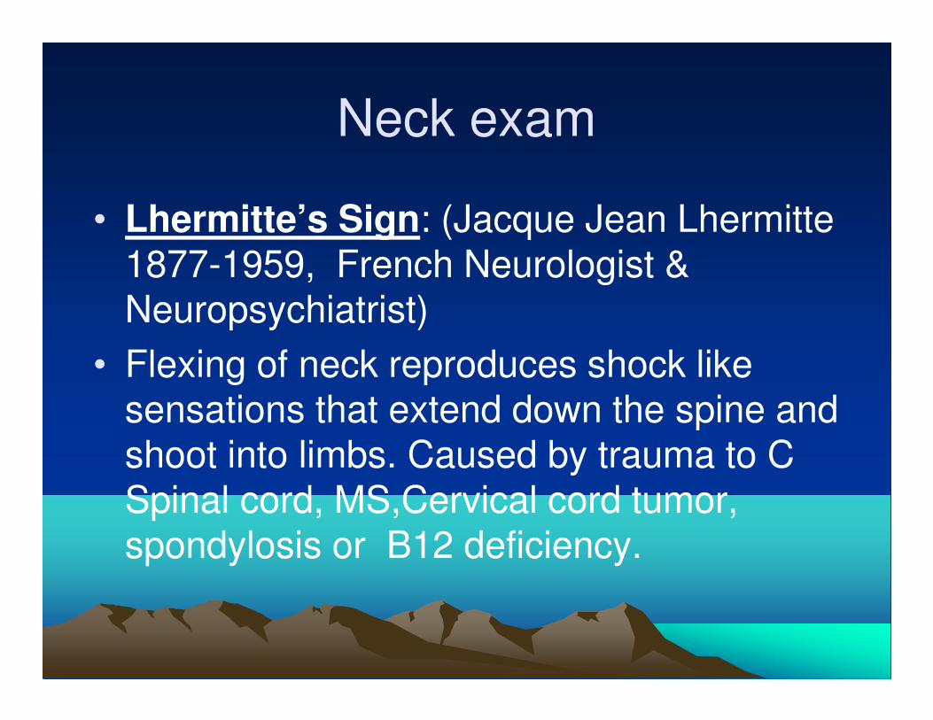
Neck exam
• Lhermitte’s Sign: (Jacque Jean Lhermitte 1877-1959, French Neurologist & Neuropsychiatrist)
• Flexing of neck reproduces shock like sensations that extend down the spine and shoot into limbs. Caused by trauma to C Spinal cord, MS,Cervical cord tumor, spondylosis or B12 deficiency.
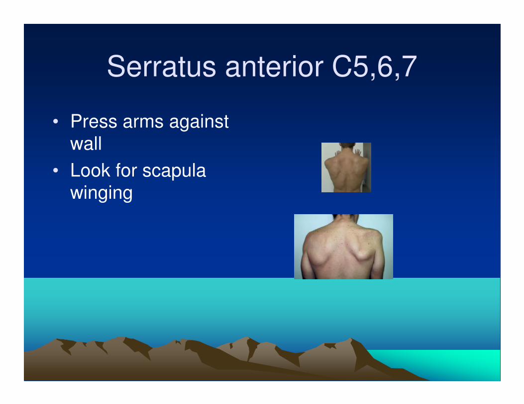
Serratus anterior C5,6,7
• Press arms against
wall
• Look for scapula
winging
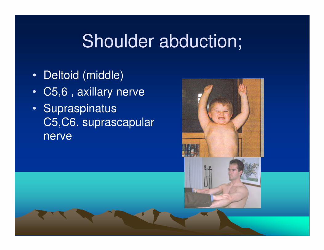
Shoulder abduction;
• Deltoid (middle)
• C5,6 , axillary nerve
• Supraspinatus
C5,C6. suprascapular
nerve
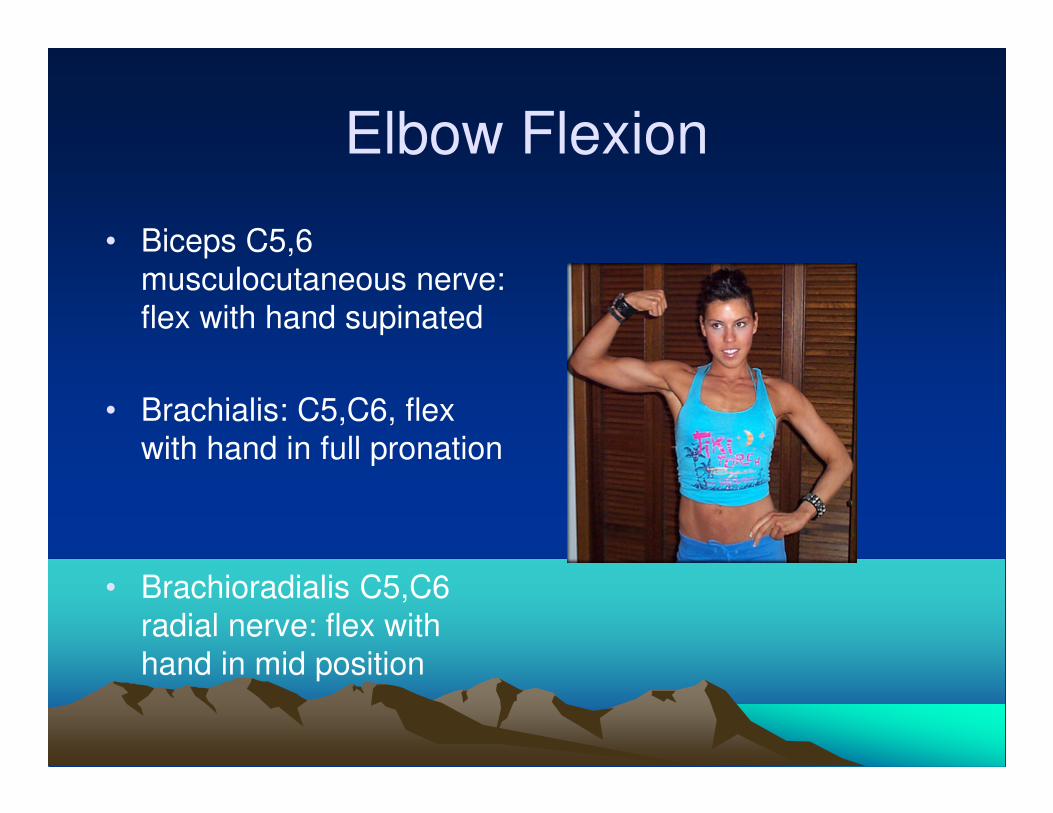
Elbow Flexion
• Biceps C5,6
musculocutaneous nerve:
flex with hand supinated
• Brachialis: C5,C6, flex
with hand in full pronation
• Brachioradialis C5,C6
radial nerve: flex with
hand in mid position
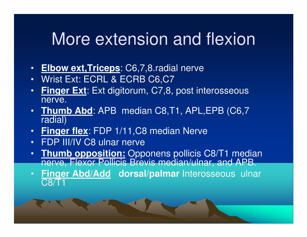
More extension and flexion
• Elbow ext,Triceps: C6,7,8.radial nerve
• Wrist Ext: ECRL & ECRB C6,C7
• Finger Ext: Ext digitorum, C7,8, post interosseous nerve.
• Thumb Abd: APB median C8,T1, APL,EPB (C6,7 radial)
• Finger flex: FDP 1/11,C8 median Nerve
• FDP III/IV C8 ulnar nerve
• Thumb opposition: Opponens pollicis C8/T1 median nerve, Flexor Pollicis Brevis median/ulnar, and APB.
• Finger Abd/Add dorsal/palmar Interosseous ulnar C8/T1
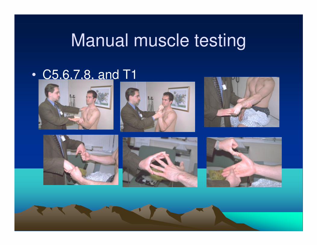
Manual muscle testing
• C5,6,7,8, and T1
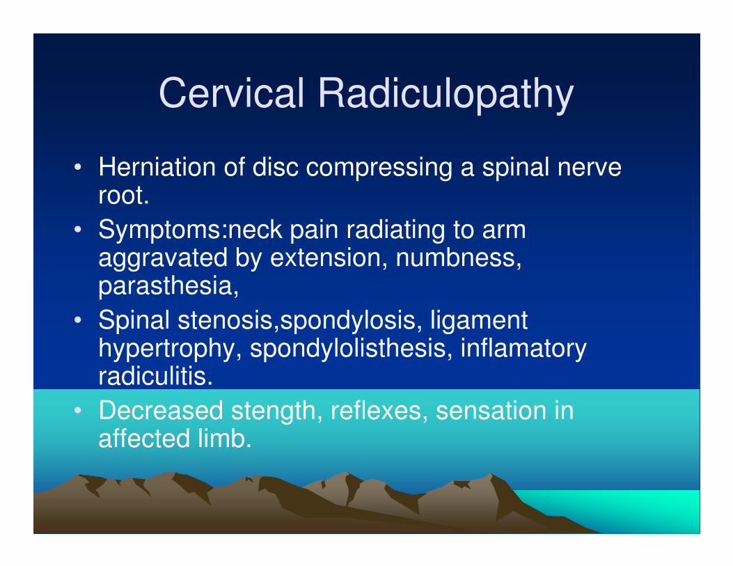
Cervical Radiculopathy
• Herniation of disc compressing a spinal nerve root.
• Symptoms:neck pain radiating to arm aggravated by extension, numbness, parasthesia,
• Spinal stenosis,spondylosis, ligament hypertrophy, spondylolisthesis, inflamatory radiculitis.
• Decreased stength, reflexes, sensation in affected limb.
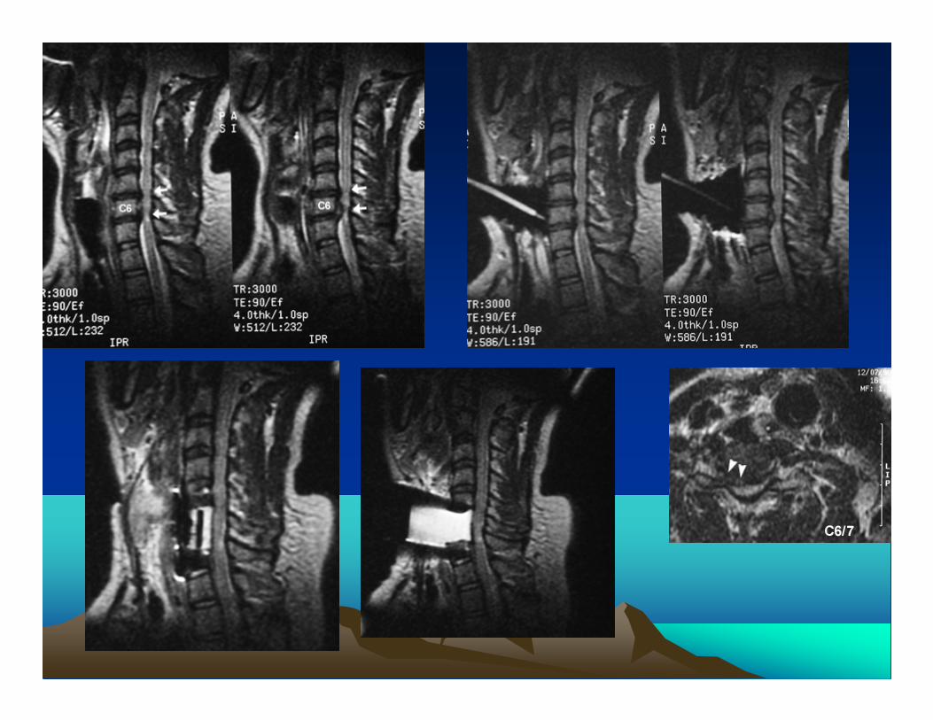
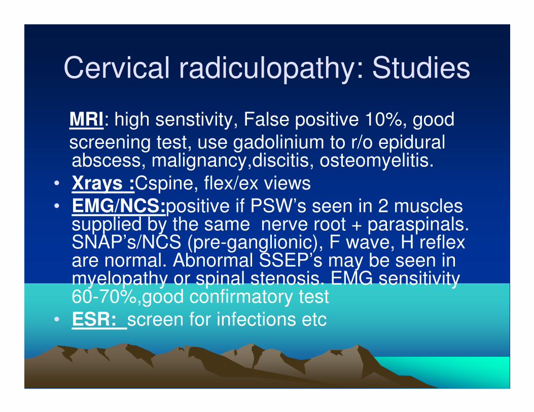
Cervical radiculopathy: Studies
MRI: high senstivity, False positive 10%, good
screening test, use gadolinium to r/o epidural abscess, malignancy,discitis, osteomyelitis.
• Xrays :Cspine, flex/ex views
• EMG/NCS:positive if PSW’s seen in 2 muscles supplied by the same nerve root + paraspinals. SNAP’s/NCS (pre-ganglionic), F wave, H reflex are normal. Abnormal SSEP’s may be seen in myelopathy or spinal stenosis. EMG sensitivity 60-70%,good confirmatory test
• ESR: screen for infections etc
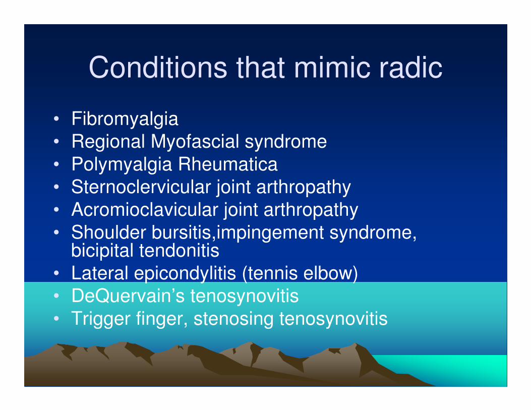
Conditions that mimic radic
• Fibromyalgia
• Regional Myofascial syndrome
• Polymyalgia Rheumatica
• Sternoclervicular joint arthropathy
• Acromioclavicular joint arthropathy
• Shoulder bursitis,impingement syndrome, bicipital tendonitis
• Lateral epicondylitis (tennis elbow)
• DeQuervain’s tenosynovitis
• Trigger finger, stenosing tenosynovitis

Brachial Plexus syndromes
• Klumpke paralysis
• Augusta Marie Klumpke:1859-1927 French neurologist
• Lower Plexus(C8,T1)
• Paralysis of hand intrinsics, claw hand, sensory loss and a horners syndrome if T1 is involved
Brachial plexus syndromes
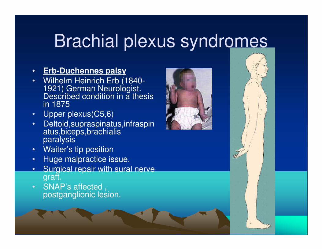
• Erb-Duchennes palsy
• Wilhelm Heinrich Erb (1840-1921) German Neurologist. Described condition in a thesis in 1875
• Upper plexus(C5,6)
• Deltoid,supraspinatus,infraspinatus,biceps,brachialis paralysis
• Waiter’s tip position
• Huge malpractice issue.
• Surgical repair with sural nerve graft.
• SNAP’s affected , postganglionic lesion.
Burners/ Stingers
• Traction neuropathy of upper plexus during sports injuries.
• Mainly football,wrestling.
• Numbness, tingling , parasthesia & proximal muscle weakness.
• Symptoms usually resolve in minutes.
• If symptoms persist order MRI, EMG/NCS to proximal shoulder muscles.
• C5/6 muscles affected, shoulder wasting may occur.
Thoracic outlet syndrome
• Compression of brachial plexus C8-T1 fibers, subclavian artery and vein due to fibrous band or cervical rib
• Neck/shoulder pain, parasthesia in the forearm, made worse by carrying suitcase/bag
• Adson’s sign: loss of radial pulse on abduction and external rotation of the shoulder.
• EMG/NCS: True neurogenic TOS, medial antebrachial cutaneous and APB, more involved than Ulnar motor and sensory and vice-versa for post cardiac surgery sternotomy.
Brachial Neuritis
• AKA neuralgic amyotrophy, Parsonage-Turner syndrome.
• Incidence :1.64:100,000 pts, 2.4:1 M/F ratio
• Viral, vaccination, strenous exercise, IVDA
• Acute shoulder pain, rapid onset,proximal muscle weakness.
• 66% unilateral, 34% bilateral involvement
• Minor sensory loss, outer aspect of shoulder
• Wasting in 3-6wks.
• 89% recover in 3 yrs.
Pancoasts Tumour
• Apical squamous cell Cancer
• Involves lower cervical &upper thoracic roots
• Severe pain around shoulder and down inside
arm
• Weak wasted hand muscles
• Sensory loss in C8/T1dermatomes
• Horners Syndrome(stellate ganglion,
sympathetic chain) involved.
Neoplastic Vs Radiation Induced
Plexopathy.• Horners syndrome reported more in tumor pts
• Pain reported in 80% of tumor pts Vs 19% in radiation pts
• Radiation tends to involve upper trunk(78%) and tumor lower trunk (72%)
• Other radiation plexoapthy peals;
• Radiotherapy of axilla > 6000 rads have higher risk of fibrosis.
• Lower plexus damage seen in 1-3% pts
• Lymphedema is a common problem, makes EMG’s difficult
• Delayed onset 5-30 mths
• Resultant fibrosis causes entrapment of brachial plexus
• Myokymia and fasciculations seen on EMG in radiation plexopathy.

Long thoracic nerve(C5,6,7)
• Serratus anterior
• Damaged by carrying
heavy
objects,strapping the
shoulder, limited
brachial neuritis,DM
• Scapula winging
Suprascapular Nerve(C5,6)
• Supraspinatus and infraspinatus
• Weakness in arm abduction (supraspinatus) and external rotation(infraspinatus).
• Entrapment at suprascapular notch involves both muscles Vs spinoglenoid notch that involves only infraspinatus
Musculocutaneous nerve(C5,6)
• Supplies biceps, brachilais and coracobrachilais
• Sensory; lateral border of arm
• Damaged by humerus fx
• Weakness in Elbow flexion, forearm supination and absent biceps reflex
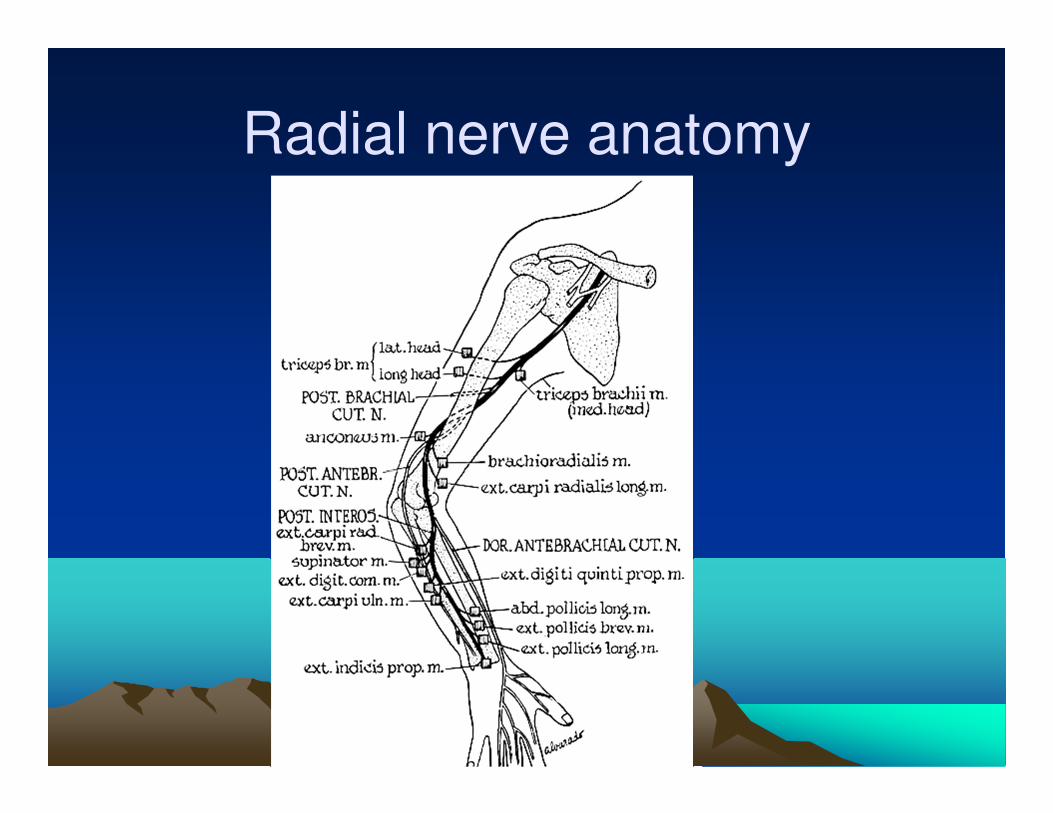
Radial nerve anatomy
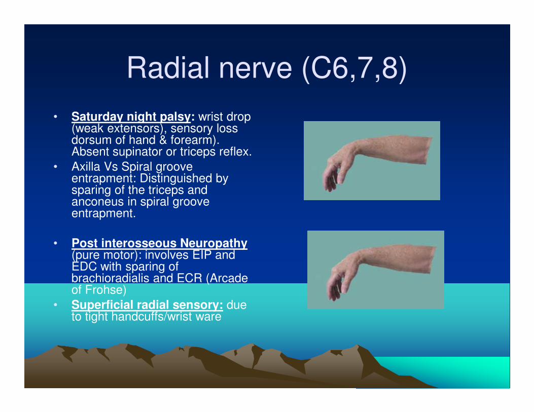
Radial nerve (C6,7,8)
• Saturday night palsy: wrist drop (weak extensors), sensory loss dorsum of hand & forearm). Absent supinator or triceps reflex.
• Axilla Vs Spiral groove entrapment: Distinguished by sparing of the triceps and anconeus in spiral groove entrapment.
• Post interosseous Neuropathy(pure motor): involves EIP and EDC with sparing of brachioradialis and ECR (Arcade of Frohse)
• Superficial radial sensory: due to tight handcuffs/wrist ware
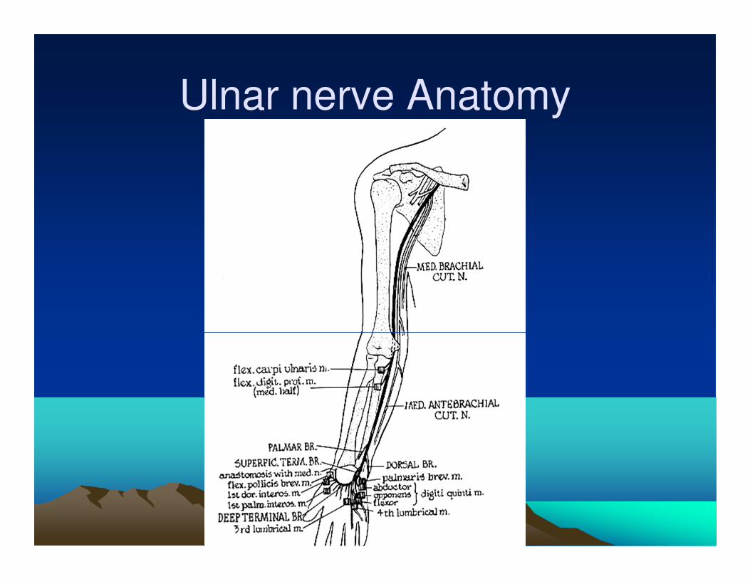
Ulnar nerve Anatomy
Ulnar nerve entrapment
• Medial cord C7/8.
• Cubital tunnel syndrome
• Injury or entrapment at elbow or distal to medial epicondyle.
• Weakness results in claw hand (ring and small fingers) or benediction sign.
• Positive Froment’s test. weakness of adductor pollicis.
• Atrophy of 1st dorsal interosseous
• Positive Tinel’s at elbow.
• EMG/NCS, drop in ampitude and conduction velocity (>10m/s) across elbow. Inching technique sometimes used to find focal conduction block.
• Surgical transposition/elbow pads used to treat.
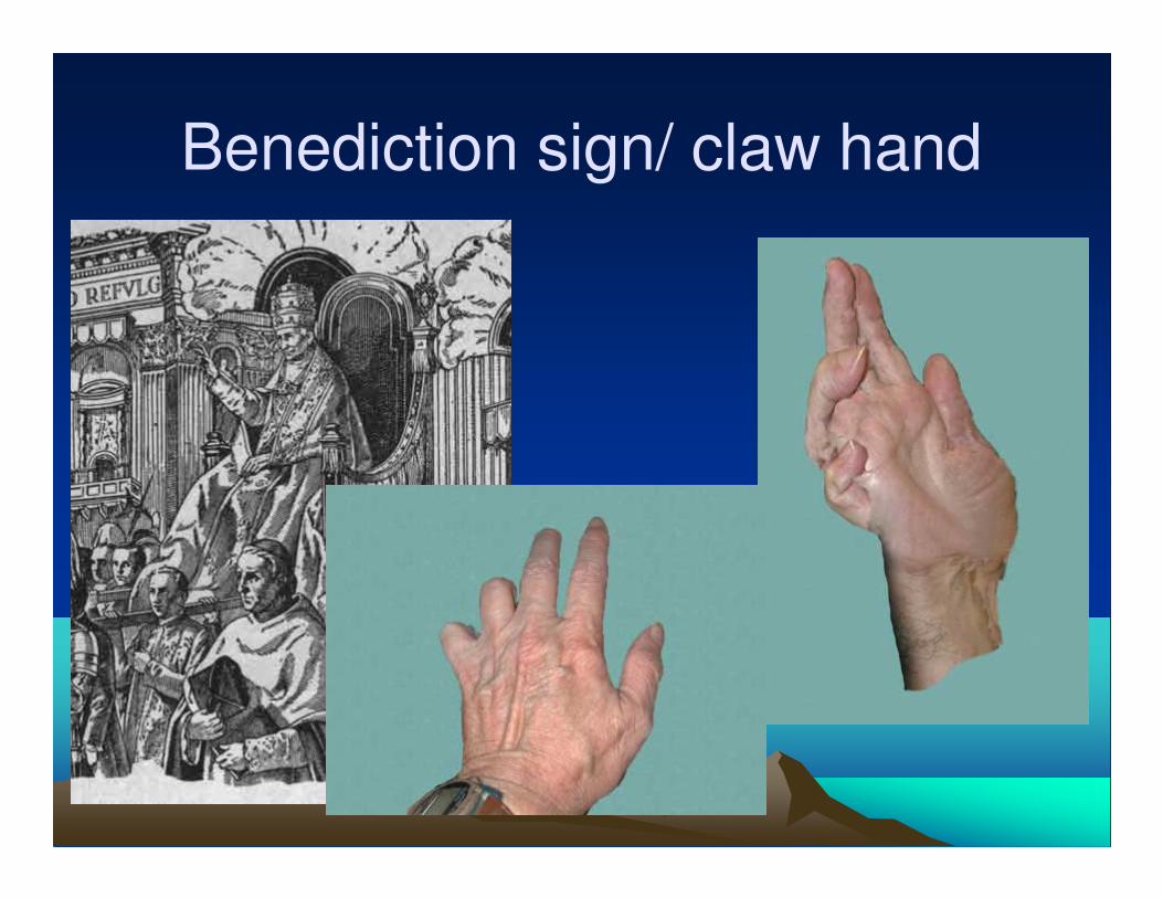
Benediction sign/ claw hand
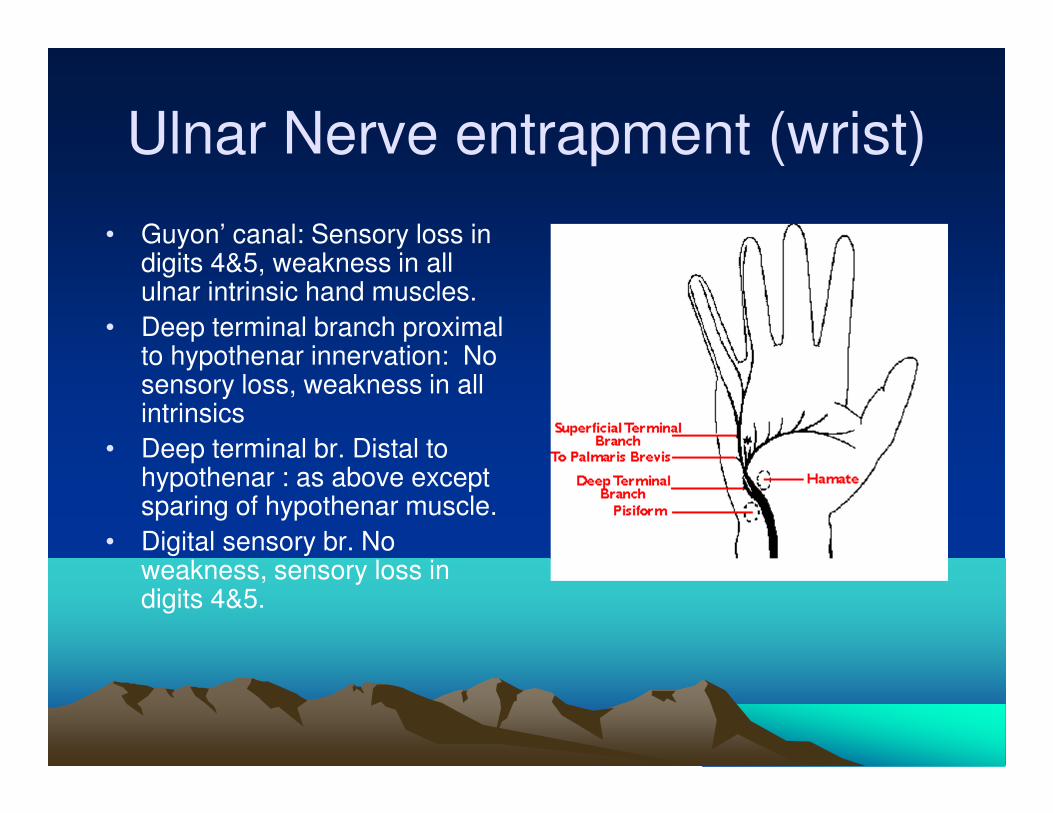
Ulnar Nerve entrapment (wrist)
• Guyon’ canal: Sensory loss in digits 4&5, weakness in all ulnar intrinsic hand muscles.
• Deep terminal branch proximal to hypothenar innervation: No sensory loss, weakness in all intrinsics
• Deep terminal br. Distal to hypothenar : as above except sparing of hypothenar muscle.
• Digital sensory br. No weakness, sensory loss in digits 4&5.
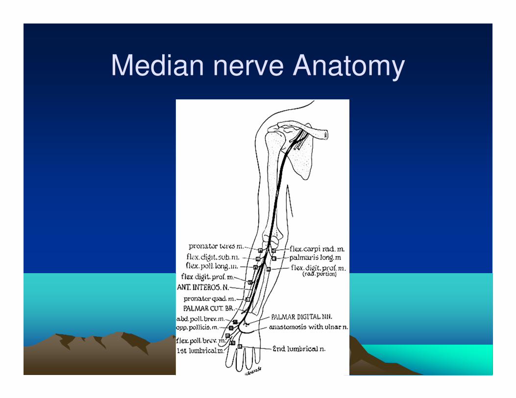
Median nerve Anatomy
Median Nerve (C7,8)
• Sites of entrapment;Carpal tunnel, pronator teres heads,ligament of struthers, bicipital aponeurosis
• Weakness abd/opp of thumb, pronation of forearm, wasting of thenar muscles
• Sensory loss in index and middle finger.( variable)

Anterior Interosseous Syndrome
• Kiloh-Nevin Syndrome.
• Leslie Gordon Kiloh Austrian Physician, Samuel Nevin English neurologist. 20th century.
• Purely motor nerve
• Supplies FDP digits2&3, FPL, and pronator quadratus
• Trauma,Parsonnage turner syndrome (NAM).
• OK sign.
Pronator Teres Syndrome
• Median nerve entrapment between heads of pronator teres, first muscle supplied
• Diffuse forearm pain & parasthesia
• Positive tinels, decreased wrist flex, finger flex,thumb opposition.
• Differentiate from CTS, by wrist weakness and more diffuse sensory loss
Carpal Tunnel Syndrome
• Symptoms:
• Median nerve distribution, numbness & parasthesias.
• Nocturnal pain and numbness
• Weakness of grip, dropping things easily.
• Worsening of symptoms with use of hands e.g. driving
CTS
• Signs:
• Weakness of thumb abduction (APB)
• Loss of median nerve sensation.
• Positive Phalen’s or reverse Phalen’s sign, tested by holding hands in flexion or extension of wrist for 60 sec with symptom reproduction. (highly sensitive/specific)
CTS
• Tinels Sign: reproduction of median parasthesias by pressing or tapping the median nerve at the wrist.(not highly sensitive/specific)
• Positive Flick sign: ask pt if she/he wakes up at night with pain & numbness. If pt says yes, ask what she does, if pt shakes of flicks hand that is a positive sign. Do not give the patient any visual or verbal cues.
Risk factors
• Diabetes
• Repetitive hand movments
• Rheumatoid Arthritis
• Pregnancy
• Obesity
• Thyroid disease
• Common in women 50’s and 60’s
EMG/NCS for CTS
• Distal motor latencies to APB: 40% sensitive, down from 80% in 60’s.
• SENSORY STUDIES:
• Numb thumb: Median/radial diff >0.5ms
• P8 Mid palm/Orthodromic stimulation : median/ulnar diff >0.3ms
• Split ring finger: median/ ulnar diff >0.4 ms
• Combined sensory Index(CSI) of all three studies > 1 is 98% sensitive.
• EMG controversial, will see PSW’s in thenar muscles, usually used to r/o radiculopathy
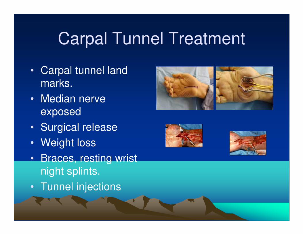
Carpal Tunnel Treatment
• Carpal tunnel land
marks.
• Median nerve
exposed
• Surgical release
• Weight loss
• Braces, resting wrist
night splints.
• Tunnel injections
Ischemic Insult
• Muscles can tolerate ischemia up to 4 hrs, at 6 hrs effect is uncertain, 8 hrs irreversible damage
• Peripheral nerves can survive up to 4 hrs w/only neurapraxic damage, at 8 hrs changes are irreversible
• Acute compartment syndrome
• Subacute Compartment syndrome
• Volkmann’s ischemic contracture
• Recurrent compartment syndrome
• Compartmental syndrome
Compartment Syndrome
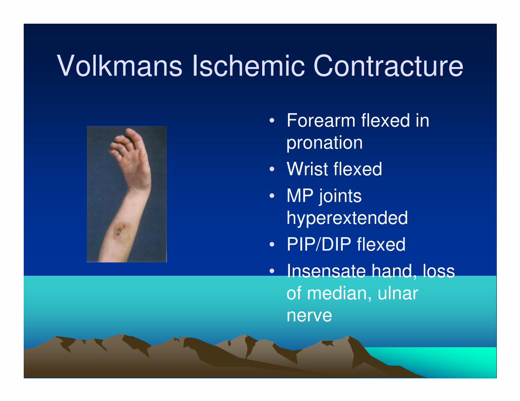
Volkmans Ischemic Contracture
• Forearm flexed in
pronation
• Wrist flexed
• MP joints
hyperextended
• PIP/DIP flexed
• Insensate hand, loss
of median, ulnar
nerve
CRPS:reflex sympathetic dystrophy
• Complex regional pain syndrome, AKA causalgia, shoulder hand syndrome,sympathetically maintained pain.(SMP).
• CRPS type 1: due to trauma, soft tissue injury, sprain, strain, surgery, fx, surgery.
• CRPS type 2: due to injury to large peripheral nerve.
• 3 stages: 1.acute (hyperemic),2:dystrophic (ischemic), 3: Atrophic stage
CRPS
• Symptoms:constant burning pain, allodynia, hyperpathia, local edema, warmth and skin changes.
• Xray; patchy osteoporosis, 3 phase bone scan( flow and static phase inc uptake)
• Tx: PT, prednisone,alpha blockers,, TCA’s, Neurontin, CBZ, stellate block, Bier block, neurolysis, sympathectomies, etc.
References
• Neurology & Neurosurgery by Lindsay
• PMR pocket pedia
• EMG secrets
• Rehabilitation medicine By De Lisa
• PMR secrets
• Internet sites( multiple) for images.
• Guide to neuropathic pain.
• Braddom PM&R


![Chapter 2 Evolution of Foot Orthoses in Sports · for the treatment of painful pathologies and deformities within the foot and lower extremity [1, 4]. The early literature describes](https://img.pdfslide.net/doc/110x75/601c1ba23dff0d3680114a86/chapter-2-evolution-of-foot-orthoses-in-sports-for-the-treatment-of-painful-pathologies.jpg)