Embed Size (px)
Citation preview

Discover the Clinical Significance of
Upright Weight-Bearing MRI
Rotate the bed from
recumbent to upright
and compare MRI scans
in different
patient positions
Acquire MRI scans in both
flexion & extension
positions since there
is nothing in front
of the patient’s face
FONAR Corporation
110 Marcus Drive, Melville, NY, 11747
631.694.2929 www.fonar.com
Weight-Bearing MRI
Multi-Position MRI

The Upright Weight-Bearing MRI actually does something
clinically valuable that a high-field MRI cannot do!
Yes, we offer exceptional patient comfort, but this MRI is
not just for claustrophobic patients.
There is considerable evidence that the Upright Weight-Bearing MRI
provides medical benefits that are not duplicated by any other MRI.
• Patient positioning plays a critical role in detecting
clinically significant pathology.
• Recumbent-only imaging can underestimate the
maximum degree of pathology.
• Peer-reviewed publications demonstrate the impact
on treatment.
Unique Benefits
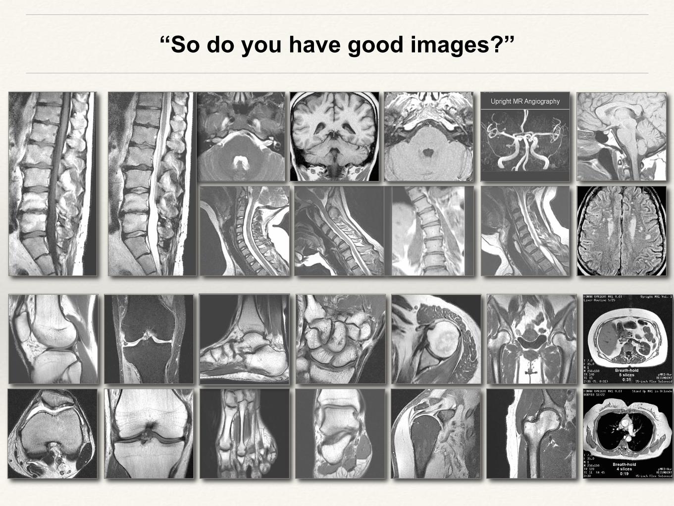
“So do you have good images?”
Liliarue05

Why an upright, weight-bearing MRI scan?
The high-field MRIs may not show the abnormalities which account for symptoms experienced while in a seated, standing, extending, flexing or lateral bending position.
An examination of the spine in the seated or upright position is important because the spine changes under the effects of both gravity and position.
A patient’s hips and knees are often flexed and the knees supported
by a pillow to take the strain off the back in order to help the patient lie
down in a traditional high-field MRI. This is an unnatural position and
not at all optimized to visualize the cause of spinal pathology for which
symptoms are often experienced when sitting or standing.
Finally, many patients with spinal issues are not able to lie down
without experiencing pain.

Let’s review some Clinical Case Studies
You need the upright position to see the pathology highlighted in red.
Each box compares the same patient in different positions
on the same day in the same MRI scanner
Recumb
ent Upright
Recumbent
CSF Flow Upright CSF
Flow

What happens when we stand or sit upright?
A. Nachemson, MD in SPINE (1976)
reported on measuring the significant
increase in disc pressure when the
patient is not lying down.
Rela
tive S
cale
The Consequences of Gravity
Recumbent Upright

Case Courtesy
of FW Smith,
MD University of
Aberdeen, UK
Nuclear
extrusio
n
Position-dependent disc
herniation
You need the upright weight-bearing position to see
the pathology highlighted in red.

You need the upright weight-bearing position to see
the pathology highlighted in red.
Case Courtesy of M Rose, MD Tampa, FL
Post-operative spinal instability in a patient with recurrent low back pain
following a fusion from L4 to S1.

Recumbent Upright
An unsuspected anterolisthesis at L3/4 in a patient with prior L4-S1 fusion changed
the patient management from conservative treatment to ligament stabilization at the
level above (i.e., at L2/3).
Recumbent Upright
Positional alterations include anterior displacement of the upper cauda equina against the posterior surface of a protruding disc at L1/2.
You need the upright weight-bearing position to see
the pathology highlighted in red.

European Journal of Orthopedic Surgery &
Traumatology 17:2 (2007)
J.P. Elsig, MD Zurich, Switzerland
Recumbent Upright
The increased downward herniation of the cerebellar tonsils required
neurosurgery to eliminate the patient’s drop attacks.
The upright CSF flow image shows an unobstructed dorsal CSF flow
(black arrow) but demonstrates the obstruction of ventral CSF flow.
You need the upright weight-bearing position to see
the pathology highlighted in red.

“The dominant motions at both the lower cervical and entire lumbar spine,
where most clinical pathology occurs, are flexion-extension”
~ AMA Guides to the Evaluation of Permanent Impairment ~
Flexion Extension

Extension Neutral In extension, the anterior longitudinal ligament becomes
taut, and the posterior longitudinal ligament becomes
lax ... so a disc with internal derangement that fluctuates will
tend to move posteriorly.
Biomechanics
Extension Neutral
You need the extension position to see
the pathology highlighted in red.
Case Courtesy of
R Marks, MD
Dallas,TX
Additional disc herniation at C4/5

You need flexion, extension and lateral bending to
see
the pathologies highlighted in red.
Cases courtesy of FW Smith, MD, University of Aberdeen, UK Ligamentous rupture at L4/5
Dynamic fluctuation of spinal neural foramen
stenosis showing narrowing of the neural
foramen at L5/S1 in extension and
generalized widening in flexion at all levels.
This spinal instability
is best visualized
with upright lateral
bending

425 lbs.
Large
Patient
Scanning
Hard-to-Scan Patients
Patients that are unable to lie down.
Cases Courtesy of FW Smith, MD University of Aberdeen, UK

Pediatric MRI: No sedation
5 yr old
10 yr old
A child sitting on his mother’s lap
watching the television during his spine
MRI scan

More Consequences of Gravity: It’s Not Just for the Spine
Case Courtesy
of P Barrance,
PhD
University of
Delaware
Tibial Subluxation Bladder & Uterine
Prolapse

Twenty-five (25) chronic low back pain patients with prior
“negative” recumbent-only MRIs …
What percentage showed
abnormalities in one or
more of the upright
positions,
and still nothing in their
recumbent position?

52%
So What?
Twenty-five (25) chronic low back pain patients with prior
“negative” recumbent-only MRIs …
Each of these
patients had
surgery and
six months later
they remain
symptom-free
What percentage showed
abnormalities in one or
more of the upright
positions,
and still nothing in their
recumbent position?

Clinical MRI (2006) 15:3 [ESSR (2005) Oxford, UK]
“Positional Upright Imaging of the Lumbar Spine Modifies the Management
of Low Back Pain and Sciatica”
Neurosurgeons examining 20 patients with symptoms consistent with cervical radiculopathy
or myelopathy concluded that “when only static supine MRI is performed ... the true
abnormality may be overlooked and inappropriate surgical plans instituted
because of a lack of illustration of the changes that occur with movement.”
“Dynamic Weight-Bearing Cervical Magnetic Resonance Imaging: Technical
Review and Preliminary Results”
Southern Medical Journal (2004)
T Vitaz MD et.al., Dept. of Neurosurgery, University of Louisville, KY
52% of a group of 25 chronic low back pain patients with prior “negative”
recumbent-only MRIs demonstrated abnormalities in one or more seated
postures “that were not evident in the recumbent position, and each of
these patients has undergone appropriate surgery and 6 months post-
surgery they remain symptom-free.”
FW Smith MD et.al., Dept. of Radiology, University of Aberdeen, UK
What’s the Clinical Relevance?
Peer-reviewed Scientific Publications: Effects on Patient Treatment

SPINE Volume 33, Number 5 (2008)
“Missed Lumbar Disc Herniations Diagnosed With Kinetic
Magnetic Resonance Imaging”
In a study of 553 patients with symptomatic back pain, in those with normal or less
than a 3 mm bulge in the neutral position, 19% demonstrated an increase in
herniation to greater than 3 mm in extension. Further, 15% demonstrated an
increase in herniation to greater than 3 mm in flexion. J. Zou, M.D. et al., Department of Orthopedic Surgery, UCLA
Clinical Radiology (2008) 63, 1035-1048
“Upright Positional MRI of the Lumbar Spine”
“... there is no doubt that clinically relevant spinal canal stenosis can be
uncovered by imaging the erect position. In cases where conventional MRI
shows no evidence of cauda equina or lumbar nerve root compression in the
setting of convincing clinical symptoms that warrant surgical intervention, re-
imaging in the upright position, with the addition of flexion and extension, is
recommended.” F. Alyas, et. al., Dept. of Radiology, Royal National Orthopaedic Hospital NHS Trust,
Stanmore, Middlesex, UK
What’s the Clinical Relevance?
Peer-reviewed Scientific Publications: Effects on Patient Treatment

Brain Injury 24:7-8 (2010)
“A Case-Controlled Study of Cerebellar Tonsillar Ectopia (Chiari)
and Head/Neck Trauma (Whiplash)”
A multi-center study of 1200 patients with neck pain showed recumbent MRI
underestimates the incidence of herniated cerebellar tonsils. The incidence of tonsillar
herniation in non-traumatic neck pain patients was about the same, 5.3-5.7%, for both
recumbent and upright positions, while in whiplash patients, 23.3% examined upright
showed herniation of the cerebellar tonsils, whereas only 9.3% examined
recumbent showed this abnormality.
M Freeman et al., Oregon University School of Medicine, Univ. of Aarhus, Univ. of
Aberdeen, Spinal Injury Foundation, Columbia Univ., Univ. of Nebraska, Wisconsin
Chiari Center
The Spine Journal (2007) Volume 7, Number 5S
“Missed Spondylolisthesis in Static MRIs But Found in Dynamic MRIs in
Patients with Low Back Pain” S.W. Hong, M.D. et al., UCLA
“In [510] patients with back pain, missed spondylolisthesis in neutral MRIs but
found in flexion MRIs is 18.1% for all the levels if the spondylolisthesis is
considered as more than 3 mm translation.”
What’s the Clinical Relevance?
Peer-reviewed Scientific Publications: Effects on Patient Treatment

What’s the Clinical Relevance?
Peer-reviewed Scientific Publications: Effects on Patient Treatment
Radiologists reported that “weight-bearing imaging of the forefoot ... demonstrated position-
related changes of the neurovascular bundles relative to the metatarsal heads ...”
D Weishaupt MD et.al., University of Zurich, Switzerland
Orthopedic surgeons concluded that “patellofemoral joint contact areas should
be measured under loaded conditions ... when trying to understand potential
mechanisms of patellofemoral pain” to account for cartilage deformation and
changes in patellar alignment. TF Beiser MD et.al., Stanford University
Journal of Orthopaedic Research 23 (2005)
“Patellofemoral Joint Contact Area Increases with Knee Flexion and Weight-bearing”
Journal of MRI 16 (2002) “MR Imaging of the Forefoot under Weight-Bearing
Conditions: Position-Related Changes of the Neurovascular Bundles and the
Metatarsal Heads in Asymptomatic Volunteers”
A radiologist concludes that “the main drawback of MRI is supine imaging that can
limit the dynamic component of the examination … upright scanners may
ultimately lead to MRI being the one imaging test for PFD.”
H Pannu MD, Johns Hopkins University
WomansImagingOnline (2007) “Pelvic Floor Dysfunction”

Concerned about the Upright MRI’s 0.6 Tesla magnetic field strength?
Compare these images from the same patient, same day, same imaging center
3:51 scan time
22 slices
5.0 mm thick
23 cm FOV
5:03 scan time
24 slices
5.0 mm thick
25 cm FOV
3.0 Tesla Upright® MRI
0.6 Tesla
4:41 scan time
24 slices
5.0 mm thick
25 cm FOV
1:30 scan time
30 slices
4.0 mm thick
23 cm FOV
3.0 Tesla Upright® MRI
0.6 Tesla

(3DFT SSFP-FID GRE) 6:45 scan time, 320 slices, 1.5 mm thick, no gap (0.75 mm slice
overlap)
3DFT Clinical Case Study: Whole Head
1.5 mm thick slices

No additional
scan time
3DFT Clinical Case Study: Whole Head

No additional
scan time
3DFT Clinical Case Study: Whole Head

Clinical 3DFT: Weight-Bearing Kneeling Knee
(3DFT SSFP-FID GRE) 5:04 scan time, 224 slices, 2.0 mm thick, no gap (0.75 mm slice
overlap)
2.0 mm thick slices

No additional
scan time
Clinical 3DFT: Weight-Bearing Kneeling Knee

Clinical 3DFT: Upright Weight-Bearing MRI Scoliosis Evaluation
• 0.5 mm isotropic voxels
• Curved MPR
• Cobb angle calculation
• T7 rotation
This example of an Upright MRI Scoliosis examination
courtesy of Stand-Up MRI of Melville
Standing Rapid MRI with Sagittal Multi-
planar Reconstruction (SMMR)
Ten subjects (10-18 years) who had undergone radiographic imaging for idiopathic
scoliosis were also evaluated using a rapid upright MRI with SMMR ... “Scoliosis
may now be accurately and reliably quantified using MRI technology, thereby
decreasing radiation exposure and its inherent risks.’’
IMAST (2009) Vienna 16th International Meeting on Advanced Spine Techniques
“Evaluation of Scoliosis with Standing Rapid MRI with SMMR: An Alternative to Plain Radiography” Q. Hammouri, J. Grauer et. al. Yale Univ. School of Medicine

Water-Fat
Separation
Direct
FatSat
Fat Suppression
Knee
Shoulder Spine Orbits
STIR
(Dixon)

The Upright MRI is twice as strong as most Open MRIs.
The Upright MRI is dramatically different from an Open MRI because physics allows the Upright MRI to use the same RF receiver coil as a high-field MRI to image the spine. The Open MRI is unable to do this.
So is our 0.6 Tesla magnetic field strong enough?
The Upright MRI has a competitive advantage for post-operative patients since artifacts from metal surgical screws diminish as the MRI’s field strength is reduced.
The position-dependent pathology we detect will be invisible or underestimated at higher field strengths … sometimes patient positioning trumps a small increase in resolution or a small decrease in scan time.
✓
✓
✓
✓

Upright MRI (0.6T) Competitive Advantage: Reduced Metal Artifacts
0.6T Upright
MRI
1.5 T
MRI
Compared to the high-field (1.5 T) MRI scan on the far right,
the structures in and around anatomy adjacent to the implanted titanium pedicle screws are less
obscured with the 0.6T Upright MRI.
The parasagittal slices show the
pedicle screws used in a L4-S1 fusion do not generate a large
metal artifact
Images from the Same Patient
at Different Field Strengths
you can see the threads!

Recumbent Patient
S
Solenoid
RF
Receiver
“Belt” Coil
“Classic” Open MRI
N
Stand-Up MRI
Planar RF Receiver Coil
Rule of MRI: The axis of symmetry of the RF receiver coil should be
perpendicular to the direction of the main magnetic field.
Planar RF Receiver Coil
“High-Field” MRI

Which RF Receiver
Coil is compatible
with which type of
MRI?
Planar (flat) Solenoid (“belt”)
High-Field MRI ✓
Open MRI ✓
Stand-Up (Upright) MRI ✓ ✓
RF Coil Magnet
More Competitive Advantages of the Upright Weight-Bearing MRI
The Upright MRI can use the same type
of RF receiver coil as a high-field MRI
(i.e., planar configuration)
An Open MRI cannot do this,
so the Upright MRI is
dramatically different than an
Open MRI

Misunderstanding Field Strength
A: “I don’t want to miss anything”
Possibly requires:
• Particular anatomical region
• Specific type of image
contrast
• Demanding spatial resolution
• Extreme image clarity
• Artifact-free images
• Specialized imaging
applications
• Different patient positions
“I must have the best images”
Q: Why?
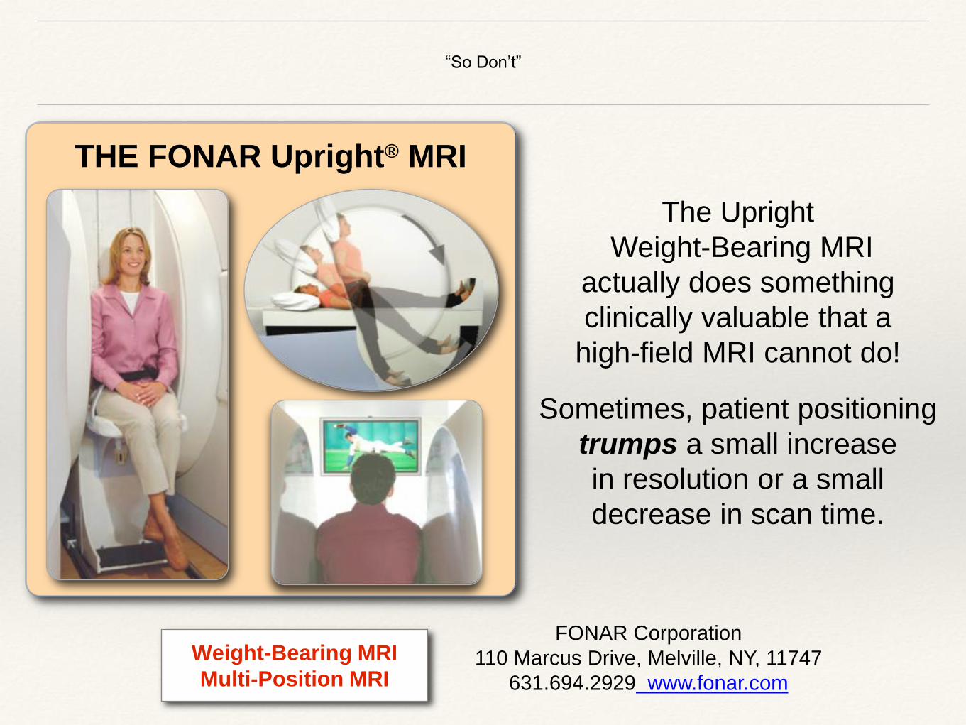
“So Don’t”
THE FONAR Upright® MRI
The Upright
Weight-Bearing MRI
actually does something
clinically valuable that a
high-field MRI cannot do!
Sometimes, patient positioning
trumps a small increase
in resolution or a small
decrease in scan time.
FONAR Corporation
110 Marcus Drive, Melville, NY, 11747
631.694.2929 www.fonar.com
Weight-Bearing MRI
Multi-Position MRI