Embed Size (px)
Citation preview

Urine CYTOLOGY
The 25th Congress of the International Academy of
Pathology / Arab Division
The 5th International Conference of the Jordanian
Society of Pathologists
November 5-9, 2013
Mousa Al-Abbadi, MD, FIAC, FCAP
Professor of Pathology & Cytopathology
King Fahad Specialist Hospital – Dammam
Saudi Arabia
Outline
• Introduction
• Specimens & processing
• Reporting and adequacy
• Accuracy
• Normal elements
• Benign
• Ancillary tests

Introduction
• Bladder cancer is the ninth most common cancer worldwide
• The age standardized incidence for north Africa is >5.3 and <9.2 /100,000 population
• The incidence is higher in Sudan and Saudi Arabia
• Egypt and Sudan also have a higher proportion of squamous cell carcinoma due to Schistosoma hematobium
Introduction
• At this point, there is no routine, effective bladder cancer screening test.
• However, urine cytology (consult) can be used as a surveillance tool for: – populations at high risk
– symptomatic patients
– patients with known prior urothelial malignancy.
• Urine cytology is feared because of low predictive values
• The low efficiency of urine cytology is inherent in the features of the urothelial lesions
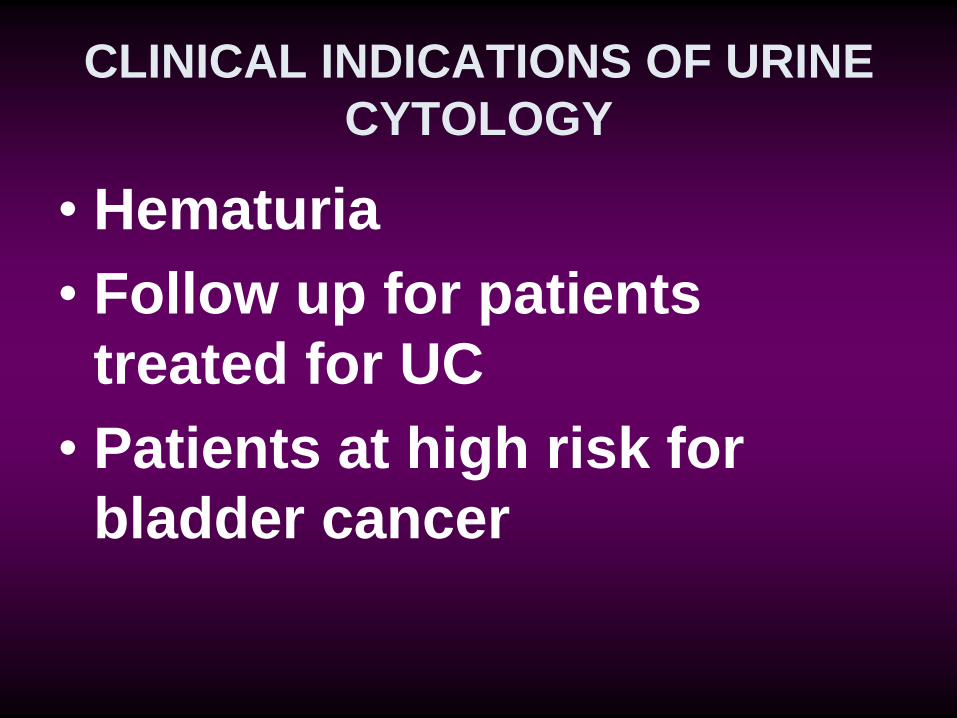
CLINICAL INDICATIONS OF URINE
CYTOLOGY
• Hematuria
• Follow up for patients
treated for UC
• Patients at high risk for
bladder cancer
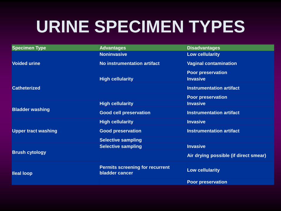
URINE SPECIMEN TYPES Specimen Type Advantages Disadvantages
Voided urine
Noninvasive Low cellularity
No instrumentation artifact Vaginal contamination
Poor preservation
Catheterized
High cellularity Invasive
Instrumentation artifact
Poor preservation
Bladder washing
High cellularity Invasive
Good cell preservation Instrumentation artifact
Upper tract washing
High cellularity Invasive
Good preservation Instrumentation artifact
Selective sampling
Brush cytology
Selective sampling Invasive
Air drying possible (if direct smear)
Ileal loop
Permits screening for recurrent
bladder cancer Low cellularity
Poor preservation

PROCESSING
• Fresh (1-12 hours), otherwise need
fixation
• Refrigeration if more
• Fixation with equal volume of alcohol
(50-70% ethanol).
• Cytocentrifugation, LBC, Cell block,
smears
• Papanicolaou stain (H&E)
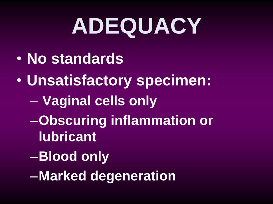
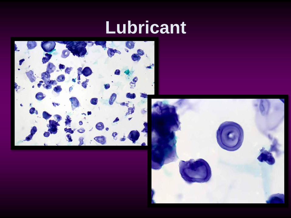
ADEQUACY
• No standards
• Unsatisfactory specimen:
– Vaginal cells only
–Obscuring inflammation or
lubricant
–Blood only
–Marked degeneration
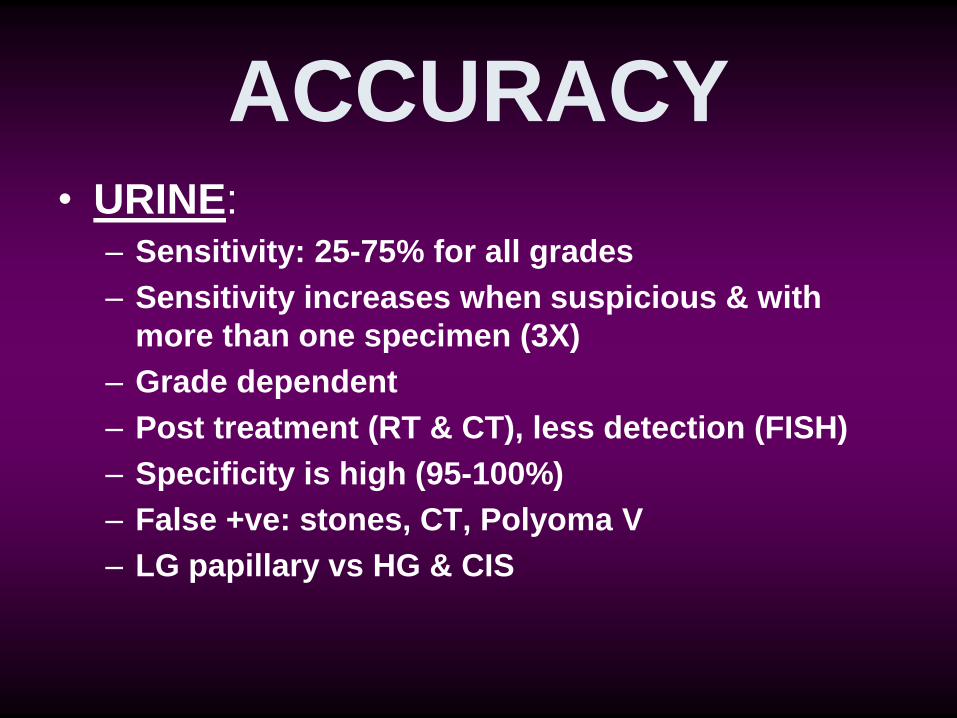
ACCURACY • URINE:
– Sensitivity: 25-75% for all grades
– Sensitivity increases when suspicious & with
more than one specimen (3X)
– Grade dependent
– Post treatment (RT & CT), less detection (FISH)
– Specificity is high (95-100%)
– False +ve: stones, CT, Polyoma V
– LG papillary vs HG & CIS
ACCURACY
• BLADDER WASHINGS:
–Sensitivity: 66-77%
–More false positive than urine
–Ureter and pelvic washings 70-
80% sensitivity
CONVENTIONAL WISDOM
“Low grade urothelial carcinoma are usually missed by the cytologist but seen by the urologist; while high grade carcinoma are easily identified by the cytologist but difficult to locate by the urologist”
NORMAL ELEMENTS:
• Urothelial cells:
• intermediate and superficial (umbrella) cells (voided urine) • intermediate, superficial, and basal cells (catheterized urine, washings)
• Squamous cells
• Seminal vesicle epithelial cells (rare)
• Degenerated intestinal epithelial cells (ileal conduit specimens)
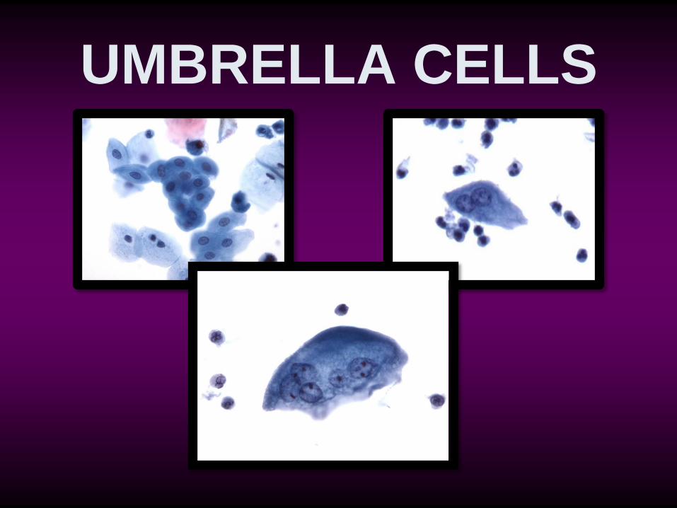
UMBRELLA CELLS
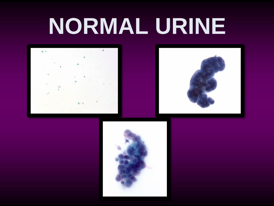
NORMAL URINE
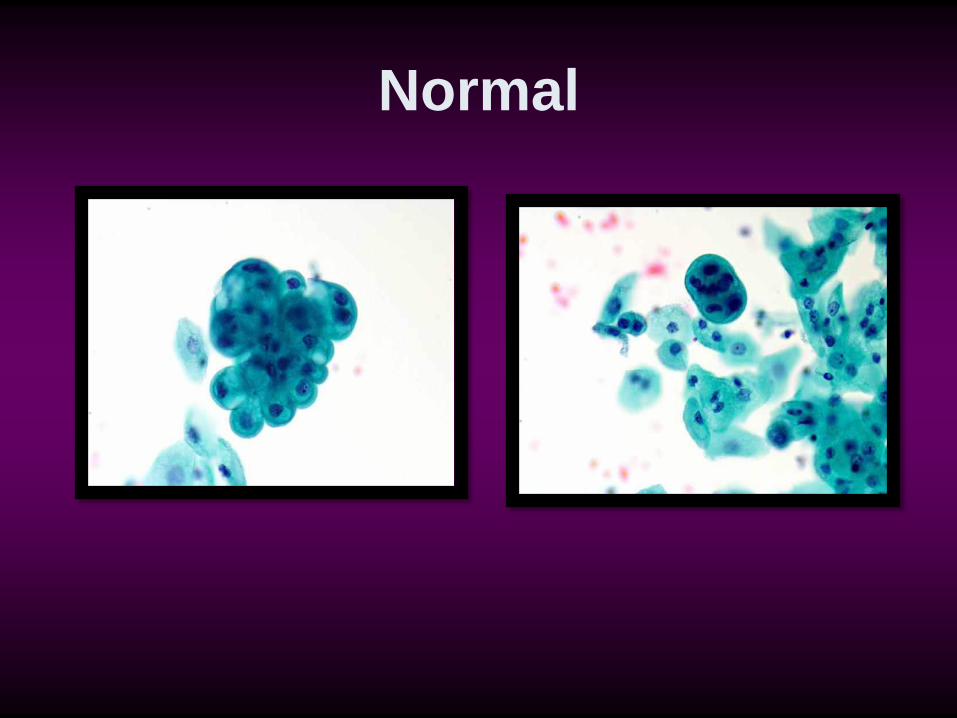
Normal
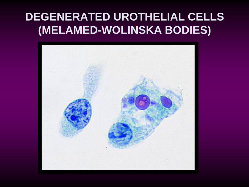
DEGENERATED UROTHELIAL CELLS
(MELAMED-WOLINSKA BODIES)
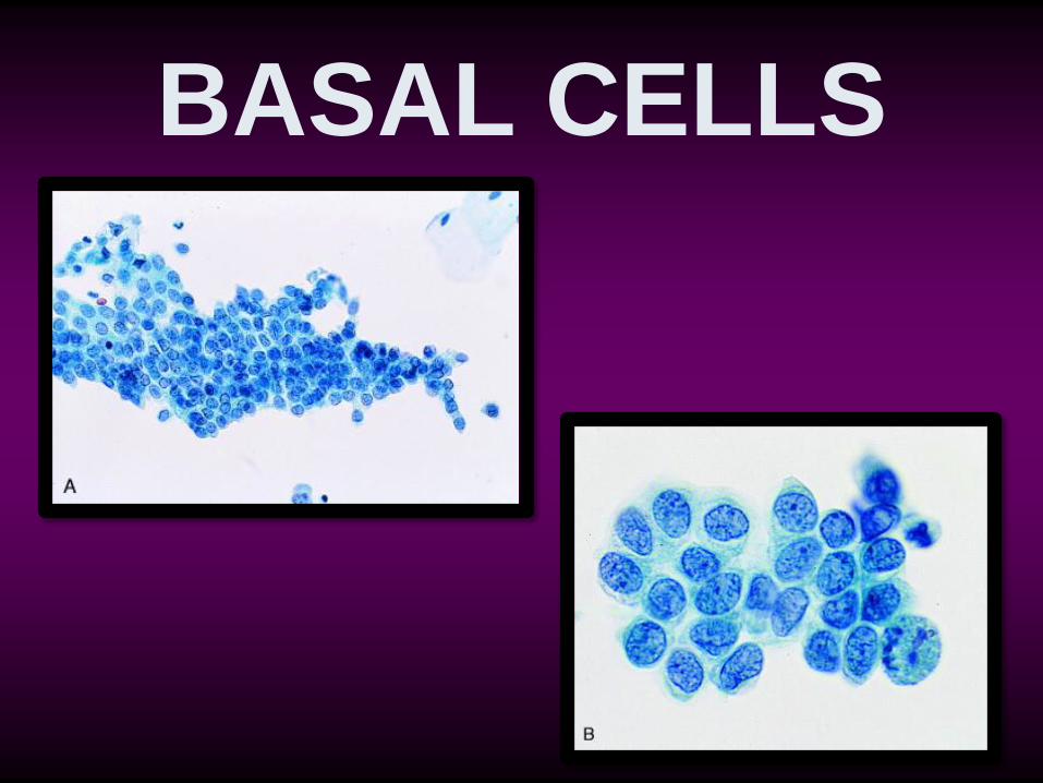
BASAL CELLS
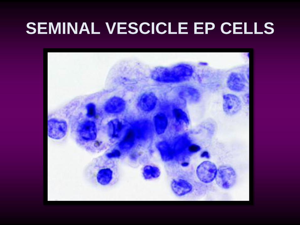
SEMINAL VESCICLE EP CELLS
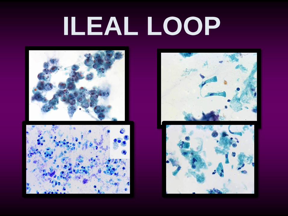
ILEAL LOOP
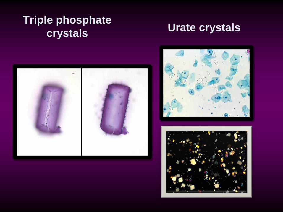
Triple phosphate
crystals
Urate crystals
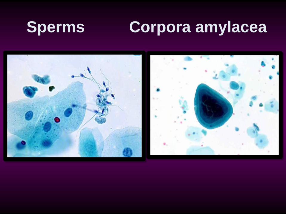
Sperms Corpora amylacea
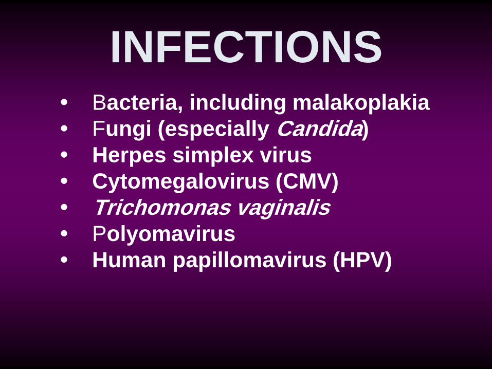
INFECTIONS • Bacteria, including malakoplakia
• Fungi (especially Candida)
• Herpes simplex virus
• Cytomegalovirus (CMV)
• Trichomonas vaginalis
• Polyomavirus
• Human papillomavirus (HPV)
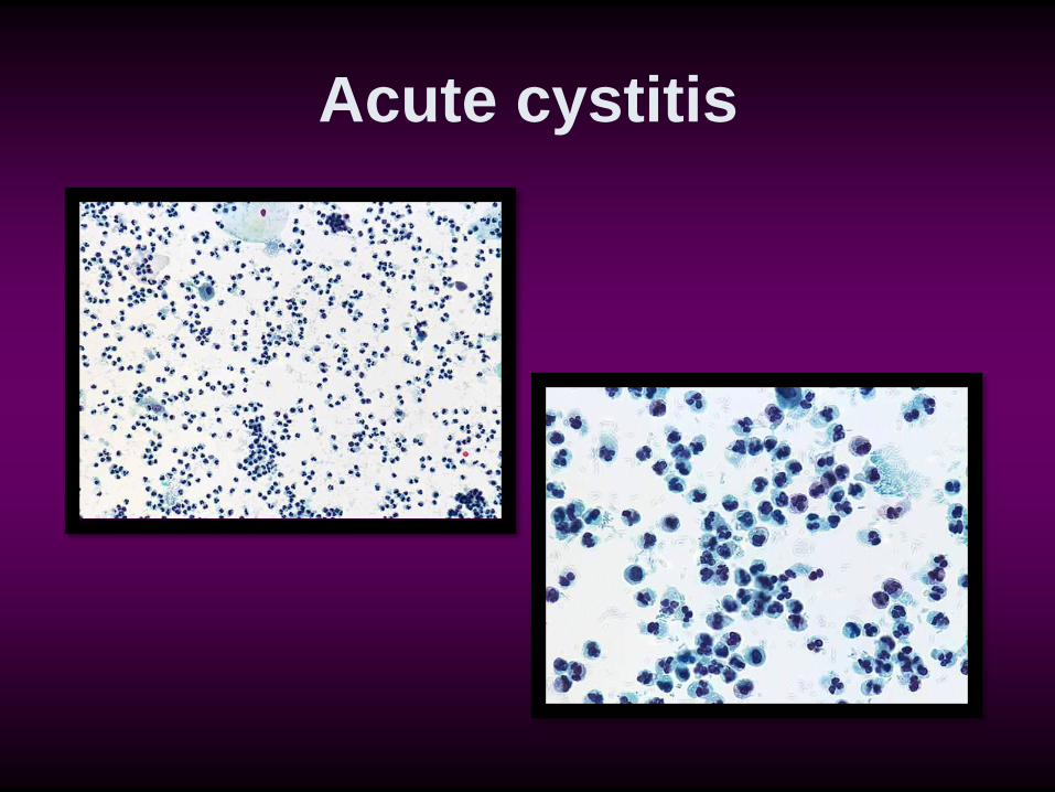
Acute cystitis
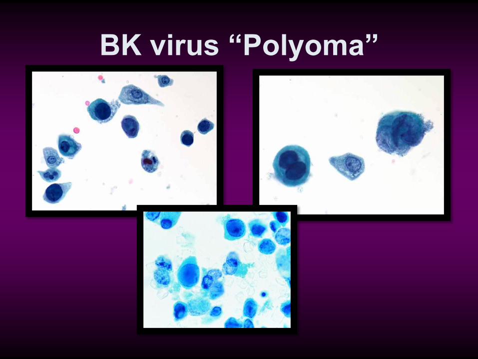
BK virus “Polyoma”
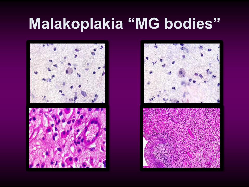
Malakoplakia “MG bodies”
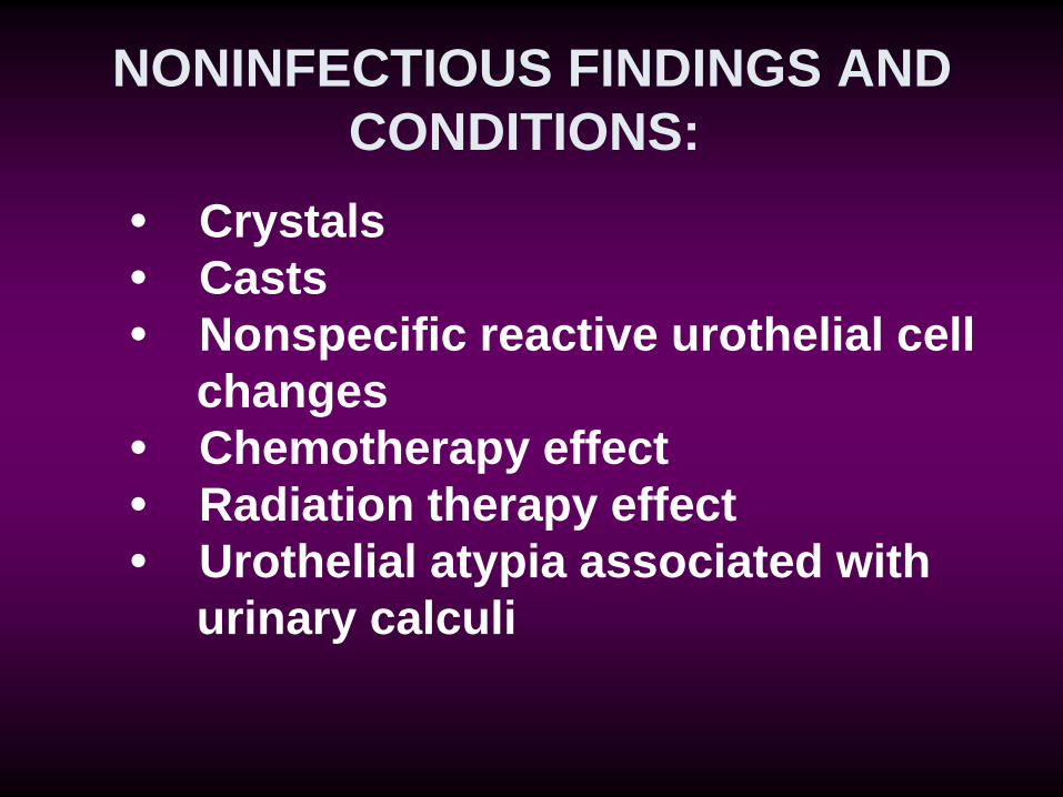
NONINFECTIOUS FINDINGS AND
CONDITIONS:
• Crystals
• Casts
• Nonspecific reactive urothelial cell
changes
• Chemotherapy effect
• Radiation therapy effect
• Urothelial atypia associated with
urinary calculi
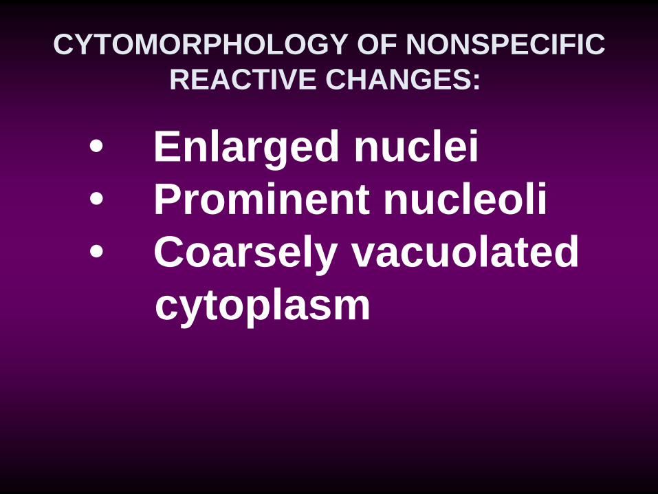
CYTOMORPHOLOGY OF NONSPECIFIC
REACTIVE CHANGES:
• Enlarged nuclei
• Prominent nucleoli
• Coarsely vacuolated
cytoplasm
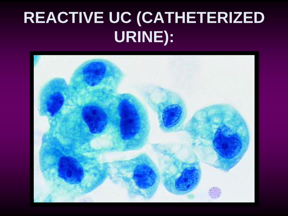
REACTIVE UC (CATHETERIZED
URINE):
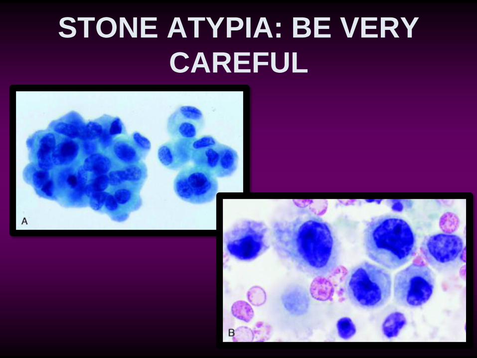
STONE ATYPIA: BE VERY
CAREFUL
UROTHELIAL
NEOPLASMS
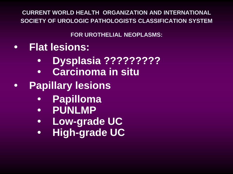
CURRENT WORLD HEALTH ORGANIZATION AND INTERNATIONAL
SOCIETY OF UROLOGIC PATHOLOGISTS CLASSIFICATION SYSTEM
FOR UROTHELIAL NEOPLASMS: • Flat lesions:
• Dysplasia ????????? • Carcinoma in situ
• Papillary lesions
• Papilloma • PUNLMP • Low-grade UC • High-grade UC
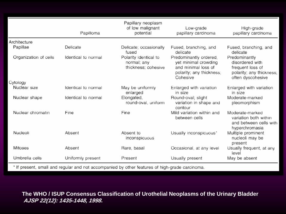
The WHO / ISUP Consensus Classification of Urothelial Neoplasms of the Urinary Bladder
AJSP 22(12): 1435-1448, 1998.
LOW GRADE
UROTHELIAL
CARCINOMA
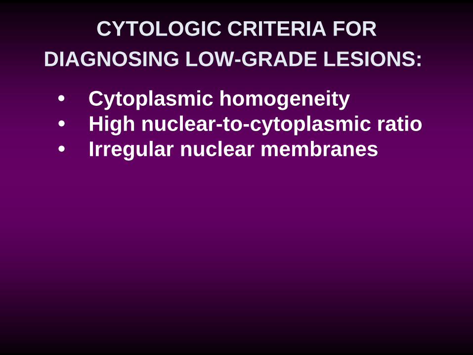
CYTOLOGIC CRITERIA FOR
DIAGNOSING LOW-GRADE LESIONS:
• Cytoplasmic homogeneity
• High nuclear-to-cytoplasmic ratio
• Irregular nuclear membranes
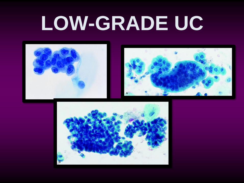
LOW-GRADE UC
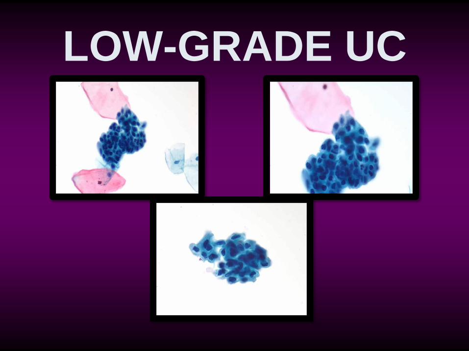
LOW-GRADE UC
HIGH GRADE
UROTHELIAL
CARCINOMA
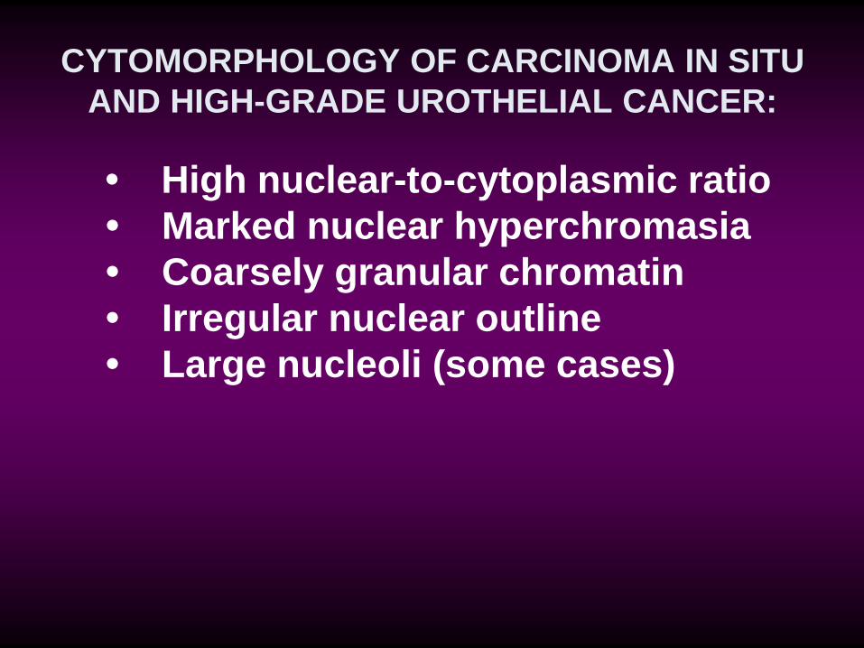
CYTOMORPHOLOGY OF CARCINOMA IN SITU
AND HIGH-GRADE UROTHELIAL CANCER:
• High nuclear-to-cytoplasmic ratio
• Marked nuclear hyperchromasia
• Coarsely granular chromatin
• Irregular nuclear outline
• Large nucleoli (some cases)
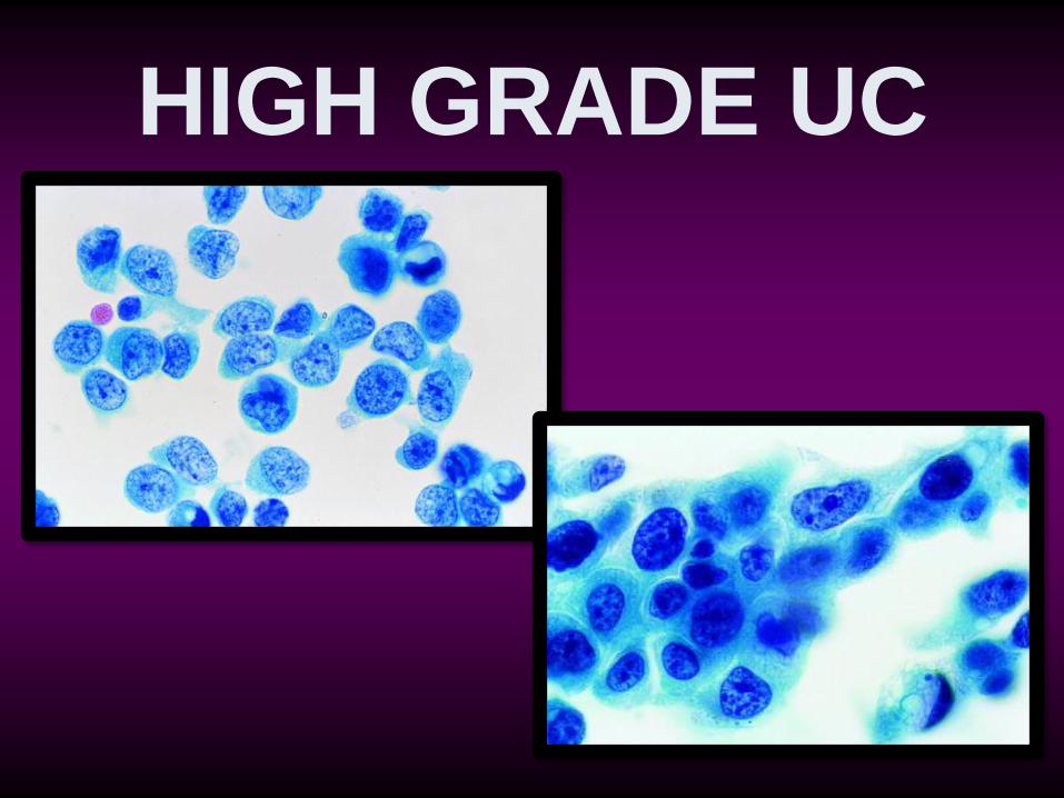
HIGH GRADE UC
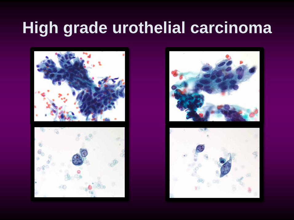
High grade urothelial carcinoma
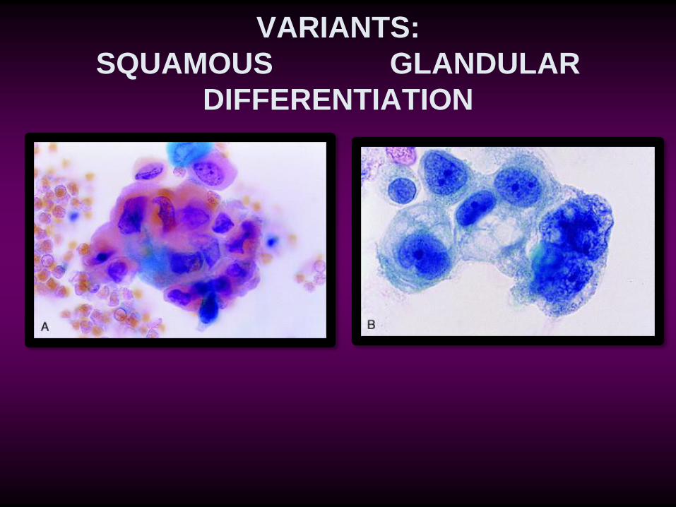
VARIANTS:
SQUAMOUS GLANDULAR
DIFFERENTIATION
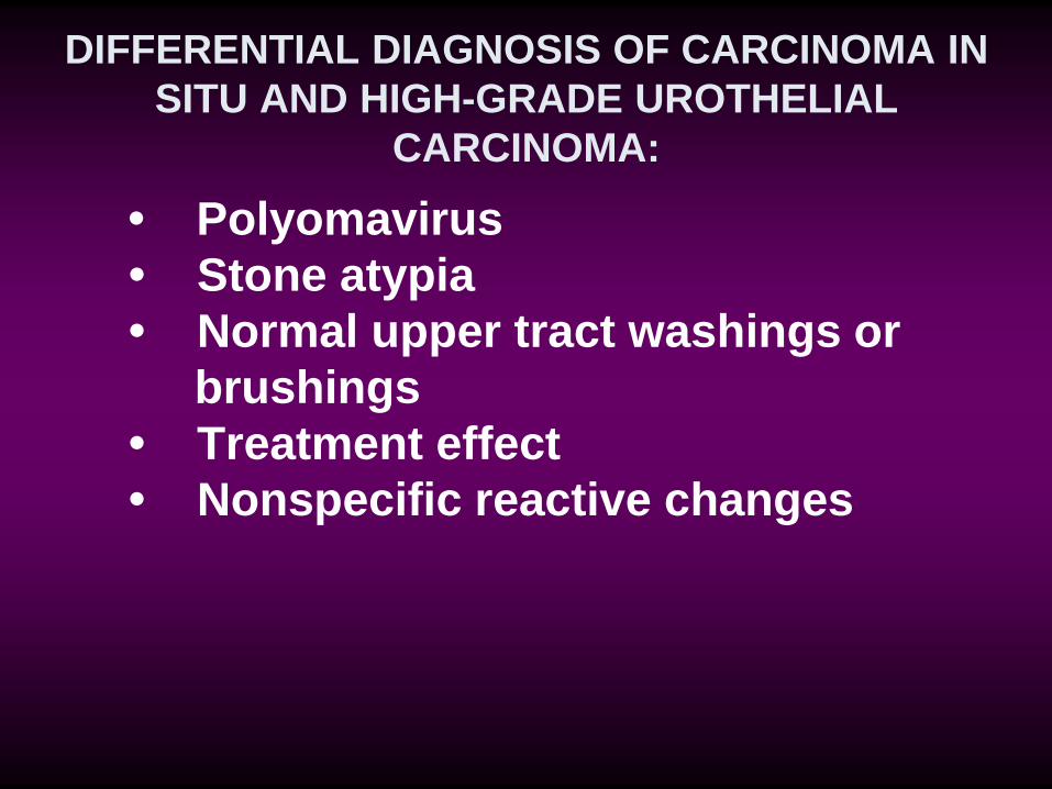
DIFFERENTIAL DIAGNOSIS OF CARCINOMA IN
SITU AND HIGH-GRADE UROTHELIAL
CARCINOMA:
• Polyomavirus
• Stone atypia
• Normal upper tract washings or
brushings
• Treatment effect
• Nonspecific reactive changes
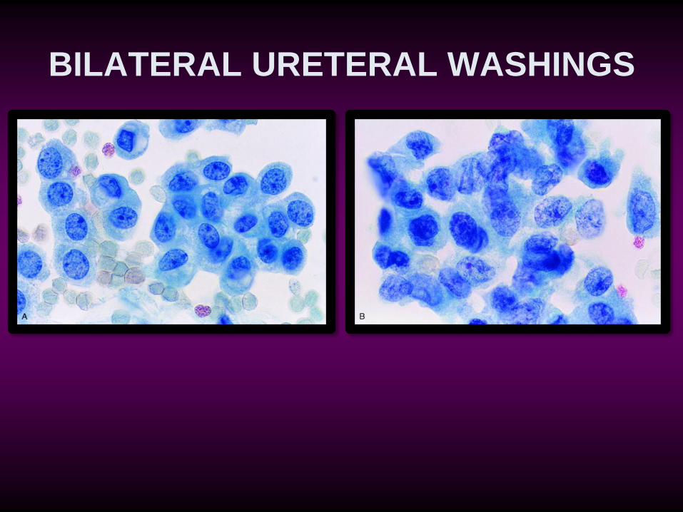
BILATERAL URETERAL WASHINGS
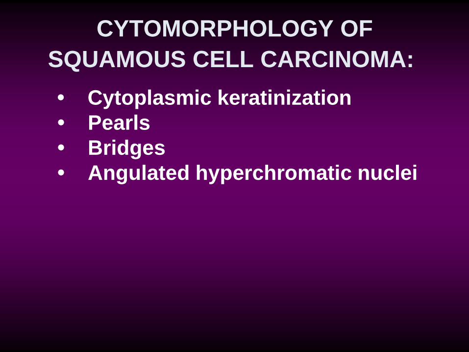
CYTOMORPHOLOGY OF
SQUAMOUS CELL CARCINOMA:
• Cytoplasmic keratinization
• Pearls
• Bridges
• Angulated hyperchromatic nuclei
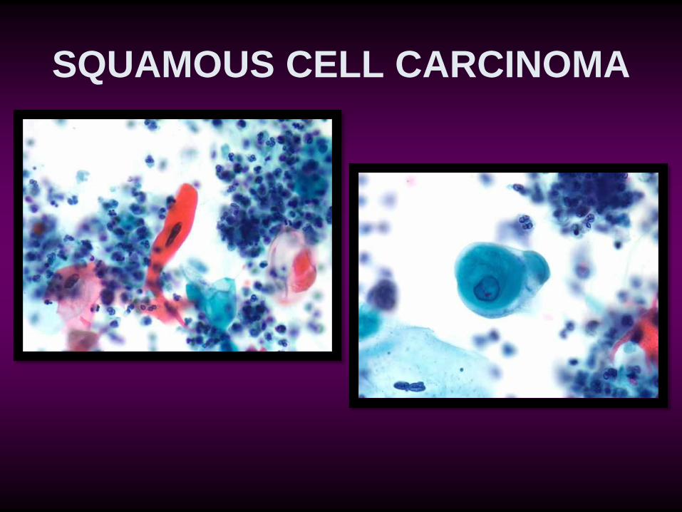
SQUAMOUS CELL CARCINOMA
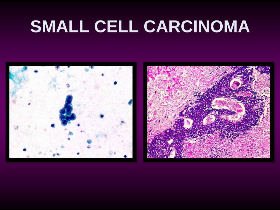
SMALL CELL CARCINOMA
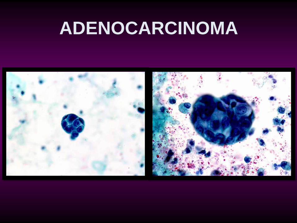
ADENOCARCINOMA
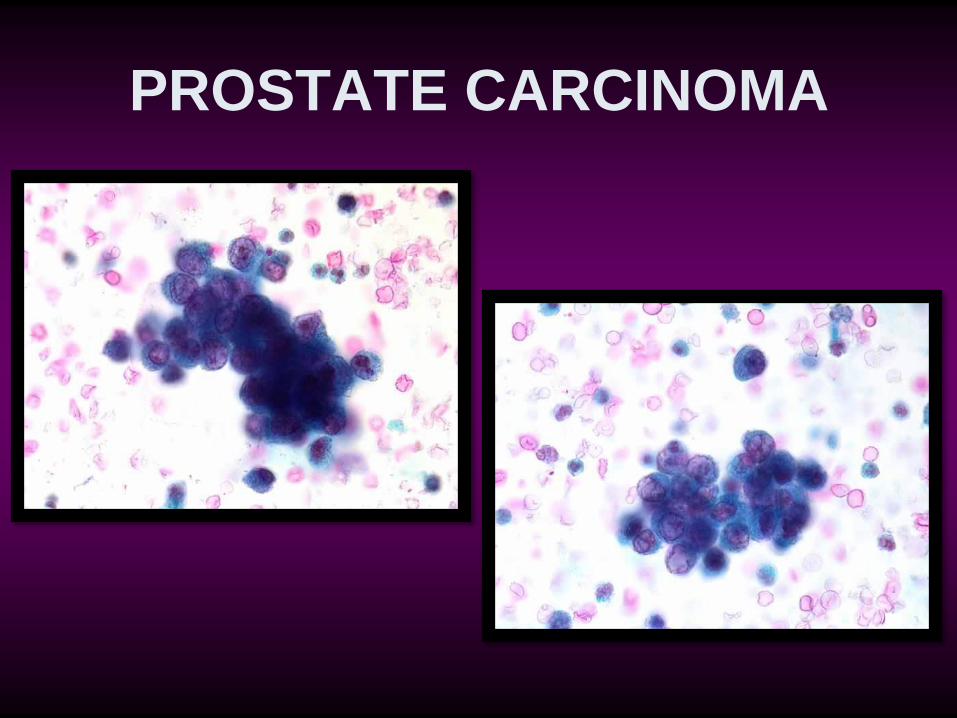
PROSTATE CARCINOMA
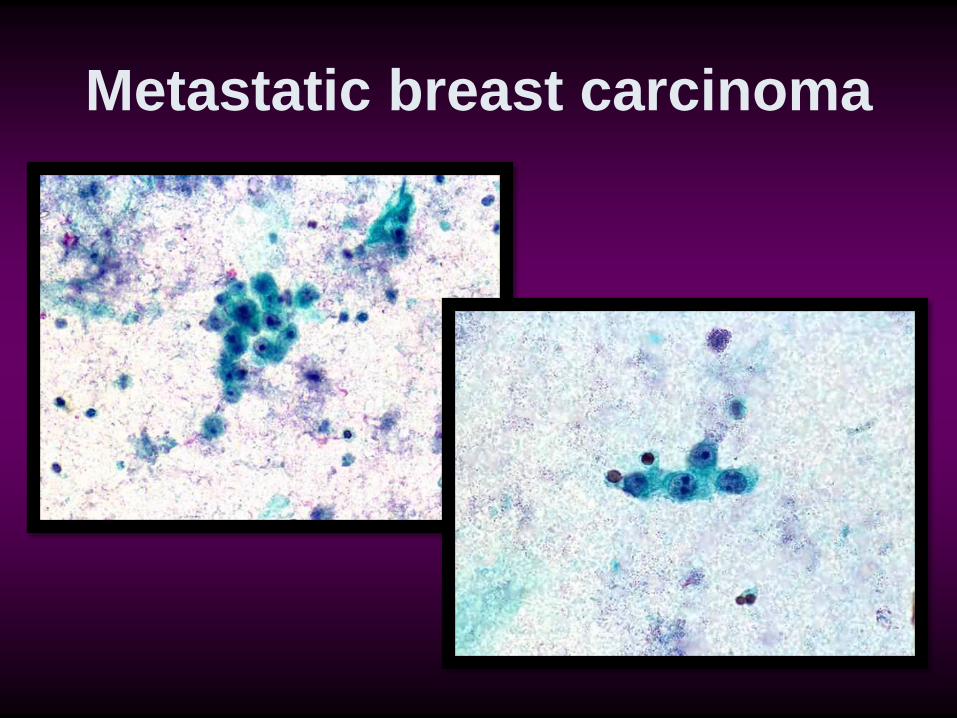
Metastatic breast carcinoma
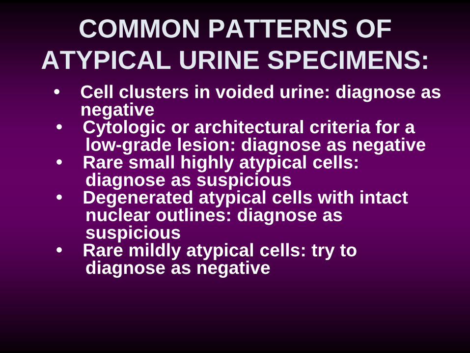
COMMON PATTERNS OF
ATYPICAL URINE SPECIMENS: • Cell clusters in voided urine: diagnose as
negative • Cytologic or architectural criteria for a low-grade lesion: diagnose as negative • Rare small highly atypical cells: diagnose as suspicious • Degenerated atypical cells with intact nuclear outlines: diagnose as suspicious • Rare mildly atypical cells: try to diagnose as negative
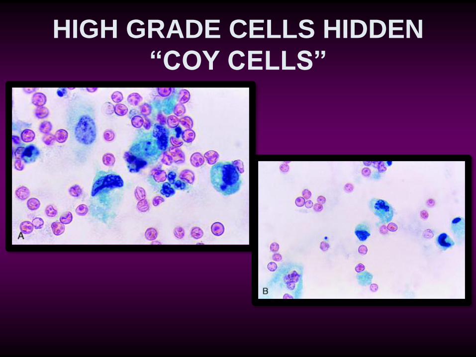
HIGH GRADE CELLS HIDDEN
“COY CELLS”
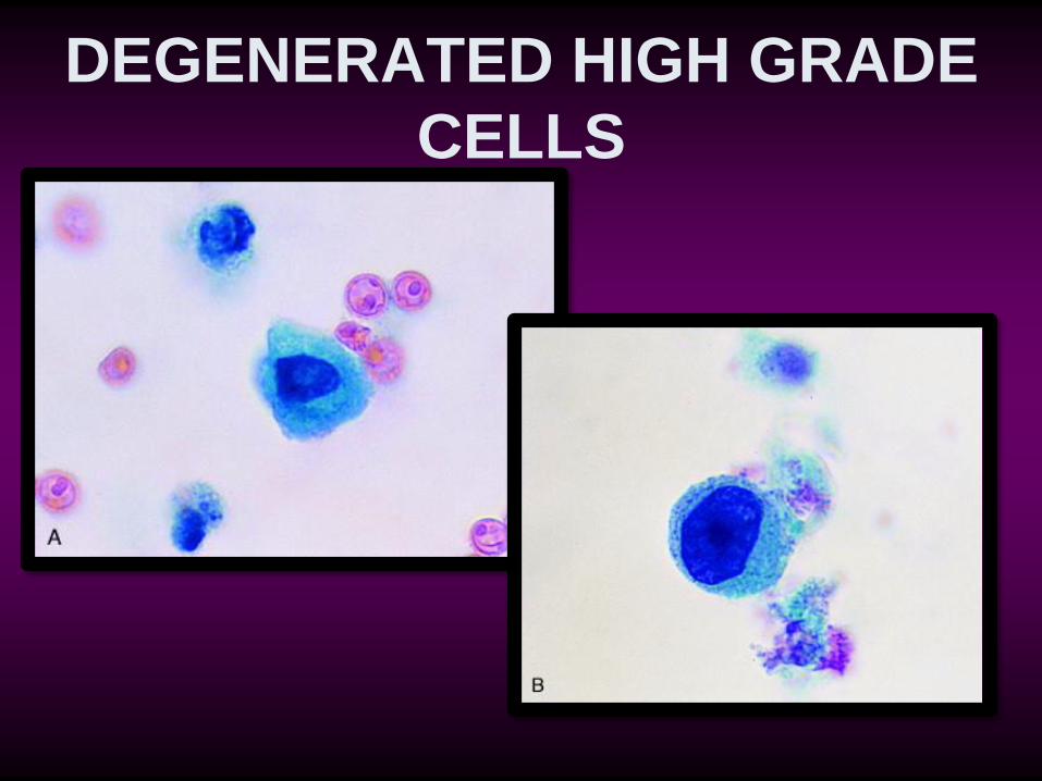
DEGENERATED HIGH GRADE
CELLS
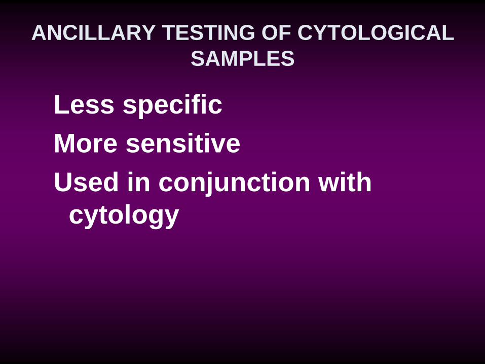
ANCILLARY TESTING OF CYTOLOGICAL
SAMPLES
Less specific
More sensitive
Used in conjunction with
cytology
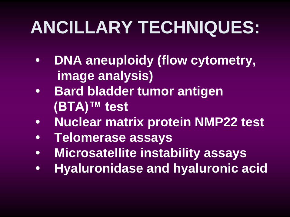
ANCILLARY TECHNIQUES:
• DNA aneuploidy (flow cytometry,
image analysis)
• Bard bladder tumor antigen
(BTA)™ test
• Nuclear matrix protein NMP22 test
• Telomerase assays
• Microsatellite instability assays
• Hyaluronidase and hyaluronic acid
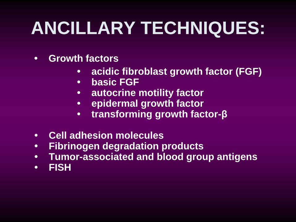
ANCILLARY TECHNIQUES:
• Growth factors
• acidic fibroblast growth factor (FGF) • basic FGF • autocrine motility factor • epidermal growth factor • transforming growth factor-β • Cell adhesion molecules • Fibrinogen degradation products • Tumor-associated and blood group antigens • FISH
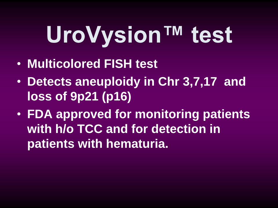
UroVysion™ test • Multicolored FISH test
• Detects aneuploidy in Chr 3,7,17 and
loss of 9p21 (p16)
• FDA approved for monitoring patients
with h/o TCC and for detection in
patients with hematuria.
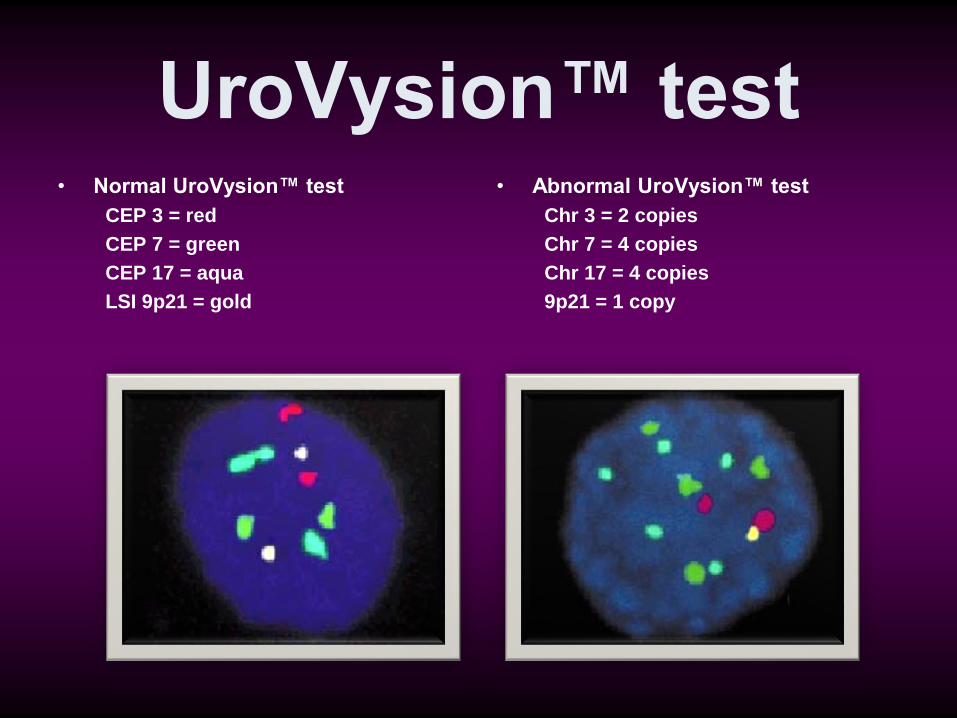
UroVysion™ test • Normal UroVysion™ test
CEP 3 = red
CEP 7 = green
CEP 17 = aqua
LSI 9p21 = gold
• Abnormal UroVysion™ test
Chr 3 = 2 copies
Chr 7 = 4 copies
Chr 17 = 4 copies
9p21 = 1 copy
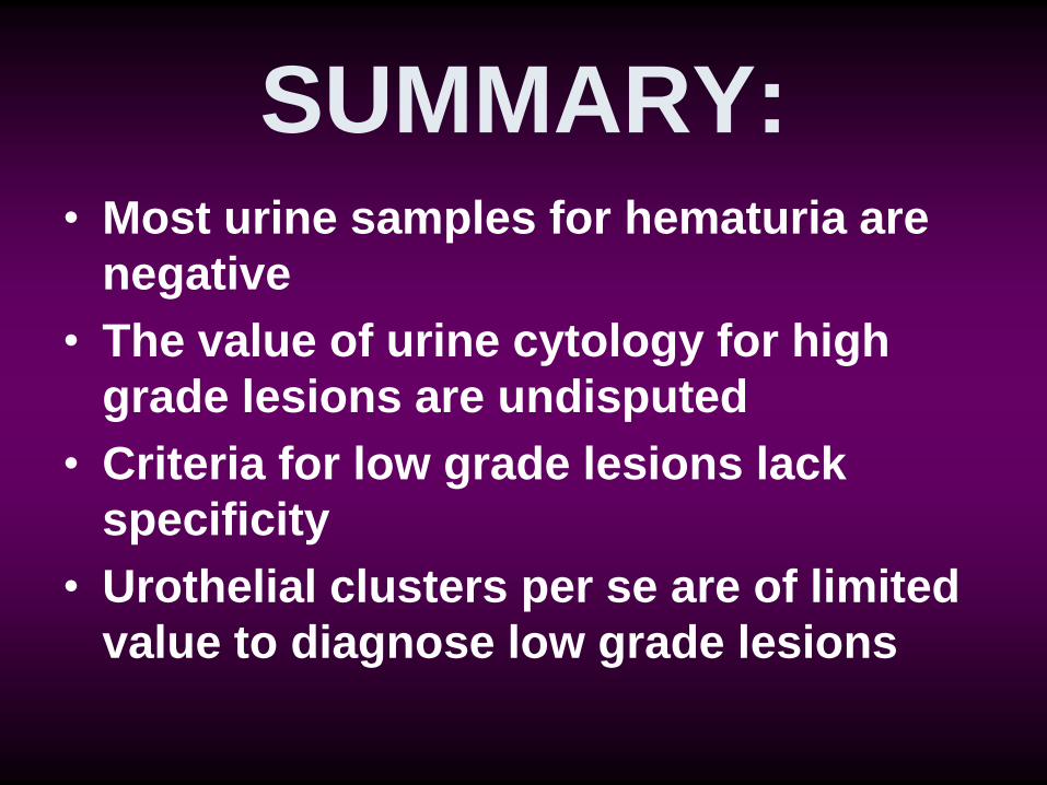
SUMMARY: • Most urine samples for hematuria are
negative
• The value of urine cytology for high
grade lesions are undisputed
• Criteria for low grade lesions lack
specificity
• Urothelial clusters per se are of limited
value to diagnose low grade lesions
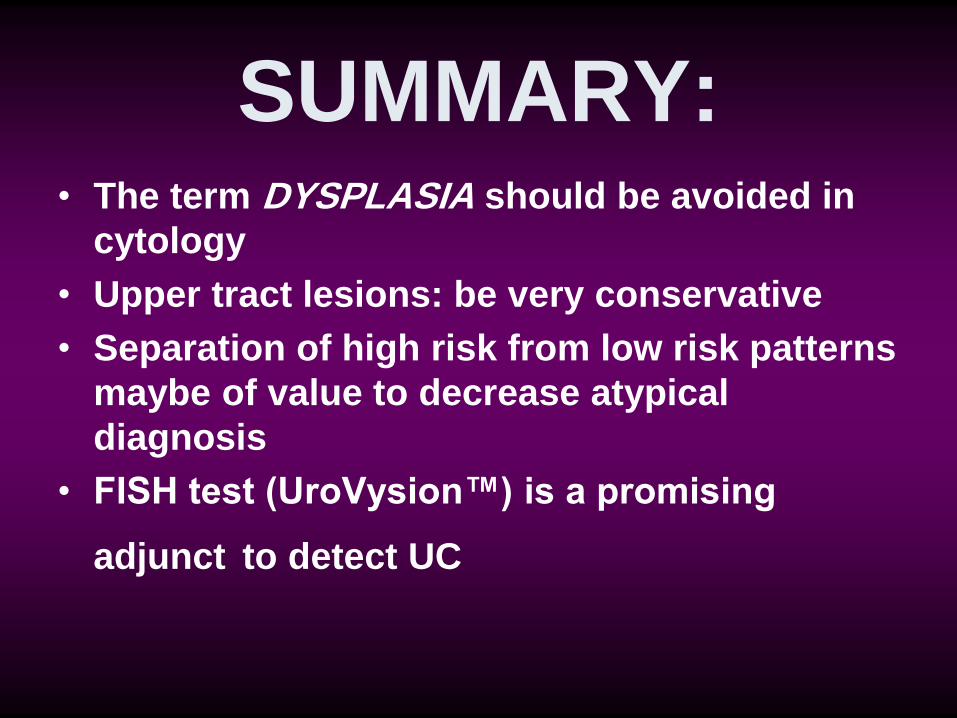
SUMMARY: • The term DYSPLASIA should be avoided in
cytology
• Upper tract lesions: be very conservative
• Separation of high risk from low risk patterns
maybe of value to decrease atypical
diagnosis
• FISH test (UroVysion™) is a promising
adjunct to detect UC

THANK
YOU
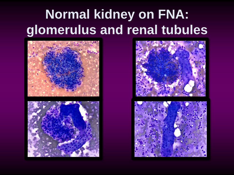
Normal kidney on FNA:
glomerulus and renal tubules
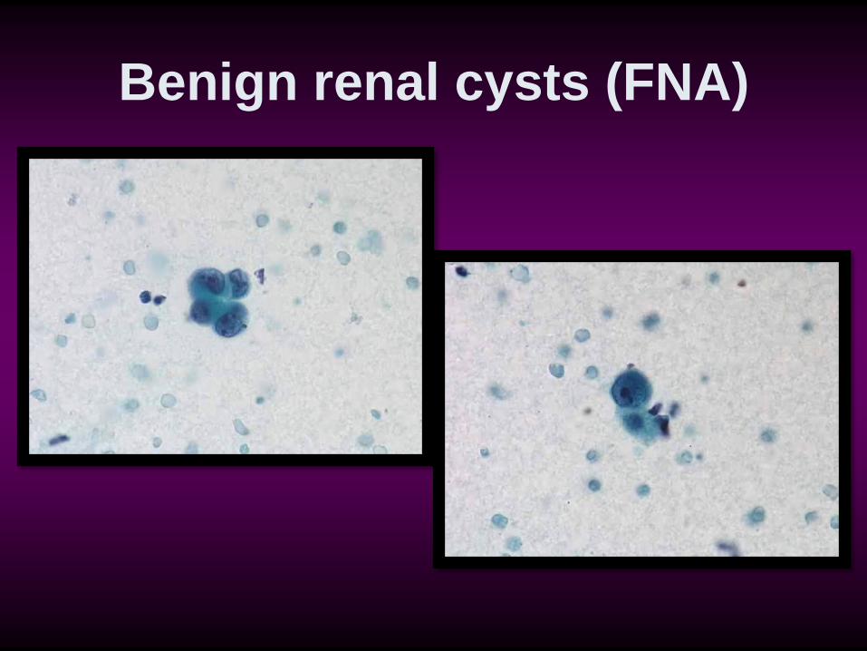
Benign renal cysts (FNA)
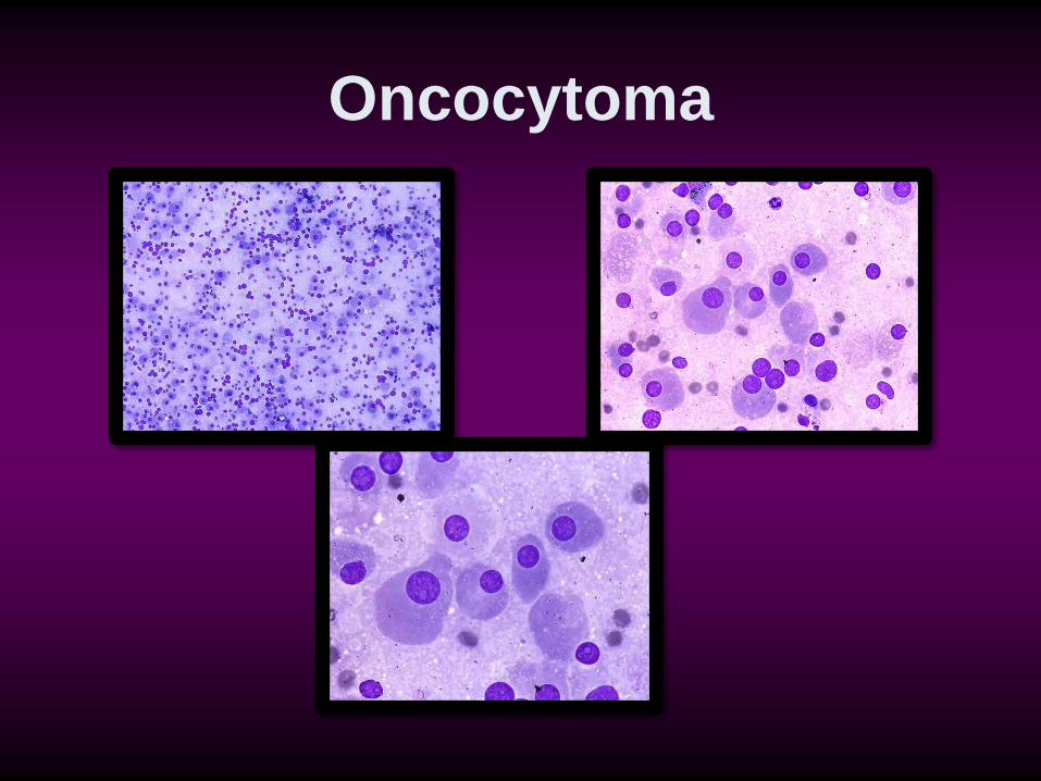
Oncocytoma
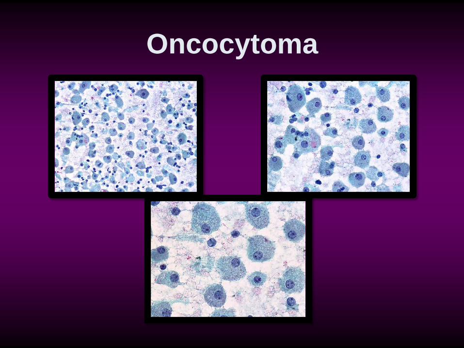
Oncocytoma
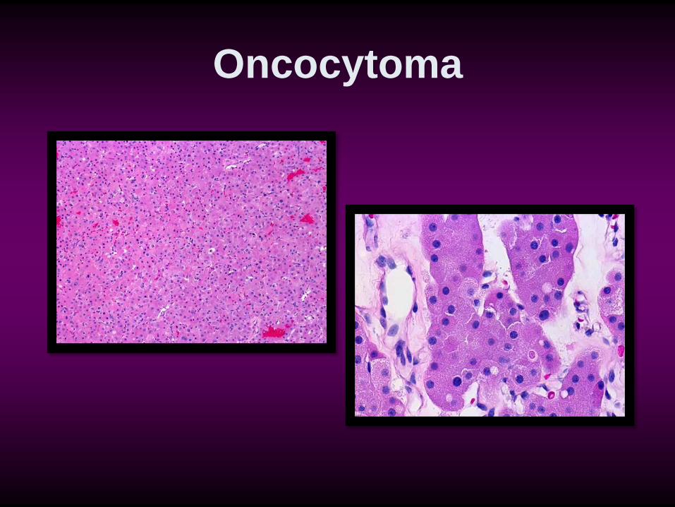
Oncocytoma
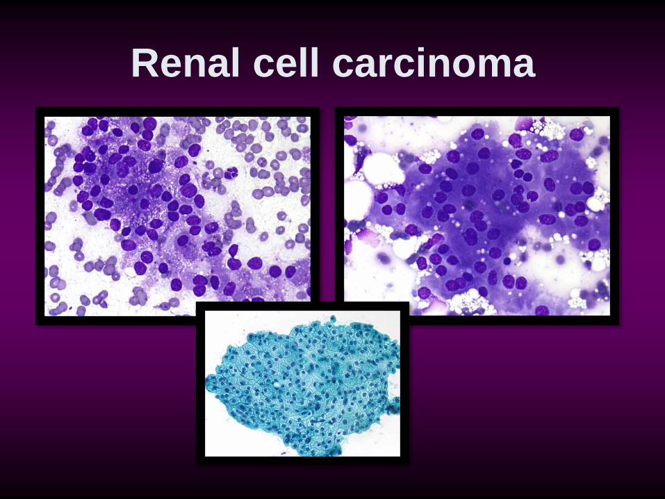
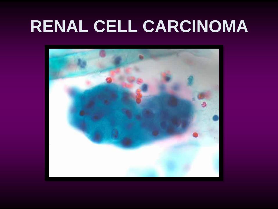
Renal cell carcinoma
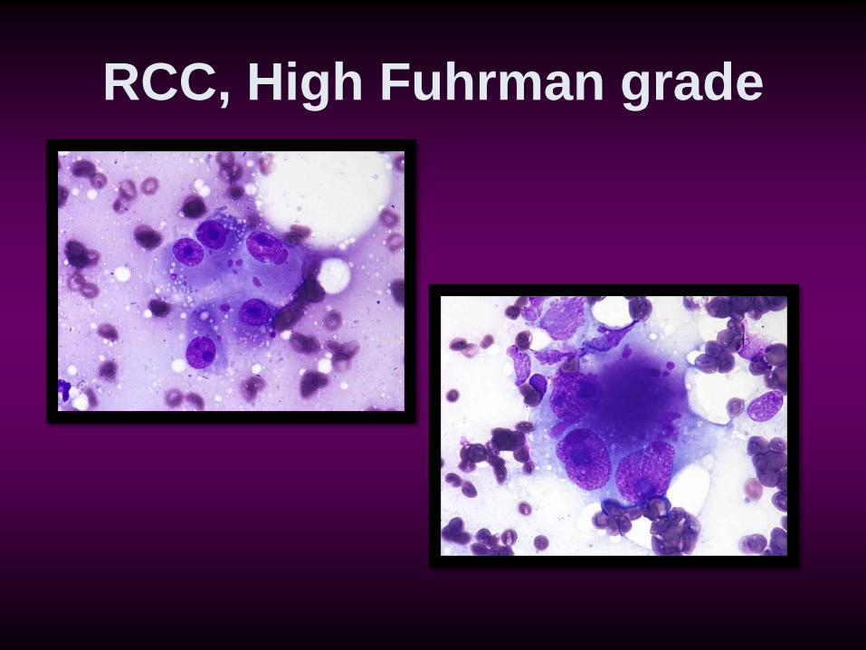
RCC, High Fuhrman grade
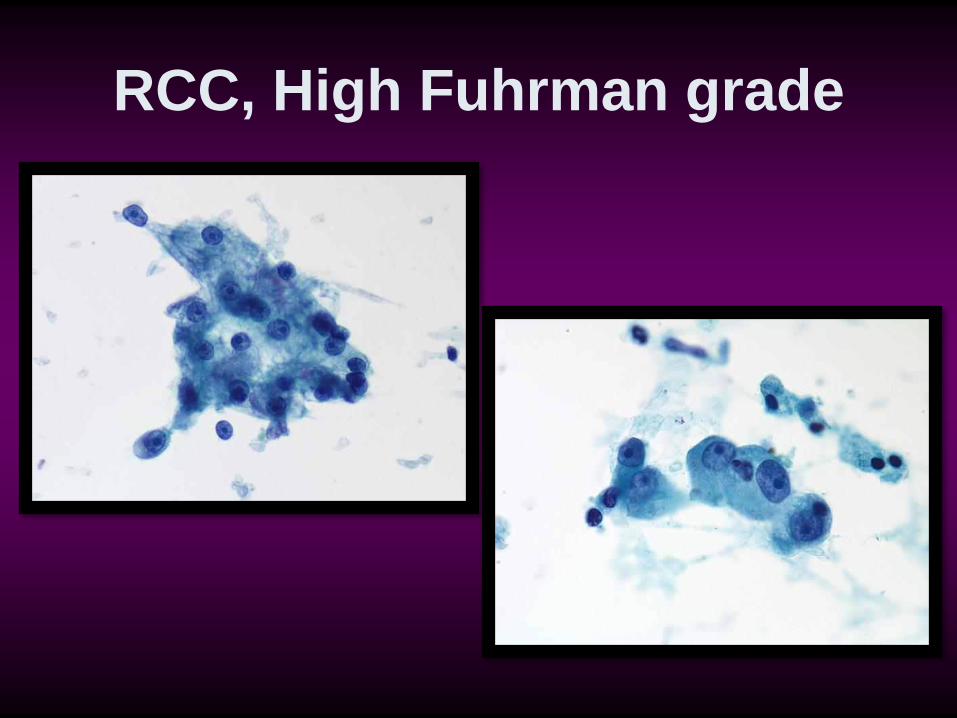
RCC, High Fuhrman grade
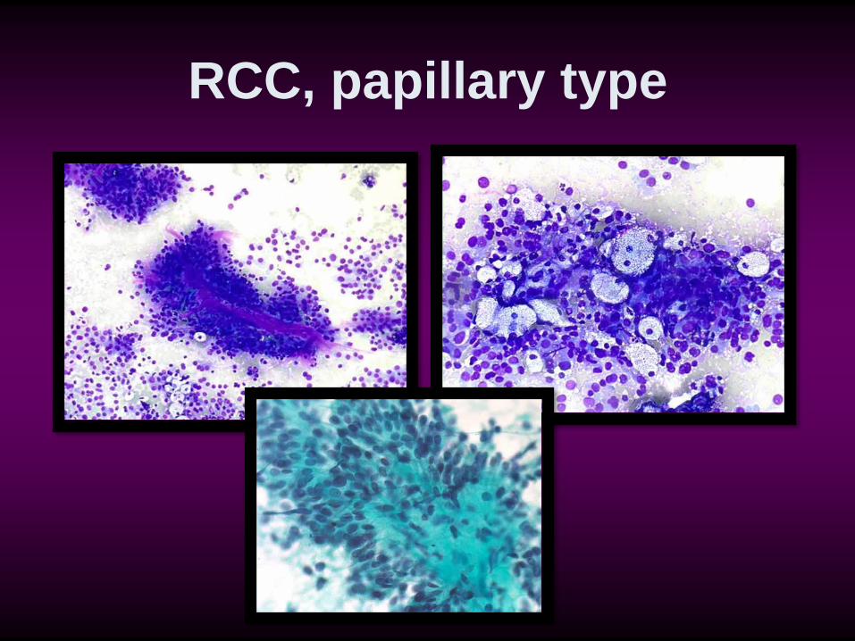
RCC, papillary type