Embed Size (px)
Citation preview
Small Molecule Therapeutics
Urokinase Plasminogen Activator System–Targeted Deliveryof Nanobins as a Novel Ovarian Cancer Therapy
Yilin Zhang1, Hilary A. Kenny1, Elden P. Swindell3,4, Anirban K. Mitra1, Patrick L. Hankins3,5, RichardW. Ahn3,5,Katja Gwin2, Andrew P. Mazar3,6,7, Thomas V. O'Halloran3,5,7, and Ernst Lengyel1
AbstractThe urokinase system is overexpressed in epithelial ovarian cancer cells and is expressed at low levels in
normal cells. To develop a platform for intracellular and targeted delivery of therapeutics in ovarian cancer,we
conjugated urokinase plasminogen activator (uPA) antibodies to liposomal nanobins. The arsenic trioxide–
loadednanobins had favorable physicochemical properties and the ability to bind specifically to uPA.Confocal
microscopy showed that the uPA-targeted nanobins were internalized by ovarian cancer cells, whereas both
inductively coupled plasma optical mass spectrometry (ICP-MS) and fluorescence-activated cell sorting
(FACS) analyses confirmed more than four-fold higher uptake of targeted nanobins when compared with
untargetednanobins. In a coculture assay, the targetednanobins showedefficient uptake inovarian cancer cells
but not in the normal primary omental mesothelial cells. Moreover, this uptake could be blocked by either
downregulating uPA receptor expression in the ovarian cancer cells using short-hairpin RNA (shRNA) or by
competitionwith free uPAor uPA antibody. In proof-of-concept experiments,mice bearing orthotopic ovarian
tumors showedagreater reduction in tumorburdenwhen treatedwith targetednanobins thanwithuntargeted
nanobins (47% vs. 27%; P < 0.001). The targeted nanobins more effectively inhibited tumor cell growth both in
vitro and in vivo compared with untargeted nanobins, inducing caspase-mediated apoptosis and impairing
stem cell marker, aldehyde dehydrogenase-1A1 (ALDH1A1), expression. Ex vivo fluorescence imaging of
tumors and organs corroborated these results, showing preferential localization of the targeted nanobins to the
tumor. These findings suggest that uPA-targeted nanobins capable of specifically and efficiently delivering
payloads to cancer cells could serve as the foundation for a new targeted cancer therapy using protease
receptors. Mol Cancer Ther; 12(12); 2628–39. �2013 AACR.
IntroductionOvarian cancer, which usually presents at an advanced
clinical stage, is the most aggressive gynecologic malig-nancy, accounting for 6% of all cancer-related deaths inwomen in the United States (1). Themost common sites ofovarian cancer metastasis are the other ovary, the perito-neum, and the omentum, whereas distant, extra-abdom-inalmetastases are rare (2, 3). The current standard of careincludes debulking surgery and subsequent adjuvantchemotherapy with taxane and platinum-containing
drugs. This chemotherapy combination clearly prolongsthe lives of patients with ovarian cancer; however, almostall tumors will eventually become chemoresistant (1).
Unfortunately, no new drugs have been approved forthe treatment of ovarian cancer since 1999, when lipo-somal doxorubicin (Doxil) was introduced for the treat-ment of recurrent disease. Doxil, when compared withfree doxorubicin, has a longer half-life due to its encap-sulation in a polyethylene glycol (PEG)–coated liposome(4). The small size of the Doxil particles (�100 nm)increases drug accumulation by escape through fenestrat-ed tumor vasculature. The favorable clinical pharmaco-kinetics and toxicity profile of Doxil, which causes lesscardiotoxicity and nausea than doxorubicin (5), has con-tributed to its clinical efficacy in recurrent ovarian cancer.Doxil represents one of the first examples of the successfultransfer of nanotechnology into the clinical setting basedon a rationale developed using a human ovarian cancertumor xenograft model (4, 6) and sets a precedent forliposomal delivery as a means to deliver cancer drugswith more efficacy and fewer side effects (7, 8).
For nanoparticles to reach their full potential in cancertherapy, they should be designed with reduced off-targettoxicity and increased circulation half-life for improvedpassive tumor uptake. These particles should ideally use a
Authors' Affiliations: Departments of 1Obstetrics and Gynecology/Sec-tion of Gynecologic Oncology and 2Pathology, University of Chicago,Chicago; 3Chemistry of Life Processes Institute; Departments of 4Chemicaland Biological Engineering, 5Chemistry, and 6Molecular Biosciences; and7Robert H. Lurie Comprehensive Cancer Center, Northwestern University,Evanston, Illinois
Note: Supplementary data for this article are available at Molecular CancerTherapeutics Online (http://mct.aacrjournals.org/).
Corresponding Author: Ernst Lengyel, Department of Obstetrics andGynecology, University of Chicago, MC 2050, 5841 South MarylandAvenue, Chicago, IL 60637. Phone: 773-7026722; Fax: 773-7025411;E-mail: [email protected]
doi: 10.1158/1535-7163.MCT-13-0204
�2013 American Association for Cancer Research.
MolecularCancer
Therapeutics
Mol Cancer Ther; 12(12) December 20132628
on May 30, 2018. © 2013 American Association for Cancer Research. mct.aacrjournals.org Downloaded from
Published OnlineFirst September 23, 2013; DOI: 10.1158/1535-7163.MCT-13-0204
cancer cell–targeted deliverymethod to specifically deliv-er large doses of a therapeutic payload directly into tumorcells. Indeed, nanoparticles can be modified for tumorcell-specific delivery with cancer-targeting ligands suchas antibodies (9), aptamers (10), peptides (11), or folate(12) that recognize receptors or antigens overexpressed onthe surface of cancer cells. There are particular advantagesof using antibodies for targeting. These include ease ofconjugation, high target specificity, and the potential forantibody-directed cellular cytotoxicity (13).In our search for new treatments ofwomen’s cancer, we
had previously encapsulated arsenic trioxide (As2O3, As),an U.S. Food and Drug Administration (FDA)–approveddrug for acute promyelocytic leukemia, into nanoparti-cles. This novel formulation strategy allowed encapsula-tion of a high concentration of As in a stable precipitatewith nickel acetate (Ni) that we designated as a nanobin(Ni,As; refs. 12, 14, 15). We showed that these nanobinssignificantly inhibited tumor cell growth in an orthotopictriple-negative humanbreast cancer xenograftmodel (14).However, the poor pharmacokinetic profile of As2O3,including a short plasma half-life and dose-limiting tox-icity, had prevented its development for solid tumorindications (16, 17). The nanobin formulation of As2O3
resolved these issues, resulting in increased tumor accu-mulation, activity, plasma half-life, and improved toler-ability when compared with free As2O3 (12, 14).To further improve antitumor activity, we conjugated
an antibody raised against the urokinase plasminogenactivator (uPA; ATN-291) to the arsenic nanobins (18).Both uPA and its receptor, u-PAR, are highly expressedin epithelial tumors (19) including ovarian cancer,where it is detected in as many as 90% of clinical ovariancancer specimens but not in untransformed ovarian/tubal epithelium [summarized in Kenny and colleagues(20)]. The antibody, ATN-291, binds to the kringledomain of uPA, even when it is bound to u-PAR, andthe entire complex is internalized (18). Because uPA isinternalized as a complex (21, 22), we reasoned that anantibody against uPA conjugated to nanobins wouldbind to tumor cells displaying receptor-bound or solu-ble uPA and, by leading to the internalization of thenanobin/uPA/u-PAR complex, results in intracellulardelivery of a large arsenic payload.In this study, we explore the hypothesis that targeting a
nanobin formulation of As2O3 to the uPA system that ishighly expressed in ovarian cancer will improve thedrug’s antitumor activity. The targeted nanobin formu-lation may revive the development of a promising cancerdrug by combining specific deliverywith improved phar-macokinetics. We report here that the targeted nanobinsinternalize into u-PAR–expressing cancer cells, and effec-tively impair ovarian cancer tumor growth.
Materials and MethodsReagents and cell linesAntibodies against u-PAR (ATN-658 for immunoblot,
and ATN-615 for immunohistochemistry) and the uPA
antibody ATN-291 were manufactured by SDIX (18),whereas antibody (UK-1) for immunoblotwas fromAmer-ican Diagnostica Inc. aldehyde dehydrogenase-1A1(ALDH1A1; B-5) antibody was obtained from Santa CruzBiotechnology. Cleaved caspase-3 (Asp175) and glyceral-dehyde-3-phosphate dehydrogenase (GAPDH; 14C10)antibodies were from Cell Signaling Technology, andmatrix metalloproteinase (MMP)-2 (IM33) antibody wasfrom Millipore. Hoechst 33342 and JC-1 dye were pur-chased from Invitrogen. Collagen type I (rat-tail) waspurchased from BD Bioscience and 1,2-distearoyl-sn-gly-cero-3-phosphocholine (DSPC) and 1,2-distearoyl-sn-gly-cero-3-phosphoethanolamine-N-[amino(polyethylenegly-col)-2000] (DSPE-PEG2000) were from Avanti Polar Lipids.Cell culture grade endotoxin-free water was from ThermoScientific. As2O3, nickel (II) acetate, cholesterol, and otherchemical reagents were purchased from Sigma-Aldrichunless otherwise indicated.
The CaOV3 and ES-2 human ovarian cancer cell lineswere from American Type Culture Collection (ATCC).SKOV3ip1 and HeyA8 cells were provided by Dr. Gor-don Mills (MD Anderson Cancer Center, Houston, TX).All human ovarian cancer cell lines (HeyA8, SKOV3ip1,and ES-2, CaOV3) and the primary ovarian cancer cloneMONTY-1 were maintained in Dulbecco’s modifiedEagle medium (DMEM, Invitrogen). MONTY-1 wasisolated from human omental metastases and used atan early passage (23). All cell lines were validated byshort-tandem repeat (STR) DNA fingerprinting usingthe AmpF‘STR Identifier Kit (Applied Biosystems). TheSTR profiles were compared with known ATCC finger-prints, to the Cell Line Integrated Molecular Authenti-cation database (CLIMA), and to the MD Andersonfingerprint database.
Preparation of alkyne-functional nanobinsArsenic/nickel complex–loaded nanobins, NB(Ni,
As), and empty nanobin controls NB(NaCl) were pre-pared and described in the Supplementary Materialsand Methods as previously reported (14, 18). DSPE-PEG3400-NH2 (Laysan Bio) was reacted with 4-penty-noic acid in the presence of 1-ethyl-3-(3-dimethylami-nopropyl) carbodiimide (EDC) and n-NHS in chloro-form at 60�C overnight. The resulting alkyne-functionallipid (DSPE-PEG3400-Alkyne) was dried, dissolved indeionized water, and lyophilized. The DSPE-PEG3400-alkyne lipid was then incubated with preformed nano-bins at a DSPE-PEG3400-alkyne to nanobin phospholipidmolar ratio of 0.01 for 1 hour at 60�C, allowing it toinsert into the lipid bilayer. Meanwhile, the total arseniccontent of the nanobin solution was adjusted to 25mmol/L to ensure that the drug loading does notchange during heating. The alkyne functional nanobinswere purified by diafiltration against 20 mmol/LHEPES and 150 mmol/L NaCl, pH 7.4. The phospho-lipid, arsenic, and nickel molar concentrations werethen measured by inductively coupled plasma opticalemission spectrometry (ICP-OES).
uPA-Targeted Nanobin Therapy in Ovarian Cancer
www.aacrjournals.org Mol Cancer Ther; 12(12) December 2013 2629
on May 30, 2018. © 2013 American Association for Cancer Research. mct.aacrjournals.org Downloaded from
Published OnlineFirst September 23, 2013; DOI: 10.1158/1535-7163.MCT-13-0204
Preparation of urokinase antibody–conjugatednanobins
The uPA antibody (ATN-291) was conjugated to thesurface of the nanobins using a copper (I)-catalyzed azide-alkyne cycloaddition reaction (24). The alkyne-functiona-lized nanobins described above were incubated with theazide-functionalized Alexa Fluor 647 (AF647)–labeledATN-291 at a molar ratio of 3 � 10�4 moles antibody permole of phospholipid. The preparation of azide-functio-nalized Alexa Fluor 647–labeled ATN-291 was describedin the SupplementaryMaterials andMethods. Copper (II)sulfate, tris(3-hydroxypropyltriazolylmethyl) amine, andsodium ascorbate were added to the reaction mixture toa final concentration of 200 mmol/L, 1 and 2 mmol/L,respectively. The reaction proceeded for 90 minutes at37�C, when fresh sodium ascorbate was added, bring-ing the total concentration to 3 mmol/L. The reactioncontinued for an additional 90 minutes at 37�C. Theunconjugated antibody was removed from the solutionusing diafiltration against 20 mmol/L HEPES and150 mmol/L NaCl, pH 7.4.
Quantification of nanobin-conjugated antibodiesThe number of uPA antibodies conjugated per nanobin
was determined by UV/Vis after proteolysis. Urokinaseantibody–conjugated nanobins were incubated with 4mg/mL of pronase for 2 hours at 37�C. Digested antibodywas then separated from intact nanobins using anAmiconUltra spin filter (50 k MWCO; Millipore). The concentra-tion of Alexa Fluor 647–labeled ATN-291was determinedby measuring absorbance at 650 nm with a spectropho-tometer (PerkinElmer) and was used as a proxy for ATN-291 concentration.
Transmission electron microscopyNanobins were visualized using the negative staining
technique via a TECNAI F30 transmission electronmicro-scope (FEI Company). The nanobin suspension wasdropped onto a glow-discharged carbon-coated coppergrid and stained with 1% uranyl acetate. Images wereexamined under 300 KV with Gatan CCD digital micro-graph (25).
Dynamic light scattering and zeta potentialThe measurement of hydrodynamic size and polydis-
persity index (PDI) of nanobins was performed using a384-well microplate reader using a DynaPro Nanostar(Wyatt Technology Corporation). The zeta potentialassessment was carried out in a clear disposable zeta cellusing a ZetasizerNanoZS analyzer (Malvern InstrumentsLtd.). Both dynamic light scattering (DLS) and zeta poten-tial were performed in PBS at 25�C, and the results werethe average of at least 10 measurements from three inde-pendent batches.
Inductively coupled plasma-optical emissionspectroscopy and -mass spectrometry
For ICP-OES, all nanobin samples and standards wereprepared in a matrix composed of deionized water, 3%
nitric acid, 2% acetic acid, 0.1% Triton X-100, and 0.5 partsper million (ppm) indium internal standards. Knownquantitative standards were prepared at concentrationsranging from 0.50 to 10 ppm arsenic, nickel, and phos-phorous. A matrix only standard was also included.
For inductively coupled plasma optical mass spectrom-etry (ICP-MS), the tissueswere rinsedwithPBSand frozenat �80�C until analysis. For elemental biodistribution,frozen tissueswere submerged in concentratednitric acid,anddigested in aMilestoneEthosEZmicrowavedigestionsystem. Samples and standards were then diluted into anultrapure deionized water matrix containing 3% nitricacid, 2% acetic acid, 0.1% Triton X-100, containing 5 partsper billion (ppb) indium as an internal standard.
ImmunohistochemistrySlides containing human ovarian cancer tissue were
deparaffinized. The u-PAR was detected with a primaryantibody (ATN-615, 1:200) and imaged through an avi-din-biotin–free detection system (Dako Envision;refs. 20, 26). For HeyA8 xenograft tumors, a representa-tive tumor area was first confirmed by a gynecologicpathologist (K. Gwin). Immunohistochemical staining ofmouse tumor sections were performed to assess markersof apoptosis (cleaved caspase-3, Asp175; 1:20), and ovar-ian cancer stem cell markers (ALDH1A1, B5; 1:400).Terminal deoxynucleotidyl transferase–mediated dUTPnick end labeling (TUNEL) was performed using theApopTag Plus Peroxidase In Situ Apoptosis Kit (Milli-pore). The stained cells were imaged at �100 and �400magnification from five random fields (25), and quanti-fied by NIH ImageJ software. Immunohistochemicalstaining of microvessel density (CD31, M-20; 1:50), pro-liferation (Ki67, SP6; 1:300) and the mouse macrophage(F4/80, A3-1; 1:500) was described in the SupplementaryMaterials and Methods.
ImmunoblotImmunoblot was performed as previously described
(20, 26). The following antibodies were used: uPA (UK-1;1:1,000), u-PAR (ATN-658; 1:2,000), cleaved caspase-3(Asp175; 1:1,000), ALDH1A1 (B-5; 1:250), MMP-2 (IM33;1:1,000), and GAPDH (14C10; 1:1,000). For cytotoxicitystudies, the HeyA8 cells were treated for 1 day with NB(Ni,As; 200 mmol/L of As), ATN-291-NB(Ni,As; 200mmol/L of As), As2O3 (20 mmol/L of As), or ATN-291-NB(NaCl).
Confocal microscopyCells were cultured for 3 days and then treated with
nanobins (25 mmol/L, lipid concentration). To evaluateinternalization, Z-stack scanning was performed byadjusting the focal plane from the bottom to the top ofthe cells, followed by orthogonal three-dimensional (3D)projection. For the ovarian cancer cell–targeted deliverystudy, primary human mesothelial cells isolated fromomentum of patients (27) were cocultured with HeyA8-GFP cells andwere treatedwithATN-291AF647-NB(Ni,As)
Zhang et al.
Mol Cancer Ther; 12(12) December 2013 Molecular Cancer Therapeutics2630
on May 30, 2018. © 2013 American Association for Cancer Research. mct.aacrjournals.org Downloaded from
Published OnlineFirst September 23, 2013; DOI: 10.1158/1535-7163.MCT-13-0204
for 24 hours. For apoptosis assessment, HeyA8 cells weretreatedwith 25mmol/L ofAs2O3,NB(Ni,As), orATN-291-NB(Ni,As) for 2 hours to measure mitochondrial mem-brane potential with the JC-1 dye, or for 18 hours tomeasure DNA fragmentation with Hoechst. Staining wasdonewith 2 mmol/L of JC-1 or 4 mmol/L of Hoechst 33342dyes for 30 minutes, respectively. The images wereacquired with a Leica SP5 confocal laser scanning micro-scope (oil lens-63X/1.4N). The excitation and emissionwavelengths were set for the detection of JC-1 aggregates(488/514 nm), JC-1 monomer (561/592 nm), Alexa Fluor647 (633/650 nm), and Hoechst (360/450 nm).
Flow cytometryOvarian cancer cells were treatedwith 25 mmol/L (lipid
concentration) of ATN-291-NB(Calcein) or NB(Calcein).For time-course studies,HeyA8 cellsweremonitoredover1 to 48 hour. In competition assays, HeyA8 cells werepretreated with either ATN-291 or single chain uPA(scuPA) with the indicated concentrations for 2 hoursbefore further incubation with nanobins for 24 hours. Inreceptor-competition assays, ES-2 wild-type (WT) andknockdown cells were incubated with nanobins for 24hours. After incubation, cells were washed, detached,resuspended in PBS, fixed by 2% paraformaldehyde, andstored at 4�C until analysis. A total of 10,000 events werecollected for each sample using an LSRFortessa cell ana-lyzer (calcein: 488/515 nm, Alexa Fluor 647: 641/670 nm).Data were analyzed by FlowJo software (TreeStar Inc.).The analysis of JC-1 stained cells was described in theSupplementary Materials and Methods.
Animal studiesHey8-GFP (5 � 105) cells (28) were injected intraperi-
toneally into 5- to 6-week-old athymic female nude mice.Four days after inoculation, the mice were randomizedinto five groups (5 mice/group): 200 mL of As2O3, NB(Ni,As), ATN-291-NB(Ni,As), PBS, or ATN-291-NB(NaCl)were injected intraperitoneally and injectionswere repeat-ed five times every other day at arsenic concentration of 4mg/kg (14). The mice were dissected 2 days after the lasttreatment. All animal experiments described wereapproved by Institutional Animal Care and Use Commit-tee (IACUC). To study nanobin biodistribution, organs(liver, heart, lung, spleen, and kidney) and the tumorweredissected, rinsedwith PBS, and imagedusing anOlympusfluorescence molecular imaging system. Blood toxicitystudies were described in the Supplementary Materialsand Methods.
Cell fractionationHeyA8 cells were treated with nanobins for 24 hours
followed by fractionation using theQproteome Cell Com-partment Kit (Qiagen). The cell compartments were iso-lated according to the manufacturer’s instructions. Thecytosolic, membrane/organelle, and nuclear compart-ments were verified by immunoblot before ICP-MS anal-ysis for arsenic content.
Statistical analysisResults were analyzed as mean� SEM. Statistical anal-
yses were performed using two-tailed t test or one-wayANOVA at P < 0.05 level of significance with GraphPadPrism version 5.04. The Cancer Genome Atlas (TCGA)database was queried for u-PAR expression in 580patients with ovarian cancer (29) and analyzed using thecBio cancer genomics portal (http://www.cbioportal.org/public-portal/index.do; ref. 30).
ResultsSynthesis and characterization of urokinaseantibody–conjugated nanobins loaded with As2O3
Epithelial ovarian cancer cells homogeneously expresselevated levels of u-PAR in the primary tumor and allabdominal metastasis (Fig. 1A). Analysis of the TCGAovarian cancer database (29) at the cBio cancer genomicsportal (30) showed that patients with ovarian cancer withu-PAR gene expression alterations had a significantlyworse overall (19.5 vs. 44.3 months) and disease-freesurvival (12.0 vs. 17.5 months; Supplementary Fig.S1A). And our previous tissue microarray studies dem-onstrated that u-PAR is highly expressed in epithelialtumors (19) including more than 90% of clinical ovariancancer specimens (20). This raises the possibility that highu-PAR expression might not only function as a marker ofaggressiveness for epithelial ovarian cancer cells but couldalsobeused to specificallydeliver a therapeuticpayload tothose cells. For the experiments described below, weidentified ovarian cancer cell lines with high (HeyA8,ES-2) and with low (SKOV3ip1, MONTY-1, and CaOV3)u-PAR expression, all of which also express uPA (Fig. 1B).Because the uPA system is highly expressed in ovariancancer (18, 20, 31), we sought to develop novel nanobinsthat would actively target uPA/u-PAR–expressing ovar-ian cancer cells. Amonoclonal antibody raised against thekringle domain of uPA inmice (ATN-291)was chosen as atargeting ligand as it binds tightly to human uPA with aKd � 0.5 nmol/L and does not disrupt uPA/u-PARbinding (18). The nanobins were assembled from choles-terol, DSPC, and DSPE-PEG, and loadedwith an arsenic/nickel coprecipitate (Fig. 1C; Supplementary Fig. S1B;refs. 12, 14, 15). Next, DSPE-PEG-alkynewas postinsertedinto the bilayer to facilitate the conjugation of the azide-functionalized uPA antibody using click chemistry. Thismethod ensures that the targeting antibodies are notexposed to potentially denaturing high temperatures (Fig.1C; ref. 32). To facilitate characterization of antibody–nanobin conjugates, the antibody was labeled with AlexaFluor 647 before click chemistry.UV/Vis analysis ofAlexaFluor 647 after proteolysis showed that the arsenic-loadednanobinsweredecoratedwith anaverageof 8.5 antibodiesper nanobin (Table 1). We also found that, after conjuga-tion, the ATN-291–targeted nanoparticle still bound uPAwith nanomolar binding affinity (data not shown).
The lipid composition of the nanobinswas optimized toensure that the nanobin surfacewas neutral at physiologicpH and to minimize nonspecific cellular binding. The
uPA-Targeted Nanobin Therapy in Ovarian Cancer
www.aacrjournals.org Mol Cancer Ther; 12(12) December 2013 2631
on May 30, 2018. © 2013 American Association for Cancer Research. mct.aacrjournals.org Downloaded from
Published OnlineFirst September 23, 2013; DOI: 10.1158/1535-7163.MCT-13-0204
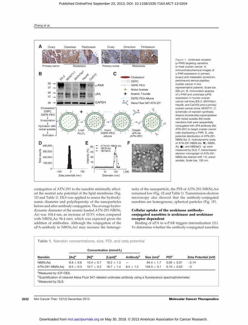
conjugation of ATN-291 to the nanobin minimally affect-ed the neutral zeta potential of the lipid membrane (Fig.1D and Table 1). DLS was applied to assess the hydrody-namic diameter and polydispersity of the nanoparticlesbefore andafter antibody conjugation. Theaveragehydro-dynamic diameter of the arsenic loaded ATN-291-NB(Ni,As) was 104.4 nm, an increase of 10.5% when comparedwith NB(Ni,As; 94.4 nm), which was expected given theaddition of antibodies. Although the conjugation of theuPA-antibody to NB(Ni,As) may increase the heteroge-
neity of the nanoparticle, the PDI of ATN-291-NB(Ni,As)remained low (Fig. 1E andTable 1). Transmission electronmicroscopy also showed that the antibody-conjugatednanobins are homogenous, spherical particles (Fig. 1F).
Cellular uptake of the urokinase antibody–conjugated nanobins is urokinase and urokinasereceptor dependent
Binding of uPA to u-PAR triggers internalization (21).To determine whether the antibody-conjugated nanobins
Table 1. Nanobin concentrations, size, PDI, and zeta potential
Concentration (mmol/L)
Nanobin [As]a [Ni]a [Lipid]a Antibodyb Size (nm)c PDIc Zeta Potential (mV)
NB(Ni,As) 8.8 � 0.6 10.4 � 0.7 18.2 � 1.3 — 94.4 � 1.7 0.05 � 0.01 �2.14ATN-291-NB(Ni,As) 8.5 � 0.3 10.7 � 0.2 18.7 � 1.4 8.5 � 1.2 104.5 � 3.1 0.10 � 0.02 �3
aMeasured by ICP-OES.bQuantification of cleaved Alexa Fluor 647–labeled urokinase antibody using a fluorescence spectrophotometer.cMeasured by DLS.
Figure 1. Urokinase receptor(u-PAR) targeting nanobinsto treat ovarian cancer. A,immunohistochemical images ofu-PAR expression in primary(ovary) and metastatic (omentum,peritoneum) serous-papillaryovarian cancer in tworepresentative patients. Scale bar,200 mm. B, immunoblot analysisof u-PAR and urokinase (uPA)expression in human ovariancancer cell lines (ES-2, SKOV3ip1,HeyA8, and CaOV3) and a primaryovarian cancer clone, MONTY1. C,schematic of nanobin synthesis.Arsenic trioxide (As) coprecipitatedwith nickel acetate (Ni) insidenanobins that were sequentiallyconjugated with uPA antibody (Ab;ATN-291) to target ovarian cancercells expressing u-PAR. D, zetapotential distribution of ATN-291-NB(Ni,As). E, hydrodynamic sizesof ATN-291-NB(Ni,As; !), NB(Ni,As; &), and NB(NaCl; ) weremeasured by DLS. F, transmissionelectron micrograph of ATN-291-NB(Ni,As) stained with 1% uranylacetate. Scale bar, 100 nm.
Zhang et al.
Mol Cancer Ther; 12(12) December 2013 Molecular Cancer Therapeutics2632
on May 30, 2018. © 2013 American Association for Cancer Research. mct.aacrjournals.org Downloaded from
Published OnlineFirst September 23, 2013; DOI: 10.1158/1535-7163.MCT-13-0204
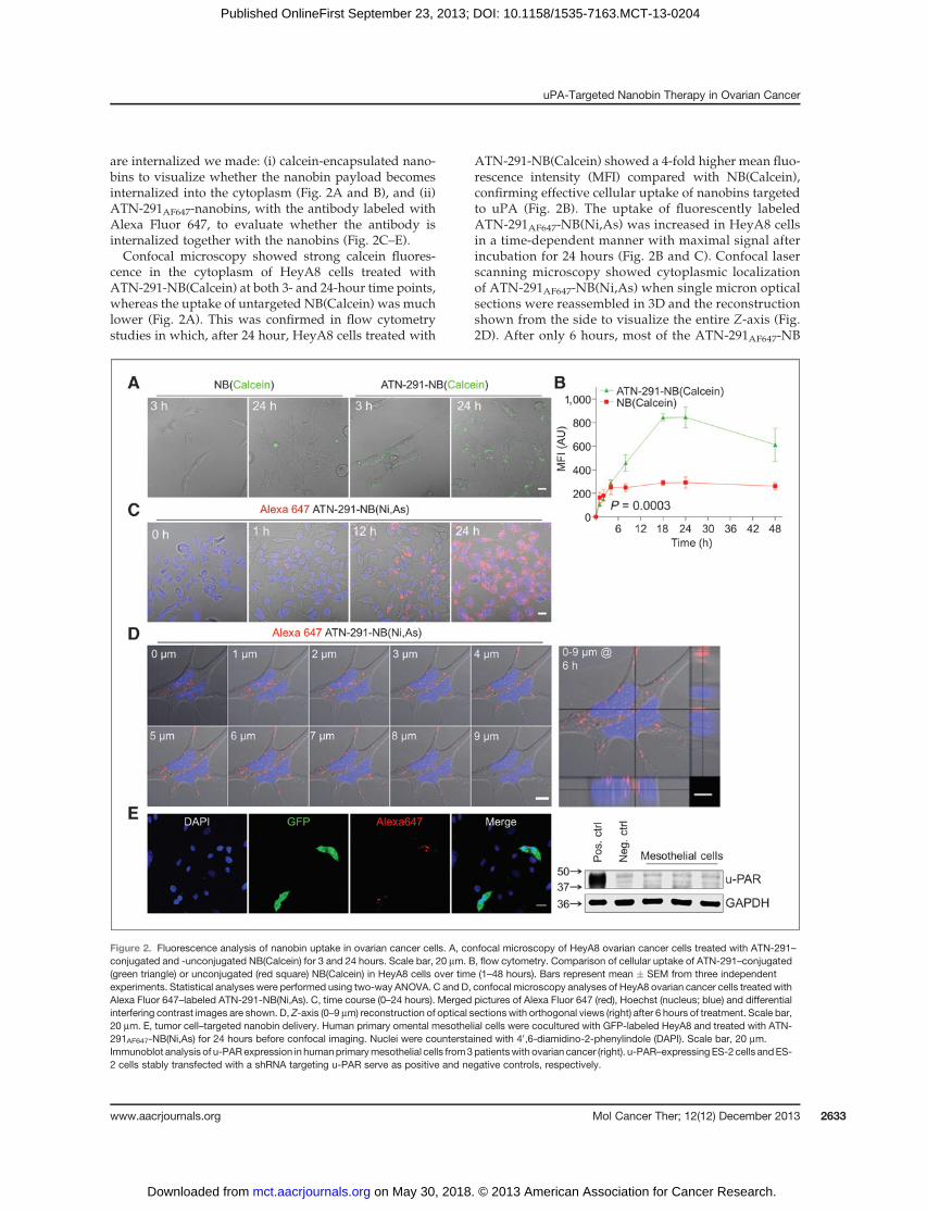
are internalized we made: (i) calcein-encapsulated nano-bins to visualize whether the nanobin payload becomesinternalized into the cytoplasm (Fig. 2A and B), and (ii)ATN-291AF647-nanobins, with the antibody labeled withAlexa Fluor 647, to evaluate whether the antibody isinternalized together with the nanobins (Fig. 2C–E).Confocal microscopy showed strong calcein fluores-
cence in the cytoplasm of HeyA8 cells treated withATN-291-NB(Calcein) at both 3- and 24-hour time points,whereas the uptake of untargeted NB(Calcein) was muchlower (Fig. 2A). This was confirmed in flow cytometrystudies in which, after 24 hour, HeyA8 cells treated with
ATN-291-NB(Calcein) showed a 4-fold higher mean fluo-rescence intensity (MFI) compared with NB(Calcein),confirming effective cellular uptake of nanobins targetedto uPA (Fig. 2B). The uptake of fluorescently labeledATN-291AF647-NB(Ni,As) was increased in HeyA8 cellsin a time-dependent manner with maximal signal afterincubation for 24 hours (Fig. 2B and C). Confocal laserscanning microscopy showed cytoplasmic localizationof ATN-291AF647-NB(Ni,As) when single micron opticalsections were reassembled in 3D and the reconstructionshown from the side to visualize the entire Z-axis (Fig.2D). After only 6 hours, most of the ATN-291AF647-NB
Figure 2. Fluorescence analysis of nanobin uptake in ovarian cancer cells. A, confocal microscopy of HeyA8 ovarian cancer cells treated with ATN-291–conjugated and -unconjugated NB(Calcein) for 3 and 24 hours. Scale bar, 20 mm. B, flow cytometry. Comparison of cellular uptake of ATN-291–conjugated(green triangle) or unconjugated (red square) NB(Calcein) in HeyA8 cells over time (1–48 hours). Bars represent mean � SEM from three independentexperiments. Statistical analyses were performed using two-way ANOVA. C and D, confocal microscopy analyses of HeyA8 ovarian cancer cells treated withAlexa Fluor 647–labeled ATN-291-NB(Ni,As). C, time course (0–24 hours). Merged pictures of Alexa Fluor 647 (red), Hoechst (nucleus; blue) and differentialinterfering contrast images are shown. D, Z-axis (0–9 mm) reconstruction of optical sections with orthogonal views (right) after 6 hours of treatment. Scale bar,20 mm. E, tumor cell–targeted nanobin delivery. Human primary omental mesothelial cells were cocultured with GFP-labeled HeyA8 and treated with ATN-291AF647-NB(Ni,As) for 24 hours before confocal imaging. Nuclei were counterstained with 40,6-diamidino-2-phenylindole (DAPI). Scale bar, 20 mm.Immunoblot analysis of u-PARexpression in humanprimarymesothelial cells from3patientswith ovarian cancer (right). u-PAR–expressingES-2 cells andES-2 cells stably transfected with a shRNA targeting u-PAR serve as positive and negative controls, respectively.
uPA-Targeted Nanobin Therapy in Ovarian Cancer
www.aacrjournals.org Mol Cancer Ther; 12(12) December 2013 2633
on May 30, 2018. © 2013 American Association for Cancer Research. mct.aacrjournals.org Downloaded from
Published OnlineFirst September 23, 2013; DOI: 10.1158/1535-7163.MCT-13-0204
(Ni,As) was detected in a perinuclear position, shownpreviously to be caused by u-PAR internalization (22).Specificity of the targeted nanobins could also be shownby coculturing primary human mesothelial cells withGFP-expressing ovarian cancer cells (Fig. 2E). The AlexaFluor 647–labeled antibody (red) colocalized with thehigh u-PAR–expressing ovarian cancer cells (green) butwas not detected in the u-PAR–negative mesothelialcells (Fig. 2E; refs. 20, 31).
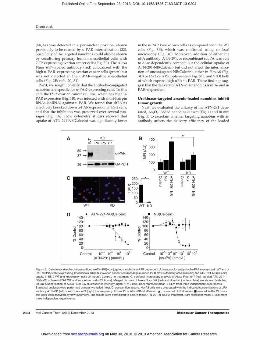
Next, we sought to verify that the antibody-conjugatednanobins are specific for u-PAR–expressing cells. To thisend, the ES-2 ovarian cancer cell line, which has high u-PAR expression (Fig. 1B), was infected with short-hairpinRNAs (shRNA) against u-PAR. We found that shRNAseffectively knocked down u-PAR expression in ES-2 cells,and that the inhibition was preserved over several pas-sages (Fig. 3A). Flow cytometry studies showed thatuptake of ATN-291-NB(Calcein) was significantly lower
in the u-PAR knockdown cells as compared with the WTcells (Fig. 3B), which was confirmed using confocalmicroscopy (Fig. 3C). Moreover, addition of either theuPA antibody, ATN-291, or recombinant scuPA was ableto dose-dependently compete out the cellular uptake ofATN-291-NB(Calcein) but did not affect the internaliza-tion of unconjugated NB(Calcein), either in HeyA8 (Fig.3D) or ES-2 cells (Supplementary Fig. S1C and S1D) bothof which express high uPA/u-PAR. These findings sug-gest that the delivery ofATN-291-nanobins is uPA- andu-PAR–dependent.
Urokinase-targeted arsenic-loaded nanobins inhibittumor growth
Next, we evaluated the efficacy of the ATN-291 deco-rated, As2O3-loaded nanobins in vitro (Fig. 4) and in vivo(Fig. 5) to ascertain whether targeting nanobins with anantibody affects the delivery efficiency of the loaded
Figure 3. Cellular uptake of urokinase antibody (ATN-291)–conjugated nanobin is u-PARdependent. A, immunoblot analysis of u-PARexpression inWTandu-PAR shRNA stably expressing (knockdown, KD) ES-2 ovarian cancer cells (passage number, P). B, flow cytometry of NB(Calcein) and ATN-291-NB(Calcein)uptake in ES-2 WT and knockdown cells (24 hours). Control, no treatment. C, confocal microscopy analysis of Alexa Fluor 647 (red)–labeled ATN-291-NB(NaCl) uptake in ES-2 WT and knockdown cells (24 hours). Merged pictures of Alexa Fluor 647 (red) and Hoechst (nucleus; blue) are shown. Scale bar,20 mm. Quantification of Alexa Fluor 647 fluorescence intensity (right). �, P < 0.05. Bars represent mean � SEM from three independent experiments.Statistical analyses were performed using a two-tailed t test. D, competition assays. HeyA8 cells were pretreated with the indicated concentrations of uPAantibody ATN-291 (left) or with the scuPA (right). Subsequently, 25 mmol/L of ATN-291-NB(Calcein;~), or as control NB(Calcein;&) was added for 24 hoursand cells were analyzed by flow cytometry. The results were normalized to cells without ATN-291 or scuPA treatment. Bars represent mean � SEM fromthree independent experiments.
Zhang et al.
Mol Cancer Ther; 12(12) December 2013 Molecular Cancer Therapeutics2634
on May 30, 2018. © 2013 American Association for Cancer Research. mct.aacrjournals.org Downloaded from
Published OnlineFirst September 23, 2013; DOI: 10.1158/1535-7163.MCT-13-0204
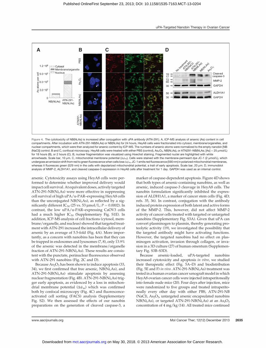
arsenic. Cytotoxicity assays using HeyA8 cells were per-formed to determine whether improved delivery wouldimpact cell survival.At equivalentdoses, actively targetedATN-291-NB(Ni,As) were more effective in suppressingcell survival of high uPA/u-PAR–expressingHeyA8 cellsthan the unconjugated NB(Ni,As), as reflected by a sig-nificantly different IC50 (25 vs. 53 mmol/L; P ¼ 0.0002). Incontrast, the low uPA/u-PAR–expressing CaOV3 cellshad a much higher IC50 (Supplementary Fig. S1E). Inaddition, ICP-MS analysis of cell fractions (cytosol, mem-brane/organelle, andnucleus) showed that targeted treat-mentwithATN-291 increased the intracellular delivery ofarsenic by an average of 5.5-fold (Fig. 4A). More impor-tantly, as a concern with nanobins has been that they canbe trapped in endosomes and lysosomes (7, 8), only 13.9%of the arsenic was detected in the membrane/organellefraction of ATN-291-NB(Ni,As). These results are consis-tent with the punctate, perinuclear fluorescence observedwith ATN-291 nanobins (Fig. 2C and D).Because As2O3 has been shown to induce apoptosis (33,
34), we first confirmed that free arsenic, NB(Ni,As), andATN-291-NB(Ni,As) stimulate apoptosis by assessingnuclear fragmentation (Fig. 4B). ATN-291-NB(Ni,As) trig-ger early apoptosis, as evidenced by a loss in mitochon-drial membrane potential (Dcm) which was confirmedboth by confocal microscopy (Fig. 4C) and fluorescence-activated cell sorting (FACS) analysis (SupplementaryFig. S2). We then assessed the effects of our nanobinpreparations on the generation of cleaved caspase-3, a
marker of caspase-dependent apoptosis. Figure 4D showsthat both types of arsenic-containing nanobins, as well asarsenic, induced caspase-3 cleavage in HeyA8 cells. Thenanobin formulation significantly inhibited the expres-sion of ALDH1A1, a marker of cancer stem cells (Fig. 4D;refs. 35, 36). In contrast, conjugation with the antibodyinducedprotein expression of both latent and active formsof the MMP-2. This, however, did not affect MMP-2activity of cancer cells treatedwith targeted or untargetednanobins (Supplementary Fig. S3A). Given that uPA canconvert plasminogen to plasmin, thereby generating pro-teolytic activity (19), we investigated the possibility thatthe targeted antibody might have activating functions.However, the targeted nanobins had no effect on plas-minogen activation, invasion through collagen, or inva-sion in a 3D culture (27) of human omentum (Supplemen-tary Fig. S3B–S3D).
Because arsenic-loaded, uPA-targeted nanobinsincreased cytotoxicity and apoptosis in vitro, we studiedtheir therapeutic effect (Fig. 5A–D) and biodistribution(Fig. 5E and F) in vivo. ATN-291-NB(Ni,As) treatment wastested in ahumanovarian cancer xenograftmodel inwhichHeyA8 ovarian cancer cells were injected intraperitoneallyinto female nude mice (20). Four days after injection, micewere randomized to five groups and treated intraperito-neally every other day with either PBS, ATN-291-NB(NaCl), As2O3, untargeted arsenic encapsulated nanobinsNB(Ni,As), or targeted ATN-291-NB(Ni,As) at an As2O3
concentration of 4 mg/kg (14). All treated mice continued
Figure 4. The cytotoxicity of NB(Ni,As) is increased after conjugation with uPA antibody (ATN-291). A, ICP-MS analysis of arsenic (As) content in cellcompartments. After incubation with ATN-291-NB(Ni,As) or NB(Ni,As) for 24 hours, HeyA8 cells were fractionated into cytosol, membrane/organelles, andnuclear compartments, which were then analyzed for arsenic content by ICP-MS. The numbers of arsenic atoms were normalized to the empty nanobin [NB(NaCl)] control. B and C, confocal microscopy. HeyA8 cells were treated with either PBS (control), As2O3, NB(Ni,As), or ATN291-NB(Ni,As; [As]¼ 25 mmol/L)for 18 hours (B), or 2 hours (C). B, nuclear fragmentation was visualized using Hoechst staining. Fragmented nuclei are highlighted with whitearrowheads. Scale bar, 10 mm. C, mitochondrial membrane potential (Dcm). Cells were stained with the membrane-permeant dye JC-1 (2 mmol/L), whichundergoes anemission shift from red togreen fluorescencewhencells loseDcm, JC-1 emits red fluorescence (590nm) in polarizedmitochondrialmembranes,whereas it fluoresces green (529 nm) in the cells with depolarized mitochondrial potential, a trait of early apoptosis. Scale bar, 20 mm. D, immunoblotanalysis of MMP-2, ALDH1A1, and cleaved caspase-3 expression in HeyA8 cells after treatment for 1 day. GAPDH was used as an internal control.
uPA-Targeted Nanobin Therapy in Ovarian Cancer
www.aacrjournals.org Mol Cancer Ther; 12(12) December 2013 2635
on May 30, 2018. © 2013 American Association for Cancer Research. mct.aacrjournals.org Downloaded from
Published OnlineFirst September 23, 2013; DOI: 10.1158/1535-7163.MCT-13-0204
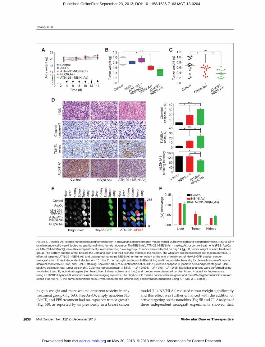
to gain weight and there was no apparent toxicity in anytreatment group (Fig. 5A). FreeAs2O3, empty nanobinsNB(NaCl), andPBS treatment hadno impact on tumorgrowth(Fig. 5B), as reported by us previously in a breast cancer
model (14). NB(Ni,As) reduced tumorweight significantlyand this effect was further enhanced with the addition ofactive targetingon thenanobins (Fig. 5B andC).Analysis ofthree independent xenograft experiments showed that,
Figure 5. Arsenic (As)-loaded nanobin reduced tumor burden in an ovarian cancer xenograft mousemodel. A, bodyweight and treatment timeline. HeyA8-GFPovarian cancer cellswere injected intraperitoneally into female nudemice. TheNB(Ni,As), ATN-291-NB(Ni,As; 4mg/kg, As), or control treatments (PBS,As2O3,or ATN-291-NB(NaCl)) were also intraperitoneally injected (arrow; 5 mice/group). Tumors were collected on day 14 (~). B, tumor weight of each treatmentgroup. The bottom and top of the box are the 25th and 75th and the line in the middle is the median. The whiskers are the minimum and maximum value. C,effect of targeted ATN-291-NB(Ni,As) and untargeted nanobins NB(Ni,As) on tumor weight at the end of treatment of HeyA8-GFP ovarian cancerxenografts from three independent studies, n¼ 15mice. D, hematoxylin and eosin (H&E) staining and immunohistochemistry for cleaved caspase-3, ovarianstemcellmarker (ALDH1A1) andTUNEL staining. Scale bar, 100mm.Quantification of ALDH1A1, cleavedcaspase-3–positive cells andpercentageof TUNEL-positive cells over total tumor cells (right). Columns represent mean� SEM. ���, P < 0.001; ��, P < 0.01; �, P < 0.05. Statistical analyses were performed usingtwo-tailed t test. E, individual organs (i.e., heart, liver, kidney, spleen, and lung) and tumors were dissected on day 14 and imaged for fluorescenceusing an OV100 Olympus fluorescence molecular imaging systems. The HeyA8-GFP ovarian cancer cells are green and the uPA-targeted nanobins are red(Alexa Fluor 647). F, the same experiment as in D was repeated and arsenic (As) concentration quantified using ICP-MS (n ¼ 6 mice).
Zhang et al.
Mol Cancer Ther; 12(12) December 2013 Molecular Cancer Therapeutics2636
on May 30, 2018. © 2013 American Association for Cancer Research. mct.aacrjournals.org Downloaded from
Published OnlineFirst September 23, 2013; DOI: 10.1158/1535-7163.MCT-13-0204
compared with the control treatment group, tumor weightwas reduced by 27% (P ¼ 0.04) with the nontargeted NB(Ni,As) treatment and 47% (P ¼ 0.0005) with the targetedATN-291-NB(Ni,As) treatments (Fig. 5C). The additionaldecrease in tumor burden after treatment with ATN-291-NB(Ni,As), comparedwith treatmentwithNB(Ni,As), wasstatistically significant (P ¼ 0.02).Pathologic review of the tumors at the end of treatment
showed undifferentiated, high-grade tumors (Fig. 5D),which did not differ in angiogenesis (microvessel density,CD31), proliferation rate (Ki-67; Supplementary Fig. S4),or u-PAR expression (data not shown). Both targeted anduntargeted nanobins induced macrophage infiltrationinto the tumor. Untargeted nanobins recruited moremacrophages than the uPA-targeted nanobins (Supple-mentary Fig. S4). Treatment with the untargeted arsenicnanobins induced active caspase-3 and DNA fragmenta-tion (TUNEL stain) in the tumors. These effectsweremorepronounced in ATN-291-NB(Ni,As)–treated tumors (Fig.5D), suggesting, as is consistent with the in vitro results(Fig. 4B–D), that arsenic-loaded nanobins reduce tumorburden by inducing apoptosis rather than by inhibitingtumor growth. Also consistent with the in vitro results isthatATN-291-NB(Ni,As) treatment reduced expressionofALDH1A1 (Fig. 4D; ref. 35). Analysis of kidney and liverfunction in serum showed no significant toxicity withATN-291-NB(Ni,As) treatment (Supplementary Fig. S5).Together, these results corroborate the therapeutic effec-tiveness of NB(Ni,As) treatment and the improved anti-tumoral effect of targeted ATN-291-NB(Ni,As) treatment.To study biodistribution of nanobins using a fluores-
cence imaging system, HeyA8-GFP ovarian cancer xeno-grafts were established and then injected with nanobins.The ATN-291AF647-NB(Ni,As) homed to the tumor,spleen, lung, and liver (Fig. 5E), common accumulationsites for liposome nanoparticles (11, 37). Ex vivo imagesfrom ATN-291AF647-NB(Ni,As)–treated animals showedthat the tumors had a higher fluorescence intensity thanthe organs, indicating the efficient delivery of nanobins tothe tumorwith active targeting. The arsenic concentrationin organs/tumor detected by ICP-MS showed no signif-icant difference in the total amount of As2O3 delivered tothe tumors (Fig. 5F).
DiscussionOvarian cancer is generally confined to the peritoneal
cavity, and intraperitoneal chemotherapy treatment is awell-established clinical treatment modality (38). There-fore, nanoparticles that efficiently target tumor cells intra-peritoneally, deliver anticancer drugs potently, and min-imize the exposure of normal tissue to chemotherapeuticsthat could lead to significant treatment improvements.Many reports show that the uPA system is consistentlyoverexpressed in ovarian cancer, and that its expression innormal cells is low (18, 20, 31). Given that the u-PAR isinternalized upon uPA binding, we developed a uPA/u-PAR–dependent targeted delivery using uPA anti-body–targeted nanobins.
We found that targeted single-agent treatment withnanobins [ATN-291-NB(Ni,As)] suppressed tumorgrowth in an orthotopic ovarian cancer mouse model byalmost 50%, whereas the untargeted NB(Ni,As), at equi-molar arsenic doses, were half as effective (Fig. 5C).Several of our observations are consistentwith the specificuptake of arsenic-loadednanobins byovarian cancer cells.When u-PAR expression was downregulated in cancercells with shRNA, we observed reduced intracellularuptake of targeted nanobins (Fig. 3A–C). Consistent withthis observation, confocal microscopy showed that thetargeted nanobins were internalized (Fig. 2). Adding freeantibody or competition with recombinant single-chainuPA to u-PAR–overexpressing cells also inhibited specificuptake of nanobins (Fig. 3D). Finally, the targeted nano-bins were taken up in vivo by the uPA/u-PAR–expressingtumor cells (Fig. 5E).
Antibodies are not the only targeting agents that canefficiently deliver intracellular payloads. We have previ-ously shown that folate-targeted liposomes containing As2O3 are internalized by folate-receptor–positive tumor cellsbut not by tumor cells without the receptor (12). In adifferent study, u-PAR–targeted delivery of iron-oxidenanoparticles enhanced detection of human breast cancerusingMRI (39).Targeting transferrinnanoparticlesnotonlydelivered siRNA but allowed selective imaging of subcu-taneous neuroblastoma tumors using a positron emissiontomography/computed tomography (PET/CT) scan (40).Conversely, targeting tumor vasculature using Arginine-glycine-aspartic acid (RGD)-linked nanogels containingdocetaxel inhibited tumor growth in a breast cancer model(41). These studies, together with our study, highlight theimportance of specific intracellular delivery of payloadsto achieve a therapeutic and diagnostic advantage.
Like integrin and folate receptors, the uPA/u-PAR iswidely expressed in epithelial cancers. However, unlikeother receptors, their expression is found in tumor-asso-ciated stromal cells as well as in tumor cells (18, 19).Further clinical development of this targeted nanomedi-cine hinges on leveraging uPA/u-PAR expression intumor cells with this effective delivery system. A centralchallenge to clinical translation of targeted nanoparticlesis the ability to reliably scale up and reproducibly char-acterize the drug carrier. The inclusion of antibodies to thenanobin surface, using copper-catalyzed alkyne-azidecycloaddition click chemistry, has several advantagesbecause it involves a reaction between alkyne and azidefunctional groups that are not otherwise found in biomo-lecules. Unlike other common conjugationmethods usingamine-coupling or thiol-coupling chemistries, this clickreaction is chemically orthogonal to reactions that canoccur between protein side chains, ensuring that no reac-tions occur that may lead to particle-particle cross-linkingor nonspecifically alter the surface properties of the nano-bins. In addition, the functional groups are not susceptibleto hydrolysis. Click chemistry exposes only the thermallystable alkyne functional groups on the DSPE-PEG-alkyneto high temperatures, avoiding thermal deactivation of
uPA-Targeted Nanobin Therapy in Ovarian Cancer
www.aacrjournals.org Mol Cancer Ther; 12(12) December 2013 2637
on May 30, 2018. © 2013 American Association for Cancer Research. mct.aacrjournals.org Downloaded from
Published OnlineFirst September 23, 2013; DOI: 10.1158/1535-7163.MCT-13-0204
the antibody. Because the ATN-291 antibody is labeledwith Alexa Fluor 647 at a known ratio, we can use fluor-ophore concentration as a proxy for antibody concentra-tion. Visible light spectrometricmeasurements are subjectto light-scattering interference in the presence of nano-particles. By digesting the protein in a small sample ofnanobins, then probing the liberated fluorophore afterfiltration of the liposomes, we can quantify the precisenumber of antibodies per particle without light-scatteringinterference from the lipid particles.
A landmark article showed that As2O3 is a very potentdrug for the treatment of patients with acute promyelo-cytic leukemia (42). It is currently being considered for thetreatment of solid human tumors because it has demon-strated potent cytotoxic effects against epithelial tumorcell lines in vitro (16). However, development in solidtumor indications has been limited by the poor pharma-cokinetics and dose-limiting toxicity of the free drug. Themost commonly described mechanisms of action(reviewed in ref. 34) of As2O3 are inhibition of tumorangiogenesis, selective killing of cancer stem cells, inhi-bition ofMMPs, and induction of apoptosis (14, 16, 17, 33).We found that ATN-291-NB(Ni,As) did not affect tumorcell proliferation or angiogenesis (Supplementary Fig. S4),but very efficiently induced ovarian cancer cell apoptosis(Figs. 4B–D, 5D; Supplementary Fig. S2). This explained,at least in part, the observed significant cytotoxicity of theconjugated nanobins and is consistent with the mechan-isms of arsenic function elucidated in a murine model ofbreast cancer (14). Liu and colleagues demonstrated thattreatment with As2O3 induced apoptosis and G2–M cell-cycle arrest in acute promyelocytic leukemia cells (33).This effect was more pronounced in leukemia cells with amutatedp53 gene, an observation of potential significancefor the treatment of ovarian cancer as almost all serousovarian cancer have mutations within the p53 gene (43).We also showed inhibition of the cancer stem cell markerALDH1A1 both in vitro and in vivo (Figs. 4D and 5D) byATN-291-NB(Ni,As). These data are consistent with astudy (36) reporting that ALDH1A1 is expressed in 73%of all human ovarian cancer and that its silencing inovarian cancer cell lines blocked tumor cell growth invitro and in vivo. The blocking of ALDH1A1 sensitizedcells to platinum and taxane-based chemotherapy (36),which suggests that ATN-291-NB(Ni,As) may have a rolein combination therapy for ovarian cancer, as thesetumors frequently develop chemotherapy resistance (1).
More than 60 years after the discovery of urokinase (44),the promise that tumor growth could be blocked byinhibiting specific proteases has not come to fruition. Thisis in part because other proteases compensate for theproteolytic function when a single protease is inhibited.
However, given that most epithelial cancers stronglyexpress uPA and u-PAR, it may be possible to harnessthe tumor-associated uPA system as an entry point todeliver therapeutic or diagnostic payloads specifically tocancer cells rather than as a target for antiproteolytictherapy. Although, in this report, we have focused onformulating As2O3 in a nanobin, our delivery systemcould also be loaded with other chemotherapeutics orimaging agents andused to target any cancers that expressuPA and u-PAR.
Disclosure of Potential Conflicts of InterestA.P. Mazar is employed as a Managing Member in Tactic Pharma and
has ownership interest (including patents) in the same. T. V O’Halloranhas ownership interest in US Patent #8,246,983, Valence Therapeutics, andTactic Pharma. No potential conflicts of interest were disclosed by theother authors.
Authors' ContributionsConception and design: Y. Zhang, E.P. Swindell, R.W. Ahn, A.P. Mazar,T.V. O’Halloran, E. LengyelDevelopment of methodology: Y. Zhang, E.P. Swindell, P.L. Hankins,R.W. Ahn, A.P. Mazar, T.V. O’Halloran, E. LengyelAcquisition of data (provided animals, acquired and managed patients,provided facilities, etc.): E.P. Swindell, A.K. Mitra, P.L. Hankins, T.V.O’Halloran, E. LengyelAnalysis and interpretation of data (e.g., statistical analysis, biostatis-tics, computational analysis): Y. Zhang, H.A. Kenny, E.P. Swindell, A.K.Mitra, P.L. Hankins, K. Gwin, T.V. O’Halloran, E. LengyelWriting, review, and/or revision of the manuscript: Y. Zhang, H.A.Kenny, E.P. Swindell, P.L. Hankins, R.W. Ahn, A.P. Mazar, T.V. O’Hal-loran, E. LengyelAdministrative, technical, or material support (i.e., reporting or orga-nizing data, constructing databases): E.P. Swindell, T.V. O’Halloran, E.LengyelStudy supervision:H.A. Kenny, A.P. Mazar, T.V. O’Halloran, E. Lengyel
AcknowledgmentsThe authors thank the immunohistochemistry (Terri Li), light micros-
copy (Christine Labno), and electron microscopy (Yimei Chen) corefacilities at the University of Chicago for their expert technical support.The authors also acknowledge the help of Dr. Marion Curtis in analyzingthe TCGA data, Gail Isenberg for carefully editing the article and theIntegrated Molecular Structure Education and Research Center at North-western University for ICP-OES.
Grant SupportThis work was supported by National Cancer Institute (NCI) Alliance
for Nanotechnology through a Cancer Nanotechnology Platform Partner-ship U01CA151461 (to A.P. Mazar, E. Lengyel, and T.V. O’Halloran), aNational Science Foundation Graduate Research Fellowship Grant No.DGE-0824162 (to P.L. Hankins), and a Clinical Translational ResearchAward from the Burroughs Wellcome fund (to E. Lengyel). Arsenicbiodistribution analysis was performed at the Northwestern UniversityQuantitative Bioelemental Imaging Center generously supported by theNational Aeronautics and Space Administration (NASA) Ames ResearchCenter NNA06CB93G.
The costs of publication of this article were defrayed in part by thepayment of page charges. This article must therefore be hereby markedadvertisement in accordance with 18 U.S.C. Section 1734 solely to indicatethis fact.
Received March 19, 2013; revised August 28, 2013; accepted September17, 2013; published OnlineFirst September 23, 2013.
References1. Jelovac D, Armstrong DK. Recent progress in the diagnosis and
treatment of ovarian cancer. CA Cancer J Clin 2011;61:183–203.2. Lengyel E. Ovarian cancer development and metastasis. Am J Pathol
2010;177:1053–64.
Zhang et al.
Mol Cancer Ther; 12(12) December 2013 Molecular Cancer Therapeutics2638
on May 30, 2018. © 2013 American Association for Cancer Research. mct.aacrjournals.org Downloaded from
Published OnlineFirst September 23, 2013; DOI: 10.1158/1535-7163.MCT-13-0204
3. Vaughn S, Coward JI, Bast RC, Berchuck A, Berek JS, Brenton JD,et al. Rethinking ovarian cancer: recommendations for improvingoutcomes. Nat Rev 2011;11:719–25.
4. Gabizon A, Shmeeda H, Barenholz Y. Pharmacokinetics of pegylatedliposomal doxorubicin: review of animal and human studies. ClinPharmacokinet 2003;42:419–36.
5. Gordon AN, Fleagle JT, Guthrie D, Parkin DE, Gore ME, Lacave AJ.Recurrent epithelial ovariancarcinoma: a randomizedphase III study ofpegylated liposomal doxorubicin versus topotecan. J Clin Oncol2001;19:3312–22.
6. Vaage J, Donovan D, Mayhew E, Abra R, Huang A. Therapy of humanovarian carcinoma xenografts using doxorubicin encapsulated insterically stabilized liposomes. Cancer 1993;72:3671–5.
7. Kim BY, Rutka JT, Chan WC. Nanomedicine. N Engl J Med 2010;363:2434–43.
8. ZamboniWC, Torchilin V, Patri AK, Hrkach J, Stern S, Lee R, et al. Bestpractices in cancer nanotechnology: perspective from NCI nanotech-nology alliance. Clin Cancer Res 2012;18:3229–41.
9. KirpotinDB,DrummondDC,ShaoY,ShalabyMR,HongK,NielsenUB,et al. Antibody targeting of long-circulating lipidic nanoparticles doesnot increase tumor localization but does increase internalization inanimal models. Cancer Res 2006;66:6732–40.
10. Salva R, Taratula O, GarbuzenkoO,Minko T. Tumor targeted quantumdot-mucin 1 aptamer-doxorubicin conjugate for imaging and treat-ment of cancer. J Control Release 2011;153:16–22.
11. Xiao K, Li Y, Lee JS, Gonik AM, Dong T, Fung G, et al. "OA02" peptidefacilitates the precise targeting of paclitaxel-loaded micellar nanopar-ticles to ovarian cancer in vivo. Cancer Res 2012;72:2100–10.
12. Chen H, Ahn R, Van den Bossche J, Thompson DH, O'Halloran TV.Folate-mediated intracellular drug delivery increases the anticancerefficacy of nanoparticulate formulation of arsenic trioxide. Mol CancerTher 2009;8:1955–63.
13. Kirpotin DB, Noble CO, Hayes ME, Huang Z, Kornaga T, Zhou Y, et al.Building and characterizing antibody-targeted lipidic nanotherapeu-tics. Methods Enzym 2012;502:139–66.
14. AhnRW,ChenF,ChenH,SternST,Clogston JD,Patri AK, et al. Anovelnanoparticulate formulation of arsenic trioxide with enhanced thera-peutic efficacy in a murine model of breast cancer. Clin Cancer Res2010;16:3607–17.
15. Chen H, MacDonald R, Li S, Krett NL, Rosen ST, O'Halloran TV. Lipidencapsulation of arsenic trioxide attenuates cytotoxicity and allowsfor controlled anticancer drug release. J Am Chem Soc 2006;128:13348–9.
16. Uslu R, Sanli UA, Sezgin C, Karabulut B, Terzioglu E, Omay SB, et al.Arsenic trioxide-mediated cytotoxicity and apoptosis in prostate andovarian carcinoma cell lines. Clin Cancer Res 2000;6:4957–64.
17. Zhang J, Wang B. Arsenic trioxide (As2O3) inhibits peritoneal invasionof ovarian carcinoma cells in vitro and in vivo. Gynecol Oncol2006;103:199–206.
18. Mazar AP, Ahn RW, O'Halloran TV. Development of novel therapeuticstargeting the urokinase plasminogen activator receptor (uPAR) andtheir translation toward the clinic. Curr Pharm Des 2011;17:1970–8.
19. Mazar AP. Urokinase plasminogen activator receptor choreographsmultiple ligand interactions: implications for tumor progression andtherapy. Clin Cancer Res 2008;14:5649–55.
20. Kenny HA, Leonhardt P, Ladanyi A, Yamada SD, Montag AG, Im HK,et al. Targeting the urokinase plasminogen activator receptor inhibitsovarian cancer metastasis. Clin Cancer Res 2011;17:459–71.
21. Cubellis M, Wun T, Blasi F. Receptor-mediated internalization anddegradation of urokinase is caused by its specific inhibitor PAI-1.EMBO J 1990;9:1079–85.
22. Conese M, Nykjaer A, Petersen CM, Andreasen PA, Gliemann J,Christensen EI, et al. a-2Macroglobulin receptor/LDL receptor-relatedprotein (Lrp)-dependent internalization of the urokinase receptor. JCellBiol 1995;131:1609–22.
23. Kaur S, Kenny HA, Jagadeeswaran S, Zillhardt M, Montag AG, KistnerE, et al. b3-Integrin expression on tumor cells inhibits tumor progres-
sion, reducesmetastasis, and is associatedwith a favorable prognosisin patients with ovarian cancer. Am J Pathol 2009;175:2184–96.
24. Hong V, Presolski SI, Ma C, Finn MG. Analysis and optimization ofcopper-catalyzed azide-alkkyne cycloaddition for bioconjugation.Angewandte Chem 2009;121:10063–7.
25. Nieman KM, Kenny HA, Penicka CV, Ladanyi A, Buell-Gutbrod R,Zillhardt M, et al. Adipocytes promote ovarian cancer metastasis andprovide energy for rapid tumor growth. Nat Med 2011;17:1498–503.
26. Sawada K, Radjabi AR, Shinomiya N, Kistner E, Kenny HA, Salgia R,et al.C-Met overexpression is aprognostic factor in ovarian cancer andan effective target for inhibition of peritoneal dissemination and inva-sion. Cancer Res 2007;67:1670–80.
27. Kenny HA, Krausz T, Yamada SD, Lengyel E. Use of a novel 3D culturemodel to elucidate the role of mesothelial cells, fibroblasts and extra-cellular matrices on adhesion and invasion of ovarian cancer cells. Int JCancer 2007;121:1463–72.
28. Mitra AK, Zillhardt M, Hua YJ, Tiwari P, Murmann A, Peter ME, et al.MicroRNAs reprogram normal fibroblasts into cancer-associatedfibroblasts in ovarian cancer. Cancer Discov 2012;2:1100–8.
29. The Cancer Genome Atlas Network. Integrated genomic analyses ofovarian carcinoma. Nature 2011;474:609–15.
30. Cerami E, Gao J, Dogrusoz U, Gross BE, Sumer S, Arman B, et al. ThecBio cancer genomics portal: an open platform for exploring multidi-mensional cancer genomics data. Cancer Discov 2012;2:401–4.
31. Al-Hassan NN, Behzadian A, Caldwell R, Ivanova VS, Syed V, MotamedK, et al. Differential roles of uPAR in peritoneal ovarian carcinomatosis.Neoplasia 2012;14:259–70.
32. VermeerW, NordeW. The thermal stability of immunoglobulin: unfold-ing and aggregation of a muti-domain protein. Biophys J 2000;78:394–404.
33. Liu Q, Hilsenbeck S, Gazitt Y. Arsenic trioxide-induced apoptosis inmyeloma cells: p53-dependent G1 or G2/M cell cycle arrest, activationof caspase-8 or caspase-9, and synergy with AP02/TRAIL. Blood2003;101:4078–87.
34. Miller WH, Schipper HM, Lee JS, Singer J, Waxman S. Mechanisms ofaction of arsenic trioxide. Cancer Res 2002;62:3893–903.
35. Chenna V, Hu C, Pramanik D, Aftab BT, Karikari C, Campbell NR, et al.A polymeric nanoparticle encapsulated small-molecule inhibitor ofhedgehog signaling (nanoHHI) bypasses secondary mutational resis-tance to smoothened antagonists. Mol Cancer Ther 2011;11:165–73.
36. Landen CN, Goodman B, Katre AA, Steg AD, Nick AM, Stone RL, et al.Targeting aldehyde dehydrogenase cancer stem cells in ovarian can-cer. Mol Cancer Ther 2010;9:3186–99.
37. Dadashzadeh S, Mirahmadi N, Babaei MH, Vali AM. Peritoneal reten-tion of liposomes:Eeffects of lipid composition, PEG coating andlipsome charge. J Control Release 2010;148:177–86.
38. Armstrong DK, Bundy B, Wenzel L, Huang H, Baergen R, Lele S, et al.Intraperitoneal cisplatin and paclitaxel in ovarian cancer. N Engl J Med2006;353:34–43.
39. Yang L, Peng XH, Wang X, Cao Z, Ni C, Karna P, et al. Receptor-targeted nanoparticles for in vivo imaging of breast cancer. ClinCancerRes 2009;15:4722–32.
40. Barlett DW, Su H, Hildebrandt IJ, Weber WA, Davis ME. Impact oftumor-specific targeting on the biodistribution and efficacy of siRNAnanoparticles measured by multimodality in vivo imaging. Proc NatlAcad Sci U S A 2007;104:15549–54.
41. Murphy EA, Majeti BK, Mukhavaram R, Acevedo LM, Barnes LA,Cheresh D. Targeted nanogels: a versatile platform for drug deliveryto tumors. Mol Cancer Ther 2011;10:972–82.
42. Soignet SL, Maslak P, Wang ZG, Jhanwar S, Calleja E, Dardashti LJ,et al. Complete remission after treatment of acute pro-myelocyticleukemia with arsenic trioxide. N Engl J Med 1998;339:1341–8.
43. Ahmet AA, Etemadmoghadam D, Temple J, Lynch AG, Riad M,Sharma R, et al. Driver mutations in TP53 are ubiquitous in high gradeserous carcinoma of the ovary. J Pathol 2010;221:49–56.
44. MacFarlane RG, Pilling J. Fibrinolytic activity of normal urine. Nature1947;159:779.
uPA-Targeted Nanobin Therapy in Ovarian Cancer
www.aacrjournals.org Mol Cancer Ther; 12(12) December 2013 2639
on May 30, 2018. © 2013 American Association for Cancer Research. mct.aacrjournals.org Downloaded from
Published OnlineFirst September 23, 2013; DOI: 10.1158/1535-7163.MCT-13-0204
2013;12:2628-2639. Published OnlineFirst September 23, 2013.Mol Cancer Ther Yilin Zhang, Hilary A. Kenny, Elden P. Swindell, et al. Nanobins as a Novel Ovarian Cancer Therapy
Targeted Delivery of−Urokinase Plasminogen Activator System
Updated version
10.1158/1535-7163.MCT-13-0204doi:
Access the most recent version of this article at:
Material
Supplementary
http://mct.aacrjournals.org/content/suppl/2013/09/23/1535-7163.MCT-13-0204.DC1
Access the most recent supplemental material at:
Cited articles
http://mct.aacrjournals.org/content/12/12/2628.full#ref-list-1
This article cites 44 articles, 19 of which you can access for free at:
Citing articles
http://mct.aacrjournals.org/content/12/12/2628.full#related-urls
This article has been cited by 1 HighWire-hosted articles. Access the articles at:
E-mail alerts related to this article or journal.Sign up to receive free email-alerts
Subscriptions
Reprints and
To order reprints of this article or to subscribe to the journal, contact the AACR Publications Department at
Permissions
Rightslink site. Click on "Request Permissions" which will take you to the Copyright Clearance Center's (CCC)
.http://mct.aacrjournals.org/content/12/12/2628To request permission to re-use all or part of this article, use this link
on May 30, 2018. © 2013 American Association for Cancer Research. mct.aacrjournals.org Downloaded from
Published OnlineFirst September 23, 2013; DOI: 10.1158/1535-7163.MCT-13-0204


![Inhibition of Urokinase by 4-substituted Benzo[ft]thiophene-2 ......ICANCER RESEARCH 5.1. 255.1-2559. June I. I993| Inhibition of Urokinase by 4-substituted Benzo[ft]thiophene-2-carboxamidines:](https://img.pdfslide.net/doc/110x75/60ed1628b0255529c8139734/inhibition-of-urokinase-by-4-substituted-benzoftthiophene-2-icancer-research.jpg)