Embed Size (px)
Citation preview
14 | OFTALMOLOG | DECEMBER 2020
Acanthamoeba keratitis– use of in vivo confocal microscopy in clinical examination and diagnosis
Neil Lagali
NEIL LAGALI, PROFESSOR OF EXPERIMENTAL OPHTHALMOLOGY,
DEPARTMENT OF BIOMEDICAL AND CLINICAL SCIENCES, LINKÖPING UNIVERSITY
Acanthamoeba, originating from the Greek word acanth, meaning ‘spikes’ and amoeba, refers to a genus of free-living single-celled amoebae that possess spine-like structures (known as acanthopodia) on their surface. Acanthamoeba are ubiquitous organisms found in the air, soil, drinking water, tap water, swimming pools, and in both saltwater and freshwater environments [1]. The Acanthamoeba genus comprises about 20 different species including the species most prevalent in the environment and most commonly found to infect the cornea, A. castellanii (genotype T4), although several other Acanthamoeba species have also been documented to infect the cornea [2].
Corneal infection by Acanthamoebacan lead to the clinical entity Acanthamoeba keratitis, or AK. Contact lens wear-related infection is the most frequent etiology of AK, accounting for up to 90% of all cases [3, 4]. Given the growth in popularity of contact lens wear in recent years and the high
penetration of contact lenses in the Scandinavian countries in particular (with almost 15% of adults in Sweden wearing contact lenses [5]), AK, once considered a rare entity, is becoming more common. A search on PubMed
for scientific articles using the term ‘Acanthamoeba keratitis’ in early November 2020 yielded 1893 results and a clear increase in publications over time (Figure 1).
Figure 1. The distribution of published scientifi c articles over time, listed in PubMed as of November 2020 with the words ‘Acanthamoeba keratitis’ as the search terms. Image by Neil Lagali.
1975
1980
1981
1984
1985
1986
1987
1988
1989
1990
1991
1992
1993
1994
1995
1996
1997
1998
1999
2000
2001
2002
2003
2004
2005
2006
2007
2008
2009
2010
2011
2012
2013
2014
2015
2016
2017
2018
2019
2020
Year
Num
ber
of a
rtic
les
in P
ubM
ed
140
120
100
80
60
40
20
0

DECEMBER 2020 | OFTALMOLOG | 15
Accordingly, the number of suspected AK cases presenting in clinics is rising. AK infection begins with attachment of Acanthamoeba to the corneal epithelium in its active, trophozoite form, followed by secretion of proteinases by the Acanthamoeba. These proteinases have cytotoxic effects on the corneal epithelial cells and allow the amoebae to degrade the epithelium, penetrate Bowman’s layer and proceed deeper into the stroma, where their cytotoxic factors can then kill the keratocytes and eventually the corneal endothelial cells [6]. Trophozoites also tend to cluster around corneal nerves, producing radial keratoneuritis which is likely related to the severe pain often reported by AK patients, and is also related to a deficit of nerves in the AK-infected cornea, due to direct trophozoite-induced nerve apoptosis [6]. This nerve deficit can, in turn, hinder epithelial healing and accelerate stromal ulceration.
The trophozoite form has characteristic spine-like pseudopodia and an irregularly -shaped cell body, and it is this form of Acanthamoebathat proliferates by cell division.
As long as the Acanthamoeba has an adequate food supply and is within an environment with neutral pH, appropriate temperature and osmolarity, it is maintained in the trophozoite state [1]. Harsh conditions, however, such as lack of food, hyper/hypo-osmolarity, the presence of pharmaceutical agents or more extreme pH or temperatures, cause the trophozoite to transform into a cyst form. This transformation first occurs as the trophozoite expels excess food, water, and particles and condenses itself into a compact, rounded structure called the pre-cyst form. This pre-cyst then matures into a double-walled cyst. The outer wall is a shell that serves to protect the Acanthamoeba so that it can survive hostile environmental conditions, including the presence of topical medications. The cyst can survive for up to years in this dormant, non-dividing state; however, once environmental conditions are again favorable, the trophozoite emerges from the cyst, to actively reproduce, leaving the outer shell behind. This life cycle is illustrated in Figure 2.
Figure 2. Stages in the Acanthamoeba life cycle. The trophozoite form is the active form which proliferates by cell division. When conditions in the microenvironment are no longer favorable for the trophozoite, it transforms into a circular cyst form, fi rst passing through the pre-cyst stage where a single wall is present, and later forming a mature cyst with a tough, double-wall that protects the Acanthamoeba from the harsh microenvironment. This process is called encystation. When conditions are again favorable, the Acanthamoeba transforms back into a trophozoite and extracts itself from the cyst, a process called excystation. Illustration by Sara T. Nøland.
Although the gold standard method for AK diagnosis is by microbiological culture of a corneal sample obtained by swab/scraping or small biopsy, there are major limitations to this technique. First, sampling of the correct material is critical. As Acanthamoebae penetrate the epithelium, superficial samples have a high probability of yielding a negative result, especially in later, more advanced stages of infection [2]. Secondly, only a limited area of the cornea is biopsied or sampled, and the sample may not contain the parasite, again leading to a false negative result. Even if a true positive result is confirmed by microbiological culture, the confirmation can take one week to recieive, during which time the parasite can become more invasive and established in the cornea. Newer and more sensitive methods such as real-time polymerase chain reaction (PCR) can detect the species by genotype in a rapid manner; however, PCR is again dependent on the presence of the Acanthamoeba in the sample. It is also important to keep in mind that some substances such as fluorescein and topical anesthetics (e.g., oxybuprocaine), are potent inhibitors of PCR, and thus contamination of samples with these substances can lead to false negative PCR results [7].
Confocal microscopy, a powerful tool in the ophthalmologist’s arsenalA complimentary approach to culture or PCR methods is the use of direct in vivo confocal microscopy (IVCM) for examination of patients with suspected

16 | OFTALMOLOG | DECEMBER 2020
AK. Although not yet considered sufficient by itself as a stand-alone diagnostic method, IVCM has in practice demonstrated its utility for real-time detection of Acanthamoeba trophozoites and cysts, to enable rapid AK diagnosis and initiation of treatment [8-11]. As such, IVCM can be considered as a confirmatory technique in cases of suspected AK based on the clinical history and symptoms, or in cases where a false negative result from PCR or culture methods may be suspected based on a continued clinical progression and/or non-responsiveness to treatments. Additionally, IVCM
examination can be used to rule out AK, but ideally only after repeated examinations and only definitively in combination with negative clinical and microbiological evidence.
A major advantage of IVCM is that in a subset of cases where AK is suspected, clear and unambiguous microscopic images of Acanthamoeba trophozoites and/or cysts can result in a rapid positive diagnosis. In many clinics with IVCM, these early positive findings are sufficient to initiate specific treatment for AK, while waiting for further confirmation by culture or PCR methods. This course of action is
Figure 3. Recommended integration of IVCM in clinical examination and follow-up of keratitis cases of unknown etiology. At the initial visit (upper panel), slit lamp examination should be complemented with IVCM and patient sample acquisition for culture and PCR. Conservative or targeted treatment (the latter in the case of definite positive evidence by IVCM) can then be initiated. At subsequent follow-up visits (lower panel), slit lamp examination to assess the status of the eye should again be complemented with microbiology and PCR results (if available) and IVCM examination to detect and/or track the progression or regression of infection and associated tissue damage, inflammation, and healing. Illustrations by Sara T. Nøland.
based on the rationale that time is of the essence in treating AK, to prevent further proliferation and progression of Acanthamoeba, particularly in its transformation to the dormant and often treatment-resistant double-walled cyst form. Even a mild, superficial infection can rapidly progress to a deeper infection and a treatment-resistant state. For these reasons, it is recommended that all cases of keratitis, particularly in early stages should be considered as possible cases of AK [12], and these should be indicated for a first IVCM examination (Figure 3).
DECEMBER 2020 | OFTALMOLOG | 17
There are, however, several limitations regarding the use of IVCM in practice. It has been shown that the utility of IVCM for diagnosis of AK is dependent on the experience of the examiner [8, 13] and upon the quality of interpretation of the images. In addition, the manner in which the IVCM examination is performed, and the timing and frequency of IVCM examinations can also be critical. Because no diagnostic method for AK is 100% sensitive and specific, a first IVCM examination in a case of suspected AK may not yield a positive identification of trophozoites or cysts. In this case, repeated IVCM examinations are recommended at subsequent visits (Figure 3). In our clinic, patients with an acute keratitis can visit the clinic several times in the early stages of treatment, and until a definitive diagnosis can be confirmed, several IVCM examinations are performed. This is partly because a single IVCM examination may not yield enough images of a broad area of the cornea to be able to detect distinctive features, and/or the examination may not capture the appropriate depth of the tissue where the Acanthamoeba is present. Another aspect to consider is
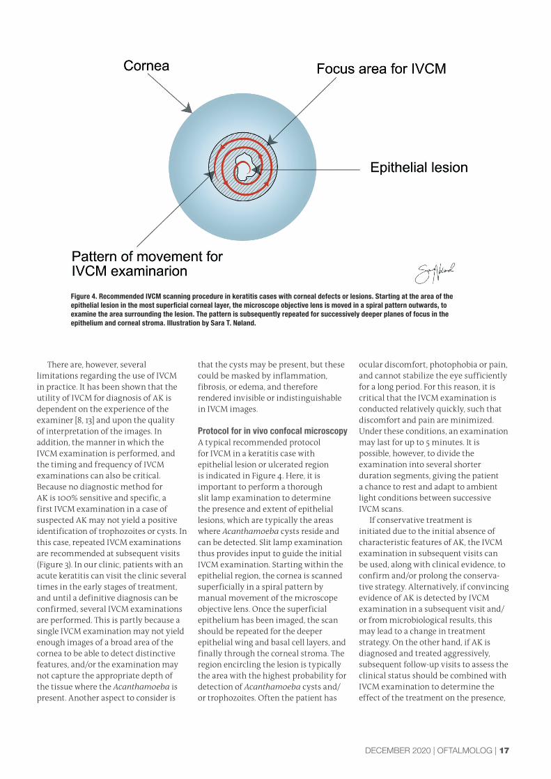
Figure 4. Recommended IVCM scanning procedure in keratitis cases with corneal defects or lesions. Starting at the area of the epithelial lesion in the most superficial corneal layer, the microscope objective lens is moved in a spiral pattern outwards, to examine the area surrounding the lesion. The pattern is subsequently repeated for successively deeper planes of focus in the epithelium and corneal stroma. Illustration by Sara T. Nøland.
that the cysts may be present, but these could be masked by inflammation, fibrosis, or edema, and therefore rendered invisible or indistinguishable in IVCM images.
Protocol for in vivo confocal microscopyA typical recommended protocol for IVCM in a keratitis case with epithelial lesion or ulcerated region is indicated in Figure 4. Here, it is important to perform a thorough slit lamp examination to determine the presence and extent of epithelial lesions, which are typically the areas where Acanthamoeba cysts reside and can be detected. Slit lamp examination thus provides input to guide the initial IVCM examination. Starting within the epithelial region, the cornea is scanned superficially in a spiral pattern by manual movement of the microscope objective lens. Once the superficial epithelium has been imaged, the scan should be repeated for the deeper epithelial wing and basal cell layers, and finally through the corneal stroma. The region encircling the lesion is typically the area with the highest probability for detection of Acanthamoeba cysts and/or trophozoites. Often the patient has
ocular discomfort, photophobia or pain, and cannot stabilize the eye sufficiently for a long period. For this reason, it is critical that the IVCM examination is conducted relatively quickly, such that discomfort and pain are minimized. Under these conditions, an examination may last for up to 5 minutes. It is possible, however, to divide the examination into several shorter duration segments, giving the patient a chance to rest and adapt to ambient light conditions between successive IVCM scans.
If conservative treatment is initiated due to the initial absence of characteristic features of AK, the IVCM examination in subsequent visits can be used, along with clinical evidence, to confirm and/or prolong the conserva-tive strategy. Alternatively, if convincing evidence of AK is detected by IVCM examination in a subsequent visit and/or from microbiological results, this may lead to a change in treatment strategy. On the other hand, if AK is diagnosed and treated aggressively, subsequent follow-up visits to assess the clinical status should be combined with IVCM examination to determine the effect of the treatment on the presence,

18 | OFTALMOLOG | DECEMBER 2020
Figure 5. Typical morphology of Acanthamoeba cysts detected by IVCM, indicating the typical form and refl ectivity of features. Note that there can be considerable variability in the appearance and size of the mature cyst form. Illustrations by Sara T. Nøland.
density, and morphology of trophozoite or cyst features in the cornea. Cases can differ dramatically in the response to treatment, and it is therefore useful to assess the morphological status of the cornea when considering a change or continuation of treatment. Cases where AK has been successfully resolved are consistent with an IVCM examination where no cyst features are observed and where the epithelium has regenerated and is intact with the epithelial cell mo-saic clearly visible [14]. Resolved cases, however, often reveal a deficit of corneal subbasal nerves (those residing in a dense plexus just below the epithelium) and a deficit of stromal keratocytes in the affected stromal regions.
Features of Acanthamoeba keratitis as detected by in vivo confocal microscopy The key feature of AK most often detected by IVCM examination is the pre-cyst or cyst form of Acanthamoeba (Figure 5). In an early stage of the Acanthamoeba life cycle, often the pre-cysts or cysts lacking a clear double-wall are observed by IVCM. These features are typically highly reflective,
corresponding to bright, white circular features in IVCM images [15]. Careful examination of these features can reveal the spike-like protrusions on the cyst surface. In a subset of cases, the cysts or pre-cysts can aggregate in linear ‘chains’ or ‘cluster’ forms, and this finding gives a high confidence of positive IVCM detection of AK [12].
Other important characteristics are the ‘dead space’ between the cysts, na-mely a dark region devoid of reflective features around the cysts, such that the cysts do not densely occupy an entire region of the IVCM image, but are typi-cally separated by linear distances of up to several hundred micrometers. Overall cyst density, however, can vary from less than one cyst per image, to very many cysts in a single image. Likewise, the size of the cysts can vary, although the cysts without a double wall tend to be quite uniform in size, about 10-25 µm in diameter, as pictured (Figure 6).
The more mature, double-walled cyst form can have a varying size and appearance, that can partially depend upon depth of focus [15] in the image (Figure 7). In such cases, the cyst can
Figure 6. IVCM images representing positive detection of AK. (A) Trophozoites appear oval or amoeboid in shape and are diffusely, non-uniformly refl ective. (B) Pre-cyst or cyst forms often appear uniform in size, are circular, and uniformly highly refl ective, sometimes visible in chain formations. (C) The mature, double-walled cyst form can have varying size and appearance, with thin or thick walls and a brightly refl ective center. (D) Image indicating a cyst and trophozoite present in an epithelial cell layer where epithelial cells are closely packed and have small, bright nuclei, not to be confused with cysts. (E) The ‘target sign’ cysts, with thick dark wall and bright central region, present in the epithelial wing cell layer with the cellular mosaic clearly apparent. (F) Large diffusely refl ective and oval-shaped trophozoites. All images, 400 x 400 µm, provided by Neil Lagali.
Figure 7. A depth-series of IVCM images depicting the same Acanthamoeba cyst, but at different depth layers, from superfi cial (A) to deep (F). The cyst is a large, mature double-walled Acanthamoeba about 50 µm in diameter, that only clearly exhibits a double wall when the plane of focus is in the equatorial region. More anterior or posterior to this, the cyst appears more uniformly refl ective. In the deepest image (F), the spikes or ‘acanthopodia’ are visible on the outer surface of the cyst. All images, 400 x 400 µm, provided by Neil Lagali.

DECEMBER 2020 | OFTALMOLOG | 19

20 | OFTALMOLOG | DECEMBER 2020
be relatively small without a visible double wall (that appears as a ring or halo formation), or the cyst may appear very large with a clear, thick double wall. Another type of cyst morphology has also been recently identified, and consists of a thick, dark walled cyst with a bright center, that is embedded within an epithelial mosaic, known as the ‘tar-get sign’ (Figure 6) [12]. It is important to remember, however, that although these double-walled features are often a telltale sign of AK, they are not neces-sarily always present in AK cases, and their morphologic features can depend on the stage of infection, depth in the tissue, microscopic focal plane, microen-vironmental conditions, and the specific species/genotype of the organism. More research is needed in determining
genotype-phenotype correlations of the various Acanthamoeba species, which may enable an improved reliability of IVCM detection of AK in the future.
Infl ammation – a potential confounderIn many cases, inflammation accompanies the infection and is a confounding factor in detecting the presence of small trophozoites or cysts in the epithelium or corneal stroma. Early corneal inflammation is often accompanied by invasion of neutrophil granulocytes, which have a characteristic appearance, size, and configuration [16], with typically large numbers invading the cornea (Figure 8). These should not be mistaken as cysts in chain or cluster forms. Other potential confounders
include leukocytes of varying size and reflectivity, and epithelial cells of varying size, shape and reflectivity, where nuclei are often clearly visible. With practice, it is possible to distinguish cysts and trophozoites from general inflammation; however, in some cases this distinction is difficult, and thus repeated IVCM examinations are desirable.
Recommendations and conclusionsThe incidence of AK is on the rise in the Nordic countries, and this may be due to the increasing popularity and prevalence contact lens wear. IVCM should be considered as an important tool in the ophthalmologist’s arsenal, to complement or supplement a tentative diagnosis or to provide confirmatory evidence of AK. Because IVCM is not limited to detection of AK but can also be used to distinguish fungal and certain types of bacterial keratitis, it is a valuable tool to have in the clinic, and ideally all cases of keratitis should be examined with IVCM early in their clinical course. IVCM is presently under-utilized in the Nordic countries, with only a few clinics having the microscope and trained operators. Utilizing IVCM in clinical practice represents an economic investment and a commitment of time and personnel; however, the potential benefits in terms of savings of ophthalmologist time and patient visits, along with the ability to apply image-guided treatment and follow-up, can directly translate into improved clinical outcomes and better quality of life and patient satisfaction. These benefits can more than compensate for the investment in resources required, particularly for clinics with significant numbers of keratitis patients.
Neil Lagali performs clinical confocal microscopy of the cornea at the Eye Clinic, Linköping University Hospital, in Linköping, Sweden.
References:www.oftalmolog.com
Figure 8. IVCM images exhibiting confounding morphologic characteristics that should not be mistaken for a positive Acanthamoeba fi nding. (A) Infi ltration of neutrophil granulocytes in the early phase of infl ammation. Note the presence of an intact background layer of basal epithelial cells. (B) Small epithelial cells and leukocytes observed in general infl ammation. A mosaic of epithelial cells is present in the background. (C) Superfi cial squamous epithelial cells with polygonal shape and prominent nucleus. (D) General epithelial infl ammation with largely intact wing cell layer. All images, 400 x 400 µm, provided by Neil Lagali.
DECEMBER 2020 | OFTALMOLOG | 21
References
1. Khan, N.A., Acanthamoeba: biology and increasing importance in human health. FEMS microbiology reviews, 2006. 30(4): p. 564-595.
2. Lorenzo-Morales, J., N.A. Khan, and J. Walochnik, An update on Acanthamoeba keratitis: diagnosis, pathogenesis and treatment. Parasite, 2015. 22.
3. Thebpatiphat, N., et al., Acanthamoeba keratitis: a parasite on the rise. Cornea, 2007. 26(6): p. 701-706.
4. Ledee, D., et al., Molecular identification of T4 and T5 genotypes in isolates from Acanthamoeba keratitis patients. Journal of clinical microbiology, 2009. 47(5): p. 1458-1462.
5. Powell, S., European contact lens market grows 4.3%. Optometry Today, 2018.
6. Clarke, D.W. and J.Y. Niederkorn, The pathophysiology of Acanthamoeba keratitis. Trends in parasitology, 2006. 22(4): p. 175-180.
7. Goldschmidt, P., et al., Effects of topical anaesthetics and fluorescein on the real-time PCR used for the diagnosis of Herpesviruses and Acanthamoeba keratitis. British Journal of Ophthalmology, 2006. 90(11): p. 1354-1356.
8. Goh, J.W., et al., Comparison of in vivo confocal microscopy, PCR and culture of corneal scrapes in the diagnosis of acanthamoeba keratitis. Cornea, 2018. 37(4): p. 480-485.
9. Pfister, D.R., et al., Confocal microscopy findings of Acanthamoeba keratitis. American journal of ophthalmology, 1996. 121(2): p. 119-128.
10. Tu, E.Y., et al., The relative value of confocal microscopy and superficial corneal scrapings in the diagnosis of Acanthamoeba keratitis. Cornea, 2008. 27(7): p. 764-772.
11. Vaddavalli, P.K., et al., Role of confocal microscopy in the diagnosis of fungal and acanthamoeba keratitis. Ophthalmology, 2011. 118(1): p. 29-35.
12. De Craene, S., et al., Assessment of confocal microscopy for the diagnosis of polymerase chain reaction–positive Acanthamoeba keratitis: A case-control study. Ophthalmology, 2018. 125(2): p. 161-168.
13. Hau, S.C., et al., Diagnostic accuracy of microbial keratitis with in vivo scanning laser confocal microscopy. British Journal of Ophthalmology, 2010. 94(8): p. 982-987.
14. Pettersson, M.N., et al., High fluence PACK-CXL as adjuvant treatment for advanced Acanthamoeba keratitis. American journal of ophthalmology case reports, 2019. 15: p. 100499.
15. Alomar, T., et al., In vivo confocal microscopy in the diagnosis and management of acanthamoeba keratitis showing new cystic forms. Clinical & experimental ophthalmology, 2009. 37(7): p. 737-739.
16. Lagali, N., et al., Laser-scanning in vivo confocal microscopy of the cornea: imaging and analysis methods for preclinical and clinical applications. Confocal Laser Microscopy—Principles and Applications in Medicine, Biology, and the Food Sciences, 2013.