Embed Size (px)
Citation preview
Leukemia Research Vol. 12, No. 10, pp. 823--831, 1988. 0145--2126/88 $3.00 + .00 Printed in Great Britain, Pergamon Press plc
USE OF THE MTT ASSAY FOR RAPID DETERMINATION OF CHEMOSENSITIVITY OF HUMAN LEUKEMIC BLAST CELLS*
BARBARA G. CAMPLING,t JOHN PVM,~ PETER R. GALBRAITH§ and SUSAN P. C. COLE[[
tDepartments of Oncology and Medicine, ~tDepartment of Surgery, §Departments of Medicine, Oncology and Paediatrics and [IDepartments of Oncology, and Microbiology and Immunology,
Queen's University, Kingston, Ontario, Canada
(Received 24 February 1988. Accepted 9 July 1988)
Abstract--A microcytotoxicity assay employing a tetrazolium salt has been adapted for testing the response of human leukemic blast cells to a variety of chemotherapeutic agents. After exposure to various concentrations of drugs, the viability of fresh leukemic blast cells was measured using a tetrazolium salt, MTT, which is converted to blue formazan crystals by living cells. The amount of formazan produced was quantitated using a microtitre plate spectrophotometer. In the present study, optimal conditions for chemosensitivity testing of human leukemia samples were determined, and the relative chemosensitivity of five patient samples was tested.
Key words: Chemosensitivity, MTr, human leukemia.
I N T R O D U C T I O N
A NUMBER of in-vitro assays are available for testing the response of human tumours to chemotherapeutic agents. The most widely used has been the clonogenic assay [1], but recently there has been renewed inter- est in short term tests of drug sensitivity such as dye exclusion assays [2, 3], or tests of inhibition of radiolabelled precursor incorporation [4-7]. The pre- dictive accuracy of these tests is quite good (reviewed by Weisenthal [8]), but numerous practical limi- tations have restricted their application to routine clinical practice. Thus there remains a need for a predictive assay of chemosensitivity that is rapid, simple, inexpensive, and available to most patients.
A simple colorimetric assay was developed by Mos- mann [9] as a test for cell proliferation and survival. It has been used to quantitate cytotoxic and growth inhibitory lymphokines [10], and has been found to have advantages over tritiated thymidine incor- poration as a measure of cell proliferation [11-13]. Cole [14], and others [15-19] have adapted this test for the measurement of chemosensitivity and
* Supported by grants from the Cancer Research Society, the National Cancer Institute of Canada, and the Clare Nelson Bequest of Kingston General Hospital.
Abbreviations: FBS, fetal bovine serum; PBS, phosphate buffered saline.
Correspondence to: Dr B. G. Campling, The Ontario Cancer Treatment and Research Foundation, Kingston Regional Cancer Centre, King Street West, Kingston, Ontario K7L 2V7, Canada.
radiosensitivity of human malignant cell lines. The assay utilizes a tetrazolium salt, MTT (3-[4,5- dimethylthiazol-2-yl)]-2,5-diphenyl-tetrazolium bro- mide), which is converted to a blue formazan product by dehydrogenase enzymes which are active only in living cells. The blue colour reaction is a measure of cell activation as well as viability [9, 13]. The assay is done in microtitre plates, and results are read on a multiwell scanning spectrophotometer. In the present study, we have adapted this test to quantitate the chemosensitivity of fresh human leukemic cells.
823
M A T E R I A L S AND M E T H O D S
Peripheral blood or bone marrow was obtained from five patients with acute leukemia (one acute lymphoblastic leukemia, one chronic myelogenous leukemia in myelo- blast crisis, two acute myelomonocytic leukemia, and one acute myelogenous leukemia). Mononuclear cells were separated on a Ficoll-Hypaque density gradient, washed, and resuspended in RPMI 1640 medium (Gibco, Grand Island, NY) supplemented with 5% FBS. In preliminary experiments, concentrations of 0, 10, and 20% FBS were used as well. In all five cases, the resultant population of cells consisted entirely of blast cells, as determined by morphology in tissue culture and by cytocentrifuge preparations.
Fresh leukemic cells from three of the patients were used to define the optimal conditions for the assay, and then the relative chemosensitivity of the five patient samples was tested to a variety of drugs.
Cells were plated in 96 well microtitre plates (Linbro 76- 032-05) at various densities ranging from 10 4 to 10 6 cells per well, using a 100 ~tl multichannel pipette. MTr (Sigma) was dissolved in PBS to a concentration of 2 mg/ml, and
824 B.G. CAMPLING et aL
25 Ixl was added to each well. To determine the optimal length of time of incubation with MTI', plates were set up with cells at 2 x 10⁵ cells per well, and various durations of exposure to MTT were tested, ranging from 15 min to 8 h. To solubilize the formazan crystals after the appro- priate incubation time, 100~tl of 1 N HCl:isopropanol (1:24) was added to each well and mixed vigorously using a multichannel pipette and the plates were incubated at 37°C for selected periods of time after addition of the HCl:isopropanol to attempt to enhance the solubilization of the formazan crystals.
The effect of cell proliferation and cell death over time on absorbance values was examined by setting up several plates at various cell densities, and adding MTI" to measure cell viability at times ranging from one day to two weeks.
Drugs tested Most of the drugs were obtained from the pharmacy at
Kingston General Hospital, already dissolved in the appropriate diluent. In a few cases (melphalan, hydroxy- urea and lomustine (CCNU)), drugs were obtained in pill or capsule form, which was then dissolved in sterile RPMI 1640 medium. Drugs were then diluted in serum free medium to the desired concentration, and stored at -20°C. They were not filter-sterilized, to avoid problems with binding to filters [20]. Drugs tested included cytosine arab- inoside, 6-thioguanine, 8-azaguanine, methotrexate, daunomycin, adriamycin, menagaril, epirubicin, mitoxan- trone, amsacrine (m-AMSA), vincristine, vinblastine, etoposide (VP-16), vumon (VM-26), hydrocortisone, cisplatinum, melphalan, lomustine (CCNU), nitrogen mus- tard and hydroxyurea.
Drugs were added as soon as the plates were made, or 16 h later. To determine the range of sensitivity, drugs were added in serial dilutions ranging from 1000 to 0.01 ~tg/ ml. Drugs were diluted to twice the desired final con- centration in RPMI 1640 medium containing 5% FBS. Each drug concentration was added to quadruplicate wells in 100 ~tl volumes, so that the final volume in each well was 200 p.l. For all of the chemosensitivity studies, cell densities of 1-5 x 10⁵ cells per well were used. Controls included wells with cells and no drug, wells with drug and no cells, and wells with no cells and the highest concentrations of drug.
Plates were incubated at 37°C in a humidified atmosphere with 5% CO2 for various lengths of time ranging from 2 to 5 days. After the appropriate incubation time, 100 ~tl of medium was removed from each well, and 25 ~tl of MTT (2 mg/ml) was added to each well using a multichannel pipette. For all of the drug induced cytotoxicity assays, a 6-h incubation with MTT was used. After this, 100 ~xl of 1 N HCl:isopropanol (1:24) was added to each well with vigorous mixing to dissolve the formazan crystals. The plates were kept at 37°C for 1 h, and then absorbance values at 570 nm were determined on a Dynatech MR600 microtitre plate reader. The spectrophotometer was cali- brated to zero absorbance using wells that had only medium and MqT.
Data analysis Data were entered on a Digital Microvax II mini-
computer using a simple form based system, stored in a relational database (Vax Rdb), and retrieved with a simple query language, Datatrieve. Custom software was used to calculate means and standard deviations for each drug
concentration and generate dose response curves for each drug, to be displayed on either a graphics terminal or Hewlett-Packard plotter.
RESULTS
Optimization of assay conditions
After incubation of the cells with MT/ ' , and prior to addition of HCl:isopropanol, blue formazan crys- tals could be visualized microscopically around viable blast cells, as shown in Fig. 1. The MTI" was difficult to solubilize, and required very vigorous mixing of the wells. In preliminary experiments, different con- centrations of FBS were used. With concentrations of FBS of 10 or 20%, a precipitate was formed when the isopropanol was added to solubilize the formazan crystals. When medium with no FBS was used, there was loss of cell viability in the untreated control wells over the usual two-day incubation period. Therefore for all subsequent experiments, culture medium with 5% FBS was used.
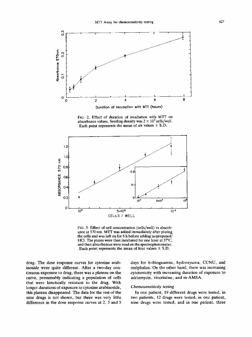
The effect of length of time of incubation with MTT on absorbance values was examined, and the results are shown in Fig. 2. It can be seen that the absorbance values increase with time up to 8 h.
To determine the relationship between cell density and absorbance values, a standard curve was gen- erated with one of the leukemia samples, as shown in Fig. 3. It can be seen that the relationship between cell number and absorbance is linear over the range of cell concentrations tested. Higher absorbance values were obtained by incubation of the plates for one hour at 37°C after addition of the HCl:isopropanol, presumably because of enhanced solubilization of the formazan crystals. Further incubation at 37°C did not improve the absorbance readings.
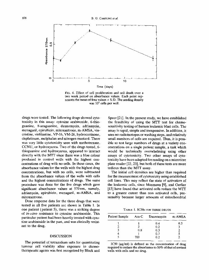
The effect on absorbance values of cell pro- liferation and cell death over a two-week period was determined on one sample, and the results are shown in Fig. 4. As can be seen, over the usual two-day incubation period, there was a doubling of the ab- sorbance values. Absorbances continued to increase up to 5 days, and then began to decrease.
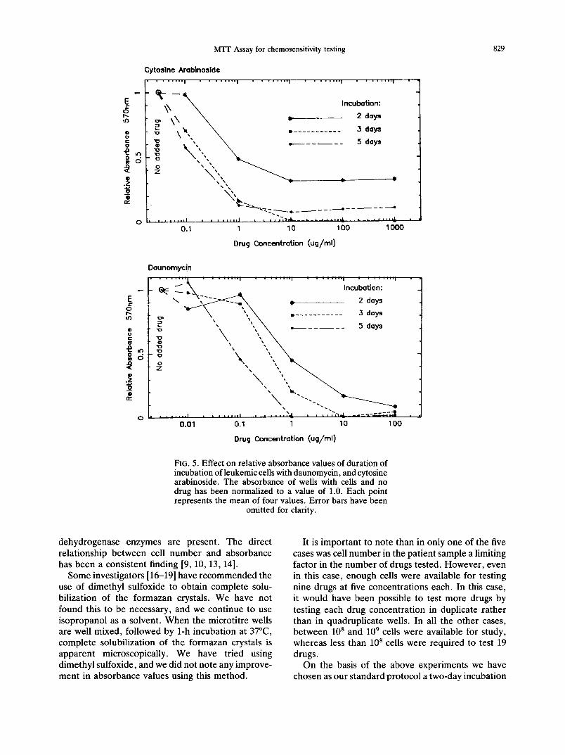
The effect of duration of incubation with drug on cytotoxocity was determined on one patient sample for nine drugs, namely, 6-thioguanine, cyto- sine arabinoside, daunomycin, adriamycin, hydroxy- urea, CCNU, melphalan, m-AMSA, vincristine. Ex- posures of 2, 3 and 5 days were tested, and the results for daunomycin and cytosine arabinoside are shown in Fig. 5. As can be seen, for daunomycin, there was greater cytotoxocity with increasing length of exposure to drug, but at the highest drug con- centration (1001ag/ml) there was complete cyto- toxicity regardless of the duration of exposure to
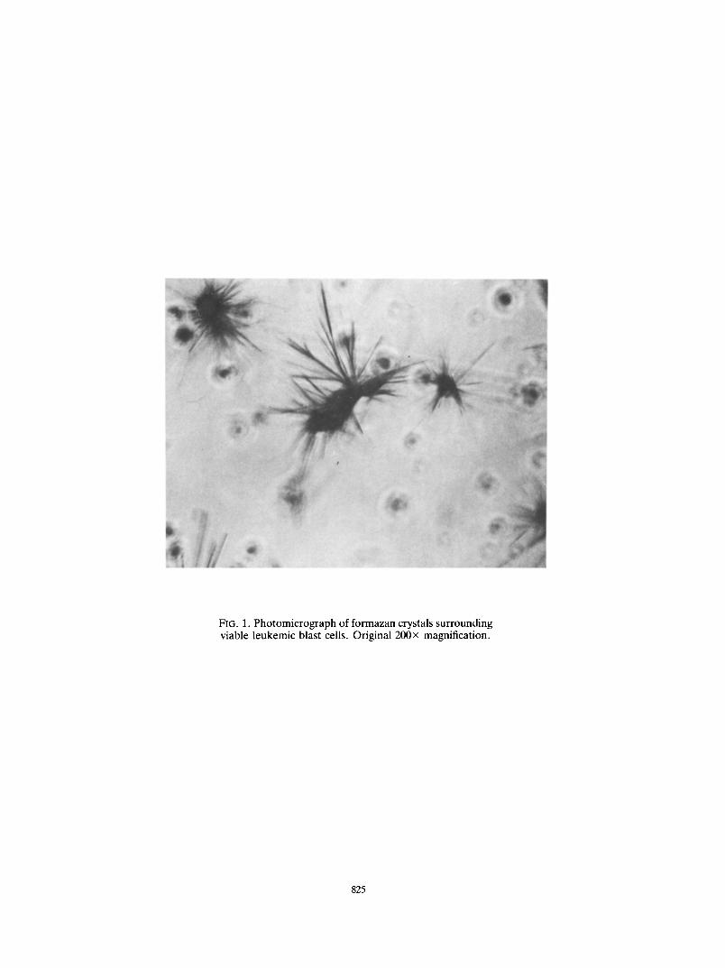
FIG. 1. Photomicrograph of formazan crystals surrounding viable leukemic blast cells. Original 200× magnification.
825

MTT Assay for chemosensitivity testing 827
E ¢N gd t O
l )
0
1'3 d
0
! !
J J /
= 1 ~ I = I
2 4 6
Durotion of Incubotion with MTT (hours)
J
FIG. 2, Effect of duration of incubation with MTT on absorbance values. Seeding density was 2 x 10⁵ cells/well. Each point represents the mean of six values - S.D.
I
8
1.2
1.0 E c-
O 0.8 tO
h i o Q6 Z
rn r r 0 . 4 O (/) nn
0.2 I0 ~ 5x lO* I 0 =
0 I I I I | | I I i
I 8 5 x l O 5 10 6
CELLS / WELL
FIG. 3. Effect of cell concentration (cells/well) vs absorb- ance at 570 nm. MTT was added immediately after plating the cells and was left on for 6 h before adding isopropanol/ HCI. The plates were then incubated for one hour at 37°C, and then absorbances were read on the spectrophotometer. Each point represents the mean of four values -+ S.D.
drug. The dose response curves for cytosine arab- inoside were quite different. After a two-day con- tinuous exposure to drug, there was a plateau on the curve, presumably indicating a population of cells that were kinetically resistant to the drug. With longer durations of exposure to cytosine arabinoside, this plateau disappeared. The data for the rest of the nine drugs is not shown, but there was very little difference in the dose response curves at 2, 3 and 5
days for 6-thioguanine, hydroxyurea, CCNU, and melphalan. On the other hand, there was increasing cytotoxicity with increasing duration of exposure to adriamycin, vincristine, and m-AMSA.
Chemosensitivity testing In one patient, 19 different drugs were tested, in
two patients, 12 drugs were tested, in one patient, nine drugs were tested, and in one patient, three
828 B.G. CAMPLING et al.
E ¢-
~6 I N
O
C O
O
. a ~C
O
o
T ' i i - i I i
j / / / ~
/
/ /
/ , /
a ~ ± _ _ . L i J t .L___ I I
5 10
"13rne (doys)
FIG. 4. Effect of cell prol i ferat ion and cell death over a two week period on absorbance values. Each point rep- resents the mean of four values - S.D. The seeding density
was 105 cells per well.
drugs were tested. The following drugs showed cyto- toxicity in this assay: cytosine arabinoside, 6-thio- guanine, 8-azaguanine, daunomycin, adriamycin, menagaril, epirubicin, mitoxantrone, m-AMSA, vin- cristine, vinblastine, VP-16, VM-26, hydrocortisone, cisplatinum, melphalan and nitrogen mustard. There was very little cytotoxicity seen with methotrexate, CCNU, or hydroxyurea. Two of the drugs tested, 6- thioguanine and hydroxyurea, appeared to interact directly with the MT-F since there was a blue colour produced in control wells with the highest con- centrations of drug with no cells. In these cases, the absorbance values for the wells with the highest drug concentrations, but with no cells, were subtracted from the absorbance values of the wells with cells and the highest concentrations of drugs. The same procedure was done for the five drugs which gave significant absorbance values at 570 nm, namely, adriamycin, epirubicin, menagaril, m-AMSA, and mitoxantrone.
Dose response data for the three drugs that were tested in all five patients are shown in Table 1. In one patient (patient 5), there was a striking degree of i n - v i t r o resistance to cytosine arabinoside. This particular patient had been heavily treated with cyto- sine arabinoside in the past, and was clinically resist- ant to the drug.
DISCUSSION
The potential of tetrazolium salts for quantitating tumour cell viability after exposure to chemo- therapeutic agents was first recognized by Black and
Speer [21]. In the present study, we have established the feasibility of using the MTT test for chemo- sensitivity testing of human leukemic blast cells. The assay is rapid, simple and inexpensive. In addition, it uses no radioisotopes or washing steps, and relatively small numbers of cells are required. Thus, it is poss- ible to test large numbers of drugs at a variety con- centrations on a single patient sample, a task which would be technically overwhelming using other assays of cytotoxicity. Two other assays of cyto- toxicity have been adapted for reading on a microtitre plate reader [22, 23], but both of these tests are more tedious than the MT-[ assay.
The initial cell densities are higher than required for the measurement of cytotoxicity using established cell lines. This may reflect the state of activation of the leukemic cells, since Mosmann [9], and Gerlier [13] have found that activated cells reduce the MT1 ~ to a greater extent than non activated cells, pre- sumably because larger amounts of mitochondrial
TABLE I. IC50s FOR THREE DRUGS
Patient Sample Ara-C Daunomycin m-AMSA
1 1 0.4 0.3 2 2 0.2 3 3 0.8 0.3 2 4 1 0.7 5 5 700 0.2 2
IC50 (Ixg/ml) is defined as the concentration of drug required to reduce the absorbance to 50% of that of control wells with cells and no drug.
MTF Assay for chemosensitivity testing 829
E o
Q U C 0
0
E p -
o t'-
® c; c o
Y, Q
d
O
u 3
d
O
Cytosine Arabinoside
\ \ ~ Incubation:
<,, \ - .ooo, , , ' , ,
~'~ ~ ~ ' - - e - - - - - . - - - e - - - - - - - - e
, i J , . = , l i | t . J , , , l • = . i ¢ ' ~ I r , 4 k . _ ~ _ ~ _ L * ~ . . . = Z , j . l , l = . Z
0.1 1 10 100 1000
Dru 9 ConcentraUon (ucj/rnl)
Daunomyc[n . . . . . . . I , . . . . . . . I . . . . . . . . I . . . . . . . . I . . . . . . . . I •
6~: ~ ~ • Incu~adtiaO ~ ~" \. "\ ~ • ........... 3 days
"\ \ \ . . . . . . 5 oy, \ ' , , \ , ,
0.01 0.1 1 10 1 O0
Drug Concentration (ug/rnl)
FIG. 5. Effect on relative absorbance values of duration of incubation of leukemic cells with daunomycin, and cytosine arabinoside. The absorbance of wells with cells and no drug has been normalized to a value of 1.0. Each point represents the mean of four values. Error bars have been
omitted for clarity.
dehydrogenase enzymes are present. The direct relationship between cell number and absorbance has been a consistent finding [9, 10, 13, 14].
Some investigators [16-19] have recommended the use of dimethyl sulfoxide to obtain complete solu- bilization of the formazan crystals. We have not found this to be necessary, and we continue to use isopropanol as a solvent. When the microtitre wells are well mixed, followed by 1-h incubation at 37°C, complete solubilization of the formazan crystals is apparent microscopically. We have tried using dimethyl sulfoxide, and we did not note any improve- ment in absorbance values using this method.
It is important to note than in only one of the five cases was cell number in the patient sample a limiting factor in the number of drugs tested. However, even in this case, enough cells were available for testing nine drugs at five concentrations each. In this case, it would have been possible to test more drugs by testing each drug concentration in duplicate rather than in quadruplicate wells. In all the other cases, between 10 s and 10 9 cells were available for study, whereas less than 10 8 cells were required to test 19 drugs.
On the basis of the above experiments we have chosen as our standard protocol a two-day incubation
830 B.G. CAMPLING et al.
with drug, a 6-h incubation with MTT, followed by a 1-h incubation of the plates at 37°C after addition of the HCl:isopranol. The two-day incubation period seems appropriate , since it is long enough to see cytotoxocity with all of the drugs that are active in this assay system, but short enough to minimize the variable effects of cell proliferation and cell death over the assay period.
We have reported experiments on a tumour that is relatively simple to examine, since leukemic cells are easily obtainable, and available in pure form in a single cell suspension. Studies are ongoing to determine whether the MTT assay can be adapted to the chemosensitivity testing of solid tumours.
It is not possible to determine from data on only five patients whether this test will be predictive of clinical sensitivity or resistance to drugs. In other studies, the M T T assay has been used to distinguish between drug sensitive and drug resistant cell lines [14, 24]. Fur thermore , there have been numerous reports of positive correlations between clinical response to chemotherapeut ic agents in acute leu- kemia, and results of in-vitro chemosensitivity testing using the clonogenic assay [25-34]. We anticipate that similar correlations will be found using the Mq-T test, but this question can only be addressed in a prospective clinical trial.
Acknowledgements--The assistance of Mrs L. Campling is gratefully acknowledged. We thank Mr John McBride, Pharmacist at Kingston General Hospital for providing most of the drugs.
R E F E R E N C E S
1. Salmon S. E., Hamburger A. W., Soehnlen B., Durie B. G. M., Albert D. S. & Moon T. E. (1978) Quanti- tation of differential sensitivity of human-tumor stem cells to anticancer drugs. New Engl. J. Med. 298, 1321.
2. Weisenthal L. M., Marsden J. A., Dill P. L. & Maca- luso C. K. (1983) A novel dye exclusion method for testing in-vitro chemosensitivity of human tumors. Cancer Res. 43, 749.
3. Bosanquet A. G., Bird M. C., Price W. J. P. & Gilby E. D. (1983) An assessment of a short-term tumour chemosensitivity assay in chronic lymphocytic leukemia Br. J. Cancer 47, 781.
4. Group for Sensitivity Testing of Tumors (KSST). (1981) In-vitro short-term test to determine the resist- ance of human tumors to chemotherapy. Cancer 48, 2127.
5. Maddox A-M., Johnston D. A., Barlogie B., Haq M., Keating M. J. & Freireich E. J. (1984) In-vitro suppression of DNA synthesis by a remission induction agent and its correlation with response in adult acute leukemia. Eur. J. Cancer clin. Oncol. 20, 507.
6. Sanfilippo O., Daidone M. G., Zaffaroni N. & Sil- vestrini R. (1984) Development of a nucleotide pre- cursor incorporation assay for testing drug sensitivity
of human tumors. Rec. Res. Cancer Res. 94, 127. 7. Sondak V. K., Bertelson C. A., Kern D. H. & Morton
D. L. (1985) Evolution and clinical application of a rapid chemosensitivity assay. Cancer 55, 1367.
8. Weisenthal L. M. & Lippman M. E. (1985) Clonogenic and nonclonogenic in vitro chemosensitivity assays. Cancer Treat. Rep. 69, 615.
9. Mosmann T. (1983) Rapid colorimetric assay for cell proliferation and survival: application to proliferation and cytotoxicity assays. J. Immun. Methods 65, 55.
10. Green L. M., Reade J. L. & Ware C. F. (1984) Rapid colorimetric assay for cell viability: application to the quantitation of cytotoxic and growth inhibitory lymphokines. J. Immun. Methods 70, 257.
11. Denizot R. & Lang R. (1986) Rapid colorimetric assay for cell growth and survival. Modifications to the tetra- zolium dye procedure giving improved sensitivity and reliability. J. Immun. Methods 89, 271.
12. Heeg K., Reimann J., Kabelitz D., Hardt C. & Wagner H. (1985) A rapid colorimetric assay for the deter- mination of IL-2-producing helper T-cell frequencies. J. Immun. Methods 77, 237.
13. Gerlier D. & Thomasset N. (1986) Use of MTF color- imetric assay to measure cell activation. J. Immun. Methods 94, 57.
14. Cole S. P. C. (1986) Rapid chemosensitivity testing of human lung tumor cells using the MTT assay. Cancer Chemother. Pharmac. 17, 259.
15. Finlay G. J., Wilson W. R. & Baguley B. D. (1986) Comparison of in-vitro activity of cytotoxic drugs toward human carcinoma and leukaemia cell lines. Eur. J. Cancer clin. Oncol. 22, 655.
16. Carmichael J., Degraff W. G., Gazdar A. F., Minna J. D. & Mitchell J. B. (1987) Evaluation of a tetrazotium- based semiautomated coiorimetric assay: assessment of chemosensitivity testing. Cancer Res. 47, 936.
17. Carmichael J., Degraff W. G., Gazdar A. F., Minna J. D. & Mitchell J. B. (1987) Evaluation of a tetrazolium- based semiautomated colorimetric assay: assessment of radiosensitivity. Cancer Res. 47, 943.
18. Twentyman P. R. & Luscombe M. (1987) A study of some variables in a tetrazolium dye (MTT) based assay for cell growth and chemosensitivity. Br. J. Cancer 56, 279.
19. Park J-G., Kramer B. S., Steinberg S. M., Carmichael J., Collins J. M., Minna J. D. & Gazdar A. F. (1987) Chemosensitivity testing of human colorectal car- cinoma cell lines using a tetrazolium-based colorimetric assay. Cancer Res. 47, 5875.
20. Bosanquet A. G. (1985) Stability of solutions of anti- neoplastic agents during preparation and storage for in-vitro assays. Cancer Chemother. Pharmac. 14, 83.
21. Black M. M. & Speer F. D. (1953) Effects of cancer chemotherapeutic agents of dehydrogenase activity of human cancer tissue in vitro. Am. J. clin. Path. 23, 218.
22. Landegren U. (1984) Measurement of cell numbers by means of the endogenous enzyme hexosaminidase. Applications to detection of lymphokines and cell sur- face antigens. J. Immun. Methods 67, 379.
23. Finlay G. J., Baguley B. C. & Wilson W. R. (1984) A semiautomated microculture method for investigating growth exhibitory effects of cytotoxic compounds on exponentially growing carcinoma cells. Analyt. Bio- chem. 139, 272.
24. Mirski S. E. L., Gerlach J. H. & Cole S. P. C. (1987)
MTF Assay for chemosensitivity testing 831
Multidrug resistance in a human small cell lung cancer cell line selected in adriamycin. Cancer Res. 47, 2594.
25. Park C. H., Amare M., Savin M. A., Goodwin J. W., Newcomb M. M. & Hoogstraten B. (1980) Prediction of chemotherapy response in human leukemia using an in-vitro chemotherapy sensitivity test on the leukemic colony-forming cells. Blood 55, 595.
26. Preisler H. D. (1980) Prediction of response to chemo- therapy in acute myelocytic leukemia. Blood 56, 361.
27. Park C. H., Amare M., Morrison F. S., Maloney T. R. & Goodwin J. W. (1982) Chemotherapy sensitivity assessment of leukemic colony-forming cells with in- vitro simultaneous exposure to multiple drugs: clinical correlations in acute lymphocytic leukemia. Cancer Treat. Rep. 66, 1257.
28. Park C. H., Wiernik P. H., Morrison F. S., Amare M., Van Sloten K. & Maloney T. R. (1983) Clinical correlations of leukemic clonogenic cell chemosensi- tivity assessed by in-vitro continuous exposure to drugs. Cancer Res. 43, 3246.
29. Lihou M. G. & Smith P. J. (1983) Quantitation of chemosensitivity in acute myelocytic leukaemia. Br. J. Cancer 48, 559.
30. Browman G., Goldberg J., Gottleib A. J., Preisler H. D., Azarnia N., Priore R. L., Brennan J. K., Vogler W. R., Winton E. F., Miller K. B. & Grunwald H. (1983) The clonogenic assay as a reproducible in-vitro system to study predictive parameters of treatment outcome in acute nonlymphoblastic leukemia. Am. J. Hemat. 15, 227.
31. Marie J-P., Zittoun R., Thevenin D., Mathieu M. & Viguie F. (1983) In-vitro culture of clonogenic leu- kaemia cells in acute myeloid leukaemia: growth pat- tern and drug sensitivity. Br. J. Haemat. 55, 427.
32. Gustavson A. & Olofson T. (1984) Prediction of response to chemotherapy in acute leukemia by in- vitro drug sensitivity testing on leukemic stem cells. Cancer Res. 44, 4648.
33. Lihou M. G. & Smith P. J. (1985) Adriamycin and cytosine arabinoside contribute equally to prediction of response in acute myelocytic leukemia with improved confidence level. Cancer Chemother. Pharmac. 14, 116.
34. Dow L. W., Dahl G. V., Kalwinsky D. K., Mirro J., Nash M. B. & Roberson P. K. (1986) Correlation of drug sensitivity in vitro with clinical responses in childhood acute leukemia. Blood 68, 400.


![An Integrated Database of Chemosensitivity to 55 ...[CANCER RESEARCH 62, 1139–1147, February 15, 2002] An Integrated Database of Chemosensitivity to 55 Anticancer Drugs and Gene](https://img.pdfslide.net/doc/110x75/5ed99d59801c872007065f4a/an-integrated-database-of-chemosensitivity-to-55-cancer-research-62-1139a1147.jpg)