Embed Size (px)
Citation preview
288
Copyright © 2015 The Korean Society of Plastic and Reconstructive SurgeonsThis is an Open Access article distributed under the terms of the Creative Commons Attribution Non-Commercial License (http://creativecommons.org/ licenses/by-nc/3.0/) which permits unrestricted non-commercial use, distribution, and reproduction in any medium, provided the original work is properly cited. www.e-aps.org
Orig
inal
Art
icle
INTRODUCTION
Surgical ablation of locally advanced breast cancer often results in huge defects, however immediate reconstruction of the breast mound is controversial, particularly its relationship to clinical indications and type of reconstruction. Adequately covering any large chest wall defect is the main clinical issue, and a variety of techniques have been implemented over the last four decades, including skin grafts, local skin or fasciocutaneous flaps, omen-
tal flaps, and myocutaneous flaps such as pectoralis major, rectus abdominis, latissimus dorsi, and external oblique flaps [1-8]. Generally, flaps are advantageous over skin grafts in terms of aesthetics and durability (Fig. 1), especially when adjuvant radi-ation therapy is indicated [1,9]. Skin flaps are usually preferred to myocutaneous flaps because of their relative simplicity and comparable results [2,9]. We have used three types of skin flaps to cover large soft tissue defects after the surgical ablation of lo-cally advanced breast cancer. The purpose of this study was to
Using Local Flaps in a Chest Wall Reconstruction after Mastectomy for Locally Advanced Breast CancerJoo Seok Park1, Sei Hyun Ahn2, Byung Ho Son2, Eun Key Kim1
Departments of 1Plastic Surgery and 2Surgery, Asan Medical Center, University of Ulsan College of Medicine, Seoul, Korea
Background Surgical ablation for locally advanced breast cancer results in large chest wall defects, which can then be managed with local flaps or skin grafts. The purpose of this article is to evaluate the outcomes of three types of local skin flaps.Methods Among 25 local flaps in 24 patients, 6 were bilateral advancement (BA) flaps, 9 were thoracoabdominal (TA) flaps, and 10 were thoracoepigastric (TE) flaps. Clinical outcomes were compared including complications, the need for a secondary surgical intervention, and the timing of adjuvant therapy. Results The mean defect size was 436.2 cm². Two patients with TA flaps and 6 patients with TE flaps developed distal flap necrosis, and skin grafts were needed to treat 2 patients with TE flaps. Radiation was administered to the BA, TA, and TE patients after average postoperative durations of 28, 30, or 41 days, respectively. The incidence of flap necrosis tended to be higher in TE patients, which lead to significant delays in adjuvant radiation therapy (P=0.02).Conclusions Three types of local skin flaps can be used to treat large chest wall defects after the excision of locally advanced breast cancer. Each flap has its own merits and demerits, and selecting flaps should be based on strict indications based on the dimensions and locations of the defects.
Keywords Breast neoplasms / Mammaplasty / Thoracic wall / Surgical flaps
Correspondence: Eun Key KimDepartment of Plastic Surgery,Asan Medical Center, University of Ulsan College of Medicine,88 Olympic-ro 43-gil, Songpa-gu, Seoul 138-736, Korea Tel: +82-2-3010-3600Fax: +82-2-476-7471E-mail: [email protected]
This article was presented at the 2013 International Congress of the Korea Society of Plastic & Reconstructive Surgeons on November 1–3, 2013 in Seoul, Korea.
No potential conflict of interest relevant to this article was reported.
Received: 9 May 2014 • Revised: 30 Jul 2014 • Accepted: 31 Jul 2014pISSN: 2234-6163 • eISSN: 2234-6171 • http://dx.doi.org/10.5999/aps.2015.42.3.288 • Arch Plast Surg 2015;42:288-294
Vol. 42 / No. 3 / May 2015
289
detail our experiences using bilateral advancement (BA), thora-coabdominal (TA), and thoracoepigastric (TE) flaps with a specific focus on outcomes, advantages, disadvantages, and proper patient selection.
METHODS
All mastectomies that required immediate reconstruction by a plastic surgeon at a single center between June 2008 and October 2013 were retrospectively reviewed. Breast mound reconstruc-tions that used flaps and/or implants were excluded. Forty-three patients (45 breasts) were referred to plastic surgeons during the study period to receive chest wall reconstructions because of failed direct wound closure after mastectomy. Of these cases, 14 breasts received split-thickness skin grafts and 6 breasts received full-thickness grafts. A total of 25 local flaps were performed on 24 patients: 6 BA flaps, 9 TA flaps, and 10 TE flaps (a flap and a skin graft were performed on each breast in 1 patient) (Table 1). Chart review was performed to obtain data on sex, age, diagnosis, oncological status, adjuvant therapy, location and size of the de-fects, and complications. Outcomes were compared between all three groups based on the flap type. Due to small number of pa-tients in each group, statistical comparisons were only performed to assess the overall incidence of complications and duration be-fore the initiation of the adjuvant therapy. Analyses were perform-ed using SPSS software (SPSS Inc., Chicago, IL, USA).
Shown is a 55-year-old female patient during adjuvant radiation therapy who subsequently received a split-thickness skin graft to cover the 14×12 cm skin defect after mastectomy.
Fig. 1. Split-thickness skin graft after mastectomy
Patient no.
Age(yr)
Defectsize (cm²) Operation method Pathology Start of
adjuvant therapy Stage Mastectomyweight (g) Complication
1 44 20×15 Bilateral advancement Phyllodes Observe Phyllodes 774.0 None2 40 12×12 Bilateral advancement IDC POD 28 T1N0M0 78.5 None3 51 23×12 Bilateral advancement IDC Observe T4N1M0 580.0 None4 53 29×24 Bilateral advancement Phyllodes Observe Phyllodes 3,916.0 Wound dehiscence5 45 22×21 Bilateral advancement IDC POD 25 T4N3M0 1,525.0 None6 47 14×14 Bilateral advancement IDC POD 31 T4N1M0 221.0 None7 53 21×15 Thoracoabdominal IDC POD 50 T3N0M1 432.0 Tip necrosis8 36 15×11 Thoracoabdominal IDC POD 35 T4N2M0 110.0 None9 44 26×26 Thoracoabdominal IDC POD 27 T3N2M0 837.0 None
10 34 22×17 Thoracoabdominal IDC POD 31 T4N2M1 827.0 None11 40 21×20 Thoracoabdominal Phyllodes POD 27 Phyllodes 1,184.0 None12 41 21×19 Thoracoabdominal IDC POD 40 T4N0M0 1,950.0 Tip necrosis13 36 26×20 Thoracoabdominal IPLC POD 26 T3N0M0 1,095.0 None14 41 18×17 Thoracoabdominal Angiosarcoma POD 18 T3N0M0 587.0 None15 38 23×20 Thoracoabdominal IDC POD 23 T3N0M0 1,093.0 None16 34 25×23 Thoracoepigatric IDC POD 37 T4N1M0 661.0 None17 42 24×22 Thoracoepigatric IDC POD 36 T3N0M0 728.0 None18 32 21×20 Thoracoepigatric IDC POD 21 T4N3M0 664.0 Tip necrosis19 48 19×19 Thoracoepigatric Phyllodes Observe Phyllodes 434.0 None20 53 22×21 Thoracoepigatric IDC POD 38 T4N3M0 1,525.0 Tip necrosis21 46 14×14 Thoracoepigatric IDC POD 55 T4N1M0 221.0 Tip necrosis22 39 24×23 Thoracoepigatric IDC POD 35 T4N3M0 1,031.0 Tip necrosis23 51 25×20 Thoracoepigatric IDC POD 46 T3N0M0 330.0 None24 38 24×24 Thoracoepigatric IDC POD 44 T2N3M0 408.0 Tip necrosis25 45 19×18 Thoracoepigatric IDC POD 47 T4N3M0 209.0 Tip necrosis
IDC, intraductal carcinoma; POD, postoperative day; TNM, tumor, node, metastasis; IPLC, invasive pleomorphic lobular carcinoma.
Table 1. Patient data

Park JS et al. Local flaps for advanced breast cancer
290
Surgical techniquesBA flapFor the BA flap, sufficient dissection begins at the margins of the mastectomy defect and progresses upward over the clavicle and downward almost to the level of the umbilicus without addition-al incisions. The plane of dissection is prefascial, and perforators from the epigastric and intercostal vessels are preserved whenev-er possible. The created cephalic and caudal flaps are sutured to-gether, leaving a horizontal scar (Fig. 2). Trimming of “the dog ear” is usually necessary. This flap is indicated when the vertical dimensions of the flap do not exceed approximately 15 cm and its shape is approximately elliptical. If excessive tension develops during closure, a TA or TE flap should be considered.
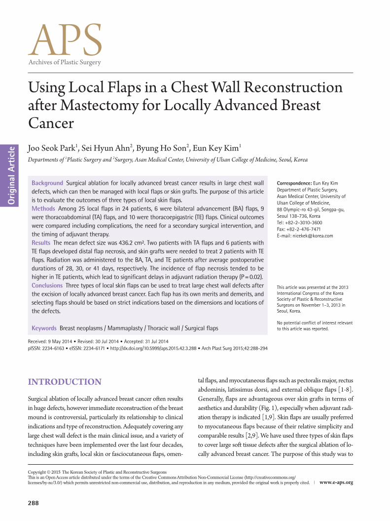
TA flapThe TA flap is basically a rotation-advancement flap that uses the lateral intercostals, subcostal, and lumbar arteries. An incision is made at the midline of the abdomen all the way down to the um-bilicus, and further dissection proceeds inferiorly and laterally across a prefascial plane. The pedicle of this flap can be identified at the medial edge of the external oblique muscle and preserved. The flap is rotated clockwise for left chest wall defects, or coun-terclockwise for right chest wall defects (Fig. 3). This flap is usu-ally indicated when a higher portion of the defect lies medial, or a large amount of medial advancement is required.
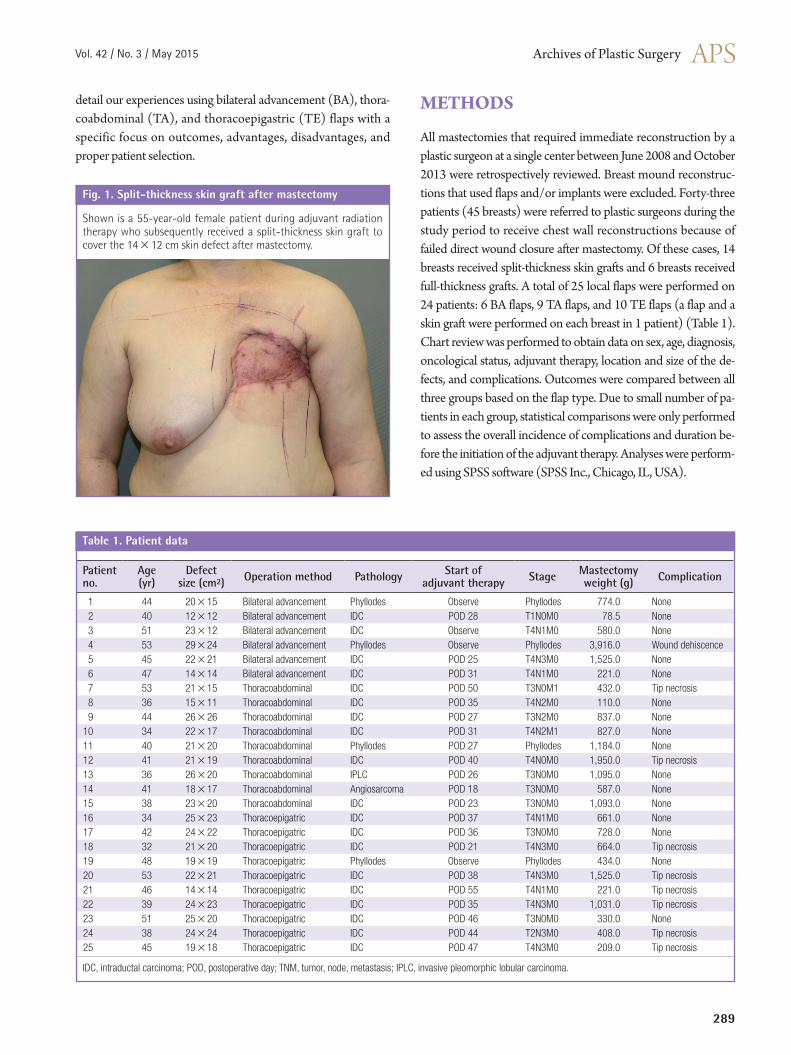
TE flapThe TE flap is like a mirror image of the TA flap and uses perfo-rators from the superior epigastric artery. The incision starts at the lower lateral angle of the defect and curves laterally down along the midaxillary line. Dissection continues medially and in-feriorly, thereby preserving the superior epigastric perforators that pierce the rectus abdominis fascia (Fig. 4). This flap is usu-ally indicated when the required medial advancement is relative-ly small and a higher portion of the defect lies laterally toward the axilla.
RESULTS
Between June 2008 and October of 2013, 25 local flaps were performed on 24 patients to cover chest wall defects after surgi-cal ablation for locally advanced breast cancer. Among 24 pa-tients, 23 were female and the mean age was 42.6 years (range, 32–53 years). Nineteen patients were diagnosed with invasive ductal carcinoma of the breast, 4 with an invasive phyllodes tu-mor, and 1 with a bilateral recalcitrant invasive phyllodes tumor. The mean follow-up period was 14 months (range, 4–66 years). The mean specimen weight was 1,382.5 g (range, 110–7,500 g; median, 894.5 g). The mean defect size was 400.1 cm2 (range, 90–696 cm2): 321 cm2 in BA flap group (n = 6) vs. 462 cm2 in TA flap group (n = 9) vs. 391 cm2 in TE flap group (n = 10). In
With the bilateral advancement flap, the cephalic and caudal flaps are elevated and sutured together, which leaves a horizontal scar.
Fig. 2. Bilateral advancement flap after mastectomy
Vol. 42 / No. 3 / May 2015
291
The thoracoepigastric flap uses the superior epigastric vessels as perforators. The vertical scar is at the midaxillary line.
Fig. 4. Thoracoepigastric flap after mastectomy
The thoracoabdominal flap uses the lateral intercostal vessels and leaves a vertical midline scar.
Fig. 3. Thoracoabdominal flap after mastectomy
total, 9 complications were recorded (36% of patients): 1 case of wound dehiscence (16.6%) in the BA flap group; 2 cases (22%) of distal flap necrosis in the TA flap group; and 6 cases (60%) of
distal flap necrosis in TE flap group (P = 0.17; Fisher exact test). All complications (except in 3 patients in the TE group) sponta-neously healed in less than 3 weeks with conservative wound
Park JS et al. Local flaps for advanced breast cancer
292
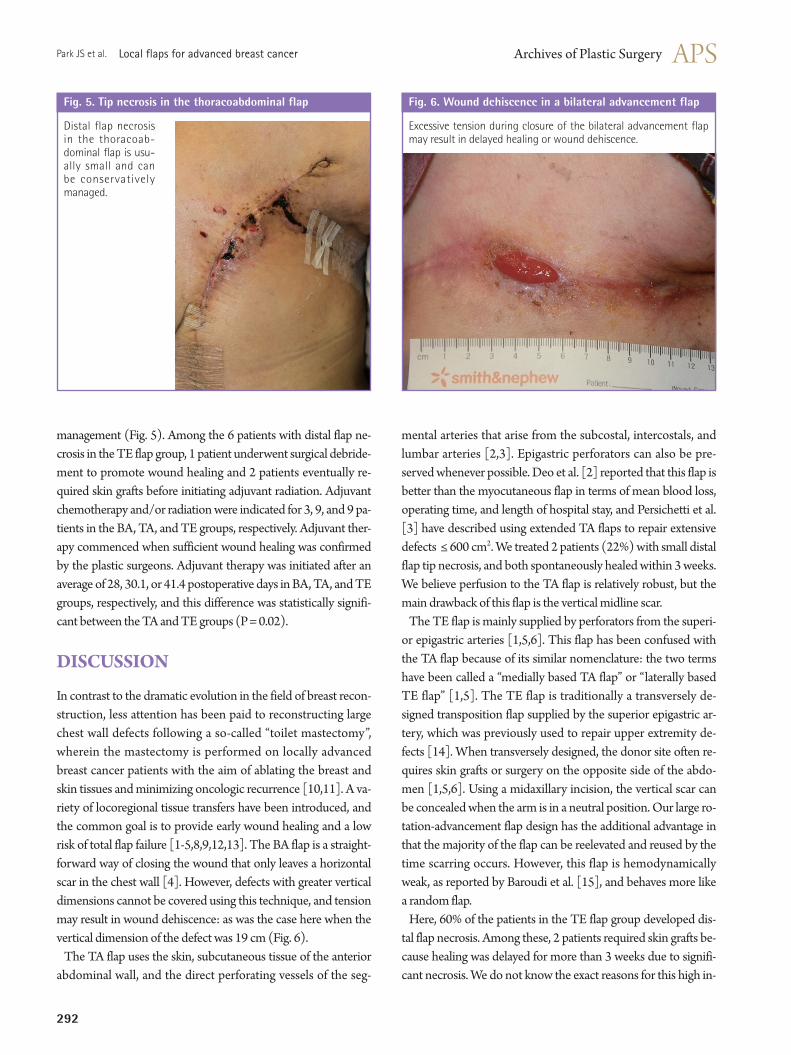
Distal flap necrosis in the thoracoab-dominal flap is usu-ally small and can be conservatively managed.
Fig. 5. Tip necrosis in the thoracoabdominal flap
management (Fig. 5). Among the 6 patients with distal flap ne-crosis in the TE flap group, 1 patient underwent surgical debride-ment to promote wound healing and 2 patients eventually re-quired skin grafts before initiating adjuvant radiation. Adjuvant chemotherapy and/or radiation were indicated for 3, 9, and 9 pa-tients in the BA, TA, and TE groups, respectively. Adjuvant ther-apy commenced when sufficient wound healing was confirmed by the plastic surgeons. Adjuvant therapy was initiated after an average of 28, 30.1, or 41.4 postoperative days in BA, TA, and TE groups, respectively, and this difference was statistically signifi-cant between the TA and TE groups (P = 0.02).
DISCUSSION
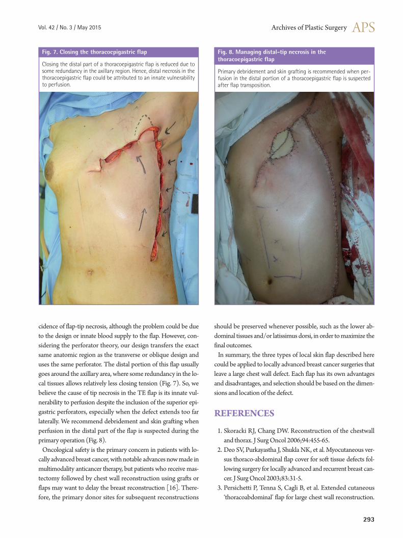
In contrast to the dramatic evolution in the field of breast recon-struction, less attention has been paid to reconstructing large chest wall defects following a so-called “toilet mastectomy”, wherein the mastectomy is performed on locally advanced breast cancer patients with the aim of ablating the breast and skin tissues and minimizing oncologic recurrence [10,11]. A va-riety of locoregional tissue transfers have been introduced, and the common goal is to provide early wound healing and a low risk of total flap failure [1-5,8,9,12,13]. The BA flap is a straight-forward way of closing the wound that only leaves a horizontal scar in the chest wall [4]. However, defects with greater vertical dimensions cannot be covered using this technique, and tension may result in wound dehiscence: as was the case here when the vertical dimension of the defect was 19 cm (Fig. 6).
The TA flap uses the skin, subcutaneous tissue of the anterior abdominal wall, and the direct perforating vessels of the seg-
mental arteries that arise from the subcostal, intercostals, and lumbar arteries [2,3]. Epigastric perforators can also be pre-served whenever possible. Deo et al. [2] reported that this flap is better than the myocutaneous flap in terms of mean blood loss, operating time, and length of hospital stay, and Persichetti et al. [3] have described using extended TA flaps to repair extensive defects ≤ 600 cm2. We treated 2 patients (22%) with small distal flap tip necrosis, and both spontaneously healed within 3 weeks. We believe perfusion to the TA flap is relatively robust, but the main drawback of this flap is the vertical midline scar.
The TE flap is mainly supplied by perforators from the superi-or epigastric arteries [1,5,6]. This flap has been confused with the TA flap because of its similar nomenclature: the two terms have been called a “medially based TA flap” or “laterally based TE flap” [1,5]. The TE flap is traditionally a transversely de-signed transposition flap supplied by the superior epigastric ar-tery, which was previously used to repair upper extremity de-fects [14]. When transversely designed, the donor site often re-quires skin grafts or surgery on the opposite side of the abdo-men [1,5,6]. Using a midaxillary incision, the vertical scar can be concealed when the arm is in a neutral position. Our large ro-tation-advancement flap design has the additional advantage in that the majority of the flap can be reelevated and reused by the time scarring occurs. However, this flap is hemodynamically weak, as reported by Baroudi et al. [15], and behaves more like a random flap.
Here, 60% of the patients in the TE flap group developed dis-tal flap necrosis. Among these, 2 patients required skin grafts be-cause healing was delayed for more than 3 weeks due to signifi-cant necrosis. We do not know the exact reasons for this high in-
Excessive tension during closure of the bilateral advancement flap may result in delayed healing or wound dehiscence.
Fig. 6. Wound dehiscence in a bilateral advancement flap
Vol. 42 / No. 3 / May 2015
293
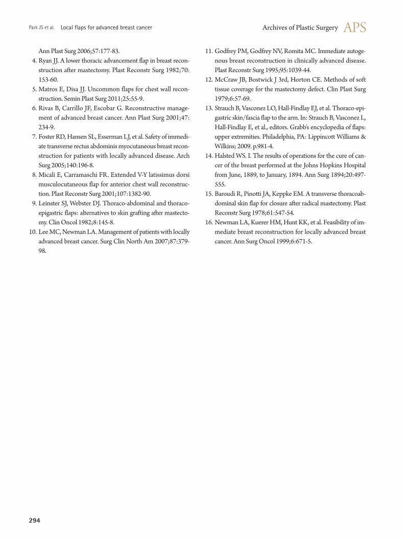
cidence of flap-tip necrosis, although the problem could be due to the design or innate blood supply to the flap. However, con-sidering the perforator theory, our design transfers the exact same anatomic region as the transverse or oblique design and uses the same perforator. The distal portion of this flap usually goes around the axillary area, where some redundancy in the lo-cal tissues allows relatively less closing tension (Fig. 7). So, we believe the cause of tip necrosis in the TE flap is its innate vul-nerability to perfusion despite the inclusion of the superior epi-gastric perforators, especially when the defect extends too far laterally. We recommend debridement and skin grafting when perfusion in the distal part of the flap is suspected during the primary operation (Fig. 8).
Oncological safety is the primary concern in patients with lo-cally advanced breast cancer, with notable advances now made in multimodality anticancer therapy, but patients who receive mas-tectomy followed by chest wall reconstruction using grafts or flaps may want to delay the breast reconstruction [16]. There-fore, the primary donor sites for subsequent reconstructions
should be preserved whenever possible, such as the lower ab-dominal tissues and/or latissimus dorsi, in order to maximize the final outcomes.
In summary, the three types of local skin flap described here could be applied to locally advanced breast cancer surgeries that leave a large chest wall defect. Each flap has its own advantages and disadvantages, and selection should be based on the dimen-sions and location of the defect.
REFERENCES
1. Skoracki RJ, Chang DW. Reconstruction of the chestwall and thorax. J Surg Oncol 2006;94:455-65.
2. Deo SV, Purkayastha J, Shukla NK, et al. Myocutaneous ver-sus thoraco-abdominal flap cover for soft tissue defects fol-lowing surgery for locally advanced and recurrent breast can-cer. J Surg Oncol 2003;83:31-5.
3. Persichetti P, Tenna S, Cagli B, et al. Extended cutaneous ‘thoracoabdominal’ flap for large chest wall reconstruction.
Closing the distal part of a thoracoepigastric flap is reduced due to some redundancy in the axillary region. Hence, distal necrosis in the thoracoepigastric flap could be attributed to an innate vulnerability to perfusion.
Fig. 7. Closing the thoracoepigastric flap
Primary debridement and skin grafting is recommended when per-fusion in the distal portion of a thoracoepigastric flap is suspected after flap transposition.
Fig. 8. Managing distal-tip necrosis in the thoracoepigastric flap
Park JS et al. Local flaps for advanced breast cancer
294
Ann Plast Surg 2006;57:177-83.4. Ryan JJ. A lower thoracic advancement flap in breast recon-
struction after mastectomy. Plast Reconstr Surg 1982;70: 153-60.
5. Matros E, Disa JJ. Uncommon flaps for chest wall recon-struction. Semin Plast Surg 2011;25:55-9.
6. Rivas B, Carrillo JF, Escobar G. Reconstructive manage-ment of advanced breast cancer. Ann Plast Surg 2001;47: 234-9.
7. Foster RD, Hansen SL, Esserman LJ, et al. Safety of immedi-ate transverse rectus abdominis myocutaneous breast recon-struction for patients with locally advanced disease. Arch Surg 2005;140:196-8.
8. Micali E, Carramaschi FR. Extended V-Y latissimus dorsi musculocutaneous flap for anterior chest wall reconstruc-tion. Plast Reconstr Surg 2001;107:1382-90.
9. Leinster SJ, Webster DJ. Thoraco-abdominal and thoraco-epigastric flaps: alternatives to skin grafting after mastecto-my. Clin Oncol 1982;8:145-8.
10. Lee MC, Newman LA. Management of patients with locally advanced breast cancer. Surg Clin North Am 2007;87:379-98.
11. Godfrey PM, Godfrey NV, Romita MC. Immediate autoge-nous breast reconstruction in clinically advanced disease. Plast Reconstr Surg 1995;95:1039-44.
12. McCraw JB, Bostwick J 3rd, Horton CE. Methods of soft tissue coverage for the mastectomy defect. Clin Plast Surg 1979;6:57-69.
13. Strauch B, Vasconez LO, Hall-Findlay EJ, et al. Thoraco-epi-gastric skin/fascia flap to the arm. In: Strauch B, Vasconez L, Hall-Findlay E, et al., editors. Grabb’s encyclopedia of flaps: upper extremities. Philadelphia, PA: Lippincott Williams & Wilkins; 2009. p.981-4.
14. Halsted WS. I. The results of operations for the cure of can-cer of the breast performed at the Johns Hopkins Hospital from June, 1889, to January, 1894. Ann Surg 1894;20:497-555.
15. Baroudi R, Pinotti JA, Keppke EM. A transverse thoracoab-dominal skin flap for closure after radical mastectomy. Plast Reconstr Surg 1978;61:547-54.
16. Newman LA, Kuerer HM, Hunt KK, et al. Feasibility of im-mediate breast reconstruction for locally advanced breast cancer. Ann Surg Oncol 1999;6:671-5.