Embed Size (px)
Citation preview

USU Water Quality Extension 435-797-2580 http://extension.usu.edu/waterquality
Utah Water Watch Harmful Algal Bloom Monitoring Program A COOPERATIVE EFFORT BETWEEN UTAH WATER WATCH, NATIONAL OCEANIC AND ATMOSPHERIC ADMINISTRATION, AND THE UTAH DIVISION OF WATER QUALITY.

USU Water Quality Extension 435-797-2580 http://extension.usu.edu/waterquality
USU Water Quality Extension 435-797-2580 http://extension.usu.edu/waterquality
Table of Contents Introduction .................................................................................................................................................. 2
Program Overview .................................................................................................................................... 2
Time Commitment .................................................................................................................................... 5
Process Diagram ........................................................................................................................................ 6
Field Identification ....................................................................................................................................... 7
Safety Precautions .................................................................................................................................... 7
bloomWatch Operation ............................................................................................................................ 9
Sample Collection ....................................................................................................................................... 12
Safety ...................................................................................................................................................... 12
Collection ................................................................................................................................................ 12
Transport ................................................................................................................................................. 12
Scope Locations ....................................................................................................................................... 13
Lab Analysis ................................................................................................................................................ 15
Lab Setup and Supplies ........................................................................................................................... 15
Microscope Setup ................................................................................................................................... 16
Microscope Operation ............................................................................................................................ 17
Analysis and Reporting ............................................................................................................................ 18
Test Strip Protocol ................................................................................................................................... 19
2
USU Water Quality Extension 435-797-2580 http://extension.usu.edu/waterquality
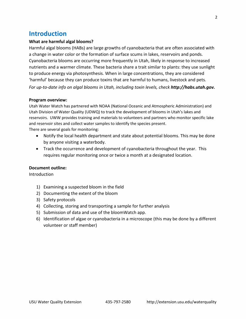
Introduction What are harmful algal blooms? Harmful algal blooms (HABs) are large growths of cyanobacteria that are often associated with a change in water color or the formation of surface scums in lakes, reservoirs and ponds. Cyanobacteria blooms are occurring more frequently in Utah, likely in response to increased nutrients and a warmer climate. These bacteria share a trait similar to plants: they use sunlight to produce energy via photosynthesis. When in large concentrations, they are considered ‘harmful’ because they can produce toxins that are harmful to humans, livestock and pets.
For up-to-date info on algal blooms in Utah, including toxin levels, check http://habs.utah.gov. Program overview: Utah Water Watch has partnered with NOAA (National Oceanic and Atmospheric Administration) and Utah Division of Water Quality (UDWQ) to track the development of blooms in Utah’s lakes and reservoirs. UWW provides training and materials to volunteers and partners who monitor specific lake and reservoir sites and collect water samples to identify the species present. There are several goals for monitoring:
• Notify the local health department and state about potential blooms. This may be done by anyone visiting a waterbody.
• Track the occurrence and development of cyanobacteria throughout the year. This requires regular monitoring once or twice a month at a designated location.
Document outline: Introduction
1) Examining a suspected bloom in the field 2) Documenting the extent of the bloom 3) Safety protocols 4) Collecting, storing and transporting a sample for further analysis 5) Submission of data and use of the bloomWatch app. 6) Identification of algae or cyanobacteria in a microscope (this may be done by a different
volunteer or staff member)
3
USU Water Quality Extension 435-797-2580 http://extension.usu.edu/waterquality
What causes blooms? Cyanobacteria blooms can occur in both pristine mountain lakes and polluted urban waterbodies. The makeup of a pond or lake’s phytoplankton population naturally shifts over the course of the year as different nutrients become available. Certain conditions, usually warmer waters and high concentrations of phosphorus and nitrogen, can increase the likelihood of blooming. As global temperatures increase, scientists have observed blooms occurring at higher frequencies. Nitrogen and phosphorus are common pollutants from sewage treatment plants, urban and agricultural runoff and erosion. Proper land management and investment in new technologies to treat wastewater can reduce the likelihood of blooms.
Are they dangerous? Certain species of cyanobacteria can release toxins into the water which can be dangerous to humans and pets. Some neurotoxins can cause immediate sickness or death while others can lead to long-term kidney or liver damage. Interestingly, not all blooms release toxins and it is not known what triggers cyanobacteria to produce toxins. Nonetheless, even blooms that do not contain detectable levels of toxins, dense concentrations of cyanobacteria can have health effects such as itching, rashes, and intestinal distress. If you notice a bloom, keep yourself and pets out of the water. What do they look like? Cyanobacteria blooms can be differentiated from blooms of ordinary green algae or growths of duckweed relatively easily using a simple guide found on the Utah Water Watch website. Not cyanobacteria:
• Green algae tend to be stringy, bubbly and often float on the surface in blobs. • Duckweed are tiny floating water plants; look closely and you will see small leaves and roots.
Cyanobacteria: • Cyanobacteria blooms often look like green pea soup, spilled paint, green blobs or streaks. • Some benthic (bottom-dwelling) blooms may not be detectable at the surface.
How you can help?
• Become a steward of your local lakes, ponds and reservoirs. Use the resources in the manual to become familiar with the visual differences between green algae and cyanobacteria. If something looks suspicious, follow protocols to collect a sample and notify your local health department.
• Come to a Utah Water Watch training and help monitor a location on a regular basis and become a NOAA volunteer and document the changes in the cyanobacteria community throughout the year. This is important research to develop satellite imagery.
Want to participate in Utah Water Watch or NOAA’s Harmful Algal Bloom monitoring program? Contact [email protected] or call the Water Quality Extension at (435) 797-2580.
4
USU Water Quality Extension 435-797-2580 http://extension.usu.edu/waterquality
Utah Water Watch Harmful Algal Bloom Monitoring
Utah Water Watch harmful algal bloom monitoring is a merger of two programs, NOAA’s (National Oceanic and Atmospheric Administration) Freshwater Phytoplankton Monitoring Network (PMN) and the state Division of Water Quality’s Harmful Algal Bloom (HAB) monitoring.
Volunteers may play multiple roles for harmful algal bloom monitoring:
• Scan for potential issues while out recreating • Monitor locations on a regular basis for Utah Water Watch • Collect samples and report blooms to the state and local health department • Monitor a site monthly – bimonthly, and check a sample in a microscope - important
research for NOAA
Volunteers may participate in different ways - see the time commitment below. Water samples are collected when a suspicious bloom is present or on a regular basis for volunteers monitoring for NOAA. The samples are analyzed at centrally located microscopes located across the state on scopes equipped with cameras that can be connected to computers. After reviewing the slides, any cyanobacteria species found is reported to Utah Water Watch and may be recorded in NOAA’s database. Procedures for analyzing collected samples will be outlined below.
Volunteers are provided a Harmful Algal Bloom notebook, with ID guides and official protocols for both NOAA and Utah, sample collection bottles, gloves and labels at a Utah Water Watch training. Volunteers participating in the NOAA monitoring will attend an additional webinar provided by NOAA to become more familiar with cyanobacteria species and their program.
5
USU Water Quality Extension 435-797-2580 http://extension.usu.edu/waterquality
Time Commitment
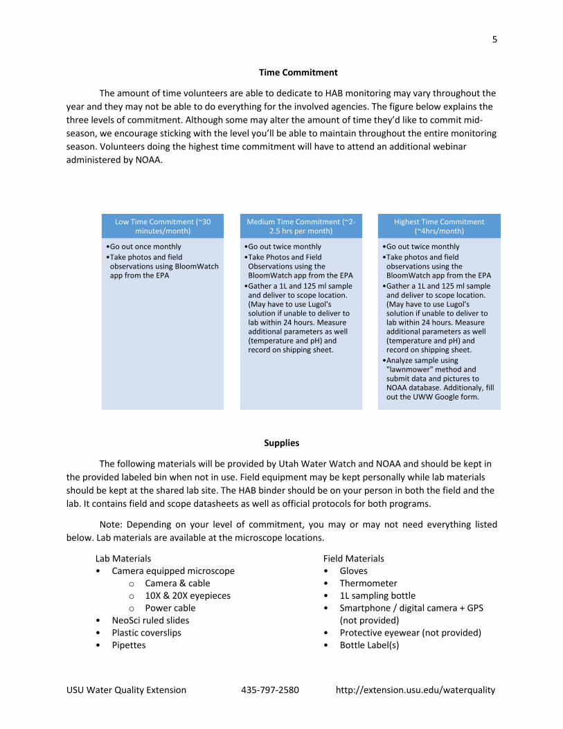
The amount of time volunteers are able to dedicate to HAB monitoring may vary throughout the year and they may not be able to do everything for the involved agencies. The figure below explains the three levels of commitment. Although some may alter the amount of time they’d like to commit mid-season, we encourage sticking with the level you’ll be able to maintain throughout the entire monitoring season. Volunteers doing the highest time commitment will have to attend an additional webinar administered by NOAA.
Supplies
The following materials will be provided by Utah Water Watch and NOAA and should be kept in the provided labeled bin when not in use. Field equipment may be kept personally while lab materials should be kept at the shared lab site. The HAB binder should be on your person in both the field and the lab. It contains field and scope datasheets as well as official protocols for both programs.
Note: Depending on your level of commitment, you may or may not need everything listed below. Lab materials are available at the microscope locations.
Lab Materials • Camera equipped microscope
o Camera & cable o 10X & 20X eyepieces o Power cable
• NeoSci ruled slides • Plastic coverslips • Pipettes
Field Materials • Gloves • Thermometer • 1L sampling bottle • Smartphone / digital camera + GPS
(not provided) • Protective eyewear (not provided) • Bottle Label(s)
Low Time Commitment (~30 minutes/month)
•Go out once monthly•Take photos and field
observations using BloomWatch app from the EPA
Medium Time Commitment (~2-2.5 hrs per month)
•Go out twice monthly •Take Photos and Field
Observations using the BloomWatch app from the EPA
•Gather a 1L and 125 ml sample and deliver to scope location. (May have to use Lugol's solution if unable to deliver to lab within 24 hours. Measure additional parameters as well (temperature and pH) and record on shipping sheet.
Highest Time Commitment (~4hrs/month)
•Go out twice monthly•Take photos and field
observations using the BloomWatch app from the EPA
•Gather a 1L and 125 ml sample and deliver to scope location. (May have to use Lugol's solution if unable to deliver to lab within 24 hours. Measure additional parameters as well (temperature and pH) and record on shipping sheet.
•Analyze sample using "lawnmower" method and submit data and pictures to NOAA database. Additionaly, fill out the UWW Google form.
6
USU Water Quality Extension 435-797-2580 http://extension.usu.edu/waterquality
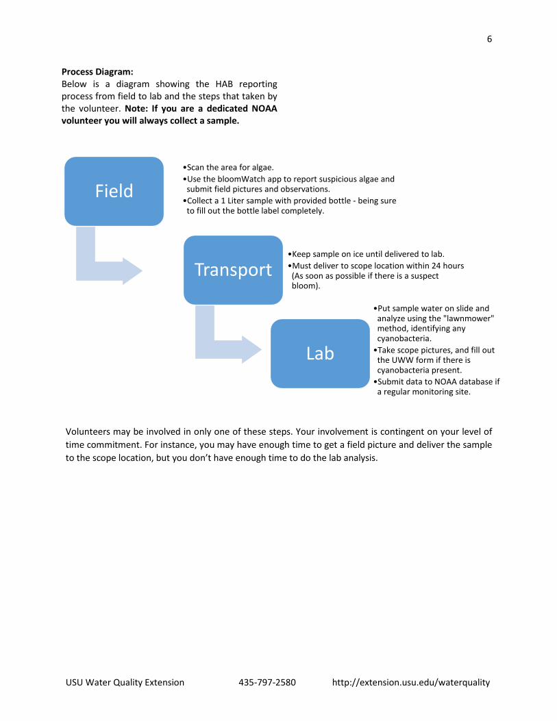
Process Diagram: Below is a diagram showing the HAB reporting process from field to lab and the steps that taken by the volunteer. Note: If you are a dedicated NOAA volunteer you will always collect a sample.
Volunteers may be involved in only one of these steps. Your involvement is contingent on your level of time commitment. For instance, you may have enough time to get a field picture and deliver the sample to the scope location, but you don’t have enough time to do the lab analysis.
Field•Scan the area for algae.•Use the bloomWatch app to report suspicious algae and
submit field pictures and observations.•Collect a 1 Liter sample with provided bottle - being sure
to fill out the bottle label completely.
Transport•Keep sample on ice until delivered to lab. •Must deliver to scope location within 24 hours
(As soon as possible if there is a suspect bloom).
Lab
•Put sample water on slide and analyze using the "lawnmower" method, identifying any cyanobacteria.
•Take scope pictures, and fill out the UWW form if there is cyanobacteria present.
•Submit data to NOAA database if a regular monitoring site.
7
USU Water Quality Extension 435-797-2580 http://extension.usu.edu/waterquality
Field Identification
Differentiating between cyanobacteria and other types of algae:
Field identification is the first step; it is important for all to be familiar with the difference between cyanobacteria and other types of algae. This is imperative not just for identification purposes, but for safety reasons as well. If it is determined in the field that what you are observing is cyanobacteria, you will want to take extra safety precautions (See below).
Determining if your scum is cyanobacteria:
Verify the suspect bloom is not filamentous green algae or duckweed using the guides below.
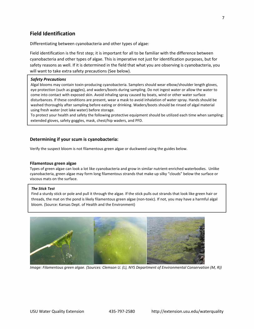
Filamentous green algae Types of green algae can look a lot like cyanobacteria and grow in similar nutrient-enriched waterbodies. Unlike cyanobacteria, green algae may form long filamentous strands that make up silky “clouds” below the surface or viscous mats on the surface.
Image: Filamentous green algae. (Sources: Clemson U. (L), NYS Department of Environmental Conservation (M, R))
Safety Precautions Algal blooms may contain toxin-producing cyanobacteria. Samplers should wear elbow/shoulder length gloves, eye protection (such as goggles), and waders/boots during sampling. Do not ingest water or allow the water to come into contact with exposed skin. Avoid inhaling spray caused by boats, wind or other water surface disturbances. If these conditions are present, wear a mask to avoid inhalation of water spray. Hands should be washed thoroughly after sampling before eating or drinking. Waders/boots should be rinsed of algal material using fresh water (not lake water) before storage. To protect your health and safety the following protective equipment should be utilized each time when sampling: extended gloves, safety goggles, mask, chest/hip waders, and PFD.
The Stick Test Find a sturdy stick or pole and pull it through the algae. If the stick pulls out strands that look like green hair or threads, the mat on the pond is likely filamentous green algae (non-toxic). If not, you may have a harmful algal bloom. (Source: Kansas Dept. of Health and the Environment)
8
USU Water Quality Extension 435-797-2580 http://extension.usu.edu/waterquality
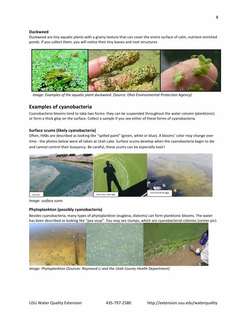
Duckweed Duckweed are tiny aquatic plants with a grainy texture that can cover the entire surface of calm, nutrient-enriched ponds. If you collect them, you will notice their tiny leaves and root structures.
Image: Examples of the aquatic plant duckweed. (Source: Ohio Environmental Protection Agency).
Examples of cyanobacteria Cyanobacteria blooms tend to take two forms: they can be suspended throughout the water column (planktonic) or form a thick glop on the surface. Collect a sample if you see either of these forms of cyanobacteria. Surface scums (likely cyanobacteria) Often, HABs are described as looking like “spilled paint” (green, white or blue). A blooms’ color may change over time - the photos below were all taken at Utah Lake. Surface scums develop when the cyanobacteria begin to die and cannot control their buoyancy. Be careful, these scums can be especially toxic!
Image: surface sums
Phytoplankton (possibly cyanobacteria) Besides cyanobacteria, many types of phytoplankton (euglena, diatoms) can form planktonic blooms. The water has been described as looking like “pea soup”. You may see clumps, which are cyanobacterial colonies (center pic).
Image: Phytoplankton (Sources: Raymond Li and the Utah County Health Department)
9
USU Water Quality Extension 435-797-2580 http://extension.usu.edu/waterquality
Additional Resources: Refer to the “Field Guide to Scums” produced by the United States Geological Survey (located on the Utah Water Watch website) for more info on common types of algae and cyanobacteria. (See http://extension.usu.edu/utahwaterwatch/monitoring/lakes/hab)
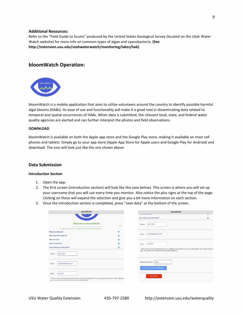
bloomWatch Operation:
bloomWatch is a mobile application that aims to utilize volunteers around the country to identify possible harmful algal blooms (HABs). Its ease of use and functionality will make it a great tool in disseminating data related to temporal and spatial occurrences of HABs. When data is submitted, the relevant local, state, and federal water quality agencies are alerted and can further interpret the photos and field observations.
DOWNLOAD
bloomWatch is available on both the Apple app store and the Google Play store, making it available on most cell phones and tablets. Simply go to your app store (Apple App Store for Apple users and Google Play for Android) and download. The icon will look just like the one shown above.
Data Submission
Introduction Section
1. Open the app. 2. The first screen (introduction section) will look like this (see below). This screen is where you will set up
your username that you will use every time you monitor. Also notice the plus signs at the top of the page. Clicking on these will expand the selection and give you a bit more information on each section.
3. Once the Introduction section is completed, press “save data” at the bottom of the screen.
10
USU Water Quality Extension 435-797-2580 http://extension.usu.edu/waterquality
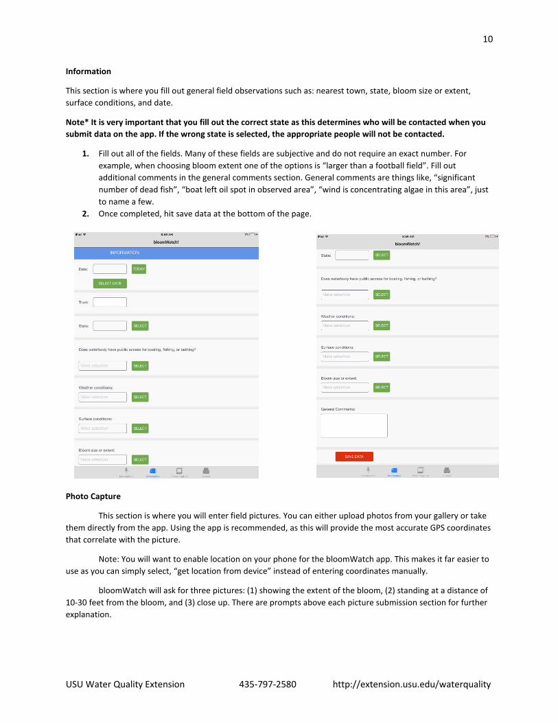
Information
This section is where you fill out general field observations such as: nearest town, state, bloom size or extent, surface conditions, and date.
Note* It is very important that you fill out the correct state as this determines who will be contacted when you submit data on the app. If the wrong state is selected, the appropriate people will not be contacted.
1. Fill out all of the fields. Many of these fields are subjective and do not require an exact number. For example, when choosing bloom extent one of the options is “larger than a football field”. Fill out additional comments in the general comments section. General comments are things like, “significant number of dead fish”, “boat left oil spot in observed area”, “wind is concentrating algae in this area”, just to name a few.
2. Once completed, hit save data at the bottom of the page.
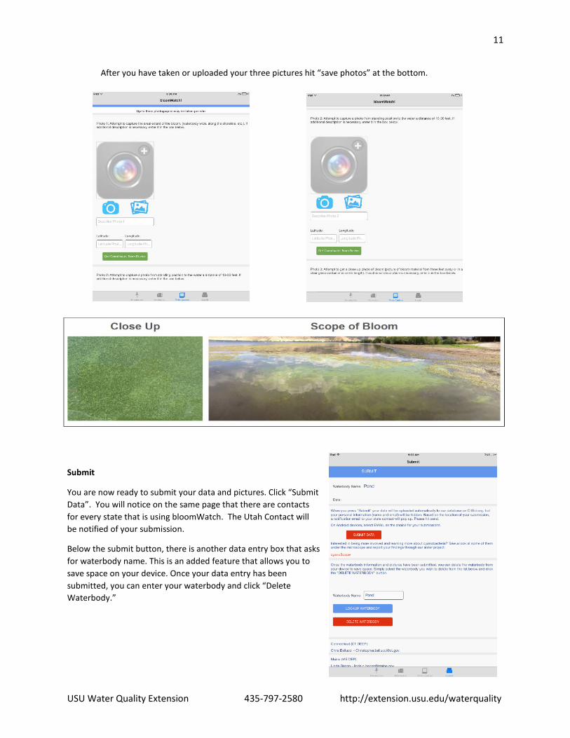
Photo Capture
This section is where you will enter field pictures. You can either upload photos from your gallery or take them directly from the app. Using the app is recommended, as this will provide the most accurate GPS coordinates that correlate with the picture.
Note: You will want to enable location on your phone for the bloomWatch app. This makes it far easier to use as you can simply select, “get location from device” instead of entering coordinates manually.
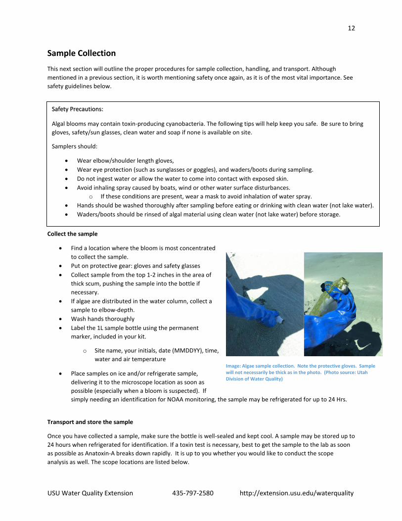
bloomWatch will ask for three pictures: (1) showing the extent of the bloom, (2) standing at a distance of 10-30 feet from the bloom, and (3) close up. There are prompts above each picture submission section for further explanation.
11
USU Water Quality Extension 435-797-2580 http://extension.usu.edu/waterquality
After you have taken or uploaded your three pictures hit “save photos” at the bottom.
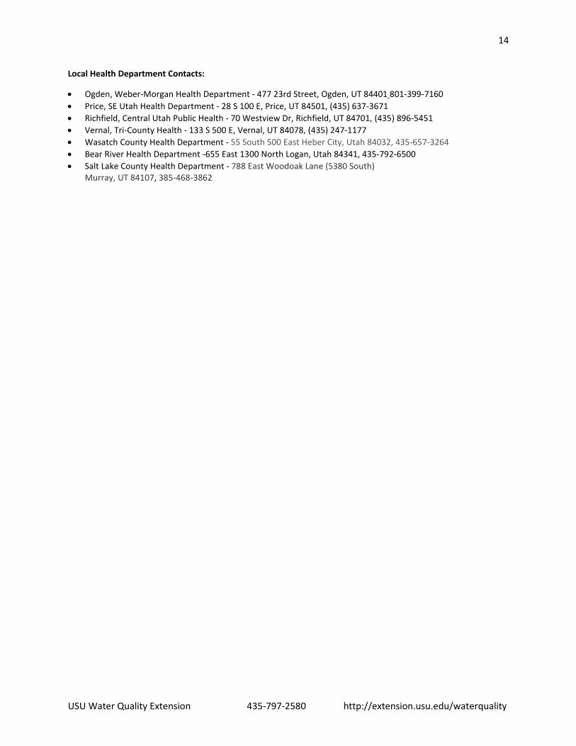
Submit
You are now ready to submit your data and pictures. Click “Submit Data”. You will notice on the same page that there are contacts for every state that is using bloomWatch. The Utah Contact will be notified of your submission.
Below the submit button, there is another data entry box that asks for waterbody name. This is an added feature that allows you to save space on your device. Once your data entry has been submitted, you can enter your waterbody and click “Delete Waterbody.”
12
USU Water Quality Extension 435-797-2580 http://extension.usu.edu/waterquality
Sample Collection
This next section will outline the proper procedures for sample collection, handling, and transport. Although mentioned in a previous section, it is worth mentioning safety once again, as it is of the most vital importance. See safety guidelines below.
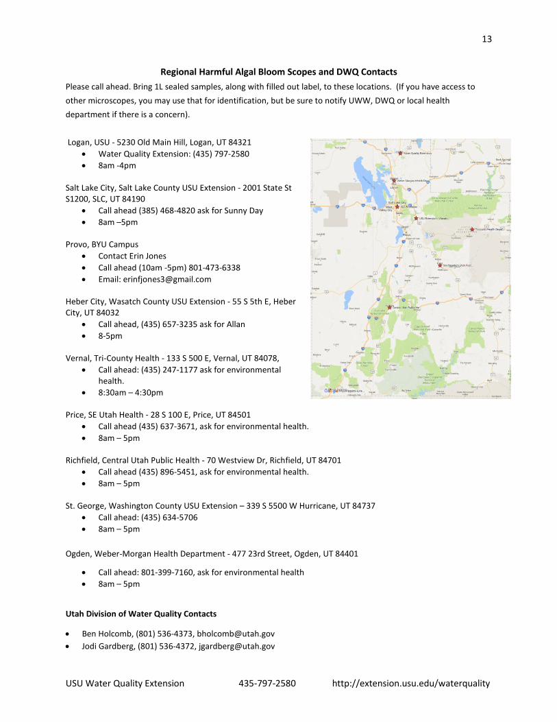
Collect the sample
• Find a location where the bloom is most concentrated to collect the sample.
• Put on protective gear: gloves and safety glasses • Collect sample from the top 1-2 inches in the area of
thick scum, pushing the sample into the bottle if necessary.
• If algae are distributed in the water column, collect a sample to elbow-depth.
• Wash hands thoroughly • Label the 1L sample bottle using the permanent
marker, included in your kit.
o Site name, your initials, date (MMDDYY), time, water and air temperature
• Place samples on ice and/or refrigerate sample, delivering it to the microscope location as soon as possible (especially when a bloom is suspected). If simply needing an identification for NOAA monitoring, the sample may be refrigerated for up to 24 Hrs.
Transport and store the sample
Once you have collected a sample, make sure the bottle is well-sealed and kept cool. A sample may be stored up to 24 hours when refrigerated for identification. If a toxin test is necessary, best to get the sample to the lab as soon as possible as Anatoxin-A breaks down rapidly. It is up to you whether you would like to conduct the scope analysis as well. The scope locations are listed below.
Safety Precautions:
Algal blooms may contain toxin-producing cyanobacteria. The following tips will help keep you safe. Be sure to bring gloves, safety/sun glasses, clean water and soap if none is available on site.
Samplers should:
• Wear elbow/shoulder length gloves, • Wear eye protection (such as sunglasses or goggles), and waders/boots during sampling. • Do not ingest water or allow the water to come into contact with exposed skin. • Avoid inhaling spray caused by boats, wind or other water surface disturbances.
o If these conditions are present, wear a mask to avoid inhalation of water spray. • Hands should be washed thoroughly after sampling before eating or drinking with clean water (not lake water). • Waders/boots should be rinsed of algal material using clean water (not lake water) before storage.
Image: Algae sample collection. Note the protective gloves. Sample will not necessarily be thick as in the photo. (Photo source: Utah Division of Water Quality)
13
USU Water Quality Extension 435-797-2580 http://extension.usu.edu/waterquality
Regional Harmful Algal Bloom Scopes and DWQ Contacts Please call ahead. Bring 1L sealed samples, along with filled out label, to these locations. (If you have access to other microscopes, you may use that for identification, but be sure to notify UWW, DWQ or local health department if there is a concern).
Logan, USU - 5230 Old Main Hill, Logan, UT 84321
• Water Quality Extension: (435) 797-2580 • 8am -4pm
Salt Lake City, Salt Lake County USU Extension - 2001 State St S1200, SLC, UT 84190
• Call ahead (385) 468-4820 ask for Sunny Day • 8am –5pm
Provo, BYU Campus
• Contact Erin Jones • Call ahead (10am -5pm) 801-473-6338 • Email: [email protected]
Heber City, Wasatch County USU Extension - 55 S 5th E, Heber City, UT 84032
• Call ahead, (435) 657-3235 ask for Allan • 8-5pm
Vernal, Tri-County Health - 133 S 500 E, Vernal, UT 84078,
• Call ahead: (435) 247-1177 ask for environmental health.
• 8:30am – 4:30pm Price, SE Utah Health - 28 S 100 E, Price, UT 84501
• Call ahead (435) 637-3671, ask for environmental health. • 8am – 5pm
Richfield, Central Utah Public Health - 70 Westview Dr, Richfield, UT 84701
• Call ahead (435) 896-5451, ask for environmental health. • 8am – 5pm
St. George, Washington County USU Extension – 339 S 5500 W Hurricane, UT 84737
• Call ahead: (435) 634-5706 • 8am – 5pm
Ogden, Weber-Morgan Health Department - 477 23rd Street, Ogden, UT 84401
• Call ahead: 801-399-7160, ask for environmental health • 8am – 5pm
Utah Division of Water Quality Contacts
• Ben Holcomb, (801) 536-4373, [email protected] • Jodi Gardberg, (801) 536-4372, [email protected]
14
USU Water Quality Extension 435-797-2580 http://extension.usu.edu/waterquality
Local Health Department Contacts:
• Ogden, Weber-Morgan Health Department - 477 23rd Street, Ogden, UT 84401 801-399-7160 • Price, SE Utah Health Department - 28 S 100 E, Price, UT 84501, (435) 637-3671 • Richfield, Central Utah Public Health - 70 Westview Dr, Richfield, UT 84701, (435) 896-5451 • Vernal, Tri-County Health - 133 S 500 E, Vernal, UT 84078, (435) 247-1177 • Wasatch County Health Department - 55 South 500 East Heber City, Utah 84032, 435-657-3264 • Bear River Health Department -655 East 1300 North Logan, Utah 84341, 435-792-6500 • Salt Lake County Health Department - 788 East Woodoak Lane (5380 South)
Murray, UT 84107, 385-468-3862
15
USU Water Quality Extension 435-797-2580 http://extension.usu.edu/waterquality
Lab analysis
When doing the lab analysis, be sure to follow the safety guidelines and wear the appropriate Personal Protection Equipment (PPE); namely, protective eyewear and nitrile or latex gloves. Sample Storage
After receiving the sample, keep it in a refrigerator or on ice until ready for the lab procedure. If there is a suspected bloom, look at the sample as soon as possible. A toxin test may be required, which is best done within a few hours of collection.
Accompanying Documentation These documents below are included in the binder accompanying the microscope. • NOAA Freshwater Phytoplankton ID Sheet: Quick ID for toxin producing cyanobacteria
https://goo.gl/RS9zmb
• USGS Field Guide: In-depth guide for cyanobacteria and other algae https://goo.gl/e2a4aE
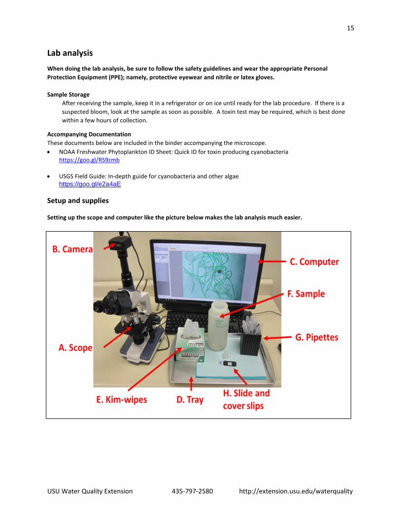
Setup and supplies Setting up the scope and computer like the picture below makes the lab analysis much easier.
16
USU Water Quality Extension 435-797-2580 http://extension.usu.edu/waterquality
Replacement Lab Supplies (notify the scope administrator or contact Utah Water Watch – [email protected] or 435-797-2580)
• 5 ml pipettes, Amazon, 100 for $7. • Gridded microscope slides, http://labscientific.com/ Catalog number 4456 one box costs $65. • Glass Cover slips 18X18 mm, http://labscientific.com/ catalog number 7781 $7 • Microscope Parts (AmScope)
http://www.amscope.com/ or call 1-888-950-2888 for help ordering a specific part. o The scope product ID is T120B-5M
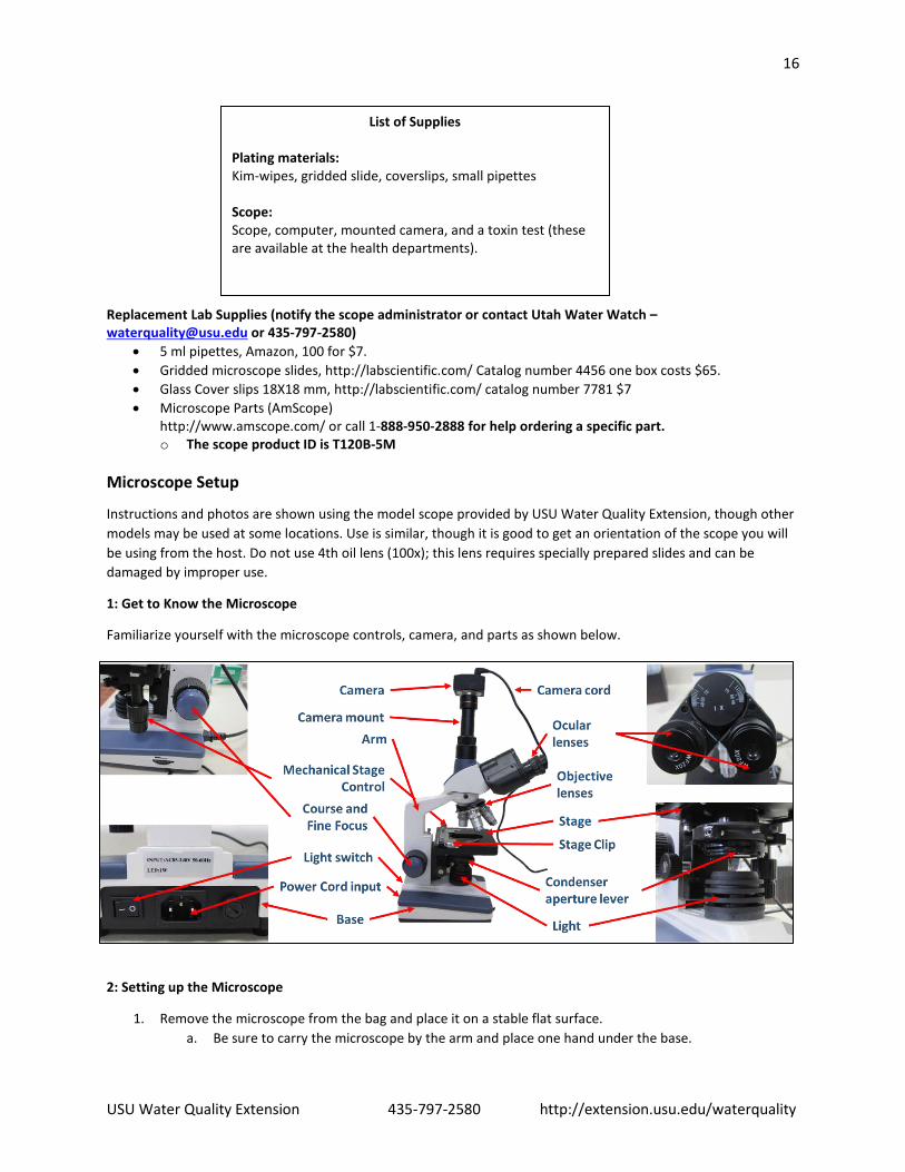
Microscope Setup
Instructions and photos are shown using the model scope provided by USU Water Quality Extension, though other models may be used at some locations. Use is similar, though it is good to get an orientation of the scope you will be using from the host. Do not use 4th oil lens (100x); this lens requires specially prepared slides and can be damaged by improper use.
1: Get to Know the Microscope
Familiarize yourself with the microscope controls, camera, and parts as shown below.
2: Setting up the Microscope
1. Remove the microscope from the bag and place it on a stable flat surface. a. Be sure to carry the microscope by the arm and place one hand under the base.
List of Supplies Plating materials: Kim-wipes, gridded slide, coverslips, small pipettes Scope: Scope, computer, mounted camera, and a toxin test (these are available at the health departments).

17
USU Water Quality Extension 435-797-2580 http://extension.usu.edu/waterquality
2. Rotate ocular lenses so they are facing you, make sure the 20x lenses are installed 3. Screw on the camera mount and then slide in the camera attachment. 4. Plug in the USB cord to the camera and the computer. 5. Plug in the power cord for the microscope into the nearest outlet and turn on the scope light, using the
switch on the back of the microscope.
3: Launching the Software
Select the software app on your computer compatible to the microscope you are using. Make sure the USB is plugged into the camera. Launch the software and open the camera.
By connecting the scope to the computer, you can view the slide in real time on the screen and take pictures of the sample.
AmScope instructions: This software can be downloaded and installed from goo.gl/KL1Q7i . Plug in the USB, launch the software and select the camera, from the camera list in the top left corner.
Microscope Operation 1: Preparing the Slide
1. Put on protective gloves. 2. Use pipette to collect a small amount from the algal sample. 3. Slide Preparation:
Place a drop from the pipette onto the middle of the top surface of the gridded slide.
Gently lay cover slip at an angle to avoid air bubbles.
2: Operating the Microscope 1. Lower the stage using the coarse knob on the right-hand side. 2. Rotate the objective lens carousel to the lowest magnification lens (4x), and move the stage to its lowest
point. 3. Place the prepared slide on the stage, using the stage clip to hold it in place.
o Use the two vertical knobs to the right to move the stage 4. Raise the stage to its highest point (it will not hit on lowest magnification) 5. Now look through the eyepiece and lower the stage (fine knob) until the sample comes into focus. 6. If further magnification is needed rotate the objective carousel to the next highest magnification. Use the fine
focus knob to return the sample to focus.
18
USU Water Quality Extension 435-797-2580 http://extension.usu.edu/waterquality
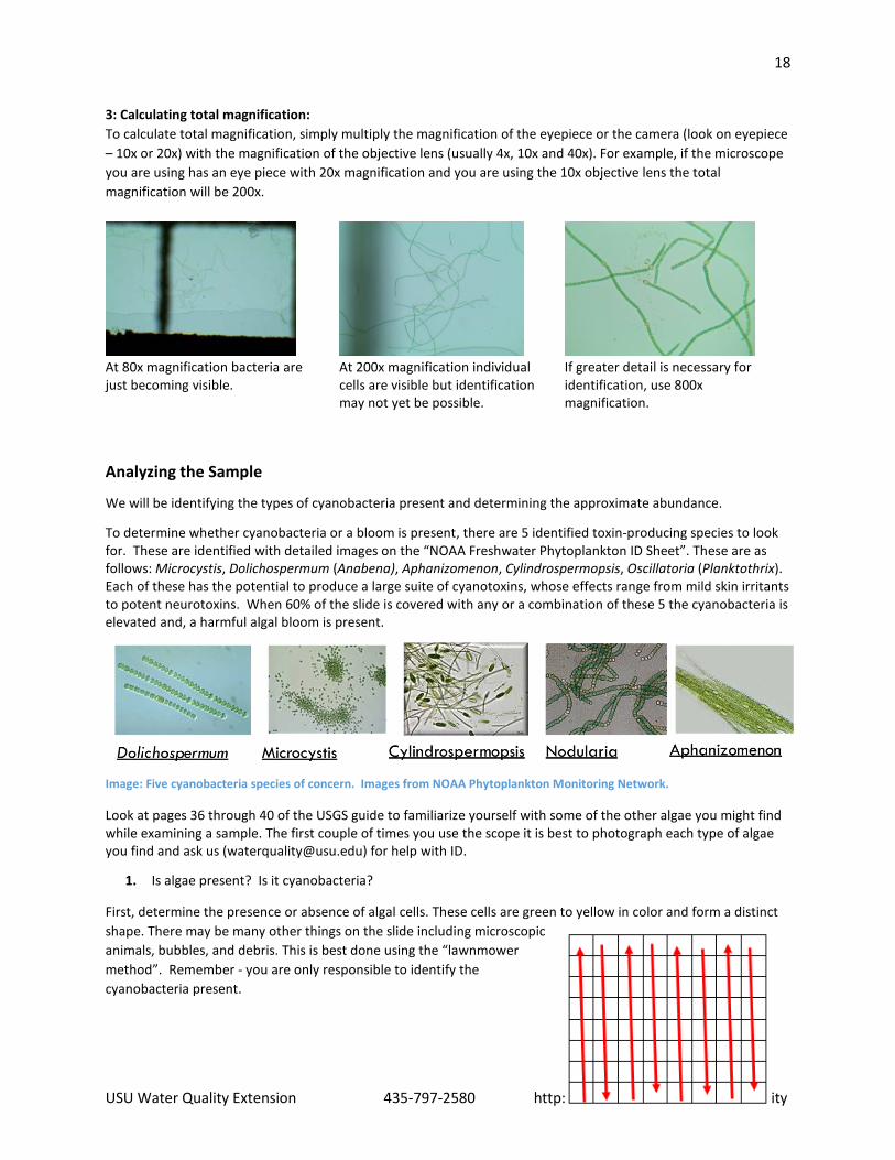
3: Calculating total magnification: To calculate total magnification, simply multiply the magnification of the eyepiece or the camera (look on eyepiece – 10x or 20x) with the magnification of the objective lens (usually 4x, 10x and 40x). For example, if the microscope you are using has an eye piece with 20x magnification and you are using the 10x objective lens the total magnification will be 200x.
At 80x magnification bacteria are just becoming visible.
At 200x magnification individual cells are visible but identification may not yet be possible.
If greater detail is necessary for identification, use 800x magnification.
Analyzing the Sample
We will be identifying the types of cyanobacteria present and determining the approximate abundance.
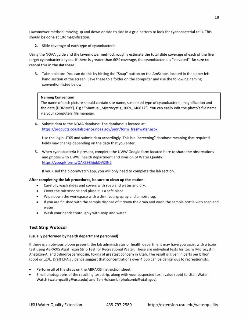
To determine whether cyanobacteria or a bloom is present, there are 5 identified toxin-producing species to look for. These are identified with detailed images on the “NOAA Freshwater Phytoplankton ID Sheet”. These are as follows: Microcystis, Dolichospermum (Anabena), Aphanizomenon, Cylindrospermopsis, Oscillatoria (Planktothrix). Each of these has the potential to produce a large suite of cyanotoxins, whose effects range from mild skin irritants to potent neurotoxins. When 60% of the slide is covered with any or a combination of these 5 the cyanobacteria is elevated and, a harmful algal bloom is present.
Image: Five cyanobacteria species of concern. Images from NOAA Phytoplankton Monitoring Network.
Look at pages 36 through 40 of the USGS guide to familiarize yourself with some of the other algae you might find while examining a sample. The first couple of times you use the scope it is best to photograph each type of algae you find and ask us ([email protected]) for help with ID.
1. Is algae present? Is it cyanobacteria?
First, determine the presence or absence of algal cells. These cells are green to yellow in color and form a distinct shape. There may be many other things on the slide including microscopic animals, bubbles, and debris. This is best done using the “lawnmower method”. Remember - you are only responsible to identify the cyanobacteria present.
19
USU Water Quality Extension 435-797-2580 http://extension.usu.edu/waterquality
Lawnmower method: moving up and down or side to side in a grid pattern to look for cyanobacterial cells. This should be done at 10x magnification.
2. Slide coverage of each type of cyanobacteria
Using the NOAA guide and the lawnmower method, roughly estimate the total slide coverage of each of the five target cyanobacteria types. If there is greater than 60% coverage, the cyanobacteria is “elevated”. Be sure to record this in the database.
3. Take a picture. You can do this by hitting the “Snap” button on the AmScope, located in the upper left-hand section of the screen. Save these to a folder on the computer and use the following naming convention listed below
4. Submit data to the NOAA database. The database is located at: https://products.coastalscience.noaa.gov/pmn/form_freshwater.aspx
Use the login UT05 and submit data accordingly. This is a “screening” database meaning that required fields may change depending on the data that you enter.
5. When cyanobacteria is present, complete the UWW Google form located here to share the observations and photos with UWW, health department and Division of Water Quality: https://goo.gl/forms/OAKS98hjubDVi2IN2
If you used the bloomWatch app, you will only need to complete the lab section.
After completing the lab procedures, be sure to clean up the station. • Carefully wash slides and covers with soap and water and dry. • Cover the microscope and place it is a safe place. • Wipe down the workspace with a disinfecting spray and a moist rag. • If you are finished with the sample dispose of it down the drain and wash the sample bottle with soap and
water. • Wash your hands thoroughly with soap and water.
Test Strip Protocol
(usually performed by health department personnel)
If there is an obvious bloom present, the lab administrator or health department may have you assist with a toxin test using ABRAXIS Algal Toxin Strip Test for Recreational Water. These are individual tests for toxins Microcystin, Anatoxin-A, and cylindrospermopsin, toxins of greatest concern in Utah. The result is given in parts per billion (ppb) or µg/L. Draft EPA guidance suggest that concentrations over 4 ppb can be dangerous to recreationists. • Perform all of the steps on the ABRAXIS instruction sheet. • Email photographs of the resulting test strip, along with your suspected toxin value (ppb) to Utah Water
Watch ([email protected]) and Ben Holcomb ([email protected]).
Naming Convention The name of each picture should contain site name, suspected type of cyanobacteria, magnification and the date (DDMMYY). E.g.: “Mantua _Mycrocystis_100x_140817”. You can easily edit the photo’s file name via your computers file manager.

Notes about Aphanizomenon:
Toxin: Saxitoxin N-fixation: YesCyanophyta – Cyanophyceae – Nostocales
4 described species
Trichomes solitary or gathered in small or large fascicles (clusters) with trichomes arranged in parallel layers.
Notes about Anabaena:
Toxin: Anatoxin-a N-fixation: YesCyanophyta – Cyanophyceae – Nostocales
More than 80 known species
Trichomes are straight, curved or coiled, in some species with mucilaginous colorless envelopes, mat forming.
Notes about Cylindrospermopsis:
Toxin: Cylindrospermopsin N-fixation: Yes Cyanophyta – Cyanophyceae – Nostocales
Around 10 known species
Trichomes are straight, bent or spirally coiled. Cells are cylindrical or barrel-shaped pale blue-green or yellowish, with aerotypes. Heterocytes and akinetes are terminal.
Notes about Microcystis: Toxin: Microcystin N-fixation: NoCyanophyta – Cyanophyceae – Chroococcales
Around 25 known species
Colonies are irregular, cloud-like with hollow spaces and sometimes with a well developed outer margin. Cells are spherical with may aerotopes.
Freshwater Phytoplankton
ID SHEET
TARGET ALGAE
K
Cylindrospermopsis spp. Curved morphotype
Aphanizomenon spp.
Microcystis spp. Microcystis spp.
Dillard, G. (1999).
Dillard, G. (1999).
Dillard, G. (1999).
Dillard, G. (1999).
Credit: GreenWater Laboratories/CyanoLab Credit: GreenWater Laboratories/CyanoLab
Credit: GreenWater Laboratories/CyanoLab Cr
edit:
Gre
enW
ater
Lab
orat
orie
s/Cy
anoL
ab
Credit: GreenWater Laboratories/CyanoLab
Anabaena spp.(now Dolichospermum)
Cylindrospermopsis spp. Straight morphotype
Anabaena spp. (now Dolichospermum)
akinete
heterocyte
akinete
heterocyte
Notes about Planktothrix:
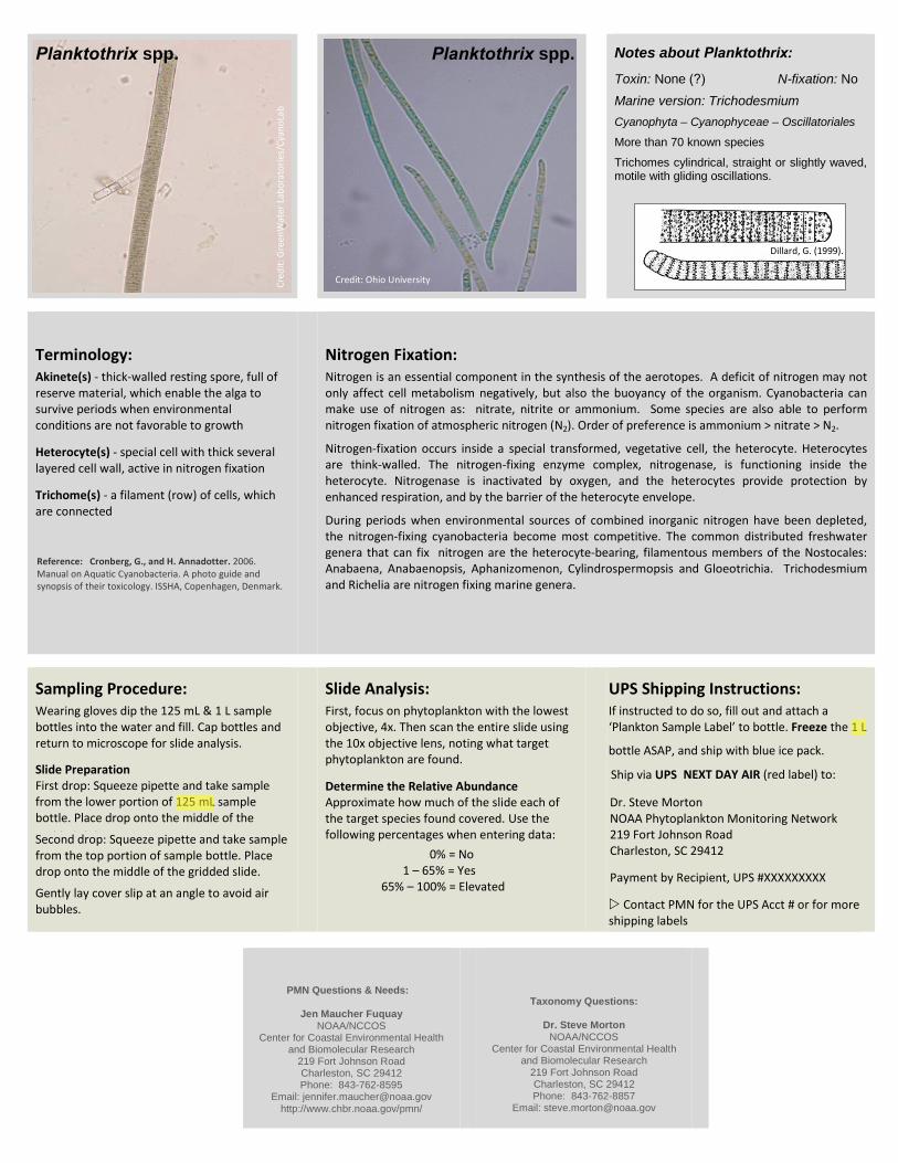
Toxin: None (?) N-fixation: NoMarine version: Trichodesmium Cyanophyta – Cyanophyceae – Oscillatoriales
More than 70 known species
Trichomes cylindrical, straight or slightly waved, motile with gliding oscillations.
Terminology:
Akinete(s) - thick-walled resting spore, full of reserve material, which enable the alga to survive periods when environmental conditions are not favorable to growth
Heterocyte(s) - special cell with thick several layered cell wall, active in nitrogen fixation
Trichome(s) - a filament (row) of cells, which are connected
Nitrogen Fixation:
Nitrogen is an essential component in the synthesis of the aerotopes. A deficit of nitrogen may not only affect cell metabolism negatively, but also the buoyancy of the organism. Cyanobacteria can make use of nitrogen as: nitrate, nitrite or ammonium. Some species are also able to perform nitrogen fixation of atmospheric nitrogen (N2). Order of preference is ammonium > nitrate > N2.
Nitrogen-fixation occurs inside a special transformed, vegetative cell, the heterocyte. Heterocytes are think-walled. The nitrogen-fixing enzyme complex, nitrogenase, is functioning inside the heterocyte. Nitrogenase is inactivated by oxygen, and the heterocytes provide protection by enhanced respiration, and by the barrier of the heterocyte envelope.
During periods when environmental sources of combined inorganic nitrogen have been depleted, the nitrogen-fixing cyanobacteria become most competitive. The common distributed freshwater genera that can fix nitrogen are the heterocyte-bearing, filamentous members of the Nostocales: Anabaena, Anabaenopsis, Aphanizomenon, Cylindrospermopsis and Gloeotrichia. Trichodesmium and Richelia are nitrogen fixing marine genera.
Sampling Procedure:
Wearing gloves dip the 125 mL & 1 L sample bottles into the water and fill. Cap bottles and return to microscope for slide analysis.
Slide Preparation First drop: Squeeze pipette and take sample from the lower portion of 125 mL sample bottle. Place drop onto the middle of the gridded slide. Second drop: Squeeze pipette and take samplefrom the top portion of sample bottle. Place drop onto the middle of the gridded slide.
Gently lay cover slip at an angle to avoid air bubbles.
Slide Analysis:
First, focus on phytoplankton with the lowest objective, 4x. Then scan the entire slide using the 10x objective lens, noting what target phytoplankton are found.
Determine the Relative Abundance Approximate how much of the slide each of the target species found covered. Use the following percentages when entering data:
0% = No 1 – 65% = Yes
65% – 100% = Elevated
UPS Shipping Instructions:
If instructed to do so, fill out and attach a ‘Plankton Sample Label’ to bottle. Freeze the 1 L
bottle ASAP, and ship with blue ice pack.
Ship via UPS NEXT DAY AIR (red label) to:
Dr. Steve Morton NOAA Phytoplankton Monitoring Network 219 Fort Johnson Road Charleston, SC 29412
Payment by Recipient, UPS #XXXXXXXXX
Contact PMN for the UPS Acct # or for moreshipping labels
PMN Questions & Needs:
Jen Maucher Fuquay NOAA/NCCOS
Center for Coastal Environmental Health and Biomolecular Research
219 Fort Johnson Road Charleston, SC 29412 Phone: 843-762-8595
Email: [email protected] http://www.chbr.noaa.gov/pmn/
Taxonomy Questions:
Dr. Steve Morton NOAA/NCCOS
Center for Coastal Environmental Health and Biomolecular Research
219 Fort Johnson Road Charleston, SC 29412 Phone: 843-762-8857
Email: [email protected]
Planktothrix spp.
Dillard, G. (1999).
Credit: Ohio University Cred
it: G
reen
Wat
er L
abor
ator
ies/
Cyan
oLab
Planktothrix spp.
Reference: Cronberg, G., and H. Annadotter. 2006. Manual on Aquatic Cyanobacteria. A photo guide and synopsis of their toxicology. ISSHA, Copenhagen, Denmark.