Embed Size (px)
Citation preview

UvA-DARE is a service provided by the library of the University of Amsterdam (http://dare.uva.nl)
UvA-DARE (Digital Academic Repository)
Autonomic nervous control of white adipose tissue : studies on the role of the brain inbody fat distributionKreier, F.H.K.
Link to publication
Citation for published version (APA):Kreier, F. (2005). Autonomic nervous control of white adipose tissue : studies on the role of the brain in body fatdistribution
General rightsIt is not permitted to download or to forward/distribute the text or part of it without the consent of the author(s) and/or copyright holder(s),other than for strictly personal, individual use, unless the work is under an open content license (like Creative Commons).
Disclaimer/Complaints regulationsIf you believe that digital publication of certain material infringes any of your rights or (privacy) interests, please let the Library know, statingyour reasons. In case of a legitimate complaint, the Library will make the material inaccessible and/or remove it from the website. Please Askthe Library: http://uba.uva.nl/en/contact, or a letter to: Library of the University of Amsterdam, Secretariat, Singel 425, 1012 WP Amsterdam,The Netherlands. You will be contacted as soon as possible.
Download date: 10 May 2018
CHAPTER 8
Perspectives for follow-up studies
'The brain has a role in body fat distribution and its associated metabolic diseases' is the general hypothesis of this thesis.
An increasing number of researchers have been studying the role of the hypothalamus as the central region that controls energy homeostasis; they initially showed that manipulations of the hypothalamus can induce major changes in energy homeostasis. But what really launched the research on the hypothalamic role in metabolism was the discovery of leptin in 1995, when it became clear to what extent the hypothalamus integrates information of the body's state'4. The hypothalamus translates its drive into the body by means of pituitary hormones, and only recently we have started to appreciate the contribution of the autonomic nervous system (ANS). In chapters 2-5 we describe a part of the interaction between the hypothalamus and metabolic organs. In this chapter we aim to add some comments with respect to the developed hypothesis and the essential experiments that still need to be done for investigating this hypothesis.
The present thesis consists of two main parts: findings and perspectives.
FINDING #1 The brain uses two neuronal systems w i th an antagonist ic
effect on metabol ic organs (chapter 2 and 4)
We show that fat tissue is innervated by (pre-)sympathetic and (pre-)parasympathetic neurons, running from the biological clock (SCN), via the PVN and LH through the ANS to fat tissue. Bartness and co-workers proved that sympathetic tone stimulates lipolysis; we demonstrated that parasympathetic tone induces lipogenesis5. Until now, lipogenesis was believed to be solely under hormonal control. Insulin and Cortisol have been shown to stimulate lipogenesis, whereas estrogen and growth hormone induce lipolysis6,7. However, humoral factors cannot completely explain fat tissue metabolism in vivo*.
Cantu demonstrated in 1967 that sympathetic denervation of the retroperitoneal fat
pad results in impaired lipolysis after fasting9. In the i98oies, Stock and Rothwell dis
covered that sympathetic tone induces mitochondrial oxidation in brown fat tissue10-
".In the same decade, Trayhurn and Dalziel refined the theoretical frame on the auto
nomic innervation of fat tissue12"14. In the i99oies Bartness and coworkers substantiated
in a series of experiments that the hypothalamus is connected to white adipose tissue
97

PART B DISCUSSION AND PERSPECTIVES
by the sympathetic nervous system exerting a lipolytic effect515. However, lipogenesis
was believed not be controlled by the ANS.
Earlier, our group applied neuroanatomical and physiological techniques to study
the hypothalamic autonomic control of heart, liver, pancreas, thyroid and adrenall6"25.
Except the adrenal gland, these organs appeared to receive both sympathetic and para
sympathetic innervation, originating in the biological clock and other hypothalamic
nuclei.
We injected PRV into intact and locally sympathectomized fat pads (chapter 2)2".
Transneuronal tracing from intact fat pads revealed a dominant sympathetic labeling
as compared to the number of parasympathetic neurons. However, if prior to the
tracer injection the sympathetic nerves where cut, the sympathetic motor neurons in
the spinal cord where empty, while the dorsal motor nucleus of vagus and the nucleus
ambiguous where strongly labeled. Finally, we conducted a series of control experi
ments to exclude false positive labeling due to leakage into the abdominal cavity and
blood stream. (See supplemental material chapter 3 for a detailed description.) A point
of discussion remains whether PRV transport and replication is partly depending on
neuronal activity17'2fi. We speculate that the dominant sympathetic labeling in our
non-denervated animals the consequence of a higher sympathetic activity.
After the neuroanatomical evidence for parasympathetic innervation was estab
lished, we tested the hypothesis whether the vagal input would be antagonistic to the
lipolytic action of the sympathetic arm. In a study with a hyperinsulinemic euglycemic
clamp we compared the insulin dependent uptake of glucose and fatty acids (FA) be
tween vagotomized and intact fat within the same animal. The vagotomized fat pads
took up 33% less glucose and 36% less FA, moreover the lipolytic hormone sensitive
lipase (HSL) was increased by 51%. In other words, the vagus plays an anabolic role in
fat tissue by inducing lipogenesis.
In chapter 4, we aimed to reveal neural connections between the hypothalamus and
the autonomic motor neurons projecting to fat tissue. We used the bilateral retroperi
toneal fat pads as a model by ipsilateral sympathetic and contralateral parasympathetic
denervations. Injection of two differently labeled PRV strains into the fat pads allowed
us to follow both branches upstream through the brain at the same time. We found
two parallel chains of pre-sympathetic and pre-parasympathetic neurons that appeared
neighbored but separated in the PVN, LH and SCN.
This finding implies that the biological clock, the neuroendocrine and autonomic
nucleus of the hypothalamus (i.e., the PVN) and the feeding center (LH) all have di
rect access to both antagonistic branches and therefore the potency to drive fat tissue
metabolism into an anabolic or catabolic direction.
98

CHAPTER 8 PERSPECTIVES FOR FOLLOW-UP STUDIES
Follow-up studies
What is the proportional contribution of hormones versus neurons in the regulation of fat tissue metabolism?
This remains a fundamental question in neuroendocrine research. While it is crucial to find the answer in order to estimate the impact on clinical patient care, it depends strongly on the experimental setting. Our studies on the vagal impact on fat tissue -not to forget the first - were done after a 24 hours fasting and 2 hours refeeding by intravenous glucose in combination with hyperinsulinemia. This situation is not a perfect image of normal physiology. Future experiments should aim to develop methods that imitate normal physiology, e.g. study the non-fasted state and the reaction of the body on a standard meal.
Does the ANS control circulation in addition to adipocyte metabolism? Histological studies have shown that nerve endings are present on fat cells29. Active fat cells need increased blood flow to exchange metabolites, both during lipogenesis and lipolysis. We know that parasympathetic tone stimulates lipogenesis and sympathetic tone lipolysis, but also vasoconstriction. Future research needs to investigate how the ANS arranges the control of both parenchymal tissue and its blood vessels.
What are the neurotransmitters of the vagal input to fat tissue? Histology studies on fat tissue have not been successful yet due to the cell structure of the white adipocytes. Traditionally, acetylcholine is assumed to be present in vagal neurons. Alternately, vasoactive intestinal peptide (VIP) or nitric oxide (NO) might be present alone or in combination with acetylcholine30". Histological analysis of content in brown fat tissue -feasible due to relatively less intracellular lipid drops and a higher proportion of mitochondria- revealed acetylcholine as vagal transmitter32. In the liver, acetylcholine, VIP and NO are known to be present3335.
Surgical sympathetic denervation of subcutaneous fat tissue
We did not succeed to develop a surgical method to denervate subcutaneous fat tissue. Investment in the development of a local surgical or chemical denervation will be needed to study the functional role of subcutaneous fat tissue metabolism in the future.
Could the activity dependent transport ofPRV be used as a marker of neuronal activity?
Future experiments might address whether the infection rate is indeed depending
on neuronal activity and if yes, whether this finding could be used as a tool to assess
neuronal activity in vivo. We assume however that due to the many variables that may
99

PART B DISCUSSION AND PERSPECTIVES
determine virus uptake that it will be difficult to absolutely ascertain that variation in
uptake and transport are due to the activity of the neurons.
FINDING #2 The brain distinguishes between organs f rom di f ferent body
compar tments and combines the organs w i th in one body compar tment
(chapter 2 and 3)
We show that intra-abdominal fat and liver, as well as liver and pancreas share input
from the same vagal motor neurons (chapter 3). In addition we show that subcutane
ous and intra-abdominal fat pads are innervated by two parallel chains of neurons,
running from the biological clock (SCN), the PVN and MPO through the sympathetic
nervous system and from vagal motor neurons to fat tissue.
Assessment of ANS function is of central interest in studies on the role of the brain
in the control of vital functions. Autonomic tone on the heart has been well-studied
by the analysis of electrocardiograms36. Skin, eye and muscle are organs that are easy
to approach and therefore they are often used in ANS studies as well.
Most of those studies assume that local autonomic tone in a certain organ is equal to
the general autonomic tone in the whole body. This is surprising because the advantage
of nerves in contrast to hormones (in fact integrated whole body signals) is their ability
to point to a specific target in the body. As an example, blood distribution is depending
on the local constriction of blood vessels. Blood flow is low in the abdomen and high
in muscle during exercise, while it is opposite during rest due to local differences in
autonomic tone37"4".
Therefore, we hypothesized that organs from different body compartments are con
trolled by different neurons. We used retrograde tracers to label the central origin of
autonomic input to intra-abdominal and subcutaneous fat pads. In chapter 2, we stud
ied the sympathetic motor control by injection of two PRV strains injected into intact
retroperitoneal and subcutaneous fat and the assumed lesser activity of the parasym
pathetic neurons. We found that sympathetic motor neurons in the intermediolateral
column of the spinal cord are specialized to project either to the intra-abdominal or
the subcutaneous compartment.
Also in chapter 2 we aimed at the vagal motor neurons, here we used the transneu-
ronal PRV in sympathectomized retroperitoneal fat and the non-transneuronal tracer
fluorogold in subcutaneous fat pads. Since fluorogold cannot cross a synapse and the
peripheral parasympathetic ganglia are located in the organs itself, the only central
labeling to be expected are the vagal motor neurons. We found distinct groups of vagal
neurons projecting to either intra-abdominal or subcutaneous fat tissue.
In chapter 3 we followed the neuronal pathways upstream to the hypothalamus. We
injected two PRV strains into vagotomized retroperitoneal and intact subcutaneous
100

CHAPTER 8 PERSPECTIVES FOR FOLLOW-UP STUDIES
fat pads. We found that in addition to the IML and brain stem, also in the SCN, PVN, MPO and amygdala the neurons are divided by body region. In the control experiment, we sympathectomized both retroperitoneal fat pads and injected two PRV strains, resulting in double labeled neurons in SCN, PVN, MPO and amygdala, demonstrating that both intra-abdominal fat pads share neuronal control.
In chapter 3, we tested whether the abdominal organs are innervated separately. We hypothesized a shared autonomic control of functional groups of organs. Organs with an anabolic function in the intra-abdominal cavity might share input, such as visceral fat, pancreas and liver. In the first experiment, we injected PRV into the sympathectomized liver and Cholera Toxin B (CTB), a non-transneuronal tracer, into intraabdominal fat tissue. We found an overlapping group of neurons in the vagal motor nucleus. In the second experiment, we injected two differently labeled CTB's into the pancreas and the liver and found double labeled neurons in the vagal motor neurons.
In conclusion, we demonstrated that intra-abdominal fat, pancreas and liver share control in the vagal motor nucleus and that intra-abdominal and subcutaneous fat are separated through the sympathetic nervous system up to the biological clock.
Follow-up studies What is the relationship between the autonomic control of the thorax
compartment and the intra-abdominal cavity?
Since circulation and heart are central parts of our metabolic system we need to know whether the thorax is controlled by different neurons than the intra-abdominal and movement compartment. Moreover, we need this information because the heart is the most studied organ of the body to measure autonomic tone. We draw the conclusion that the body is controlled by body compartments. However, future research has to investigate the neuronal control in more detail to test whether the system is in any detail different from what we found in the first approach.
How could we develop tests that measure autonomic tone simultaneously in
multiple body compartments?
An autonomic test battery should be cheap, fast and as non-invasive as possible. The
finding of autonomic functions test especially for the abdominal region in human
would be of significant clinical impact. The autonomic test battery is mandatory for
fundamental and clinical research to understand the mechanisms of ANS function
and evaluate future therapies. To measure autonomic tone in the thorax compartment,
electrocardiography might be used. Sympathetic tone in muscle can be measured by
postganglionic muscle sympathetic nerve activity (microneurography). For the intra
abdominal compartment, no validated method is available yet. Six parameters that are
under autonomic control might be promising to validate:
101

PART B DISCUSSION AND PERSPECTIVES
a. Ultrasound of gall bladder contraction after oral mineral water.
b. Doppler ultrasound of brachial versus splanchnic blood vessels.
c. Standard meal and cephalic insulin response.
d. Electrogastroentergraphy.
e. Pulsatility of insulin secretion and.
f. Radiological assessment of gastrointestinal transition time41"47.
What is the physiological relevance of hypothalamic compartmental body control?
If we combine the results of our experiments, we might assume that the hypothalamus
can control the body compartments separately. However, the physiological impact of
hypothalamic control has to be tested. E.g., SCN lesions combined with assessment of
autonomic tone in the abdominal, thorax and movement compartment (for method see
above) should demonstrate if the biological clock indeed oscillates the compartments
in an antiphasic circadian rhythm.
FINDING #3 Sex and stress hormones can access central neurons project
ing to fat t issue (chapter 5)
We demonstrate that estrogen alpha and glucocorticoid receptors are expressed on neurons in hypothalamus and brainstem that are connected to fat tissue.
Body fat distribution has clear sex differences: visceral fat causes the android apple form and subcutaneous fat the gynoid peer form. In addition, patients suffering from Cushing syndrome (hypercortisolism) show the remarkable combination of visceral obesity and peripheral (subcutaneous) lipoatrophy6. As a first step to test whether the hormonal effect on fat tissue might be centrally mediated, we stained brainstem and hypothalamus for PRV and for either estrogen receptor alpha (ER alpha) or glucocorticoid receptor (GR). In DMV, PVN, LH and amygdala we found a strong colocaliza-tion with GR, indicating that glucocorticoids might affect fat tissue metabolism via (pre)-autonomic neurons in the CNS. These findings might give a new view on body fat distribution in Cushing syndrome: we show that a neuroanatomical network is present that might mediate the effect of stress hormone on fat tissue. In contrast, in male rats, we only found less colocalization with ER alpha (see future research).
Follow-up studies
Are ER alpha/beta/testosterone receptors expressed on fat projecting neurons in
females?
The sparse presents of ER alpha on fat projecting neurons suggest a minor effect of
estrogen on fat metabolism in male rats. How is this organized in female rats, and what
is the effect of testosterone in males?
1 0 2
CHAPTER 8 PERSPECTIVES FOR FOLLOW-UP STUDIES
What is the distribution of ER alpha/beta and GR on fat projecting neurons
depending on their compartment?
To draw any conclusion about the effect of sex- and stress hormones on fat tissue
metabolism, a comparison of the receptors on neurons projecting to visceral versus
subcutaneous fat tissue is mandatory. Currently experiments in our group try to answer
these questions.
What is the physiological impact ofER alpha and GR on fat projecting neurons?
To assess this, in a first step, intracerebral estrogen or Cortisol versus peripheral hor
mone suppletion might be applied. The results could be measured as the uptake of
substrate or the body fat distribution as detected by MRI.
PERSPECTIVE #1 The metabol ic syndrome migh t be a brain disease
(chapter 6).
The biological clock can oscillate the activity of individual body compartments according to the day night rhythm. Disturbed circadian rhythms are a prominent feature of the metabolic syndrome. We speculate that a metabolically flattened environment induces flattened endogenous rhythms with high sympathetic tone in the thorax and movement compartment, leading to hypertension and insulin resistance in muscle and high parasympathetic tone in the intra-abdominal compartment leading to visceral obesity, hyperinsulinemia and fatty liver disease.
Recently, new insights in the field support our hypothesis. Epidemiological analysis of a large cohort study on sleep behavior revealed a positive relationship between to short or to long sleep with obesity and cardiovascular disease4*. In mutant mice, the metabolic syndrome develops if the key transcription factor of the biological rhythms CLOCK is missing'9. New clinical studies revealed that obese patients suffer from significant circadian dysfunction, especially the non-dipping of blood pressure at night, while weight loss restores the normal circadian blood pressure rhythm50,51.
Follow-up studies
How can we test the hypothesis of an arrhythmic biological clock and an
unbalanced ANS as the cause of the metabolic syndrome in animals?
Such a study should aim to resolve whether the biological clock is involved, whether
the autonomic tone has a parasympathetic overweight in the abdominal compartment
and a sympathetic overweight in the thorax compartment.
During the course of this thesis, we conducted experiments with retrograde neurotox
ins in rats to test the hypothesis (see figure 6.1-6.5). We aimed to create this autonomic
shift by a deactivation of sympathetic motor neurons by neurotoxins52. After a surgical
103
PART B DISCUSSION AND PERSPECTIVES
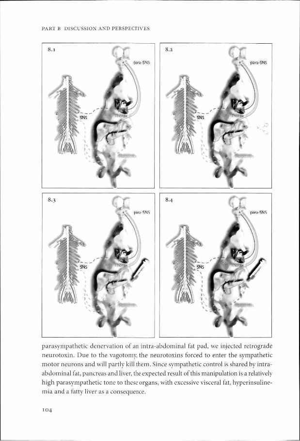
parasympathetic denervation of an intra-abdominal fat pad, we injected retrograde
neurotoxin. Due to the vagotomy, the neurotoxins forced to enter the sympathetic
motor neurons and will partly kill them. Since sympathetic control is shared by intra
abdominal fat, pancreas and liver, the expected result of this manipulation is a relatively
high parasympathetic tone to these organs, with excessive visceral fat, hyperinsuline-
mia and a fatty liver as a consequence.
104

CHAPTER 8 PERSPECTIVES FOR FOLLOW-UP STUDIES
Figure 8.1 The abdominal compartment receives both sympathetic and parasympathetic shared input via the sympathetic and parasympathetic nervous system (chapter 2, 3 and 4). We assume that the thorax compartment receives sympathetic and parasympathetic input from a separate group of neurons (chapter 6).
Figure 8.2 The retroperitoneal fat pad is vagotomized (see for technique supplemental material in chapter 3).
Figure 8.3 Neurotoxin conjugated to a non-transneuronal retrograde tracer (e.g. saporin - CTB) is injected into the vagotomized fat pad.
Figure 8.4 Since the fat pad is vagotomized and the neurotoxin has access to the sympathetic input only, the motor neurons in the peripheral sympathetic ganglion projecting to the abdomen will be killed.
Figure 8.5 Since the abdominal organs share neuronal control, the sympathetic input to the abdomen will be decreased and the net effect will be a parasympathetic overweight in the abdominal cavity, but not in the thorax compartment. If our hypothesis is right, the animal should develop visceral obesity in the intact abdominal fat pads, hyperglycemia and hyper-insulinemia. See color section.
Two types of retrograde neurotoxins have been studied: Cholera toxin B / saporin
conjugate (CTB-SAP) and Pseudorabies Virus gl l (PRV-gll). In a first step we aimed
to demonstrate the presents of retrograde neurotoxins in the CNS with sympathetic
or parasympathetic and without surgical denervation.
CTB is a neuronal tracer that fills autonomic motor neurons after injection into an
organ and can be visualized in the CNS by immunohistochemistry; saporin is a ri-
105
PART B DISCUSSION AND PERSPECTIVES
bosome inactivating neurotoxin53,54. Saporin conjugated to a retrograde tracer, such as
CTB, has been used in to retrogradely kill autonomic motor neurons in rodents" 55~57.
We applied several types of CTB-SAP that were conjugated in our lab and by com
mercial suppliers. We could not detect CTB in the CNS with immunohistochemical
techniques, nor did we find changes in glucose tolerance test after CTB-SAP treatment.
In a next step, we tested a different retrograde neurotoxin. PRV-gll is a swine neuro
trophic a herpes virus that infects the synapse, is being retrogradely transported and
replicates in the cell body until the neuron disintegrates. In contrast to PRV-bartha, a
transneuronal tracer, PRV-gll has not the ability to enter a neighboring neuron and
spread upstream through the CNS586". Therefore, PRV-gll is a neuroanatomically a
marker and physiologically a neurotoxin at the same time. In a similar approach to
CTB-SAP, we injected PRV-gll into abdominal organs. Here, sympathetic denervation
was chosen because it forces the virus to the DMV where the presence or neuronal
damage is easier to establish than in the celiac ganglion. Using immunohistochemistry
staining against PRV, we could not detect PRV-gll in the CNS.
Transport of CTB-SAP and PRV-gll has been described after dipping in cut nerve
endings or intra-neuronal injections52,56'58. To develop successful retrograde transport
from intra-abdominal fat tissue, the percentage of neurotoxin as well as the medium in
the dilution might be changed. Alternatively, a more aggressive variant of PRV could
be used, with other insertions and deletions.
In summary, further experiments with different doses or compounds are necessary
to test the physiological impact of the shared neuronal control of the intra-abdominal
cavity. Since we did not succeed to have central labeling of a retrograde neurotoxin,
we cannot verify or reject the hypothesis about the involvement of the ANS in type 2
diabetes.
How can we test the hypothesis of an arrhythmic biological clock and an
unbalanced ANS as the cause of the metabolic syndrome in humans?
In patients, the circadian rhythms and ANS function should be assessed in prospec
tive cohort studies. If circadian and autonomic malfunctioning is present before the
symptoms of the metabolic syndrome develop, our hypothesis would be supported.
Until now, prospective cohort studies show an strong association between disturbed
sleep, ANS function and the development of the metabolic syndrome36,48,61"66.
How could we treat the metabolic syndrome based on this hypothesis?
The reintroduction of physiological circadian rhythmicity with high maximum energy
intake and utilization in the morning and sufficient sleeping time should ameliorate the
metabolic syndrome, as discussed in chapter 6. Treatment with drugs that emphasize
the natural rhythmicity such as melatonin might have important additional value67.
106

CHAPTER 8 PERSPECTIVES FOR FOLLOW-UP STUDIES
PERSPECTIVE #2 HIV-related l ipodyst rophy migh t be a brain disease
(chapter 7)
Patients with the HIV-1-associated adipose redistribution syndrome (HARS) suffer from visceral fat accumulation, peripheral fat atrophy and features of the metabolic syndrome. We speculate that this unbalance might be a result of a high parasympathetic tone to the visceral fat tissue and high sympathetic tone to peripheral fat tissue due to selective neurotoxicity of the HIV-medication, possibly in combination with the HIV virus itself.
Follow-up studies
Is autonomic function disturbed in HARS?
An autonomic test battery is needed to evaluate the autonomic status of these patients (see finding #2).
Does HIV medication has an neurotoxic effect on (pre-)sympathetic or (pre-)parasympathetic neurons projecting to fat tissue?
In an animal model, the effect of HIV medication on the brain could be tested by comparing peripheral and intra-cranial administration, followed by measurement of body fat distribution by MRI, assessment of autonomic tone, and histological analysis of (pre)autonomic neurons projecting to fat tissue. These experiments will show if the drugs indeed have a central effect on body fat distribution that is mediated by the ANS.
REFERENCES
1. Barinaga, M. "Obese"protein slims mice. Science 269, 475-6 (1995).
2. Campfield, L. A., Smith, F. J., Guisez, Y., Devos, R. & Burn, P. Recombinant mouse OB protein: evidence for a peripheral signal linking adiposity and central neural networks. Science 269, 546-9 (i995)-
3. Haiaas, J. L. et al. Weight-reducing effects of the plasma protein encoded by the obese gene. Science 269, 543-6(i995)-
4. Pelleymounter, M. A. et al. Effects of the obese gene product on body weight regulation in ob/ob mice. Science 269, 540-3 (1995).
5. Bartness, T. J. & Bamshad, M. Innervation of mammalian white adipose tissue: implications for the regulation of total body fat. Am J Physiol 275, R1399-411 (1998).
6. Bjorntorp, P. Hormonal control of regional fat distribution. Hum Reprod 12 Suppl 1, 21-5.
(i997)-
7. Bouchard, C , Despres, J. P. & Mauriege, P. Genetic and nongenetic determinants of regional fat distribution. Endocr Rev 14, 72-93 (1993).
8. Wajchenberg, B. L. Subcutaneous and visceral adipose tissue: their relation to the metabolic syndrome. Endocr Rev 21, 697-738. (2000).
9. Cantu, R. C. & Goodman, H. M. Effects of denervation and fasting on white adipose tissue. Am J Physiol 212, 207-12 (1967).
107

PART B DISCUSSION AND PERSPECTIVES
10. Rothwell, N. J. & Stock, M. J. A role for brown adipose tissue in diet-induced thermogenesis. Nature 281, 31-5 (1979)-
11. Brooks, S. L., Rothwell, N. J., Stock, M. J., Goodbody, A. E. & Trayhurn, R Increased proton conductance pathway in brown adipose tissue mitochondria of rats exhibiting diet-induced thermogenesis. Nature 286, 274-6 (1980).
12. Perkins, M. N., Rothwell, N. J., Stock, M. J. & Stone, T. W. Activation of brown adipose tissue thermogenesis by the ventromedial hypothalamus. Nature 289, 401-2 (1981).
13. Trayhurn, P. & Ashwell, M. Control of white and brown adipose tissues by the autonomic nervous system. Proc Nutr Soc 46, 135-42 (1987).
14. Dal/.iel, K. The nervous system and adipose tissue. Clin Dermatol 7, 62-77 (1989). 15. Bamshad, M., Aoki, V. T., Adkison, M. G., Warren, W. S. & Bartness, T.). Central nervous system
origins of the sympathetic nervous system outflow to white adipose tissue. Am J Physiol 275, R291-9 (1998).
16. Scheer, F. A., Ter Horst, G. J., van Der Vliet, J. & Buijs, R. M. Physiological and anatomic evidence for regulation of the heart by suprachiasmatic nucleus in rats. Am J Physiol Heart Circ Physiol
280, H1391-9. (2001). 17. Buijs, R. M. & Kalsbeek, A. Hypothalamic integration of central and peripheral clocks. Nat Rev
Neurosci 2, 521-6 (2001).
18. Kalsbeek, A. & Strubbe, J. H. Circadian control of insulin secretion is independent of the temporal distribution of feeding. Physiol Behav 63, 553-8. (1998).
19. La Fleur, S. E., Kalsbeek, A., Wortel, J. & Buijs, R. M. A suprachiasmatic nucleus generated rhythm in basal glucose concentrations. J Neuroendocrinol 11, 643-52. (1999).
20. la Fleur, S. E., Kalsbeek, A., Wortel, J., Fekkes, M. L. & Buijs, R. M. A daily rhythm in glucose tolerance: a role for the suprachiasmatic nucleus. Diabetes 50,1237-43 (2001).
21. Ruiter, M. et al. The daily rhythm in plasma glucagon concentrations in the rat is modulated by the biological clock and by feeding behavior. Diabetes 52, 1709-15 (2003).
22. la Fleur, S. E., Kalsbeek, A., Wortel, J. & Buijs, R. M. Polysynaptic neural pathways between the hypothalamus, including the suprachiasmatic nucleus, and the liver. Brain Res 871, 50-6 (2000).
23. Buijs, R. M., Chun, S. J., Niijima, A., Romijn, H. J. & Nagai, K. Parasympathetic and sympathetic control of the pancreas: A role for the suprachiasmatic nucleus and other hypothalamic centers that are involved in the regulation of food intake. J Comp Neurol 431, 405-23. (2001).
24. Kalsbeek, A., Fliers, E., Franke, A. N., Wortel, J. & Buijs, R. M. Functional connections between the suprachiasmatic nucleus and the thyroid gland as revealed by lesioning and viral tracing techniques in the rat. Endocrinology 141, 3832-41. (2000).
25. Buijs, R. M. et al. Anatomical and functional demonstration of a multisynaptic suprachiasmatic nucleus adrenal (cortex) pathway. Eur J Neurosci 11,1535-44 (1999).
26. Kreier, F. et al. Selective parasympathetic innervation of subcutaneous and intra-abdominal fat -functional implications.} Clin Invest 110, 1243-1250 (2002).
27. Horvath, S., Kis, Z., Boldogkoi, Z., Nogradi, A. & Toldi, J. Oestrogen-dependent tracing in the rat CNS after pseudorabies virus infection. Eur J Neurosci 15, 937-43 (2002).
28. Lee, J. W. & Erskine, M. S. Pseudorabies virus tracing of neural pathways between the uterine cervix and CNS: effects of survival time, estrogen treatment, rhizotomy, and pelvic nerve transection. J Comp Neurol 418, 484-503. (2000).
29. Cinti, S. Anatomy of the adipose organ. Eat Weight Disord 5, 132-42 (2000). 30. van der Velden, V. H. & Hulsmann, A. R. Autonomic innervation of human airways: structure,
function, and pathophysiology in asthma. Neuroimmunomodulation 6, 145-59 (i999)-31. Onuoha, G. N., Alpar, E. K., Chukwulobelu, R. & Nicholls, D. P. Distributions of VIP, substance
P, neurokinin A and neurotensin in rat heart: an immunocytochemical study. Neuropeptides 33, 19-25 (1999).
108

CHAPTER 8 PERSPECTIVES FOR FOLLOW-UP STUDIES
32. Giordano, A., Frontini, A., Castellucci, M. & Cinti, S. Presence and distribution of cholinergic nerves in rat mediastinal brown adipose tissue. J Histochem Cytochem 52, 923-30 (2004).
33. Latour, M. G. & Lautt, W. W. The hepatic vagus nerve in the control of insulin sensitivity in the rat. Auton Neurosci 95, 125-30 (2002).
34. Ueno, T. et al. Distribution of substance P and vasoactive intestinal peptide in the human liver: light and electron immunoperoxidase methods of observation. Am J Gastroenterol 86, 1633-7 (i99i)-
35. McCuskey, R. S. Anatomy of efferent hepatic nerves. Anat Rec A Discov Mol Cell Evol Biol 280, 821-6 (2004).
36. Frontoni, S. et al. Early autonomic dysfunction in glucose-tolerant but insulin-resistant offspring of type 2 diabetic patients. Hypertension 41, 1223-7 (2003).
37. Yardley, C. R & Hilton, S. M. Vasodilatation in hind-limb skeletal muscle evoked as part of the defence reaction in the rat. J Auton Nerv Syst 19, 127-36 (1987).
38. Musch, T. I. et al. Regional distribution of blood flow of dogs during graded dynamic exercise. J Appl Physiol 63, 2269-77 (1987).
39. Rosell, S. & Belfrage, E. Blood circulation in adipose tissue. Physiol Rev 59, 1078-1104. (1979). 40. Ngai, S. H., Rosell, S. & Wallenberg, L. R. Nervous regulation of blood flow in the subcutaneous
adipose tissue in dogs. Acta Physiol Scand 68, 397-403 (1966).
41. Gutenbrunner, C , El-Cherid, A., Gehrke, A. & Fink, M. Orcadian variations in the responsiveness of human gallbladder to sulfated mineral water. Chronobiol Int 18, 1029-39 (2001).
42. Richard, C , Thuillez, C , Depret, J., Auzepy, P. & Giudicelli, J. F. Regional hemodynamic effects of perindopril in congestive heart failure. Am Heart J 126, 782-8 (1993).
43. Herath, C. B., Reynolds, G. W., MacKenzie, D. D., Davis, S. R. & Harris, P. M. Vagotomy suppresses cephalic phase insulin release in sheep. Exp Physiol 84, 559-69 (1999).
44. Gupta, S., Elder,}. B. & Kay, A. W. Exocrine secretory responses of the pancreas to insulin and to a standard meal in dogs. Br J Surg 58, 857-8 (1971).
45. Kaneoke, Y. et al. Gastrointestinal dysfunction in Parkinson's disease detected by electrogastroen-terography.J Auton Nerv Syst 50, 275-81 (1995).
46. Porksen, N. Early changes in beta-cell f unction and insulin pulsatility as predictors for type 2 diabetes. Diabetes Nutr Metab 15, 9-14 (2002).
47. Cummings, J. H., Jenkins, D. J. & Wiggins, H. S. Measurement of the mean transit time of dietary residue through the human gut. Gut 17, 210-8 (1976).
48. Gangwisch, J. E. & Heymsfield, S. B. in NAASO (2005).
49. Turek, F. W. et al. Obesity and metabolic syndrome in circadian Clock mutant mice. Science 308, 1043-5 (2005).
50. Czupryniak, L., Strzelczyk, J., Pawlowski, M. & Loba, J. Circadian blood pressure variation in morbidly obese hypertensive patients undergoing gastric bypass surgery. Am J Hypertens 18, 446-51 (2005).
51. Kotsis, V. et al. Impact of obesity on 24-hour ambulatory blood pressure and hypertension. Hypertension 45, 602-7 (2005).
52. Llewellyn-Smith, I. J., Martin, C. L., Arnolda, L. F. & Minson, J. B. Tracer-toxins: cholera toxin B-saporin as a model. J Neurosci Methods 103, 83-90. (2000).
53. Hyland, N. R, Abrahams, T. P., Fuchs, K., Burmeister, M. A. & Hornby, P. J. Organization and neurochemistry of vagal preganglionic neurons innervating the lower esophageal sphincter in ferrets. J Comp Neurol 430, 222-34 (2001).
54. Fordham-Skelton, A. P., Taylor, P. N., Hartley, M. R. & Croy, R. R. Characterisation of saporin genes: in vitro expression and ribosome inactivation. Mol Gen Genet 229, 460-6 (1991).
55. Wiley, R. G. Neural lesioning with ribosome-inactivating proteins: suicide transport and immu-nolesioning. Trends Neurosci 15, 285-90. (1992).
56. Llewellyn-Smith, I. J., Martin, C. L., Arnolda, L. F. & Minson, J. B. Retrogradely transported CTB-saporin kills sympathetic preganglionic neurons. Neuroreport 10, 307-12. (1999).
109

PART B DISCUSSION AND PERSPECTIVES
57. Wiley, R. G. & Kline, I. V. Neuronal lesioning with axonally transported toxins. J Neurosci Methods 103, 73-82. (2000).
58. Babic, N., Mettenleiter, T. C , Flamand, A. & Ugolini, G. Role of essential glycoproteins glI and gp$o in transneuronal transfer of pseudorabies virus from the hypoglossal nerves of mice. J Virol 67,4421-6(1993).
59. Jansen, A. S., Nguyen, X. V, Karpitskiy, V, Mettenleiter, T. C. & Loewy, A. D. Central command neurons of the sympathetic nervous system: basis ofthefight-or-flight response. Science 270, 644-6
(i995). 60. Sams, J. M., Jansen, A. S., Mettenleiter, T. C. & Loewy, A. D. Pseudorabies virus mutants as tran
sneuronal markers. Brain Res 687,182-90. (1995). 61. Meigs, J. B., Cupples, L. A. & Wilson, P. W. Parental transmission of type 2 diabetes: the Framing-
ham Offspring Study. Diabetes 49, 2201-7. (2000).
62. Ayas, N. T. et al. A prospective study of self-reported sleep duration and incident diabetes in women. Diabetes Care 26, 380-4 (2003).
63. Schroeder, E. B. et al. Diabetes, glucose, insulin, and heart rate variability: the Atherosclerosis Risk in Communities (ARIC) study. Diabetes Care 28, 668-74 (2005).
64. Carnethon, M. R., Jacobs, D. R., Jr., Sidney, S. & Liu, K. Influence of autonomic nervous system dysfunction on the development of type 2 diabetes: the CARD1A study. Diabetes Care 26, 3035-41 (2003).
65. I.indmark, S., Wiklund, U., Bjerle, P. & Eriksson, J. W. Does the autonomic nervous system play a role in the development of insulin resistance? A study on heart rate variability in first-degree relatives of Type 2 diabetes patients and control subjects. Diabet Med 20, 399-405 (2003).
66. Carnethon, M. R., Golden, S. EL, Folsom, A. R., Haskell, W. & Liao, D. Prospective investigation of autonomic nervous system function and the development of type 2 diabetes: the Atherosclerosis Risk In Communities study, 1987-1998. Circulation 107, 2190-5 (2003).
67. Scheer, E A., Van Montfrans, G. A., van Someren, E. J., Mairuhu, G. & Buijs, R. M. Daily nighttime melatonin reduces blood pressure in male patients with essential hypertension. Hypertension 43, 192-7 (2004).
110