Embed Size (px)
Citation preview
UvA-DARE is a service provided by the library of the University of Amsterdam (http://dare.uva.nl)
UvA-DARE (Digital Academic Repository)
Cytoreduction and hyoerthermic intraperitoneal chemotherapy in peritonealcarcinomastosis of colorectal originVerwaal, V.J.
Link to publication
Citation for published version (APA):Verwaal, V. J. (2004). Cytoreduction and hyoerthermic intraperitoneal chemotherapy in peritonealcarcinomastosis of colorectal origin
General rightsIt is not permitted to download or to forward/distribute the text or part of it without the consent of the author(s) and/or copyright holder(s),other than for strictly personal, individual use, unless the work is under an open content license (like Creative Commons).
Disclaimer/Complaints regulationsIf you believe that digital publication of certain material infringes any of your rights or (privacy) interests, please let the Library know, statingyour reasons. In case of a legitimate complaint, the Library will make the material inaccessible and/or remove it from the website. Please Askthe Library: http://uba.uva.nl/en/contact, or a letter to: Library of the University of Amsterdam, Secretariat, Singel 425, 1012 WP Amsterdam,The Netherlands. You will be contacted as soon as possible.
Download date: 03 May 2018
Chapterr two
Randomizedd trial of cytoreduction, hyperthermic
intraperitoneall chemotherapy and systemic chemotherapy
versuss systemic chemotherapy and palliative surgery in
patientss with peritoneal carcinomatosis of colorectal cancer
Vicc J. Verwaai1, Serge van Ruth1, Eelco de Bree1, Gooike W van Slooten1,
Harmm van Tinteren2, Henk Boot3 and Frans A.N. Zoetmulder1
11 Department of Surgery, 2 Department of Biometrics, 33 Department of Gastroenterology
Thee Netherlands Cancer Institute - Antoni van Leeuwenhoek Hospital
Amsterdam,, the Netherlands
Purpose:Purpose: To confirm the findings from uncontrolled studies that aggressive cytoreduction in combination withh hyperthermic intraperitoneal chemotherapy (HIPEC) is superior to standard treatment in patients with peritoneall carcinomatosis of colorectal cancer origin.
PatientsPatients and Methods: Between February 1998 and August 2001, 105 patients were randomly assigned to re-ceivee either standard treatment consisting of systemic chemotherapy (fluorouracil-leucovorin) with or with-outt palliative surgery, or experimental therapy consisting of aggressive cytoreduction with HIPEC, followed byy the same systemic chemotherapy regime. The primary end point was survival.
Results:Results: After a median follow-up period of 21.6 months, the median survival was 12.6 months in the stan-dardd therapy arm and 22.3 months in the experimental therapy arm (log-rank test, P - 0.032). The treat-ment-relatedd mortality in the aggressive therapy group was 8%. Most complications from HIPEC were re-latedd to bowel leakage. Subgroup analysis of the HIPEC group showed that patients with 0 to 5 of the 7 regionss of the abdominal cavity involved by tumor at the time of the cytoreduction had a significantly better survivall than patients with 6 or 7 affected regions (log-rank test, P < 0.0001). If the cytoreduction was macroscopicallyy complete (R-l), the median survival was also significant better than in patients with limited (R-2a),, or extensive residual disease (R-2b; log-rank test, P < 0.0001).
Conclusion:Conclusion: Cytoreduction followed by HIPEC improves survival in patients with peritoneal carcinomatosis off colorectal origin. However, patients with involvement of six or more regions of the abdominal cavity, orr grossly incomplete cytoreduction, had still a grave prognosis. JournalJournal of Clinical Oncology 2003; 21:3737-3743.
15 5

16 6
Randomizedd trial
Introduction n
Peritoneall carcinomatosis (PC) of colorectal origin is common and is the second-most frequent
causee of death in colorectal cancer after metastatic disease to the liver. In an estimated 25% of
patients,, no other tumor locations can be found, even when a detailed diagnostic work-up is per-
formed.11 3 Sugarbaker45 has suggested that PC of colorectal origin should probably not be equated
withh generalized disease, but can be a first step of dissemination, not unlike the situation with liver
metastasess of colorectal origin. ,
Basedd on this concept, attempts have been made to achieve long-term survival in patients with
PCC by combining surgery and intraperitoneal chemotherapy to eradicate microscopic residual dis-
ease.. Advances in surgical techniques and improved anesthesiology have made it possible to re-
movee most or all macroscopic tumor in PC.6 In theory, intraperitoneal chemotherapy could eradi-
catee limited residual tumor and should have an optimal chance to succeed if it would immediately
followw surgery (to avoid regrowth of tumor cells), and if exposure of the peritoneal surface at risk
couldd be guaranteed. To achieve these goals, peritoneal lavage, as part of the surgical procedure,
hass been developed.7 Others and our group have shown that peritoneal lavage containing Mitomy-
cinn C (MMC) results in a drug exposure to the peritoneal surface that is 20 times higher than else-
wheree in the body.8-9 This degree of pharmacokinetic advantage is thought to result in optimal cir-
cumstancess for tumor cell kill . In addition, enhancement of MMC cytotoxity at temperatures
higherr than 39°C has been demonstrated in animals and in vitro models.10-11 The addition of intra-
peritoneall hyperthermia has been shown to be technically feasible in the surgical setting.12-13 This
approachh of aggressive cytoreduction in combination with intraperitoneal chemotherapy, often em-
ployingg MMC and hyperthermia, has been studied in 11 phase II studies on patients with PC of co-
lorectall origin.12-1423 The results of these studies show a strikingly long median survival and, more
importantly,, a 20% to 30% long-term (5-year) survival rate, reminiscent of that of surgery for iso-
latedd liver metastases. It has been advocated that these favorable results justify such an aggressive
treatment,, particularly since long-term survival is hardly ever seen after systemic chemodierapy
alone.. It remains to be shown that these encouraging results of uncontrolled studies are not the re-
sultt of patient selection. The need for a controlled study was recently re-emphasized in an editorial
inn the Journal of Clinical Oncology by Sugarbaker,24 who originally pioneered the approach. This
needd is particularly urgent because HIPEC is associated with significant morbidity and treatment-
relatedd mortality. In this article, we report the results of a randomized single-institution phase II I
study,, and present data that may aid in better selection of patients for aggressive treatment of peri-
toneall carcinomatosis.
Patientss and methods
PatientPatient Selection and Study Design
Patientss with histologically proven peritoneal metastases of colorectal adenocarcinoma (CRC) or
positivee cytology of ascites, who were diagnosed either at first presentation or at recurrence of
CRC,, were eligible. No signs of distant metastases (liver, lung) on computed tomography (CT-scan)
17 7
Chapterr 2
off abdomen and chest x-ray were allowed. Patients had to be younger than 71 years and fit for ma-
jorr surgery (normal bone marrow indices, and normal renal and liver functions). Initially, patients
whoo had received fluorouracil (FU) within 12 months before random assignment were excluded. In
thee first year of the study, an amendment to the protocol was made to allow inclusion of these pa-
tients. .
Patientss were randomly allocated to either standard treatment or to the experimental treatment.
Thee randomization was performed centrally by computer, and stratified for presentation (primary
orr recurrence) and site (appendix, colon or rectum). The medical ethical committee of the Nether-
landss Cancer Institute approved the study, and written informed consent was obtained from all pa-
tients. .
StandardStandard Treatment
Surgeryy was only performed in cases of symptoms of intestinal obstruction, and consisted of ei-
therr bypass or stoma surgery. Often, this type of surgery had already been performed before refer-
rall for random assignment. Patients started chemotherapy immediately after random assignment or
afterr recovery from surgery. Chemotherapy was given in the local setting, usually by the patients'
ownn medical oncologist, and consisted of FU (intravenous (IV) push-dose of 400 mg/m2) and leu-
covorinn (TV 80 mg/m2) on an outpatient basis (modified Laufman regimen).25 Treatment was given
weeklyy for 26 weeks, or until progression, death, or unacceptable toxicity. Patients who had already
beenn treated with FU within 12 months before random assignment were treated with irinotecan
(3500 mg/m2) at three-weekly intervals for six months or until progression or intolerable toxicity.
ExperimentalExperimental Treatment CytoreductiveCytoreductive surgery
Alll procedures were carried out in the Netherlands Cancer Institute. Laparotomy under general
anesthesiaa was performed from xyphoid to pubis. After opening the abdomen, the presence of
macroscopicc tumor deposits was recorded in seven abdominal regions: pelvis and sigmoid; right
lowerr abdomen; small bowel and mesentery; omentum and transverse colon; subhepatic space and
stomach;; right subphrenic space; and left subphrenic space. The maximal tumor size was recorded
inn each region as: none, less than one cm, one to five cm, or more than five cm.
Thee objective of cytoreduction was to leave no macroscopic tumor behind, or at least to have
limitedd residual tumor (< 2.5 mm in thickness). To achieve this, the stripping of the parietal perito-
neumm was carried out as described by Sugarbaker et al.26 Infiltrated viscera were resected if this was
compatiblee with retaining function. Most often this concerned the rectum, parts of small bowel and
colon,, the gall bladder, parts of the stomach, and the spleen. The greater omentum was routinely
removed.. Reconstruction of gastrointestinal continuity was postponed until after the lavage, to pre-
ventt entrapment of tumor cells in suture lines. At completion of cytoreduction, the absence of re-
siduall tumor was recorded as R-l. If the largest residual tumor was smaller than 2.5 mm, it was re-
gardedd as an R-2a resection. In cases of residual tumor larger than 2.5 mm, cytoreductive surgery
wass scored as R-2b. The total length of the operation, and blood loss were also recorded.
18 8
Randomizedd trial
HyperthermicHyperthermic intraperitoneal chemotherapy (HIPEC)
Too increase the volume of the abdominal cavity and to prevent spillage of lavage fluid, the skin of
thee laparotomy wound was pulled up against a retractor. A plastic sheet covered the laparotomy
openingg to reduce heat loss and to avoid drug spilling. A central aperture was made to allow ma-
nipulationn to achieve optimal drug and heat distribution. The perfusion circuit consisted of a cen-
trallyy placed inflow catheter, outflow cadieters, placement in the pelvis below left and right dia-
phragm,, a roller pump, and a heat exchanger. Temperature probes were attached to inflow and
outfloww catheters. Perfusion was started with a minimum of three liter of isotonic dialysis fluid, at
onee to two 1/min, and an inflow temperature of 41 °C to 42°C. As soon as the temperature in die
abdomenn was stable above 40°C, MMC was added to the perfusate at a dose of 17.5 mg/m2 fol-
lowedd by 8.8 mg/m2 every 30 minutes. The total dose was limited to 70 mg at maximum. If the
coree temperature exceeded 39°C, the inflow temperature was reduced. After 90 minutes, the perfu-
sionn fluid was drained from the abdomen, and bowel continuity was restored. A temporary colos-
tomyy was made in most cases if the rectum was resected. A draining gastrostomy and transgastric
jejunall feeding tube were inserted. The outflow catheters were used for postoperative drainage of
thee abdomen cavity.
PostoperativePostoperative Care
Patientss stayed in the intensive care unit for three days. In cases of abdominal sepsis (faecal flora
inn drain fluid, high fever, sepsis), a laparotomy was performed to correct bowel leakage at an early
stage.Jejunall tube feeding was begun on day 1. Parenteral nutrition was given until jejunal feeding
couldd cover all nutritional needs. Oral fluid and food intake was resumed as soon as the gas-
trostomyy production dropped below 500 raL per 24 hours.
AdjuvantAdjuvant Chemotherapy
Systemicc chemotherapy was intended to start after six weeks beyond cytoreduction followed by
HIPEC,, and before three months after cytoreduction followed by HIPEC. The regimens as de-
scribedd in the standard therapy arm were used.
Toxicity'/ComplicaToxicity'/Complica tions
Chemotherapy-relatedd toxicity was recorded using the World Health Organization (WHO) scale.
Al ll postoperative complications were noted, and were graded as toxicity according to the WHO
scale. .
Follow-Up Follow-Up
Alll patients were seen at the outpatient clinic once every three months for two years, and every
sixx months thereafter. The follow-up consisted of physical examination and serum CEA every
threee months and an abdominal CT scan of the abdomen every six months, starting three months
afterr randomization in the standard arm and three months after cytoreduction followed by HIPEC
inn the experimental arm.
StatisticalStatistical Analysis
Thee main end point was survival, measured as time from randomization to death from any cause.
19 9
Chapterr 2
Patientss alive at the time of analysis were censored at their last follow-up examination. The survival
wass estimated by the Kaplan Meier method and tested with the log-rank test following the inten-
tion-to-treatt principle. The analysis was planned at a median follow-up of two years to have 80%
powerr to detect a 20% absolute difference in survival. To detect this difference, with P<0.05 (two-
tailedd test), at least 100 patients had to be entered.
Too improve patient selection in the future, additional exploratory analyses were performed to
identifyy potential prognostic factors. Presentation (primary v recurrence), site (appendix vs colon vs
rectum),, number of regions involved (<5 regions vs >5 regions), and completeness of cytoreduc-
tionn (R-l vs R-2a vs R-2b) were included in a Cox proportional hazards regression model in order
too obtain hazard ratios and 95% confidence intervals. All P values are two-sided.
Results s
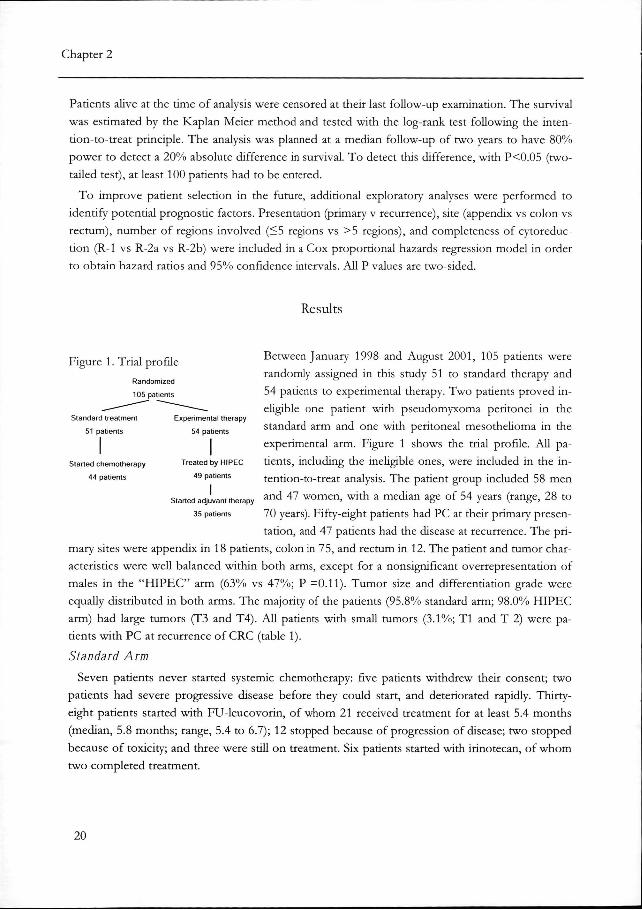
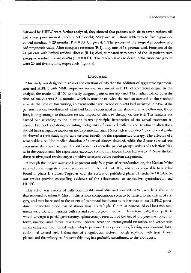
Figuree 1. Trial profile
Randomized d
105 5
Standardd treatment
511 patients
I I I I
Startedd chemotherapy 444 patients
3at t ents s
E E xperimentall therapy
544 patients
l l I I
Treatedd by HIPEC 499 patients
I I
Betweenn January 1998 and August 2001, 105 patients were
randomlyy assigned in this study 51 to standard therapy and
544 patients to experimental therapy. Two patients proved in-
eligiblee one patient with pseudomyxoma peritonei in the
standardd arm and one with peritoneal mesothelioma in the
experimentall arm. Figure 1 shows the trial profile. All pa-
tients,, including the ineligible ones, were included in the in-
tention-to-treatt analysis. The patient group included 58 men
startedd adjuvant therapy a nd 4 7 women, with a median age of 54 years (range, 28 to
355 patients 70 years). Fifty-eight patients had PC at their primary presen-
tation,, and 47 patients had the disease at recurrence. The pri-
maryy sites were appendix in 18 patients, colon in 75, and rectum in 12. The patient and tumor char-
acteristicss were well balanced within both arms, except for a nonsignificant overrepresentation of
maless in the "HIPEC" arm (63% vs 47%; P =0.11). Tumor size and differentiation grade were
equallyy distributed in both arms. The majority of the patients (95.8% standard arm; 98.0% HIPEC
arm)) had large tumors (T3 and T4). All patients with small tumors (3.1%; Tl and T 2) were pa-
tientss with PC at recurrence of CRC (table 1).
StandardStandard Arm
Sevenn patients never started systemic chemotherapy: five patients withdrew their consent; two
patientss had severe progressive disease before they could start, and deteriorated rapidly. Thirty-
eightt patients started with FU-leucovorin, of whom 21 received treatment for at least 5.4 months
(median,, 5.8 months; range, 5.4 to 6.7); 12 stopped because of progression of disease; two stopped
becausee of toxicity; and three were still on treatment. Six patients started with irinotecan, of whom
twoo completed treatment.
20 0
Randomizedd trial
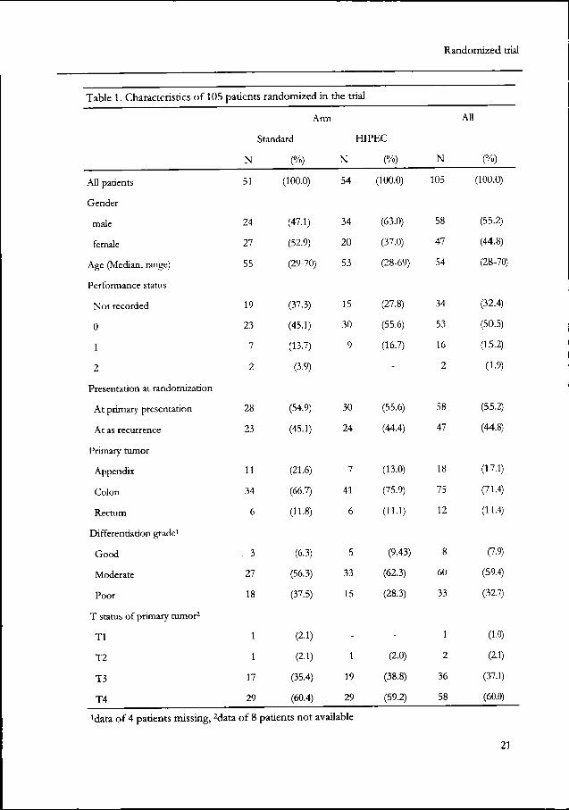
Tablee 1. Characteristics of 105
Al ll patients
Gender r
male e
female e
Agee (Median, range)
Performancee status
Nott recorded
0 0
1 1
2 2
Presentationn at randomization
Att primary presentation
Att as recurrence
Primaryy tumor
Appendix x
Colon n
Rectum m
Differentiationn grade1
Good d
Moderate e
Poor r
TT status of primary tumor2
Tl l
T2 2
T3 3
T4 4
patients s randomizedd in
Arm m
Standard d
N N
51 1
24 4
27 7
55 5
19 9
23 3
7 7
2 2
28 8
23 3
11 1
34 4
6 6
.. 3
27 7
18 8
1 1
1 1
17 7
29 9
(%) )
(100.0) )
(47.1) )
(52.9) )
(29-70) )
(37.3) )
(45.1) )
(13.7) )
(3.9) )
(54.9) )
(45.1) )
(21.6) )
(66.7) )
(11.8) )
(6.3) )
(56.3) )
(37.5) )
(2.1) )
(2.1) )
(35.4) )
(60.4) )
thee trial
HIPEC C
N N
54 4
34 4
20 0
53 3
15 5
30 0
9 9
--
30 0
24 4
7 7
41 1
6 6
5 5
33 3
15 5
--
1 1
19 9
29 9
(%) )
(100.0) )
(63.0) )
(37.0) )
(28-69) )
(27.8) )
(55.6) )
(16.7) )
--
(55.6) )
(44.4) )
(13.0) )
(75.9) )
(11.1) )
(9.43) )
(62.3) )
(28.3) )
--
(2.0) )
(38.8) )
(59.2) )
N N
105 5
58 8
47 7
54 4
34 4
53 3
16 6
2 2
58 8
47 7
18 8
75 5
12 2
8 8
60 0
33 3
1 1
2 2
36 6
58 8
Al l l
(%) )
(100.0) )
(55.2) )
(44.8) )
(28-70) )
(32.4) )
(50.5) )
(15.2) )
(1.9) )
(55.2) )
(44.8) )
(17.1) )
(71.4) )
(11.4) )
(7.9) )
(59.4) )
(32.7) )
(1.0) )
(2.1) )
(37.1) )
(60.0) )
'dataa of 4 patients missing, 2data off 8 patients not available
21 1

Chapterr 2
ExperimentalExperimental Arm
Fivee patients did not undergo cytoreduction followed by HIPEC treatment. While waiting for
surgery,, one died due to rapid tumor progression; two patients developed lung and liver metastases,
forr which they were treated with palliative chemotherapy; and in one patient, a primary lung cancer
wass detected shortly after randomization. This patient died shortly after randomization. One pa-
tientt withdrew consent. The median time between randomization and surgery was six weeks (range,
66 days to 14 weeks). The median hospital stay of the 49 patients operated on was 29 days (range, 6
too 166 days). The median duration of the cytoreduction and HIPEC was 485 minutes (range, 315
too 765 minutes), while the median blood loss was 3.9 L (range, 0.5 to 30.0 L; for seven patients,
dataa were not available). Of the seven possible affected regions, six or seven were involved in 16
patients.. Those patients had a median operation time of 585 minutes (range, 440 to 765 minutes)
andd a median blood loss of 6.0 L (range, 3.5 to 30.0 L). In two patients, no macroscopic tumor was
foundd at all, and peritoneal metastases had been resected at a previous laparotomy. Both received
HIPECC without cytoreduction. The median hospital admission duration was 23 days (range, 13 to
900 days) for zero to five affected regions, and 38 days (range, 6 to 166 days) for six to seven re-
gions. .
Ann average of 1.8 visceral resections were performed per patient. Most often, parts of small
bowell (45 patients) and rectum (25 patients) were resected. Twenty-four patients needed a colos-
Tablee 2. Major toxicity and complications of 48 patients1 with peritoneal carcinomatosis treated by cytoreductionn plus HIPEC
gradee 3 grade 4
33 (6%)
77 (15%) 1 (2%)
22 (4%)
22 (4%)
11 (2%)
22 (4%)
11 (2%)
33 (6%)
22 (4%)
11 (2%)
44 (8%) 2 (4%)
77 (15%)
11 (2%)
33 (6%)
33 (6%) 4 (8%)
33 (6%) 2 (4%)
dataa of complications missing in one patient, GI: gastro-intestinal
22 2
Fever r
Leukopenia a
Thrombocytopenia a
Neuropathyy (paresis)
Pleurall effusion
Pulmonaryy embolus (within 3 months after surgery)
Pneumonia a
Anuriaa (acute tubular necrosis)
Renall obstruction
Cardiacc arrhythmia
Heartt failure
GII Fistula
Pancreatitis s
Catheterr infections
Haemorrhage e
Psychologicall disorders

Randomizedd trial
tomy.. The median number of bowel anastomoses was two (range, zero to seven anastomoses). In
188 patients, no macroscopic residual disease was left behind (R-l); in 21, the residual deposits were
smallerr than 2.5 mm (R-2a); and in 10 cases, residual deposits were < 2.5 mm (R-2b).
Gradee 3 and 4 toxicity, as well as complications, are shown in table 2. The surgical complications
aree recorded as toxicity, as described in the WHO criteria. Only bone marrow toxicity (14% grade
3,, and 5% grade 4) is definitely attributable to MMC. The nadir was between 10 and 12 days. AH
otherr toxicity is most likely due to surgery or to an interaction between MMC and major surgery.
Thee most important complications were small bowel leakage and abdominal sepsis. Four patients
(8%)) died as a result of the treatment. Two patients (4%) died of abdominal sepsis within 30 days
afterr cytoreduction followed by HIPEC. Two other patients (4%) never recovered and died of a
complicatedd postoperative course.
Fourteenn patients never started adjuvant chemotherapy after cytoreduction followed by HIPEC.
Thiss was because of early progression (seven patients), toxicity due to HIPEC (four patients) or
refusall (three patients). All 35 patients who started chemotherapy received FU-leucovorin. Twenty
completedd six months of therapy, five stopped early because of disease progression, two stopped
becausee of toxicity, and one withdrew consent. At the time of closing the database, seven patients
weree still receiving the treatment..
Survival Survival
Onee patient was lost during follow-up after seven months, while the follow-up was complete for
alll other patients. After a median follow-up of 21.6 months, 20 patients were still alive in the stan-
dardd treatment group, compared with 30 patients in the HIPEC group. Cytoreduction followed by
HIPECC significantly reduced the risk of dying (hazard ratio, 0.55; 95% CI, 0.32 to 0.95; log-rank
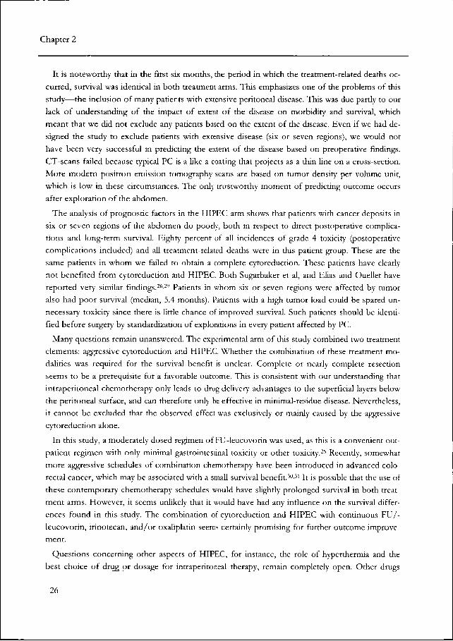
Figuree 2. Kaplan Meier survival curve, comparing standard treatment to HIPEC
0.80--
to o
0.00--
^ " ^ ^
\ \ _ _ L L
i. .
i i
1 1 j _ _
—— standard treatment - -- HIPEC
L L
122 18 24 survivall in months
30 0 36 6
23 3

Chapterr 2
PP = 0.032). Median survival in the standard arm was 12.6 months, compared with 22.4 months in
thee HIPEC arm (P = 0.032; figure 2). Exploratory subgroup analysis did not reveal any particular
subgroupp in which the effect of cytoreduction followed by HIPEC was better or worse compared
withh standard treatment (figure 3). When the data of the patients who underwent cytoreduction
Figuree 3. Explorative subgroup analysis on survival of all 105 patients randomized Forestt plot shows the hazard ratio for various subgroups of patients. The diamond indicating the overalll result corresponds with the 95% confidence interval
Subgroupp estimates 99%, Overall 95% contidence interval-
Subgroup p Sex x m^ln n female e
A g e e
«=== 50 yrs 511 - 60 yrs >> 60 yrs
Ki tee of tumor appendix x colon n rectum m
Or ig in n primary y recurrence e
Overa l ll result
HIPEC C events/N N
17/34 4 7/20 0
10/22 2 9/16 6 5/16 6
2/7 7
20/41 1 2/6 6
14/30 0 10/24 4
24/54 4
Contro' ' events/f\ \
18/24 4 13/27 7
9/16 6 12/20 0 10/15 5
7/11 1 21/34 4
3/6 6
16/28 8 15/23 3
31/51 1 0.555 ( 0.321 , 0.951
Figuree 4. Kaplan Meier survival curve of 49 patientss with peritoneal carcinomatosis treated byy cytoreduction followed by HIPEC, compar-ingg number of regions affected with PC
Figuree 5. Kaplan Meier survival curve of 49 pa-tientss with peritoneal carcinomatosis treated by cytoreductionn followed by HIPEC, comparing numberr of regions with residual tumor
1.00--
0.75--
prob
abili
ty y
o
o
o o
0.25--
o.oo--
jj L_
l l i i
L_ _ 1 1 1 1
24 4
12 2
0-55 regions -6-77 regions
\ \
— i — — 24 4 36 6
survivall in months
1.00 0
0.75 5
SS 0.50-O O
0.25 5
0.00 0
R-1 1 R-2a a R-2b b
ii 1
122 24 survivall in months
36 6
Randomizedd trial
followedd by HIPEC were further analyzed, they showed that patients with six to seven regions still
hadd a very poor survival (median, 5.4 months) compared with those with zero to five regions in-
volvedd (median, > 29 months; P < 0.0001, figure 4; ). The success of the surgical procedure also
hadd prognostic value. After complete resection (R-l), only one of 18 patients died. Fourteen of the
211 patients with limited residual disease (R-la) died, compared with seven of the 10 patients with
extensivee residual disease (R-2b) (P < 0.0001). The median times to death in the latter two groups
weree 20 and five months, respectively (figure 5).
Discussion n
Thiss study was designed to answer the question of whether the addition of aggressive cytoreduc-
tionn and HIPEC with MMC improves survival in patients with PC of colorectal origin. In this
analysis,, the results of all 105 randomly assigned patients are reported. The median follow-up at the
timee of analysis was 21.6 months, which is more than twice the median survival in the standard
arm.. At the time of this writing, an event (either recurrence or death) had occurred in 61% of the
patients,, almost two-thirds of what had been experienced in the standard arm. Follow-up, there-
fore,, is long enough to demonstrate any impact of this new therapy on survival. The analysis was
carriedd out according to the intention-to-treat principle, irrespective of the actual treatment re-
ceived.. Protocol violations, including ineligibility of enrolled patients and treatment alterations,
shouldd have a negative impact on the experimental arm. Nevertheless, Kaplan-Meier survival analy-
siss showed a statistically significant survival benefit for the experimental therapy. The effect is of a
remarkablee size. The median duration of survival almost doubled, while the 2-year survival was
evenn more than twice as high. The difference between the patient groups withstands selection bias,
ass in the control arm, life expectancy exceeded six months known from literature.27'28 Nevertheless,
thesee relative good results suggest positive selection before random assignment.
Althoughh the longest survivor is at present only four years after randomization, the Kaplan-Meier
survivall curve suggests a 5-year survival rate in the order of 20%, which is comparable to survival
foundd in phase II studies. Together with die results of published phase II studies12-14"23 (table 3),
ourr results provide compelling evidence of the effectiveness of aggressive cytoreduction and
HIPEC. .
Thiss effect was associated with considerable morbidity and mortality (8%), which is similar to
thatt reported by others.21 Most of the serious complications seem to be related to the extent of sur-
gery,, and may be related to the extent of peritoneal involvement, rather than to the HIPEC proce-
dure.. The median blood loss of almost four liter is high. The most extreme blood loss measure-
mentss were found in patients with six and seven regions involved. Characteristically, these patients
wouldd undergo a partial gastrectomy, splenectomy, resection of the tail of the pancreas, omentec-
tomy,, multiple small bowel resections, ileocecal resection, rectosigmoid resection, and uterus with
adnexx extirpation combined with multiple peritonectomy procedures, leaving an enormous intra-
abdominall wound bed. Exhaustion of coagulations factors, though replaced with fresh frozen
plasmaa and thrombocytes if measurably low, has probably contributed to the blood loss.
25 5
Chapterr 2
I tt is noteworthy that in the first six months, the period in which the treatment-related deaths oc-
curred,, survival was identical in both treatment arms. This emphasizes one of the problems of this
s tudy—thee inclusion of many patients with extensive peritoneal disease. This was due partly to our
lackk of understanding of the impact of extent of the disease on morbidity and survival, which
meantt that we did not exclude any patients based on the extent of the disease. Even if we had de-
signedd the study to exclude patients with extensive disease (six or seven regions), we would not
havee been very successful in predicting the extent of the disease based on preoperative findings.
CT-scanss failed because typical PC is a like a coating that projects as a thin line on a cross-section.
Moree modern positron emission tomography scans are based on tumor density per volume unit,
whichh is low in these circumstances. The only trustworthy moment of predicting outcome occurs
afterr exploration of the abdomen.
Thee analysis of prognostic factors in the HIPEC arm shows that patients with cancer deposits in
sixx or seven regions of the abdomen do poorly, both in respect to direct postoperative complica-
tionss and long-term survival. Eighty percent of all incidences of grade 4 toxicity (postoperative
complicationss included) and all treatment-related deaths were in this patient group. These are the
samee patients in whom we failed to obtain a complete cytoreduction. These patients have clearly
nott benefited from cytoreduction and HIPEC. Both Sugarbaker et al, and Elias and Ouellet have
reportedd very similar findings.26-29 Patients in whom six or seven regions were affected by tumor
alsoo had poor survival (median, 5.4 months). Patients with a high tumor load could be spared un-
necessaryy toxicity since there is litde chance of improved survival. Such patients should be identi-
fiedfied before surgery by standardization of explorations in every patient affected by PC.
Manyy questions remain unanswered. The experimental arm of this study combined two treatment
elements:: aggressive cytoreduction and HIPEC. Whether the combination of these treatment mo-
dalitiess was required for the survival benefit is unclear. Complete or nearly complete resection
seemss to be a prerequisite for a favorable outcome. This is consistent with our understanding that
intraperitoneall chemotherapy only leads to drug delivery advantages to the superficial layers below
thee peritoneal surface, and can therefore only be effective in minimal-residue disease. Nevertheless,
i tt cannot be excluded that the observed effect was exclusively or mainly caused by the aggressive
cytoreductionn alone.
I nn this study, a moderately dosed regimen of FU-leucovorin was used, as this is a convenient out-
patientt regimen with only minimal gastrointestinal toxicity or other toxicity.25 Recendy, somewhat
moree aggressive schedules of combination chemotherapy have been introduced in advanced colo-
rectall cancer, which may be associated with a small survival benefit.w-31 It is possible that the use of
thesee contemporary chemotherapy schedules would have slighdy prolonged survival in both treat-
mentt arms. However, it seems unlikely that it would have had any influence on the survival differ-
encess found in this study. The combination of cytoreduction and H IPEC with continuous FU /-
leucovorin,, irinotecan, and /or oxaliplatin seems certainly promising for further outcome improve-
ment. .
Quest ionss concerning other aspects of HIPEC, for instance, the role of hyperthermia and die
bestt choice of drug or dosage for intraperitoneal therapy, remain completely open. Other drugs
26 6

Randomizedd trial
suchh as oxaliplatin32 and floxuridine33 have been studied and may be incorporated alone or in novel
combinations. .
I nn this study, the open coliseum technique is used. This open system presents the possibility to
maintainn optimal distribution by manual stirring. Recently, this system has been tested for safety by
operatingg room personnel.34 In this study, MM C was found neither in the operating room air, nor in
thee urine of the surgeon or perfusionist, and is therefore safe.
Thiss study shows that the therapeutic nihilism that has dominated the care for patients with PC for
suchh a long time may not be appropriate. Limited PC may represent a situation analogous to that of
isolatedd liver metastases, in which long-term survival can be achieved in some patients by the surgical
removall of macroscopic disease and by systemic treatment to deal with microscopic residual disease.
Wit hh the appropriate patient selection and a determined locoregional treatment effort, PC of colo-
rectall origin may even be a potentially curable disease in patients with limited peritoneal involve-
ment. .
References s
1.. Minsky BD, Mies C, Rich TA, et al: Potentially curative surgery of colon cancer: Patterns of failure and survival.. J Clin Oncol 1988: 6: 106-118.
2.. Russell AH, Tong D, Dawson LE, et al: Adenocarcinoma of the retroperitoneal ascending and descend-ingg colon: Sites of initial dissemination and clinical patterns of recurrence following surgery alone. Int J Radiat OncolOncol Biol Phys 1983; 9: 361-365.
3.. Tong D, Russell AH, Dawson LE, et al: Adenocarcinoma of the cecum: Natural history and clinical pat-ternss of recurrence following radical surgery. Int J Radiat Oncol Biol Phys 1983; 9: 357-360.
4.. Sugarbaker PH: Intraperitoneal chemotherapy and cytoreductive surgery for the prevention and treat-mentt of peritoneal carcinomatosis and sarcomatosis. Semin Surg Oncol 1998; 14: 254-261.
5.. Sugarbaker PH: Management of peritoneal-surface malignancy: The surgeon's role. Langenbecks Arch Surg 1999;; 384: 576-587.
6.. Sugarbaker PH: Peritonectomy procedures. Ann Surg 1995; 221: 29-42.
7.. Spratt JS, Adcock RA, Muskovin M, et al: Clinical delivery system for intraperitoneal hyperthermic che-motherapy.. Cancer Res 1980; 40: 256-260.
8.. Fernandez-Trigo V, Stuart OA, Stephens AD, et al: Surgically directed chemotherapy: Heated intraperito-neall lavage with mitomycin C. Cancer Treat Res 1996; 81: 51-61.
9.. Witkamp AJ, van Coevorden F, Kaag MM, et al: Dose finding study of hyperthermic intraperitoneal che-motherapyy with mitomycin C in patients with carcinosis of colorectal origin. Eur J Surg Oncol 1998; 24: 214, (abstrr F74).
10.. Cavaliere R, Ciocatto EC, Giovanella BC, et al: Selective heat sensitivity of cancer cells: Biochemical and clinicall studies. Cancer \961; 20: 1351-1381.
11.. Isacoff WH, Borud K: Chemotherapy for the treatment of patients with metatastic colorectal cancer: An overview.. WorldJSutg 1997; 21: 748-762.
12.. Fujimura T, Yonemura Y, Fujita H, et al: Chemohyperthermic peritoneal perfusion for peritoneal dis-seminationn in various intra-abdominal malignancies. Int Surg 1999; 84: 60-66.
13.. Storm FK: Clinical hyperthermia and chemotherapy. Radiol Clin North Am 1989; 27: 621-627,
14.. Beaujard AC, Glehen O, Caillot JL, et al: Intraperitoneal chemohyperthermia with mitomycin C for di-gestivee tract cancer patients with peritoneal carcinomatosis. Cancer 2000: 88; 2512-2519.
15.. Cavaliere F, Perri P, Di Filippo F, et al: Treatment of peritoneal carcinomatosis with intent to cure. J
27 7

Chapterr 2
SurgSurg Oncol 2000; 74: 41-44. 16.. Elias D, Blot F, El Otmany A, et at: Curative treatment of peritoneal carcinomatosis arising from colo-
rectall cancer by complete resection and intraperitoneal chemotherapy. Cancer 2001; 92: 71-76.
17.. Loggie BW, Fleming RA, McQuellon RP, et al: Cytoreductive surgery with intraperitoneal hyperther-micc chemotherapy for disseminated peritoneal cancer of gastrointestinal origin. Am Surg 2000; 66: 561-568.
18.. Piso P, Bektas H, Werner U, et al: Improved prognosis following peritonectomy procedures and hy-perthermicc intraperitoneal chemotherapy for peritoneal carcinomatosis from appendiceal carcinoma. Eur J SurgSurg Oncol'2001; 27: 286-290.
19.. Rey Y, Porcheron J, Talabard JN, et al: Peritoneal carcinomatosis treated by cytoreductive surgery and intraperitoneall chemohyperthermia. Ann Chir2000; 125: 631-642.
20.. Schneebaum S, Arnold MW, Staubus A, et al: Intraperitoneal hyperthermic perfusion with mitomycin CC for colorectal cancer with peritoneal metastases. Ann Surg Oncol 1996; 3: 44-50.
21.. Shen P, Levine EA, Hall J, et al: Factors predicting survival after intraperitoneal hyperthermic chemo-therapyy with mitomycin C after cytoreductive surgery for patients with peritoneal carcinomatosis. Arch Surg 2003;; 138: 26-33.
22.. Sugarbaker PH, Chang D: Results of treatment of 385 patients with peritoneal surface spread of ap-pendiceall malignancy. Ann Surg Oncol \999; \999; 6: 727-731.
23.. Witkamp AJ, de Bree E, Kaag MM, et al: Extensive cytoreductive surgery followed by intra-operative hyperthermicc intraperitoneal chemotherapy with mitomycin-C in patients with peritoneal carcinomatosis of colorectall origin. EurJ Cancer-2001; 37: 979-984.
24.. Sugarbaker PH: Carcinomatosis: Is cure an option? J Clin Oncol 2003; 21: 762-764.
25.. Laufman LR, Krzeczowski KA , Roach R, et al: Leucovorin plus 5-fluorouracil: An effective treatment forr metastatic colon cancer. J Clin Oncol\981; 5: 1394-1400.
26.. Sugarbaker PH, Schellinx ME, Chang D, et al: Peritoneal carcinomatosis from adenocarcinoma of the colon.. World J Surg \996; 20: 585-591.
27.. Jayne DG, Fook S, Loi C, et al: Peritoneal carcinomatosis from colorectal cancer. Br J Surg 2002; 89: 1545-1550. .
28.. Sadeghi B, Arvieux C, Glehen O, et al: Peritoneal carcinomatosis from non-gynecologic malignancies: Resultss of the EVOCAPE 1 multicentric prospective study. Cancer 2000; 88: 358-363.
29.. Elias DM, Ouellet JF: Intraperitoneal chemohyperthermia: Rationale, technique, indications, and re-sults.. Surg Oncol Clin North Am 2001; 10: 915-933.
30.. Andre T, Louvet C, Maindrault-Goebel F, et al: CPT-11 (irinotecan) addition to bimonthly, high-dose leucovorinn and bolus and continuousinfusion 5-fluorouracil (FOLFIRI) for pretreated metastatic colorectal cancerr GERCOR. Eur J Cancer \999\ 35: 1343-1347.
31.. de Gramont A, Figer A, Seymour M, et al: Leucovorin and fluorouracil with or without oxaliplatin as first-linee treatment in advanced colorectal cancer. J Clin Oncol 2000; 18: 2938-2947.
32.. Elias D, Bonnay M, Puizillou JM, et al: Heated intra-operative intraperitoneal oxaliplatin after com-pletee resection of peritoneal carcinomatosis: Pharmacokinetics and tissue distribution. Ann Oncol 2002; 13: 267-272. .
33.. Culliford AT, Brooks AD, Sharma S et al.: Surgical debulking and intraperitoneal chemotherapy for establishedd peritoneal metastases from colon and appendix cancer. Ann Surg Oncol 2001; 8: 787-795.
34.. Stuart OA, Stephens AD, Welch L, Sugarbaker PH: Safety monitoring of the coliseum technique for heatedd intraoperative intraperitoneal chemotherapy with mitomycin C. Ann Surg Oncol 2002; 9: 186-191.
28 8
Randomizedd trial
Communicationn in the Journal of Clincal Oncology after publication of the
randomi2edd trial.
Letterr to the editor by Maurie Markman
Verwaall et al1 are to be congratulated for their efforts to evaluate in a phase II I randomized trial
thee clinical utility of "aggressive surgical cytoreduction" followed by hyperthermic intraperitoneal
chemotherapyy (HIPEC) as a management strategy for peritoneal carcinomatosis resulting from a
colorectall malignancy.
Unfortunately,, a major conceptual flaw in the study's design prevents any meaningful conclusions
too be drawn from the results of this otherwise interesting study. The investigators elected to com-
paree a strategy that combined aggressive surgery with HIPEC versus standard intravenous chemo-
therapyy plus palliative surgery (if necessary). However, if the aim of the study was to evaluate the
highlyy experimental, complex, costly, and potentially very morbid regional chemotherapy strategy,
thiss trial has failed to address this important question.
AA more appropriate trial design would have been to randomly assign patients with peritoneal car-
cinomatosiss to aggressive surgical cytoreduction with or without HIPEC. With the present study
design,, the demonstrated survival benefit may have been due principally, if not totally, to the exten-
sivee surgery, with the regional chemotherapy only adding toxicity. The favorable impact on survival
associatedd with an attempt at "interval surgical cytoreduction" (without the subsequent administra-
tionn of intraperitoneal chemotherapy) has been documented in ovarian cancer,2 and the major
influencee of the volume of residual disease on survival has been clearly shown in the current study
(mediann survival of 20 months versus 5 months for patients with "limited" versus "extensive" resi-
duall cancer, respectively).
Ass appropriately noted by the authors: "... it cannot be excluded that the observed effect was ex-
clusivelyy or mainly caused by the aggressive cytoreduction alone." In sum, this study fails to provi-
dee any support for the routine (as opposed to investigative) use of HIPEC in this clinical setting.
Itt is hoped that these investigators will follow their important initial efforts with a phase II I ran-
domizedd trial that directly addresses the unique contribution (if any) of the regional chemotherapy
componentt of this intensive management strategy.
References s
1.. Verwaal VJ, van Ruth S, de Bree E, et al: Randomized trial of cytoreduction and hyperthermic intraperi-toneall chemotherapy versus systemic chemotherapy and palliative surgery in patients with peritoneal carci-nomatosiss of colorectal cancer. J Clin Oncol 2003; 21: 3737-3743.
2.. Van der Burg MEL, Van Lent M, Buyse M, et al: The effect of debulking surgery after induction che-motherapyy on the prognosis in advanced epithelial ovarian cancer. N Engl J Med1995; 332: 629-634.
29 9
Chapterr 2
Letterr to the editor bv Bert Hildebrandt, Beate Rau Johanna, Gellermann, Peter
Wustt and Hanno Riess
Vi cc }. Verwaai and colleagues1 demonstrated that an improvement of survival is achieved in colo-
rectall cancer patients with peritoneal carcinosis treated by surgical cytoreduction and hyperthermic
intraperitoneall chemotherapy (HIPEC) when compared with schedules with palliative chemothera-
pyy (with or without surgery) alone. This study, for the first time, demonstrated a survival benefit
forr adjunctive H IPEC in the scope of a randomized trial, but this result was counterbalanced by a
16%% early death rate and an excessively high rate of severe side effects in the experimental arm
( W H OO 3: 6 5% of patients; W H O 4: 4 5% of patients). In our opinion, such high complication rates
aree hardly acceptable in a palliative treatment concept that yielded an overall survival benefit of 10
monthss for the entire patient population. In addition, there was virtually no clinically relevant im-
provementt in subgroups of patients with far advanced disease. Indeed, a median survival of less
thann 6 months was reported in 3 3% of patients treated in the experimental arm.
I tt is plausible that a number of side effects associated with surgery plus H IPEC have been caused
orr promoted by either the intraperitoneal application of mitomycin C or the hyperthermic conditi-
onss of its administration (eg, hematotoxicity, 27%; fistuke, 15%; infection, 6%; pancreatitis, 2% of
patientss treated). Certain other complications may rather be assigned to the surgical procedure or
rapidd disease progression (eg, pulmonary embolism within 3 months after surgery). However, 24%
off patients treated with H I P EC suffered from severe or life-threatening complications that caused
damagee to vital organs (heart failure/arrhythmia, 14%; terminal renal failure, 6%; paresis: 4% of pa-
tients),tients), and it is difficul t to ascribe these events to any of the single modality applied. Therefore,
thesee high rates of severe ad-verse events appear to result from the topical application of surgery
andd adjunctive chemotherapy under hyperthermic conditions. Unfortunately, the study was not de-
signedd to precisely define the role of H I P EC by comparing treatment with surgery alone with sur-
geryy plus HIPEC.
Wee would like to emphasize that it is largely unknown if elevated (eg, > 37°C) temperatures actu-
allyy contribute to the efficacy of intraperitoneal chemotherapy. On the one hand, 10 randomized
trialss on different locoregional hyperthermia approaches have already demonstrated a benefit when
hyperthermiaa is added to standard radiotherapy and/or chemotherapy. Most of these studies have
beenn performed with radiofrequency hyperthermia techniques in patients with superficial or pelvic
tumors,, and a number of concomitant analyses have revealed a clear-cut correlation between ther-
mall dose and clinical outcome.2 On the other hand, such a dose-response relationship has never
beenn duplicated for any of the "convect ive" hyperthermia techniques HIPEC or hyperthermic iso-
latedd limb perfusion (HIEP), respectively. In addition, a randomized comparison on normothermic
versuss hyperthermic chemotherapy in the scope of HIPEC or HIEP has never been performed.
Thee occurrence of organ failure in the context of H IPEC raises suggestions on the experiences
withh chemotherapy and whole-bod)- hyperthermia (WBH). In particular extracorporal WBH, in
whichh the patient's core body temperature is raised bv convective heating of blood, bears the risk
30 0

Randomizedd trial
off severe renal failure,3 but heart failure, arrhythmias, multiorgan failure, or pareses have also been
reportedd with the use of modern radiant-heat WBH devices.4
I nn our opinion, it cannot be ruled out that intraoperative H IPEC may produce additional toxicity
withoutt being more effective than normothermic intraperitoneal chemotherapy. One strategy to
resolvee the question of whether the efficacy of intraperitoneal chemotherapy is actually enhanced
byy application as HIPEC may be a randomized comparison between normothermic and hyperther-
micc intraperitoneal drug application. Another policy may be the application of hyperthermic che-
motherapyy in the postoperative or palliative setting by employing novel regional radiofrequency hy-
perthermiaa (RHT) applicators that enable effective heat delivery to the entire abdomen ("partbody
hyperthermia").. After a phase of preclinical evaluation, such magnetic resonance— guided R HT de-
vicess have recently been introduced into clinical practice in our institution, as well as a very few
others.. First clinical experience and phase I-data indicate that effective hyperthermia can be safely
deliveredd to patients with peritoneal masses by this technology, with only littl e additional toxic-ity.5
AA first phase II trial on intravenous chemotherapy (folinic acid/nuorouracil/oxaliplatin) plus RHT
inn patients with peritoneal carcinosis has recently been initiated.
References s
1.. Verwaai VJ, van Ruth S, de Bree E, et al: Randomized trial of cytoreduction and hyperthermic intraperi-
toneall chemotherapy versus systemic chemotherapy and palliative surgery in patients with peritoneal carci-
nomatosiss of colorectal cancer. / Clin Oncol 2003; 21: 3737-3743.
2.. Wust P, Hildebrandt B, Sreenivasa G, et al: Hyperthermia in combined treatment of cancer. Lancet Oncol
2002;; 3: 487-497.
3.. Wiedemann GJ, Robins HI, Gutsche S, et al: Ifosfamide, carboplatin and etoposide (ICE) combined
withh 41.8 degrees C whole body hyperthermia in patients with refractory sarcoma. Eur J Cancer 1996; 32A:
888-892 2
4.. Kerner T, Hildebrandt B, Ahlers O, et al: Anaesthesiological experiences with whole body hyperther-
mia.. Int J Hyperthermia 2003; 19: 1-12.
5.. Gellermann J, Wlodarczyk W, Seebass M, et al: MR-thermometry during radiofrequeny hyperthermia
(part-bodyy hyperthermia) in pelvic tumors. Radiology 2002; 225(P): 510 (abstr).
I nn rep ly by V i c J Verwaai
Thee comments in the letters to the editor are well taken. We agree with both writers that our stu-
dyy could not answer the question of whether adding hyperthermic, intraperitoneal chemotherapy to
cytoreductionn is better than cytoreduction alone. To answer this question, an appropriate study de-
signn would, indeed, be a randomized trial comparing cytoreduction alone with cytoreduction plus
hyperthermicc intraperitoneal chemotherapy.
Att the current state of art in the treatment of carcinomatosis, neither cytoreduction alone nor
hyperthermiaa plus chemotherapy is common practice or a proven treatment. This means that a
studyy design comparing single elements of the treatment would be a comparison of two new
31 1
Chapterr 2
therapiess and would not answer the question whether either treatment is better than the regular
treatment. .
Att this moment, the gold standard in the treatment of peritoneal carcinomatosis has not yet been
established.. In practice, most patients affected by peritoneal carcinomatosis face a therapeutic nihi-
lismm and do not undergo major treatments. In this perspective, a novel therapy like cytoreduction
withh hyperthermic intraperitoneal chemotherapy should be compared with a nihilistic treatment re-
gime,, as being the standard.
Noww that our randomized trial has been completed, the question of which element of the combi-
nedd treatment is effective becomes relevant, as suggested in both letters. The design proposed in
thee first letter is an excellent suggestion. In such a study, cytoreduction plus hyperthermic intraperi-
toneall chemotherapy is seen as standard treatment. Therefore, this would be a noninferiority study
design.. A study of this nature is currendy being prepared.
Thee suggestions in the second letter could follow if the oncoming trial will prove a benefit of hy-
perthermicc intraperitoneal chemotherapy after cytoreduction. This trial would compare cytoreduc-
tionn plus hyperthermic intraperitoneal chemotherapy, with cytoreduction plus intraperitoneal che-
motherapyy without hyperthermia.
Ourr randomized trial showed a survival benefit of cytoreduction plus hyperthermic intraperito-
neall in patients affected by peritoneal carcinomatosis of colorectal origin. The value of any other
neww therapies should be tested either with the standard therapy or with cytoreduction plus hy-
perthermicc intraperitoneal chemotherapy in a phase II I setting. Only in this way we will be able to
determinatee which treatment or combination of treatments is optimal.
32 2