Embed Size (px)
Citation preview
Valeri PetkovDepartment of Physics, Central Michigan University, Mt. Pleasant, MI 48859
Atomic-scale structure of disordered pharmaceuticalmaterials by high-energy XRD and atomic PDF analysis

This document was presented at PPXRD -Pharmaceutical Powder X-ray Diffraction Symposium
Sponsored by The International Centre for Diffraction Data
This presentation is provided by the International Centre for Diffraction Data in cooperation with the authors and presenters of the PPXRD symposia for the express purpose of educating the scientific community.
All copyrights for the presentation are retained by the original authors.
The ICDD has received permission from the authors to post this material on our website and make the material available for viewing. Usage is restricted for the purposes of education and scientific research.
ICDD Website - www.icdd.comPPXRD Website – www.icdd.com/ppxrd

Why do materials behave the way they do ? It has a lot to do with howatoms are arranged in materials, i.e. with the atomic-scale “structure”,
and that is why we need to know it well.
Diamond (crystal) - hard,transparent, insulating andexpensive.
Graphite (crystal) - soft, black,conducts heat and electricityand cheap.
It is all just Carbon
Carbon nanotubesStronger than steel
AmorphousCarbon
Catalysis

Atomic ordering in materials is determined by X-ray diffraction: Principles
PHY 101: Diffraction of light
CRYST 101: Diffraction of x-rays from a single row ofatoms (Klug and Alexander in “X-ray diffractionprocedures”)
No Intensity loss, just Intensity re-distribution !

Structure of long-range ( μm-range) ordered materials: Crystals
The atomic scale structure of crystals may be described on the basis of 3D
periodic lattices in terms of a few parameters: Lattice type and symmetryUnit cell parameters a, b, c, , , Atomic positions inside the unit cell: (x,y,z).
Diamond Graphite
These parameters/numbers allow to compute, understand and predict properties of crystals;also allow to “patent” crystalline pharmaceutical materials.

Example - Aspirin
C.A. Medendorp et al. J. Pharm. Science 97 (2008) 1361.

Non-crystalline materials: glasses, polymers, composites..
Example: again carbon but“amorphous”, i.e. coal.
Example: again carbonbut “nanotube”..
Atoms in non-crystals do not sit on thevertices of 3D periodic lattices.Such materials are called “structurallydisordered” yet they have a very well definedlocal (short + often intermediate) atomicordering.Why ? Nature of chemical bonding does notchange (usually) between crystalline andamorphous state (e.g. quartz – silica glass)We still need structure modelsparameters/numbersparameters/numbers to describe thisordering.
Could we use x-rays ?
Crystals
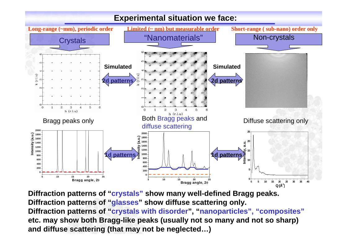
Bragg peaks only Both Bragg peaks anddiffuse scattering
5 10 15 20 25
0
200
400
600
800
1000
1200
1400
1600
1800
2000
Inte
ns
ity
(a.u
.)
Bragg angle, 2
“Nanomaterials”
Diffraction patterns of “crystals” show many well-defined Bragg peaks.Diffraction patterns of “glasses” show diffuse scattering only.Diffraction patterns of “crystals with disorder”, “nanoparticles”, “composites”etc. may show both Bragg-like peaks (usually not so many and not so sharp)and diffuse scattering (that may not be neglected…)
Simulated
2d patterns
1d patterns
Non-crystalsLong-range (~mm), periodic order Limited (~ nm) but measurable order Short-range ( sub-nano) order only
Diffuse scattering only
Simulated
2d patterns
1d patterns
0 5 10 15 20 25 30 35 400
5
10
15
20
25
Inte
ns
ity,
a.u
.
Q (Å-1)
5 10 15 20 25
0
200
400
600
800
1000
1200
1400
1600
1800
2000
Inte
ns
ity
(a.u
.)
Bragg angle, 2
Experimental situation we face:

What can be done ? Total XRD = High-energy XRD and real-space data analysis
Q=4sin()/=1.0135sin()E[keV]
S(Q)=1+ 22. )(/)()( QfcQfcQI iiiiel
G(r) = (2/)
max
,)sin(]1)([Q
oQ
dQQrQSQ
G(r) = 4r[(r) - o](r) is the local ando the average atomic density
Diffraction
experiment
5 1 0 1 5 2 0 2 5
0
2 0 0
4 0 0
6 0 0
8 0 0
1 0 0 0
1 2 0 0
1 4 0 0
1 6 0 0
1 8 0 0
2 0 0 0
Inte
ns
ity
(a.u
.)
B ra g g a n g l e , 2
i) The atomic PDF peaks at characteristic interatomic distances reflecting the 3D structure ofmaterials. No long-range order or periodicity implied, i.e. well suited for any material.ii) Total XRD, and its Fourier transform, the atomic PDF takes into account both the Bragg-likepeaks and the diffuse scattering component of the XRD data. Both carry structural information !
Need x-rays of higher energy – to reach higher QNeed stronger flux & more efficient detectors – to measure the
diffuse component of XRD patterns with good statisticsWhat do we need ?

Q: Where is the PDF’s definition coming from ?A: See below (after Klug and Alexander..)
For a collection of N atoms..
Here s=
For a multicomponent system si(s) is“replaced” by F(Q) where:

Welcome to real space world: Si standard
0 20 40 60 80 100 120
0
15000
30000
45000
60000
75000
90000
0 2 4 6 8 10 12 14 16
0
2
4
Str
uctu
refu
nctio
nQ
[S(Q
)-1
]
Wave vector Q[A-1]
(....)
(400)
(222)
(311)
(220)
(111)Si standardMo Ka, X'Pert
Inte
nsi
ty
Bragg angle, 2 theta
(....)
(222)
(400)
(311)
(220)
(111)
SiS.G: F d 3 m (227)Structure: diamond typeCell parameters:a=b=c=5.4309 A
α= β= γ= 90.0° Si (8a) 0.125, …
Reciprocal/diffraction space Real space
0 10 20 30 40 50
-0.4
-0.2
0.0
0.2
0.4
0.6
0 2 4 6 8 10
-0.4
-0.2
0.0
0.2
0.4
0.6
PD
FG
(r)
Radial distance [A-]
PD
FG
(r)
Radial distance r [A]
(....)
(12)
(6)
(12)(12)
CN= (4)
Fouriercouple
S(Q)=1+ 22. )(/)()( QfcQfcQI iiiiel G(r) = (2/)
max
,)sin(]1)([Q
oQ
dQQrQSQ
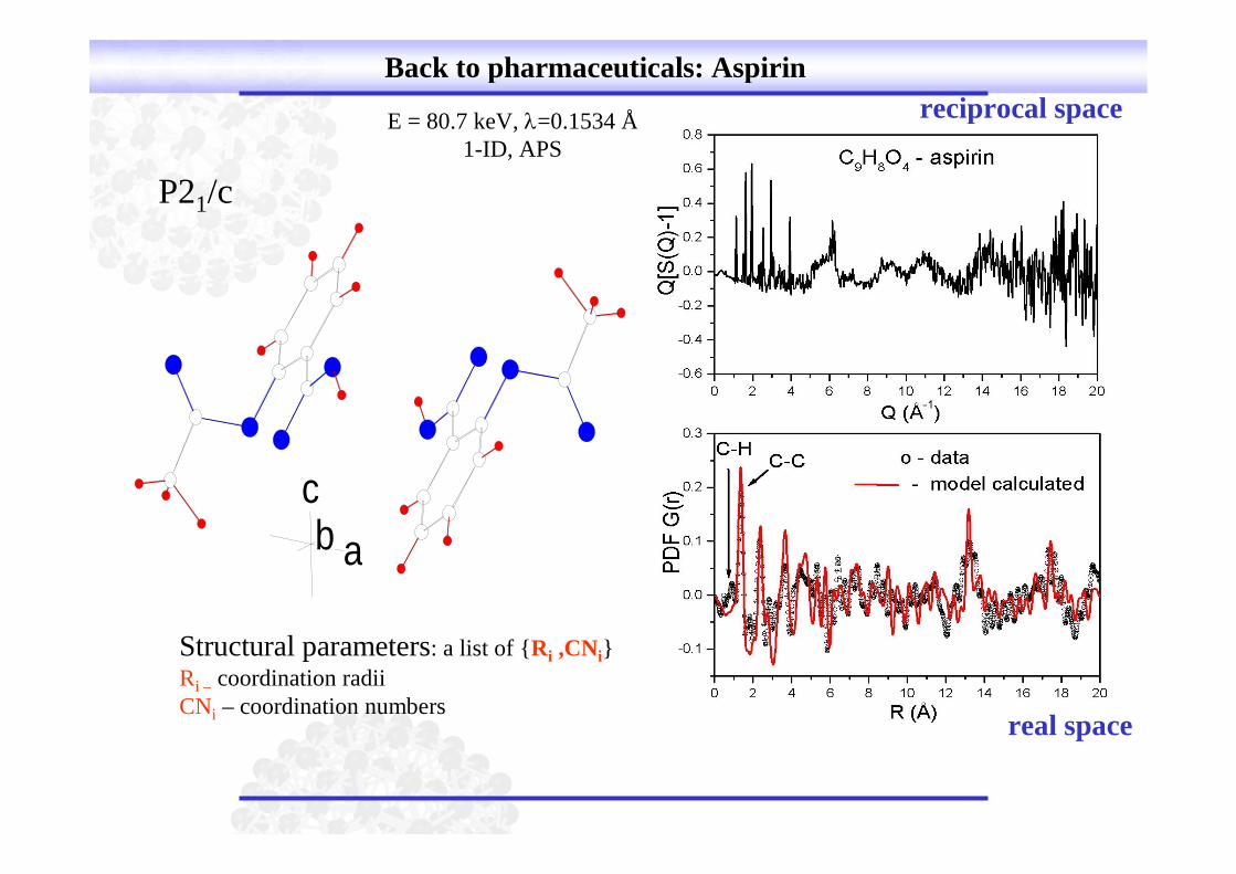
Back to pharmaceuticals: Aspirin
abc
E = 80.7 keV, =0.1534 Å1-ID, APS
P21/c
Structural parameters: a list of {Ri ,CNi}
Ri – coordination radiiCNi – coordination numbers
real space
reciprocal space

Bragg peaks Unit cell Indexing Extinctions Space group
Structural model Rietveld refinement
Si-standard material11ID-C – APS/Argonneλ = 0.1083Å
Structural determination assuming perfect 3D atomic ordering. Crystallinematerials
Interatomic Distances,angles
0 2 4 6 8 10 12
-50
0
50
100
150
200
250
300
350
400
450
Inte
nsity
(Arb
.U
nits)
2 (deg) nd sin2Bragg’s Law:
Crystalline materials produce well defined Bragg peaks that can be used to solve theaverage, long-range structure of material. Bragg peaks come from a stack of regularatomic planes (Miller indexes) !
A useful comparison: Traditional XRD/Rietveld Analysis (reciprocal space)

0 10 20 30 40 50
-1.0
-0.5
0.0
0.5
1.0
1.5
Ato
mic
PD
FG
(r)
Radial distance (Å)
High-resolution G(r) can be obtained by collecting diffraction data in wide Q-ranges High-energy x-rays.
Si-standard material11ID-C – APS/Argonneλ = 0.1083Å, Qmax = 25Å-1
However, traditional (reciprocal space) and PDF (real space) analyses have differentsensitivity to the atomic ordering in materials.Traditional – Long-range order and translational periodicity (Bragg peaks only)PDF – Any atomic ordering, periodic or not….does not matter.
Structure refinement using PDF
Pair Distribution Function Analysis (real space)
PDF peaks =Interatomic distances
Structural model (search andrefinement)
If necessary maycompute the XRDpattern
PDFs peaks reflect frequently occurring atomic pairs/coordination distances/spheresand not atomic planes ! Any material has a particular/unique coordination spheresdistribution..…may serve as a “structural fingerprint” !
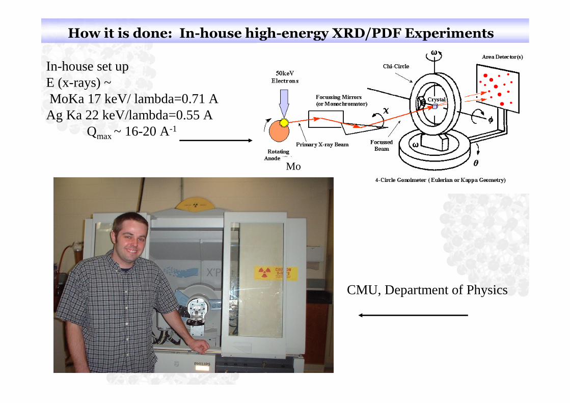
How it is done: In-house high-energy XRD/PDF Experiments
In-house set upE (x-rays) ~MoKa 17 keV/ lambda=0.71 A
Ag Ka 22 keV/lambda=0.55 AQmax ~ 16-20 A-1
CMU, Department of Physics
Mo

High-energy XRD/PDF experiments at synchrotrons
Synchrotron x-rays
Continuum of wavelengthsEnergy range (0 ~ 150 keV vs 8 keV from Cu tube)
(Advanced Photon Source, Argonne, Chicago)
390 meters (1,225 feet)

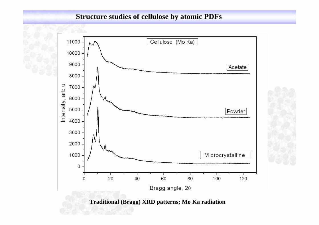
Example 1: Structure studies of cellulose by atomic PDFs

Structure studies of cellulose by atomic PDFs
Cellulose IICellulose IbCellulose Ia
Traditional (Bragg) XRD patterns; Cu Ka radiation
Collaboration with ICDD
Structure studies of cellulose by atomic PDFs
Traditional (Bragg) XRD patterns; Mo Ka radiation

Structure studies of cellulose by atomic PDFs
Atomic PDFs from synchrotron data
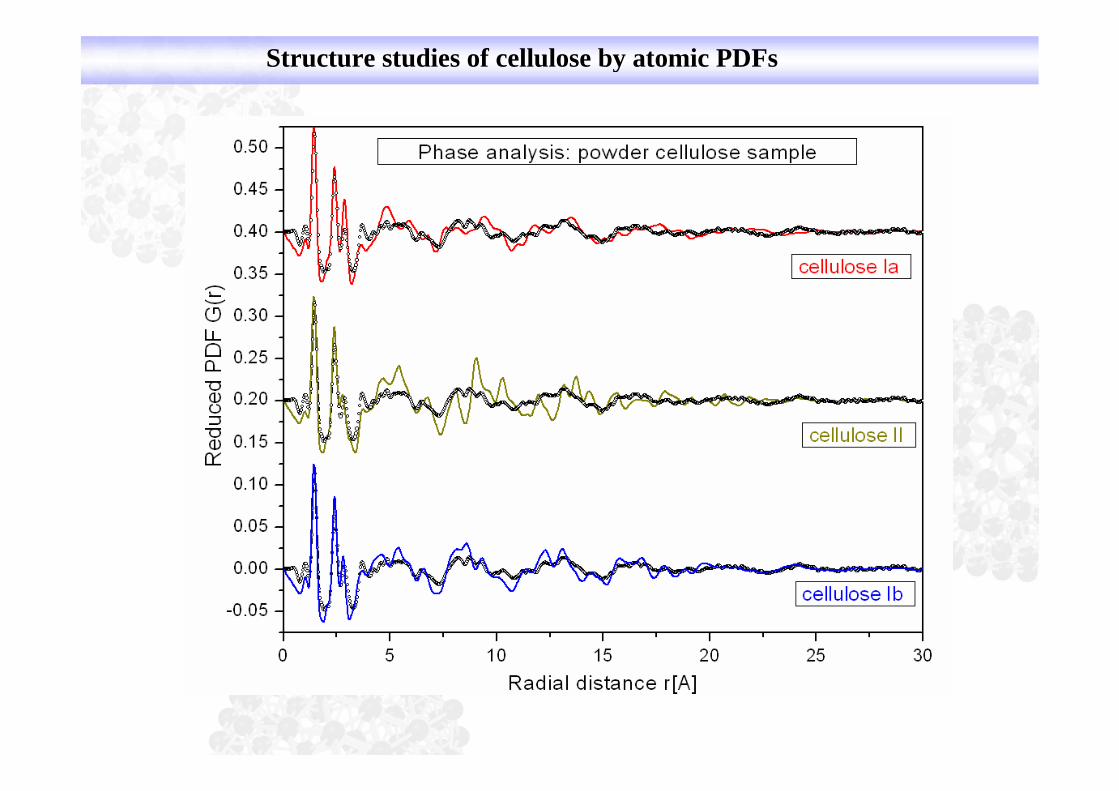
Structure studies of cellulose by atomic PDFs

Cellaburate is a reaction product of cellulose,acetic anhydride or acetic acid, and butyric acid orbutyric anhydride....
Example 2: Structure studies of other organic polymeric products…
Collaboration with ICDD

Structure studies of other organic polymeric products…
Atomic PDFs from synchrotron data

Example 3: Crystallization of lactose
XRD patterns/synchrotron
Sample dried at different conditions and crystallized for different timesCollaboration with Sandra Weiling, U. Bonn and Panalytical
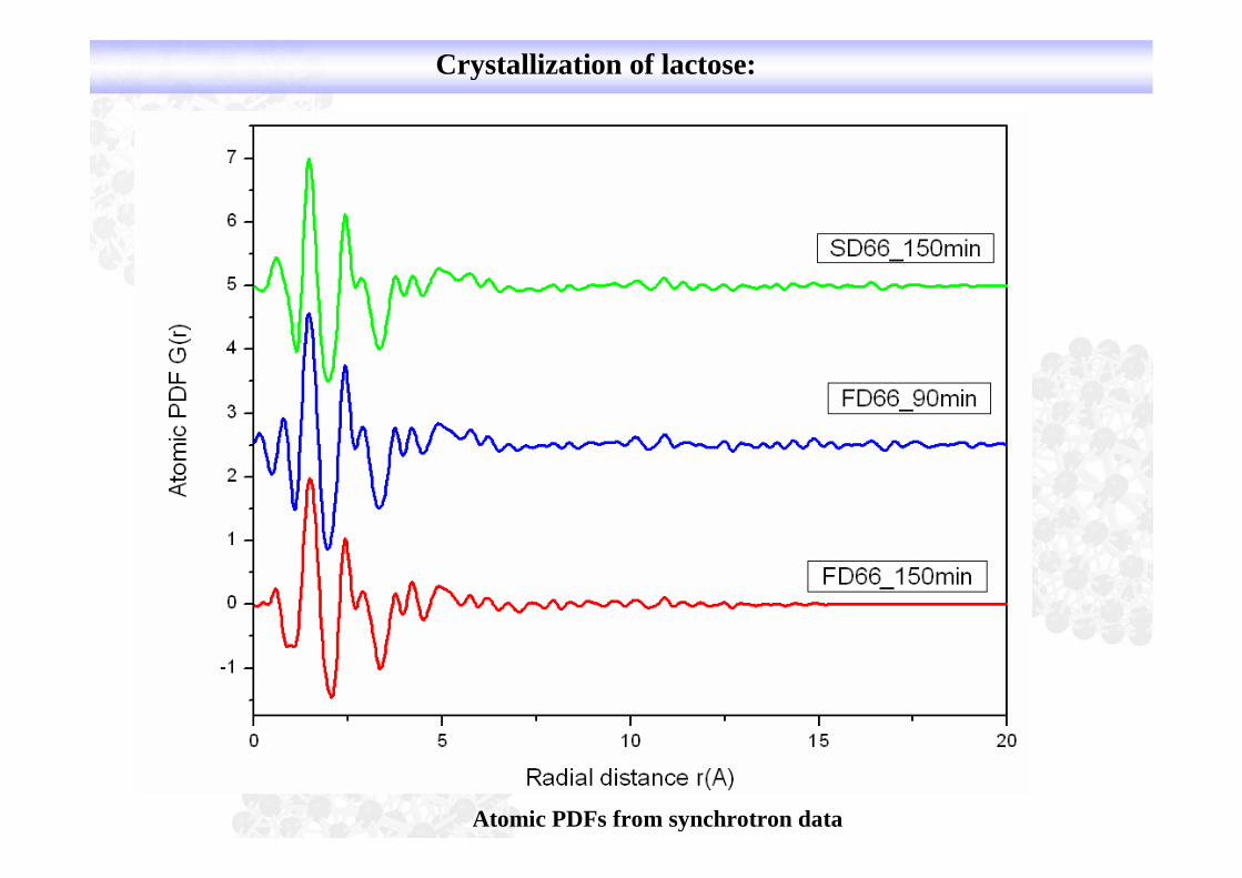
Crystallization of lactose:
Atomic PDFs from synchrotron data

Dendrimers: consist of a series of
chemical shells built on a small coremolecule. Can be designed with avariety of organic and inorganic coresand branches, with tunable branchlength, multiplicity and surfacefunctionality:
Applications:Polymer mimics of globular proteinsBuilding blocks of multifunctionalnanocompositesHosts of guests molecules andnanoparticles
Example 4: Organic macromolecules for drug delivery
Questions: Is the interior hollow ?How big is the free volume, if any ?
Collaboration with DNT

Organic macromolecules for drug delivery

PAMAM dendrimers in more detail
core
branches
3D structure depends on thepreparation technique employed(divergent/convergent,solvent), repetitive units,generation number etc.
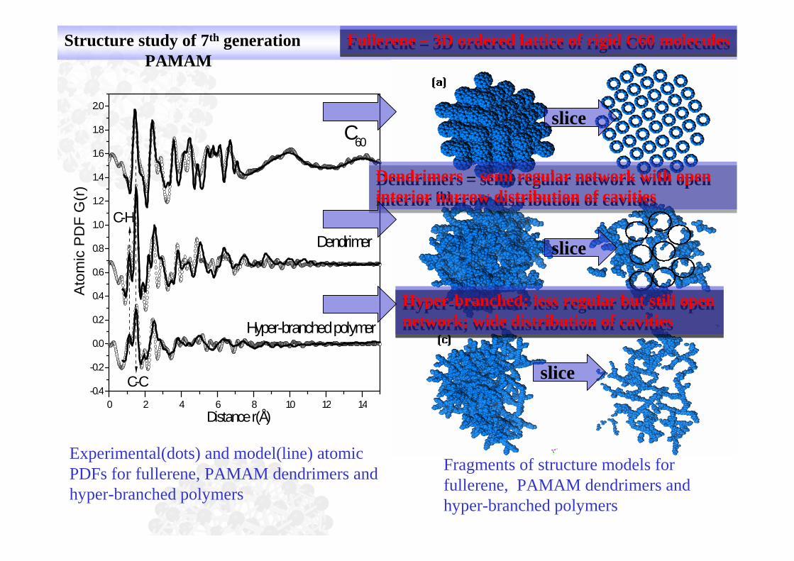
Structure study of 7th generationPAMAM
0 2 4 6 8 10 12 14
-0.4
-0.2
0.0
0.2
0.4
0.6
0.8
1.0
1.2
1.4
1.6
1.8
2.0
C-H
C-C
Hyper-branchedpolymer
Dendrimer
C60
Ato
mic
PD
FG
(r)
Distancer(Å)
Experimental(dots) and model(line) atomicPDFs for fullerene, PAMAM dendrimers andhyper-branched polymers
Fragments of structure models forfullerene, PAMAM dendrimers andhyper-branched polymers
Dendrimers – semi regular network with openinterior narrow distribution of cavities
Dendrimers – semi regular network with openinterior narrow distribution of cavities
Hyper-branched: less regular but still opennetwork; wide distribution of cavities
Hyper-branched: less regular but still opennetwork; wide distribution of cavities
slice
slice
slice
Fullerene – 3D ordered lattice of rigid C60 moleculesFullerene – 3D ordered lattice of rigid C60 molecules

PDF study of 7th generation PAMAM
3D models of PAMAM dendrimers
Too regular
Realistic
Collapsed.
.
.
Exp. data – symbolsModel data – line in red

PDF study of 7th generation PAMAM

Example 5: Chemical specificity: PtPd nanoparticles in solution
PtPd
Pure Pt Pure Pd
PtPd random alloy
Pt-core/Pd-shell
Pd-core/Pt-shell
I. Sanchez et al. JACS 131 (2009) 8683.
Collaboration with ICDD
Collaboration with R. Nuzzo, U. Illinois

Total vs Pt differential PDFs
Cores and shells in PtPd are fcc-type but structurally incoherent !
How would this affect their properties ? Pt Pd

Resonant XRD application in pharmaceuticals:
Traditional anti-cancer drugs……

MRI ……
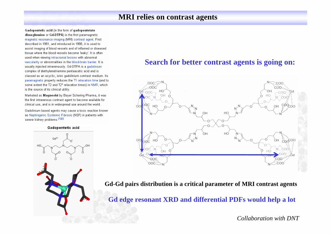
MRI relies on contrast agents
Search for better contrast agents is going on:
Gd-Gd pairs distribution is a critical parameter of MRI contrast agents
Gd edge resonant XRD and differential PDFs would help a lot
Collaboration with DNT
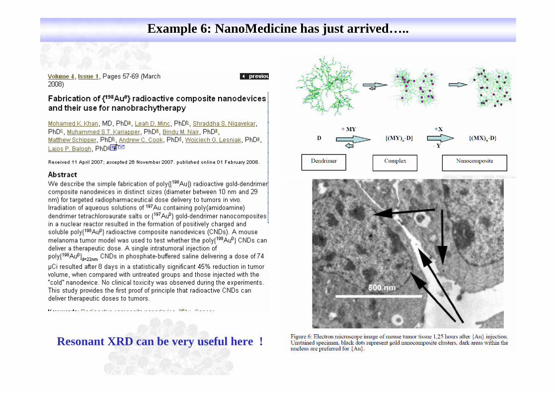
Example 6: NanoMedicine has just arrived…..
Resonant XRD can be very useful here !
0 5 1 0 1 5 2 0 2 5 3 0
- 0 . 3
0 . 0
0 . 3
0 . 6
Ato
mic
PD
FG
(r)
R a d i a l d is t a n c e r ( Å )
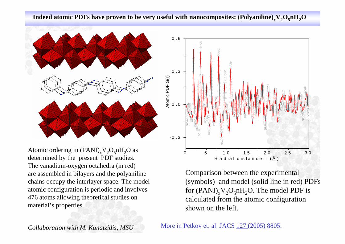
Indeed atomic PDFs have proven to be very useful with nanocomposites: (Polyaniline)xV2O5nH2O
Atomic ordering in (PANI)xV2O5nH2O asdetermined by the present PDF studies.The vanadium-oxygen octahedra (in red)are assembled in bilayers and the polyanilinechains occupy the interlayer space. The modelatomic configuration is periodic and involves476 atoms allowing theoretical studies onmaterial’s properties.
Comparison between the experimental(symbols) and model (solid line in red) PDFsfor (PANI)xV2O5nH2O. The model PDF iscalculated from the atomic configurationshown on the left.
More in Petkov et. al JACS 127 (2005) 8805.Collaboration with M. Kanatzidis, MSU

NanoMedicine will be around for a long time…..
Can Atomic PDFs be used to characterizemetal-organic frameworks,nanocomposites etc ?

and with metal-organic frameworks, and many more…

Conclusions:
Accurate structural characterization of disordered bulk and nanosizedpharmaceutical materials can be done by high-energy XRD coupled toPDF (real space) data analysis. The approach succeeds because it relieson total scattering data (Bragg plus diffuse) measured over an extendedrange of wave vectors. It probes the material as a whole (i.e. it is notsurface only sensitive/imaging like TEM) over its entire length (not onlythe first/second coordination sphere like EXAFS/spectroscopy) ofstructural coherence. It is flexible with respect to sample’s state,morphology, amount, phase homogeneity and environment. Could bedone using in-house or synchrotron sources of x-rays, and offer chemicalspecificity (resonant XRD/differential PDFs) The approach has all thepotential to become a “standard structural characterization tool” inpharmaceutical research and industry.
The work would have been impossible without the help of NSF, DoE, ARLand an army of collaborators and students ! Thank you all !
Acknowledgements:
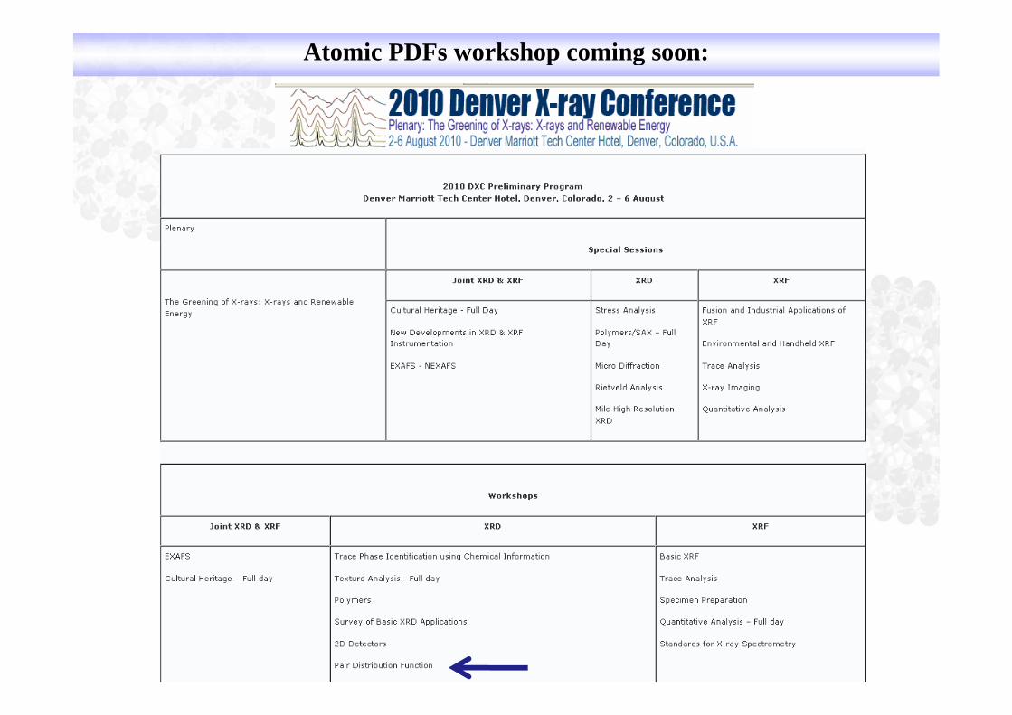
Atomic PDFs workshop coming soon: