Embed Size (px)
Citation preview
Valvular Valvular DisordersDisorders
&&Infecive Infecive
EndocarditisEndocarditis
Normal Valve FunctionNormal Valve Function Prevent backward flow of bloodPrevent backward flow of blood Permit forward flow of bloodPermit forward flow of blood
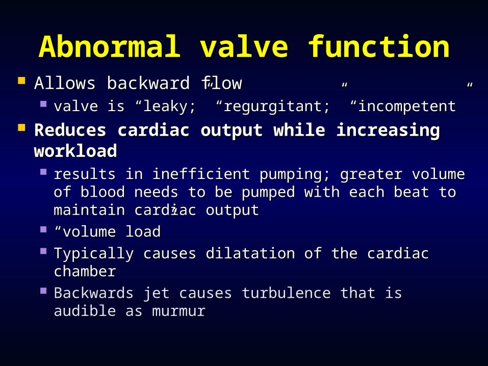
Abnormal valve functionAbnormal valve function Allows backward flowAllows backward flow
valve is “leaky;” “regurgitant;” “incompetent”valve is “leaky;” “regurgitant;” “incompetent” Reduces cardiac output while increasing Reduces cardiac output while increasing
workloadworkload results in inefficient pumping; greater volume of results in inefficient pumping; greater volume of
blood needs to be pumped with each beat to blood needs to be pumped with each beat to maintain cardiac outputmaintain cardiac output
““volume load”volume load” Typically causes dilatation of the cardiac chamberTypically causes dilatation of the cardiac chamber Backwards jet causes turbulence that is audible Backwards jet causes turbulence that is audible
as murmuras murmur
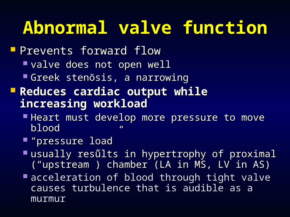
Abnormal valve functionAbnormal valve function Prevents forward flowPrevents forward flow
valve does not open wellvalve does not open well Greek stenōsis, a narrowing Greek stenōsis, a narrowing
Reduces cardiac output while Reduces cardiac output while increasing workloadincreasing workload Heart must develop more pressure to move Heart must develop more pressure to move
bloodblood ““pressure load”pressure load” usually results in hypertrophy of proximal usually results in hypertrophy of proximal
(“upstream”) chamber (LA in MS, LV in AS)(“upstream”) chamber (LA in MS, LV in AS) acceleration of blood through tight valve acceleration of blood through tight valve
causes turbulence that is audible as a murmurcauses turbulence that is audible as a murmur
Valvular Aortic StenosisValvular Aortic Stenosis
failure of valve to open normally during failure of valve to open normally during systole, requiring LV to develop excess systole, requiring LV to develop excess pressure to overcome increased resistancepressure to overcome increased resistance
pressure gradient between LV and aorta may pressure gradient between LV and aorta may be as much as 100 mm Hgbe as much as 100 mm Hg
causes concentric hypertrophycauses concentric hypertrophy symptoms of exertional chest pain, syncope, symptoms of exertional chest pain, syncope,
dyspnea dyspnea mandate valve replacement to prevent sudden mandate valve replacement to prevent sudden
deathdeath
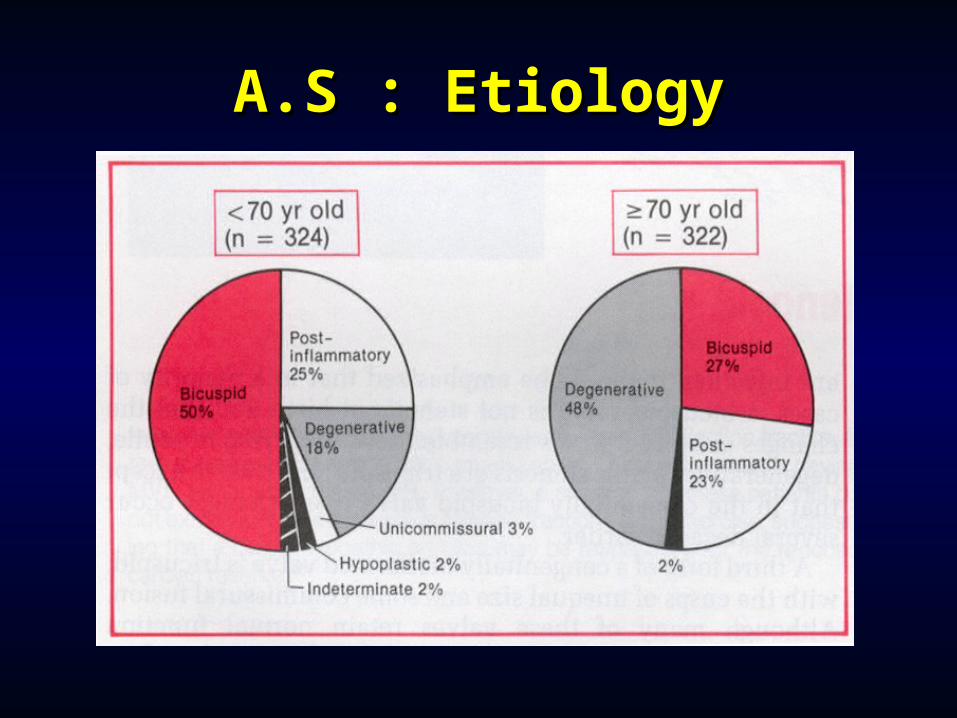
A.S : EtiologyA.S : Etiology
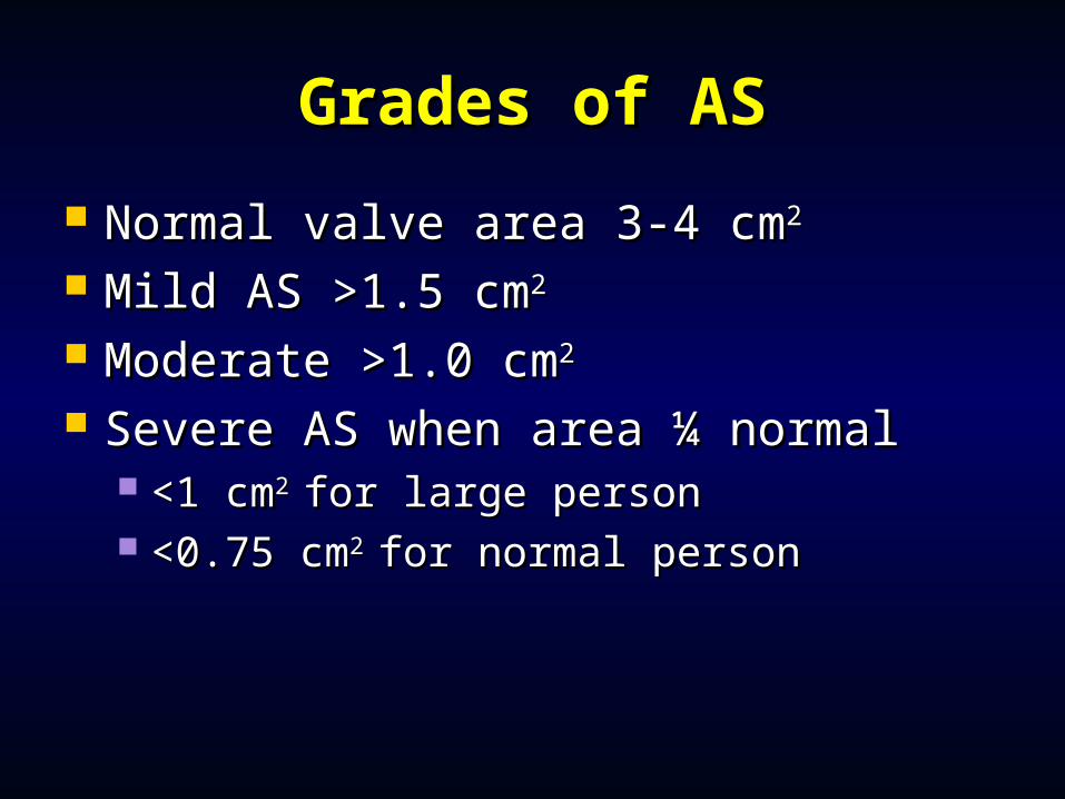
Grades of ASGrades of AS
Normal valve area 3-4 cmNormal valve area 3-4 cm22
Mild AS >1.5 cmMild AS >1.5 cm22
Moderate >1.0 cmModerate >1.0 cm22
Severe AS when area ¼ normalSevere AS when area ¼ normal <1 cm<1 cm2 2 for large personfor large person <0.75 cm<0.75 cm2 2 for normal personfor normal person
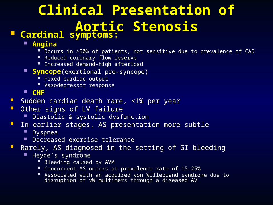
Clinical Presentation of Aortic Clinical Presentation of Aortic StenosisStenosis
Cardinal symptoms:Cardinal symptoms: AnginaAngina
Occurs in >50% of patients, not sensitive due to prevalence of CADOccurs in >50% of patients, not sensitive due to prevalence of CAD Reduced coronary flow reserveReduced coronary flow reserve Increased demand-high afterloadIncreased demand-high afterload
SyncopeSyncope(exertional pre-syncope)(exertional pre-syncope) Fixed cardiac outputFixed cardiac output Vasodepressor responseVasodepressor response
CHFCHF Sudden cardiac death rare, <1% per yearSudden cardiac death rare, <1% per year Other signs of LV failure Other signs of LV failure
Diastolic & systolic dysfunctionDiastolic & systolic dysfunction In earlier stages, AS presentation more subtleIn earlier stages, AS presentation more subtle
DyspneaDyspnea Decreased exercise toleranceDecreased exercise tolerance
Rarely, AS diagnosed in the setting of GI bleedingRarely, AS diagnosed in the setting of GI bleeding Heyde’s syndromeHeyde’s syndrome
Bleeding caused by AVMBleeding caused by AVM Concurrent AS occurs at prevalence rate of 15-25%Concurrent AS occurs at prevalence rate of 15-25% Associated with an acquired von Willebrand syndrome due to disruption of vW Associated with an acquired von Willebrand syndrome due to disruption of vW
multimers through a diseased AVmultimers through a diseased AV
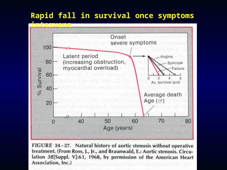
Rapid fall in survival once symptoms intervene
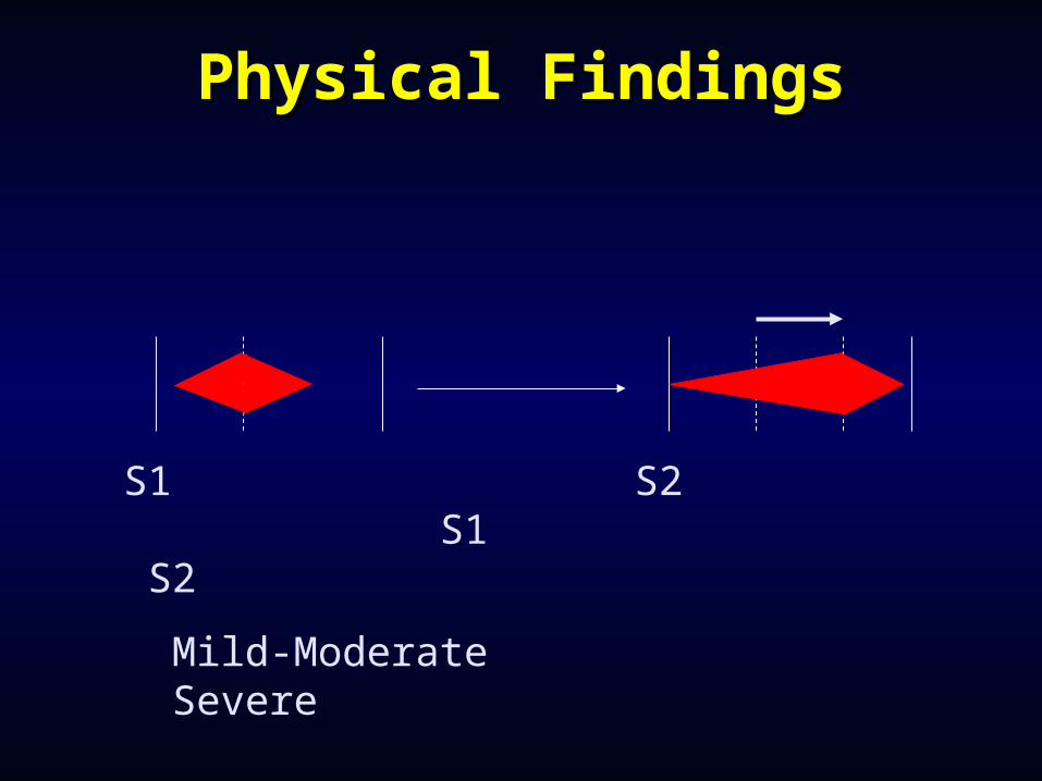
Physical FindingsPhysical Findings
S1 S2 S1 S2
Mild-Moderate Severe
Symptomatic AS- Symptomatic AS- managementmanagement
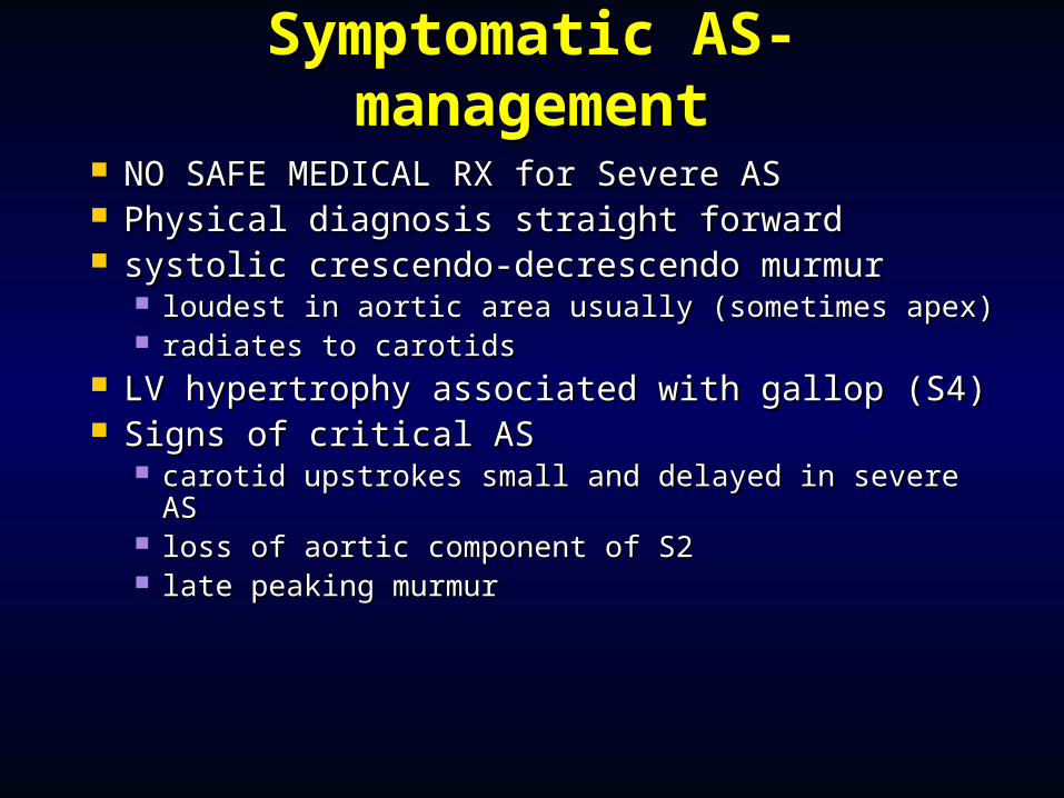
NO SAFE MEDICAL RX for Severe ASNO SAFE MEDICAL RX for Severe AS Physical diagnosis straight forwardPhysical diagnosis straight forward systolic crescendo-decrescendo murmursystolic crescendo-decrescendo murmur
loudest in aortic area usually (sometimes apex)loudest in aortic area usually (sometimes apex) radiates to carotidsradiates to carotids
LV hypertrophy associated with gallop (S4)LV hypertrophy associated with gallop (S4) Signs of critical ASSigns of critical AS
carotid upstrokes small and delayed in severe AScarotid upstrokes small and delayed in severe AS loss of aortic component of S2loss of aortic component of S2 late peaking murmurlate peaking murmur
Management of Aortic Management of Aortic StenosisStenosis
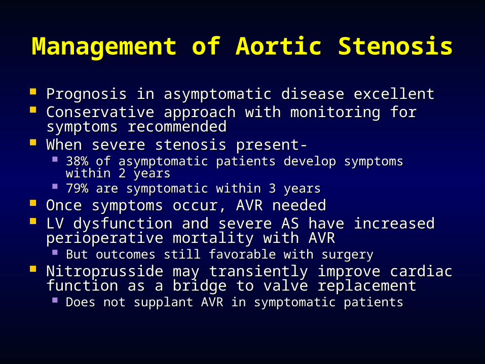
Prognosis in asymptomatic disease excellent Prognosis in asymptomatic disease excellent Conservative approach with monitoring for Conservative approach with monitoring for
symptoms recommendedsymptoms recommended When severe stenosis present-When severe stenosis present-
38% of asymptomatic patients develop symptoms within 38% of asymptomatic patients develop symptoms within 2 years2 years
79% are symptomatic within 3 years79% are symptomatic within 3 years Once symptoms occur, AVR neededOnce symptoms occur, AVR needed LV dysfunction and severe AS have increased LV dysfunction and severe AS have increased
perioperative mortality with AVRperioperative mortality with AVR But outcomes still favorable with surgeryBut outcomes still favorable with surgery
Nitroprusside may transiently improve cardiac Nitroprusside may transiently improve cardiac function as a bridge to valve replacement function as a bridge to valve replacement Does not supplant AVR in symptomatic patientsDoes not supplant AVR in symptomatic patients
Aortic Valve ReplacementAortic Valve Replacement
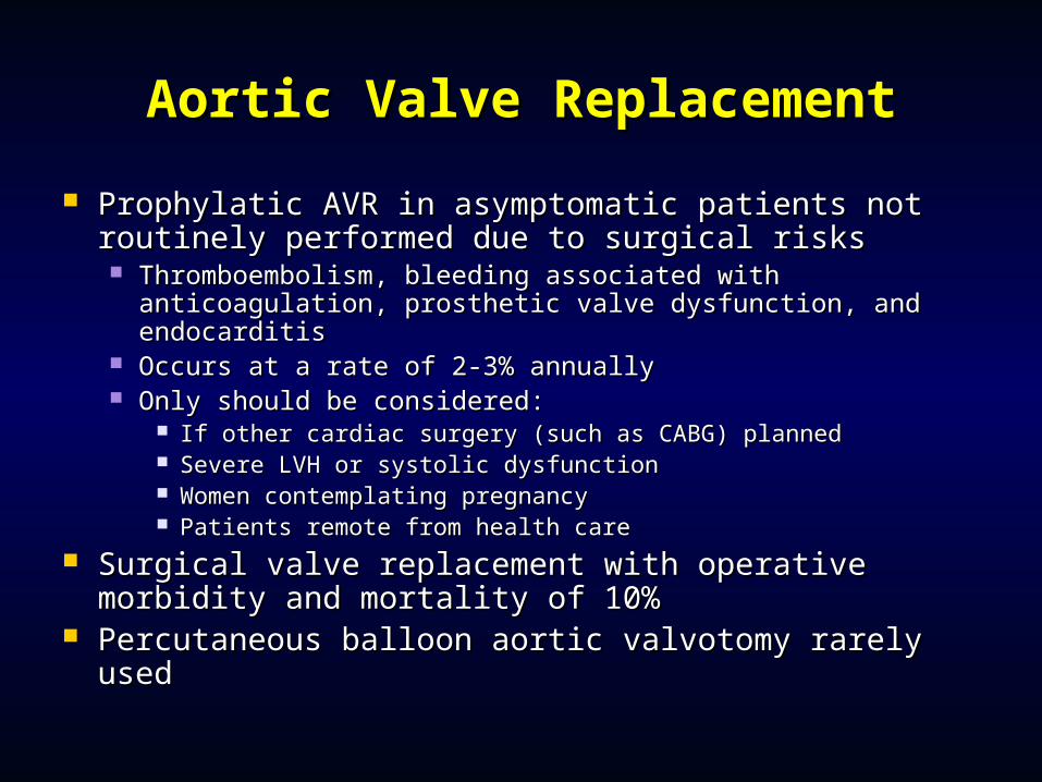
Prophylatic AVR in asymptomatic patients not Prophylatic AVR in asymptomatic patients not routinely performed due to surgical risksroutinely performed due to surgical risks Thromboembolism, bleeding associated with Thromboembolism, bleeding associated with
anticoagulation, prosthetic valve dysfunction, and anticoagulation, prosthetic valve dysfunction, and endocarditis endocarditis
Occurs at a rate of 2-3% annuallyOccurs at a rate of 2-3% annually Only should be considered:Only should be considered:
If other cardiac surgery (such as CABG) plannedIf other cardiac surgery (such as CABG) planned Severe LVH or systolic dysfunctionSevere LVH or systolic dysfunction Women contemplating pregnancyWomen contemplating pregnancy Patients remote from health carePatients remote from health care
Surgical valve replacement with operative Surgical valve replacement with operative morbidity and mortality of 10%morbidity and mortality of 10%
Percutaneous balloon aortic valvotomy rarely usedPercutaneous balloon aortic valvotomy rarely used
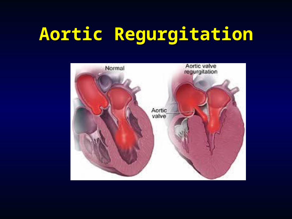
Aortic RegurgitationAortic Regurgitation
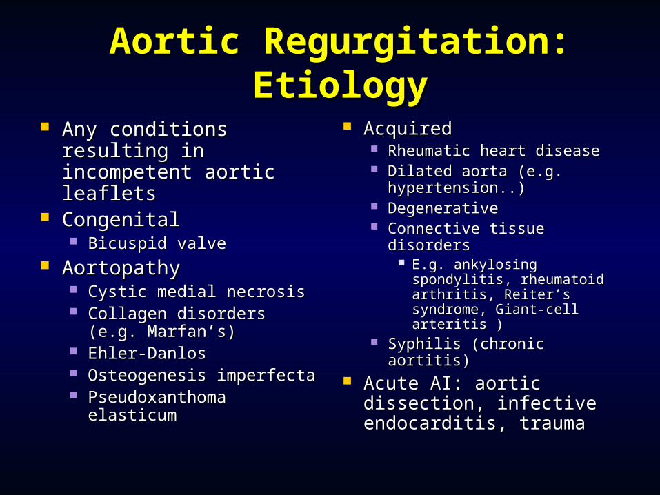
Aortic Regurgitation: Aortic Regurgitation: EtiologyEtiology
Any conditions Any conditions resulting in resulting in incompetent aortic incompetent aortic leafletsleaflets
CongenitalCongenital Bicuspid valveBicuspid valve
AortopathyAortopathy Cystic medial necrosisCystic medial necrosis Collagen disorders (e.g. Collagen disorders (e.g.
Marfan’s)Marfan’s) Ehler-DanlosEhler-Danlos Osteogenesis imperfectaOsteogenesis imperfecta Pseudoxanthoma Pseudoxanthoma
elasticumelasticum
AcquiredAcquired Rheumatic heart diseaseRheumatic heart disease Dilated aorta (e.g. Dilated aorta (e.g.
hypertension..)hypertension..) DegenerativeDegenerative Connective tissue Connective tissue
disorders disorders E.g. ankylosing E.g. ankylosing
spondylitis, rheumatoid spondylitis, rheumatoid arthritis, Reiter’s arthritis, Reiter’s syndrome, Giant-cell syndrome, Giant-cell arteritis )arteritis )
Syphilis (chronic aortitis)Syphilis (chronic aortitis) Acute AI: aortic Acute AI: aortic
dissection, infective dissection, infective endocarditis, traumaendocarditis, trauma
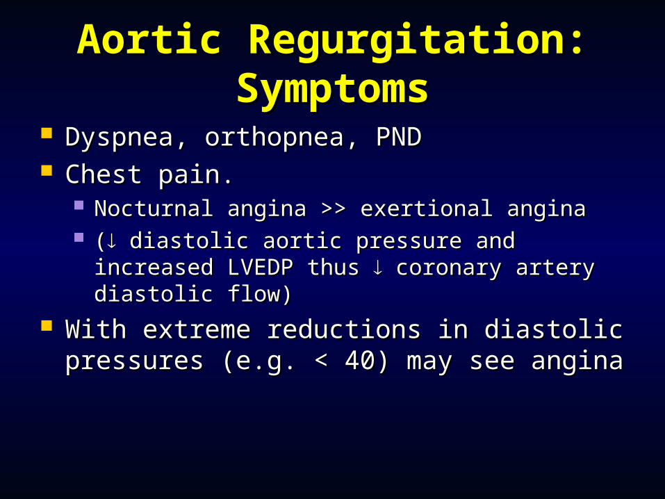
Aortic Regurgitation: Aortic Regurgitation: SymptomsSymptoms
Dyspnea, orthopnea, PNDDyspnea, orthopnea, PND Chest pain.Chest pain.
Nocturnal angina >> exertional angina Nocturnal angina >> exertional angina (( diastolic aortic pressure and increased diastolic aortic pressure and increased
LVEDP thus LVEDP thus coronary artery diastolic flow) coronary artery diastolic flow) With extreme reductions in diastolic With extreme reductions in diastolic
pressures (e.g. < 40) may see anginapressures (e.g. < 40) may see angina
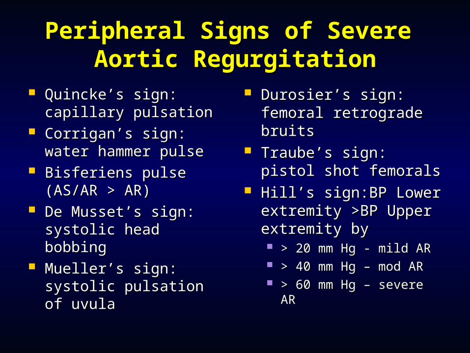
Peripheral Signs of Severe Peripheral Signs of Severe Aortic RegurgitationAortic Regurgitation
Quincke’s sign: Quincke’s sign: capillary pulsationcapillary pulsation
Corrigan’s sign: water Corrigan’s sign: water hammer pulsehammer pulse
Bisferiens pulse Bisferiens pulse (AS/AR > AR) (AS/AR > AR)
De Musset’s sign: De Musset’s sign: systolic head bobbing systolic head bobbing
Mueller’s sign: Mueller’s sign: systolic pulsation of systolic pulsation of uvulauvula
Durosier’s sign: Durosier’s sign: femoral retrograde femoral retrograde bruitsbruits
Traube’s sign: pistol Traube’s sign: pistol shot femoralsshot femorals
Hill’s sign:BP Lower Hill’s sign:BP Lower extremity >BP Upper extremity >BP Upper extremity by extremity by > 20 mm Hg - mild AR> 20 mm Hg - mild AR > 40 mm Hg – mod AR> 40 mm Hg – mod AR > 60 mm Hg – severe > 60 mm Hg – severe
ARAR
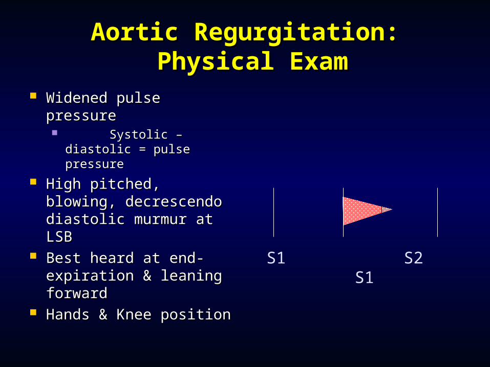
Aortic Regurgitation:Aortic Regurgitation: Physical Exam Physical Exam
Widened pulse pressure Widened pulse pressure Systolic – diastolic Systolic – diastolic
= pulse pressure= pulse pressure High pitched, blowing, High pitched, blowing,
decrescendo diastolic decrescendo diastolic murmur at LSB murmur at LSB
Best heard at end-Best heard at end-expiration & leaning expiration & leaning forwardforward
Hands & Knee positionHands & Knee positionS1 S2 S1
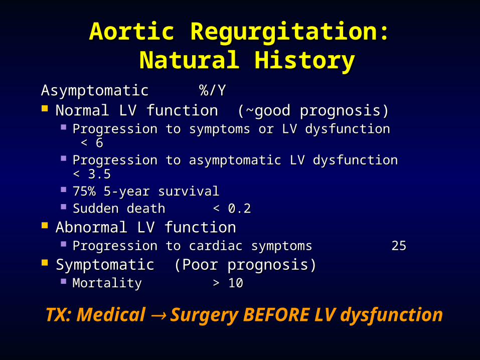
Aortic Regurgitation:Aortic Regurgitation: Natural History Natural History
AsymptomaticAsymptomatic %/Y %/Y Normal LV function (~good prognosis)Normal LV function (~good prognosis)
Progression to symptoms or LV dysfunction < 6Progression to symptoms or LV dysfunction < 6 Progression to asymptomatic LV dysfunction < Progression to asymptomatic LV dysfunction <
3.53.5 75% 5-year survival75% 5-year survival Sudden deathSudden death < 0.2 < 0.2
Abnormal LV functionAbnormal LV function Progression to cardiac symptomsProgression to cardiac symptoms 25 25
Symptomatic (Poor prognosis)Symptomatic (Poor prognosis) Mortality Mortality > 10 > 10
TX: Medical Surgery BEFORE LV dysfunction
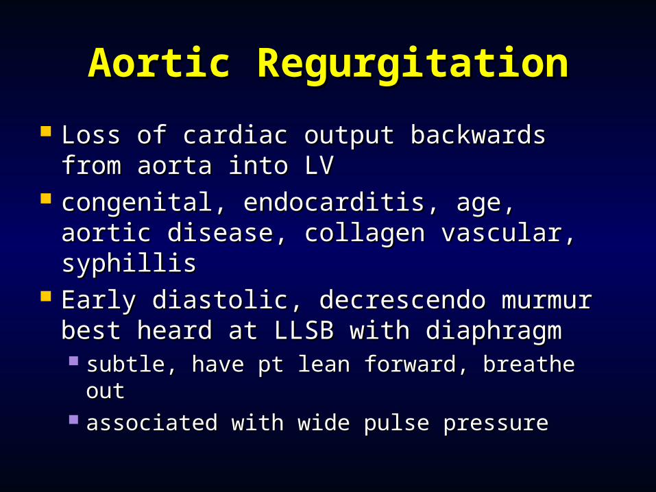
Aortic RegurgitationAortic Regurgitation
Loss of cardiac output backwards Loss of cardiac output backwards from aorta into LVfrom aorta into LV
congenital, endocarditis, age, aortic congenital, endocarditis, age, aortic disease, collagen vascular, syphillisdisease, collagen vascular, syphillis
Early diastolic, decrescendo murmur Early diastolic, decrescendo murmur best heard at LLSB with diaphragmbest heard at LLSB with diaphragm subtle, have pt lean forward, breathe subtle, have pt lean forward, breathe
outout associated with wide pulse pressureassociated with wide pulse pressure
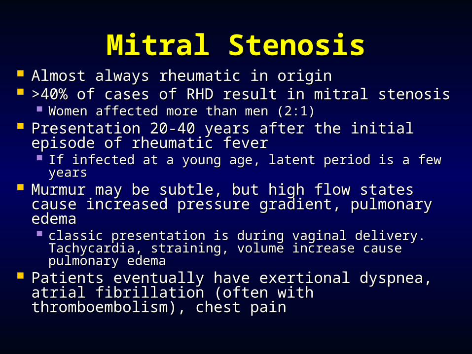
Mitral StenosisMitral Stenosis Almost always rheumatic in originAlmost always rheumatic in origin >40% of cases of RHD result in mitral stenosis>40% of cases of RHD result in mitral stenosis
Women affected more than men (2:1)Women affected more than men (2:1) Presentation 20-40 years after the initial episode Presentation 20-40 years after the initial episode
of rheumatic feverof rheumatic fever If infected at a young age, latent period is a few yearsIf infected at a young age, latent period is a few years
Murmur may be subtle, but high flow states Murmur may be subtle, but high flow states cause increased pressure gradient, pulmonary cause increased pressure gradient, pulmonary edemaedema classic presentation is during vaginal delivery. classic presentation is during vaginal delivery.
Tachycardia, straining, volume increase cause Tachycardia, straining, volume increase cause pulmonary edemapulmonary edema
Patients eventually have exertional dyspnea, Patients eventually have exertional dyspnea, atrial fibrillation (often with thromboembolism), atrial fibrillation (often with thromboembolism), chest painchest pain
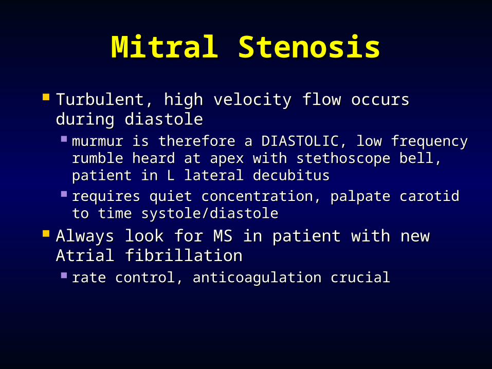
Mitral StenosisMitral Stenosis
Turbulent, high velocity flow occurs during Turbulent, high velocity flow occurs during diastolediastole murmur is therefore a DIASTOLIC, low murmur is therefore a DIASTOLIC, low
frequency rumble heard at apex with frequency rumble heard at apex with stethoscope bell, patient in L lateral decubitusstethoscope bell, patient in L lateral decubitus
requires quiet concentration, palpate carotid to requires quiet concentration, palpate carotid to time systole/diastoletime systole/diastole
Always look for MS in patient with new Always look for MS in patient with new Atrial fibrillationAtrial fibrillation rate control, anticoagulation crucialrate control, anticoagulation crucial
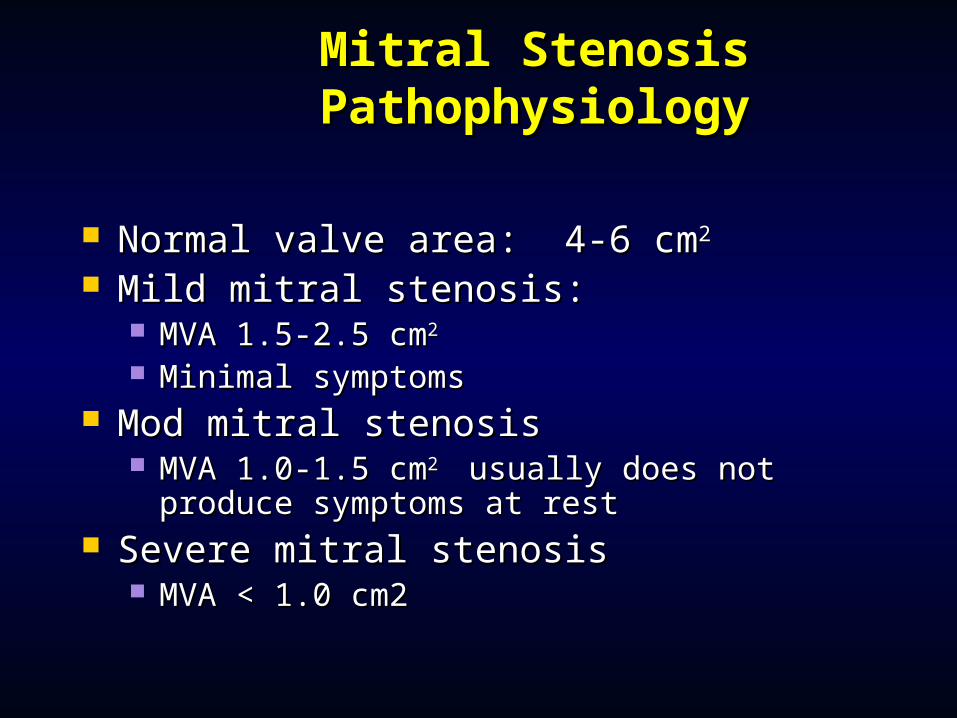
Mitral Stenosis Mitral Stenosis PathophysiologyPathophysiology
Normal valve area: 4-6 cmNormal valve area: 4-6 cm22
Mild mitral stenosis: Mild mitral stenosis: MVA 1.5-2.5 cmMVA 1.5-2.5 cm22
Minimal symptomsMinimal symptoms Mod mitral stenosisMod mitral stenosis
MVA 1.0-1.5 cmMVA 1.0-1.5 cm2 2 usually does not produce usually does not produce symptoms at restsymptoms at rest
Severe mitral stenosisSevere mitral stenosis MVA < 1.0 cm2MVA < 1.0 cm2
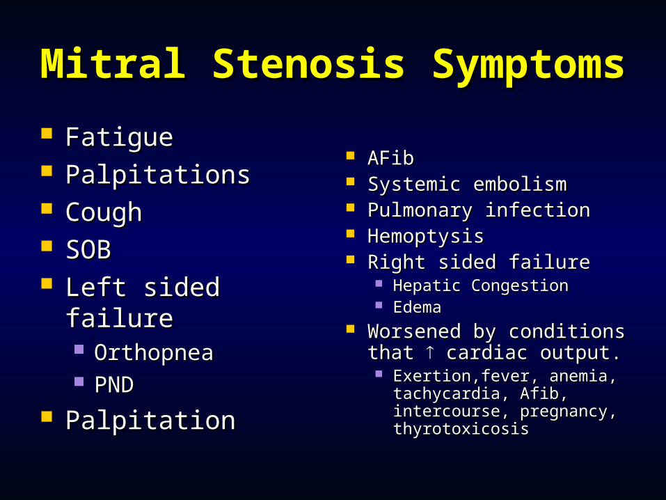
Mitral Stenosis Mitral Stenosis SymptomsSymptoms
Fatigue Fatigue PalpitationsPalpitations CoughCough SOBSOB Left sided failureLeft sided failure
OrthopneaOrthopnea PNDPND
PalpitationPalpitation
AFibAFib Systemic embolismSystemic embolism Pulmonary infectionPulmonary infection HemoptysisHemoptysis Right sided failureRight sided failure
Hepatic CongestionHepatic Congestion EdemaEdema
Worsened by conditions Worsened by conditions that that cardiac output. cardiac output. Exertion,fever, anemia, Exertion,fever, anemia,
tachycardia, Afib, tachycardia, Afib, intercourse, pregnancy, intercourse, pregnancy, thyrotoxicosisthyrotoxicosis
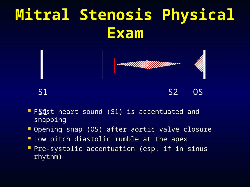
Mitral Stenosis Physical Mitral Stenosis Physical ExamExam
First heart sound (S1) is accentuated and snappingFirst heart sound (S1) is accentuated and snapping Opening snap (OS) after aortic valve closureOpening snap (OS) after aortic valve closure Low pitch diastolic rumble at the apexLow pitch diastolic rumble at the apex Pre-systolic accentuation (esp. if in sinus rhythm)Pre-systolic accentuation (esp. if in sinus rhythm)
S1 S2 OS S1
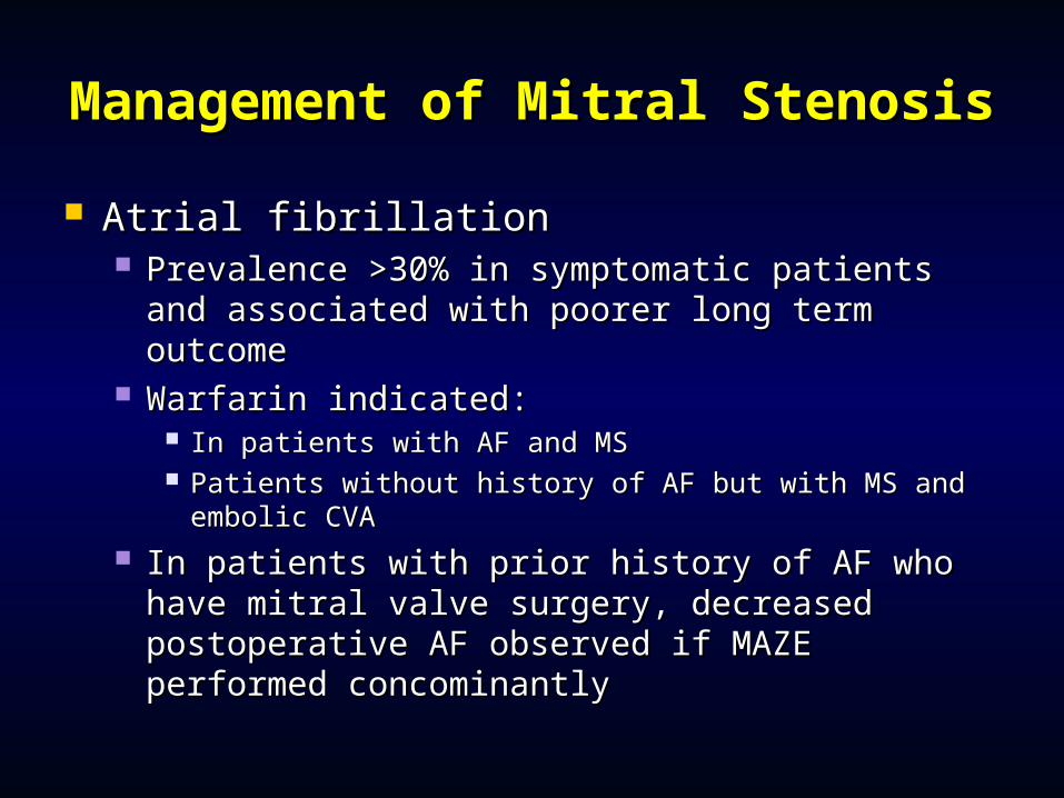
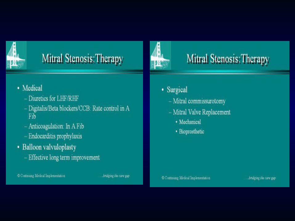
Management of Mitral Management of Mitral StenosisStenosis
Atrial fibrillationAtrial fibrillation Prevalence >30% in symptomatic patients and Prevalence >30% in symptomatic patients and
associated with poorer long term outcomeassociated with poorer long term outcome Warfarin indicated:Warfarin indicated:
In patients with AF and MSIn patients with AF and MS Patients without history of AF but with MS and Patients without history of AF but with MS and
embolic CVAembolic CVA In patients with prior history of AF who have In patients with prior history of AF who have
mitral valve surgery, decreased postoperative mitral valve surgery, decreased postoperative AF observed if MAZE performed AF observed if MAZE performed concominantlyconcominantly
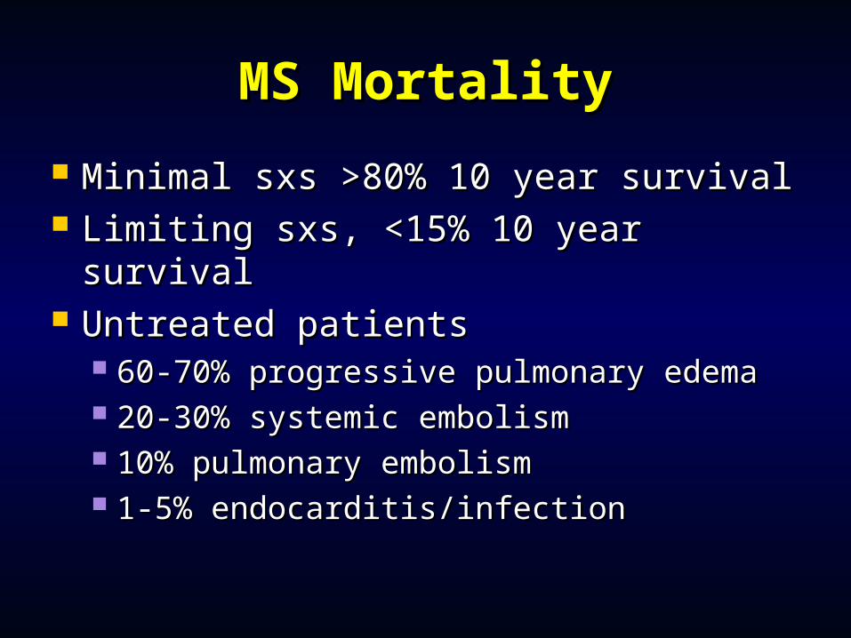
MS MortalityMS Mortality
Minimal sxs >80% 10 year survivalMinimal sxs >80% 10 year survival Limiting sxs, <15% 10 year survivalLimiting sxs, <15% 10 year survival Untreated patientsUntreated patients
60-70% progressive pulmonary edema60-70% progressive pulmonary edema 20-30% systemic embolism20-30% systemic embolism 10% pulmonary embolism10% pulmonary embolism 1-5% endocarditis/infection1-5% endocarditis/infection
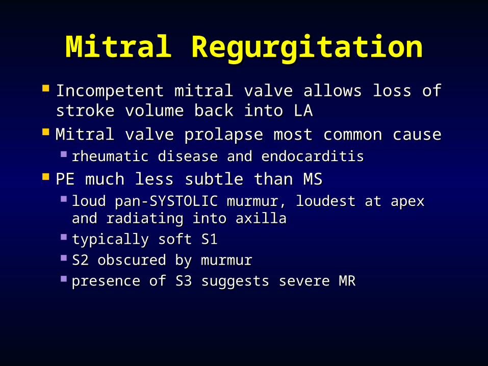
Mitral RegurgitationMitral Regurgitation Incompetent mitral valve allows loss of Incompetent mitral valve allows loss of
stroke volume back into LAstroke volume back into LA Mitral valve prolapse most common causeMitral valve prolapse most common cause
rheumatic disease and endocarditisrheumatic disease and endocarditis PE much less subtle than MSPE much less subtle than MS
loud pan-SYSTOLIC murmur, loudest at apex loud pan-SYSTOLIC murmur, loudest at apex and radiating into axillaand radiating into axilla
typically soft S1typically soft S1 S2 obscured by murmurS2 obscured by murmur presence of S3 suggests severe MRpresence of S3 suggests severe MR

Mitral Regurgitation: EtiologyMitral Regurgitation: Etiology
Valvular-leafletsValvular-leaflets Myxomatous MV Myxomatous MV
DiseaseDisease RheumaticRheumatic EndocarditisEndocarditis Congenital-cleftsCongenital-clefts
ChordaeChordae Fused/inflammatoryFused/inflammatory Torn/tTorn/traumarauma DegenerativeDegenerative IEIE
AnnulusAnnulus Calcification, Calcification, IE IE
(abcess)(abcess) Papillary MusclesPapillary Muscles
CAD (Ischemia, CAD (Ischemia, Infarction, Rupture)Infarction, Rupture)
HCMHCM Infiltrative disordersInfiltrative disorders
LV dilatation & LV dilatation & functional functional regurgitationregurgitation
TraumaTrauma
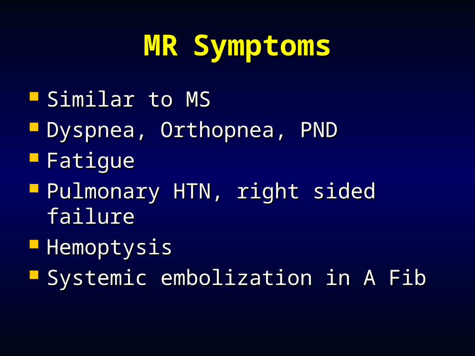
MRMR SymptomsSymptoms
Similar to MSSimilar to MS Dyspnea, Orthopnea, PNDDyspnea, Orthopnea, PND FatigueFatigue Pulmonary HTN, right sided failurePulmonary HTN, right sided failure HemoptysisHemoptysis Systemic embolization in A FibSystemic embolization in A Fib
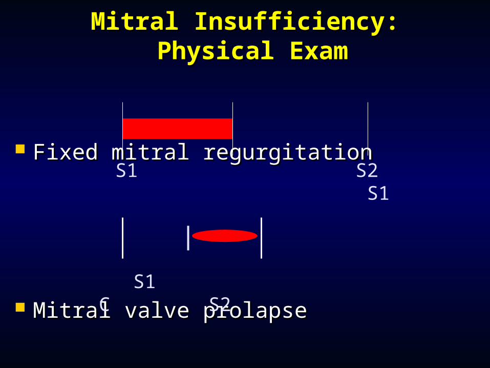
Fixed mitral regurgitationFixed mitral regurgitation
Mitral valve prolapseMitral valve prolapse
Mitral Insufficiency:Mitral Insufficiency: Physical Exam Physical Exam
S1 S2 S1
S1 C S2
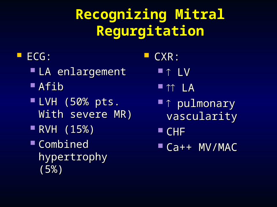
Recognizing Mitral Recognizing Mitral RegurgitationRegurgitation
ECG:ECG: LA enlargementLA enlargement AfibAfib LVH (50% pts. LVH (50% pts.
With severe MR)With severe MR) RVH (15%)RVH (15%) Combined Combined
hypertrophy hypertrophy (5%)(5%)
CXR:CXR: LVLV LALA pulmonary pulmonary
vascularityvascularity CHFCHF Ca++ MV/MACCa++ MV/MAC
MR TreatmentMR Treatment
No medical therapyNo medical therapy Most difficult clinicallyMost difficult clinically
By the time symptoms occur, it may be By the time symptoms occur, it may be too latetoo late
Drop in EF or development of atrial Drop in EF or development of atrial fibrillation enough to justify surgeryfibrillation enough to justify surgery
Mitral Valve Prolapse : Mitral Valve Prolapse : EpidemiologyEpidemiology
Affects 5-10% of population Affects 5-10% of population Most common cause of isolated severe MR Most common cause of isolated severe MR Females >> males; Ages of 14 and 30years Females >> males; Ages of 14 and 30years Strong hereditary component (? Autosomal Strong hereditary component (? Autosomal
Dominant)Dominant) 22ºº to failure of apposition/coaptation of the to failure of apposition/coaptation of the
anterior and posterior mitral valve leaflets.anterior and posterior mitral valve leaflets. Results form diverse pathologic conditions, but Results form diverse pathologic conditions, but
cause is unknown in a majority of ptscause is unknown in a majority of pts
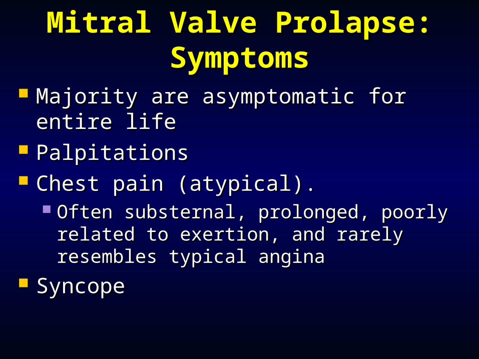
Mitral Valve Prolapse: Mitral Valve Prolapse: SymptomsSymptoms
Majority are asymptomatic for entire Majority are asymptomatic for entire lifelife
PalpitationsPalpitations Chest pain (atypical).Chest pain (atypical).
Often substernal, prolonged, poorly Often substernal, prolonged, poorly related to exertion, and rarely resembles related to exertion, and rarely resembles typical anginatypical angina
SyncopeSyncope
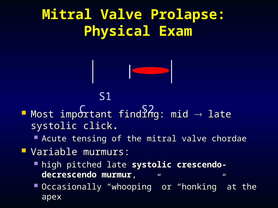
Mitral Valve Prolapse: Mitral Valve Prolapse: Physical ExamPhysical Exam
Most important finding: mid Most important finding: mid late systolic late systolic clickclick.. Acute tensing of the mitral valve chordaeAcute tensing of the mitral valve chordae
Variable murmurs:Variable murmurs: high pitched late high pitched late systolicsystolic crescendo-crescendo-
decrescendo murmurdecrescendo murmur, , Occasionally “whooping” or “honking” at the Occasionally “whooping” or “honking” at the
apexapex
S1 C S2
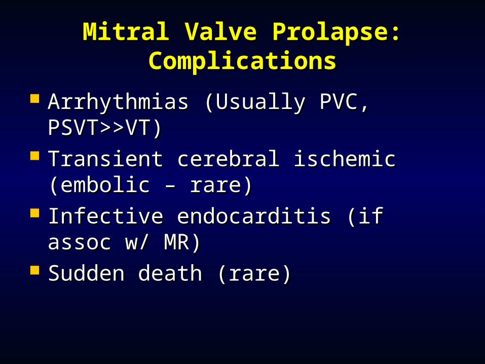
Mitral Valve Prolapse: Mitral Valve Prolapse: ComplicationsComplications
Arrhythmias (Usually PVC, Arrhythmias (Usually PVC, PSVT>>VT)PSVT>>VT)
Transient cerebral ischemic (embolic Transient cerebral ischemic (embolic – rare)– rare)
Infective endocarditis (if assoc w/ Infective endocarditis (if assoc w/ MR)MR)
Sudden death (rare)Sudden death (rare)
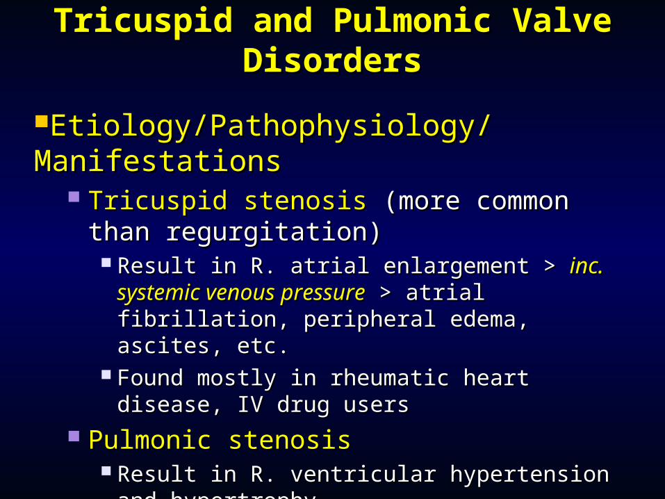
Tricuspid and Pulmonic Valve Tricuspid and Pulmonic Valve DisordersDisorders
Etiology/Pathophysiology/Etiology/Pathophysiology/ManifestationsManifestations
Tricuspid stenosisTricuspid stenosis (more common than (more common than regurgitation)regurgitation) Result in R. atrial enlargement > Result in R. atrial enlargement > inc. inc.
systemic venous pressuresystemic venous pressure > atrial fibrillation, > atrial fibrillation, peripheral edema, ascites, etc.peripheral edema, ascites, etc.
Found mostly in rheumatic heart disease, IV Found mostly in rheumatic heart disease, IV drug usersdrug users
Pulmonic stenosisPulmonic stenosis Result in R. ventricular hypertension and Result in R. ventricular hypertension and
hypertrophyhypertrophy Fatigue , loud midsystolic murmurFatigue , loud midsystolic murmur
Uncommon valve disordersUncommon valve disorders
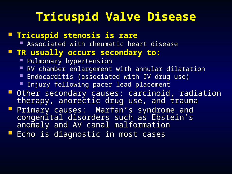
Tricuspid Valve DiseaseTricuspid Valve Disease Tricuspid stenosis is rareTricuspid stenosis is rare
Associated with rheumatic heart diseaseAssociated with rheumatic heart disease TR usually occurs secondary to:TR usually occurs secondary to:
Pulmonary hypertensionPulmonary hypertension RV chamber enlargement with annular dilatationRV chamber enlargement with annular dilatation Endocarditis (associated with IV drug use)Endocarditis (associated with IV drug use) Injury following pacer lead placementInjury following pacer lead placement
Other secondary causes: carcinoid, radiation Other secondary causes: carcinoid, radiation therapy, anorectic drug use, and traumatherapy, anorectic drug use, and trauma
Primary causes: Marfan’s syndrome and congenital Primary causes: Marfan’s syndrome and congenital disorders such as Ebstein’s anomaly and AV canal disorders such as Ebstein’s anomaly and AV canal malformationmalformation
Echo is diagnostic in most casesEcho is diagnostic in most cases
Tricuspid RegurgitationTricuspid Regurgitation
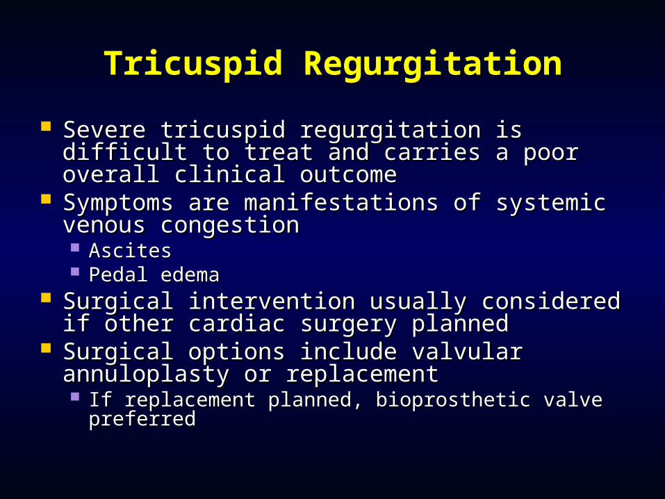
Severe tricuspid regurgitation is difficult to Severe tricuspid regurgitation is difficult to treat and carries a poor overall clinical treat and carries a poor overall clinical outcomeoutcome
Symptoms are manifestations of systemic Symptoms are manifestations of systemic venous congestionvenous congestion AscitesAscites Pedal edemaPedal edema
Surgical intervention usually considered if Surgical intervention usually considered if other cardiac surgery plannedother cardiac surgery planned
Surgical options include valvular Surgical options include valvular annuloplasty or replacementannuloplasty or replacement If replacement planned, bioprosthetic valve If replacement planned, bioprosthetic valve
preferredpreferred
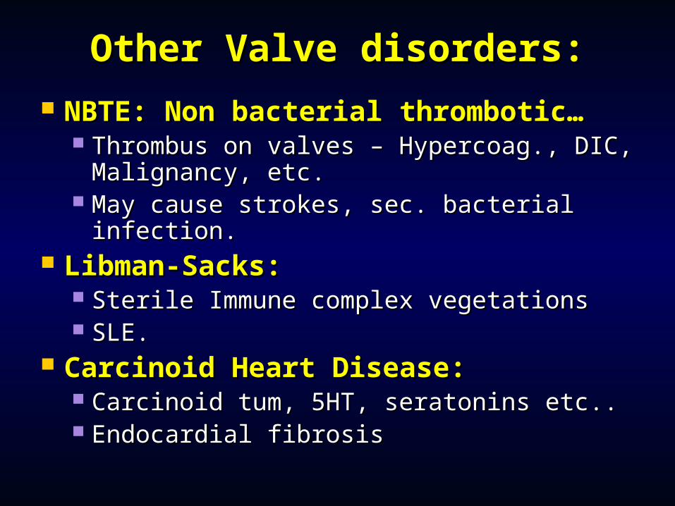
Other Valve disorders:Other Valve disorders: NBTE: Non bacterial thrombotic…NBTE: Non bacterial thrombotic…
Thrombus on valves – Hypercoag., DIC, Thrombus on valves – Hypercoag., DIC, Malignancy, etc.Malignancy, etc.
May cause strokes, sec. bacterial infection.May cause strokes, sec. bacterial infection. Libman-Sacks:Libman-Sacks:
Sterile Immune complex vegetations Sterile Immune complex vegetations SLE.SLE.
Carcinoid Heart Disease:Carcinoid Heart Disease: Carcinoid tum, 5HT, seratonins etc..Carcinoid tum, 5HT, seratonins etc.. Endocardial fibrosisEndocardial fibrosis
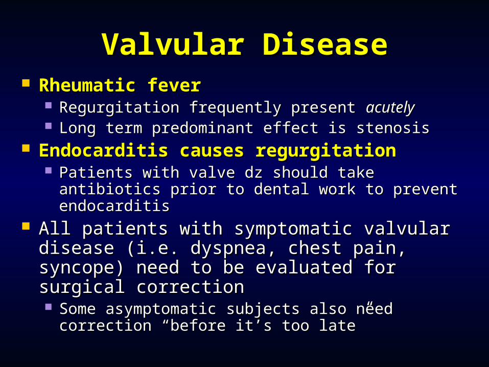
Valvular DiseaseValvular Disease Rheumatic feverRheumatic fever
Regurgitation frequently present Regurgitation frequently present acutelyacutely Long term predominant effect is stenosisLong term predominant effect is stenosis
Endocarditis causes regurgitationEndocarditis causes regurgitation Patients with valve dz should take antibiotics Patients with valve dz should take antibiotics
prior to dental work to prevent endocarditisprior to dental work to prevent endocarditis All patients with symptomatic valvular All patients with symptomatic valvular
disease (i.e. dyspnea, chest pain, syncope) disease (i.e. dyspnea, chest pain, syncope) need to be evaluated for surgical correctionneed to be evaluated for surgical correction Some asymptomatic subjects also need Some asymptomatic subjects also need
correction “before it’s too late”correction “before it’s too late”
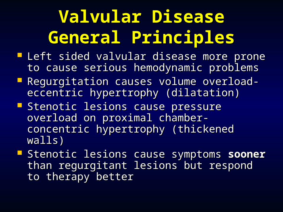
Valvular DiseaseValvular DiseaseGeneral PrinciplesGeneral Principles
Left sided valvular disease more prone to Left sided valvular disease more prone to cause serious hemodynamic problemscause serious hemodynamic problems
Regurgitation causes volume overload- Regurgitation causes volume overload- eccentric hypertrophy (dilatation)eccentric hypertrophy (dilatation)
Stenotic lesions cause pressure overload Stenotic lesions cause pressure overload on proximal chamber- concentric on proximal chamber- concentric hypertrophy (thickened walls)hypertrophy (thickened walls)
Stenotic lesions cause symptoms Stenotic lesions cause symptoms soonersooner than regurgitant lesions but respond to than regurgitant lesions but respond to therapy bettertherapy better
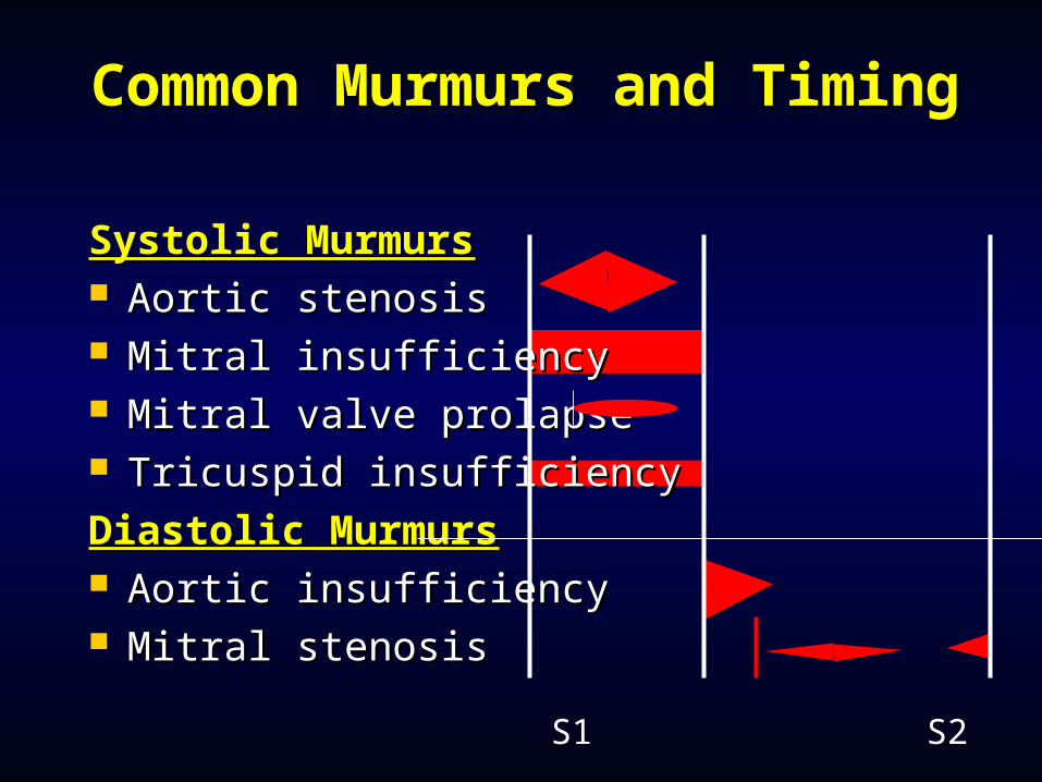
Common Murmurs and Common Murmurs and TimingTiming
Systolic MurmursSystolic Murmurs Aortic stenosisAortic stenosis Mitral insufficiencyMitral insufficiency Mitral valve prolapseMitral valve prolapse Tricuspid insufficiency Tricuspid insufficiency
Diastolic MurmursDiastolic Murmurs Aortic insufficiencyAortic insufficiency Mitral stenosisMitral stenosis
S1 S2 S1
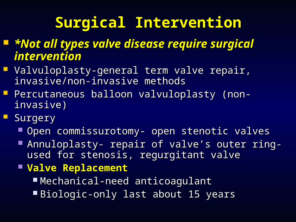
Surgical InterventionSurgical Intervention *Not all types valve disease require *Not all types valve disease require
surgical interventionsurgical intervention Valvuloplasty-general term valve repair, invasive/non-Valvuloplasty-general term valve repair, invasive/non-
invasive methodsinvasive methods Percutaneous balloon valvuloplasty (non-invasive)Percutaneous balloon valvuloplasty (non-invasive) SurgerySurgery
Open commissurotomy- open stenotic valvesOpen commissurotomy- open stenotic valves Annuloplasty- repair of valve’s outer ring-used for Annuloplasty- repair of valve’s outer ring-used for
stenosis, regurgitant valvestenosis, regurgitant valve Valve ReplacementValve Replacement
Mechanical-need anticoagulantMechanical-need anticoagulant Biologic-only last about 15 yearsBiologic-only last about 15 years
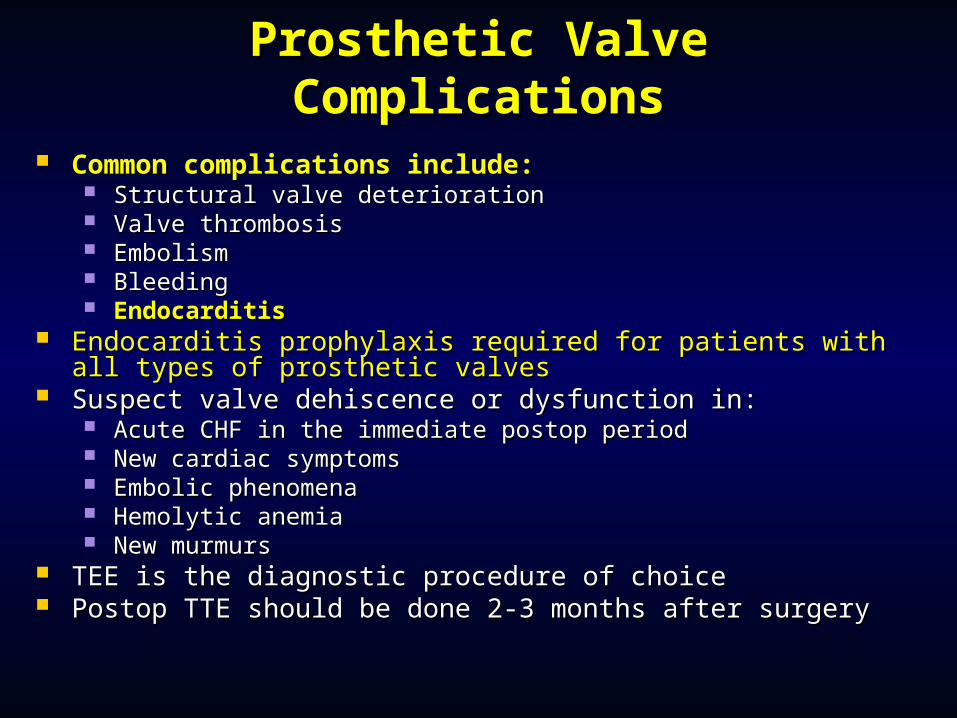
Prosthetic Valve ComplicationsProsthetic Valve Complications
Common complications include:Common complications include: Structural valve deteriorationStructural valve deterioration Valve thrombosisValve thrombosis EmbolismEmbolism BleedingBleeding EndocarditisEndocarditis
Endocarditis prophylaxis required for patients with all types of Endocarditis prophylaxis required for patients with all types of prosthetic valvesprosthetic valves
Suspect valve dehiscence or dysfunction in:Suspect valve dehiscence or dysfunction in: Acute CHF in the immediate postop periodAcute CHF in the immediate postop period New cardiac symptomsNew cardiac symptoms Embolic phenomenaEmbolic phenomena Hemolytic anemiaHemolytic anemia New murmursNew murmurs
TEE is the diagnostic procedure of choiceTEE is the diagnostic procedure of choice Postop TTE should be done 2-3 months after surgeryPostop TTE should be done 2-3 months after surgery
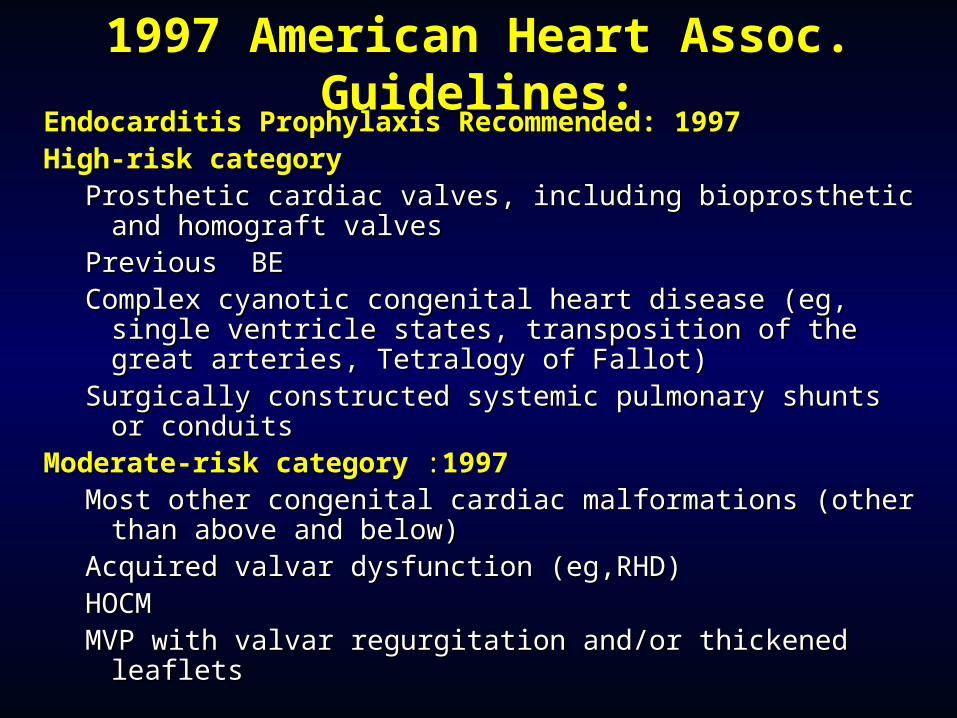
1997 American Heart Assoc. 1997 American Heart Assoc. Guidelines:Guidelines:
Endocarditis Prophylaxis Recommended:Endocarditis Prophylaxis Recommended: 19971997 High-risk category High-risk category
Prosthetic cardiac valves, including bioprosthetic and Prosthetic cardiac valves, including bioprosthetic and homograft valves homograft valves
Previous BE Previous BE Complex cyanotic congenital heart disease (eg, single Complex cyanotic congenital heart disease (eg, single
ventricle states, transposition of the great arteries, ventricle states, transposition of the great arteries, Tetralogy of Fallot) Tetralogy of Fallot)
Surgically constructed systemic pulmonary shunts or Surgically constructed systemic pulmonary shunts or conduits conduits
Moderate-risk categoryModerate-risk category : :19971997 Most other congenital cardiac malformations (other Most other congenital cardiac malformations (other
than above and below) than above and below) Acquired valvar dysfunction (eg,RHD) Acquired valvar dysfunction (eg,RHD) HOCM HOCM MVP with valvar regurgitation and/or thickened leafletsMVP with valvar regurgitation and/or thickened leaflets
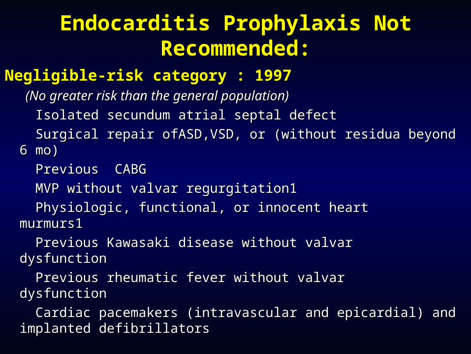
Endocarditis Prophylaxis Not Endocarditis Prophylaxis Not Recommended:Recommended:
Negligible-risk category : 1997Negligible-risk category : 1997 (No greater risk than the general population) (No greater risk than the general population)
Isolated secundum atrial septal defect Isolated secundum atrial septal defect
Surgical repair ofASD,VSD, or (without residua beyond Surgical repair ofASD,VSD, or (without residua beyond 6 mo) 6 mo)
Previous CABG Previous CABG
MVP without valvar regurgitation1 MVP without valvar regurgitation1
Physiologic, functional, or innocent heart murmurs1 Physiologic, functional, or innocent heart murmurs1
Previous Kawasaki disease without valvar Previous Kawasaki disease without valvar dysfunction dysfunction
Previous rheumatic fever without valvar dysfunction Previous rheumatic fever without valvar dysfunction
Cardiac pacemakers (intravascular and epicardial) and Cardiac pacemakers (intravascular and epicardial) and implanted defibrillatorsimplanted defibrillators
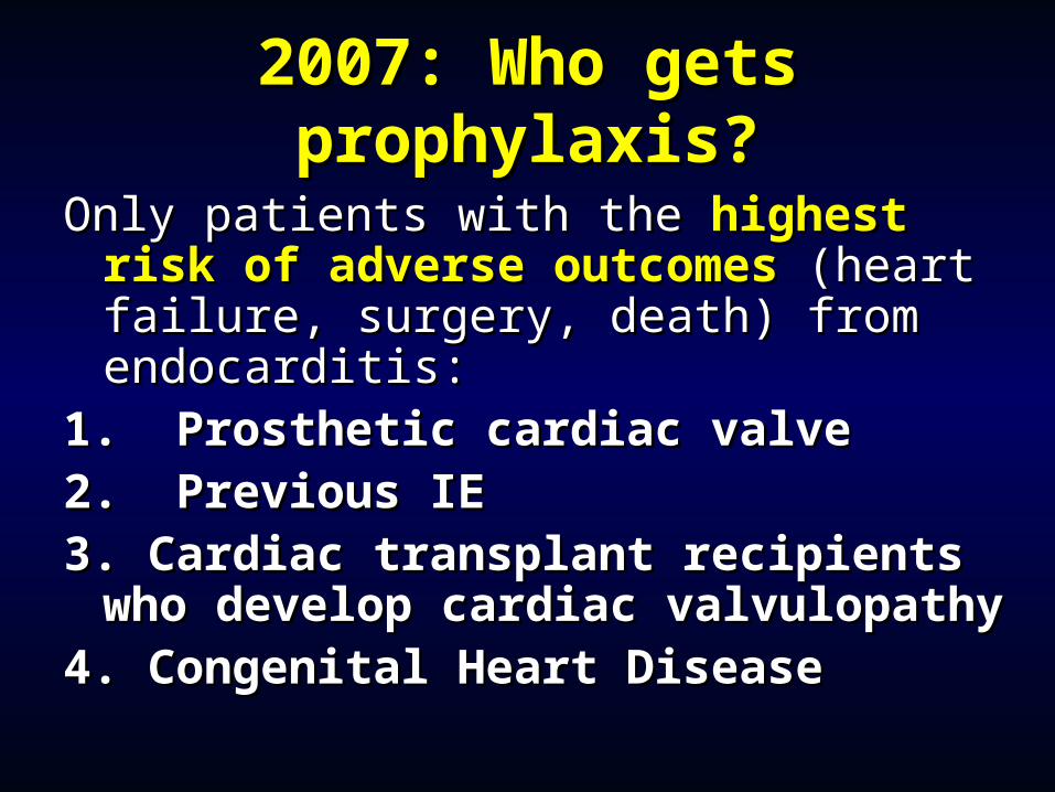
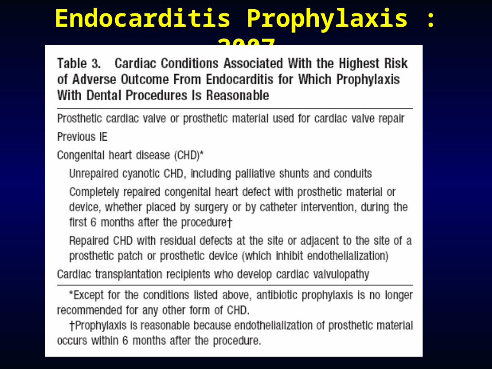
2007: Who gets 2007: Who gets prophylaxis?prophylaxis?
Only patients with the Only patients with the highest risk of highest risk of adverse outcomesadverse outcomes (heart failure, (heart failure, surgery, death) from endocarditis:surgery, death) from endocarditis:
1. Prosthetic cardiac valve1. Prosthetic cardiac valve2. Previous IE2. Previous IE3. Cardiac transplant recipients who 3. Cardiac transplant recipients who
develop cardiac valvulopathydevelop cardiac valvulopathy4. Congenital Heart Disease4. Congenital Heart Disease
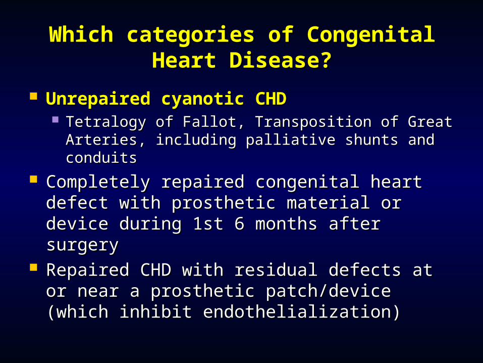
Which categories of Congenital Which categories of Congenital Heart Disease?Heart Disease?
Unrepaired cyanotic CHDUnrepaired cyanotic CHD Tetralogy of Fallot, Transposition of Great Tetralogy of Fallot, Transposition of Great
Arteries, including palliative shunts and Arteries, including palliative shunts and conduitsconduits
Completely repaired congenital heart Completely repaired congenital heart defect with prosthetic material or device defect with prosthetic material or device during 1st 6 months after surgeryduring 1st 6 months after surgery
Repaired CHD with residual defects at or Repaired CHD with residual defects at or near a prosthetic patch/device (which near a prosthetic patch/device (which inhibit endothelialization)inhibit endothelialization)
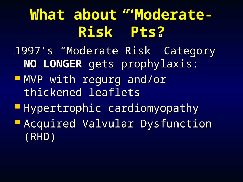
What about “Moderate-What about “Moderate-Risk” Pts?Risk” Pts?
1997’s “Moderate Risk” Category 1997’s “Moderate Risk” Category NO NO LONGERLONGER gets prophylaxis: gets prophylaxis:
MVP with regurg and/or thickened MVP with regurg and/or thickened leafletsleaflets
Hypertrophic cardiomyopathyHypertrophic cardiomyopathy Acquired Valvular Dysfunction Acquired Valvular Dysfunction
(RHD)(RHD)
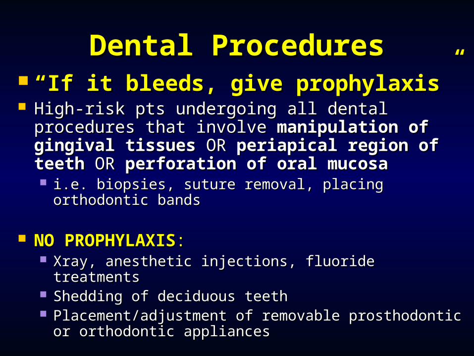
Dental ProceduresDental Procedures ““If it bleeds, give prophylaxis”If it bleeds, give prophylaxis” High-risk pts undergoing all dental High-risk pts undergoing all dental
procedures that involve procedures that involve manipulation of manipulation of gingival tissues gingival tissues OROR periapical region of periapical region of teeth teeth OROR perforation of oral mucosa perforation of oral mucosa i.e. biopsies, suture removal, placing orthodontic i.e. biopsies, suture removal, placing orthodontic
bandsbands
NO PROPHYLAXISNO PROPHYLAXIS:: Xray, anesthetic injections, fluoride treatmentsXray, anesthetic injections, fluoride treatments Shedding of deciduous teethShedding of deciduous teeth Placement/adjustment of removable prosthodontic Placement/adjustment of removable prosthodontic
or orthodontic appliancesor orthodontic appliances
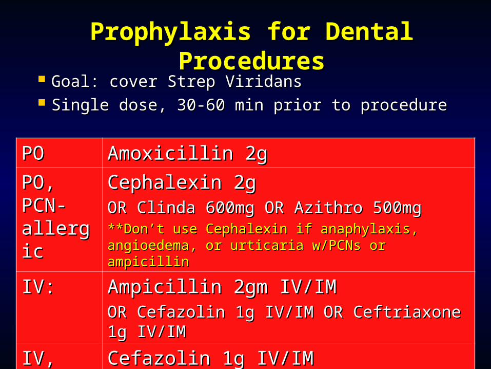
Prophylaxis for Dental Prophylaxis for Dental ProceduresProcedures
Goal: cover Strep ViridansGoal: cover Strep Viridans Single dose, 30-60 min prior to procedureSingle dose, 30-60 min prior to procedure
POPO Amoxicillin 2gAmoxicillin 2g
PO, PO, PCN-PCN-allergicallergic
Cephalexin 2g Cephalexin 2g OR Clinda 600mg OR Azithro 500mgOR Clinda 600mg OR Azithro 500mg**Don’t use Cephalexin if anaphylaxis, angioedema, **Don’t use Cephalexin if anaphylaxis, angioedema, or urticaria w/PCNs or ampicillinor urticaria w/PCNs or ampicillin
IV:IV: Ampicillin 2gm IV/IM Ampicillin 2gm IV/IM OR Cefazolin 1g IV/IM OR Ceftriaxone 1g OR Cefazolin 1g IV/IM OR Ceftriaxone 1g IV/IMIV/IM
IV, IV, PCN-PCN-allergicallergic
Cefazolin 1g IV/IM Cefazolin 1g IV/IM OR Ceftriaxone 1g IV/IM OR Clinda 600mg OR Ceftriaxone 1g IV/IM OR Clinda 600mg IV/IM IV/IM
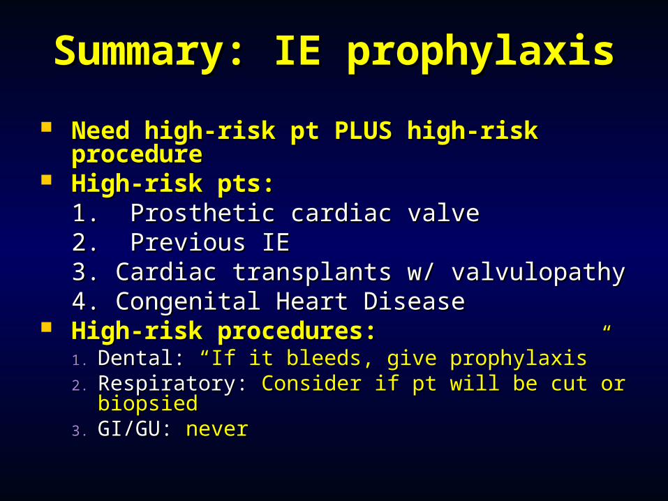
Summary: IE prophylaxisSummary: IE prophylaxis
Need high-risk pt PLUS high-risk Need high-risk pt PLUS high-risk procedureprocedure
High-risk pts: High-risk pts: 1. Prosthetic cardiac valve1. Prosthetic cardiac valve2. Previous IE2. Previous IE3. Cardiac transplants w/ valvulopathy3. Cardiac transplants w/ valvulopathy4. Congenital Heart Disease4. Congenital Heart Disease
High-risk procedures:High-risk procedures:1.1. Dental: Dental: “If it bleeds, give prophylaxis”“If it bleeds, give prophylaxis”2.2. Respiratory: Respiratory: Consider if pt will be cut or Consider if pt will be cut or
biopsiedbiopsied3.3. GI/GU: GI/GU: nevernever
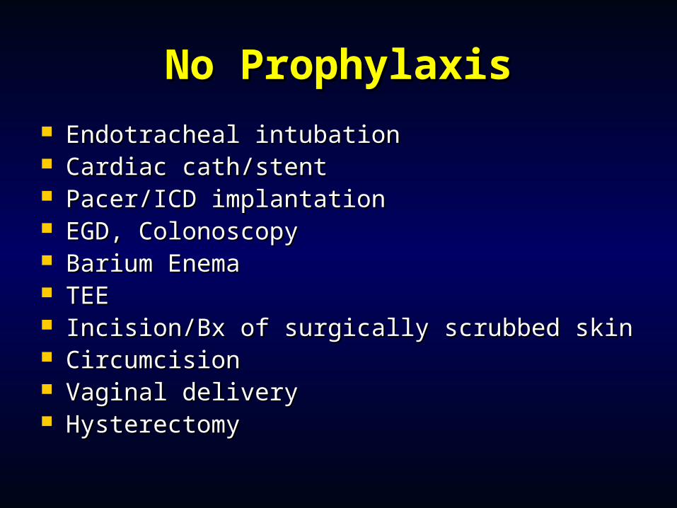
No ProphylaxisNo Prophylaxis Endotracheal intubationEndotracheal intubation Cardiac cath/stentCardiac cath/stent Pacer/ICD implantationPacer/ICD implantation EGD, ColonoscopyEGD, Colonoscopy Barium EnemaBarium Enema TEETEE Incision/Bx of surgically scrubbed skinIncision/Bx of surgically scrubbed skin CircumcisionCircumcision Vaginal deliveryVaginal delivery HysterectomyHysterectomy
Endocarditis Prophylaxis : Endocarditis Prophylaxis : 20072007
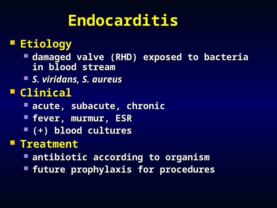
EndocarditisEndocarditis EtiologyEtiology
damaged valve (RHD) exposed to bacteria in damaged valve (RHD) exposed to bacteria in blood streamblood stream
S. viridans, S. aureusS. viridans, S. aureus ClinicalClinical
acute, subacute, chronicacute, subacute, chronic fever, murmur, ESRfever, murmur, ESR (+) blood cultures (+) blood cultures
TreatmentTreatment antibiotic according to organismantibiotic according to organism future prophylaxis for proceduresfuture prophylaxis for procedures
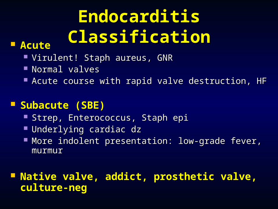
Endocarditis Endocarditis ClassificationClassification AcuteAcute
Virulent! Staph aureus, GNRVirulent! Staph aureus, GNR Normal valvesNormal valves Acute course with rapid valve destruction, HFAcute course with rapid valve destruction, HF
Subacute (SBE)Subacute (SBE) Strep, Enterococcus, Staph epiStrep, Enterococcus, Staph epi Underlying cardiac dzUnderlying cardiac dz More indolent presentation: low-grade fever, More indolent presentation: low-grade fever,
murmurmurmur
Native valve, addict, prosthetic valve, Native valve, addict, prosthetic valve, culture-negculture-neg
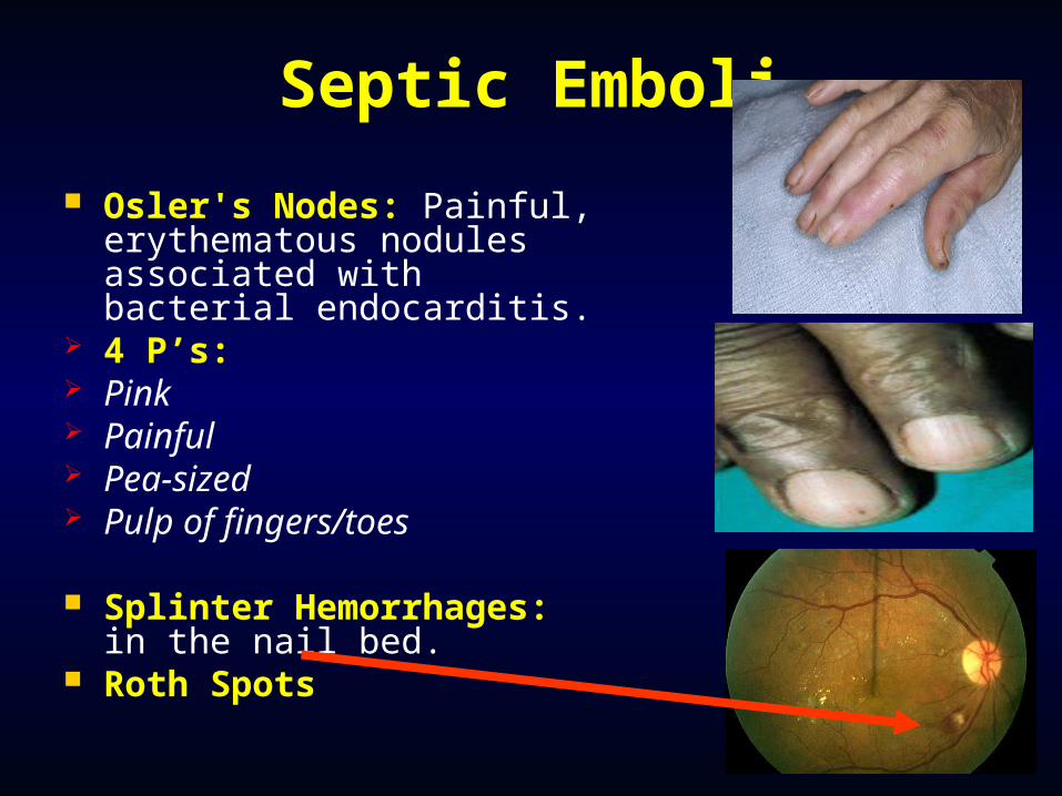
Septic Emboli
Osler's Nodes: Painful, erythematous nodules associated with bacterial endocarditis.
4 P’s: Pink Painful Pea-sized Pulp of fingers/toes
Splinter Hemorrhages: in the nail bed.
Roth Spots
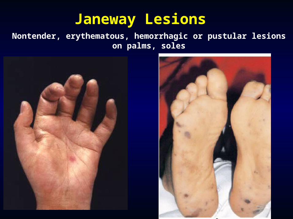
Janeway LesionsNontender, erythematous, hemorrhagic or pustular lesions
on palms, soles
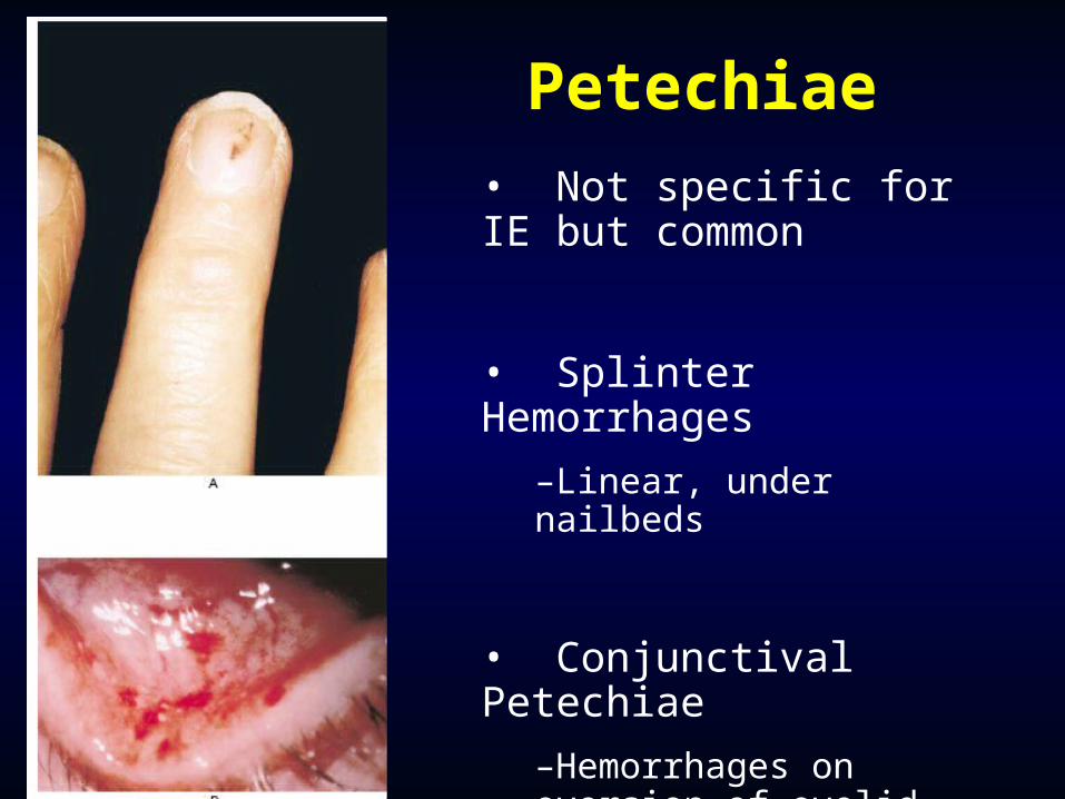
Petechiae• Not specific for IE but common
• Splinter Hemorrhages
–Linear, under nailbeds
• Conjunctival Petechiae
–Hemorrhages on eversion of eyelid
Prosthetic Valve Prosthetic Valve EndocarditisEndocarditis
Risk of IERisk of IE 1% at 12 mos, 2-3% at 60 mos post-op1% at 12 mos, 2-3% at 60 mos post-op
EARLY:EARLY: < 2 months post-op< 2 months post-op #1 cause: Staph aureus (used to be staph epi)#1 cause: Staph aureus (used to be staph epi) Usually acquired in the hospital: direct intra-op Usually acquired in the hospital: direct intra-op
contamination or post-op hematogenous spreadcontamination or post-op hematogenous spread Anchoring sutures and valve ring are not yet Anchoring sutures and valve ring are not yet
endothelialized, so more vulnerableendothelialized, so more vulnerable LATE:LATE: > 2 months post-op> 2 months post-op
Endothelialization occurs over months, making it Endothelialization occurs over months, making it more difficult for bugs to adhere. Community-more difficult for bugs to adhere. Community-acquired.acquired.
IVDUIVDU
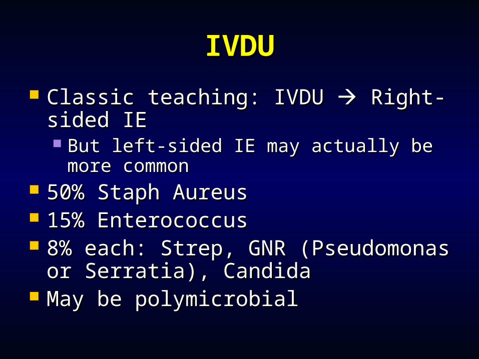
Classic teaching: IVDU Classic teaching: IVDU Right- Right-sided IEsided IE But left-sided IE may actually be more But left-sided IE may actually be more
commoncommon 50% Staph Aureus50% Staph Aureus 15% Enterococcus15% Enterococcus 8% each: Strep, GNR (Pseudomonas 8% each: Strep, GNR (Pseudomonas
or Serratia), Candidaor Serratia), Candida May be polymicrobialMay be polymicrobial
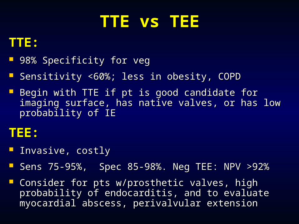
TTE vs TEETTE vs TEETTE:TTE: 98% Specificity for veg98% Specificity for veg
Sensitivity <60%; less in obesity, COPDSensitivity <60%; less in obesity, COPD
Begin with TTE if pt is good candidate for imaging Begin with TTE if pt is good candidate for imaging surface, has native valves, or has low probability of IEsurface, has native valves, or has low probability of IE
TEE:TEE: Invasive, costlyInvasive, costly
Sens 75-95%, Spec 85-98%. Neg TEE: NPV >92%Sens 75-95%, Spec 85-98%. Neg TEE: NPV >92%
Consider for pts w/prosthetic valves, high probability Consider for pts w/prosthetic valves, high probability of endocarditis, and to evaluate myocardial abscess, of endocarditis, and to evaluate myocardial abscess, perivalvular extensionperivalvular extension
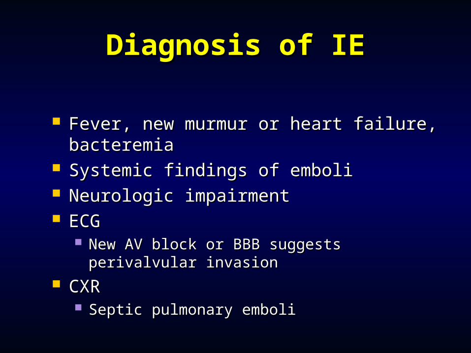
Diagnosis of IEDiagnosis of IE
Fever, new murmur or heart failure, Fever, new murmur or heart failure, bacteremiabacteremia
Systemic findings of emboliSystemic findings of emboli Neurologic impairmentNeurologic impairment ECGECG
New AV block or BBB suggests perivalvular New AV block or BBB suggests perivalvular invasioninvasion
CXRCXR Septic pulmonary emboliSeptic pulmonary emboli
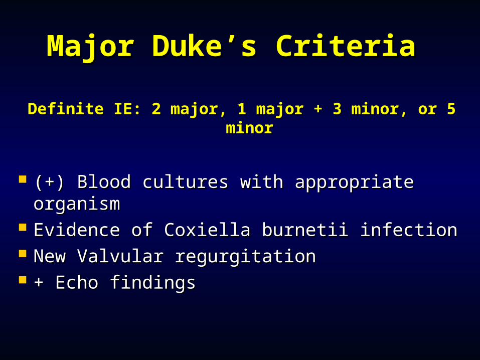
Major Duke’s CriteriaMajor Duke’s Criteria
Definite IE: 2 major, 1 major + 3 minor, or 5 Definite IE: 2 major, 1 major + 3 minor, or 5 minorminor
(+) Blood cultures with appropriate (+) Blood cultures with appropriate organismorganism
Evidence of Coxiella burnetii infectionEvidence of Coxiella burnetii infection New Valvular regurgitationNew Valvular regurgitation + Echo findings+ Echo findings
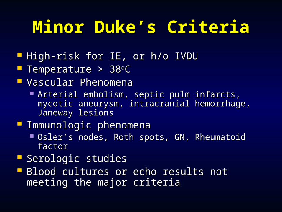
Minor Duke’s CriteriaMinor Duke’s Criteria High-risk for IE, or h/o IVDUHigh-risk for IE, or h/o IVDU Temperature > 38Temperature > 38ooCC Vascular PhenomenaVascular Phenomena
Arterial embolism, septic pulm infarcts, mycotic Arterial embolism, septic pulm infarcts, mycotic aneurysm, intracranial hemorrhage, Janeway aneurysm, intracranial hemorrhage, Janeway lesionslesions
Immunologic phenomenaImmunologic phenomena Osler’s nodes, Roth spots, GN, Rheumatoid factorOsler’s nodes, Roth spots, GN, Rheumatoid factor
Serologic studiesSerologic studies Blood cultures or echo results not meeting Blood cultures or echo results not meeting
the major criteriathe major criteria
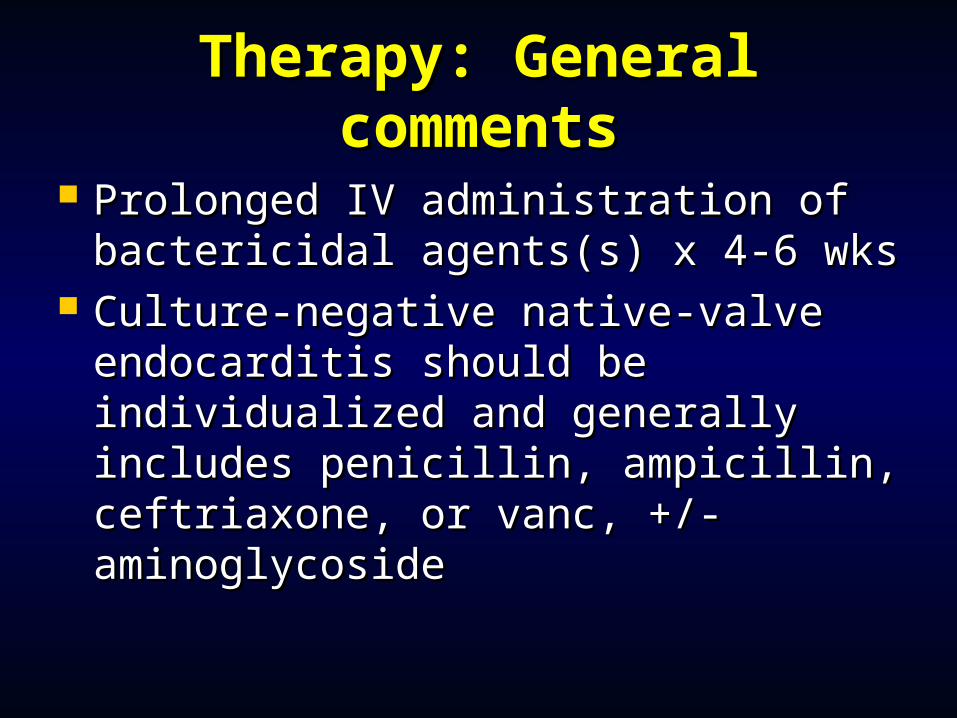
Therapy: General Therapy: General commentscomments
Prolonged IV administration of Prolonged IV administration of bactericidal agents(s) x 4-6 wksbactericidal agents(s) x 4-6 wks
Culture-negative native-valve Culture-negative native-valve endocarditis should be endocarditis should be individualized and generally includes individualized and generally includes penicillin, ampicillin, ceftriaxone, or penicillin, ampicillin, ceftriaxone, or vanc, +/- aminoglycosidevanc, +/- aminoglycoside
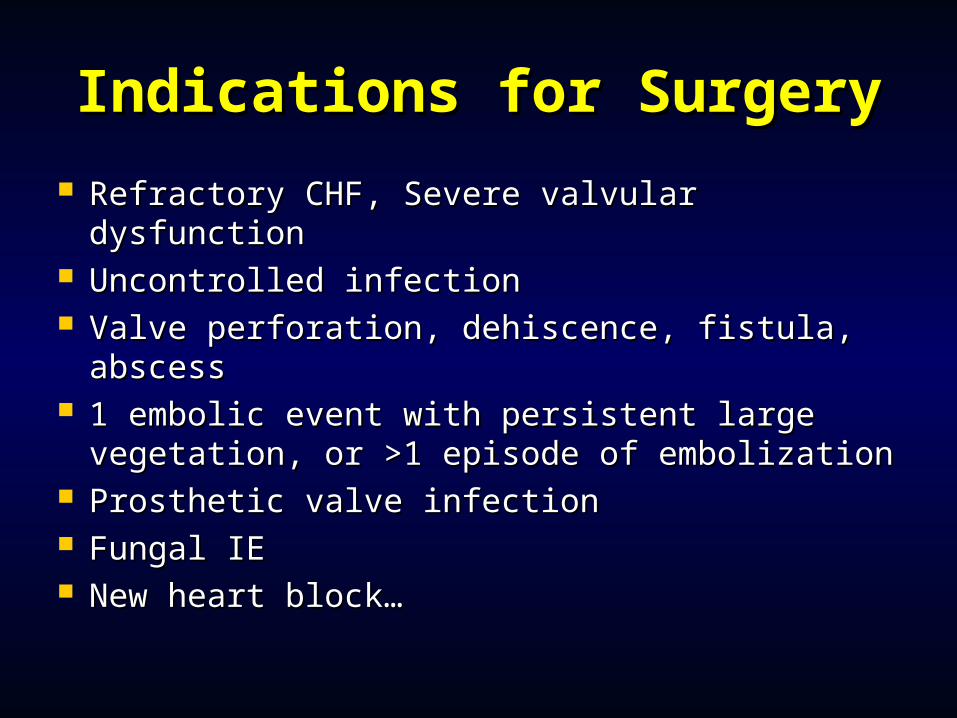
Indications for SurgeryIndications for Surgery
Refractory CHF, Severe valvular Refractory CHF, Severe valvular dysfunctiondysfunction
Uncontrolled infectionUncontrolled infection Valve perforation, dehiscence, fistula, Valve perforation, dehiscence, fistula,
abscessabscess 1 embolic event with persistent large 1 embolic event with persistent large
vegetation, or >1 episode of embolizationvegetation, or >1 episode of embolization Prosthetic valve infectionProsthetic valve infection Fungal IEFungal IE New heart block…New heart block…
ConclusionsConclusions
Valvular heart disease associated Valvular heart disease associated with spectrum of presentationswith spectrum of presentations
Recognition prior to the onset of Recognition prior to the onset of symptoms may be life savingsymptoms may be life saving
Careful physical exam almost always Careful physical exam almost always diagnosticdiagnostic
““Pearls”Pearls” Diastolic murmurs usually represent pathological Diastolic murmurs usually represent pathological
conditions as do most continuous murmurs.conditions as do most continuous murmurs. Most important issue in patient with a cardiac murmur is Most important issue in patient with a cardiac murmur is
the presence or absence of symptoms. the presence or absence of symptoms. Many asymptomatic children and young adults with grade 2/6 Many asymptomatic children and young adults with grade 2/6
midsystolic murmurs and no other cardiac physical findings midsystolic murmurs and no other cardiac physical findings need no further cardiac evaluationneed no further cardiac evaluation
Many asymptomaticMany asymptomatic e elderly patients have midsystolic lderly patients have midsystolic murmurs related to sclerotic aortic valve leaflets, flow into murmurs related to sclerotic aortic valve leaflets, flow into tortuous, noncompliant great vessels tortuous, noncompliant great vessels
Such murmurs must be distinguished from murmurs caused by Such murmurs must be distinguished from murmurs caused by mild to severe valvular aortic stenosis (AS) which is prevalent in mild to severe valvular aortic stenosis (AS) which is prevalent in this age group.this age group.