Embed Size (px)
Citation preview
120 Korean J Radiol 8(2), April 2007
Variable CT Findings of Epithelial OriginOvarian Carcinoma According to theDegree of Histologic Differentiation
Objective: We wanted to evaluate the CT findings of epithelial origin ovariancarcinoma according to the degree of histologic differentiation.
Materials and Methods: This study enrolled 124 patients with 31 well differen-tiated, 44 moderately differentiated and 95 poorly differentiated carcinomas withepithelial origin. The CT images were retrospectively evaluated with regard tobilateral ovarian involvement, the tumor’s nature, lymphadenopathy, adjacentorgan invasion, peritoneal tumor seeding, a large amount of ascites and distantmetastasis. In cystic, predominantly cystic and mixed tumors, the tumor wall,septa, papillary projection and necrosis in the solid portion were assessed.
Results: Bilateral ovarian involvement was more common in the poorly (48%)and moderately (42%) differentiated carcinomas than in the well differentiatedcarcinomas (7%) (p < 0.05). The frequency of a predominantly solid or solidnature was greater in the moderately and poorly differentiated carcinomas than inthe well differentiated carcinomas (p < 0.0001). In the 87 tumors with a cystic,predominantly cystic or mixed nature, septa greater than 3 mm, papillary projec-tion and necrosis in the solid portion were more common in the poorly differentiat-ed carcinoma (91%, 91% and 77%, respectively) than in the moderately (64%,68% and 34%, respectively) and well differentiated carcinomas (63%, 47% and27%, respectively) (p < 0.05). Lymphadenopathy, organ invasion, tumor seedingand a large amount of ascites were more common in the poorly differentiated car-cinomas (38%, 27%, 73% and 69%, respectively) than in the moderately (13%,10%, 48% and 45%, respectively) and well differentiated carcinomas (3%, 0%,10% and 17%, respectively) (p < 0.05).
Conclusion: Epithelial origin ovarian carcinoma shows different CT findingsaccording to the degree of histologic differentiation.
varian cancer is the second most common gynecologic malignancy, and itis one of the leading causes of death from cancer in women (1). Physiciansplay important roles for both early detection and making an accurate
diagnosis of malignant tumors, based on proper tissue characterization, for optimallymanaging these patients. Many previous studies have investigated the imaging findingsfor differentiating malignant ovarian tumors from benign tumors. According to thosereports, irregular wall thickening, large size, irregular septa, papillary projections andnecrosis in the solid portion favor the diagnosis of ovarian malignancy (2 9). In theearly 2000s, Hricak et al. (9) postulated that necrosis in a solid lesion and vegetations ina cystic lesion are the most predictive MR imaging findings of ovarian malignancy.However, most previous studies had classified ovarian tumor simply as benign ormalignancy, but the studies had not considered the degree of histologic differentiation
Yun-Jin Jang, MDJeong Kon Kim, MDSung Bin Park, MDKyoung-Sik Cho, MD
Index terms:Ovarian cancer, differentiationComputed tomography (CT)
Korean J Radiol 2007;8:120-126Received October 19, 2005; accepted after revision February 9, 2006.
Department of Radiology and ResearchInstitute of Radiology, University of UlsanCollege of Medicine, Asan MedicalCenter, Seoul 138-736, Korea
Address reprint requests to:Jeong Kon Kim, MD, Department ofRadiology and Research Institute ofRadiology, University of Ulsan College ofMedicine, Asan Medical Center, Seoul138-736, South Korea, 388-1 Poongnap-dong, Songpa-gu, Seoul 138-736, Korea.Tel. (822) 3010-4355Fax. (822) 476-0090e-mail: rialto@ amc.seoul.kr
O
in malignant tumors. The degree of differentiation ofovarian carcinoma is thought to be related to the grossmorphology. Therefore, it can be hypothesized that theimaging findings of ovarian carcinoma may vary accordingto their degree of differentiation.
Epithelial origin ovarian tumors consist of the largestportion of ovarian tumor.
Despite the inferiority of CT compared to MR for charac-terizing ovarian tumor, CT is still widely used for makingthe diagnosis of ovarian carcinoma. The purpose of thisstudy is to investigate the variable CT findings of epithelialorigin ovarian cancer according to the histologic degree ofdifferentiation.
MATERIALS AND METHODS
Our institutional review board approved this study;however, informed consents from the subjects were notrequired for this retrospective study.
Patient PopulationA computerized search of the medical records between
January 1997 and March 2003 at our institution revealed164 patients who had undergone surgery for epithelialorigin ovarian carcinoma. From those patients, wecollected the records of 124 patients (mean age: 52 years)who had available abdomen and pelvic CT imagesobtained before surgery (1 30 days), and who had no pasthistory of other malignant disease before the diagnosis ofovarian carcinoma. None of these patients had undergonechemotherapy for ovarian cancer prior to the CT examina-tion. All the tumors included in this study arose fromepithelial cells, and the histologic diagnoses aresummarized in Table 1.
Based on the pathologic reports of these patients thatwere written by various staff pathologists at our institution,each epithelial origin ovarian carcinoma was classified aswell, moderately or poorly differentiated according to thedegree of differentiation by referring to the modifiedBroder’s grading system (10, 11). According to this system,well differentiated cells were more than 75% of the tumorcells in grade 1 (well differentiated) carcinoma, they were25% 75% of the tumor cells in grade 2 (moderatelydifferentiated) carcinoma and they were less than 25% ofthe tumor cells in grade 3 (poorly differentiated) carcinoma(10, 11). Consequently, the number of patients with well,moderately and poorly differentiated carcinoma was 29,31 and 64, respectively.
CT ExaminationOne hundred and eleven patients underwent CT
examination at our institution, but 13 patients with one welldifferentiated carcinoma, eight with moderately differenti-ated carcinoma and four with poorly differentiatedcarcinoma had CT images obtained at other institutions.The imaging acquisition parameters of the CT imagesobtained at other institutions were variable, but all theimages included contrast-enhanced scans with a 5 10 mmslice thickness. At our institution, the CT examinationswere performed using a single-detector row helical CTscanner (Somatom Plus-S; Siemens Medical Systems,Erlangen, Germany) for 88 patients, including 26 with welldifferentiated carcinoma, 16 with moderately differentiatedcarcinoma and 46 with poorly differentiated carcinoma.Multi-detector row helical CT scanners (LightSpeed QX/i,General Electric Medical System, Milwaukee, WI) wereused for the remaining 23 patients, including two with welldifferentiated carcinoma, seven with moderately differenti-ated carcinoma and 14 with poorly differentiatedcarcinoma. All patients received 500 900 mL of oralcontrast material (E-Z-CAT [2% barium sulfate suspen-sion]; E-Z-EM, Westbury, NY) 30 minutes prior to the CTexamination. Intravenous contrast material (Ultravist 300[iopromide]; Schering, Berlin, Germany or Iopamiro 300[iopamidol]; Bracco, Milano, Italy) was administered intothe antecubital vein with using a power injector at a dose of2 mL/kg to a maximum dose of 160 mL at a rate of 3mL/sec. The scan delay for contrast-enhanced scanning was100 120 seconds for the single-detector row helical CTscanning and 90 100 seconds for the multi-detector rowhelical CT scanning. Scan coverage was from the diaphrag-matic dome to the ischial tuberosities.
The scanning parameters for single-detector row helicalCT were section collimation: 7 mm, pitch: 1.5, table speed:7.5 mm per rotation (10 mm/sec), reconstruction interval:5 mm, 120 kV and 210 mA. For multi-detector row helicalCT, a section collimation of 5 mm 4, a beam pitch of 1.5,a reconstruction interval of 5 mm, a X-ray tube voltage of120 kV and a tube current of 210 240 mA were used.
Analysis of CT FindingsTwo radiologists, who were unaware of the pathologic
CT Findings vs. Histologic Differentiation in Ovarian Epithelial Carcinomas
Korean J Radiol 8(2), April 2007 121
Table 1. Histopathologic Diagnosis in 124 Patients
Diagnosis Number
Serous adenocarcinoma 65Mucinous adenocarcinoma 22Endometrioid carcinoma 22Transitional cell carcinoma 01Mixed subtype 05Undifferentiated carcinoma 09
reports of ovarian carcinoma, but who knew about thepresence of ovarian cancer in each patient, evaluated theCT findings in a consensus fashion on the picture archivingand communication system, PetaVision (Asan MedicalCenter, Seoul, Korea); this made it possible to measure thetumor diameter and attenuation in a particular region ofinterest.
The reviewers evaluated the presence or absence ofbilateral ovarian involvement and the nature of the tumoraccording to the ratio of the cystic portion in the tumor:this included cystic tumor (approximately 100%), predom-inantly cystic tumor (> 70%), mixed tumor (30 70%),predominantly solid tumor (< 30%), and purely solidtumor (approximately 0%). The presence of a solid portionwithin the tumor was determined when an area showed anattenuation value greater than 60 HU. Then, in tumorswith cystic, predominantly cystic or mixed nature, thepresence or absence of septa, papillary projection andnecrosis in the solid portion were evaluated. Furthermore,the presence or absence of a wall and/or septa greater than3 mm at the maximum thickness was also evaluated. Theancillary findings were also evaluated, includinglymphadenopathy, tumor seeding, organ invasion,metastasis and a large amount of ascites.Lymphadenopathy was considered to be present when theminimum diameter of a lymph node was greater than 1cm. The presence of organ invasion was regarded to existwhen an ovarian tumor encased more than 50% of thesurface of an adjacent organ. A large amount of ascites wasindicated when ascites was noted in both the abdomen andpelvis.
Statistical AnalysisThe frequency of bilateral ovarian involvement, the
tumor nature, septa, papillary projection, necrosis,lymphadenopathy, tumor seeding, organ invasion, ascitesand metastasis were all compared in well, moderately, andpoorly differentiated ovarian carcinomas by using Fisher’sexact test. P values < 0.05 were considered statisticallysignificant.
RESULTS
Bilateral Ovarian InvolvementAmong 124 patients, 46 patients had bilateral ovarian
involvement. Only two (7%) of the 29 patients with welldifferentiated carcinoma had bilateral ovarian involve-ment, whereas 13 (42%) of the 31 patients withmoderately differentiated carcinoma and 31 (48%) of the64 patients with poorly differentiated ovarian carcinomashowed bilateral ovarian involvement. The frequency of
bilateral ovarian involvement was greater in themoderately and poorly differentiated carcinomas than inthe well differentiated carcinomas (p = 0.002 and p <0.0001, respectively), whereas the frequency was notsignificantly different between moderately and poorlydifferentiated carcinomas (p = 0.662) (Figs. 1, 2).
Tumor NatureA total of 170 ovarian tumors from 124 patients were
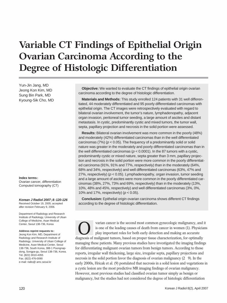
analyzed. The number of well, moderately and poorlydifferentiated carcinomas was 31, 44 and 95, respectively.Comparison of the tumor nature in the well, moderatelyand poorly differentiated carcinomas is summarized inTable 2. Well differentiated carcinomas showed a strongtendency to be cystic or predominantly cystic nature, aswas demonstrated by 25 (81%) of 31 tumors (Fig. 1). Incontrast, the moderately or poorly differentiated carcino-mas tended to be mixed, predominantly solid or solid innature with a frequency of 73% (32/44) for themoderately differentiated carcinomas and 88% (84/95) for
Jang et al.
122 Korean J Radiol 8(2), April 2007
Fig. 1. 30-year-old female patient with well differentiated ovariancarcinoma. Transverse CT image shows 12-cm-sized unilocularcystic mass (arrows). Mass originates from the right ovary andthe left ovary (not shown) is normal.
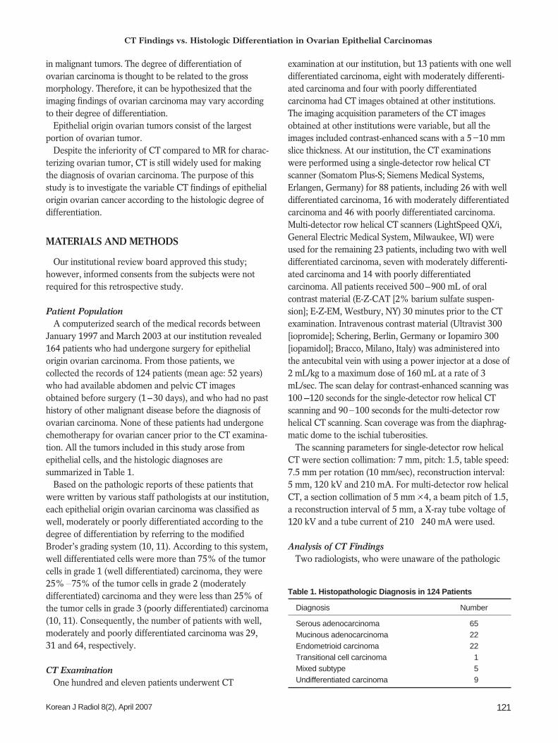
Fig. 2. 46-year-old female patient with poorly differentiatedovarian carcinoma. Transverse CT image shows two massesinvolving bilateral ovaries (arrows). Both masses show solidnature.
the poorly differentiated carcinomas (Fig. 2). Thefrequency of a cystic or predominantly cystic nature wasgreater in the well differentiated carcinomas than in themoderately differentiated (p < 0.0001) or poorly differenti-ated carcinomas (p < 0.0001). This frequency was alsogreater in the moderately differentiated carcinomas than inthe poorly differentiated carcinomas (p = 0.028). Incontrast, the frequency of a predominantly solid or solidnature was greater in the moderately and poorly differenti-ated carcinomas than in the well differentiated carcinomas(p < 0.0001).
Comparison of having a septa or wall in the 87 tumorswith a cystic, predominantly cystic or mixed nature issummarized in Table 3. The distribution of having amaximum wall thickness was similar in these three groups(p > 0.05). A septa greater than 3 mm for the maximumthickness was more frequently noted in the poorly differ-entiated carcinomas (91%) than in the well (63%) ormoderately differentiated carcinomas (64%) (p < 0.0001and p = 0.015, respectively) (Figs. 3, 4).
In 87 cystic, predominantly cystic and mixed tumors,papillary projection was noted in 14 (47%) of the 30 welldifferentiated carcinomas, in 15 (68%) of the 22
moderately differentiated carcinomas and in 32 (91%) ofthe 35 poorly differentiated carcinomas. Papillary projec-tion was more frequently noted in the poorly differentiatedcarcinomas than in the well differentiated (p < 0.0001) ormoderately differentiated carcinomas (p = 0.035), whereasthis frequency was similar in well differentiated andmoderately differentiated carcinomas (p = 0.162).
In 87 cystic, predominantly cystic and mixed tumors,necrosis in the solid portion was observed in eight (27%)
CT Findings vs. Histologic Differentiation in Ovarian Epithelial Carcinomas
Korean J Radiol 8(2), April 2007 123
Table 3. Comparison of the Walls and Septa in Well,Moderately and Poorly Differentiated Carcinoma
Differentiation Wall Thickness < 3 mm Wall Thickness > 3 mm
Well (n = 30) 3 (10) 27 (90)Moderate (n = 22) 4 (18) 18 (82)Poor (n = 35) 5 (14) 30 (86)
No septa Septa < 3 mm Septa 3 mm
Well (n = 30) 5 (17) 6 (20) 19 (63)Moderate (n = 22) 5 (23) 3 (14) 14 (64)Poor (n = 35) 1 (2)0 2 (6)0 32 (91)
Note. Numbers in parentheses are percentages.
Table 2. Comparison of the Tumor Nature in Well, Moderately and Poorly Differentiated Carcinomas
Differentiation Cystic Predominantly Cystic Mixed Predominantly Solid Solid
Well (n = 31) 11 (35) 14 (45) 05 (16) 1 (3) 0 (0)Moderate (n = 44) 06 (14) 06 (14) 10 (23) 08 (18) 14 (32)Poor (n = 95) 4 (4) 7 (7) 24 (25) 17 (18) 43 (45)
Note. Numbers in parentheses are percentages.
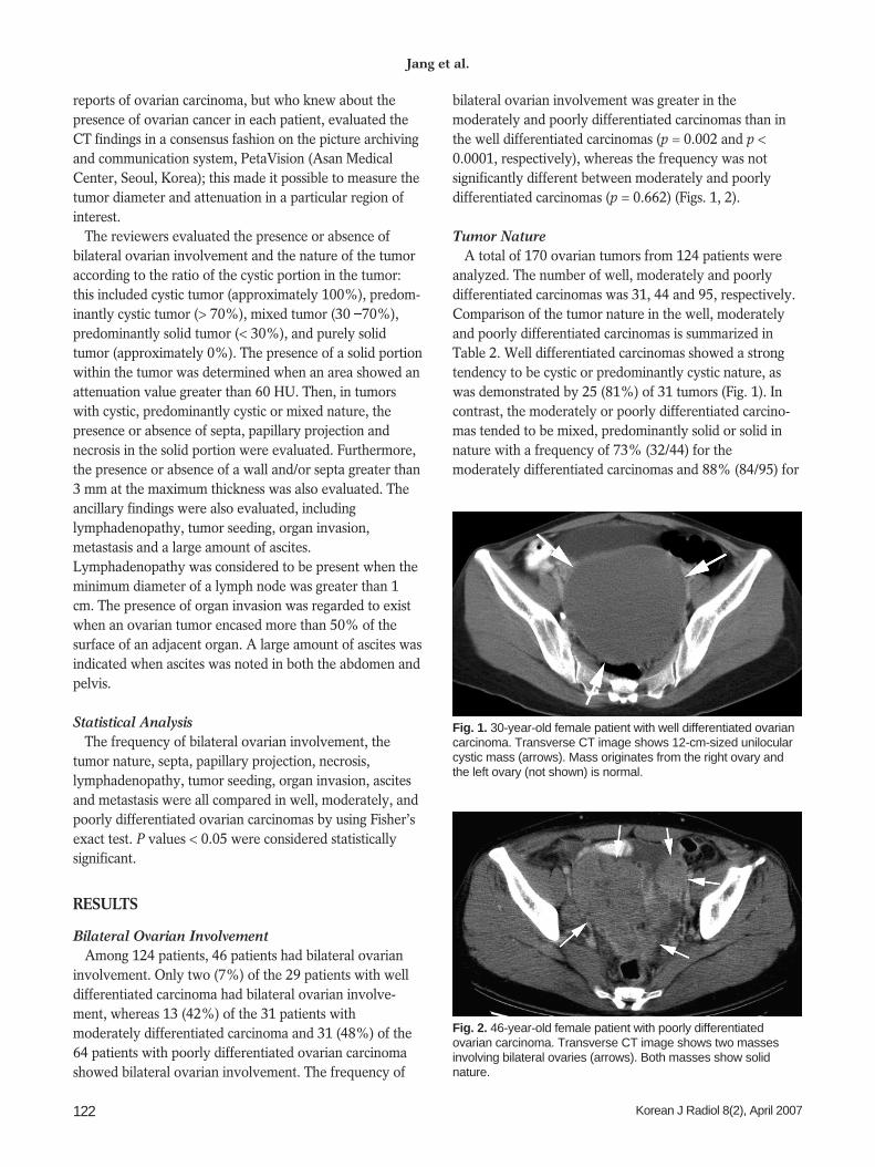
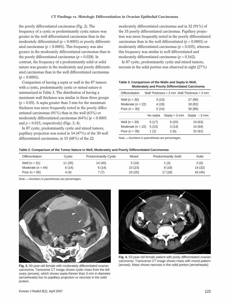
Fig. 4. 53-year-old female patient with pooly differentiated ovariancarcinoma. Transverse CT image shows mass with mixed pattern(arrows). Mass shows necrosis in the solid portion (arrowheads).Fig. 3. 50-year-old female with moderately differentiated ovarian
carcinoma. Tansverse CT image shows cystic mass from the leftovary (arrows), which shows septa thinner than 3 mm in diameter(arrowheads) but no papillary projection or necrosis in the solidprotion.
of the 30 well differentiated carcinomas, in eight (34%) ofthe 22 moderately differentiated carcinomas, and in 27(77%) of the 35 poorly differentiated carcinomas. Thefrequency of necrosis was greater in the poorly differenti-ated carcinomas than in the well differentiated (p <0.0001) or moderately differentiated carcinomas (p =0.005). This frequency was similar in the well differenti-ated and moderately differentiated carcinomas (p = 0.548)(Figs. 3, 4).
Ancillary FindingsThe results of the ancillary findings are summarized in
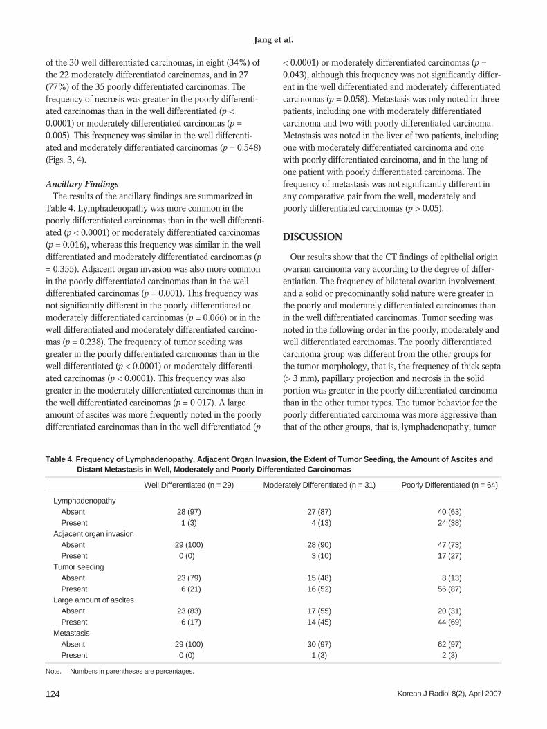
Table 4. Lymphadenopathy was more common in thepoorly differentiated carcinomas than in the well differenti-ated (p < 0.0001) or moderately differentiated carcinomas(p = 0.016), whereas this frequency was similar in the welldifferentiated and moderately differentiated carcinomas (p= 0.355). Adjacent organ invasion was also more commonin the poorly differentiated carcinomas than in the welldifferentiated carcinomas (p = 0.001). This frequency wasnot significantly different in the poorly differentiated ormoderately differentiated carcinomas (p = 0.066) or in thewell differentiated and moderately differentiated carcino-mas (p = 0.238). The frequency of tumor seeding wasgreater in the poorly differentiated carcinomas than in thewell differentiated (p < 0.0001) or moderately differenti-ated carcinomas (p < 0.0001). This frequency was alsogreater in the moderately differentiated carcinomas than inthe well differentiated carcinomas (p = 0.017). A largeamount of ascites was more frequently noted in the poorlydifferentiated carcinomas than in the well differentiated (p
< 0.0001) or moderately differentiated carcinomas (p =0.043), although this frequency was not significantly differ-ent in the well differentiated and moderately differentiatedcarcinomas (p = 0.058). Metastasis was only noted in threepatients, including one with moderately differentiatedcarcinoma and two with poorly differentiated carcinoma.Metastasis was noted in the liver of two patients, includingone with moderately differentiated carcinoma and onewith poorly differentiated carcinoma, and in the lung ofone patient with poorly differentiated carcinoma. Thefrequency of metastasis was not significantly different inany comparative pair from the well, moderately andpoorly differentiated carcinomas (p > 0.05).
DISCUSSION
Our results show that the CT findings of epithelial originovarian carcinoma vary according to the degree of differ-entiation. The frequency of bilateral ovarian involvementand a solid or predominantly solid nature were greater inthe poorly and moderately differentiated carcinomas thanin the well differentiated carcinomas. Tumor seeding wasnoted in the following order in the poorly, moderately andwell differentiated carcinomas. The poorly differentiatedcarcinoma group was different from the other groups forthe tumor morphology, that is, the frequency of thick septa(> 3 mm), papillary projection and necrosis in the solidportion was greater in the poorly differentiated carcinomathan in the other tumor types. The tumor behavior for thepoorly differentiated carcinoma was more aggressive thanthat of the other groups, that is, lymphadenopathy, tumor
Jang et al.
124 Korean J Radiol 8(2), April 2007
Table 4. Frequency of Lymphadenopathy, Adjacent Organ Invasion, the Extent of Tumor Seeding, the Amount of Ascites andDistant Metastasis in Well, Moderately and Poorly Differentiated Carcinomas
Well Differentiated (n = 29) Moderately Differentiated (n = 31) Poorly Differentiated (n = 64)
LymphadenopathyAbsent 28 (97) 27 (87) 40 (63)Present 1 (3) 04 (13) 24 (38)
Adjacent organ invasionAbsent 29 (100) 28 (90) 47 (73)Present 0 (0)0 03 (10) 17 (27)
Tumor seedingAbsent 23 (79) 15 (48) 08 (13)Present 06 (21) 16 (52) 56 (87)
Large amount of ascitesAbsent 23 (83) 17 (55) 20 (31)Present 06 (17) 14 (45) 44 (69)
MetastasisAbsent 29 (100) 30 (97) 62 (97)Present 0 (0)0 1 (3) 2 (3)
Note. Numbers in parentheses are percentages.
seeding and a large amount of ascites were morefrequently noted in the poorly differentiated carcinomathan in the other tumor types.
The presence of papillary projection and a solid portionin the tumor are known as the most predictive findings forovarian carcinoma (9). In this study, these findings werenoted in less than half of the well differentiated carcino-mas, whereas most of the cases of poorly differentiatedcarcinoma showed these findings. Moreover, the frequencyof these findings in the moderately differentiatedcarcinoma was between those frequencies of the welldifferentiated and poorly differentiated carcinomas. Withthese results, our study shows that the frequency of thewell-known CT findings that are predictive of ovariancarcinoma is heavily related to the degree of differentia-tion.
The fact that many cases of well differentiated carcinomado not show papillary projection or necrosis in the solidportion means that there may be a high risk for misinter-preting well differentiated carcinoma from benign cysticneoplasms in daily practice. Therefore, any decision ondistinguishing benign from malignant ovarian neoplasmsshould be reached not only according to the imagingfindings, but also according to the clinical findings such asthe CA-125 level and the patient’s signs and symptoms.
In our study, nearly half the cases of moderately orpoorly differentiated carcinomas showed bilateral ovarianinvolvement. This result suggests the difficulty in differen-tiating primary ovarian carcinoma from metastatic ovariancancer. Brown et al. (12) reported that the frequency ofmultilocularity was significantly higher in primary ovariantumors than in the metastatic ovarian cancers in bilateralovarian tumors. However, many cases of moderately andpoorly differentiated carcinomas in this study had apredominantly solid or purely solid nature that seemed tobe unrelated to multilocularity. Therefore, this differentia-tion is very difficult when bilateral ovarian masses show asolid nature, and in these cases, more effort must be givenin these cases to detect primary tumors in organs otherthan the ovaries.
Various imaging modalities such as ultrasonography, CTand MR are used for evaluating ovarian tumors. It hasbeen reported that gadolinium-enhanced MR imaging issuperior to CT and US for tissue characterization and thatCT may miss a small solid portion; further, it cannot differ-entiate solid nodules from high-attenuation fluid such asmucin or hemorrhage (6, 9). Nevertheless, we believe thatCT is still widely used for patients with presumed ovariantumor because it is superior to MR in terms of the scanningtime, cost and range of scan coverage. Furthermore, CT isundoubtedly better than MR for detecting a possibly
hidden primary tumor in organs other than the ovaries.Therefore, radiologists should be mindful of the spectrumof CT imaging of ovarian carcinoma according to thedegree of differentiation.
There are limitations and possible controversy regardingthis study. First, this study evaluated only cases ofmalignant ovarian epithelial neoplasms and it did notinclude other malignant tumors such as granulosa celltumor and germ cell tumors, which usually manifest aspredominantly solid or purely solid tumors (13, 14).Therefore, the CT findings of solid ovarian masses do notalways suggest moderately or poorly differentiated ovariancarcinoma, and it is mandatory to consider the clinical orserologic findings such as CA-125, alphafetoprotein andbeta human chorioglobin levels when interpreting CTimages.
Second, CT has a widely known limitation for assessingsolid lesions. The decision for a solid lesion can be made onthe basis of enhancement between the unenhanced andcontrast-enhanced scans. Unfortunately, as all of ourpatients did not have both these types of images, we couldnot accurately estimate the extent of the solid portions ofthe tumors, although we considered a portion with a CTnumber greater than 60 HU as a solid portion. Therefore,there may have been a potential study drawback thatmucin or hemorrhage in the tumor was interpreted as asolid portion.
Third, as we judged the presence of adjacent organinvasion and peritoneal carcinomatosis only by the CTfindings, but not by the pathology reports, there mighthave been false negative or positive cases when evaluatingthose findings.
Last, for determining the grade of differentiation inovarian carcinoma, several grading systems have beenintroduced based on various findings such as the percent-age of undifferentiated or well differentiated cells, thearchitectural features and the nuclear features (15 18).This study used Broder’s grading system, which is mainlybased on the percentage of well differentiated cells,because this system is generally used in the pathologydepartment of our institution. Therefore, the compositionof our study population might have changed if anothergrading system were applied.
In summary, our results demonstrate that epithelialorigin ovarian carcinoma has various imaging findingsaccording to the degree of differentiation. We suggestradiologists should keep in mind that well differentiatedcarcinoma may not show the typical findings of ovariancarcinomas and that moderately or poorly differentiatedcarcinomas may mimic metastatic ovarian cancers.
CT Findings vs. Histologic Differentiation in Ovarian Epithelial Carcinomas
Korean J Radiol 8(2), April 2007 125
Jang et al.
126 Korean J Radiol 8(2), April 2007
References1. Landis SH, Murray T, Bolden S, Wingo PA. Cancer statistics,
1998. CA Cancer J Clin 1998;48:6-292. Herrmann UJ, Jr, Locher GW, Goldhirsch A. Sonographic
patterns of ovarian tumors: prediction of malignancy. ObstetGynecol 1987;69:777-781
3. Ghossain MA, Buy JN, Ligneres C, Bazot M, Hassen K, MalbecL, et al. Epithelial tumors of the ovary: comparison of MR andCT findings. Radiology 1991;181:863-870
4. Komatsu T, Konishi I, Mandai M, Togashi K, Kawakami S,Konishi J, et al. Adnexal masses: transvaginal US and gadolin-ium-enhanced MR imaging assessment of intratumoral structure.Radiology 1996;198:109-115
5. Yamashita Y, Hatanaka Y, Torashima M, Takahashi M,Miyazaki K, Okamura H. Characterization of sonographicallyindeterminate ovarian tumors with MR imaging: a logisticregression analysis. Acta Radiol 1997;38:572-577
6. Kurtz AB, Tsimikas JV, Tempany CM, Hamper UM, Arger PH,Bree RL, et al. Diagnosis and staging of ovarian cancer: compar-ative values of Doppler and conventional US, CT, and MRImaging correlated with surgery and histopathologic analysis--Report of the Radiology Diagnostic Oncology Group. Radiology1999;212:19-27
7. Kawamoto S, Urban BA, Fishman EK. CT of epithelial ovariantumors. Radiographics 1999;19:S85-102
8. Jeong YY, Outwater EK, Kang HK. Imaging evaluation ofovarian masses. Radiographics 2000;20:1445-1470
9. Hricak H, Chen M, Coakley FV, Kinkel K, Yu KK, Sica G, et al.
Complex adnexal masses: detection and characterization withMR imaging-Multivariate analysis. Radiology 2000;214:39-46
10. Khoo SK, Battistutta D, Hurst T, Sanderson B, Ward BG, FreeK. The prognostic value of clinical, pathologic, and biologicparameters in ovarian cancer. Cancer 1993;72:531-537
11. Nascimento AG, Meis-Kindblom J.M. Recent advances andcontroversies in soft tissue pathology. Rev Esp Patol1999;32:424-430
12. Brown DL, Zou KH, Tempany CM, Frates MC, Silverman SG,McNeil BJ, et al. Primary versus secondary ovarian malignancy:imaging findings of adnexal masses in the Radiology DiagnosticOncology Group Study. Radiology 2001;219:213-218
13. Morikawa K, Hatabu H, Togashi K, Kataoka ML, Mori T,Konishi J. Granulosa cell tumor of the ovary: MR findings. JComput Assist Tomogr 1997;21:1001-1004
14. Pretorius ES, Outwater EK, Hunt JL, Siegelman ES. Magneticresonance imaging of the ovary. Top Magn Reson Imaging2001;12:131-146
15. Barber HR, Sommers SC, Synder R, Kwon TH. Histologic andnuclear grading and stromal reactions as indices for prognosis inovarian cancer. Am J Obstet Gynecol 1975;121:795-807
16. Lieberman MW, Lebovitz RM. Anderson’s pathology, 10th ed.St. Louis: Mosby, 1996:513-547
17. Cotran RS, Kumar V, Collins T. Robbins pathologic basis ofdisease, 6th ed. Philadelphia: WB Saunder, 1999:260-327
18. Giacomarra V, Tirelli G, Papanikolla L, Bussani R. Predictivefactors of nodal metastases in oral cavity and oropharynxcarcinomas. Laryngoscope 1999;109:795-799