Embed Size (px)
Citation preview

The Open Dentistry Journal, 2012, 6, 111-117 111
1874-2106/12 2012 Bentham Open
Open Access
Epithelial-Myoepithelial Carcinoma of High Grade Transformation: The Case Report in the Buccal Mucosa
Francisco Jadson Lima1,*
, Damião Edgleys Porto2, Josuel Raimundo Cavalcante
1, Salomão Cury-
Rad Oka1 and Gustavo Pina Godoy
1
1Dentistry Postgraduate Program, State University of Paraíba, Brazil
2Oral and Maxillofacial Surgeon, Pivate Practice, Brazil
Abstract: Epithelial-myoepithelial carcinoma was first described by Danath et al. in 1972 and is classified as a rare
low-grade biphasic neoplasm of the salivary glands. This case report presents a male patient who had a lesion in the oral
mucosa with a history of recurrence of the tumor. The outcome resulted in a profile consistent with an
epithelial-myoepithelial carcinoma with a high degree of transformation. The case highlights the importance of
histopathological evaluation of oral lesions, which occasionally may not present typical clinical aspects of malignant
lesion.
Keywords: Dentistry, Diagnosis, Oral Cancer, Oral Diagnosis, Pathology, Salivary Glands.
INTRODUCTION
Epithelial-myoepithelial carcinoma (EMC) was first
described by Danath et al. in 1972 and classified by the
World Health Organization (WHO) in 1991 as a rare low
grade of malignancy tumor, representing about 1% to 2% of
all primary tumors of salivary glands [1, 2].
The average age of patients diagnosed is 60 years and
approximately 55% of them are women 1. In 80% of cases
EMC affects the parotid gland. The second most frequent
site is the submandibular gland, in 10% of the cases [1-3].
The histology of this neoplasm is unique over the others, since it is characterized by presenting two distinct cell populations: one composed by myoepithelial cells, that are apparently larger, with clear cytoplasm, and arranged around the other cell population, which features in a centrally and generally in an inferior arrangement [4].
Light microscopy and staining with hematoxylin-eosin, in general, are the basis for the diagnosis of EMC [4]. Treatment is varied and the surgical incision with a safety margin is strongly recommended. Subsequent radiation and chemotherapy may be necessary [2, 4].
This report brings a rare presentation of EMC in the oral mucosa with high-grade transformation.
CASE REPORT
A male patient, aging 61 years, presented himself to a public reference dental clinic and reported a "lump in the
*Address correspondence to this author at the Dentistry Postgraduate
Program, State University of Paraíba, Brazil Rua Juvêncio Arruda, S/N -
Bodocongó Departamento de Odontologia - Campus Bodocongó CEP:
58.429-600; Tel: 55 (83) 3315-3471; Fax: 55 (83) 9626 – 7573;
Emails: [email protected], [email protected]
cheek bothering for 8 years". This lesion had been previously removed, but presented local recurrence. The patient's medi-cal history showed no major pathologies on his systems and he denied drinking, smoking or cancer in the family.
On intra oral examination, the left buccal mucosa showed a lesion with tumor aspect. It had the same color of normal mucosa, was hard, showed slow growth and exophytic and sessile deployment. It also had a smooth surface and a well-defined boundary (Fig. 1).
Therefore, imaging examinations were requested. The
coronal CT scan with soft tissue and hard tissue window
revealed a nodular area with soft tissue density, limits par-
tially clear, measuring 2x2x1cm (Fig. 2). We conducted an
incisional biopsy and histopathological examination stained
with hematoxylin-eosin which revealed dense proliferation
of oval or elongated cells, consistent with a picture of un-
specified neoplasm of salivary gland. We also conducted an
immunohistochemical examination to clarify the histogenetic origin of the lesion.
So, under local anesthesia and through an elliptical inci-
sion, with a safety margin of 1cm we did the removal of the
lesion. Then this new specimen was sent for histopathologi-
cal and immunohistochemistry analysis to confirm the histo-genetic origin of the lesion (Fig. 3).
The histology revealed ductal formations showing an in-ner layer of cuboidal cells. It also revealed proliferation of polygonal cells arranged in a solid way (Fig. 4).
The immunohistochemical evaluation was positive for antibodies AE1/AE3, S-100, p63 and Ki-67 in about 25% of neoplastic cells, resulting in a profile consistent with an epithelial-myoepithelial carcinoma with high-grade trans-formation (Fig. 5, 6, 7 and 8). The patient was referred to an oncology service to follow up the case.

112 The Open Dentistry Journal, 2012, Volume 6 Lima et al.
Fig. (1). Clinical Overview: A - (smaller increase) tumor appearance, color of mucosa, slow and exophytic growing, sessile deployment,
smooth and well delineated surface. B - major increase.
Fig. (2). Axial CT scan showing a nodular image with sharp boundaries in part, measuring approximately 2.0x1.0 cm. A – CT soft tissue
window. B – CT hard tissue window.

Epithelial-Myoepithelial Carcinoma of High Grade Transformation The Open Dentistry Journal, 2012, Volume 6 113
Fig. (3). Excisional biopsy: A - wedge incision. B - removal of debris. C – suture. D - surgical specimen.
Fig. (4). HE, 200x: ductal formations showing inner layer of cuboidal cells and also proliferation of solidly arranged polygonal cells.
114 The Open Dentistry Journal, 2012, Volume 6 Lima et al.
DISCUSSION
Epithelial-myoepithelial carcinoma (EMC) was first de-
scribed in 1972 by Danath et al. There are only a few cases
reported in the literature until now [5, 6]. Historically, the
lesion has been reported with different terminologies, and
sometimes regarded as a benign neoplasia, until WHO, in
1991, defined it as a malignant neoplasm of the salivary
gland [1, 3, 4].
Most authors have reported that the lesion is more preva-
lent in adults, with higher incidence in the seventh decade of
life [3, 5, 7]. The females are affected in 60% of the cases [1,
3, 5, 7, 8]. Disagreeing regarding this tendency of female
gender involvement, this and some other reports affected
male patients [7].
EMC most commonly affects parotid and submandibular glands, followed by minor salivary glands. It has various
Fig. (5). Immunohistochemistry, 200x - Protein positivity for AE1/AE3 proteins in the inner cuboidal cells of the ductal formations.
Fig. (6). Immunohistochemistry, 200x - Diffuse staining for S100 protein in tumor cells.
Epithelial-Myoepithelial Carcinoma of High Grade Transformation The Open Dentistry Journal, 2012, Volume 6 115
sites of presentation as the basis and the womb of the tongue, nasal cavity, lacrimal glands [1-4, 9]. EMC also has a report involving another type of gland, as well as a report in bron-chia [10]. Stands out as rare a presentation of EMC in the oral mucosa, especially because this is a variant of high-grade transformation.
Classically, EMC presents behavior of low malignancy [1, 3, 4]. In this clinical report, it was verified an exception, when it was identified a high degree of transformation tu-mor. It agrees with another study [7], which described the histological and immunohistochemical picture of three other cases of EMC classified as high-grade transformation. Ac-
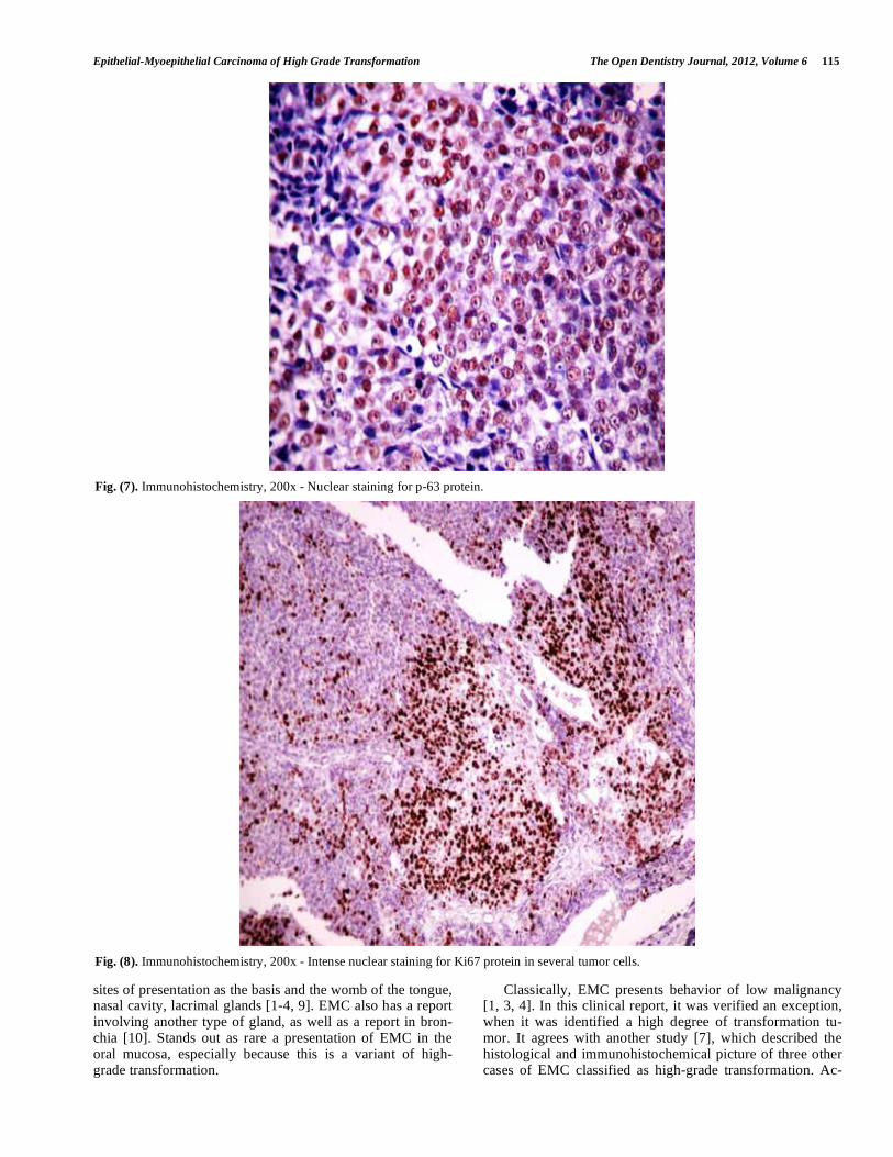
Fig. (7). Immunohistochemistry, 200x - Nuclear staining for p-63 protein.
Fig. (8). Immunohistochemistry, 200x - Intense nuclear staining for Ki67 protein in several tumor cells.

116 The Open Dentistry Journal, 2012, Volume 6 Lima et al.
cording to Roy et al. [7], the criteria for defining high degree of transformation in EMC must include margins of the lesion showing infiltration, cellular atypia, disorganization of the external myoepithelial layer and the need for complementary examination to prove its histogenetic origin. Such character-istics were identified in the case described herein.
In this report, was highlighted by the patient that the cur-rent lesion corresponded to a local recurrence, which agrees with Matos et al. [1], who reported a case of local recurrence after four years of the removal of the lesion and also agrees with Esposito et al. [4], who refereed a variation on the loco-regional recurrence in percentage terms, ranging from 28% to 50%. Additionally, these authors emphasized that the EMC has a low mortality rate, but also has a strong local and regional aggressiveness, whether it presenting high or low degree of transformation.
Histopathologically, the EMC has rounded epithelial cells, clear polygonal cells and a myoepithelial component in a solid arrangement that may resemble other salivary gland tumors. The differential diagnosis involves mainly myoepi-thelial carcinoma (MC), pleomorphic adenoma (PA) and adenoid cystic carcinoma (ACC) [2, 3, 5-7].
To properly differentiate EMC from other similar sali-vary gland neoplasms, is necessary a careful histopathologi-cal examination, which seeks to identify the presence of two cell populations: epithelial and myoepithelial cells. The iden-tification of a solid ductal arrangement with an inner layer of epithelial cells and an outer layer of myoepithelial cells, can be done on EMC. However the PA, ACC and MC may have similar structural components depending on the degree of cellular differentiation. On these occasions, special tech-niques such as immunohistochemistry should be performed [2, 5, 7].
The EMC diagnosis is usually based on microscopy, however, in cases similar to that described (with a high de-gree of transformation), the use of immunohistochemistry in the diagnosis consists in one of the prevailing alternatives for closing this framework [7, 8].
Regarding the choice of antibodies used in immunohisto-chemical assessment of this case, the anti-p63 is a selective marker for nuclear staining, with a significant markup for myoepithelial cells [3, 7]. This report corroborates the in-formation from van Tongeren et al. [3], that the marked ex-pression of p63 protein reveals its participation in the morphogenesis of the EMC.
The S-100 protein can be used for differential diagnosis between EMC and ACC, which also possesses two cell lay-ers. However, the positive staining for S-100 protein in ACC only occurs in the inner layer duct, and while in EMC it only is positive in the external ductal layer
as can be seen in this
case report [2, 3].
The Ki-67 protein is related to cell proliferation. It is found throughout the entire cell cycle, and it can bring addi-tional data regarding the biological behavior of the lesion. According Seethala, Barnes and Hunt [6], myoepithelial cells of the EMC demonstrate high proliferative and mitotic activity, indicating the significant Ki-67 positivity in the myoepithelial component. In the case presented here, it is
emphasized that this protein has facilitated the understanding that it is a variant of high degree EMC.
In a similar case to the reported here, Matos et al. [8], confirmed the diagnosis of EMC using the technique of im-munohistochemistry. They did it using cytokeratin 7 and 8, which showed positivity in the internal ductal structures and also using smooth muscle actin (SMA), which was positive in the external cells. In this case we used the antibodies to cytokeratin AE1/AE3, confirming a cell group of epithelial origin and finally observing, along with other immunohisto-chemical findings, a profile consistent with epithelial-myoepithelial carcinoma with high grade transformation [7].
According to the literature, the treatment is varied and the main method is surgery with a safety margin and if nec-essary, subsequent using of radiotherapy and chemotherapy. In some cases, treatment may include enlargement of the incision and neck dissection with lymph node emptying [2, 4, 7-9]. In the present clinical case the initial treatment was a local surgical incision with safety margin, even without prior confirmation of a malignant lesion of salivary gland. Since it was confirmed the malignant nature of the lesion, the patient was referred to an oncology service.
In conclusion, it is noteworthy, therefore, the importance
of critical and reflective evaluation by the dentist as well as
diagnostic and prognostic value of histopathology and im-
munohistochemistry, especially for cases of malignant tu-
mors that do not exhibit clinical features that may character-
ize such condition.
CONFLICT OF INTEREST
We wish to declare that the submitted work is original
and has not been submitted or published elsewhere. Moreo-
ver, all authors have read and approved the manuscript and
agree with the current submission. Finally, there are no po-
tential conflicts of interest
ACKNOWLEDGEMENTS
We are very thankful to Centro de Anatomopatologia S/C Ltda (UNILAP), Campina Grande - Paraiba / Brazil.
REFERENCES
[1] Kumai Y, Ogata N, Yumoto E. Epithelial-myoepithelial carcinoma
in the base of the tongue: a case report. Am J Otolaryngol 2006; 27: 58-60.
[2] Alves PM, Silva LAC, Godoy GP, Gomes DQC, Queiroz LMMG, Galvão HC. Unusual epithelial-myoepithelial carcinoma in palate-
case report and immunohistochemical study. J Clin Exp Dent 2010; 2: 22-5.
[3] Van Tongeren J, Creytens DHKV, Meulemans EV, Bondt RBJ, Jong J, Manni JJ. Synchronous bilateral epithelial–myoepithelial
carcinoma of the parotid gland: case report and review of the litera-ture. Eur Arch Otorhinolaryngol 2009; 266: 1495-500.
[4] Esposito E, Cassiano B, Cinquegrani F. Salivary glands: report of a rare case of myoepithelial carcinoma involving tongue base treated
by CO2 laser. Acta Otorhinolaryngol Ital 2009; 29:156-9. [5] Kusafuka K, Takizawa Y, Ueno T, et al. Dedifferentiated epithe-
lialmyoepithelial carcinoma of the parotid gland: a rare case report of immunohistochemical analysis and review of the literature. Oral
Surg Oral Med Oral Pathol Oral Radiol Endod 2008; 106: 85-91. [6] Seethala RR, Barnes EL, Hunt JL. Epithelial-myoepithelial carci-
noma: a review of the clinicopathologic spectrum and immunophe-notypic characteristics in 61 tumors of the salivary glands and up-
per aerodigestive tract. Am J Surg Pathol 2007; 31: 44-57.

Epithelial-Myoepithelial Carcinoma of High Grade Transformation The Open Dentistry Journal, 2012, Volume 6 117
[7] Roy P, Bullock MJ, Perez-Ordonez B, Dardick I, Weinreb I.
Epithelial myoepithelial carcinoma with high grade transformation. Am J Surg Pathol 2010; 34: 1258-65.
[8] Matos FR, Miranda JL, Mequita ATM, Santos CRR, Freitas RA. Epithelial-myoepithelial carcinoma in the ventral surface of the
tongue. Braz J Otorhinolaryngol 2010; 76:540.
[9] Ramraje SN, Bharambe BM, Zode R. Epithelial myoepithelial
carcinoma of the submandibular salivary gland. Bombay Hosp J 2010; 52: 53-8.
[10] Chao TY, Lin AS, Lie CH, Chung YH, Lin JW, Lin MC. Bronchial epithelial-myoepithelial carcinoma. Ann Thorac Surg 2007; 83:
689-91.
Received: May 02, 2012 Revised: June 06, 2012 Accepted: June 07, 2012
© Lima et al.; Licensee Bentham Open.
This is an open access article licensed under the terms of the Creative Commons Attribution Non-Commercial License
(http://creativecommons.org/licenses/by-nc/3.0/) which permits unrestricted, non-commercial use, distribution and reproduction in any medium, provided the
work is properly cited.


![Transcriptomic response of goat mammary epithelial cells ...€¦ · previously [Ogorevc et al. 2009b]. Luminal, myoepithelial and fibroblast cells were characterized using antibodies](https://img.pdfslide.net/doc/110x75/605fe504ab02910182502932/transcriptomic-response-of-goat-mammary-epithelial-cells-previously-ogorevc.jpg)