Embed Size (px)
Citation preview
1
Various parameters influence on electrospun herbs incorporated cellulose acetate and its
suitability for drug delivery
Thilakam R* and Elakkiya Thangaraju
Department of Chemistry, Sri Sarada College for Women (Autonomous), Salem – 16,
Tamilnadu, India
[email protected]*, and [email protected]
Abstract
In the present work, electrospun cellulose acetate membrane suitability was analyzed for
nanocarrier in drug delivery systems. Electrospinning is one of the versatile techniques to
produce interconnected porous nanofibrous membrane. Of the electrospinning process, various
parameters namely solvent, collector and solution concentration would influence the nanofibrous
membrane. One of the hydrophilic biopolymer namely cellulose acetate was used to electrospun
and the smooth nanofibrous membrane was optimized. The electrospun cellulose acetate surface
morphology was deeply analyzed by Scanning electron microscopy. The curcumin was
incorporated into the optimized cellulose acetate solution and produced curcumin loaded
cellulose acetate nanofibrous membrane in one step process. The presence of curcumin in
curcumin loaded cellulose acetate nanofibrous membrane was studied by Scanning electron
microscopy and Transmission electron microscopy. The antioxidant activity and in-vitro
curcumin release from curcumin loaded cellulose acetate nanofibrous membrane was evaluated
for drug delivery.
Keywords: Electrospinning, Nanofibers, Cellulose acetate, Curcumin, Drug delivery
IAETSD JOURNAL FOR ADVANCED RESEARCH IN APPLIED SCIENCES
VOLUME VI, ISSUE III, MARCH/2019
ISSN NO: 2394-8442
PAGE NO:34
2
Introduction
Electrospinning is one of the efficient techniques to fabricate highly interconnected,
continuous non-woven nanofibers that do not require expensive purification protocols. Choosing
the solvent, concentration of the solution and collector plays a major role on the fiber diameter
during electrospinning process [1-3]. They can be effectively used as a biomedical applications
namely tissue engineering, drug delivery, wound healing and dental implants due to their
extraordinary properties such as high porosity and large surface area to volume ratio. In addition,
it has great ability to physically resemble natural extra cellular matrix (ECM) protein structure
which also allows higher drug encapsulation efficiency [4-5]. The good biocompatible and
biodegradable food and drug administration approved polymers like cellulose acetate has drawn
great attention because they can be used in many biomedical applications including implants [6-
7]. Cellulose acetate, a derivative of cellulose obtained from natural resources can be easily
fabricated into films, fibers and membranes due to its abundant availability and biodegradability.
Cellulose acetate produced by electrospinning process has been reported with diameters in the
range of tens of nanometers to micrometers with pore sizes in the range of submicron to microns
[8-10]. Cellulose acetate based polymer blend membranes have been used in biomedical
applications especially drug delivery and tissue engineering due to its good hydrolytic stability,
excellent biocompatibility in human body and relatively low cost [11]. The herbs like curcumin,
which is also known as turmeric exhibit highly immense anti–tumor, anti-oxidant, and anti-
inflammatory properties [11-15]. Curcumin is highly hydrophobic in nature with poor water
solubility.
The present study aims in evaluating the ability of cellulose acetate nanofibrous
membrane as a nanocarrier for hydrophobic drug like curcumin. In this study, the diameter and
alignment of the cellulose acetate fibrous membrane were optimized by the process of
electrospinning based on the solvent, concentration of the polymer solution and the collector.
The surface morphology of different types of electrospun cellulose acetate was studied by
scanning electron microscopy. The curcumin present in cellulose acetate nanofibrous membrane
was studied by scanning electron microscopy and transmission electron microscopy. The
antioxidant activity of curcumin loaded cellulose acetate nanofibrous membrane was
characterized using DPPH assay. The in-vitro curcumin release from curcumin loaded cellulose
IAETSD JOURNAL FOR ADVANCED RESEARCH IN APPLIED SCIENCES
VOLUME VI, ISSUE III, MARCH/2019
ISSN NO: 2394-8442
PAGE NO:35
3
acetate nanofibrous membrane was studied by High performance liquid chromatography with
ultra-violet detector.
Experimental details
Materials
Cellulose acetate (CA; Mn ~ 50,000 by GPC) and curcumin from curcuma longa L. were
purchased from Sigma – aldrich, India. Acetic acid, 1, 2-dichloroethane and ethanol were
purchased from Sisco Research Laboratories Private Limited, India. All chemicals were used as
received without further treatment or purifications.
Electrospinning Process
A weighed amount of cellulose acetate powder was dissolved in different types of solvent
(acetic acid, dichloroethane and dichloroethane-ethanol (4:1 ratio)) in various solution
concentrations. The curcumin powder (0.5, 1 and 1.5 w/w %) and cellulose acetate powder were
co-dissolved in the mixture of dichloroethane-ethanol solvent and gently stirred continuously for
about 3 h at room temperature.
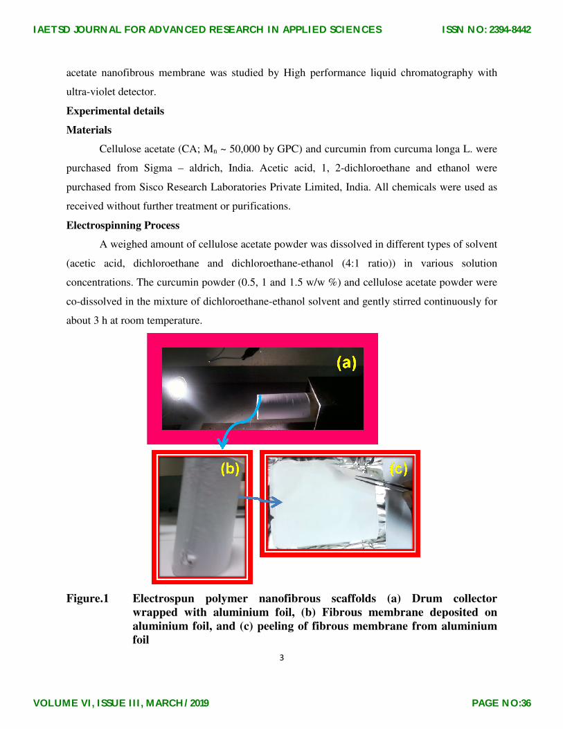
Figure.1 Electrospun polymer nanofibrous scaffolds (a) Drum collector
wrapped with aluminium foil, (b) Fibrous membrane deposited on
aluminium foil, and (c) peeling of fibrous membrane from aluminium
foil
IAETSD JOURNAL FOR ADVANCED RESEARCH IN APPLIED SCIENCES
VOLUME VI, ISSUE III, MARCH/2019
ISSN NO: 2394-8442
PAGE NO:36
4
The prepared homogeneous solution was taken in a 2 ml syringe to which a needle tip of
0.56 mm inner diameter was attached. The electrospinning set-up used here has been described
earlier [6, 16]. The electric voltage, flow rate, the tip to collector distance was optimized at 22
kV, 1ml/h and 11 cm in all solutions for electrospinning process. The electrospun nanofibrous
membrane deposited on the aluminium foil in the drum collector and after peeling from the
collector is shown in Figure 1.
Characterization
The surface morphology of the prepared nanofibrous membrane was investigated using
FEI Quanta FEG 200 – HRSEM at 15 kV and TF 20: Tecnai at 200 kV. The average diameters
were determined by analyzing the scanning electron microscopic images with UTHSCSCSA
image tool software. The 1,1 diphenyl-2-picrylhydrazyl (DPPH) radical scavenging activity of
curcumin loaded cellulose acetate nanofibrous membrane was determined by the method of
Brand-Williams, Guelier & Berset, 1995 with some modifications [17]. The in-vitro curcumin
release from curcumin loaded cellulose acetate nanofibrous membrane was analyzed by High
performance liquid chromatography with ultra-violet detector at 450 nm.
Results and Discussion
Surface morphology
Figure. 2 shows the scanning electron microscopy images of the electrospun cellulose
acetate with different solvents (acetic acid, dichloroethane and dichloroethane-ethanol mixture in
the ratio 4:1) and 1 wt % curcumin loaded cellulose acetate. Electrospun cellulose acetate in
acetic acid solution produced large amount of beads with fibers due to high boiling point of
acetic acid (117.9 °C). High boiling point of the solvent makes it difficult to evaporate during
drafting process. However, dichloroethane in cellulose acetate when electrospun also resulted in
the formation of fibers with beads. A mixture of dichloroethane and ethanol (4:1) solvent was
used to dissolve cellulose acetate and electrospun which resulted in smooth continuous beadless
nanofibrous membrane. The cellulose acetate in dichloroethane and ethanol solution at 8 wt %
was electrospun into fibers which contained a few beads and these disappeared on increasing the
cellulose acetate concentration to 10 wt %. The average diameter of these fibers was found to be
120±30 nm. However, on further increasing the concentration of cellulose acetate solution to 12
wt %, the resultant average diameter of the fibers increased to 210±30 nm respectively. To
IAETSD JOURNAL FOR ADVANCED RESEARCH IN APPLIED SCIENCES
VOLUME VI, ISSUE III, MARCH/2019
ISSN NO: 2394-8442
PAGE NO:37

5
obtain smooth fine nanofibers from beaded to non beaded morphology solution, cellulose acetate
concentration was fixed as 10 wt %. The use of suitable collector is also very important as it also
affects the fiber morphology of the electrospun cellulose acetate.
Figure. 2 Scanning electron microscopic images of the electrospun cellulose
acetate with different solvents (acetic acid, dichloroethane and dichloroethane-
ethanol) and different weight percentages (8, 10 and 12 wt %) and 1 wt % of
curcumin loaded cellulose acetate (10 wt %) using a flat collector
In the present work, two types of collectors (flat and drum) were used. On using flat
collector, electrospun cellulose acetate produced uniform porous nanofibers with rod like
IAETSD JOURNAL FOR ADVANCED RESEARCH IN APPLIED SCIENCES
VOLUME VI, ISSUE III, MARCH/2019
ISSN NO: 2394-8442
PAGE NO:38
6
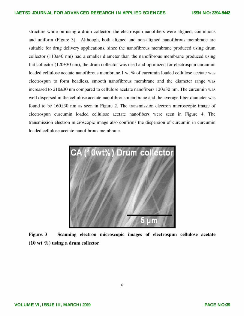
structure while on using a drum collector, the electrospun nanofibers were aligned, continuous
and uniform (Figure 3). Although, both aligned and non-aligned nanofibrous membrane are
suitable for drug delivery applications, since the nanofibrous membrane produced using drum
collector (110±40 nm) had a smaller diameter than the nanofibrous membrane produced using
flat collector (120±30 nm), the drum collector was used and optimized for electrospun curcumin
loaded cellulose acetate nanofibrous membrane.1 wt % of curcumin loaded cellulose acetate was
electrospun to form beadless, smooth nanofibrous membrane and the diameter range was
increased to 210±30 nm compared to cellulose acetate nanofibers 120±30 nm. The curcumin was
well dispersed in the cellulose acetate nanofibrous membrane and the average fiber diameter was
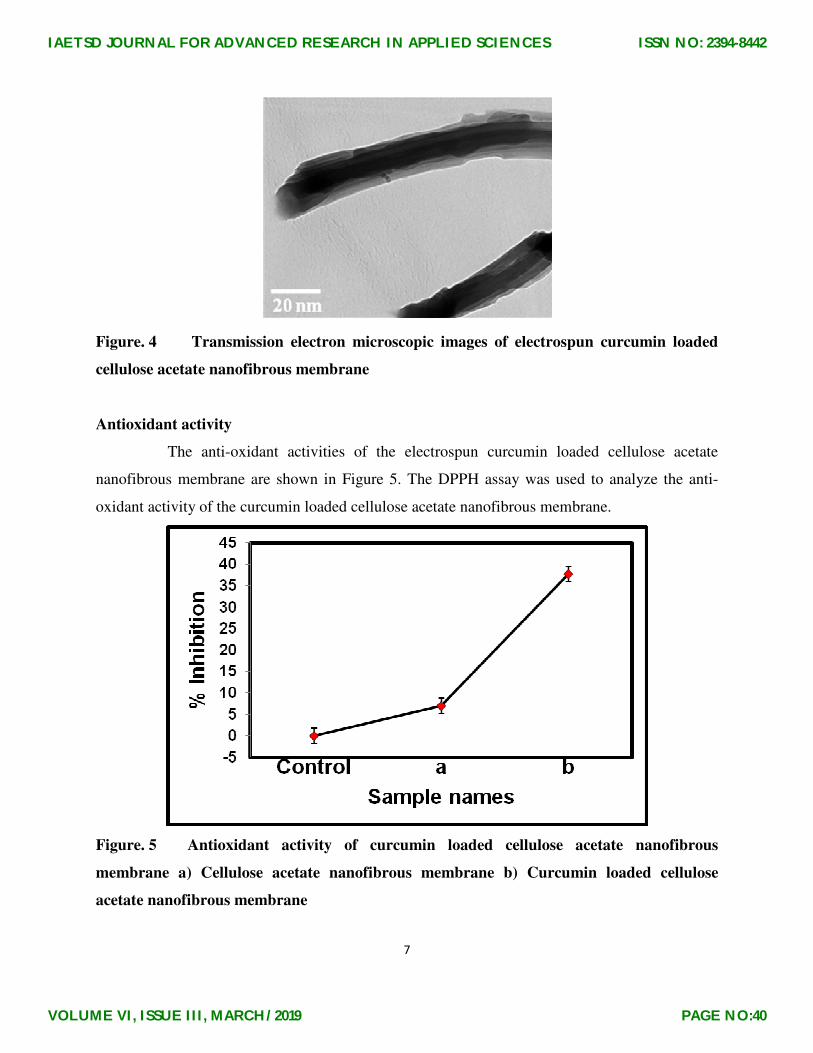
found to be 160±30 nm as seen in Figure 2. The transmission electron microscopic image of
electrospun curcumin loaded cellulose acetate nanofibers were seen in Figure 4. The
transmission electron microscopic image also confirms the dispersion of curcumin in curcumin
loaded cellulose acetate nanofibrous membrane.
Figure. 3 Scanning electron microscopic images of electrospun cellulose acetate
(10 wt %) using a drum collector
IAETSD JOURNAL FOR ADVANCED RESEARCH IN APPLIED SCIENCES
VOLUME VI, ISSUE III, MARCH/2019
ISSN NO: 2394-8442
PAGE NO:39
7
Figure. 4 Transmission electron microscopic images of electrospun curcumin loaded
cellulose acetate nanofibrous membrane
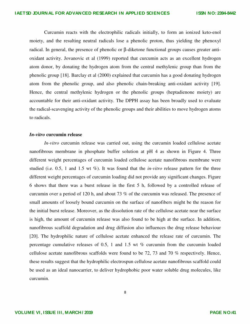
Antioxidant activity
The anti-oxidant activities of the electrospun curcumin loaded cellulose acetate
nanofibrous membrane are shown in Figure 5. The DPPH assay was used to analyze the anti-
oxidant activity of the curcumin loaded cellulose acetate nanofibrous membrane.
Figure. 5 Antioxidant activity of curcumin loaded cellulose acetate nanofibrous
membrane a) Cellulose acetate nanofibrous membrane b) Curcumin loaded cellulose
acetate nanofibrous membrane
IAETSD JOURNAL FOR ADVANCED RESEARCH IN APPLIED SCIENCES
VOLUME VI, ISSUE III, MARCH/2019
ISSN NO: 2394-8442
PAGE NO:40
8
Curcumin reacts with the electrophilic radicals initially, to form an ionized keto-enol
moiety, and the resulting neutral radicals lose a phenolic proton, thus yielding the phenoxyl
radical. In general, the presence of phenolic or β-diketone functional groups causes greater anti-
oxidant activity. Jovanovic et al (1999) reported that curcumin acts as an excellent hydrogen
atom donor, by donating the hydrogen atom from the central methylenic group than from the
phenolic group [18]. Barclay et al (2000) explained that curcumin has a good donating hydrogen
atom from the phenolic group, and also phenolic chain-breaking anti-oxidant activity [19].
Hence, the central methylenic hydrogen or the phenolic groups (heptadienone moiety) are
accountable for their anti-oxidant activity. The DPPH assay has been broadly used to evaluate
the radical-scavenging activity of the phenolic groups and their abilities to move hydrogen atoms
to radicals.
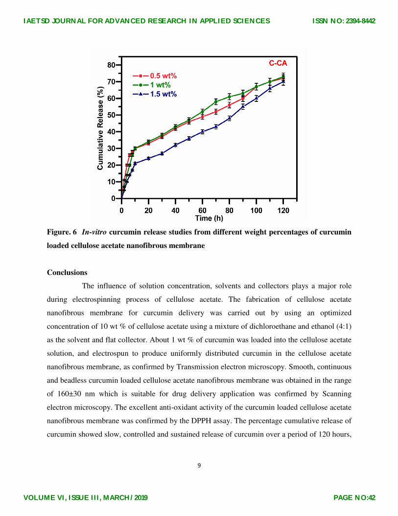
In-vitro curcumin release
In-vitro curcumin release was carried out, using the curcumin loaded cellulose acetate
nanofibrous membrane in phosphate buffer solution at pH 4 as shown in Figure 4. Three
different weight percentages of curcumin loaded cellulose acetate nanofibrous membrane were
studied (i.e. 0.5, 1 and 1.5 wt %). It was found that the in-vitro release pattern for the three
different weight percentages of curcumin loading did not provide any significant changes. Figure
6 shows that there was a burst release in the first 5 h, followed by a controlled release of
curcumin over a period of 120 h, and about 73 % of the curcumin was released. The presence of
small amounts of loosely bound curcumin on the surface of nanofibers might be the reason for
the initial burst release. Moreover, as the dissolution rate of the cellulose acetate near the surface
is high, the amount of curcumin release was also found to be high at the surface. In addition,
nanofibrous scaffold degradation and drug diffusion also influences the drug release behaviour
[20]. The hydrophilic nature of cellulose acetate enhanced the release rate of curcumin. The
percentage cumulative releases of 0.5, 1 and 1.5 wt % curcumin from the curcumin loaded
cellulose acetate nanofibrous scaffolds were found to be 72, 73 and 70 % respectively. Hence,
these results suggest that the hydrophilic electrospun cellulose acetate nanofibrous scaffold could
be used as an ideal nanocarrier, to deliver hydrophobic poor water soluble drug molecules, like
curcumin.
IAETSD JOURNAL FOR ADVANCED RESEARCH IN APPLIED SCIENCES
VOLUME VI, ISSUE III, MARCH/2019
ISSN NO: 2394-8442
PAGE NO:41
9
Figure. 6 In-vitro curcumin release studies from different weight percentages of curcumin
loaded cellulose acetate nanofibrous membrane
Conclusions
The influence of solution concentration, solvents and collectors plays a major role
during electrospinning process of cellulose acetate. The fabrication of cellulose acetate
nanofibrous membrane for curcumin delivery was carried out by using an optimized
concentration of 10 wt % of cellulose acetate using a mixture of dichloroethane and ethanol (4:1)
as the solvent and flat collector. About 1 wt % of curcumin was loaded into the cellulose acetate
solution, and electrospun to produce uniformly distributed curcumin in the cellulose acetate
nanofibrous membrane, as confirmed by Transmission electron microscopy. Smooth, continuous
and beadless curcumin loaded cellulose acetate nanofibrous membrane was obtained in the range
of 160±30 nm which is suitable for drug delivery application was confirmed by Scanning
electron microscopy. The excellent anti-oxidant activity of the curcumin loaded cellulose acetate
nanofibrous membrane was confirmed by the DPPH assay. The percentage cumulative release of
curcumin showed slow, controlled and sustained release of curcumin over a period of 120 hours,
IAETSD JOURNAL FOR ADVANCED RESEARCH IN APPLIED SCIENCES
VOLUME VI, ISSUE III, MARCH/2019
ISSN NO: 2394-8442
PAGE NO:42
10
and the curcumin release percentage were found to be 72, 73 and 70 % for 0.5, 1 and 1.5 wt % of
curcumin in the curcumin loaded cellulose acetate nanofibrous membrane.
Acknowledgement
We acknowledge UGC for the autonomy grant 2017-18 under which we have purchased
ESPIN NANO Electrospinning apparatus. Model : V1VHC, Sr. No. 20012018 in the Department
of Chemistry, Sri Sarada College for Women (Salem – 16).
References
1. P Quynh, S Upma and G M Antonios, Tissue Eng., (2006), 12, 1197-1211.
2. W E Teo, and S Ramakrishna, Nanotechnology, (2006), 17, R89-R106.
3. P Supaphol, O Suwantong, P Sansanoh, S Sowmya, R Jayakumar, and S V Nair, Advances in
Polymer Science, (2012), 246, 213-240.
4. M Prabaharan, R Jayakumar, and S V Nair, Advances in Polymer Science, (2012), 246, 241-
262.
5. E Thangaraju, N T Srinivasan, R Kumar, P K Sehgal, and R Sheeja, Fibers and Polymers,
(2012), 13, 823-830.
6. J Gunn, and M Zhang, Trends in Biotechnology, (2010), 28, 189-197.
7. M Biondi, F Ungaro, F Quagli, P A Netti, Advanced Drug Delivery Reviews, (2008), 60,
229-242.
8. S O Han, J H Youk, K D Min, Y O Kang, and W H Park, Materials Letters, (2008), 62, 759-
762.
9. A Celebioglu and T Uyar, Materials Letters, (2011), 65, 2291-2294.
10. A Anitha, S Maya, N Deepa, K P Chennazhi, S V Nair, H Tamura, and R Jayakumar,
Carbohydrate Polymers, (2011), 83, 452-461.
11. H Y Zhou, and X G Chen, Frontier Material Science China, (2008), 2, 417-425.
12. C A C Araujo, L L Leon, Memorias do Instituto Oswaldo Cruz, Rio de Janeiro, (2001), 96,
723-728.
13. J Miquel, A Bernd, J M Sempere, J Diaz-Alperi, and A Ramirez, Archives of Gerontology
and Geriatrics, (2002), 34, 37-46.
14. B B Aggarwal, C Sundaram, N Malani, N and H Ichikawa, Advances in Experimental
Medicine and Biology, (2007), 595, 1-75.
IAETSD JOURNAL FOR ADVANCED RESEARCH IN APPLIED SCIENCES
VOLUME VI, ISSUE III, MARCH/2019
ISSN NO: 2394-8442
PAGE NO:43
11
15. B S Vinod, J Antony, H H Nair, V T Puliyappadamba, M Saikia, S Shyam Narayanan, A
Bevin, and R John Anto, Cell Death and Disease, (2013), 4, 1-13.
16. S Bibekananda, V Subramanian, and T S Natarajan, Applied Physics Letters, (2004), 84,
1222-1224.
17. W Brand-Williams, M E Cuvelier, and C Berset, LWT-Food Science and Technology,
(1995), 28, 25-30.
18. S V Jovanovic, S Steenken, C W Boone, and M G Simic, Journal of American Chemical
Society, (1999), 121, 9677-9681.
19. L R C Barclay, M R Vinqvist, K Mukai, H Goto, Y Hashimoto, A Tokuanga, and H Uno,
Organic Letters, (2000), 2, 2841-2843.
20. XM Wu, C J Branford-White, L M Zhu, N P Chatterton, and D G Yu, Journal of Material
Science; Materials in Medicine, (2010), 21, 2403-2411.
IAETSD JOURNAL FOR ADVANCED RESEARCH IN APPLIED SCIENCES
VOLUME VI, ISSUE III, MARCH/2019
ISSN NO: 2394-8442
PAGE NO:44