Embed Size (px)
Citation preview

Journal of Case Reports and Images in Obstetrics and Gynecology, Vol. 4, 2018.
J Case Rep Images Obstet Gynecol 2018;4:100041Z08JN2018. www.ijcriog.com
Ng et al. 1
CASE REPORT OPEN ACCESS
Vasa previa in unrecognized placenta accreta
June Ng, Haylea Sweat-Patrick
ABSTRACT
Introduction: Vasa previa and placenta accreta are both associated with significant morbidity and mortality. Vasa previa poses the risk of fetal exsanguination from ruptured fetal vessels with potential for devastating fetal effects including perinatal death. Placenta accreta is associated with maternal hemorrhage and mortality. This is the first report of the co-existance of vasa previa and placenta accreta. Case Report: We present a case of an antenatally suspected vasa previa in which a concurrent placenta accreta was diagnosed intraoperatively at the time of cesarean delivery. Although morbidly adherent placentation was not suspected preoperatively, appropriate intraoperative management with cesarean hysterectomy resulted in good maternal and neonatal outcomes. Conclusion: This is the first published case, to our knowledge, of a vasa previa with concurrent placenta accreta. While morbidity for both these conditions is high, prompt recognition and management in this case resulted in good outcomes.
Keywords: Bilobed placenta, Placenta accreta, Placenta previa, Vasa previa
June Ng1, Haylea Sweat-Patrick2
Affiliations: 1Department of Obstetrics, Gynecology, and Reproductive Sciences, Rutgers – Robert Wood Johnson Medical School, Clinical Academic Building, New Brun-swick, New Jersey, USA; 2Division of Maternal-Fetal Medi-cine, Department of Obstetrics, Gynecology, and Repro-ductive Sciences, Rutgers – Robert Wood Johnson Medical School, Clinical Academic Building, New Brunswick, New Jersey, USA.Corresponding Author: Haylea Sweat-Patrick, MD, Division of Maternal-Fetal Medicine, Department of Obstetrics, Gy-necology, and Reproductive Sciences, Rutgers – Robert Wood Johnson Medical School, Clinical Academic Building, 125 Paterson Street, #4200, New Brunswick, NJ 08901, USA; Email: [email protected]
Received: 10 October 2018Accepted: 04 December 2018Published: 31 December 2018
How to cite this article
Ng J, Patrick HS. Vasa previa in unrecognized placenta accreta. J Case Rep Images Obstet Gynecol 2018;4:100041Z08JN2018.
Article ID: 100041Z08JN2018
*********
doi: 10.5348/100041Z08JN2018CR
INTRODUCTION
Vasa previa and placenta accreta are associated with significant maternal and fetal morbidity and mortality. Vasa previa occurs when fetal vessels are identified sonographically to run over or within 2 cm of the internal cervical os. These vessels are unprotected by placenta and Wharton’s jelly and, thus are vulnerable to injury with common intrapartum events such as uterine contractions and rupture of fetal membranes. Injury to fetal vessels may lead to rapid fetal exsanguination within minutes [1]. Placenta accreta, a form of morbidly adherent placentation, is associated with risk of massive maternal hemorrhage, with an average blood loss of 2–5 litres [2]. While vasa previa occurs in 1 in 2,500 to 5,000 pregnancies, placenta accreta is more common, occurring in 1 in 300 to 500 pregnancies. The incidence of both of these conditions is increasing [1]. In this report, we present a case of a vasa previa with concurrent placenta accreta. Based on a Pubmed search using the terms “vasa previa” and “placenta accreta,” we believe that this is the first published case of a vasa previa with co-existing placenta accreta.
CASE REPORT
A 36-year-old woman, gravida three para two, was observed to have a bilobed placenta and vasa previa on transvaginal ultrasound at 20 weeks gestation (Figure 1). The patient had two prior uncomplicated vaginal deliveries. Serial ultrasounds were repeated every 4
CASE REPORT PEER REVIEWED | OPEN ACCESS
Journal of Case Reports and Images in Obstetrics and Gynecology, Vol. 4, 2018.
J Case Rep Images Obstet Gynecol 2018;4:100041Z08JN2018. www.ijcriog.com
Ng et al. 2
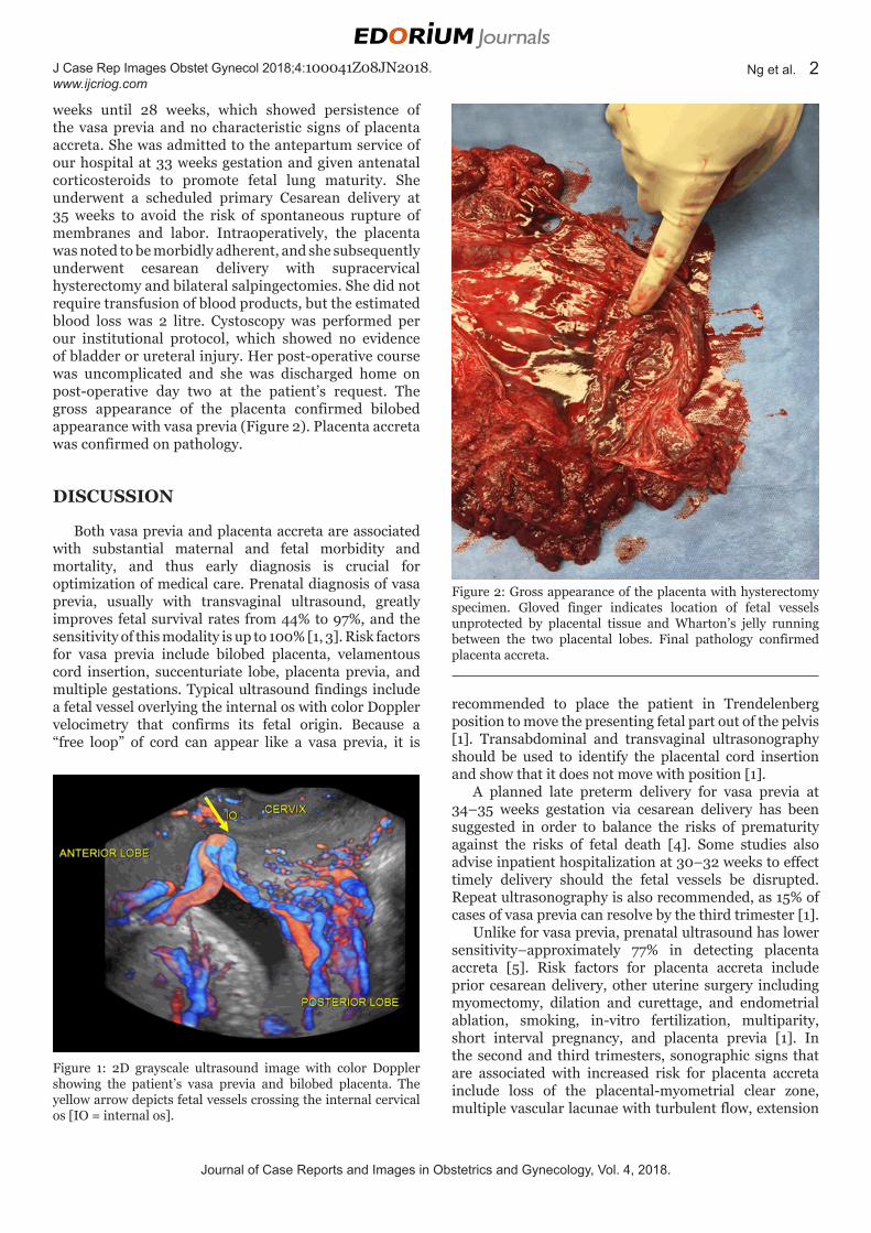
weeks until 28 weeks, which showed persistence of the vasa previa and no characteristic signs of placenta accreta. She was admitted to the antepartum service of our hospital at 33 weeks gestation and given antenatal corticosteroids to promote fetal lung maturity. She underwent a scheduled primary Cesarean delivery at 35 weeks to avoid the risk of spontaneous rupture of membranes and labor. Intraoperatively, the placenta was noted to be morbidly adherent, and she subsequently underwent cesarean delivery with supracervical hysterectomy and bilateral salpingectomies. She did not require transfusion of blood products, but the estimated blood loss was 2 litre. Cystoscopy was performed per our institutional protocol, which showed no evidence of bladder or ureteral injury. Her post-operative course was uncomplicated and she was discharged home on post-operative day two at the patient’s request. The gross appearance of the placenta confirmed bilobed appearance with vasa previa (Figure 2). Placenta accreta was confirmed on pathology.
DISCUSSION
Both vasa previa and placenta accreta are associated with substantial maternal and fetal morbidity and mortality, and thus early diagnosis is crucial for optimization of medical care. Prenatal diagnosis of vasa previa, usually with transvaginal ultrasound, greatly improves fetal survival rates from 44% to 97%, and the sensitivity of this modality is up to 100% [1, 3]. Risk factors for vasa previa include bilobed placenta, velamentous cord insertion, succenturiate lobe, placenta previa, and multiple gestations. Typical ultrasound findings include a fetal vessel overlying the internal os with color Doppler velocimetry that confirms its fetal origin. Because a “free loop” of cord can appear like a vasa previa, it is
recommended to place the patient in Trendelenberg position to move the presenting fetal part out of the pelvis [1]. Transabdominal and transvaginal ultrasonography should be used to identify the placental cord insertion and show that it does not move with position [1].
A planned late preterm delivery for vasa previa at 34–35 weeks gestation via cesarean delivery has been suggested in order to balance the risks of prematurity against the risks of fetal death [4]. Some studies also advise inpatient hospitalization at 30–32 weeks to effect timely delivery should the fetal vessels be disrupted. Repeat ultrasonography is also recommended, as 15% of cases of vasa previa can resolve by the third trimester [1].
Unlike for vasa previa, prenatal ultrasound has lower sensitivity–approximately 77% in detecting placenta accreta [5]. Risk factors for placenta accreta include prior cesarean delivery, other uterine surgery including myomectomy, dilation and curettage, and endometrial ablation, smoking, in-vitro fertilization, multiparity, short interval pregnancy, and placenta previa [1]. In the second and third trimesters, sonographic signs that are associated with increased risk for placenta accreta include loss of the placental-myometrial clear zone, multiple vascular lacunae with turbulent flow, extension
Figure 1: 2D grayscale ultrasound image with color Doppler showing the patient’s vasa previa and bilobed placenta. The yellow arrow depicts fetal vessels crossing the internal cervical os [IO = internal os].
Figure 2: Gross appearance of the placenta with hysterectomy specimen. Gloved finger indicates location of fetal vessels unprotected by placental tissue and Wharton’s jelly running between the two placental lobes. Final pathology confirmed placenta accreta.

Journal of Case Reports and Images in Obstetrics and Gynecology, Vol. 4, 2018.
J Case Rep Images Obstet Gynecol 2018;4:100041Z08JN2018. www.ijcriog.com
Ng et al. 3
of the villi to the myometrium, interruption of the serosa-bladder interface, placenta previa, and irregular subplacental vascularity. MRI has been used to improve diagnostic accuracy. However, MRI is not routinely used at all institutions due to high cost, lack of accessibility, and unclear incremental diagnostic yield over ultrasound.
Prenatal diagnosis is important to improve outcomes for both of these conditions. In this case, vasa previa was suspected. This resulted in hospitalization and management intended to minimize fetal risks. However, we did not suspect morbidly adherent placentation due to lack of classic ultrasound findings and traditional risk factors for this condition. Fortunately, prompt recognition in the operating room and decisive care resulted in definitive surgical management with limited morbidity for the patient.
CONCLUSION
In this report, we present the first case of concurrent vasa previa with an unrecognized placenta accreta. Although vasa previa and placenta accreta are associated with significant maternal and fetal morbidity and mortality, prenatal diagnosis may be associated with improved outcomes. Prompt recognition of placenta accreta and decisive surgical management in this case resulted in good outcome, but we hope this case will draw attention to this potentially comorbid situation.
REFERENCES
1. Silver RM. Abnormal placentation: Placenta previa, vasa previa, and placenta accreta. Obstet Gynecol 2015;126(3):654–68.
2. Rao KP, Belogolovkin V, Yankowitz J, Spinnato JA 2nd. Abnormal placentation: Evidence-based diagnosis and management of placenta previa, placenta accreta, and vasa previa. Obstet Gynecol Surv 2012;67(8):503–19.
3. Ruiter L, Kok N, Limpens J, et al. Systematic review of accuracy of ultrasound in the diagnosis of vasa previa. Ultrasound Obstet Gynecol 2015;45(5):516–22.
4. Robinson BK, Grobman WA. Effectiveness of timing strategies for delivery of individuals with vasa previa. Obstet Gynecol 2011;117(3):542–9.
5. Committee on obstetric practice. Committee opinion no. 529: Placenta accreta. Obstet Gynecol 2012;120(1):207–11.
*********
AcknowledgementsThe authors would like to thank Justin Brandt MD, and Natalia Kartashov, RDMS, for their guidance in the editing of this manuscript and the acquisition of the figures published here.
Author ContributionsJune Ng – Substantial contributions to conception and design, Acquisition of data, Drafting the article, Final approval of the version to be publishedHaylea Sweat-Patrick – Substantial contributions to conception and design, Acquisition of data, Revising it critically for important intellectual content, Final approval of the version to be published
Guarantor of SubmissionThe corresponding author is the guarantor of submission.
Source of SupportNone.
Consent StatementWritten informed consent was obtained from the patient for publication of this clinical image.
Conflict of InterestAuthors declare no conflict of interest.
Data AvailabilityAll relevant data are within the paper and its Supporting Information files.
Copyright© 2018 June Ng et al. This article is distributed under the terms of Creative Commons Attribution License which permits unrestricted use, distribution and reproduction in any medium provided the original author(s) and original publisher are properly credited. Please see the copyright policy on the journal website for more information.
Access full text article onother devices
Access PDF of article onother devices