Embed Size (px)
Citation preview

Vascular CD39/ENTPD1 DirectlyPromotes Tumor Cell Growthby Scavenging ExtracellularAdenosine Triphosphate1,2
Lili Feng*,†,3, Xiaofeng Sun*,3, Eva Csizmadia*,Lihui Han*, Shu Bian*, Takashi Murakami‡,Xin Wang†, Simon C. Robson*,4 and Yan Wu*,4
*Department of Medicine, Transplantation Institute,Beth Israel Deaconess Medical Center, Harvard MedicalSchool, Boston, MA, USA; †Department of Hematology,Provincial Hospital Affiliated to Shandong University, Jinan,PR China; ‡Division of Bioimaging Sciences, Center forMolecular Medicine, Jichi Medical University, Shimotsuke,Tochigi, Japan
AbstractExtracellular adenosine triphosphate (ATP) is known to boost immune responses in the tumor microenvironmentbut might also contribute directly to cancer cell death. CD39/ENTPD1 is the dominant ectonucleotidase expressedby endothelial cells and regulatory T cells and catalyzes the sequential hydrolysis of ATP to AMP that is furtherdegraded to adenosine by CD73/ecto-5′-nucleotidase. We have previously shown that deletion of Cd39 results indecreased growth of transplanted tumors in mice, as a result of both defective angiogenesis and heightened in-nate immune responses (secondary to loss of adenosinergic immune suppression). Whether alterations in localextracellular ATP and adenosine levels as a result of CD39 bioactivity directly affect tumor growth and cytotoxicityhas not been investigated to date. We show here that extracellular ATP exerts antitumor activity by directly inhibit-ing cell proliferation and promoting cancer cell death. ATP-induced antiproliferative effects and cell death are, inlarge part, mediated through P2X7 receptor signaling. Tumors in Cd39 null mice exhibit increased necrosis in as-sociation with P2X7 expression. We further demonstrate that exogenous soluble NTPDase, or CD39 expression bycocultured liver sinusoidal endothelial cells, stimulates tumor cell proliferation and limits cell death triggered byextracellular ATP. Collectively, our findings indicate that local expression of CD39 directly promotes tumor cellgrowth by scavenging extracellular ATP. Pharmacological or targeted inhibition of CD39 enzymatic activity mayfind utility as an adjunct therapy in cancer management.
Neoplasia (2011) 13, 206–216
IntroductionAdenosine triphosphate (ATP) mediates multiple physiological reac-tions and plays a crucial role in cellular metabolism, inclusive of rolesin bioenergetics [1–3]. Extracellular ATP acts on type 2 purinergic(P2) receptors to exert signaling effects. There are two P2 families:seven P2X ion channel receptors recognizing ATP (P2X1-7) and eightP2Y G protein–coupled receptors (P2Y1, 2, 4, 6, 11-14) that bind sev-eral nucleoside triphosphates and diphosphates [4–6]. Documentedcytotoxic effects of extracellular ATP on various malignant cells haveelicited attention to this signaling pathway [2,7–10]. Five P2 recep-tor subtypes have been considered to be involved in the antitumoractions of ATP, namely P2X5, P2X7, P2Y1, P2Y2, and P2Y11 (exclu-sively in human), but precise roles for these receptors are not well de-fined [2,9,11].
Abbreviations: ATP, adenosine triphosphate; ADP, adenosine diphosphate; AMP, aden-osine monophosphate; ENTPD1, ectonucleoside triphosphate diphosphohydrolase 1;LSEC, liver sinusoidal endothelial cell; Treg, regulatory T cells; TLC, thin-layer chro-matography; wt, wild type; Luc-B16/F10, luciferase-expressing B16/F10 cellsAddress all correspondence to: Yan Wu, PhD, Beth Israel Deaconess Medical Center, 330Brookline Ave, E/CLS-614, Boston, MA 02215. E-mail: [email protected] study was supported by funds from National Institutes of Health (National Heart,Lung, and Blood Institute grants PO1-HL076540 and RO1-HL094400). L. Feng wasa recipient of a scholarship from the China Scholarship Council. The authors discloseno conflicts.2This article refers to supplementary materials, which are designated by Table W1 andFigures W1 to W4 and are available online at www.neoplasia.com.3These authors contributed equally to the work.4These authors share senior coauthorship.Received 13 September 2010; Revised 19November 2010; Accepted 29November 2010
Copyright © 2011 Neoplasia Press, Inc. All rights reserved 1522-8002/11/$25.00DOI 10.1593/neo.101332
www.neoplasia.com
Volume 13 Number 3 March 2011 pp. 206–216 206

Intracellular ATP concentrations are typically of the order of 3 to10 mM. Basal concentrations of extracellular ATP, in contrast, areconsidered to be around 10 nM. The latter levels are maintainedby ectonucleotidases, which hydrolyze released ATP sequentially toadenosine diphosphate (ADP), adenosine monophosphate (AMP),and further to adenosine [12]. These ectoenzymes result in a 106-foldgradient for potential ATP efflux. Therefore, the release of a smallamount of intracellular ATP could elicit a dramatic elevation ofextracellular ATP concentration thereby affecting purinergic signal-ing [13].Anticancer chemotherapies directly induce tumor cell death. Dying
tumor cells release mediators that signal cellular damage (e.g., uric acid,nucleic acids, alum, high mobility group box 1 protein) [14,15]. Thesesignals may be recognized by dendritic cells, which further provokeanticancer immune responses [16–18]. ATP has been recently identi-fied as a novel danger signal emitted by dying tumor cells and is alsoreleased by immune cells. ATP is considered important for the effi-cient immune responses required for the successful anticancer ther-apies [19]. ATP can also be released from the cytosol of necroticcells, which are always present in the center of fast-growing tumors[11], such as in transplanted melanomas [20,21].CD39/ENTPD1 (ectonucleoside triphosphate diphosphohydro-
lase 1) is the dominant ectonucleotidase expressed by endothelial cells(ECs) and regulatory T cells (Treg) [22–24]. We have previously dem-onstrated that deletion of Cd39 results in reduction of melanomagrowth and inhibition of pulmonary metastases, associated with abro-gation of angiogenesis [20]. We have also recently shown that CD39expression on Treg inhibits NK cell–mediated antitumor activity andis permissive for hepatic metastatic tumor growth, whereas vascularCD39 boosts angiogenesis [21]. When ATP appears in the extracel-lular space of tumor microenvironment, it is quickly metabolized byCD39 to AMP. Therefore, in Cd39 null mice, failure of removal ofATP released by necrotic tumor cells in the center of fast-growing tu-mors might cause acute increases in levels of local extracellular ATPand result in killing of adjacent tumor cells.Given that CD39 has been implicated in promoting tumor growth
and metastases through the suppression of antitumor immune re-sponses and enhancement of angiogenesis [20,21], we further hypoth-esized that CD39 expression by ECs might directly protect tumor cellsfrom high levels of extracellular ATP (from whatever source). In thisstudy, we demonstrate that extracellular ATP directly limits tumor cellgrowth and that these antitumor effects could be mitigated by pro-vision of CD39/apyrase or by the intrinsic EC expression of CD39.Targeting the expression and/or ectoenzymatic activity of CD39 incombination with other chemotherapy regimens might provide a novelapproach to cancer therapy.
Materials and Methods
MiceEight- to twelve-week-old male Cd39 null and Cd73 null mice on
the C57BL/6 background (have been interbred and backcrossed ×12)were used [23,25]. Age-, sex-, and strain-matched wild-type mice werepurchased from Taconic (Hudson, NY). All experimental mice werekept in a temperature-controlled room with alternating 12-hour dark-light cycles. Animal experimentation protocols were reviewed andapproved by the Institutional Animal Care and Use Committees ofBeth Israel Deaconess Medical Center.
Tumor Cell LinesLuciferase-expressing B16/F10 (luc-B16/F10), a genetically modi-
fied C57BL/6 mouse melanoma cell line, was established as previouslydescribed [26]. Syngeneic C57BL/6 murine MCA38 colon cancer cellswere provided by Dr Nicholas P. Restifo (National Cancer Institute).All cell lines were tested forMycoplasma and other infections by mouseIMPACT III PCR Profile using RADIL (Columbia, MO) and weremaintained as described previously [21].
Antibodies and ReagentsRabbit anti-P2X7 antibodywas purchased fromAlomone Laboratories
( Jerusalem, Israel) [27,28]. Mouse anti–β-actin monoclonal antibodywas from Abcam (Cambridge, MA). The rabbit antibodies againstcleaved caspase-3 (Asp175), cleaved caspase-9 (Asp353), caspase-3(8G10), and caspase-9 were purchased from Cell Signaling Technol-ogy (Danvers, MA). Rat anti–mouse CD31 antibody was obtainedfrom R&D Systems (Minneapolis, MN). The production of rabbitpolyclonal anti–mouse CD39 antibody (C9F) has been described previ-ously [29]. 3H-thymidine was purchased from Perkin-Elmer (Waltham,MA). All chemicals were obtained from Sigma-Aldrich (St Louis,MO), unless otherwise stated.
Assessment of Cell Proliferation and Cell ViabilityCells (5 × 10³) were seeded into 96-well plates and cultured for
24 hours. Nucleotides were then added into cultures. Sixteen hourslater, cell viability was analyzed using Cell Counting Kit-8 (DojindoMolecular Technologies, Inc, Rockville, MD) following the manufac-turer’s instructions. In parallel, 3H-thymidine (1 μCi/well) was addedinto the cultures immediately after addition of nucleotides, and cellproliferation was evaluated 16 hours later using 3H-TdR incorpora-tion method as described previously [30].
In Situ Cellular AnalysisCells (5 × 10³) were seeded into Corning 3603 Black 96-well plates
and cultured for 24 hours before being exposed to ATP. Sixteen hourslater, cell growth was evaluated using the Celigo Cytometer (Cyntellect,Inc, San Diego, CA). Cells were imaged and counted using the CeligoCell Counting application.
Liver Sinusoidal Endothelial Cell CultureLiver sinusoidal endothelial cells (LSECs) were isolated, and cell pu-
rity was assayed using acetylated low-density lipoprotein labeled with1,1′-dioctadecyl-3,3,3′3′-tetramethylindo-carbocyanine perchlorate(10 μg/ml) following the manufacturer’s instructions (BiomedicalTechnologies, Inc, Stoughton, MA) as previously described [31,32].The purity of LSECs was greater than 99%.
Cotreatment or Coculture ExperimentsFor experiments with apyrase or antagonist treatment, luc-B16/F10
cells were pretreated with apyrase, KN-62 (Tocris Bioscience, Ellisville,MO), MRS-2500, or suramin for 30 minutes before being exposed totreatment of ATP. For coculture experiments, LSECs were seeded to-gether with luc-B16/F10 cells (3 × 10³) at indicated ratios of cellnumbers into fibronectin-coated plates and cultured in 1:1 mixturesof LSEC medium and luc-B16/F10 medium for 24 hours before beingexposed to further treatments.
Neoplasia Vol. 13, No. 3, 2011 ATP and Tumor Growth Feng et al. 207
Immunoblot AnalysisCultured cells were lysed inmodified RIPA buffer containing 50mM
Tris-HCl (pH 7.4), 150 mM NaCl, 0.5% sodium deoxycholate,0.1% SDS, 1% NP-40, phosphatase inhibitors (Sigma-Aldrich), andprotease inhibitor cocktail tablets (Roche Applied Science, Mannheim,Germany). The measurement of protein concentrations and detailedprocedures of immunoblot analysis were described previously [33].
Reverse Transcription–Polymerase Chain Reaction andReal-time Quantitative PCRTotal RNA were extracted and purified from cells using an RNeasy
kit (Qiagen, Valencia, CA). Reverse transcription was conducted on1 μg of total RNA using ABI Prism TaqMan reverse transcription re-agents (Applied Biosystems, Foster City, CA). Reverse transcription–polymerase chain reaction (RT-PCR) and real-time quantitative PCR(RQ-PCR) analyses were performed as described previously [33,34].Specific primers for RT-PCR were obtained from Invitrogen (Carlsbad,CA), and the primer sequences were shown in Table W1. Primer probesets of P2X7 and GAPDH used for RQ-PCR were purchased fromApplied Biosystems.
Flow Cytometric AnalysisAfter treatment of luc-B16/F10 cells with ATP or together with
KN-62 for the indicated periods, apoptotic cells and necrotic cellswere analyzed by staining the cells with fluorescein isothiocyanate(FITC)–annexin V and propidium iodide (PI), according to the manu-facturer’s instructions (apoptosis kit; BD Pharmingen, San Diego, CA).Briefly, an aliquot of 105 cells was incubated with FITC–annexin Vand PI for 15 minutes at room temperature in the dark. Cells werethen immediately analyzed by LSR II (BD Biosciences). Viable cellsare not stained with FITC–annexin V or PI. The necrotic cells wereFITC–annexin V and PI-positive, whereas apoptotic cells were an-nexin V–positive and PI-negative [35,36]. FACS data were analyzedusing FlowJo software (TreeStar, Inc, Ashland, OR).
Tumor Supernatant PreparationLuc-B16/F10 cells (5 × 10⁵) were injected (s.c.) into flanks of wild-
type C57BL/6 mice as previously established [20]. On day 14, tumorswere separated, weighed, excised, and washed once with completeRPMI 1640 medium. Media were collected as “prewash” media.The tumor tissues supplemented with fresh media were then passedthrough a 100-μm cell strainer. The filtrates were then subjected tosnap-freeze (in liquid nitrogen)–thaw cycles (twice to disrupt cellmembranes), followed by centrifugation at 14,000 rpm for 30 minutesat 4°C. Supernatants were collected as “tumor supernatants” and im-mediately added into luc-B16/F10 cell cultures.
Measurement of ATP Levels in Biologic SamplesThe “prewash” media and “tumor supernatants” were subjected to
a deproteinizing sample preparation kit (BioVision, Mountain View,CA) to remove the proteins, followed by assays of ATP levels usingthe ATP Colorimetric/Fluorometric Assay Kit (BioVision) in accor-dance with the manufacturer’s instructions.
Hepatic Metastatic Melanoma ModelThis was preformed as described previously [21]. Briefly, luc-B16/
F10 cells (2 × 10⁵) were infused into liver through portal vein ofC57BL/6 mice. After 14 days, the mice were killed and examined fortumor growth in the liver.
Immunocytohistochemistry and ImmunofluorescenceThese procedures were performed as previously [20,21,33,37]. Luc-
B16/F10 cells (1 × 10³) were seeded on poly-D-lysine/laminin–coatedglass coverslips (BD Biosciences) and cultured for 4 days before beingexposed to treatment.
Thin-Layer Chromatography AnalysisEnzymatic activity of freshly isolated LSECs or luc-B16/F10 cells
was analyzed using thin-layer chromatography (TLC), as previouslydescribed [30,34]. A total of 3 × 10⁵ cells were analyzed.
Statistical AnalysisAll data are represented as means ± SD of values (obtained from at
least three independent experiments in triplicates). All histologic andimmunohistochemical images are representative of at least four miceper group. All statistical analyses were performed using the 2-tailedStudent’s t test. Significance was defined as P < .05.
Results
Antiproliferative Functions of ATP Are Mediated throughP2X7 ReceptorLuc-B16/F10 cells were used for the present study. We first exam-
ined the effects of extracellular ATP at high concentrations on theproliferation of these B16 melanoma cells. As shown in Figure 1A,cell proliferation was inhibited by exposure (16 hours) of ATP in aconcentration-dependent manner. BzATP (synthetic nonhydrolyz-able and potent ATP analogue) had more potent inhibitory effectson melanoma cell proliferation (Figure 1B), as expected. Similar in-hibitory effects of extracellular ATP on other MCA38 colon cancercells were also observed, albeit with differential dose-response curves(Figure W1).To investigate involvement of P2 receptor(s) in the effects on ATP-
induced proliferation, we first examined mRNA expression of all P2receptors by RT-PCR analysis using total RNA from luc-B16/F10cells (Figure 1C). Luc-B16/F10 cells expressed mRNA of several P2receptors but not P2X6, P2Y4, and P2Y11.It has been recently reported that P2X7 and P2Y1 might be the
major P2 receptors responsible for the antimelanoma activity ofATP in human cells [38]. Next, therefore luc-B16/F10 cells were in-cubated with P2 antagonists including KN-62 (to P2X7), MRS-2500(to P2Y1), and suramin (nonselective to P2Rs), together with ATP(2.5 mM) for 16 hours. Changes in cell proliferation were then eval-uated. Coincubation of cells with KN-62 decreased the extent ofATP-induced inhibition on melanoma cell proliferation in a dose-dependent manner (60% at 1 μM and 70% at 2.5 μM) with notedcytotoxicity of 2.5 μM KN-62 alone (Figure 1D). MRS-2500 (Fig-ure 1E ), or suramin (data not shown), failed to block the inhibitoryeffects of ATP.We next performed Western blot analysis using total cell lysates
from luc-B16/F10 cells to examine the protein expression of P2X7in these cells. For comparison, a human embryonic kidney (HEK293)cells line, which does not express P2X7 [39], was also tested as a nega-tive control. As shown in Figure 1F, P2X7 was expressed in luc-B16/F10 cells. Taken together, our data suggest that P2X7 receptor medi-ates, at least in part, the antiproliferation action of ATP in B16 mela-noma cells.
208 ATP and Tumor Growth Feng et al. Neoplasia Vol. 13, No. 3, 2011

Figure 1. Antiproliferative effects of ATP on B16 melanoma cells are mediated through the P2X7 receptor. High levels of extracellularATP and BzATP inhibited melanoma cell proliferation in a dose-dependent manner. (A and B) Luc-B16/F10 cells were treated with ATP(A) or BzATP (B) at the indicated concentrations for 16 hours, and cell proliferation was determined by 3H-TdR incorporation assay.Columns indicate mean of triplicate determinations; bars, SD. (C) mRNA expression of P2 receptors in luc-B16/F10 cells were deter-mined by RT-PCR. The PCR products were visualized by agarose gel electrophoresis. The size standards (Std) are shown in the left lane.(D and E) Luc-B16/F10 cells were exposed to antagonists of P2Rs (KN-62, selective for P2X7; MRS-2500, selective for P2Y1), at indicatedconcentrations, in the presence or absence of ATP (2.5 mM) for 16 hours. Cell proliferation was assessed by 3H-TdR incorporation andindicated as a percentage of untreated control cells. Points indicate mean of triplicate determinations; bars, SD. (F) Immunoblot analysisof the P2X7 expression in luc-B16/F10 cells. A sample of total cell lysates (20 μg of protein) from luc-B16/F10 cells was run in parallel witha sample of HEK293 cell lysates (20 μg, negative control) and probed with P2X7 antibody (top). β-Actin was used as loading control(bottom). Size standards are shown in the left lane.
Neoplasia Vol. 13, No. 3, 2011 ATP and Tumor Growth Feng et al. 209

ATP Promotes Tumor Cell Death through P2X7 ReceptorBesides inhibition of cell proliferation, other mechanisms for anti-
tumor function of ATP include direct induction of cell death. Dra-matic decreases in cell growth caused by ATP may be also associatedwith cell death. We next examined the cell death after ATP exposure(Figure 2). ATP induced cytotoxicity on melanoma cells (Figure 2A).In situ cellular analysis demonstrated that melanoma cell death (Fig-
ure 2, B-D) with decreases in cell confluency and cell count was trig-gered on ATP stimulation, in a dose-dependent manner.Similar effects were observed in MCA38 colon cancer cells albeit
with different sensitivity to ATP (Figure W2 and data not shown), inkeeping with data shown in Figure W1.To precisely examine patterns of apoptosis and necrosis in the set-
ting of ATP-induced cell death, the staining pattern of the cells were
Figure 2. ATP-induced B16 melanoma cell death. Luc-B16/F10 cells were treated with ATP at the indicated concentrations for 16 hours.(A) Cell viability was measured using Cell Counting Kit-8. Cells were also imaged and counted using the Celigo Cell Counting application.(B) Representative brightfield images of live luc-B16/F10 cells. (C) Confluency of cells per well is expressed as a percentage of untreatedcontrol cells (left). Representative image of control cells with gating of confluency is shown on the right. (D) Cell count per well. Columnsindicate mean of triplicate determinations; bars, SD.
210 ATP and Tumor Growth Feng et al. Neoplasia Vol. 13, No. 3, 2011

analyzed with fluorescein isothiocyanate (FITC)–conjugated annexin Vand PI by flow cytometry [35,36]. In this experiment, luc-B16/F10 cells treated with ATP (2.5 mM) alone or together with KN-62(2.5 μM), or MRS-2500 (2.5 μM), or suramin (250 μM) were stainedsimultaneously with both FITC–annexin V and PI. Induction ofboth apoptosis and necrosis were observed in cells exposed to ATP ina time-dependent manner (Figure 3A, top). In addition, ATP-stimulatedapoptotic/necrotic cell death could be blocked by coincubation witha P2X7 antagonist, KN-62 (2.5 μM; Figure 3A, bottom), but not bycoincubation of MRS-2500 or suramin (data not shown).Next, we tested the cleavage of caspase-3 and caspase-9 as markers
of apoptosis. Figure 3B showed that the levels of cleaved caspase-3(Figure 3B, top), and caspase-9 (Figure 3B, bottom) increased over timeafter exposure to ATP. These observations were further confirmed byWestern blot analysis using anti–cleaved caspase-3 and caspase-9 anti-bodies (Figure 3C , left). Furthermore, coincubation of cells with KN-62(2.5 μM) blocked ATP-induced cleavage of caspase-3 and caspase-9(Figure 3C , right).Quantitative real-time PCR revealed that treatment with ATP
(2.5 mM) for 16 hours significantly increased the mRNA expressionlevel of P2X7, whereas cotreatment with KN-62 (2.5 μM) abrogatedthe increase in P2X7 expression (Figure 3D). These findings indicatethat ATP-induced cell death in B16 melanoma cells is associated withboth apoptosis and necrosis and is at least partly mediated throughthe P2X7 receptor.
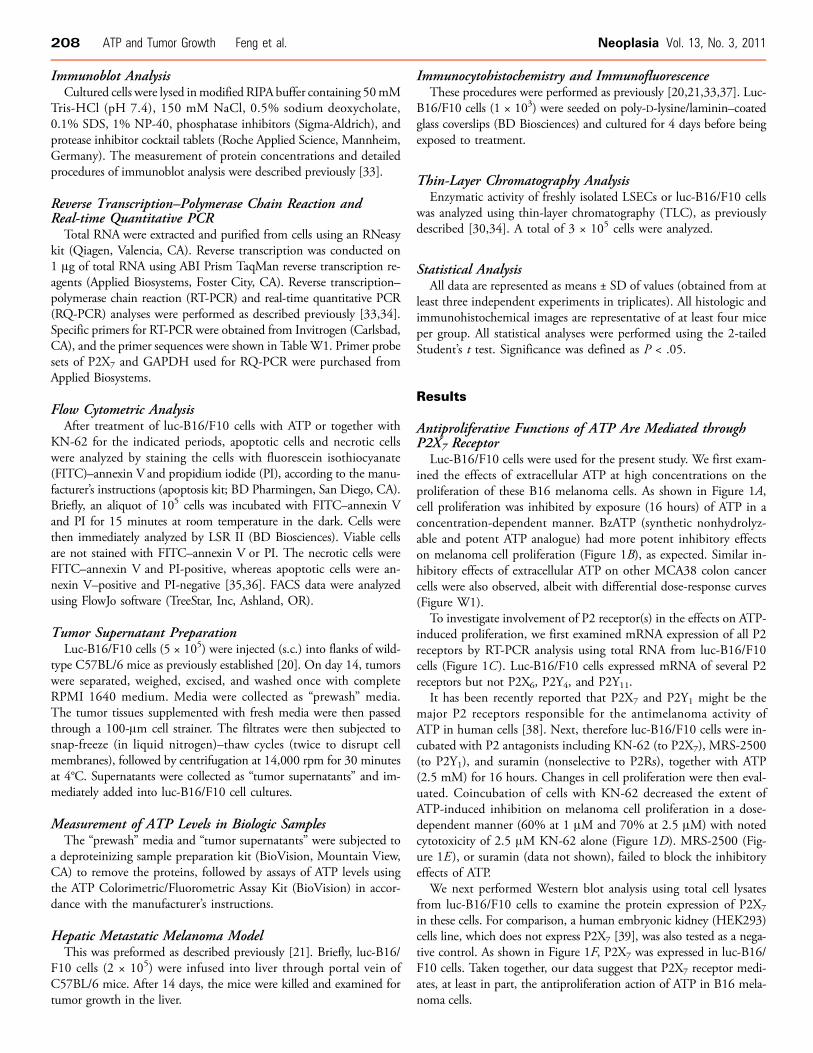
Apyrase (Soluble NTPDase) or Vascular Cell CD39 Expressedby LSECs Abrogates/Reverses Antitumor Activity of ATPNext, we sought to determine whether scavenging of extracellular
ATP by apyrase, a soluble form of NTPDase with ATPase and ADPaseactivity at a 1:1 ratio, could rescue ATP-stimulated growth inhibitionof tumor cells. Figure 4A showed that tumor cell growth inhibitiontriggered by ATP (2.5 mM) was completely abrogated by coincubationof cells with apyrase (10 U/ml). The rescue of tumor cells by apyrasewas dose dependent.CD39 and CD73 are the major ectonucleotidases expressed by
LSECs. Next, LSECs were purified from wt, Cd39 null, or Cd73 nulllivers and were cocultured with luc-B16/F10 cells for 24 hours, atvarious ratios of cell numbers, before being exposed to ATP (2.5 mM)for 16 hours.Successful isolation of healthy LSECs was verified by uptake of
DiI-labeled Ac-LDL and FACS analysis for endothelium makers (in-cluding CD31, CD34, and Flk-1) (data not shown), as establishedpreviously [31,32].As shown in Figure 4B, wt LSECs attenuated the inhibitory effects
of ATP on tumor cell growth, whereas Cd39 null LSECs did notretain this capacity. Interestingly, growth inhibition by ATP was re-versed by Cd73 null LSECs to a greater extent, when compared withwt LSECs (Figure 4B). The rescue observed with wt and Cd73 nullLSECs was dose dependent.We also noted that extracellular ATP inhibited growth of LSECs
(Figure W3 for wt cells and data not shown for null cells).To further investigate the mechanisms underlying the observations
in Figure 4B, purine metabolism by ectonucleotidase activity of freshlyisolated LSECs was examined by TLC analysis using ADP-C14 as sub-strate. ADP was first hydrolyzed to AMP and then to adenosine bywt LSECs. Adenosine was further degraded to hypoxanthine by wtLSECs (Figure 4C ). Cd73 null LSECs could only generate AMPbut not adenosine (Figure 4C ). Cd39 null LSECs and luc-B16/F10
cells had minimal nonspecific ectonucleotidase activity, in contrast towt LSECs (Figure 4C). These findings clearly explain how wt, Cd73null, and Cd39 null LSECs exhibit differential salvage abilities onATP-induced growth inhibition as observed in Figure 4B.
Defective Angiogenesis Is Associated with Heightened TumorNecrosis and Increased P2X7 Expression in Cd39 NullTumor-Bearing MiceWenext determined whether injured tumor cells could release endog-
enous mediators that directly result in cellular damage of contiguous/adjacent tumor cells. Luc-B16/F10 cells were injected (s.c.) intoflanks of wild type C57BL/6 mice for 14 days, tumors were excised,and tumor supernatants were prepared (see Materials and Methods),these were then added to luc-B16/F10 cell cultures. In Figure 5A, weshow that melanoma cell proliferation was inhibited by the addi-tion of tumor supernatants in a concentration-dependent manner.Dramatic increases in ATP levels were also noted in these tumorsupernatants compared with the prewash media (Figure W4). How-ever, coincubation with apyrase alone failed to rescue the growth in-hibitory effects triggered by these crude tumor supernatants (data notshown) as previously noted with exogenous ATP (Figure 4A). Thesedata suggest that other cytotoxic constituents besides nucleotidescontribute to the tumor killing activity of supernatants.We have recently demonstrated the effect of Cd39 deletion on mel-
anoma growth in vivo using a murine model of hepatic metastasesof B16/F10 melanoma [21]. We noted that immune cell as well asvascular CD39 expression promote tumor growth, whereas pharma-cological inhibition of CD39 enzymatic activity (in contrast) abrogatestumor growth [21].We stained these liver tumor sections using anti-CD31 (a marker for
endothelium) and anti-CD39 antibodies. We observed that CD39 wasexpressed on tumor-associated endothelial cells (ECs) in wt livers. Incontrast, in Cd39 null tumor-bearing livers, lack of CD39 expression(suggesting decreased ATP scavenging in the tumor microenvironment)was associated with defective angiogenesis and larger areas of necrosiswithin the centers of tumors (Figure 5B). Immunofluorescent stainingon tumor sections further showed that protein expression of P2X7 wasincreased on melanoma cells in Cd39 null tumor-bearing livers com-pared with wt livers (Figure 5C).
DiscussionThe present study clearly demonstrates that tumor-derived mediators,inclusive of ATP, directly exert growth inhibitory effects on tumor cells.The P2X7 receptor is functionally expressed in B16/F10 melanomacells and is responsible, at least in part, for such ATP-induced growthinhibition and cell death. Importantly, coculture of tumor cells withLSECs demonstrates that expression of ecto-enzyme CD39 by endo-thelial cells counteracts tumoricidal actions stimulated by extracellu-lar ATP. Collectively, in light of ATP-induced tumor suppression, ourresults indicate novel purinergic mechanisms implicated in tumor biol-ogy: 1) danger signals (including ATP) released by necrotic tumor cellsresult in subsequent death of neighboring tumor cells and 2) CD39expressed on ECs promotes tumor cell growth by scavenging extracel-lular ATP in the tumor microenvironment.Different P2 receptor subtypes have been shown to modulate dif-
ferent cellular functions such as proliferation, differentiation, and ap-optosis. P2Y1 and P2X7 receptors are expressed in human melanomacells in situ and mediate apoptotic and necrotic actions of ATP [38].
Neoplasia Vol. 13, No. 3, 2011 ATP and Tumor Growth Feng et al. 211

The antitumor actions of these receptors contain three processes: in-hibition of cell proliferation, promotion of cell differentiation (result-ing in inhibition of cell proliferation), and cell death [2]. Here, weshow that antitumor activity of ATP is largely due to the combina-tion of inhibition of cell proliferation and induction of cell death.
These two processes are both mediated largely through the expressionof P2X7.When ATP appears in the extracellular space of tumor microen-
vironment, this mediator is rapidly hydrolyzed by ectonucleotidasesto ADP, AMP, and, finally, adenosine [12]. Most studies to date
212 ATP and Tumor Growth Feng et al. Neoplasia Vol. 13, No. 3, 2011
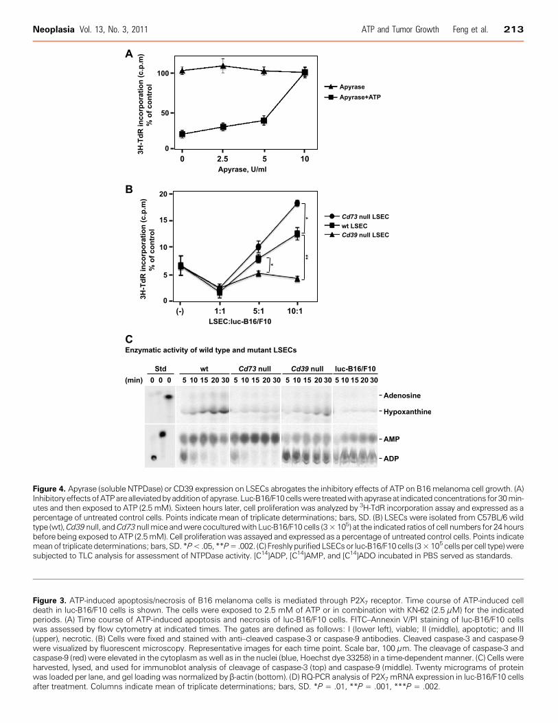
Figure 4. Apyrase (soluble NTPDase) or CD39 expression on LSECs abrogates the inhibitory effects of ATP on B16melanoma cell growth. (A)InhibitoryeffectsofATParealleviatedbyadditionof apyrase. Luc-B16/F10cellswere treatedwithapyraseat indicatedconcentrations for30min-utes and then exposed to ATP (2.5 mM). Sixteen hours later, cell proliferation was analyzed by 3H-TdR incorporation assay and expressed as apercentage of untreated control cells. Points indicate mean of triplicate determinations; bars, SD. (B) LSECs were isolated from C57BL/6 wildtype (wt),Cd39null, andCd73nullmice andwere coculturedwith Luc-B16/F10 cells (3×10⁵) at the indicated ratios of cell numbers for 24 hoursbefore being exposed to ATP (2.5mM). Cell proliferationwas assayed and expressed as a percentage of untreated control cells. Points indicatemeanof triplicatedeterminations; bars, SD. *P<.05, **P=.002. (C) Freshly purified LSECsor luc-B16/F10cells (3×10⁵ cells per cell type)weresubjected to TLC analysis for assessment of NTPDase activity. [C14]ADP, [C14]AMP, and [C14]ADO incubated in PBS served as standards.
Figure 3. ATP-induced apoptosis/necrosis of B16 melanoma cells is mediated through P2X7 receptor. Time course of ATP-induced celldeath in luc-B16/F10 cells is shown. The cells were exposed to 2.5 mM of ATP or in combination with KN-62 (2.5 μM) for the indicatedperiods. (A) Time course of ATP-induced apoptosis and necrosis of luc-B16/F10 cells. FITC–Annexin V/PI staining of luc-B16/F10 cellswas assessed by flow cytometry at indicated times. The gates are defined as follows: I (lower left), viable; II (middle), apoptotic; and III(upper), necrotic. (B) Cells were fixed and stained with anti–cleaved caspase-3 or caspase-9 antibodies. Cleaved caspase-3 and caspase-9were visualized by fluorescent microscopy. Representative images for each time point. Scale bar, 100 μm. The cleavage of caspase-3 andcaspase-9 (red) were elevated in the cytoplasm aswell as in the nuclei (blue, Hoechst dye 33258) in a time-dependentmanner. (C) Cells wereharvested, lysed, and used for immunoblot analysis of cleavage of caspase-3 (top) and caspase-9 (middle). Twenty micrograms of proteinwas loaded per lane, and gel loading was normalized by β-actin (bottom). (D) RQ-PCR analysis of P2X7 mRNA expression in luc-B16/F10 cellsafter treatment. Columns indicate mean of triplicate determinations; bars, SD. *P = .01, **P = .001, ***P = .002.
Neoplasia Vol. 13, No. 3, 2011 ATP and Tumor Growth Feng et al. 213

have focused on the alterations of purinergic receptors in tumors,whereas ectonucleotidases are much less investigated. Purinergic sig-naling can be modulated by modifying the expression and/or ac-tivity of these ectoenzymes in addition to changes in P2 receptorlevels [11,40].
This study shows for the first time that deletion of Cd39 on ECsenhances antitumor activity of ATP, whereas deletion of Cd73 on ECshas different effects. These data suggest a feasible approach to augmentanticancer therapy by modulating expression and/or enzymatic activ-ity of NTPDase-type ectonucleotidases. This approach might be easierto achieve and more efficacious than to independently target severalpurinergic receptors.CD39 is also expressed by immune cells inclusive of Treg (CD4+
Foxp3+) and memory cells (CD4+CD44+CD62L−Foxp3−) [21,41].These cells often infiltrate into solid tumors [21]. Whether theseCD39+ infiltrating immune cell populations also contribute to degra-dation of cytotoxic ATP in the tumor microenvironment and therebyindependently promote tumor growth remains unclear.Exposure of ECs to elevated levels of ATP has been shown to pro-
mote apoptosis in vitro [42]. We have previously demonstrated thatinhibition of tumor growth in Cd39 null mice is associated with de-fects of tumor angiogenesis [20,21]. Moreover, in this study, we showthat ATP also exhibits direct growth inhibitory effects on LSECs (Fig-ure W3 and data not shown) that further compromises cell-associatedNTPDase activity. Therefore, the reduction of tumor size and volumein Cd39 null tumor-bearing mice might result from dual actions ofATP on tumor cells as well as on ECs.The phosphohydrolysis of ATP to adenosine has a complex mod-
ulatory effect on tumor cell proliferation and growth [43–45]. Aden-osine ultimately derived from ATP may be responsible for some ofthe observed effects as this nucleoside has been shown to affect tumorgrowth in a cell-specific manner determined by concentrations andkinetics of exposure [43–45] (and unpublished observations in ourlaboratory). Tumor cell expressions of A2A and A2B have been shownto be proapoptotic and have antitumor activity [44,45]. The actions ofA3 are contradictory. Most studies have demonstrated that A3 agonistsinduce apoptosis and tumor growth inhibition [45–48], whereasothers show that A3 stimulation blocks A2A-induced cell death andensures cell survival [43].Therefore, the ambient vascular nucleotide/nucleoside milieu as
regulated by ectonucleotidases and ectonucleotidases dictates the ef-ficacy of antitumor activity of ATP in vivo. In this study, the differ-ential salvage abilities on ATP-triggered tumor cell growth inhibitionand NTPDase activity exhibited by wt, Cd73 null, and Cd39 nullLSECs (Figure 4, B and C) are indicative of the participation of ATP-derived adenosine in the antitumor function of ATP.Dzhandzhugazyan et al. [49] have shown that CD39 is the major
ectonucleotidase in human melanocytes and melanoma cell lines andCD39 is overexpressed in differentiated human melanomas. It hasbeen recently reported that CD73 is expressed on various tumor cells(e.g., ID8 ovarian cancer cells) and participates in adenosine genera-tion, thereby suppressing antitumor immune responses [50], but po-tentially also affecting cancer cell apoptosis.We therefore also examined the expression of CD39 and CD73
on tumor cells used for this study. Neither CD39 nor CD73 expres-sion was observed on cultured B16/F10 or MCA38 cells as well ason malignant cells in metastatic tumors in the livers and lungs at anyprogression stage after tumor challenge [20,21] (data not shown).Lastly, we have also shown that ATP exhibits cytotoxic effects on
MCA38 colon cancer cells as well, suggesting general feature of anti-tumor capability of ATP (Figures W1 and W2; data not shown).In summary, we postulate an intriguing mechanism by which ex-
tracellular ATP released by dying tumor cells accumulates to highconcentrations that not only function as danger signals to the im-
Figure 5. Metastatic melanoma growth in Cd39 null mice. (A) Dose-dependent inhibitory effects of tumor supernatants (16 hours oftreatment) on luc-B16/F10 melanoma cell proliferation. (B) Repre-sentative immunohistochemical staining on tumor tissue sectionsobtained from melanoma metastasized to mouse livers using anti-CD31 (a marker for endothelium) and anti-CD39 antibodies andTUNEL staining (TdT) for apoptosis. (C) P2X7 expression on tumortissues. Representative fluorescence immunohistochemical stain-ing using anti-P2X7 antibody: Hoechst dye 33258 staining nucleiin blue and P2X7 in red. Columns indicate mean of triplicate deter-minations; bars, SD. Scale bar, 100 μm.
214 ATP and Tumor Growth Feng et al. Neoplasia Vol. 13, No. 3, 2011

mune system but also can directly kill adjacent tumor cells. Our datashowing that the antitumor activities of ATP are dose-dependent andcan be amplified by inhibition of ectonucleotidase, such as CD39,open new avenues for investigation in cancer management.
AcknowledgmentsThe authors sincerely thank Nicholas P. Restifo (National CancerInstitute) for the MCA38 cells.
References[1] Burnstock G and Knight GE (2004). Cellular distribution and functions of P2
receptor subtypes in different systems. Int Rev Cytol 240, 31–304.[2] Shabbir M and Burnstock G (2009). Purinergic receptor–mediated effects
of adenosine 5′-triphosphate in urological malignant diseases. Int J Urol 16,143–150.
[3] Burnstock G (2006). Purinergic P2 receptors as targets for novel analgesics.Pharmacol Ther 110, 433–454.
[4] Burnstock G and Kennedy C (1985). Is there a basis for distinguishing twotypes of P2-purinoceptor? Gen Pharmacol 16, 433–440.
[5] Burnstock G (2006). Purinergic signalling—an overview. Novartis Found Symp276, 26–48; discussion 48–57, 275–281.
[6] Burnstock G (2007). Purine and pyrimidine receptors. Cell Mol Life Sci 64,1471–1483.
[7] Rapaport E (1983). Treatment of human tumor cells with ADP or ATP yieldsarrest of growth in the S phase of the cell cycle. J Cell Physiol 114, 279–283.
[8] Rapaport E (1988). Experimental cancer therapy in mice by adenine nucleo-tides. Eur J Cancer Clin Oncol 24, 1491–1497.
[9] White N and Burnstock G (2006). P2 receptors and cancer. Trends PharmacolSci 27, 211–217.
[10] Shabbir M, Thompson C, Jarmulowiczc M, Mikhailidis D, and Burnstock G(2008). Effect of extracellular ATP on the growth of hormone-refractory pros-tate cancer in vivo. BJU Int 102, 108–112.
[11] Deli T and Csernoch L (2008). Extracellular ATP and cancer: an overview withspecial reference to P2 purinergic receptors. Pathol Oncol Res 14, 219–231.
[12] Zimmermann H (2000). Extracellular metabolism of ATP and other nucleo-tides. Naunyn Schmiedebergs Arch Pharmacol 362, 299–309.
[13] Trautmann A (2009). Extracellular ATP in the immune system: more than just a“danger signal”. Sci Signal 2, pe6.
[14] Behrens MD, Wagner WM, Krco CJ, Erskine CL, Kalli KR, Krempski J, GadEA, Disis ML, and Knutson KL (2008). The endogenous danger signal, crystal-line uric acid, signals for enhanced antibody immunity. Blood 111, 1472–1479.
[15] Apetoh L, Ghiringhelli F, Tesniere A, Obeid M, Ortiz C, Criollo A, Mignot G,Maiuri MC, Ullrich E, Saulnier P, et al. (2007). Toll-like receptor 4–dependentcontribution of the immune system to anticancer chemotherapy and radiother-apy. Nat Med 13, 1050–1059.
[16] Mariathasan S, Weiss DS, Newton K, McBride J, O’Rourke K, Roose-GirmaM, Lee WP, Weinrauch Y, Monack DM, and Dixit VM (2006). Cryopyrinactivates the inflammasome in response to toxins and ATP. Nature 440,228–232.
[17] Martins I, Tesniere A, Kepp O, Michaud M, Schlemmer F, Senovilla L, Seror C,Metivier D, Perfettini JL, Zitvogel L, et al. (2009). Chemotherapy induces ATPrelease from tumor cells. Cell Cycle 8, 3723–3728.
[18] Aymeric L, Apetoh L, Ghiringhelli F, Tesniere A, Martins I, Kroemer G, SmythMJ, and Zitvogel L (2010). Tumor cell death and ATP release prime dendriticcells and efficient anticancer immunity. Cancer Res 70, 855–858.
[19] Ghiringhelli F, Apetoh L, Tesniere A, Aymeric L, Ma Y, Ortiz C, Vermaelen K,Panaretakis T, Mignot G, Ullrich E, et al. (2009). Activation of the NLRP3inflammasome in dendritic cells induces IL-1β–dependent adaptive immunityagainst tumors. Nat Med 15, 1170–1178.
[20] Jackson SW, Hoshi T, Wu Y, Sun X, Enjyoji K, Cszimadia E, Sundberg C, andRobson SC (2007). Disordered purinergic signaling inhibits pathological angio-genesis in cd39/Entpd1-null mice. Am J Pathol 171, 1395–1404.
[21] Sun X, Wu Y, Gao W, Enjyoji K, Csizmadia E, Muller CE, Murakami T, andRobson SC (2010). CD39/ENTPD1 expression byCD4+Foxp3+ regulatory T cellspromotes hepatic metastatic tumor growth in mice. Gastroenterology 139(3),1030–1040.
[22] Plesner L (1995). Ecto-ATPases: identities and functions. Int Rev Cytol 158,141–214.
[23] Enjyoji K, Sevigny J, Lin Y, Frenette PS, Christie PD, Esch JS II, Imai M,Edelberg JM, Rayburn H, Lech M, et al. (1999). Targeted disruption of cd39/ATP diphosphohydrolase results in disordered hemostasis and thromboregulation.Nat Med 5, 1010–1017.
[24] Robson SC, Wu Y, Sun X, Knosalla C, Dwyer K, and Enjyoji K (2005). Ecto-nucleotidases of CD39 family modulate vascular inflammation and thrombosisin transplantation. Semin Thromb Hemost 31, 217–233.
[25] Mills JH, Thompson LF, Mueller C, Waickman AT, Jalkanen S, Niemela J,Airas L, and Bynoe MS (2008). CD73 is required for efficient entry of lympho-cytes into the central nervous system during experimental autoimmune enceph-alomyelitis. Proc Natl Acad Sci USA 105, 9325–9330.
[26] Sato A, Ohtsuki M, Hata M, Kobayashi E, and Murakami T (2006). Antitumoractivity of IFN-λ in murine tumor models. J Immunol 176, 7686–7694.
[27] Jun DJ, Kim J, Jung SY, Song R, Noh JH, Park YS, Ryu SH, Kim JH, Kong YY,Chung JM, et al. (2007). Extracellular ATP mediates necrotic cell swelling inSN4741 dopaminergic neurons through P2X7 receptors. J Biol Chem 282,37350–37358.
[28] Kunzli BM, Nuhn P, Enjyoji K, Banz Y, Smith RN, Csizmadia E, Schuppan D,Berberat PO, Friess H, and Robson SC (2008). Disordered pancreatic inflam-matory responses and inhibition of fibrosis in CD39-null mice. Gastroenterology134, 292–305.
[29] Kaczmarek E, Koziak K, Sevigny J, Siegel JB, Anrather J, Beaudoin AR, Bach FH,and Robson SC (1996). Identification and characterization of CD39/vascularATP diphosphohydrolase. J Biol Chem 271, 33116–33122.
[30] Deaglio S, Dwyer KM, Gao W, Friedman D, Usheva A, Erat A, Chen JF,Enjyoji K, Linden J, Oukka M, et al. (2007). Adenosine generation catalyzedby CD39 and CD73 expressed on regulatory T cells mediates immune suppres-sion. J Exp Med 204, 1257–1265.
[31] LeCouter J, Moritz DR, Li B, Phillips GL, Liang XH, Gerber HP, Hillan KJ,and Ferrara N (2003). Angiogenesis-independent endothelial protection of liver:role of VEGFR-1. Science 299, 890–893.
[32] Beldi G, Wu Y, Sun X, Imai M, Enjyoji K, Csizmadia E, Candinas D, Erb L,and Robson SC (2008). Regulated catalysis of extracellular nucleotides by vas-cular CD39/ENTPD1 is required for liver regeneration. Gastroenterology 135,1751–1760.
[33] Wu Y, Sun X, Kaczmarek E, Dwyer KM, Bianchi E, Usheva A, and Robson SC(2006). RanBPM associates with CD39 and modulates ecto-nucleotidase activ-ity. Biochem J 396, 23–30.
[34] Beldi G, Wu Y, Banz Y, Nowak M, Miller L, Enjyoji K, Haschemi A, YegutkinGG, Candinas D, Exley M, et al. (2008). Natural killer T cell dysfunction inCD39-null mice protects against concanavalin A–induced hepatitis. Hepatology48, 841–852.
[35] Koopman G, Reutelingsperger CP, Kuijten GA, Keehnen RM, Pals ST, and vanOers MH (1994). Annexin V for flow cytometric detection of phosphatidyl-serine expression on B cells undergoing apoptosis. Blood 84, 1415–1420.
[36] Vermes I, Haanen C, Steffens-Nakken H, and Reutelingsperger C (1995). Anovel assay for apoptosis. Flow cytometric detection of phosphatidylserine ex-pression on early apoptotic cells using fluorescein labelled annexin V. J ImmunolMethods 184, 39–51.
[37] Goepfert C, Sundberg C, Sevigny J, Enjyoji K, Hoshi T, Csizmadia E, andRobson S (2001). Disordered cellular migration and angiogenesis in CD39-nullmice. Circulation 104, 3109–3115.
[38] White N, Knight GE, Butler PE, and Burnstock G (2009). An in vivo model ofmelanoma: treatment with ATP. Purinergic Signal 5, 327–333.
[39] Humphreys BD, Virginio C, Surprenant A, Rice J, and Dubyak GR (1998).Isoquinolines as antagonists of the P2X7 nucleotide receptor: high selectivityfor the human versus rat receptor homologues. Mol Pharmacol 54, 22–32.
[40] Fang WG, Pirnia F, Bang YJ, Myers CE, and Trepel JB (1992). P2-purinergicreceptor agonists inhibit the growth of androgen-independent prostate carci-noma cells. J Clin Invest 89, 191–196.
[41] Zhou Q, Yan J, Putheti P, Wu Y, Sun X, Toxavidis V, Tigges J, Kassam N, EnjyojiK, Robson SC, et al. (2009). Isolated CD39 expression on CD4+ T cells denotesboth regulatory and memory populations. Am J Transplant 9, 2303–2311.
[42] Goepfert C, Imai M, Brouard S, Csizmadia E, Kaczmarek E, and Robson SC(2000). CD39 modulates endothelial cell activation and apoptosis. Mol Med 6,591–603.
[43] Merighi S, Mirandola P, Milani D, Varani K, Gessi S, Klotz KN, Leung E,Baraldi PG, and Borea PA (2002). Adenosine receptors as mediators of both
Neoplasia Vol. 13, No. 3, 2011 ATP and Tumor Growth Feng et al. 215

cell proliferation and cell death of cultured human melanoma cells. J Invest Der-matol 119, 923–933.
[44] Merighi S, Mirandola P, Varani K, Gessi S, Leung E, Baraldi PG, Tabrizi MA,and Borea PA (2003). A glance at adenosine receptors: novel target for anti-tumor therapy. Pharmacol Ther 100, 31–48.
[45] Panjehpour M and Karami-Tehrani F (2007). Adenosine modulates cell growth inthe human breast cancer cells via adenosine receptors. Oncol Res 16, 575–585.
[46] Kim SJ, Min HY, Chung HJ, Park EJ, Hong JY, Kang YJ, Shin DH, Jeong LS,and Lee SK (2008). Inhibition of cell proliferation through cell cycle arrest andapoptosis by thio-Cl-IB-MECA, a novel A3 adenosine receptor agonist, in hu-man lung cancer cells. Cancer Lett 264, 309–315.
[47] Fishman P, Bar-Yehuda S, Madi L, and Cohn I (2002). A3 adenosine receptor asa target for cancer therapy. Anticancer Drugs 13, 437–443.
[48] Fishman P, Bar-Yehuda S, Ohana G, Barer F, Ochaion A, Erlanger A, and Madi L(2004). An agonist to the A3 adenosine receptor inhibits colon carcinoma growthin mice via modulation of GSK-3 β and NF-κB. Oncogene 23, 2465–2471.
[49] Dzhandzhugazyan KN, Kirkin AF, thor Straten P, and Zeuthen J (1998). Ecto-ATP diphosphohydrolase/CD39 is overexpressed in differentiated human mela-nomas. FEBS Lett 430, 227–230.
[50] Jin D, Fan J, Wang L, Thompson LF, Liu A, Daniel BJ, Shin T, Curiel TJ, andZhang B (2010). CD73 on tumor cells impairs antitumor T-cell responses: a novelmechanism of tumor-induced immune suppression. Cancer Res 70, 2245–2255.
216 ATP and Tumor Growth Feng et al. Neoplasia Vol. 13, No. 3, 2011

Table W1. RT-PCR Primers.
Transcript Primer Sequence
Mouse P2X1 Forward 5′-tcattgccagaggctttc-3′Reverse 5′-gtggagggttgtatgtgt-3′
Mouse P2X2 Forward 5′-caaagcctatgggattcg-3′Reverse 5′-cctatgaggagttctgtt-3′
Mouse P2X3 Forward 5′-gtgaaaagctggaccattgg-3′Reverse 5′-gctgccattctccatcttgt-3′
Mouse P2X4 Forward 5′-tacgtcattgggtgggtgtt-3′Reverse 5′-cttgatctggatacccatga-3′
Mouse P2X5 Forward 5′-aggacattgacacttccctg-3′Reverse 5′-catcaggtcacggaactcta-3′
Mouse P2X6 Forward 5′-gtggtagtctacgtgatagg-3′Reverse 5′-gcctctctatccacatacag-3′
Mouse P2X7 Forward 5′-cttgccaactatgaacgg-3′Reverse 5′-cttggcctttgccaactt-3′
Mouse P2Y1 Forward 5′-ttatgtcagcgtgctggtgt-3′Reverse 5′-cgtgtctccattctgcttga-3′
Mouse P2Y2 Forward 5′-gaggacttcaagtacgtgct-3′Reverse 5′-acggagctgtaagccacaaa-3′
Mouse P2Y4 Forward 5′-aacaactgcttcctccct-3′Reverse 5′-aagtcctagaggtaggtg-3′
Mouse P2Y6 Forward 5′-cctgatgtatgcctgttcac-3′Reverse 5′-cacagccaagtaggctgtct-3′
Human P2Y11* Forward 5′-tgtggcccatactggtggttgag-3′Reverse 5′-gaagaaggggtgcacgatgccca-3′
Mouse P2Y12 Forward 5′-atatgcctggtgtcaacacc-3′Reverse 5′-ggaatccgtgcaaagtggaa-3′
Mouse P2Y13 Forward 5′-tgcagggcttcaacaagtct-3′Reverse 5′-cctttccccatctcacacat-3′
Mouse P2Y14 Forward 5′-ggaacaccctgatcacaaag-3′Reverse 5′-tgaccttccgtctgactctt-3′
Mouse actin Forward 5′-tgtacgtagccatccaggct-3′Reverse 5′-ggtaccaccagacaacactgt-3′
*Human P2Y11 was used as a negative control as mouse cells do not express P2Y11 receptor.
Figure W1. Effects of extracellular ATP on colon cancer cell prolif-eration. Extracellular ATP inhibited proliferation of colon cancercells, in a dose-dependent manner. MCA38 cells were treated withATP at the indicated concentrations for 16 hours. Cell proliferationwas determined by 3H-TdR incorporation assay. Columns indicatemean of triplicate determinations; bars, SD.
Figure W2. Effects of extracellular ATP on viability of colon cancercells. Extracellular ATP exhibited cytotoxicity on colon cancer cellsin a dose-dependent manner. MCA38 cells were treated with ATPat the indicated concentrations for 16 hours, and cell viability wasevaluated using Cell Counting Kit-8. Columns indicate mean of trip-licate determinations; bars, SD.
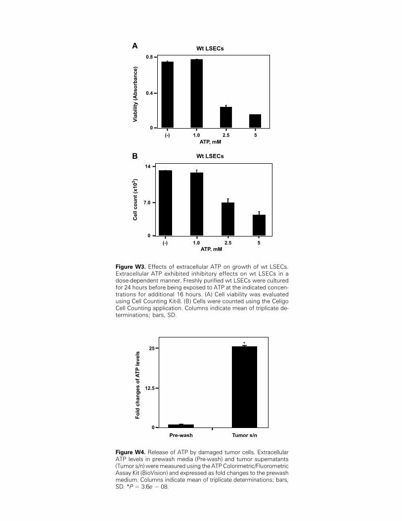
Figure W3. Effects of extracellular ATP on growth of wt LSECs.Extracellular ATP exhibited inhibitory effects on wt LSECs in adose-dependent manner. Freshly purified wt LSECs were culturedfor 24 hours before being exposed to ATP at the indicated concen-trations for additional 16 hours. (A) Cell viability was evaluatedusing Cell Counting Kit-8. (B) Cells were counted using the CeligoCell Counting application. Columns indicate mean of triplicate de-terminations; bars, SD.
Figure W4. Release of ATP by damaged tumor cells. ExtracellularATP levels in prewash media (Pre-wash) and tumor supernatants(Tumor s/n) weremeasured using the ATP Colorimetric/FluorometricAssay Kit (BioVision) and expressed as fold changes to the prewashmedium. Columns indicate mean of triplicate determinations; bars,SD. *P = 3.6e − 08.