Embed Size (px)
Citation preview

Vasculitis of the nervous systemDavid S. Younger
Purpose of review
Vasculitis refers to heterogenous clinicopathologic disorders
that share the histopathology of inflammation of blood vessels.
When unrecognized and therefore untreated, vasculitis of the
nervous system leads to pervasive injury and disability making
this a disorder of paramount importance to all clinicians.
Recent findings
Remarkable progress has been made in the pathogenesis,
diagnosis, and treatment of vasculitis of the central (CNS) and
peripheral nervous system (PNS). The classification of vasculitis
affecting the nervous system includes (1) Systemic vasculitis
disorders (necrotizing arteritis of the polyarteritis type,
hypersensitivty vasculitis, systemic granulomatous vasculitis,
giant cell arteritis, diverse connective tissue disorders; viral,
spirochete, fungal, and retroviral infection; (2) Paraneoplastic
disorders; (3) Amphetamine abuse; (4) Granulomatous angiitis
of the brain; (5) Isolated peripheral nerve vasculitis, each in the
absence of systemic involvement; and (6) diabetes mellitus,
associated wtih inflammatoy PNS vasculopathy.
Summary
Vasculitis is diagnosed with assurance after intensive
evaluation. Successful treatment follows ascertainment of the
specific vasculitic disorder and the underlying cytochemical
mechanism of pathogenesis. Clinicians must choose from
among the available immunomodulating, immunosuppressive,
and targeted immunotherapies, unfortunately without the benefit
of prospective clinical trials, tempered by the recognition of all of
the possible medication related side effects.
Keywords
nervous system, vasculitis
Curr Opin Neurol 17:317–336. # 2004 Lippincott Williams & Wilkins.
Department of Neurology, New York University School of Medicine, New York, NewYork 10016, USA
Correspondence to David S. Younger, MD, 715 Park Avenue, Ground Floor, NewYork, NY 10021, USATel: +1 212 535 4314; fax: +1 212 535 6392; e-mail: [email protected]
Current Opinion in Neurology 2004, 17:317–336
Abbreviations
ANCA antineutrophil cytoplasmic antibodyAPC antigen-presenting cellCNS central nervous systemCSF cerebrospinal fluidGANS granulomatous angiitis of the nervous systemIVIg intravenous immunoglobulinLCV leukocytoclastic vasculitisMAC membrane attack complexMNM mononeuritis multiplexMPA microscopic polyangiitisMRI magnetic resonance imagingPAN polyarteritis nodosaPMN polymorphonuclear leukocytePNS peripheral nervous systemSLE systemic lupus erythematosusSPECT single photon emission computed tomographyVZV varicella zoster virusWG Wegener granulomatosis
# 2004 Lippincott Williams & Wilkins1350-7540
IntroductionVasculitis is a spectrum of clinicopathological disorders
defined by inflammation of the blood vessels, including
arteries and veins of varying caliber, which result in a
variety of clinical neurological manifestations related to
ischemic injury of the central nervous system (CNS) and
peripheral nervous system (PNS). This article reviews
the remarkable progress that has been made in the
pathogenesis, classification, diagnosis, and management
of vasculitis of the nervous system. Several excellent
reviews have been published on this topic [1.,2].
ClassificationVasculitis in its various forms affects blood vessels of
varying caliber from the aorta to capillaries and veins
(Fig. 1). The diverse forms of vasculitis and autoimmune
diseases to be considered in this chapter are summarized
in Table 1.
Systemic necrotizing arteritis of thepolyarteritis nodosa typePolyarteritis nodosa (PAN) is the prototypic disorder of
the group of systemic necrotizing arteritis, which also
includes Churg–Strauss syndrome, microscopic polyan-
giitis (MPA) syndrome, Kawasaki disease and an overlap
syndrome.
The early history of vasculitis is debatable, but one fact
is clear, the ealiest patients with vasculitis had PAN and
neurological involvement, such as the patient described
by Kussmaul and Maier in 1886 [3], who presented with
leg pains, cramps, and tenderness so prominent that
DOI: 10.1097/01.wco.0000130301.13775.96 317

trichinosis was contemplated. At postmortem examina-
tion there was widespread arteritis that resembled
syphilitic periarteritis caused by the frequent occurrence
of internal and external elastic lamina necrosis, fibrin
deposition, aneurysmal dilatations, and intimal prolifera-
tion resulting in endarteritis obliterans of the brain,
nerves, skeletal muscle, and in systemic organs of small
and medium size vessels with a diameter of 120 mm or
more (Fig. 2). Peripheral neuropathy is the most
frequent finding, typically mononeuritis multiplex
(MNM) caused by involvement of the arteriae nervorum,
followed by CNS manifestations of two types, one
resulting from stroke and cerebral hemorrhage (Fig. 3)
and the second a diffuse encephalopathy accompanied
by seizure phenomena. The arteritis of MPA affects
small arterioles, capillaries, and venules of the kidney
and lungs; 50–80% of patients have circulating antineu-
trophil cytoplasmic antibodies (ANCA) to myeloperox-
idase or perinuclear ANCA. Kawasaki disease is
characterized by viral exanthema that ultimately leads
to PAN, focused primarily in the coronary arteries.
In 1951, Churg and Strauss [4] delineated a syndrome of
asthma, eosinophilia, extravascular granulomas and
necrotizing arteritis, involving arterioles, capillaries, and
venules that was named in their honor. The extravas-
cular granulomas, epithelioid and giant cell infiltrates
distinguish Churg–Strauss syndrome from classic PAN
and resemble MPA in the tendency to involve the lung
and kidney. Early suspected patients present with
allergic rhinitis, nasal polyposis, and asthma, followed
by tissue eosinophilia and systemic vasculitis, with CNS
involvement that includes confusion, seizures, cranial
nerve involvement, optic neuropathy, and less common
radicular and plexus PNS manifestations.
Hypersensitivity vasculitisHistorically, in the same period as PAN, Zeek et al. [5]described patients with sensitivity to sulfonamides and
Figure 1. The pathological spectrum of the major vasculitides
Vessels involved Clinical syndrome
Veins
Venules
Capillaries
Arterioles
Small musculararteries(intraorgan vessels)
Medium musculararteries (coronary,hepatic, intracerebral)
Large arteries(vertebral, temporal,carotid)
Aorta
Usually involved
Sometimes involved
Eales’disease
Hypersen-sitivityangitis
Wegener’sgranulo-matosis
Lympho-matoidgranulo-matosis
Allergicgranulo-matosis
Micro-scopicpoly-angitis
Polyarter-itisnodosa
CNSvasculitis
Temporalarteritis
Takayasu’sarteritis
Reproduced from Younger [1.] with permission of the publisher.
Inflammatory diseases and infection318

other drugs that manifested with skin rash and general-
ized vasculitis. So-called hypersensitivity vasculitis
includes syndromes related to drug reactions, Henoch–
Schonlein purpura, hypocomplementemic or urticarial
vasculitis and cryoglobulinemia. These disorders demon-
strate non-segmental infiltration of the walls of small
vessels and postcapillary venules, particularly of the
dermis and other systemic tissue by polymorphonuclear
leukocytes (PMN), which disintegrate leaving nuclear
fragments or leukocytoclastic vasculitis (LCV) (Fig. 4),
fibrinoid necrosis, micro-infarction and hemorrhages of
affected tissues, including those of the nervous system.
Drug reactions are responsible for approximately 20% of
cases of dermal vasculitis usually associated with
immune complex deposition, and abates with drug
withdrawal and all other possibly offending agents.
Affected patients present with ’flu-like constitutional
signs that include fever and headache, and progresses to
skin lesions, and if serious or advanced enough, seizures,
encephalopathy, stroke, cranial nerve signs, and myelo-
pathy. In contrast to PAN, lesions are in the same stage
of evolution.
Henoch–Schonlein purpura is characterized by non-
thrombocytopenic purpura, arthralgia, abdominal pain,
LCV, and IgA immune complex deposition with
complement activation in affected children. It is
characterized histopathologically by varying degrees of
arteriolar, capillary, and venular interstitial infiltration by
PMN cells, eosinophils, and mononuclear cells, with
variable fibrinoid necrosis and perivascular granuloma
formation. Affected children present with fever, urticaria,
arthralgia, lymphadenopathy, and headache and abdom-
inal pain so severe as to suggest meningitis and acute
surgical abdomen, usually after injection with hetero-
logous antiserum.
Hypocomplementemic or urticarial vasculitis is charac-
terized by urticaria, migratory arthralgia, angioneurotic
edema, and systemic laryngeal, renal, abdominal, and
splenic involvement, in middle-aged women. Tissue
biopsies showed immune complexes, with binding of
IgG and IgM to C1q along basement membranes, which
leads to complement activation, with normal C1 esterase
inhibitor levels.
Cryoglobulins are substances composed of IgG and IgM,
complement, lipoprotein, and antigenic moieties that
precipitate at temperatures below 378C, which in serum
excess lead to hyperviscosity. Their detection of
circulating cryoglobulin leads to consideration of one of
the three recognized clinical types, based on whether the
single monoclonal antibody is of the IgM or IgG type,
mixed, or with activity against polyclonal IgG, and
associated with lymphoproliferative disorders and hepa-
Table 1. Classification of vasculities that affect the nervous system
Systemic necrotizing arteritisPolyarteritis nodosaChurg–Strauss syndromeMicroscopic polyangiitis
Hypersensitivity vasculitisHenoch–Schonlein purpuraHypocomplementemic vasculitisCryoglobulinemia
Systemic granulomatous vasculitisWegener granulomatosisLymphomatoid granulomatosisLethal midline granuloma
Giant cell arteritisTemporal arteritisTakayasu arteritis
Granulomatous angiitis of the nervous systemConnective tissue disorders associated with vasculitis
Systemic lupus erythematosusSclerodermaRheumatoid arthritisSjogren syndromeMixed connective tissue diseaseBehcet disease
Inflammatory diabetic vasculopathyIsolated peripheral nervous system vasculitisVasculitis associated with infection
Varicella zoster virusSpirochetesTreponema pallidumBorrelia burgdorferi
FungiRickettsiaBacterial meningitisMycobacterium tuberculosisHIV-1
Central nervous system vasculitis associated with amphetamine abuseParaneoplastic vasculitis
Figure 2. This small muscular artery from muscle is from a patientwith polyarteritis nodosa
In the third, or proliferative, phase illustrated here, chronic inflammatorycells replace the neutrophils of the second phase, there is evidence ofnecrosis of the media (arrows), early intimal proliferation (arrowheads),and fibrosis. The lumen is almost completely occluded. Ultimately, in thehealing phase, this process is replaced by dense, organized connectivetissue (stain, hematoxylin and eosin; original magnification 6250).Reproduced from Younger [1.] with permission of the publisher.
Vasculitis of the nervous system Younger 319

Figure 3. Magnetic resonance imaging scans of a case of polyarteritis nodosa with cerebral involvement
a b
c
Multiple small cortical and subcortical regions of increased signal on these proton density weighted images reflect infarcts in the distribution of small,unnamed branch arteries. Reproduced from Younger [1.] with permission of the publisher.
Inflammatory diseases and infection320

titis C virus infection, respectively termed types I, II,
and III. Ischemia of affected arterioles and capillaries
results from cryoprecipitation, hyperviscosity, and in-
travascular activation of complement and the clotting
cascade by aggregated immunoglobulin and immune
complexes, secondary wall damage, cold agglutination or
erythrocytes, local tissue reaction, and vascular endothe-
lial proliferation and luminal narrowing. Clinical mani-
festations include dermatitis and palpable purpura in all
three types, and CNS and PNS manifestations in types
II and III as a result of vascular occlusion with or without
vasculitis, and hyperviscosity.
Systemic granulomatous vasculitisGranulomatous vasculitis consists of several clinicopatho-
logical disorders (Fig. 1), including Wegener granuloma-
tosis (WG), lymphomatoid granulomatosis, and lethal
midline granulomas.
In 1951, Godman and Churg [6] described a triad of
necrotizing granulomatous lesions of the sinuses and
lower respiratory tract, with systemic necrotizing vascu-
litis of the small arteries and veins, and glomerulo-
nephritis. The lesions of WG begin as minute foci of
granular necrosis and fibrinoid degeneration with PMN
cells, followed by histiocytes and giant cells along the
margins of granulomas of the upper airways and in renal
glomeruli. Necrotizing granulomatous lesions secondarily
involve small arteries, arterioles, capillaries, and venules
with segmental fibrinoid necrosis in the tissues involved
(Fig. 5). Neurological manifestations occur as a result of
systemic vasculitis, granulomatous invasion and exten-
sion from the upper airway and remote granulomatous
disease. Affected patients often present with multifocal
pain, sensory loss, and weakness caused by MNM that
can ultimately become disabling. CNS involvement is of
several types depending upon whether there is vasculitic,
contiguous extension, or remote granulomatous spread.
Stroke, intracerebral and subarachnoid hemorrhage, and
optic neuritis can result from vasculitis of the anterior and
posterior ciliary and retinal vessels. Contiguous extension
from nasal and paranasal sinus cavity granulomas can
occur through the orbit leading to pseudotumor with
exophthalmos, or may involve extraocular muscles, optic
and oculomotor nerves, whereas extension through the
temporal bone can destroy the middle ear. Patients with
WG have circulating ANCA specific for proteinase-3 or
circulating ANCA.
Lymphomatoid granulomatosis is an inflammatory ma-
lignant lymphoreticular disorder that results from angio-
centric and angiodestructive lesions of small and
medium-sized muscular arteries and their endothelia
(Fig. 6). Infiltration by unifocal and multifocal necrotiz-
ing, inflammatory masses occurs in systemic organs and
the CNS usually without fibrinoid necrosis or LCV.
Focal neurological involvement stems from the invasion
of the CNS by unifocal and multifocal necrotizing
inflammatory masses of the cerebrum, brain stem,
cerebellar parenchyma, and meninges, usually associated
with chest lesions, which raises the suspicion of WG,
sarcoidosis, fungal and mycobacterial infection.
Figure 4. This arteriole from muscle is from a patient withleukocytoclastic vasculitis
The entire vessel and perivascular tissue is infiltrated withpolymorphonuclear leukocytes and some chronic inflammatory cellswith necrosis and nuclear debris. The vascular lumen is nearlyobliterated (stain, hematoxylin and eosin; original magnification 6400).Reproduced from Younger [1.] with permission of the publisher.
Figure 5. Wegener granulomatosis
This small muscular artery is nearly completely destroyed. There is alarge confluent area of fibrinoid degeneration (arrows) surrounded byacute and chronic inflammatory cells, and some giant cells. (Stain,hematoxylin and eosin; original magnification 6250). From Younger[1.], reproduced with permission of the publisher.
Vasculitis of the nervous system Younger 321

Lethal midline granuloma is a destructive and often fatal
vasculitis of major midline structures of the head.
Historically, this disorder was likened to WG, but
systemic disease is not a major feature as in WG, and
the latter rarely causes facial mutilation. Neurological
manifestations result from direct invasion of the orbit
and face, jugular vein, sigmoid and cavernous sinus
leading to vascular thrombosis, sepsis, meningitis, and
exsanguination.
Giant cell arteritisThe concept of giant cell (temporal) arteritis was first
described in 1937 by Horton et al. [7], and was later
named for the site of granulomatous giant cell inflamma-
tion and vessel involvement by Jennings [8]. Patients
with biopsy-confirmed temporal arteritis and associated
blindness as a result of the involvement of the
ophthalmic and posterior ciliary artery were classified
as having cranial arteritis; whereas those with prominent
constitutional and musculoskeletal complaints, without
neurological involvement, were deemed to have poly-
myalgia rheumatica. Patients with giant cell lesions along
the aorta, its branches, and in other medium and large-
sized arteries at autopsy warranted the diagnosis of
generalized giant cell arteritis. Temporal arteritis is
primarily related to disease along the ophthalmic,
posterior ciliary, superficial temporal, occipital, facial,
and internal maxillary arteries, primarily in old indivi-
duals of either sex. This leads to headache, jaw
claudication, scalp tenderness, thickened, nodular, or
pulseless temporal artery, which if untreated results in
visual loss as a result of ischemic optic neuritis.
Takayasu arteritis involves the elastic branches of the
aorta and its major extracranial vessels in adolescent girls
and women, typically 50 years of age or younger. The
inflammatory cell infiltrate in temporal arteritis and
Takayasu arteritis is comprised of activated T cells,
macrophages, and multinucleated giant cells, often
arranged in granulomas, close to the fragmented internal
elastic membrane [9..]. Intimal hyperplasia leads to
concentric thrombosis and occlusion of the vessel lumen,
which in Takayasu arteritis, can lead to vessel dilation
and aneurysm formation (Fig. 7). Neurological sequelae
occur late in the obliterative phase of the disease as a
result of chronic ischemia of the ascending or descending
aorta or its major branches, as manifested by headache,
orthostatic dizziness, syncope, stroke, amaurosis fugax,
monocular blindness, optic nerve atrophy, and corneal
opacification.
Granulomatous angiitis of the nervoussystemThe concept of a vasculitis with a unique predilection for
the CNS emerged in 1959 with the classic description by
Cravioto and Fegin [10] of granulomatous angiitis of the
nervous system (GANS), and with it problems of
nomenclature for decades to come. Before its formal
delineation as a distinct clinicopathological entity, there
was difficulty in separating it from polyarteritis and
syphilitic endarteritis because of the occasional finding of
vascular necrosis, giant cells and epithelioid cells in those
disorders. The association with varicella zoster virus
(VZV) infection, lymphoproliferative tumors, sarcoidosis,
amyloid angiopathy, and HIV infection has demon-
strated the clinical heterogeneity. The pathological
heterogeneity has been exemplified by its variability in
the predilection for vessels of varying sizes, from small
leptomeningeal to large named cerebral vessels (Fig. 8)
[11]. Headache, mental change, and cerebrospinal fluid
(CSF) pleocytosis with protein content above 75 mg/dl
are noted in virtually all cases, leading to uncertainty in
the diagnosis in the absence thereof. Unrecognized and
therefore untreated, up to half the patients develop focal
signs, seizures, aphasia, and hemiparesis, progressing to
tetraparesis, and coma (Fig. 9). Granulomatous angiitis in
association with VZV infection, lymphoma, sarcoidosis,
giant cell arteritis, amyloid angiopathy, and HIV infec-
tion carries a similarly severe prognosis.
The etiology of granulomatous angiitis is not well
understood, but a brain and meningeal biopsy are the
gold standard for diagnosis. The preferred site is the
temporal tip of the non-dominant hemisphere, so guided
by the results of neuroimaging and angiography.
Neurologists have had to decide the course of therapy
of patients with GANS without the benefit of controlled
trials, and with bias in the literature that has long favored
the administration of combined prednisone and cyclo-
Figure 6. Lymphomatoid granulomatosis
The vasular lumen is markedly narrowed by the perivascular tissueinvasion without well formed granulomas or fibrinoid necrosis. (Stain,hematoxylin and eosin; original magnification 6250). From Younger[1.], reproduced with permission of the publisher.
Inflammatory diseases and infection322

phosphamide therapy. Common sense dictates that
cyclophosphamide at least be reserved for pathologically
confirmed patients who fail to improve or progress when
taking prednisone, and can be safely monitored for
potentially serious medication side-effects [12].
Connective tissue disordersThe earliest concepts of the collagen vascular or
connective tissue disorders stemmed from the apprecia-
tion of fibrinoid necrosis using collagen staining in
patients with systemic lupus erythematosus (SLE).
Necrotizing and non-necrotizing vasculitis occurs in
SLE, scleroderma, rheumatoid arthritis, Sjogren syn-
drome, mixed connective tissue disease, and Behcet
disease.
Systemic lupus erythematosus
SLE is a multisystem autoimmune disorder, with protein
dermal, joint, renal, cardiac, and hematological manifes-
tations. Vasculitis occurs in 10–15% of patients at some
time, often in the first year of diagnosis, most often optic
neuropathy, transverse myelitis, headache, stroke, and
pseudotumor cerebri caused by venous sinus thrombosis,
myelopathy, chorea, neuropathy, dementia, and affective
Figure 8. Central nervous system vasculitis
a
b
(a) The media and adventitia of this small leptomeningeal artery havebeen almost completely replaced by multinucleated giant cells(arrowheads). There is intimal proliferation with obliteration of thevascular lumen, and a dense, perivascular, mononuclear inflammatoryinfiltrate can be seen (stain, hematoxylin and eosin; original magnification6250). (b) A somewhat larger leptomeningeal vessel shows necrosis ofthe media and internal elastic lamina, with multinucleated giant cellformation (arrows), intimal proliferation (arrowhead), and lymphocyticinfiltration of the adventitia and neighboring meninges (stain, hematoxylinand eosin; original magnification 6250). Reproduced from Younger [1.]with permission of the publisher.
Figure 7. Temporal arteritis
a
b
(a) In an early lesion of a large muscular artery, necrosis, inflammation,and giant cell formation (single arrow) can be seen immediately adjacentto the internal elastic lamina (arrowhead), which is undergoingdegenerative changes, and there is some intimal proliferation (doublearrows) (stain, hematoxylin and eosin; original magnification 6100).(b) This more advanced lesion has complete segmental destructionof the internal elastic lamina and virtually the entire media (arrows).Marked intimal proliferation has nearly occluded the lumen, and fewinflammatory cells remain (stain, hematoxylin and eosin; originalmagnification 650). Reproduced from Younger [1.] with permissionof the publisher.
Vasculitis of the nervous system Younger 323

disorders. Once thought to be an important cause of
cerebral lupus, true vasculitis with disruption of the
vessel walls and internal elastic lamina and muscular
necrosis is exceedingly rare, and should be eschewed
especially in those with encephalopathy and stroke,
unless there is frank systemic vasculitic involvement.
However, when present, cerebral vasculitis results from
fibrinoid necrosis of the small arteries, arterioles, and
capillaries, with fibrinoid necrosis of collagen fibers,
which swell, fragment, and later dissolve in the course of
the disease (Fig. 10). The resulting homogenous hyaline
material contains immunoglobulins, antigen–antibody
complexes, complement, and fibrinogen. Non-vasculitic
cerebral vasculopathy in SLE may be caused by
circulating IgG and IgM antiphospholid antibodies,
which demonstrate procoagulant activity with prolonga-
tion of the activated partial thromboplastic time. The
enzyme-linked immunosorbent assay (anticardiolipin
antibody test), using cardiolipin as the antigenic probe
for antiphospholid antibodies, is abnormal in more than
50% of patients with SLE, and in high titers, especially
anticardiolipin IgG, heightens the risk of occlusive
cerebral events in cohorts matched for age-related risk
of stroke (Fig. 11). A catastrophic syndrome occurs with
SLE with or without a history of antiphospholid
antibodies, which is rapidly fatal unless treated promptly
with plasmapheresis and anticoagulation.
Scleroderma
Scleroderma or systemic sclerosis is characterized by
widespread microvasculopathy and diffuse tissue fibrosis
affecting the skin and other systemic organs, particularly
the heart, lungs, and gastrointestinal tract. Central and
peripheral neuromuscular manifestations including head-
ache, encephalopathy, seizures, and myositis, and follow
the onset of renal involvement with the development of
malignant high renin hypertension, and the CREST
syndrome, named for Raynaud phenomenon, esophageal
dysmotility, sclerodactyly, and telangiectasia, often in
association with interstitial lung disease. Systemic
necrotizing arteritis develops in less than 1% of cases,
and can be indolent or can resemble PAN with systemic
sclerosis, CREST, dermal vasculitis, stroke, and MNM
(Fig. 12). Microvascular disease in scleroderma appears
to be mediated by at least three autoantibodies, those
against centromere, SCL-70 or topoisomerase, RNA-
polymerase III determinants, and the HLA-DQB1
haplotype.
Rheumatoid arthritis
Rheumatoid arthritis is a multisystemic nodular, granu-
lomatous disease with prominent constitutional and
multiorgan involvement including the nervous system.
Rheumatoid lesions begin as proliferative joint synovitis
with infiltration of T cells and plasma cells and scattered
areas of fibrinoid necrosis. Neutrophils contribute to
tissue destruction through the release of lytic enzymes
and the production of toxic oxygen free radicals. The
inflammatory infiltrate increases in size with nodule
formation with a predilection for serous membranes, or
may take the form of sheet-like plaques of necrosis and
inflammation in the nervous system at sites of con-
nective tissue investment, such as the dura, muscle,
and nerve. Rheumatic pachymeningitis results from
dural, leptomeningeal plaques, and nodules containing
Figure 9. CNS vasculitis
MRI FLAIR sequence of a patient with biopsy-proven CNS vasculitis thatwas largely confined to the left temporal and lower frontal regions. FromYounger [1.], reproduced with permission of the publisher.
Figure 10. Systemic lupus erythematosus
This small vessel within brain parenchyma is largely necrotic. Abundantfibrin (darkly stained) is evident in vessel walls and surrounding tissues.There are a few chronic inflammatory cells indicating the presence ofvasculitis, which may be seen in 20% of patients (stain, fibrin; originalmagnification 6250). Reproduced from Younger [1.] with permission ofthe publisher.
Inflammatory diseases and infection324

Figure 11. Thrombotic-embolic cerebral microangiopathy in a patient with antiphospholipid antibody syndrome (see text for details)
a b
c
Reproduced from Younger [1.] with permission of the publisher.
Vasculitis of the nervous system Younger 325

lymphocytic inflammation and fibrinoid material that
predisposes to seizures, cerebral hemorrhage, encephalo-
pathy and myelopathy. The erosive skeletal manifesta-
tions include atlantoaxial, odontoid, atlantoaxial and
subaxial subluxation; vertebral collapse, and canal
stenosis resulting from extradural pannus that leads to
spastic quadriparesis.
Sjogren syndrome
Sjogren syndrome is recognized by keratoconjunctivitis
sicca, xerostomia, and frequent association with other
connective tissue disorders such as SLE, scleroderma,
essential mixed cryoglobulinemia, accompanied by
lymphoid invasion of the exocrine tissues throughout
the body, rarely in renal institium and muscle. Two
types of vasculitis occur, LCV of the skin, with palpable
purpura, urticaria, erythematous macules and papules,
and a second type that resembles PAN, with involve-
ment of the brain, spinal cord, muscle, nerve, and
multiple systemic organs, ultimately leading to a
heightened risk of stroke, hemorrhage, seizures, aseptic
meningoencephalitis, transverse myelitis, sensorimotor
neuropathy and myositis. Extractable RNA proteins Ro
or Sjogren syndrome (SS)-A, and intranuclear RNA-
associated antigen La or SS-B occur in the majority of
cases.
Mixed connective tissue disease
Mixed connective tissue disease is characterized by the
clinical features of SLE, scleroderma, and polymyositis
together or sequentially, with Sm and rib nucleoprotein
agglutination antibodies. Neurological manifestations
occur in 10% of patients, including headache, seizures,
encephalopathy, transverse myelitis, ataxia, aseptic
meningitis, monocular blindness, neuropathy, and gan-
glioneuritis. The pathological features of mixed con-
nective tissue disease are those expected by the
associated tissue disorder, in addition to proliferative
changes, capillary involvement and mild diffuse fibrosis.
Behcet disease
The triad of oral and genital ulceration and uveitis, with
variable arthritis, retinal and cutaneous vasculitis, throm-
bophlebitis, gastroenteritis and chondritis characterizes
Behcet disease. The essential pathological changes are
foci of LCV with or without fibrinoid necrosis, and
perivascular lymphocytic infiltration around small blood
vessels of involved tissues of the skin, mucosa and
brain,with varying gliosis. CNS involvement leads to
headache, focal brain stem meningoencephalitis, cranial
nerves, cochlear and vestibular dysfunction, with pro-
gressive dementia, seizures and aphasia, and a heigh-
tened risk of venous sinus thrombosis, pseudotumor
cerebri, axonal peripheral neuropathy and myopathy.
Inflammatory diabetic vasculopathyThe frequency of necrotizing arteritis in diabetic nerves
is not known, but as there are only a handful of reported
cases, two of which were reported by this author [13], the
occurrence of vasculitic diabetic neuropathy is probably
exceedingly rare. Non-necrotizing vasculopathy has
been noted for decades, but only recently has its
significance been appreciated. Peripheral nerve perivas-
culitis, defined as inflammation around the walls of
epineurial vessels, and microvasculitis, in which inflam-
mation invades epineurial vessel walls, occurs in the
majority of patients, with severe distal symmetrical
sensorimotor, proximal diabetic neuropathy, lumbosacral
plexopathy, and diabetic MNM (Fig. 13). The biopsied
nerves of such patients, studied intensively by a panel of
monoclonal antibodies against lymphocyte cell determi-
nants and inflammatory markers, reveal a predominance
of CD8 cytotoxic suppressor cells in the vascular
inflammatory infiltrate, with the expression of IL-2,
nerve growth factor receptor, and a-IFN, and abnormal
activation of C5b-9 membrane attack complex (MAC),
consistent with an autoimmune pathogenesis of the
neuropathy in such cases [14].
Isolated peripheral nerve vasculitisPeripheral neuropathy may rarely be the singular
manifestation of necrotizing arteritis, as demonstrated
in a nerve and muscle biopsy specimen without
evidence of systemic necrotizing vasculitis in life or at
postmortem examination. Some authorities, including
the author, question the premise of an isolated
peripheral nerve vasculitis, citing several lines of
evidence. First, the absence of long-term follow-up in
Figure 12. Progressive systemic sclerosis
This digital artery has severe intimal hyperplasia and greater than 90%luminal narrowing. There is also severe adventitial fibrosis and markedtelangiectasia of the vasa vasorum, but the media and internal elasticlamina are relatively spared (stain, trichrome; original magnification660). Reproduced from Younger [1.] with permission of the publisher.
Inflammatory diseases and infection326

most cases, with only a single autopsy-confirmed patient
reported more than 60 years ago [15]. Second, the
finding of systemic vasculitis in up to two-thirds of
patients with histologically confirmed peripheral nerve
vasculitis, usually forme fruste of PAN, WG type, or in
association with another definable dysimmune disease
such as rheumatoid arthritis, Sjogren syndrome, SLE,
scleroderma, monoclonal gammopathy, or mixed con-
nective tissue disease type.
Central nervous system vaculitis caused byinfectionVasculitis and headache occurs early in the setting of
infection by several possible mechanisms. VZV, the
spirochetes Treponema pallidum and Borrelia burgdorferi,several fungal agents, and Rickettsiae invade cerebral
blood vessels causing CNS vasculitis. Bacteria and
mycobacteria cause indirect damage to cerebral blood
vessels in fulminant meningitis as they traverse purulent
exudate in cisterns at the base of the brain, and along
foci of cerebritis. Cerebral vessel damage occurs in
association with hepatitis C viral infection through the
production of cryoglobulins and cryoprecipitate com-
prised of immunoglobulin, complement, lipoprotein, and
hepatitis C viral antigen; as well as immune complex
deposition, cold agglutinin formation, intravascular acti-
vation of complement and clotting factors, and vascular
endothelial cell proliferation. HIV-1 infection causes
neurological vasculitic complications as a result of
opportunistic infection and HIV itself.
Varicella zoster virus
Herpes zoster ophthalmicus caused by VZV infection is
associated with headache and delayed contralateral
hemiparesis as a result of granulomatous vasculitis
ipsilateral to skin lesions, especially in immunocompro-
mised patients. In pathologically studied cases, necrotiz-
ing arteritis and thrombosis is noted with viral particles
and antigens isolated in the media of affected vessels.
Spirochete infection
Two spirochete infections lead to vasculitis of the
nervous system, as described below.
Treponema pallidum
Spirochetes enter the host through a skin or mucous
membrane site, with wide dissemination to multiple
organs including the CNS, with latent reactivation and
cerebral vessel involvement leading to headache and
vasculitis. One to 2 years after asymptomatic neurosy-
philis caused by CNS seeding by T. pallidum, acute
syphilitic meningitis presents with headache, meningeal
signs, cranial nerve palsies, seizures, and other focal
deficits. Meningovascular syphilis, which occurs in
approximately 10% of patients, presents with headache,
vertigo, behavioral, and mood changes that last weeks to
months, and stroke in the setting of meningeal involve-
ment. The vasculitis is believed to result from spiroche-
tal invasion of vascular endothelial cells. Cerebral vessel
involvement occurs years later in tertiary parenchymal
syphilis syndrome of general paresis and tabes dorsalis,
in which there is considerably more neuronal degenera-
tion and gummas indicative of chronic inflammation are
present.
Borrelia burgdorferi
The spirochete B. burgdorferi is transmitted by the bite
of an infected tick leading to characteristic skin, joint,
heart, eye, and nervous system involvement. Lyme
neuroborreliosis presents with Garin’s triad of headache,
neuritis, meningitis, and radiculitis. Cerebrovascular
manifestations include vasculitis in association with
headache, stroke, transient ischemic attack, and subar-
achnoid hemorrhage caused by focal mononuclear
inflammatory cell infiltration of blood vessels, with
vascular endothelial cell swelling. Patients with late
Lyme neuroborreliosis manifest with headache, person-
ality change, and cognitive decline after symptomatic
infection and appropriate antibiotic therapy. Peripheral
nerve biopsy in patients with acute and subacute neuritis
may demonstrate perivasculitis mediated by cytotoxic
suppressor CD8 cells [16].
Fungi
Four fungal agents have a predilection for cerebral
vessels, leading to vasculitis, particularly in immuno-
compromised and neutropenic hosts. Aspergillosis in-
vades the CNS in disseminated infection and by
contiguous extension from paranasal sinuses and orbital
foci of infection, leading to headache, hyphal angiitis,
with resultant large and small vessel thromboses,
cerebral infarction, and mycotic aneurysm formation.
Figure 13. Inflammatory diabetic vasculopathy
A focal-intense collection of CD8 T cells efface the wall of a smallepineurial blood vessel (arrowheads) (hematoxylin and eosin 6400).
Vasculitis of the nervous system Younger 327
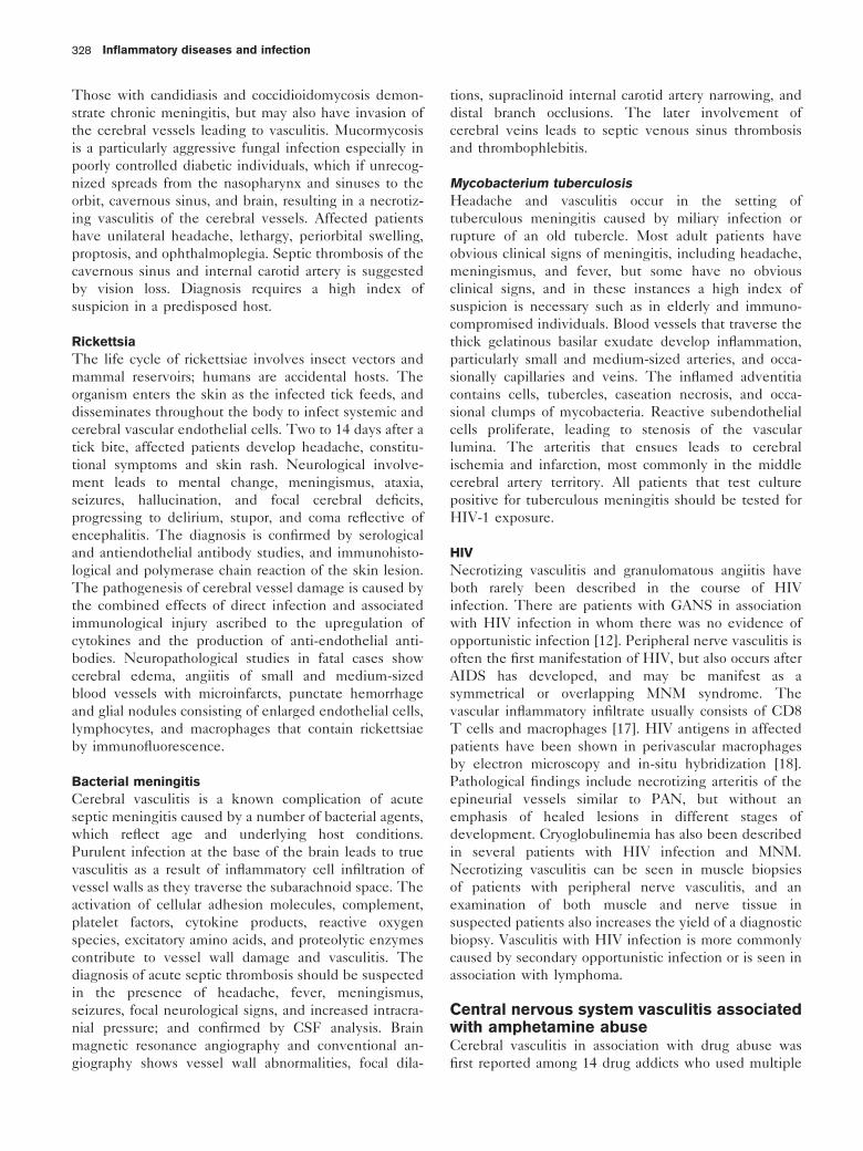
Those with candidiasis and coccidioidomycosis demon-
strate chronic meningitis, but may also have invasion of
the cerebral vessels leading to vasculitis. Mucormycosis
is a particularly aggressive fungal infection especially in
poorly controlled diabetic individuals, which if unrecog-
nized spreads from the nasopharynx and sinuses to the
orbit, cavernous sinus, and brain, resulting in a necrotiz-
ing vasculitis of the cerebral vessels. Affected patients
have unilateral headache, lethargy, periorbital swelling,
proptosis, and ophthalmoplegia. Septic thrombosis of the
cavernous sinus and internal carotid artery is suggested
by vision loss. Diagnosis requires a high index of
suspicion in a predisposed host.
Rickettsia
The life cycle of rickettsiae involves insect vectors and
mammal reservoirs; humans are accidental hosts. The
organism enters the skin as the infected tick feeds, and
disseminates throughout the body to infect systemic and
cerebral vascular endothelial cells. Two to 14 days after a
tick bite, affected patients develop headache, constitu-
tional symptoms and skin rash. Neurological involve-
ment leads to mental change, meningismus, ataxia,
seizures, hallucination, and focal cerebral deficits,
progressing to delirium, stupor, and coma reflective of
encephalitis. The diagnosis is confirmed by serological
and antiendothelial antibody studies, and immunohisto-
logical and polymerase chain reaction of the skin lesion.
The pathogenesis of cerebral vessel damage is caused by
the combined effects of direct infection and associated
immunological injury ascribed to the upregulation of
cytokines and the production of anti-endothelial anti-
bodies. Neuropathological studies in fatal cases show
cerebral edema, angiitis of small and medium-sized
blood vessels with microinfarcts, punctate hemorrhage
and glial nodules consisting of enlarged endothelial cells,
lymphocytes, and macrophages that contain rickettsiae
by immunofluorescence.
Bacterial meningitis
Cerebral vasculitis is a known complication of acute
septic meningitis caused by a number of bacterial agents,
which reflect age and underlying host conditions.
Purulent infection at the base of the brain leads to true
vasculitis as a result of inflammatory cell infiltration of
vessel walls as they traverse the subarachnoid space. The
activation of cellular adhesion molecules, complement,
platelet factors, cytokine products, reactive oxygen
species, excitatory amino acids, and proteolytic enzymes
contribute to vessel wall damage and vasculitis. The
diagnosis of acute septic thrombosis should be suspected
in the presence of headache, fever, meningismus,
seizures, focal neurological signs, and increased intracra-
nial pressure; and confirmed by CSF analysis. Brain
magnetic resonance angiography and conventional an-
giography shows vessel wall abnormalities, focal dila-
tions, supraclinoid internal carotid artery narrowing, and
distal branch occlusions. The later involvement of
cerebral veins leads to septic venous sinus thrombosis
and thrombophlebitis.
Mycobacterium tuberculosis
Headache and vasculitis occur in the setting of
tuberculous meningitis caused by miliary infection or
rupture of an old tubercle. Most adult patients have
obvious clinical signs of meningitis, including headache,
meningismus, and fever, but some have no obvious
clinical signs, and in these instances a high index of
suspicion is necessary such as in elderly and immuno-
compromised individuals. Blood vessels that traverse the
thick gelatinous basilar exudate develop inflammation,
particularly small and medium-sized arteries, and occa-
sionally capillaries and veins. The inflamed adventitia
contains cells, tubercles, caseation necrosis, and occa-
sional clumps of mycobacteria. Reactive subendothelial
cells proliferate, leading to stenosis of the vascular
lumina. The arteritis that ensues leads to cerebral
ischemia and infarction, most commonly in the middle
cerebral artery territory. All patients that test culture
positive for tuberculous meningitis should be tested for
HIV-1 exposure.
HIV
Necrotizing vasculitis and granulomatous angiitis have
both rarely been described in the course of HIV
infection. There are patients with GANS in association
with HIV infection in whom there was no evidence of
opportunistic infection [12]. Peripheral nerve vasculitis is
often the first manifestation of HIV, but also occurs after
AIDS has developed, and may be manifest as a
symmetrical or overlapping MNM syndrome. The
vascular inflammatory infiltrate usually consists of CD8
T cells and macrophages [17]. HIV antigens in affected
patients have been shown in perivascular macrophages
by electron microscopy and in-situ hybridization [18].
Pathological findings include necrotizing arteritis of the
epineurial vessels similar to PAN, but without an
emphasis of healed lesions in different stages of
development. Cryoglobulinemia has also been described
in several patients with HIV infection and MNM.
Necrotizing vasculitis can be seen in muscle biopsies
of patients with peripheral nerve vasculitis, and an
examination of both muscle and nerve tissue in
suspected patients also increases the yield of a diagnostic
biopsy. Vasculitis with HIV infection is more commonly
caused by secondary opportunistic infection or is seen in
association with lymphoma.
Central nervous system vasculitis associatedwith amphetamine abuseCerebral vasculitis in association with drug abuse was
first reported among 14 drug addicts who used multiple
Inflammatory diseases and infection328

amphetamine drugs [19]. Necrotizing arteritis of the
PAN type was found in cerebral arteries and arterioles at
postmortem examination (Fig. 14). The vascular insult
followed drug-induced vasospasm, hypertension, and
secondary vascular injury, including cerebral infarction,
aneurysm formation and rupture with cerebral hemor-
rhage, transient ischemic attack, or myelopathy as the
case is usually self-limited. Interestingly, necrotizing
arteritis is not a feature of amphetamine-induced
cerebrovascular injury in primate models; and the
beading of cerebral vessels develops 2 weeks after
parenteral administration of amphetamine, suggesting
the participation of other factors. The frequency of
cerebral vasculitis caused by drug abuse is difficult to
estimate for several reasons, and is probably over-
exaggerated as a cause of cerebral vasculitis. First, most
patients have been diagnosed by vessel ‘beading’ on
cerebral angiography without pathological verification.
Second, the vascular insults associated with ampheta-
mine drugs may be caused by factors other than
vasculitis. For example, parenteral amphetamine use
enhances the risk of endocarditis, cardiac embolism,
hemorrhage, stroke, and mycotic aneurysm formation;
and all other routes of administration predispose to acute
hypertension and cerebral hemorrhage. Third, opportu-
nistic infection associated with HIV infection and AIDS
probably accounts for most cases of cerebral vasculitis as
a result of the high frequency of HIV-1 infection in the
drug addict population.
Paraneoplastic vasculitisParaneoplastic neurological disorders are diseases of
nervous system function that occur in association with
cancer, but cannot be ascribed to metastases or direction
infiltration of the nervous system by tumor. In two-thirds
of affected patients, the neurological disorder generally
precedes the diagnosis of the tumor, and the correct
identification of the related neurological disorder directs
a search for occult and potentially curable cancer.
Necrotizing arteritis and microvasculitis have been
described in association with anti-Hu associated para-
neoplastic encephalomyelitis and sensory neuropathy
[20]. The anti-Hu antibody, when present at high titers
in the serum and CSF of affected patients, is a highly
sensitive and specific marker for paraneoplastic ence-
phalomyelitis and sensory neuropathy in association with
occult small cell lung cancer. Patients develop multifocal
neurological symptoms and signs that include headache,
limb encephalitis, brain stem deficits, cerebellar incoor-
dination, proprioceptive and tactile sensory loss leading
to imbalance of stance and gait, pseudoathetosis,
numbness and paresthesia, weakness, wasting, fascicula-
tion, active tendon reflexes, Babinski signs, Hoffman
signs, and clonus. Histopathological examination of the
neuraxis reveals widespread neuronal degeneration,
neuronal loss, and Wallerian fiber degeneration in the
hippocampus, brainstem, cerebellum, spinal cord dorsal
columns, dorsal root ganglia, and nerve roots, with
perivascular collections of chronic inflammatory cells,
also known as perivasculitis. Paraneoplastic encephalo-
myelitis should be suspected in patients with subacute
sensory neuropathy, a history of cancer or suspected
malignancy, weight loss, headache, mental change, and
multifocal involvement of the nervous system with
upper and lower motor neuron signs, central and
peripheral sensory loss, CSF pleocytosis and elevated
total protein and IgG content, contrast neuroimaging
studies that reveal meningeal or perivascular enhance-
ment, and muscle and nerve biopsy that demonstrates
microvasculitis, Wallerian nerve fiber degeneration, or
inflammatory myopathy.
Laboratory diagnosisThe laboratory diagnosis of vasculitis of the nervous
system proceeds along a systematic framework of
generally accepted principles with an extensive choice
of recommended studies as described below.
General principles
There is general agreement on four principles in the
diagnosis of vasculitis: First, vasculitis is a potentially
serious disorder with a propensity for permanent
disability as a result of tissue ischemia and infarction;
Figure 14. Cerebral vasculopathy in a case of intracerebralhemorrhage associated with the use of phenylpropanolamine asan aid to weight loss
The profound intimal hyperplasia all but obliterates the vascular lumen.Polymorphonuclear leukocytes are in all three vascular layers butparticularly the intima. The media are remarkably well preservedcompared with cases of polyarteritis nodosa and leukocytoclasticvasculitis (stain, hematoxylin and eosin; original magnification 6100).Reproduced from Younger [1.] with permission of the publisher.
Vasculitis of the nervous system Younger 329

recognition of the neurological manifestations is im-
portant in developing a differential etiological diagnosis.
Second, undiagnosed and untreated, the outcome of
vasculitis is potentially fatal. Third, a favorable response
to an empiric course of immunosuppressive and
immunomodulating therapy should never be considered
a substitute for the absolute proof of the diagnosis of
vasculitis. Fourth, histolopathological confirmation of
vasculitis in the nervous system is essential for accurate
diagnosis, such as by analysis of nerve and muscle
biopsy tissue when PNS involvement is postulated, and
by brain and meninges when there is CNS involve-
ment.
Recommended laboratory evaluation of suspected
vasculitis
The laboratory evaluation of vasculitis of the nervous
system is summarized in Table 2. The initial diagnosis of
vasculitis commences with an investigation of possible
associated serological markers in a given patient. The
emergence of specific serological studies for many of the
connective tissue diseases including vasculitis has
transformed our concepts of autoimmune disease.
However, their use should be guided by the clinical
presentation and postulated etiological diagnosis to avoid
excessive cost and spurious results.
Electrodiagnostic studies are useful in the initial
investigation of systemic vasculitis, because they can
identify areas of asymptomatic involvement and sites for
muscle and nerve biopsy and distinguish the various
neuropathic syndromes associated with peripheral nerve
and muscle involvement. A wide sampling of nerves and
muscles should be examined, both distal and proximal,
using standard recording and needle electrodes for the
performance of nerve conduction studies and needle
electromyography at skin temperatures of 348C in
comparison with normative data. Most patients with
peripheral nerve vasculitis show evidence of active
axonopathy acutely in an MNM pattern and over time
in a distal symmetric or asymmetric pattern. Quantitative
motor unit potential analysis can delineate whether
proximal wasting and weakness are caused by myopathic
or neurogenic disease.
CSF analysis, electroencephalography, and neuroima-
ging studies are integral to the diagnostic evaluation of
most CNS disorders, including vasculitis. Properly
performed, lumbar puncture carries a minimal risk and
provides potentially useful information regarding pos-
sible underlying vasculitis so suggested by pleocytosis
in excess of 5 cells/mm3, protein elevation greater
than 100 mg/dl, and evidence of intrathecal synthesis
of immunoglobulin and oligoclonal bands. Molecular
genetic, immunoassay, and direct staining techniques to
exclude spirochetal, fungal, mycobacterial and viral
infections, as well as cytospin examination of CSF for
possible malignant cells should be performed.
There are no typical electroencephalography findings in
CNS vasculitis. Magnetic resonance imaging (MRI) is
more sensitive than computed tomography, but both
methods lack specificity in histologically confirmed
cases. The most common MRI findings are multiple
bilateral cortical and deep white matter signal abnorm-
alities, and enhancement of the meninges after gadoli-
nium. Magnetic resonance angiography and functional
imaging of the brain provide complementary findings to
conventional MRI. The former is useful in the evalua-
tion of medium and large vessel disease, but misses fine
vessel contours better seen on cut-film or digital
subtraction angiography. The abnormal diffuse and focal
perfusion patterns seen on single photon emission-
computed tomography (SPECT) do not always correlate
with neurological symptoms or distinguish vasculitic
from non-vasculitic vasculopathy. Some authorities
claimed that cerebral angiography showed diagnostic
Table 2. Laboratory evaluation of headache and vasculitis
Blood studiesComplete blood countErythrocyte sedimentation rateChemistry panel including creatine phosphokinaseAntinuclear antibodyComplement levelsRheumatoid factorCryoglobulinsImmunofixation electrophoresisQuantitative immunoglobulinsT and B cell panelsAntibodies (selectively) to: Ro (SS-A), La (SS-B), Sm, SCL-70,hepatitis B and C virus
HIV-1, Borrelia burgdorferi (ELISA, Western blot), c-ANCA andp-ANCA
Radiographic studiesChestBody computed tomographMagnetic resonance imagingMagnetic resonance angiography and venographySingle photon emission computed tomographySystemic and cerebral angiography
Other neurodiagnostic studiesElectroencephalographyElectromyography and nerve conduction studiesLumbar puncture for cerebrospinal fluid analysis: protein, glucose, cellcount, IgG level, cytology, VDRL, Gram stain, culture, India ink; viralantigens, Lyme antibodies and PCR (as indicated)
Histopathological studies (as indicated)Muscle and nerve biopsyTemporal artery biopsyMeningeal and cortexSkinSystemic organsLymph nodes
ELISA, Enzyme-linked immunosorbent assay; c-ANCA, circulatingantineutrophil cytoplasmic antibody; p-ANCA, perinuclear antineutrophilcytoplasmic antibody; PCR, polymerase chain reaction; SS, Sjogrensyndrome; VDRL, Veneral Disease Research Laboratory.
Inflammatory diseases and infection330

features, but that assertion was later modified. The
beading of vessels is found in only approximately a third
of patients with histologically confirmed CNS vasculitis,
as well as in CNS infection, atherosclerosis, cerebral
embolism, and vasospasm of diverse cause (Fig. 15).
Multiple microaneurysms, often seen on visceral angio-
graphy in systemic vasculitis, are distinctly rare in CNS
vessels.
Brain and meningeal biopsy are still the gold standard for
the diagnosis of CNS vasculitis, but false-negatives occur
because of focal lesions and sampling errors. Radio-
graphic studies that guide the biopsy site towards areas
of abnormality probably improve the sensitivity, but this
has not been formally studied. The risk of serious
morbidity related to biopsy is less than 2.0% at most
centers, which is probably less than the cumulative risk
of an empiric course of long-term immunosuppressive
therapy. There are no certain guidelines as to when to
proceed to brain and meningeal biopsy. However, it
would certainly be warranted if there were no other
explanation for the progressive syndrome of fever,
headache, encephalopathy, and focal cerebral signs, in
association with CSF pleocytosis, and protein content
elevation greater than 100 mg/dl, which is suggestive of
GANS.
The importance of nerve and muscle biopsy in the
diagnosis of vasculitis cannot be overemphasized. It can
be approached with confidence when a neurologist or
surgeon skilled in nerve and muscle biopsy techniques
at centers performs the procedure with neuropathologists
trained to process and examine the specimens for all of
the diagnostic possibilities. The nerve and muscle
should be clinically and electrophysiologically affected.
However, the muscle should not be so affected, or end-
stage, as to preclude interpretation. A segment of the
sural, superficial peroneal sensory, or femoral interme-
dius sensory nerve can be surgically removed without
incurring a serious deficit, along with pieces of muscle
tissue, respectively, from the soleus, peroneus brevis, or
rectus femoris muscle, thereby providing potentially
useful information regarding the severity of the under-
lying neuropathy and increasing the yield of vasculitic
lesions (Fig. 16). Commercially available monoclonal and
polyclonal antibodies directed against T- and B-cell
subsets, macrophages, immunoglobulins, C3d, C5b-9
MAC proteins, cytokines and other inflammatory med-
iators, and main histocompatibility class I and II antigens
add precision to the analysis of peripheral nerve speci-
mens with suspected necrotizing and non-necrotizing
peripheral nerve vasculitis [21].
ImmunopathogenesisEarly progress in the understanding of vasculitis took a
major turn during a discussion of the paper by Kernohan
and Woltman [15] on PAN. At that time, in 1938, there
was no effective treatment and antemortem diagnosis
was rarely possible. Harry Lee Parker conceptualized
nerve and muscle biopsy when he commented ‘It occurs
to me that in any case in which polyarteritis nodosa may
be suspected, it is advisable to take a biopsy from a
peripheral nerve, muscle, or artery.’
The Second World War provided another opportunity
for the advancement of our understanding of the blood
supply of the peripheral nerves, and these findings in
turn guided our thoughts on vasculitis. In 1943, Coers
and Woolf [22] described the surgical approach for
biopsy of the superficial peroneal sensory nerve and
peroneus brevis muscle now performed routinely for
vasculitis. Two years later, Sunderland [23,24] provided
a detailed account of neurovascular anatomy by perform-
ing dissection of amputated limbs after injection of India
ink to opacify the vessels. His findings were summarized
in a concluding statement: ‘Each of the major nerves is
generally abundantly vascularized throughout its entire
length by a succession of vessels, which by their
repeated division and anastomosis within the nerve
outline an unbroken vascular net’ (Fig. 17).
Some nerves such as the median and ulnar nerve
between the axilla and elbow and along the sciatic in
Figure 15. Radiographic features of cerebral vasculitis
Ectasia and beading in the M1 segment and lack of flow in the A1segment of the right anterior cerebral artery (arrow).
Vasculitis of the nervous system Younger 331

the gluteal region had few or no entering nutrient
vessels, but vascular insufficiency was still an unlikely
occurrence as stated by Sunderland. Admittedly there
are instances, although uncommon, in which one vessel
supplies long stretches of a nerve without reinforcement,
but it has been demonstrated in sectioned and injected
material that even under such apparently adverse
conditions of supply, the anastomosis is of such
dimensions at the peripheral limits of the solitary
channel that segmental ischemia caused by the blocking
of such a single vessel is a remote possibility.
On the basis of such studies, there was no convincing
evidence for the presence of watershed zones of poor
vascular supply along major nerves of the arm or leg, a
contention that has permeated the literature with regard
to the clinical sequelae of vasculitis. Nonetheless, Dyck
and coworkers [25] ascribed centrofascicular nerve fiber
loss in a patient with necrotizing vasculitis to poor
vascular perfusion along presumed watershed zones of
the upper arm and proximal thigh regions. Almost a
decade later, Moore and Fauci [26] ascribed flail
weakness and mid-level sensory loss of the arms to
vasculitis of the arteria nervorum, even though the
patient was not studied pathologically.
It is now known that under normal circumstances, the
nervous system is protected from systemic immunologi-
cal reactions by the blood–brain and blood–nerve
barriers. Tight junctions between neighboring cells and
a paucity of micropinocytotic vessels are unique to the
blood–brain barrier, and along with other local determi-
Figure 16. Muscle and nerve biopsy technique
a b
c d
(a) The superficial peroneal sensory nerve is palpated laterally along the distal third of the leg along a line between the fibular head and lateral malleolusproviding markings for the incision. (b) An incision is made and the area is dissected revealing the nerve (n) obliquely traversing the field (arrow). (c)Incising the muscle aponeurosis reveals underlying peroneus brevis muscle tissue (m) in addition to nerve (n, and arrows) available for biopsy. (d) Afterthe specimens are removed and the site irrigated, a subcuticular closure is performed using absorbable sutures. Reproduced from Younger DS, editor.Motor disorders, peripheral nerve pathology. Philadelphia Lippincott Williams and Wilkins; 1999, p. 84, with permission of the publisher.
Inflammatory diseases and infection332

nants, contribute to the prevention of the early involve-
ment of the CNS in the course of systemic inflammation.
Immune activation requires the interaction of a specific
autoantigen, an MHC class II antigen-presenting cell
(APC) and an antigen-specific T cell. Macrophages are
the principal APCs of the PNS, and their role appears to
be that of a local surveillance system, taking up and
processing protein antigens and presenting them on their
surface. Their interaction with native antigen and
antigen-specific T cells leads to a proliferation of specific
helper (CD4) and cytotoxic suppressor (CD8) T cells
with the expression of HLA-DR, IL-2 receptor, and
TNF-a secretion. T cells that become sensitized early in
the course of systemic illness probably later contribute to
the cellular immune response directed against cross-
reacting epitopes present in peripheral nerves and the
brain.
Vascular endothelial cells play an important role in the
pathobiology of vascular inflammation along the blood–
nerve and blood–brain barriers, because of their potential
interaction with elements of the systemic immune
Figure 17. Vascular supply of the peripheral nerves of the limb
a b
(a) An artist view of the intraneural blood supply. (b) The injection of India ink shows the actual internal architecture of the vascular supply. FromYounger [1.], reproduced with permission of the publisher.
Vasculitis of the nervous system Younger 333

system. They are potentially active participants in
vasculitis, not simply passive targets of injury. They
satisfy the criteria for an APC because of their native
ability to express MHC class I molecules for interaction
with cytotoxic T cells; and under certain conditions, they
express MHC class II molecules and the necessary co-
stimulatory factors to induce T-cell proliferation in vivoand in vitro. The function of vascular endothelial cells is
regulated mainly by IL, TNF, and endotoxins derived
from immigrant or resident mononuclear cells. Their
action, by virtue of binding to specific receptors, is
alteration of the transcription of an array of endothelial
genes that programme cellular inflammatory secretion,
the local expression of leukocyte adhesion molecules,
the balance of prothrombotic and antithrombotic vascular
functions, the synthesis of matrix molecules and their
receptors, and the secretion of growth factors, secondary
cytokines, and enzymes related to matrix degradation.
The localization and propagation of leukocytes along
vascular endothelial cells depend on the local production
of IL-1, IL-6, and IL-8, the expression of cell adhesion
molecules, namely integrins, selectins, and the immu-
noglobulin super-family molecules (intercellular cell
adhesion molecule 1, vascular cell adhesion molecule
1, platelet-endothelial cell adhesion molecule 1) con-
stitutively expressed or induced after cellular activation,
and the expression of autoantigens such as ANCA and
anti-endothelial cell antibodies. Other substances that
contribute to vascular integrity include free radical nitric
oxide, Von Willebrand factor, tissue plasminogen acti-
vator inhibitor, thrombomodulin, and platelet-activating
factors.
Human mechanisms also contribute to the development
of vasculitis and nervous system damage through
complement-mediated injury of microvessels. The com-
plement system is composed of 11 proteins that
sequentially interact in the activated state to form an
assembly of five proteins referred to as C5b-9 or MAC.
The activation of C5b-9 along peripheral nerve micro-
vessels leads to increased local permeability, edema, and
inflammatory cell infiltration. Complement-mediated
injury appears to be an important mechanism in the
etiopathogenesis of inflammatory diabetic vasculopathy;
however, the initiating factors are still speculative. One
possibility is a defect in the expression of certain
regulatory membrane proteins in the walls of micro-
vessels, including complement receptor (CR10), decay-
accelerating factor (CD55), membrane co-factor protein
(CD46), and membrane inhibitor of reactive lysis
(CD59), which normally protect cells by limiting the
activation of the complement cascade.
Interest in the role of specific pathogenic autoantibodies
has evolved over the past two decades as, for example, in
our understanding of WG and related disorders in the
elucidation of ANCA. The pathobiology of ANCA was
understood through the use of animal models, human
neutrophil studies, and monolayers of cultured human
umbilical vein endothelial cells. MPO and PR3 antigens,
so named for the patterns of staining of normal ethanol-
fixed neutrophils with indirect immunofluorescence,
granular cytoplasmic ANCA, and perinuclear ANCA,
were found to correlate with disease activity in WG and
MPA, respectively. The MPO and PR3 antigens are
accessible for binding with circulating ANCA and Fc
receptors. ANCA-augmented chemotaxis and adhesion
bring circulating neutrophils and mononuclear phago-
cytes into close contact with endothelial cells and induce
neutrophil-mediated endothelial cell lysis and vascular
permeability.
TreatmentNeurologists treating vasculitis must choose the se-
quence and combination among available immunosup-
pressant and immunomodulating therapies, recognizing
the possible adverse effects.
A modern appreciation of the usefulness of corticosteroid
preparations in systemic vasculitis was first reported in
1950 [27]. Untreated, patients with PAN had a 5-year
survival rate of 10%; treatment with corticosteroids
increased survival to 48% [28]. Although the effective-
ness of corticosteroids is well established, there is
uncertainty even among experts as to the optimal
regimen. For example, in one analysis [29], a sustained
benefit in PAN was obtained in patients with a
minimum equivalent dosage of 31 mg of prednisone a
day for 7 months. The beneficial effects of corticoster-
oids are attributed to a multiplicity of effects on the cell
and humoral immune system, including the inhibition of
activated T and B cells, APC, and leukocytes at sites of
inflammation, IFN-g, induced MHC class II expression,
macrophage differentiation, pathogenic cytokine expres-
sion, complement interactions, and immunomodulating
cell adhesion molecules.
The effectiveness of a daily oral regimen of cyclophos-
phamide and prednisone in WG was first reported by
Fahey et al. [30] in 1954, and later by Fauci et al. [31] in1971, and served as a template for the treatment of
virtually all types of systemic vasculitis, including GANS
until its long-term side-effects were appreciated in WG.
Nonetheless, this alkylating agent is an important
adjunct in the treatment of systemic vasculitis. It leads
to preferential T-cell lysis resulting from the inhibition
of hematopoietic precursors in the bone marrow.
Azathioprine is a purine analog that metabolizes to the
cytotoxic derivative 6-mercaptopurine. It exerts favor-
able action in vasculitis by the inhibition of T-cell
activation and T-cell-dependent antibody-mediated re-
Inflammatory diseases and infection334

sponses. It is appropriate alterative therapy to corticos-
teroids and cyclophosphamide in systemic vasculitis.
There are three drawbacks to its use. First, idiosyncratic
side-effects, most often gastrointestinal and ’flu-like,
occur in approximately 10% of patients and rarely
necessitate permanent withdrawal of the medication.
However, pancreatitis and gastritis severe enough to
warrant hospitalization can occur. Second, bone marrow
suppression occurs in nearly all patients, usually
manifested by mild pancytopenia. Third, there is
typically a long delay in the onset of the therapeutic
effect of 3 months or more. Taking all these factors into
account, most clinicians concur with the slow advance-
ment of the dose over weeks, commencing with 50 mg a
day and achieving maintenance levels of 2–3 mg/kg a
day, with careful monitoring of liver and marrow
function.
Intravenous immunoglobulin (IVIg) therapy has been
widely used in the treatment of autoimmune neurolo-
gical diseases, and warrants consideration as initial or
adjunctive therapy in vasculitis of the PNS and as
adjunctive therapy in CNS. This is because of its
acknowledged salutary action through the action of
blocking antibodies, the suppression of antibody-
mediated responses, the accelerated catabolism of
pathogenic IgG antibodies, the suppression of patho-
genic cytokines, and most of all, the inhibition of
C5b-9 MAC-mediated cytolysis. The usual dosage is
400 mg/kg per day for 5 days once a month, after the
determination of adequate renal clearance and the
absence of IgA antibodies that can rarely promote
anaphylaxis. A new manufacturing process that employs
chromatographic steps in the intravenous immunoglo-
bulin purification scheme yields a purer intravenous
immunoglobulin product that more closely reflects the
IgG subclass distribution found in plasma.
ConclusionThe comprehensive therapy of patients with vasculitis
often requires the commitment of a multidisciplinary
team of health professionals and caregivers to optimize
recovery while initiating immunotherapy. Physical ther-
apy and orthosis may be warranted for disabling motor
and cognitive disorder impairments to maintain a range
of motion and strength, to improve function status, and
to maintain ambulation. Effective pain management may
be an important aspect of their care, not only to provide
overall wellbeing, but to permit more aggressive
physiotherapy. Agents such as tricyclic antidepressants,
gabapentin, mexiletine, opioids, clonazepam, and topical
anesthetic creams have all been used with varying
success. Finally, efforts should be made to limit the
ischemic-enhancing effects of other conditions, such as
with diabetes mellitus through improved glycemic
control, the regulation of blood pressure and hyperlipi-
demia, alone or as a side-effect of concomitant cortico-
steroids, and the cessation of cigarette smoking.
AcknowledgementThis work was supported by the Neurology Research Foundation, Inc.
References and recommended readingPapers of particular interest, published within the annual period of review, havebeen highlighted as:. of special interest.. of outstanding interest
1.
Younger DS. Vasculitis and connective tissue disorders. In: Griggs R, JoyntR, editors. Baker and Joynt’s clinical neurology on CD-ROM, Chapter 59.Philadelphia: Lippincott Williams and Wilkins; 2003.
This article is a comprehensive review of vasculitis of the nervous system withexcellent tables and figures.
2 Younger DS, Kass RM. Vasculitis and the nervous system. Neurol Clin 1997;15:737–758.
3 Kussmaul A, Maier R. Ueber eine bisher nicht beschriebene eigaenthumlicheArterienerkrangung (periarteritis nodosa) die mit morbus brightii und rapidfortachreitender allgemeiner muskellahmung einergeht. Deutsches Arch KlinMed 1866; 1:484–518.
4 Churg J, Strauss L. Allergic granulomatosis, allergic angiitis, and periarteritisnodosa. Am J Pathol 1951; 27:277–302.
5 Zeek PM, Smith CC, Weeter JC. Studies on periarteritis nodosa, III: thedifferentiation between the vascular lesions of periarteritis nodosa and ofhypersensitivity. Am J Pathol 1948; 24:889–917.
6 Godman GC, Churg J. Wegener’s granulomatosis: pathology and review ofthe literature. Arch Pathol 1954; 58:533–553.
7 Horton BT, Magath BT, Brown GE. Arteritis of temporal vessels: report of 7cases. Proc Staff Meet Mayo Clin 1937; 12:548–553.
8 Jennings GH. Arteritis of the temporal vessels. Lancet 1938; 1:424.
9. .
Weyland CM, Goronzy JJ. Medium- and large-vessel vasculitis. N Engl J Med2003; 349:160–169.
This is an excellent overview of giant cell arteritis with descriptions of new insightsinto the observed immunohistopathology.
10 Cravioto H, Fegin I. Non-infectious granulomatous angiitis with a predilectionfor the nervous system. Neurology 1959; 9:599–609.
11 Younger DS, Hays AP, Brust JCM, Rowland LP. Granulomatous angiitis ofthe brain: an inflammatory reaction of nonspecific etiology. Arch Neurol 1988;45:514–518.
12 Younger DS, Calabrese LH, Hays AP. Granulomatous angiitis of the nervoussystem. Neurol Clin 1997; 15:821–834.
13 Younger DS, Rosoklija G, Hays AP. Diabetic peripheral neuropathy. SeminNeurol 1998; 18:95–104.
14 Younger DS, Rosoklija G, Hays AP, et al. Diabetic peripheral neuropathy: aclinical and immunohistochemical analysis of sural nerve biopsies. MuscleNerve 1996; 19:722–727.
15 Kernohan JW, Woltman HW. Periarteritis nodosa: a clinicopathologic studywith special reference to the nervous system. Arch Neurol Psychiatry 1938;39:655–686.
16 Younger DS, Rosoklija G, Hays AP. Lyme polyradiculoneuritis: immunohis-tochemical findings in sural nerve. Muscle Nerve 1995; 18:359–360.
17 Younger DS, Rosoklija G, Hays AP, et al. HIV-1 associated sensoryneuropathy; a patient with peripheral nerve vasculitis. Muscle Nerve 1996;19:1364–1366.
18 Younger DS, Rosoklija G, Hays AP. Sensory neuropathy in AIDS:demonstration of vasculitis and HIV antigens in peripheral nerve. J Neurol1994; 241 (Suppl.):17.
19 Citron BP, Halpern M, Mccarron M, et al. Necrotizing angiitis associated withdrug abuse. N Engl J Med 1970; 283:1003–1011.
20 Younger DS, Dalmau J, Inghirami G, Hays AP. Anti-Hu-associated peripheralnerve and muscle micro vasculitis. Neurology 1994; 44:181–183.
21 Younger DS, Rosoklija G, Hays AP, Latov N. Peripheral nerve immuno-histochemistry in diabeteic neuropathy. Semin Neurol 1996; 16:139–142.
22 Coers C, Woolf AL. The innervation of muscle: a biopsy study. Oxford:Oxford University Press; 1943. pp. 2–3.
Vasculitis of the nervous system Younger 335

23 Sunderland S. Blood supply of the nerve of the upper limb in man. ArchNeurol Psychiatry 1945; 53:91–115.
24 Sunderland S. Blood supply of the sciatic nerve and its popliteal divisions inman. Arch Neurol Psychiatry 1945; 53:283–289.
25 Dyck PJ, Conn DL, Okazaki H. Necrotizing angiopathic neuropathy. Threedimensional morphology of fiber degeneration related to sites of occludedvessels. Mayo Clin Proc 1972; 47:461–475.
26 Moore PM, Fauci AS. Neurologic manifestations of systemic vasculitis. Aretrospective and prospective study of the clinicopathologic features andresponse to therapy in 25 patients. Am J Med 1981; 71:517–524.
27 Bagenstoss AH, Shick RM, Polley HF. The effect of cortisone on the lesionsof periarteritis nodosa. Am J Pathol 1950; 26:709.
28 Frohnert PP, Sheps SG. Long-term followup study of periarteritis nodosa.Am J Med 1967; 43:8–14.
29 Leib ES, Restivo C, Paulus HE. Immunosuppressive and corticosteroidtherapy of polyarteritis nodosa. Am J Med 1979; 67:941–947.
30 Fahey JL, Leonard E, Churg J, et al. Wegener’s granulomatosis. Am J Med1954; 17:168–179.
31 Fauci AS, Wolff SM, Johnson JS. Effect of cyclophosphamide upon theimmune response in Wegener’s granulomatosis. N Engl J Med 1971;285:1493–1496.
Inflammatory diseases and infection336