Embed Size (px)
Citation preview
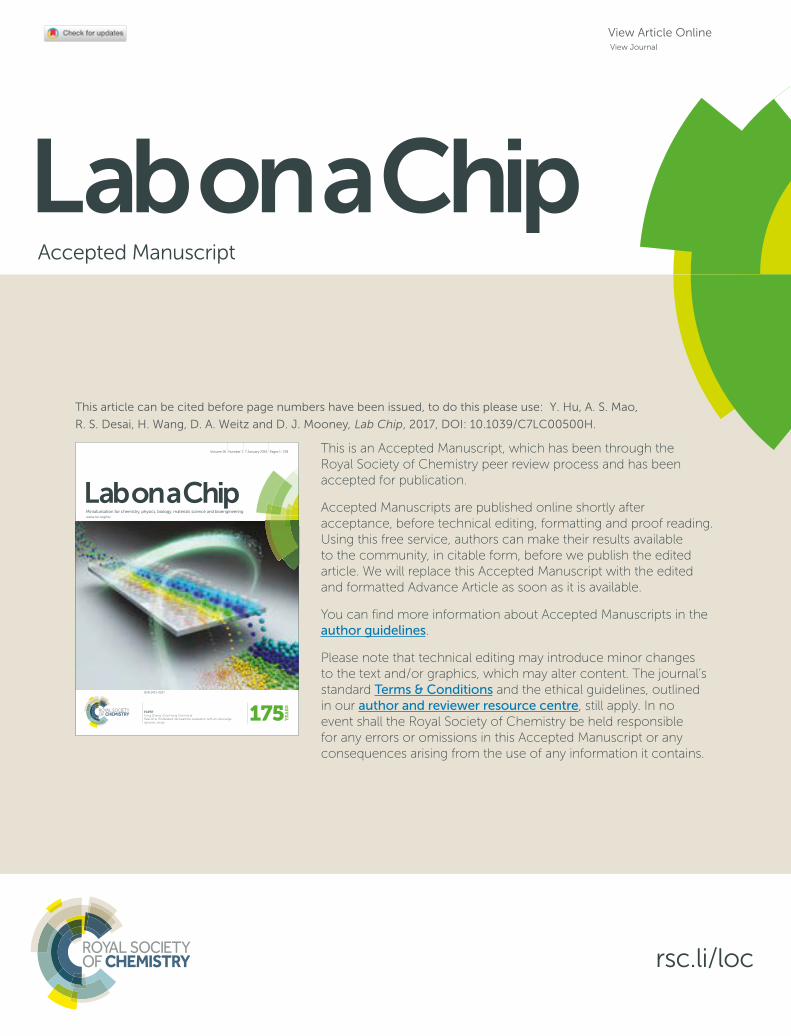
This is an Accepted Manuscript, which has been through the Royal Society of Chemistry peer review process and has been accepted for publication.
Accepted Manuscripts are published online shortly after acceptance, before technical editing, formatting and proof reading. Using this free service, authors can make their results available to the community, in citable form, before we publish the edited article. We will replace this Accepted Manuscript with the edited and formatted Advance Article as soon as it is available.
You can find more information about Accepted Manuscripts in the author guidelines.
Please note that technical editing may introduce minor changes to the text and/or graphics, which may alter content. The journal’s standard Terms & Conditions and the ethical guidelines, outlined in our author and reviewer resource centre, still apply. In no event shall the Royal Society of Chemistry be held responsible for any errors or omissions in this Accepted Manuscript or any consequences arising from the use of any information it contains.
Accepted Manuscript
rsc.li/loc
www.rsc.org/loc
ISSN 1473-0197
Lab on a ChipMiniaturisation for chemistry, physics, biology, materials science and bioengineering
PAPERYong Zhang, Chia-Hung Chen et al.Real-time modulated nanoparticle separation with an ultra-large dynamic range
Volume 16 Number 1 7 January 2016 Pages 1–218
Lab on a ChipMiniaturisation for chemistry, physics, biology, materials science and bioengineering
View Article OnlineView Journal
This article can be cited before page numbers have been issued, to do this please use: Y. Hu, A. S. Mao,
R. S. Desai, H. Wang, D. A. Weitz and D. J. Mooney, Lab Chip, 2017, DOI: 10.1039/C7LC00500H.
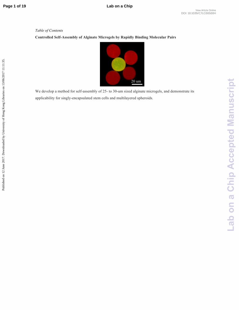
Table of Contents
Controlled Self-Assembly of Alginate Microgels by Rapidly Binding Molecular Pairs
We develop a method for self-assembly of 25- to 30-um sized alginate microgels, and demonstrate its
applicability for singly-encapsulated stem cells and multilayered spheroids.
20 um
Page 1 of 19 Lab on a Chip
Lab
ona
Chi
pA
ccep
ted
Man
uscr
ipt
Publ
ishe
d on
12
June
201
7. D
ownl
oade
d by
Uni
vers
ity o
f H
ong
Kon
g L
ibra
ries
on
13/0
6/20
17 1
1:11
:35.
View Article OnlineDOI: 10.1039/C7LC00500H
Controlled Self-Assembly of Alginate Microgels by Rapidly Binding Molecule Pairs
Yuebi Hu1*, Angelo S. Mao
1,2*, Rajiv M. Desai
1,2, Huanan Wang
1,3,4, David A. Weitz
1,3, David J.
Mooney1,2†
*Authors contributed equally to this work.
†Author to whom correspondence should be addressed. Email: [email protected]
1John A. Paulson School of Engineering and Applied Sciences, Harvard University, 29 Oxford St,
Cambridge, MA 02138, USA
2Wyss Institute for Biologically Inspired Engineering, Harvard University, 3 Blackfan Circle, Boston, MA
02115, USA
3Department of Physics, Harvard University, 17 Oxford St, Cambridge, MA 02138, USA
4School of Life Science and Biotechnology, Dalian University of Technology, Dalian 116023, P.R. China
Abstract
Controlled self-assembly of cell-encapsulating microscale polymeric hydrogels (microgels) could be
advantageous in a variety of tissue engineering and regenerative medicine applications. Here, a method of
assembly by chemical modification of alginate polymer with binding pair molecules (BPM) was explored.
Alginate was modified with several types of BPM, specifically biotin and streptavidin and click chemistry
compounds, and fabricated into 25-30-µm microgels using a microfluidic platform. These microgels were
demonstrated to self-assemble under physiological conditions. By combining complementary microgels at
a high ratio, size-defined assemblages were created, and the effects of BPM type and assembly method on
the number of microgels per assemblage and packing density were determined. Furthermore, a magnetic
process was developed to separate assemblages from single microgels, and allow formation of multilayer
spheroids. Finally, cells were singly encapsulated into alginate microgels and assembled using BPM-
modified alginate, suggesting potential applications in regenerative medicine.
Page 2 of 19Lab on a Chip
Lab
ona
Chi
pA
ccep
ted
Man
uscr
ipt
Publ
ishe
d on
12
June
201
7. D
ownl
oade
d by
Uni
vers
ity o
f H
ong
Kon
g L
ibra
ries
on
13/0
6/20
17 1
1:11
:35.
View Article OnlineDOI: 10.1039/C7LC00500H
Introduction
In the field of tissue engineering, there has been significant interest in using self-assembly to form
biological tissues in a bottom-up engineering paradigm with individual building blocks1. Biological
systems are inherently hierarchical, composed of many distinct building blocks in ordered structures, such
as nephrons in kidneys2. Engineered tissues have applications in regenerative medicine as replacements for
diseased tissues or to provide tissue mimics for in vitro drug screening3. Self-assembled tissue subunits
could potentially be combined to form more complex structures for organ transplantation4.
Self-assembly occurs when inherent interactions between components induce the spontaneous formation of
an ordered structure5. Compared to guided and directed assembly methods, self-assembly is advantageous
because building blocks can be assembled without external forces that may modify or damage building
blocks, and can potentially occur in contexts in which using directed assembly apparatuses is not feasible,
for example in some in vivo settings1. Examples of self-assembly on the nanoscale include certain proteins
and nucleic acids1, such as aromatic dipeptides
6. On the microscale scale, multicellular heterospheroids
have been shown to self-assemble by modification of cellular surfaces with biotin and streptavidin7, as have
polyethylene glycol hydrogels functionalized with DNA dimers8 and electrostatic charges
9. On a larger size
scale, millimeter-sized cell-encapsulating hydrogels were shown to assemble through paramagnetic-guided
interactions between free radicals10
, and a microrobot has been used to arrange cell-encapsulating hydrogels
for tissue culture via direct assembly11
. However, self-assembly of small, single cell-encapsulated
microscale hydrogels (microgels) has yet to be demonstrated.
Hydrogel encapsulation of cells has been widely used for regenerative medicine applications12
. Single cell
encapsulation of cells into microscale hydrogels has recently been achieved using various methods, notably
microfluidic technologies13
. These have been used for in vitro tissue culture and in vivo cell delivery14
. In
terms of in vitro applications, a thin hydrogel coating of singly-encapsulated cells may permit the
fabrication of higher resolution in vitro tissue mimics with high cell density, potentially providing a more
accurate reproduction of the in vivo microenvironment14
. The reduced diffusive barrier and smaller size
may also improve in vivo applications15
. Moreover, modification of the cell-encapsulating microgel to
mediate self-assembly rather than this being controlled by the cell membrane itself, as in some approaches7,
could prevent adverse effects on the cells themselves.
Here, we demonstrate the self-assembly of alginate microgels that were modified with three separate
combinations of binding pair molecules (BPM). These molecules bind to their complements rapidly and
under physiological conditions. Alginate was chosen for microgel fabrication as it is considered
biocompatible16
, is amenable to chemical modification17
, and can be cross-linked under gentle conditions
that are conducive to cell survival18
. Its rapid but gentle crosslinking mechanism has made it an ideal
vehicle for the fabrication of microgels and for cell encapsulation19
. The BPMs chosen were biotin and
Page 3 of 19 Lab on a Chip
Lab
ona
Chi
pA
ccep
ted
Man
uscr
ipt
Publ
ishe
d on
12
June
201
7. D
ownl
oade
d by
Uni
vers
ity o
f H
ong
Kon
g L
ibra
ries
on
13/0
6/20
17 1
1:11
:35.
View Article OnlineDOI: 10.1039/C7LC00500H
streptavidin, and two sets of complementary click compounds, tetrazine (Tz) and trans-cyclooctene (TCO),
and Tz and norbornene (Nb), respectively. Biotin and streptavidin form one of the strongest non-covalent
interactions with a Kd of 10-14
mol/L, and are stable in the presence of pH changes, organic solvents, and
denaturing agents20
. Moreover, cells functionalized with biotin and avidin were previously shown to be
capable of assembly7. The click chemistry compounds undergo bio-orthogonal Diels-Alder reactions and
do not require the addition of cytotoxic copper catalysts21
. These reactions are also highly chemoselective
and occur at physiological temperatures and pH21
. Recently, bulk alginate hydrogels cross-linked via click
chemistry compounds have been found to allow cell encapsulation21
. We also characterize the effects of
different assembly methods on the number of microgels per assemblage (Nm) and its distribution, and
microgel packing within assemblages. Finally, the assembly of cell-encapsulating BPM-modified microgels
and the fabrication of multilayered spheroids of BPM-modified microgels are demonstrated.
Methods
3-(p-benzylamino)-1,2,4,5 tetrazine synthesis and purification: 3-(p-benzylamino)-1,2,4,5-tetrazine was
synthesized as previously described21
. Briefly, 4-(aminomethyl)benzonitrile hydrochloride and
formamidine acetate were mixed in the presence of anhydrous hydrazine. After the reaction was cooled to
room temperature, NaNO2 and HCl were added, and the product was extracted using dichloromethane and
worked up with NaHCO3. The crude product was purified by high performance liquid chromatography and
lyophilized.
Tetrazine-, trans-cyclooctene, and norbornene- functionalized alginate synthesis: Ultrapure alginate
(Pronova UP MVG; FMC Biopolymer) or high molecular weight alginate (Protanal lf20/40; FMC
BioPolymer) alginate was dissolved in MES buffer solution (0.1 M MES, 0.3 M NaCl). N-
hydroxysuccinimide (NHS; Sigma-Aldrich) and 1-ethyl-3-(3-dimethylaminopropyl)-carbodiimide
hydrochloride (EDC; Thermo Scientific) were dissolved in MES buffer solution and added in 5000 molar
excess of alginate. 3-(p-benzylamino)-1,2,4,5 tetrazine or trans-cyclooctene-amine (TCO-Amine, HCl salt;
kerafast) or 1-bicyclo[2.2.1]hept-5-en-2-ylmethanamine (Norbornene Methanamine; Matrix Scientific) was
added in 250 molar excess of alginate. The conjugation reaction was stirred for 20 hours at room
temperature and quenched with hydroxylamine (Sigma-Aldrich) at 5000 molar excess of alginate. The
alginate was subsequently dialyzed, filtered (Steriflip, 0.22 µm), frozen and lyophilized for 1-2 weeks.
Biotin-functionalized alginate synthesis: Low molecular weight alginate (Protanal lf10/60; FMC
BioPolymer) was dissolved in MES buffer solution (0.1 M MES, 0.3 M NaCl) at 1% overnight while
stirring. N-hydroxysulfosuccinimide (Sulfo-NHS; Thermo Scientific) and 1-ethyl-3-(3-
dimethylaminopropyl)-carbodiimide hydrochloride (EDC; Thermo Scientific) were dissolved in MES
Page 4 of 19Lab on a Chip
Lab
ona
Chi
pA
ccep
ted
Man
uscr
ipt
Publ
ishe
d on
12
June
201
7. D
ownl
oade
d by
Uni
vers
ity o
f H
ong
Kon
g L
ibra
ries
on
13/0
6/20
17 1
1:11
:35.
View Article OnlineDOI: 10.1039/C7LC00500H
buffer solution and added at 5000 molar excess of alginate. EZ-Link Amine-PEG3-Biotin was then added at
15 molar excess of alginate. The conjugation reaction was stirred for 20 hours at room temperature,
followed by addition of hydroxylamine (Sigma-Aldrich). The alginate was subsequently processed in the
same manner as for click-chemistry modified alginates.
Biotin quantification by 2-anilinonaphthalene-6-sulfonic acid (2,6 ANS) fluorescent detection20: 2,6 ANS
was dissolved at 6 mg/mL in DMSO. Avidin was dissolved at 5 mg/mL in diH2O. Biotin-functionalized
alginate was dissolved at 2% in diH2O and combined with 2,6 ANS and avidin at different concentrations
in a black bottom 96-well plate. The final volumes of all wells were brought up to 250 uL with phosphate
buffered saline. The fluorescence was then read using a plate reader BioTek Synergy HT (excitation:
360/40 nm; emission: 460/40 nm).
Microgel fabrication: CaCO3 nanoparticles (CalEssence 70 PCC) were suspended in HEPES-buffered
DMEM (HEPES, Sigma-Aldrich; Dulbecco's Modified Eagle Medium, Gibco). TCO alginate was
dissolved at 1.8% overnight while all others were dissolved at 2% for 1-2 hours. For cell encapsulation,
cells from a murine mesenchymal stem cell line (D1) were trypsinized and combined with CaCO3
nanoparticles and alginate (aqueous phase). Fe2O3 nanoparticles (US Research Nanomaterials, Inc; 20 nm)
were added to the aqueous phase at a final concentration of 1.8% for fabrication of magnetic microgels.
Acetic acid (EMD) in 3M Novec 7500 oil was prepared and added at a final concentration of 0.03-0.04% to
3M Novec 7500 oil containing 1% fluorosurfactant22
. The aqueous and oil phases were loaded into Leur-
Lok syringes (BD Syringe) and run using a Harvard Apparatus PHD 2000 Programmable syringe pump at
1 uL/min and 3.3 uL/min, respectively, through a cross-junction microfluidic device23
. The emulsion was
incubated for 40 minutes and broken with 20% perfluorooctanol (Alfa Aesar) in 3M Novec 7500 oil. For
TCO microgel fabrication, 3M Novec 7500 fluorinated oil with 0.08% acetic acid was added and the
emulsion was incubated for 20 minutes. All microgels had a final concentration of 1% weight/volume
alginate. Microgels were filtered through 40 µm mesh before use. TCO microgels were incubated in a 20
mM CaCl2 HEPES buffer for 20 minutes before use. To fabricate streptavidin microgels, biotin microgels
(106 microgels/mL) was incubated in FITC conjugated-streptavidin (0.2 mg/mL; VWR) for 5 minutes.
Excess streptavidin was removed by centrifugation. Cell viability was determined using ethidium
homodimer-1 and calcein-AM (Invitrogen).
Self-assembly and spheroid formation: Complementary microgels were pipetted into a 0.65 mL test tube at
a concentration of 106 microgels/mL in a 30:1 or 1:1 ratio. The mixture was either followed by a five-
minute, ten-minute, or overnight incubation (suspension-mediated assembly), or centrifuged at 350 rcf for 5
minute and then incubated for five minutes (centrifugation-mediated assembly). Biotin and magnetic
streptavidin microgels, or magnetic tetrazine and TCO microgels, were self-assembled using suspension-
mediated assembly conditions at a 30:1 ratio. The test tube was then placed on top of a magnet for 5
Page 5 of 19 Lab on a Chip
Lab
ona
Chi
pA
ccep
ted
Man
uscr
ipt
Publ
ishe
d on
12
June
201
7. D
ownl
oade
d by
Uni
vers
ity o
f H
ong
Kon
g L
ibra
ries
on
13/0
6/20
17 1
1:11
:35.
View Article OnlineDOI: 10.1039/C7LC00500H
minutes. Supernatant was removed and new HEPES buffer (0.13 M NaCl, 25 mM HEPES, 2 mM CaCl2,
pH = 7.62) was added back. This step was repeated three times. Non-magnetic streptavidin or tetrazine
microgels were added in a 30:1 ratio relative to the total number of microgels to form the subsequent layer.
Assemblage dissociation: In order to monitor assemblage over time, assemblages were distributed sparsely
on a tissue culture well plate. After 2 days, assemblages were gently agitated by pipetting and the Nm was
quantified via imaging.
Image acquisition: Images were acquired using an EVOS FL microscope as well as a confocal microscope
(Upright Zeiss LSM 710).
Statistical Analysis: One-way ANOVA with pairwise comparisons adjusted with Tukey-Kramer was used
to test the statistical significance of differences between means of the groups. Mann-Whitney rank sum test
and unpaired, two-tailed Welch’s t-test with were used to test the statistical significance between means,
where appropriate. Statistical significance was defined as p < 0.05.
Results
Self-assembly using biotin- and streptavidin-modified alginates
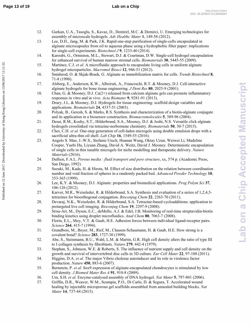
Biotin-functionalized alginate was synthesized using EDC/NHS carbodiimide crosslinker chemistry
(Supplementary Fig. 1a), and ANS fluorescence detection was used to quantify biotin conjugation20
. The
"degree of substitution" (DS) is defined as the number of chemical moieties per alginate polysaccharide
chain. Biotin-functionalized alginates with a degree of substitution of 11.8 and 2.1 were synthesized (Fig.
1a).
Spherical alginate microgels were formed with a microfluidic device (Supplementary Fig. 1b) using the
alginate that had been functionalized with biotin. The resulting microgels (biotin microgels) presumably
presented biotin-binding sites throughout their volume. To create streptavidin-functionalized microgels,
biotin-functionalized microgels were incubated with soluble streptavidin protein. As the streptavidin was
tagged with a green fluorophore and the entirety of the biotin microgels appeared green, streptavidin
binding likely occurred throughout the microgel (Fig. 1b). These microgels, formed from incubating biotin-
alginate microgels with streptavidin, are termed “streptavidin microgels.” The average diameter for both
biotin and streptavidin microgels was 25 µm (Fig. 1c).
Next, the self-assembly capabilities of biotin and streptavidin microgels were tested. Biotin and streptavidin
microgels first were pipetted into a test tube, followed by a short five-minute incubation. This process was
Page 6 of 19Lab on a Chip
Lab
ona
Chi
pA
ccep
ted
Man
uscr
ipt
Publ
ishe
d on
12
June
201
7. D
ownl
oade
d by
Uni
vers
ity o
f H
ong
Kon
g L
ibra
ries
on
13/0
6/20
17 1
1:11
:35.
View Article OnlineDOI: 10.1039/C7LC00500H
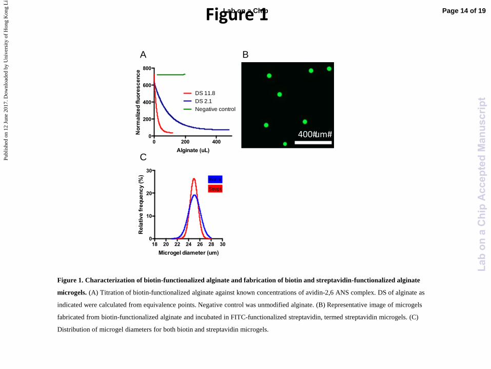
considered “suspension-mediated” assembly (Fig. 2a). Using this approach, microgel assemblages, defined
as multi-microgel structures containing one type of microgel in physical contact with microgel(s) of
complementary BPM, were formed (Fig. 2b). However, when only biotin or streptavidin microgels alone
were assembled under the same conditions, >99% of the microgels were single microgels (Fig. 2c),
indicating that assemblages were formed due to BPM-specific interactions.
Next, the effect of varying the ratio of microgels functionalized with complementary BPM on the formed
assemblages was analyzed, specifically quantifying the Nm and distribution of assemblages. Ratios of 30:1
and 1:1 were analyzed. Using suspension-mediated assembly, biotin and streptavidin microgels in a 30:1
ratio resulted in assemblages that had a mean Nm of 8.7, compared to a mean of 11.0 for the 1:1
combination (Fig. 2d). These differences were not statistically significant, but the variability in the Nm
between the two conditions was substantial. The distribution of the Nm (solid line) formed from a 30:1 ratio
of biotin to streptavidin microgels was unimodal (Fig. 2e) with a maximum at 6 microgels per assemblage,
with 97% of the starting streptavidin beads incorporated into an assemblage. In contrast, the distribution of
Nm (solid line) formed from a 1:1 combination of biotin to streptavidin was bimodal. In this condition,
31.6% of assemblages had ≥18 microgels per assemblage, and 61.5% of assemblages had ≤5 microgels per
assemblage (Fig. 2f). These differences were also reflected in the coefficient of variance of the two groups,
which were 0.67 and 1.05 for the 30:1 and 1:1 ratio, respectively. Analysis of the distribution microgels
that were incorporated into assemblages of different Nm revealed that the larger assemblages incorporated
the majority of microgels in the 1:1 ratio (dotted line). These results indicate the ratio of microgels bearing
complementary BPM controls the Nm distribution of assemblages that formed, and that a high ratio leads to
a more uniform distribution. All subsequent assembly studies were conducted using a 30:1 ratio of the
excess species to limiting species.
The effects of the number of BMP per microgel, microgel concentration, and microgel size on Nm were
then tested. Assemblages formed from microgels with a higher degree of biotin substitution resulted in a
higher mean Nm (Fig. 2g). Reducing the concentration of microgels in suspension was also found to
produce assemblages with lower Nm, suggesting that the frequency of contact between microgels in
suspension also affected assembly (Fig. 2h). To test the effect of microgel size, 10-µm streptavidin
microgels were fabricated using a microfluidic device with a smaller cross-junction aperture and used to
mediate the formation of assemblages with 25-µm biotin microgels (Fig. 2i) in a 1:30 ratio. As with equal
sized microgels, the distribution of Nm (solid line) and of microgel incorporation (dotted line) formed with
10-µm streptavidin microgels demonstrated one distinct peak (Fig. 2j). The mean Nm, however, was
significantly lower than when both biotin and streptavidin microgels were of equal sizes (Fig. 2k).
Self-assembly with click-modified alginate microgels
Page 7 of 19 Lab on a Chip
Lab
ona
Chi
pA
ccep
ted
Man
uscr
ipt
Publ
ishe
d on
12
June
201
7. D
ownl
oade
d by
Uni
vers
ity o
f H
ong
Kon
g L
ibra
ries
on
13/0
6/20
17 1
1:11
:35.
View Article OnlineDOI: 10.1039/C7LC00500H
As biotin and streptavidin coupling is not completely bio-orthogonal to other compounds, a second
microgel self-assembly system was developed using alginate functionalized with click-chemistry
compounds, specifically those that do not require cytotoxic copper catalysts. Alginate functionalized with
the click-chemistry compounds tetrazine(Tz)-, transcyclooctene(TCO), and norbornene(Nb) were
synthesized using EDC/NHS carbodiimide crosslinker chemistry (Supplementary Fig. 1c-e). As with
biotinylated alginate, these chemically modified alginates were then used to fabricate microgels. The
average diameters for tetrazine-, TCO-, and Nb-functionalized microgels were similar to those of biotin and
streptavidin microgels (Fig. 3a). Click microgels were then combined in suspension-mediated assembly
(Fig. 2a), and the type of BPM was found to influence Nm (Fig. 3b). Like the biotin and streptavidin
microgel assemblages, the click-chemistry assemblages only formed in the presence of complementary
BPM. When Tz, TCO, and Nb microgels alone were in suspension, 93-97% of microgels were single
microgels (Supplemental Fig. 2a).
With a 5-minute incubation for suspension-mediated assembly, biotin and streptavidin microgels
assemblages had a mean Nm of 6.1, Tz and TCO microgel assemblages had a mean Nm of 5.2, and Tz and
Nb microgel assemblages had a mean Nm of 2.6. Incubating complementary microgels for an additional 5
minutes created larger assemblages of Tz and TCO microgels, though not of biotin and streptavidin or Tz
and Nb microgels. An overnight incubation produced assemblages of biotin and streptavidin and Tz and
TCO microgels that were substantially larger than those produced from shorter incubation times. Tz and Nb
microgel assemblages, however, maintained the same Nm, and had a lower mean Nm than assemblages
made from other BPM at all times points (Fig. 3b).
We next explored if the assembly method could lead to different Nm. As centrifugation may bring
microgels into contact to a greater extent than in a suspension and thereby produce larger assemblages,
centrifugation-mediated assembly was tested (Fig. 3c). Like assemblages produced in suspension,
assemblages resulting from centrifugation-mediated assembly only formed when complementary BPM
microgels were combined. Microgels modified with the same BPM produced 93-99% single microgels
when centrifuged, similar to levels before centrifugation (Supplemental Fig. 2b-c). With the same overall
incubation time for suspension- and centrifugation-mediated assembly (10 minutes), centrifugation resulted
in significantly higher mean Nm across all three systems. The Nm of biotin-streptavidin assemblages was
higher than that of Tz-TCO assemblages, which were in turn was higher than that of Tz-Nb assemblages
(Fig. 3d).
To monitor assemblages over time, the mean Nm across the three systems after 2 or 5 days was measured
and compared to the baseline Nm. While the mean Nm for biotin-streptavidin and TCO-Tz assemblages did
not decrease over time, there was a 1.8 reduction in mean Nm for Nb-Tz assemblages (Fig. 3e).
Page 8 of 19Lab on a Chip
Lab
ona
Chi
pA
ccep
ted
Man
uscr
ipt
Publ
ishe
d on
12
June
201
7. D
ownl
oade
d by
Uni
vers
ity o
f H
ong
Kon
g L
ibra
ries
on
13/0
6/20
17 1
1:11
:35.
View Article OnlineDOI: 10.1039/C7LC00500H
The percentage of the limiting microgel type, defined as the microgel species present in less quantity during
assembly, incorporated into assemblages was then determined as a function of the specific BPM. Close to
100% of the limiting microgel species was incorporated into assemblages in both the biotin-streptavidin
and TCO-Tz systems under both suspension- and centrifugation- mediated conditions. However, in
suspension-mediated assembly of Nb-Tz microgels, only 18% of the limiting microgel species was
incorporated into assemblages. Centrifugation-mediated assembly was able to increase the limiting
microgel species incorporation in the Nb-Tz system to 98% (Fig. 3f).
In addition to Nm, the coordination numbers for assemblages were quantified in order to determine the
packing density of microgels. The coordinate number was defined as the number of complementary
microgels in direct contact with a central microgel of the limiting species. Under suspension-mediated
assembly, the biotin-streptavidin exhibited a higher mean coordinate number than the TCO-Tz system,
which exhibited a higher mean coordinate number than the Nb-Tz system for all three suspension-mediated
assembly timepoints (Fig. 4a). These coordination numbers were lower than those characteristic of the
thinnest regular packing of monodisperse spheres (6)24
. Larger coordination numbers resulted from
centrifugation-mediated assembly in all three systems compared to 5-10-minute suspension-mediated
assembly (Fig. 4b). The mean coordination numbers of assemblages formed from centrifugation-mediated
assembly demonstrated the same trend as those formed from suspension-mediated assembly. The biotin-
streptavidin and TCO-Tz systems are comparable to the coordinate number observed in very loose random
packings25
. However, the mean coordination number of Nb-Tz assemblages remained much lower than the
thinnest regular packing of spheres. The contact areas between microgels in spontaneous- and
centrifugation-mediated assembly were also measured (Fig. 4c-d), and was greater with centrifugation-
mediated assembly (Fig. 4e).
Assemblage separation, spheroid formation, and cell encapsulation
In order to purify assemblages from single beads and allow multilayer assemblages, a magnetic-based
sorting system was then tested. Biotin microgels were loaded with Fe2O3 nanoparticles by mixing the
alginate polymer precursor with Fe2O3 nanoparticles prior to injection into the microfluidic device. Since
the pore size of alginate hydrogels is roughly 5 nm26
, the nanoparticles, which are 15-20 nanometers, were
entrapped in the matrix. A magnetic separation process was then executed to obtain a pure population of
assemblages (Fig. 5a). After three rounds of magnetic purification, assemblages were retained while 93%
of single microgels were removed (Fig. 5b). An assemblage with three layers was then pursued by adding
microgels modified with the BPM species of the core microgel to the initial asssemblages, allowing
“growth” of microgel layers in the radial direction. A magnetic Tz microgel core was assembled with non-
magnetic TCO microgels, sorted, and then assembled with a subsequent layer of non-magnetic Tz
Page 9 of 19 Lab on a Chip
Lab
ona
Chi
pA
ccep
ted
Man
uscr
ipt
Publ
ishe
d on
12
June
201
7. D
ownl
oade
d by
Uni
vers
ity o
f H
ong
Kon
g L
ibra
ries
on
13/0
6/20
17 1
1:11
:35.
View Article OnlineDOI: 10.1039/C7LC00500H
microgels (Fig. 5c). The coordination number for the TCO layer (layer 1) was found to be 6, and the
coordination number for the non-magnetic Tz layer (layer 2) was 16 (Fig. 5d).
Finally, the ability of cell-encapsulated microgels to self-assemble was tested. Murine mesenchymal stem
cells were encapsulated into alginate microgels, with the number of cells per microgel averaging 1.4 (Fig.
6a). These cells exhibited high viability following encapsulation in all four types of microgels (Fig. 6b) as
well as upon suspension in the HEPES-buffered physiological saline used previously for self-assembly.
Cell-encapsulating microgels were then combined to form assemblages (Fig. 6c-g). As with empty
microgels, the mean Nm of cell-encapsulating TCO-Tz assemblages was lower than that of cell-
encapsulating biotin-streptavidin assemblages (Fig. 6g). There was also no statistical difference in mean Nm
between cell-encapsulating and empty microgel assemblages in both the biotin-streptavidin and TCO-Tz
systems.
Discussion
Microgels modified with BPM could be induced to self-assemble either in suspension (suspension-
mediated) or following a brief centrifugation step (centrifugation-mediated). No other external forces or
components were necessary for assembly. Using the suspension-mediated biotin-streptavidin microgel
system, methods to control assembly were explored. A number of different assembly factors appeared to
exert control over Nm, including DS of biotin, microgel concentration, and relative microgel size.
Moreover, the ratio of complementary microgels affected the distribution of Nm, with high ratios creating
assemblages with much less variance in Nm. When complementary microgels are combined at a high ratio,
steric inhibition of adjacent homotypic microgels around a single microgel of the limiting BPM species
presumably constrains Nm, preventing the formation of large, uncontrolled assemblages.
Comparison of biotin-streptavidin modified microgels with click compound modified microgels suggest
that the type of BPM affects Nm and coordination number. In suspension-mediated assembly, click-BPM
modified microgels formed assemblages with lower Nm than those modified with biotin-streptavidin.
Notably, the binding constants of the click reagents used here, 26000 M-1
s-1
for TCO-Tz27
and 1.6-1.9 M-1
s-
1 for Nb-Tz
28, respectively, are many times smaller than that of biotin and streptavidin (3.0 x 10
6 - 4.5 x 10
7
M-1
s-1
)29
. As the Nm resulting from these three systems decreased in order of decreasing binding constants
of the BPM in suspension-mediated assembly, it is possible that this parameter strongly influences the
ability of microgels to self-assembly in suspension. However, this effect appears to diminish with increased
incubation time, as biotin-streptavidin and TCO-Tz assemblages did not exhibit a different mean Nm at 10-
minute and overnight suspension-mediated assembly. Nb-Tz assemblages, on the other hand, consistently
exhibited a lower Nm compared to the other two systems.
Page 10 of 19Lab on a Chip
Lab
ona
Chi
pA
ccep
ted
Man
uscr
ipt
Publ
ishe
d on
12
June
201
7. D
ownl
oade
d by
Uni
vers
ity o
f H
ong
Kon
g L
ibra
ries
on
13/0
6/20
17 1
1:11
:35.
View Article OnlineDOI: 10.1039/C7LC00500H
Assembly method was found to provide an addition means of controlling Nm, in addition to binding
constants and incubation time. Centrifugation led to higher Nm compared to suspension-mediated assembly
with equivalent incubation times, and to values of Nm that were equivalent to suspension-mediated
assembly given a longer, overnight incubation. This suggests that centrifugation expedites the packing of
microgels that would naturally occur with extended incubations. The dissociation of assemblages may also
correlate with binding kinetics. Whereas the Nm of biotin-streptavidin and TCO-Tz assemblages remained
the same over 2-5 days, assemblages of Nb-Tz experienced a reduction in Nm over the same time frame,
suggesting that binding kinetics may be used for controlling the dissociation of assemblages.
In contrast to binding constants, there was no apparent positive correlation between bond energy and the
Nm, as the bond energies of biotin and streptavidin is 160 pN30
, compared to 1600 pN31
for both the click
systems. However, as decreased DS of biotin significantly reduced the Nm (Fig. 2i), the overall adhesion
strength between microgels does appear to play a role in self-assembly.
The coordination numbers of assemblages was quantified in order to determine packing density, as many
studies have shown that overall cell density can impact biological outcomes32,33,34,35
. The highest packing
densities achieved here, those resulting from centrifugation-mediated assembly, were equivalent to very
loose random packing. Further increasing the concentration of microgels might lead to a higher packing
density. However, initial binding of microgels is likely to be random, and strong BPM could prevent
microgels from rearranging in order to accommodate higher packing density. It is unclear if rearrangement
occurs in the systems investigated here, but the strong bond energies and the low packing density would
suggest that rearrangement does not occur. BPM that have high binding kinetics but low bond energies may
therefore enable rearrangements of microgels towards the most thermodynamically favorable configuration
in the future.
Finally, BPM-modified alginates were shown to be capable of viably encapsulating cells without losing
their assembly properties. Modification of the encapsulating polymer rather than the cell itself to promote
self-assembly may reduce negative side effects on cell viability and behavior. By exploiting assembly
parameters, such as assembly method, BPM type and concentration, and microgel size, controlled
assemblages of heterogeneous cell clusters at specific ratios may be fabricated. Modification of cell-
encapsulating microgels with BPM may also complement existing techniques using microrobotics and
other platforms for assembling microscale hydrogels into determined structures by providing a means of
stably and rapidly annealing assembled microgels1,8,11,36
. Moreover, unlike assembly strategies that rely on
nucleic acids, the assembly buffer used here—a HEPES buffered physiological saline—does not require
detergents to stabilize the assembly. These considerations may be important for in vivo applications, as the
in vivo environment will often prevent the usage of specialized buffers or assembly apparatus. Microgels
Page 11 of 19 Lab on a Chip
Lab
ona
Chi
pA
ccep
ted
Man
uscr
ipt
Publ
ishe
d on
12
June
201
7. D
ownl
oade
d by
Uni
vers
ity o
f H
ong
Kon
g L
ibra
ries
on
13/0
6/20
17 1
1:11
:35.
View Article OnlineDOI: 10.1039/C7LC00500H
have been shown to be capable of forming injectable microporous scaffolds, and incorporation of cell
encapsulation and BPM modification may enable the creation of cell-seeded microporous scaffolds in vivo
without the need of separate annealing agents37
.
Conclusion
We developed a system for cell-encapsulating microgel self-assembly using alginates modified with three
sets of BPM: biotin and streptavidin, Tz and Nb, and Tz and TCO. Microgels altered in this way could self-
assemble to form structures with packing densities comparable to very loose randomly packed
configurations. Self-assembly systems such as these could augment existing regenerative medicine
therapies, such as the fabrication of multicellular “niches” for drug screening, or encapsulation of cells for
cell infusion therapies.
Acknowledgements
This work was supported by the National Institutes of Health (NIH) Grants RO1EB014703 (D.J.M. and
D.A.W.), and the National Science Foundation (NSF) Graduate Research Fellowship Program (A.S.M.).
The authors would like to thank Tom Ferrante of the Wyss Institute for imaging support.
References
1. Guven, S. et al. Multiscale assembly for tissue engineering and regenerative medicine. Trends
Biotechnol 33, 269-79 (2015).
2. Elbert, D.L. Bottom-up tissue engineering. Curr Opin Biotechnol 22, 674-80 (2011).
3. Xu, F. et al. Microengineering methods for cell-based microarrays and high-throughput drug-
screening applications. Biofabrication 3, 034101 (2011).
4. Winter, C.C. et al. Transplantable living scaffolds comprised of micro-tissue engineered aligned
astrocyte networks to facilitate central nervous system regeneration. Acta Biomater 38, 44-58
(2016).
5. Zhang, S. Fabrication of novel biomaterials through molecular self-assembly. Nat Biotechnol 21,
1171-8 (2003).
6. Wang, Y., Qi, W., Huang, R., Su, R. & He, Z. Counterion-Directed, Structurally Tunable
Assembly of Hydrogels, Membranes, and Sacs at Aqueous Liquid–Liquid Interfaces. Advanced
Materials Interfaces 3, 1500327-n/a (2016).
7. Kojima, N., Takeuchi, S. & Sakai, Y. Establishment of self-organization system in rapidly formed
multicellular heterospheroids. Biomaterials 32, 6059-67 (2011).
8. Qi, H. et al. DNA-directed self-assembly of shape-controlled hydrogels. Nat Commun 4, 2275
(2013).
9. Han, Y.L. et al. Directed self-assembly of microscale hydrogels by electrostatic interaction.
Biofabrication 5, 035004 (2013).
10. Tasoglu, S. et al. Guided and magnetic self-assembly of tunable magnetoceptive gels. Nat
Commun 5, 4702 (2014).
11. Tasoglu, S., Diller, E., Guven, S., Sitti, M. & Demirci, U. Untethered micro-robotic coding of
three-dimensional material composition. Nat Commun 5, 3124 (2014).
Page 12 of 19Lab on a Chip
Lab
ona
Chi
pA
ccep
ted
Man
uscr
ipt
Publ
ishe
d on
12
June
201
7. D
ownl
oade
d by
Uni
vers
ity o
f H
ong
Kon
g L
ibra
ries
on
13/0
6/20
17 1
1:11
:35.
View Article OnlineDOI: 10.1039/C7LC00500H
12. Gurkan, U.A., Tasoglu, S., Kavaz, D., Demirel, M.C. & Demirci, U. Emerging technologies for
assembly of microscale hydrogels. Adv Healthc Mater 1, 149-58 (2012).
13. Lee, D.H., Jang, M. & Park, J.K. Rapid one-step purification of single-cells encapsulated in
alginate microcapsules from oil to aqueous phase using a hydrophobic filter paper: implications
for single-cell experiments. Biotechnol J 9, 1233-40 (2014).
14. Karoubi, G., Ormiston, M.L., Stewart, D.J. & Courtman, D.W. Single-cell hydrogel encapsulation
for enhanced survival of human marrow stromal cells. Biomaterials 30, 5445-55 (2009).
15. Martinez, C.J. et al. A microfluidic approach to encapsulate living cells in uniform alginate
hydrogel microparticles. Macromol Biosci 12, 946-51 (2012).
16. Smidsrod, O. & Skjak-Braek, G. Alginate as immobilization matrix for cells. Trends Biotechnol 8,
71-8 (1990).
17. Alsberg, E., Anderson, K.W., Albeiruti, A., Franceschi, R.T. & Mooney, D.J. Cell-interactive
alginate hydrogels for bone tissue engineering. J Dent Res 80, 2025-9 (2001).
18. Chan, G. & Mooney, D.J. Ca(2+) released from calcium alginate gels can promote inflammatory
responses in vitro and in vivo. Acta Biomater 9, 9281-91 (2013).
19. Drury, J.L. & Mooney, D.J. Hydrogels for tissue engineering: scaffold design variables and
applications. Biomaterials 24, 4337-51 (2003).
20. Polyak, B., Geresh, S. & Marks, R.S. Synthesis and characterization of a biotin-alginate conjugate
and its application in a biosensor construction. Biomacromolecules 5, 389-96 (2004).
21. Desai, R.M., Koshy, S.T., Hilderbrand, S.A., Mooney, D.J. & Joshi, N.S. Versatile click alginate
hydrogels crosslinked via tetrazine-norbornene chemistry. Biomaterials 50, 30-7 (2015).
22. Choi, C.H. et al. One-step generation of cell-laden microgels using double emulsion drops with a
sacrificial ultra-thin oil shell. Lab Chip 16, 1549-55 (2016).
23. Angelo S. Mao, J.-W.S., Stefanie Utech, Huanan Wang, Oktay Uzun, Weiwei Li, Madeline
Cooper, Yuebi Hu, Liyuan Zhang, David A. Weitz, David J. Mooney. Deterministic encapsulation
of single cells in thin tunable microgels for niche modelling and therapeutic delivery. Nature
Materials (2016).
24. Dullien, F.A.L. Porous media : fluid transport and pore structure, xx, 574 p. (Academic Press,
San Diego, 1992).
25. Suzuki, M., Kada, H. & Hirota, M. Effect of size distribution on the relation between coordination
number and void fraction of spheres in a randomly packed bed. Advanced Powder Technology 10,
353-365 (1999).
26. Lee, K.Y. & Mooney, D.J. Alginate: properties and biomedical applications. Prog Polym Sci 37,
106-126 (2012).
27. Karver, M.R., Weissleder, R. & Hilderbrand, S.A. Synthesis and evaluation of a series of 1,2,4,5-
tetrazines for bioorthogonal conjugation. Bioconjug Chem 22, 2263-70 (2011).
28. Devaraj, N.K., Weissleder, R. & Hilderbrand, S.A. Tetrazine-based cycloadditions: application to
pretargeted live cell imaging. Bioconjug Chem 19, 2297-9 (2008).
29. Srisa-Art, M., Dyson, E.C., deMello, A.J. & Edel, J.B. Monitoring of real-time streptavidin-biotin
binding kinetics using droplet microfluidics. Anal Chem 80, 7063-7 (2008).
30. Florin, E.L., Moy, V.T. & Gaub, H.E. Adhesion forces between individual ligand-receptor pairs.
Science 264, 415-7 (1994).
31. Grandbois, M., Beyer, M., Rief, M., Clausen-Schaumann, H. & Gaub, H.E. How strong is a
covalent bond? Science 283, 1727-30 (1999).
32. Abe, S., Steinmann, B.U., Wahl, L.M. & Martin, G.R. High cell density alters the ratio of type III
to I collagen synthesis by fibroblasts. Nature 279, 442-4 (1979).
33. Stephan, S., Johnson, W.E. & Roberts, S. The influence of nutrient supply and cell density on the
growth and survival of intervertebral disc cells in 3D culture. Eur Cell Mater 22, 97-108 (2011).
34. Higgins, D.A. et al. The major Vibrio cholerae autoinducer and its role in virulence factor
production. Nature 450, 883-6 (2007).
35. Bernstein, P. et al. Sox9 expression of alginate-encapsulated chondrocytes is stimulated by low
cell density. J Biomed Mater Res A 91, 910-8 (2009).
36. Um, S.H. et al. Enzyme-catalysed assembly of DNA hydrogel. Nat Mater 5, 797-801 (2006).
37. Griffin, D.R., Weaver, W.M., Scumpia, P.O., Di Carlo, D. & Segura, T. Accelerated wound
healing by injectable microporous gel scaffolds assembled from annealed building blocks. Nat
Mater 14, 737-44 (2015).
Page 13 of 19 Lab on a Chip
Lab
ona
Chi
pA
ccep
ted
Man
uscr
ipt
Publ
ishe
d on
12
June
201
7. D
ownl
oade
d by
Uni
vers
ity o
f H
ong
Kon
g L
ibra
ries
on
13/0
6/20
17 1
1:11
:35.
View Article OnlineDOI: 10.1039/C7LC00500H
0 200 4000
200
400
600
800
Alginate (uL)
No
rma
lize
d flu
ore
sc
en
ce
Negative control
DS 2.1
DS 11.8
18 20 22 24 26 28 300
10
20
30
Microgel diameter (um)
Re
lativ
e fre
qu
en
cy
(%
)
Biotin
Strept
400#um#
A
C
B
Figure 1
Figure 1. Characterization of biotin-functionalized alginate and fabrication of biotin and streptavidin-functionalized alginate
microgels. (A) Titration of biotin-functionalized alginate against known concentrations of avidin-2,6 ANS complex. DS of alginate as
indicated were calculated from equivalence points. Negative control was unmodified alginate. (B) Representative image of microgels
fabricated from biotin-functionalized alginate and incubated in FITC-functionalized streptavidin, termed streptavidin microgels. (C)
Distribution of microgel diameters for both biotin and streptavidin microgels.
Page 14 of 19Lab on a Chip
Lab
ona
Chi
pA
ccep
ted
Man
uscr
ipt
Publ
ishe
d on
12
June
201
7. D
ownl
oade
d by
Uni
vers
ity o
f H
ong
Kon
g L
ibra
ries
on
13/0
6/20
17 1
1:11
:35.
View Article OnlineDOI: 10.1039/C7LC00500H
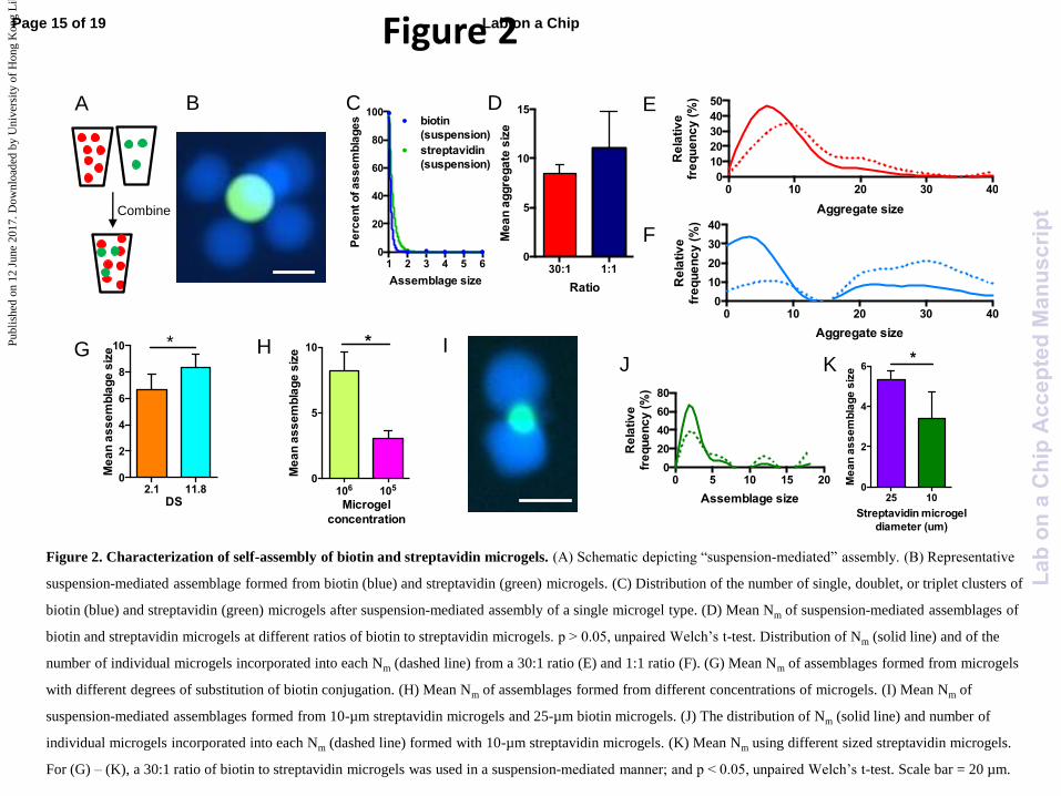
Figure 2
30:1 1:10
5
10
15
Ratio
Me
an
ag
gre
ga
te s
ize
1 2 3 4 5 60
20
40
60
80
100
Assemblage size
Pe
rce
nt o
f a
ss
em
bla
ge
s biotin
(suspension)
streptavidin
(suspension)
0 10 20 30 400
10
20
30
40
Aggregate size
Re
lativ
e
fre
qu
en
cy
(%
)
0 5 10 15 200
20
40
60
80
Assemblage size
Re
lativ
e
fre
qu
en
cy
(%
)
0 10 20 30 400
10
20
30
40
50
Aggregate size
Re
lativ
e
fre
qu
en
cy
(%
)
I
D
F
K J G H
E B A
A
Combine
C
2.1 11.80
2
4
6
8
10
DS
Me
an
as
se
mb
lag
e s
ize
*
106 1050
5
10
Microgel
concentration
Me
an
as
se
mb
lag
e s
ize
*
25 100
2
4
6
Streptavidin microgel
diameter (um)
Me
an
as
se
mb
lag
e s
ize
*
Figure 2. Characterization of self-assembly of biotin and streptavidin microgels. (A) Schematic depicting “suspension-mediated” assembly. (B) Representative
suspension-mediated assemblage formed from biotin (blue) and streptavidin (green) microgels. (C) Distribution of the number of single, doublet, or triplet clusters of
biotin (blue) and streptavidin (green) microgels after suspension-mediated assembly of a single microgel type. (D) Mean Nm of suspension-mediated assemblages of
biotin and streptavidin microgels at different ratios of biotin to streptavidin microgels. p > 0.05, unpaired Welch’s t-test. Distribution of Nm (solid line) and of the
number of individual microgels incorporated into each Nm (dashed line) from a 30:1 ratio (E) and 1:1 ratio (F). (G) Mean Nm of assemblages formed from microgels
with different degrees of substitution of biotin conjugation. (H) Mean Nm of assemblages formed from different concentrations of microgels. (I) Mean Nm of
suspension-mediated assemblages formed from 10-µm streptavidin microgels and 25-µm biotin microgels. (J) The distribution of Nm (solid line) and number of
individual microgels incorporated into each Nm (dashed line) formed with 10-µm streptavidin microgels. (K) Mean Nm using different sized streptavidin microgels.
For (G) – (K), a 30:1 ratio of biotin to streptavidin microgels was used in a suspension-mediated manner; and p < 0.05, unpaired Welch’s t-test. Scale bar = 20 µm.
Page 15 of 19 Lab on a Chip
Lab
ona
Chi
pA
ccep
ted
Man
uscr
ipt
Publ
ishe
d on
12
June
201
7. D
ownl
oade
d by
Uni
vers
ity o
f H
ong
Kon
g L
ibra
ries
on
13/0
6/20
17 1
1:11
:35.
View Article OnlineDOI: 10.1039/C7LC00500H
Figure 3
Bio-Strep TCO-Tz Nb-Tz-4
-2
0
2
4
6
Ch
an
ge
in
me
an
Nm
Biotin TCO-Tz Nb-Tz0
20
40
60
80
100
BPM
Lim
itin
g m
icro
ge
l
inc
orp
ora
tio
n (%
)
Bio-Strep TCO-Tz Nb-Tz0
5
10
15
Me
an
as
se
mb
lag
e s
ize
**
*
B
D
0 10 20 30 400
20
40
60
80
Microgel diameter (um)
Re
lativ
e fre
qu
en
cy
(%
)
Tz
TCO
Nb
A
E
C
Nb-Tz
TCO-Tz
5 min 10 min overnight0
2
4
6
8
10
12
Me
an
as
se
mb
lag
e s
ize Biotin-strept
F
Figure 3. Self-assembly of click-modified alginate microgels. (A) Distribution of microgel diameters for TCO-, Tz-, and Nb-functionalized microgels. (B)
Mean Nm of suspension-mediated assemblages of biotin and streptavidin microgels (red), TCO and Tz microgels (gray), and Nb and Tz microgels (blue) after 5-
minute, 10-minute, and overnight incubation. (C) Schematic of “centrifugation-mediated” assembly process. (D) Mean Nm of centrifugation-mediated
assemblages of biotin and streptavidin microgels (red), TCO and Tz microgels (gray), and Nb and Tz microgels (blue); p < 0.05, ANOVA, pairwise
comparisons adjusted with Tukey-Kramer. (E) Change in Nm across the three systems after 2 or 5 days. (F) Percentage of limiting microgel species incorporated
into assemblages under spontaneous-mediated (purple) and centrifugation-mediated (yellow) assembly. For (B), (D) and (F), a 30:1 ratio of microgels was used.
Page 16 of 19Lab on a Chip
Lab
ona
Chi
pA
ccep
ted
Man
uscr
ipt
Publ
ishe
d on
12
June
201
7. D
ownl
oade
d by
Uni
vers
ity o
f H
ong
Kon
g L
ibra
ries
on
13/0
6/20
17 1
1:11
:35.
View Article OnlineDOI: 10.1039/C7LC00500H
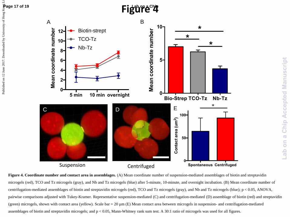
Figure 4
Spontaneous Centrifuged0
50
100
Co
nta
ct a
rea
(u
m2)
*
Suspension Centrifuged
D C E
A B
TCO-Tz
Nb-Tz
5 min 10 min overnight0
2
4
6
8
10
12
Me
an
co
ord
ina
te n
um
be
r
Biotin-strept
Bio-Strep TCO-Tz Nb-Tz0
5
10
Me
an
co
ord
ina
te n
um
be
r **
*
Figure 4. Coordinate number and contact area in assemblages. (A) Mean coordinate number of suspension-mediated assemblages of biotin and streptavidin
microgels (red), TCO and Tz microgels (gray), and Nb and Tz microgels (blue) after 5-minute, 10-minute, and overnight incubation. (B) Mean coordinate number of
centrifugation-mediated assemblages of biotin and streptavidin microgels (red), TCO and Tz microgels (gray), and Nb and Tz microgels (blue); p < 0.05, ANOVA,
pairwise comparisons adjusted with Tukey-Kramer. Representative suspension-mediated (C) and centrifugation-mediated (D) assemblage of biotin (red) and streptavidin
(green) microgels, shown with contact area (yellow). Scale bar = 20 µm (E) Mean contact area between microgels in suspension- and centrifugation-mediated
assemblages of biotin and streptavidin microgels; and p < 0.05, Mann-Whitney rank sum test. A 30:1 ratio of microgels was used for all figures.
Page 17 of 19 Lab on a Chip
Lab
ona
Chi
pA
ccep
ted
Man
uscr
ipt
Publ
ishe
d on
12
June
201
7. D
ownl
oade
d by
Uni
vers
ity o
f H
ong
Kon
g L
ibra
ries
on
13/0
6/20
17 1
1:11
:35.
View Article OnlineDOI: 10.1039/C7LC00500H
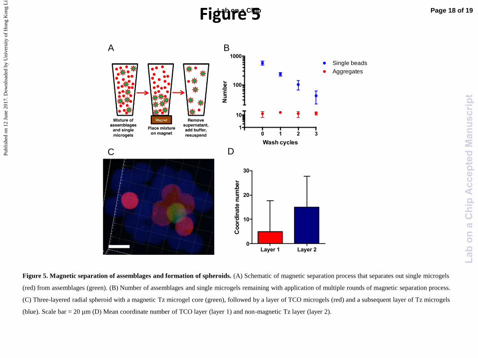
Figure 5
0 1 2 3 1
10
100
1000
Wash cycles
Nu
mb
er
Single beads
Aggregates
Magnetic Tz microgel
Non-magnetic TCO microgel
C
Non-magnetic Tz microgel
B
D C
A
Layer 1 Layer 20
10
20
30
Co
ord
ina
te n
um
be
r
Figure 5. Magnetic separation of assemblages and formation of spheroids. (A) Schematic of magnetic separation process that separates out single microgels
(red) from assemblages (green). (B) Number of assemblages and single microgels remaining with application of multiple rounds of magnetic separation process.
(C) Three-layered radial spheroid with a magnetic Tz microgel core (green), followed by a layer of TCO microgels (red) and a subsequent layer of Tz microgels
(blue). Scale bar = 20 µm (D) Mean coordinate number of TCO layer (layer 1) and non-magnetic Tz layer (layer 2).
Page 18 of 19Lab on a Chip
Lab
ona
Chi
pA
ccep
ted
Man
uscr
ipt
Publ
ishe
d on
12
June
201
7. D
ownl
oade
d by
Uni
vers
ity o
f H
ong
Kon
g L
ibra
ries
on
13/0
6/20
17 1
1:11
:35.
View Article OnlineDOI: 10.1039/C7LC00500H
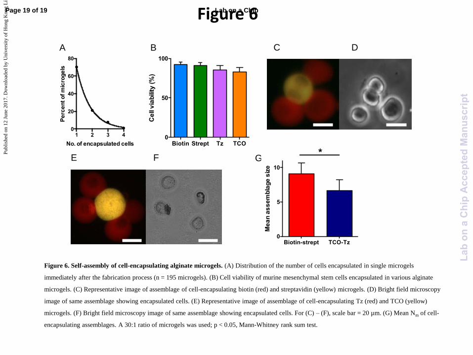
Figure 6
C D
E F G
A
1 2 3 40
20
40
60
80
No. of encapsulated cells
Pe
rce
nt o
f m
icro
ge
ls
B
Biotin-strept TCO-Tz0
5
10
Me
an
as
se
mb
lag
e s
ize
*Biotin Strept Tz TCO
0
50
100
Ce
ll v
iab
ility
(%
)
Figure 6. Self-assembly of cell-encapsulating alginate microgels. (A) Distribution of the number of cells encapsulated in single microgels
immediately after the fabrication process (n = 195 microgels). (B) Cell viability of murine mesenchymal stem cells encapsulated in various alginate
microgels. (C) Representative image of assemblage of cell-encapsulating biotin (red) and streptavidin (yellow) microgels. (D) Bright field microscopy
image of same assemblage showing encapsulated cells. (E) Representative image of assemblage of cell-encapsulating Tz (red) and TCO (yellow)
microgels. (F) Bright field microscopy image of same assemblage showing encapsulated cells. For (C) – (F), scale bar = 20 µm. (G) Mean Nm of cell-
encapsulating assemblages. A 30:1 ratio of microgels was used; p < 0.05, Mann-Whitney rank sum test.
Page 19 of 19 Lab on a Chip
Lab
ona
Chi
pA
ccep
ted
Man
uscr
ipt
Publ
ishe
d on
12
June
201
7. D
ownl
oade
d by
Uni
vers
ity o
f H
ong
Kon
g L
ibra
ries
on
13/0
6/20
17 1
1:11
:35.
View Article OnlineDOI: 10.1039/C7LC00500H