Embed Size (px)
Citation preview
MS ID#: GENOME/2013/164830
Dynamic shifts in occupancy by TAL1 are guided by GATA factors and drive large-scale
reprogramming of gene expression during hematopoiesis
Weisheng Wu 1,4, Christapher S. Morrissey 1, Cheryl A. Keller 1, Tejaswini Mishra 1, Maxim Pimkin 2,
Gerd A. Blobel 2,3, Mitchell J. Weiss 2,3, Ross C. Hardison 1
1 Center for Comparative Genomics and Bioinformatics, Department of Biochemistry and Molecular
Biology, The Pennsylvania State University, University Park, PA 168022 Division of Hematology, The Children’s Hospital of Philadelphia, Philadelphia, PA 19104, USA, 3 Perelman School of Medicine at the University of Pennsylvania, Philadelphia, PA 19104, USA4 Current address: Bioinformatics Core, Department of Computational Medicine and Bioinformatics,
University of Michigan, Ann Arbor, MI 48109-2218
Running title: Dynamics of TAL1 occupancy in mouse hematopoiesis
Key words: epigenetics, hematopoiesis, gene regulation, mouse ENCODE, transcription factor
occupancy
Address for correspondence:
Ross C. Hardison, 304 Wartik Laboratory, Penn State University, University Park, PA 16802
e-mail: [email protected]
Phone: 814-863-0113
Note: Major textual changes made in response to reviewer comments are in blue text. Additional
streamlining edits and deletions are not indicated.
1

ABSTRACT
We used mouse ENCODE data along with complementary data from other laboratories to study the
dynamics of occupancy and role in gene regulation of the transcription factor TAL1, a critical
regulator of hematopoiesis, at multiple stages of hematopoietic differentiation. We combined ChIP-
seq and RNA-seq data in six mouse cell types representing a progression from multilineage
precursors to differentiated erythroblasts and megakaryocytes. We found that sites of occupancy
shift dramatically during commitment to the erythroid lineage, vary further during terminal
maturation, and are strongly associated with changes in gene expression. In multilineage progenitors,
the likely target genes are enriched for hematopoietic growth and functions associated with the
mature cells of specific daughter lineages (such as megakaryocytes). In contrast, target genes in
erythroblasts are specifically enriched for red cell functions. Furthermore, shifts in TAL1 occupancy
during erythroid differentiation are associated with gene repression (dissociation) and induction (co-
occupancy with GATA1). Based on both enrichment for transcription factor binding site motifs and
co-occupancy determined by ChIP-seq, recruitment by GATA transcription factors appears to be a
stronger determinant of TAL1 binding to chromatin than the canonical E-box binding site motif.
Studies of additional proteins lead to the model that TAL1 regulates expression after being directed
to a distinct subset of genomic binding sites in each cell type via its association with different
complexes containing master regulators such as GATA2, ERG and RUNX1 in multilineage cells and the
lineage-specific master regulator GATA1 in erythroblasts.
2

INTRODUCTION
Dynamic changes in the locations and actions of transcription factors are thought to drive much
of the differential gene expression that determines cell fate, morphology and function (Davidson and
Erwin 2006). Recent genome-wide determinations of transcription factor occupancy in multiple
stages of hematopoiesis (Kassouf et al. 2010; Wilson et al. 2010), coupled with new data from the
mouse ENCODE project (Wu et al. 2011; Stamatoyannopoulos et al. 2012;
Mouse_ENCODE_Consortium 2014; Pimkin et al. 2014), allow us to examine in detail the patterns of
differential occupancy by key transcription factors during hematopoietic differentiation, correlate this
dynamic binding with changes in gene expression, and search for determinants of differential
occupancy.
Here we focused on TAL1 (also called SCL), a transcription factor that is indispensible at multiple
stages of hematopoiesis. This basic helix-loop-helix (bHLH) protein is required to establish
hematopoietic stem cells during embryogenesis and also to differentiate along the erythroid and
multiple myeloid cell lineages, including those leading to megakaryocytes, mast cells, and
eosinophils. The requirement for TAL1 in these processes has been demonstrated by multiple in vivo
and in vitro genetic experiments. Homozygous Tal1 null murine embryos die of anemia with failed
yolk sac hematopoiesis (Robb et al. 1995; Shivdasani et al. 1995). Furthermore, no hematopoietic
lineages were detectable from Tal1 null embryonic stem cells after in vitro differentiation or in
chimeric mice (Porcher et al. 1996). Conditional Tal1 knockout and rescue experiments show that
TAL1 is also needed for specification and differentiation of erythroid and megakaryocytic cells
(Schlaeger et al. 2005). TAL1 is expressed broadly in erythropoiesis, from highly proliferative,
committed progenitor cells (BFU-e and CFU-e) to more mature erythroblasts (Aplan et al. 1992;
Porcher et al. 1996). In contrast, TAL1 is normally absent from lymphoid cells, but its aberrant
expression in T cells leads to T-cell acute lymphocytic leukemia (Palii et al. 2011). The pleotropic
effects of Tal1 mutations in hematopoietic stem cells and in multiple hematopoietic lineages suggest
that the TAL1 protein plays unique roles in each stage and lineage. These roles could be realized in
either or both of two ways: by binding to different locations in the genome to regulate distinct sets of
genes in each cell type, and by interacting with different proteins to carry out distinct functions, such
as activation or repression.
One determinant of TAL1 binding to DNA is the sequence preference of its DNA-binding domain.
Binding-site selection experiments in solution have shown that TAL1, as a heterodimer with other
bHLH proteins such as the E-proteins E47 or E12 (Hsu et al. 1994), binds to the consensus sequence
AACAGATGGT, which contains a subset of E-box motifs (CANNTG) (Church et al. 1985). Other studies
showed preferential binding to CAGGTG (Wadman et al. 1997) and CAGCTG (Kassouf et al. 2010),
implying that CAGVTG is the preferred consensus sequence. Remarkably, the DNA binding domain is
not required for all TAL1 functions. Mutant ES cells homozygous for an intrinsic DNA binding domain
defective Tal1 allele (Tal1 rer) still support primitive erythropoiesis (Porcher et al. 1999), and mouse
3
embryos homozygous for this mutation survive past 9.5dpc, when the Tal1 homozygous null mice die
(Kassouf et al. 2008). These results show that direct binding to DNA is dispensable for some TAL1
functions in primitive erythropoiesis. Furthermore, a motif search on TAL1 binding sites in human
proerythroblasts revealed that E-boxes are absent from over one-fifth of the sites. Indeed, GATA
motifs ranked as the most overrepresented motifs, and they were closer to TAL1 peak summits than
E-boxes (Tripic et al. 2009; Palii et al. 2011). Another study compared TAL1 binding sites in primary
erythroid progenitor cells from wild type mice and from Tal1 rer/rer mice (lacking the Tal1 DNA binding
domain), and found that one-fifth of the wild type TAL1 binding sites were also occupied in the
mutant mice (Kassouf et al. 2010). This ability of DNA binding domain defective TAL1 to bind specific
genomic locations suggests that it may be recruited by other DNA-binding transcription factors.
Some of the TAL 1 in the nucleus is in a multiprotein complex with the transcription factors (TFs)
GATA1 (or GATA2), LMO2, and LDB1; this complex binds to specific cis-regulatory elements in
erythroid cells (Wadman et al. 1997; Anguita et al. 2004; Schuh et al. 2005; Cheng et al. 2009). In the
hematopoietic precursor cell line HPC7, which exhibits multilineage myeloid and erythroid potential
(Pinto do et al. 1998), additional TFs including LYL1, RUNX1, ERG, and FLI1 co-associate with the
bound TAL1-containing complex (Wilson et al. 2010). Co-binding of different TFs with TAL1 affects its
function. When bound together with GATA1, TAL1 is strongly associated with activation of gene
expression in erythroid cells. In multiple models for erythroid differentiation, a substantial majority
of induced genes are co-occupied by both GATA1 and TAL1, whereas a subset of GATA1-repressed
genes is bound by GATA1 but not TAL1 (Wozniak et al. 2008; Cheng et al. 2009; Tripic et al. 2009;
Soler et al. 2010; Wu et al. 2011). Furthermore, the activity of GATA1-occupied DNA segments
(GATA1 OSs) as enhancers is associated with co-occupancy by TAL1 (Tripic et al. 2009) and is
dependent on an intact binding site (E-box) for TAL1 (Elnitski et al. 1997). In contrast, TAL1 binding to
some genes operates as a molecular switch, leading to activation or repression under different
conditions (Huang et al. 1999; Huang and Brandt 2000; Elnitski et al. 2001). These studies indicate
that different cell type specific functions of TAL1 are regulated by the composition and activity of its
interacting proteins.
The widely differing phenotypes of cells expressing active TAL1 predict that its regulated gene
targets differ significantly. Consequently the DNA segments occupied by this protein should differ
between cell types. This prediction can now be evaluated comprehensively and quantitatively in
mouse cell models. Recent studies from our laboratory, as part of the mouse ENCODE project
(Mouse_ENCODE_Consortium 2014), and others have used chromatin immunoprecipitation followed
by second generation sequencing, ChIP-seq (Johnson et al. 2007; Robertson et al. 2007), and related
methods to map DNA segments occupied by TAL1 and other transcription factors across the
genomes of multiple human and mouse hematopoietic cells of different lineages and at progressive
stages of maturation (Cheng et al. 2009; Wilson et al. 2009; Kassouf et al. 2010; Soler et al. 2010;
Wilson et al. 2010; Palii et al. 2011; Tijssen et al. 2011; Wu et al. 2011; Dore et al. 2012; Kowalczyk et
al. 2012; Xu et al. 2012; Pimkin et al. 2014). To gain further insights into the functions carried out by
4
TAL1 in each cell type, we integrated these maps of TAL1 occupancy to establish its patterns of cell
lineage- and maturational stage-specific occupancy and correlated these with gene expression. We
studied the roles of histone modifications, matches to binding site motifs, and transcription factor
co-occupancy in determining differential TAL1 occupancy in different cell types. The results indicate
that TAL1 is a potent regulator of hematopoiesis whose specificity is directed by other hematopoietic
transcription factors.
5

RESULTS
Substantial changes in occupancy by TAL1 during differentiation
A comprehensive comparison of ChIP-seq experiments shows that the genomic positions
occupied by TAL1 shift dramatically at progressive stages of differentiation. We compared the DNA
segments occupied by TAL1 among cell types representing different stages of cell commitment and
differentiation (Fig. 1A). As summarized in Table 1, TAL1 occupancy data are available for a
multipotential hematopoietic precursor cell line, HPC7 (Wilson et al. 2009), and in a population of
Ter119- fetal liver cells, which contain erythroid progenitors (Epro) (Kassouf et al. 2010). TAL1 ChIP-
seq data were determined in our laboratory in G1E cells, G1E-ER4+E2 (ER4) cells, Ter119+
erythroblasts (Ebl) from fetal liver (Wu et al. 2011), and cultured megakaryocytes from fetal liver
(Pimkin et al. 2014). G1E cells were derived from mouse ES cells hemizygous for a Gata1 knockout;
these immortalized cells show many features of committed erythroid progenitor cells (Weiss et al.
1997; Welch et al. 2004; Pilon et al. 2011). A subline, G1E-ER4, was engineered to express an
estradiol-dependent hybrid GATA1-ER protein, which upon hormone treatment rescues the Gata1
deficiency and allows the cells to differentiate into erythroblasts (Weiss et al. 1997; Gregory et al.
1999). Hormonally treated G1E-ER4 cells do not complete erythroblast maturation, but preparations
of primary Ter119+ erythroblasts contain fully differentiated erythroblasts (Fig. 1A). Cell lines were
used as the source of some of the material and data in our study either because (in the case of HPC7
cells) they provide sufficient material for ChIP, which cannot yet be obtained from primary
hematopoietic precursor cells, or because (in the case of the G1E system) they allow us to study
synchronized, dynamic changes dependent on a specific transcription factor (GATA1) during
erythroid maturation.
A comprehensive set of 18,595 TAL1-occupied DNA segments (TAL1 OSs) across myeloid
hematopoiesis was constructed by taking the union of all the peak calls for these six cell types and
merging overlapping segments. These were used to generate a data matrix with each TAL1 OS on a
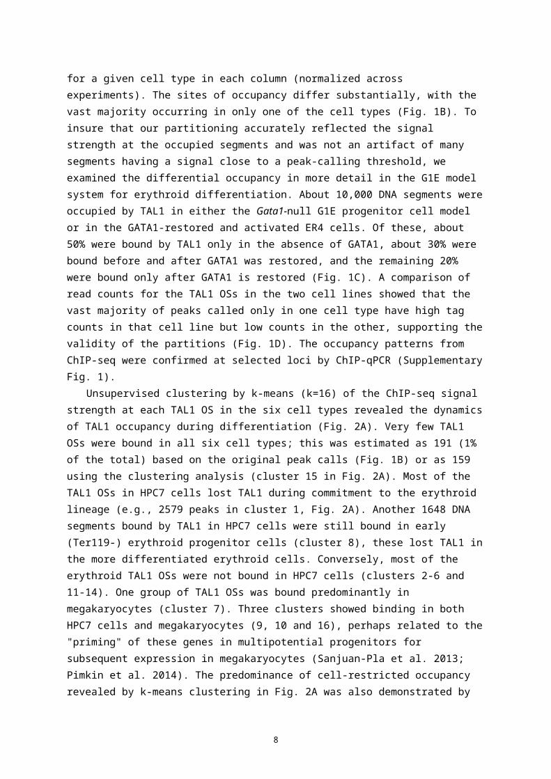
row and the value for the TAL1 ChIP-seq read count for a given cell type in each column (normalized
across experiments). The sites of occupancy differ substantially, with the vast majority occurring in
only one of the cell types (Fig. 1B). To insure that our partitioning accurately reflected the signal
strength at the occupied segments and was not an artifact of many segments having a signal close to
a peak-calling threshold, we examined the differential occupancy in more detail in the G1E model
system for erythroid differentiation. About 10,000 DNA segments were occupied by TAL1 in either
the Gata1-null G1E progenitor cell model or in the GATA1-restored and activated ER4 cells. Of these,
about 50% were bound by TAL1 only in the absence of GATA1, about 30% were bound before and
after GATA1 was restored, and the remaining 20% were bound only after GATA1 is restored (Fig. 1C).
A comparison of read counts for the TAL1 OSs in the two cell lines showed that the vast majority of
peaks called only in one cell type have high tag counts in that cell line but low counts in the other,
6
supporting the validity of the partitions (Fig. 1D). The occupancy patterns from ChIP-seq were
confirmed at selected loci by ChIP-qPCR (Supplementary Fig. 1).
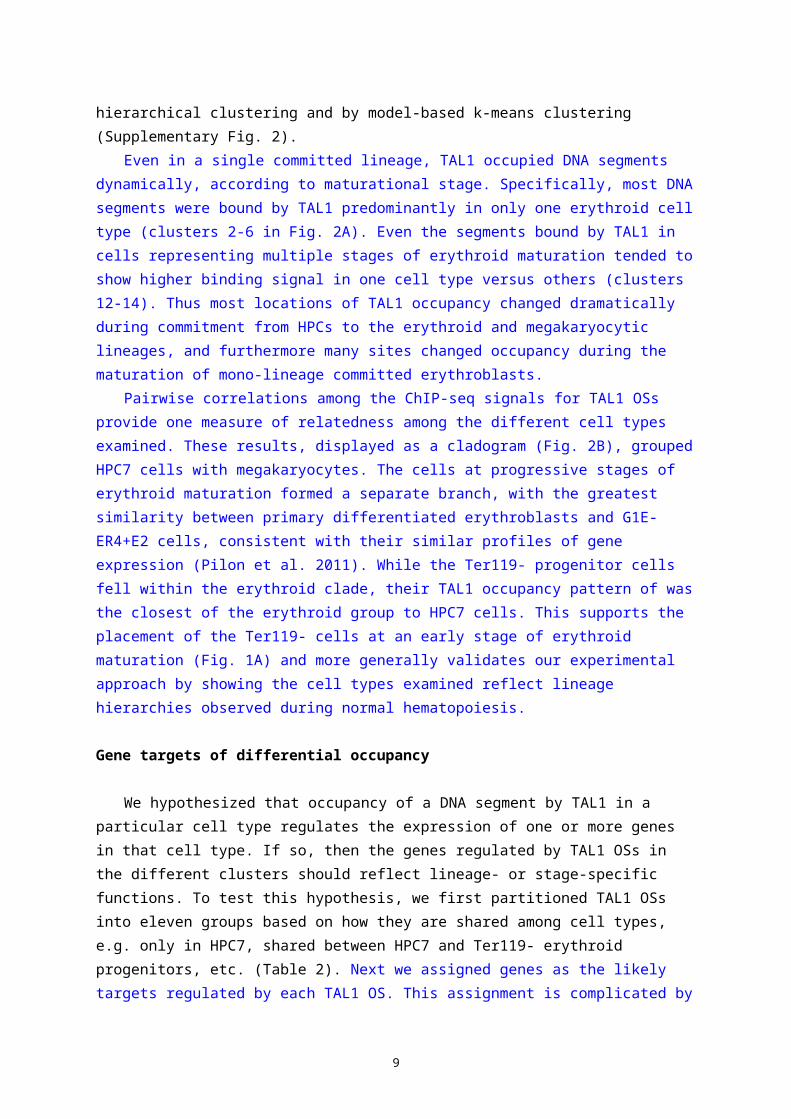
Unsupervised clustering by k-means (k=16) of the ChIP-seq signal strength at each TAL1 OS in
the six cell types revealed the dynamics of TAL1 occupancy during differentiation (Fig. 2A). Very few
TAL1 OSs were bound in all six cell types; this was estimated as 191 (1% of the total) based on the
original peak calls (Fig. 1B) or as 159 using the clustering analysis (cluster 15 in Fig. 2A). Most of the
TAL1 OSs in HPC7 cells lost TAL1 during commitment to the erythroid lineage (e.g., 2579 peaks in
cluster 1, Fig. 2A). Another 1648 DNA segments bound by TAL1 in HPC7 cells were still bound in early
(Ter119-) erythroid progenitor cells (cluster 8), these lost TAL1 in the more differentiated erythroid
cells. Conversely, most of the erythroid TAL1 OSs were not bound in HPC7 cells (clusters 2-6 and 11-
14). One group of TAL1 OSs was bound predominantly in megakaryocytes (cluster 7). Three clusters
showed binding in both HPC7 cells and megakaryocytes (9, 10 and 16), perhaps related to the
"priming" of these genes in multipotential progenitors for subsequent expression in megakaryocytes
(Sanjuan-Pla et al. 2013; Pimkin et al. 2014). The predominance of cell-restricted occupancy revealed
by k-means clustering in Fig. 2A was also demonstrated by hierarchical clustering and by model-
based k-means clustering (Supplementary Fig. 2).
Even in a single committed lineage, TAL1 occupied DNA segments dynamically, according to
maturational stage. Specifically, most DNA segments were bound by TAL1 predominantly in only one
erythroid cell type (clusters 2-6 in Fig. 2A). Even the segments bound by TAL1 in cells representing
multiple stages of erythroid maturation tended to show higher binding signal in one cell type versus
others (clusters 12-14). Thus most locations of TAL1 occupancy changed dramatically during
commitment from HPCs to the erythroid and megakaryocytic lineages, and furthermore many sites
changed occupancy during the maturation of mono-lineage committed erythroblasts.
Pairwise correlations among the ChIP-seq signals for TAL1 OSs provide one measure of
relatedness among the different cell types examined. These results, displayed as a cladogram (Fig.
2B), grouped HPC7 cells with megakaryocytes. The cells at progressive stages of erythroid maturation
formed a separate branch, with the greatest similarity between primary differentiated erythroblasts
and G1E-ER4+E2 cells, consistent with their similar profiles of gene expression (Pilon et al. 2011).
While the Ter119- progenitor cells fell within the erythroid clade, their TAL1 occupancy pattern of
was the closest of the erythroid group to HPC7 cells. This supports the placement of the Ter119- cells
at an early stage of erythroid maturation (Fig. 1A) and more generally validates our experimental
approach by showing the cell types examined reflect lineage hierarchies observed during normal
hematopoiesis.
Gene targets of differential occupancy
We hypothesized that occupancy of a DNA segment by TAL1 in a particular cell type regulates
the expression of one or more genes in that cell type. If so, then the genes regulated by TAL1 OSs in
7
the different clusters should reflect lineage- or stage-specific functions. To test this hypothesis, we
first partitioned TAL1 OSs into eleven groups based on how they are shared among cell types, e.g.
only in HPC7, shared between HPC7 and Ter119- erythroid progenitors, etc. (Table 2). Next we
assigned genes as the likely targets regulated by each TAL1 OS. This assignment is complicated by
two important factors. First, many genes are bound by TAL1 at multiple sites. While each TAL1 OS is
placed into a unique category based on the pattern of occupancy in the cell types, a gene can be
associated with TAL1 OSs in multiple categories. Second, determining the actual target(s) for TF-
bound DNA segments is challenging because the target need not be the closest gene. Nevertheless,
informative correlations between TF binding and expression have been made using simple rules for
assigning targets. We used two methods. For the more inclusive method, we assigned genes as
potential targets of each OS by using mouse enhancer-promoter units (EPUs), which were deduced
by correlating the appearance of predicted enhancers (based on histone modification patterns) with
the expression of genes (Shen et al. 2012). All genes in an EPU were assigned as potential targets of
each TAL1 OS in that EPU. This approach allows genes that are within an expression-correlated
genomic region to be considered as targets, but it can also assign multiple genes as targets an
individual TAL1 OS. In the second method, we assigned the gene with a transcription start site (TSS)
closest to a TAL1 OS as the target, requiring the TSS be within 1 Mb of the TAL1 OS. The assignment
by proximity keeps a single gene as the target for each TAL1 OS, but does not allow skipping of genes
during assignment of targets. While both methods have limitations, we present the results that were
consistent between both approaches. The genes presumptively regulated by TAL1 OSs in each cell-
type partition (based on EPUs) were evaluated using the computational tool GREAT (McLean et al.
2010) for enrichment in functional categories. A selected set of 855 terms representing the common
themes from this analysis, along with enrichment q-values and genes for all the TAL1 OS cell-type
partitions, is provided in Supplementary Table 1. The terms fell into the six major categories shown in
Table 2, which also provides specific examples, q-values, and presumptive target genes by major
category. The results obtained when using proximity of a TSS to assign presumptive gene targets are
given in Supplementary Table 2.
The presumptive gene targets of HPC7-specific TAL1 occupancy were highly enriched for
functions associated with hematopoiesis, proliferation, and apoptosis. Examples of hematopoietic
genes (Table 2) are Kit, encoding the receptor for stem cell factor, and Cbfa2t3, encoding a core-
binding factor whose ortholog in humans is rearranged in some leukemias. Examples of presumptive
target genes associated with proliferation include those encoding growth factors and receptors such
as VEGFA and its receptor FLT1, TGFB and TGFBR1, and CSF1 and CSF1R. Genes encoding proteins in
signaling pathways for proliferation, such as MAP2K3 and MYB, also are preferentially bound by TAL1
in HPC7 cells. Terms associated with apoptosis were also enriched in these presumptive target genes.
Several examples of lineage-specific occupancy of genes in these categories are shown in
Supplementary Figure 3 (Vegfa, Vegfc, Kit, Myb).
Binding of TAL1 in HPC7 cells and other less differentiated cells could participate in lineage
8
priming, i.e. the expression of lineage-specific genes in multilineage progenitors (Mansson et al.
2007; Pina et al. 2012). Genes that are presumptive targets of TAL1 occupancy in HPC7 cells, as well
as in Ter119- progenitors, were highly enriched for functions associated with the differentiated
myeloid cells, perhaps reflecting the maintenance of multiple lineage potentials. The HPC7 cells can
be induced to differentiate into several myeloid cell types such as granulocytes and monocytes (Pinto
do et al. 1998), and terms associated with innate immunity are strongly enriched for the presumptive
targets of TAL1 in these cells (Table 2). This could indicate that HPC7 cells, and by inference
multilineage hematopoietic progenitor cells, maintain expression of some genes characteristic of the
differentiated progeny cells through the binding of transcription factors such as TAL1.
The case for lineage priming by TAL1 is quite strong for megakaryocytic genes. Several pathways,
phenotypes, and Gene Ontology (GO) terms associated with megakaryocytes were enriched for the
sets of genes that are presumptive targets of TAL1 OSs observed in both HPC7 cells and in
megakaryocytes (Table 2). Thus, these genes are bound not only in the mature, differentiated cells
where the gene product participates in platelet specific functions, but also in the multilineage
progenitor cells. This precocious binding in progenitor cells could be mediating early expression of
these genes that are subsequently induced to higher levels in megakaryocytes. An example is the
gene Pf4, encoding the precursor to platelet factor 4, and the adjacent gene Ppbp, encoding platelet
basic protein. Using gene expression data from a population of hematopoietic precursors that are
not lineage committed (Sca1+Lin-), for which HPC7 cells serve as a proxy, and from primary
erythroblasts and cultured megakaryocytes (Pimkin et al. 2014), we found that Pf4 (Fig. 3A) and Ppbp
(not shown) were expressed in multilineage precursor cells and were further induced in
megakaryocytes, but expression levels were lowered in erythroblasts. Similarly, TAL1 was bound near
the transcription start site of Pf4 in HPC7 cells and megakaryocytes. A weak signal for TAL1 was seen
in erythroid progenitors, and this declined further as erythroid maturation progressed (Fig. 3A).
Moreover, in HPC7 cells, this DNA segment was bound by additional transcription factors (GATA2,
ERG and RUNX1) that are part of the heptad containing TAL1 (Wilson et al. 2010); some of these
components were also bound to distal sites. This pattern of TAL1 occupancy is consistent a role in
lineage priming of megakaryocyte-specific genes in multilineage progenitors. Numerous other
megakaryocyte genes, such as Gp1ba and Pdgfb, appear to be primed in HPC7 cells via TAL1
occupancy (Supplementary Figure 4).
In contrast, the presumptive target genes of the TAL1 OSs that are shared among Epro, G1E cells,
ER4 cells, and Ebl were significantly associated with functions and phenotypes that are characteristic
of the erythroid lineage, such as heme biosynthesis, reticulocytosis and erythrocyte physiology (Table
2). This association indicates that the set of TAL1 OSs that are stably occupied through erythroid
maturation is used to maintain the expression of erythroid lineage-specific genes. The fact that these
sites are not typically bound in HPC7 cells indicates that lineage priming is not as prevalent for
erythroid genes as it is for megakaryocytic ones. A striking example of TAL1 occupancy restricted to
the erythroid lineage but present at multiple stages of maturation is the flanking region of Cpox,
9
encoding the heme biosynthetic enzyme coproporphyrinogen-III oxidase. TAL1 binding close to the
transcription start site was observed in committed erythroid progenitors but not in HPC7 cells or
megakaryocytes. Additional sites were bound in cells representing progressively more mature
erythroblasts, where Cpox was highly expressed (Fig. 3B). GATA1 and KLF1 were also bound to the
TAL1 OSs in erythroblasts. These additional sites of binding by TAL1 and other hematopoietic
transcription factors may serve to maintain Cpox expression as most genes become repressed during
the later stages of erythroid maturation. A similar erythroid-specific pattern of occupancy is seen for
many erythroid genes (Table 2); additional examples of Fech and Hbb-b1 are shown in
Supplementary Figure 5.
Often, multiple DNA segments of the same gene were bound by TAL1 and associated proteins
with distinctive patterns of protein occupancy. Consider the gene Cbfa2t3, encoding the protein
CBFA2T3 (also known as ETO2, MTG16, and MTGR2), a co-repressor (Hug and Lazar 2004) implicated
in hematopoietic regulation (Schuh et al. 2005). Cbfa2t3 was bound by TAL1 at a minimum of eleven
sites. Some of these OSs were bound in all cell types examined, while others increased or decreased
progressively during erythroid maturation (Fig. 3C). This diversity of binding patterns indicates a
complex set of regulatory regions that are utilized dynamically during hematopoiesis. Remarkably,
these dynamic changes in occupancy are not accompanied by large changes in expression of Cbfa2t3,
suggesting that distinct sets of TF-bound DNA segments can be utilized in different lineages to
achieve similar levels of expression. Similar complex patterns of occupancy were observed for
multiple genes, including those encoding transcription factors such as RUNX1T1 (CBFA2T1, ETO),
GATA2, and RUNX1 (Supplementary Figure 6). The binding of TAL1 to distinctive sites in different cells
for a given gene means that, in our analysis, the gene was placed in multiple categories of
presumptive targets for TAL1, which contributes to the appearance of functional enrichment terms in
unexpected cell types, such as enrichment for megakaryocytic functions and innate immunity in G1E
cells (Table 2). TAL1 binding in erythroid cells to genes expressed in other lineages suggests that TAL1
may be playing a repressive role in these cases. Indeed, the genes contributing to the enrichment for
megakaryocytic function in targets of TAL1 occupancy in erythroid cells tended to be repressed in
erythroblasts (Supplementary Figure 7). Thus, despite the confounding effects of multiple TAL1 OSs
per gene and multiple targets for some OSs, the analysis by GREAT produced several meaningful
categories of functional enrichments for cell-specific occupancy.
Determinants of differential occupancy: histone modifications
The DNA segments bound by TAL1 reside in chromatin with histone modifications associated
with gene activation, such H3K4 mono- and trimethylation and H3K36 trimethylation, with little or no
signal for the repressive modifications H3K27 trimethylation or H3K9 trimethylation (Fig. 4). This
analysis was conducted for TAL1 OSs in G1E cells, ER4 cells, Ter119+ erythroblasts and
megakaryocytes, for which the histone modification data are available.
10
While no histone modification data are available for the HPC7 cells or Ter119- erythroid
progenitors, we were able to assess whether the DNA segments bound by TAL1 only in HPC7 cells
change to an inactive chromatin state in erythroid cells. The genomes of G1E, ER4, and Ter119+ cells
were segmented based on combinations of histone modification signals using a nine-state model
generated by chromHMM (Ernst and Kellis 2010; Ernst et al. 2011; Wu et al. 2011;
Mouse_ENCODE_Consortium 2014). Each of the 4597 DNA segments bound by TAL1 only in HPC7
cells was then assigned to one of the nine chromatin states in the three erythroid cell types. To
simplify the 27 possible combinations of the nine chromatin states in three cell types, each TAL1 OS
was then placed in a summary category of active chromatin, defined as being in a state enriched for
H3K4me1, H3K4me3, or H3K36m3, or inactive chromatin, defined as being in a state enriched for
H3K27me3, H3K9me3, or no histone modification. We found that about half of the DNA segments
bound by TAL1 only in HPC7 cells remained in an active chromatin state in the erythroid cell types
(Fig. 4B). About 28% of them had the opposite fate, being found in inactive chromatin in all three
erythroid cell types. Another 9% progressed to inactive states across the series G1E > G1E-ER4+E2 >
Ter119+ cells. The remaining 14% fell into a mix of active or inactive states in the three cell types.
Making the likely assumption that the DNA segments were in active chromatin states when bound in
HPC7 cells, these results indicate that almost 40% of the DNA segments that lose occupancy by TAL1
are associated with inactivation of the chromatin.
Determinants of differential occupancy: binding site motifs and binding by other proteins
Given that active chromatin states are preferred for transcription factor binding in all cell types,
we searched for signals that could help determine differential occupancy by TAL1 across cell types.
We hypothesized that the relocation of TAL1 occupancy during differentiation could be driven by
other transcription factors bound to their cognate motifs within the TAL1 OSs, and tested it by
searching for matches to binding site motifs that are distinctive for different cell types.
To find the enriched motifs and their distribution patterns on the different sets of TAL1 binding
sites, we first used MEME-ChIP and related tools (Machanick and Bailey 2011) to generate a list of
known TF binding site motifs enriched in the occupied segments in each cell type (Supplementary
Table 3). Five motifs predominated in the MEME results, corresponding to binding site motifs for
GATA factors, bHLH proteins (E-box), ETS proteins (ETS-box), Runx proteins, and Krüppel-like factors
(KLF) (Fig. 5). We then used FIMO (Grant et al. 2011) to locate all instances of these motifs in each
1kb interval centered on a TAL1 OS. The distribution of motif instances was summarized by a
histogram (Fig. 5A), and more detailed views of the patterns of motif instances across the TAL1 OSs
were generated as dot plots (Ozdemir et al. 2011) (Supplementary Fig. 7). The motif enrichment
score was computed as the logarithm (base 2) of the odds ratio determined by the number of motif
instances in the central 50 bp compared to the number in the rest of the OS, in each case normalized
for the frequency of motif instances in comparably-sized random DNA segments (Fig. 5B).
11

The preferred DNA binding site for a heterodimer of TAL1 with E-proteins in vitro is CAGVTG. A
position-specific weight matrix derived from the MEME analysis matched this consensus, and we
used it to find instances of this particular E-box in the TAL1 OSs. This motif was enriched in binding
sites from all six cell types (Fig. 5B), showing a peak in occurrence at the center of the occupied DNA
segments (Fig. 5A and Supplementary Fig. 7), as expected for a determinant of occupancy.
Surprisingly, the level of enrichment for the TAL1 E-box motif was only about half that observed
for the motif corresponding to a GATA factor binding site, which was the most strongly enriched of
any motif (Fig. 5A, B). This motif showed a substantial preference for the centers of the TAL1 OSs (Fig.
5A and Supplementary Fig. 7). This pattern was observed for the TAL1-bound DNA segments in all six
cell types, suggesting a consistent and frequent co-occupancy of TAL1 and GATA factors. However,
the same GATA factor cannot account for these enrichments, since hematopoietic precursor and
erythroid progenitor cells express GATA2, which is replaced by GATA1 during erythroid
differentiation. Hence we interpret the predominance of the GATA binding site motif in the TAL1 OSs
as reflecting the binding of TAL1 with GATA2 in earlier stages (represented by HPC7, Epro, and G1E
cells) and GATA1 in more differentiated stages (represented by ER4 and Ebl).
The interpretation of the motif enrichment as reflecting co-occupancy was tested by looking for
overlap between the TAL1 OSs in each cell type and occupancy data for other transcription factors.
This analysis strongly supported a role for GATA2 and GATA1 in determining TAL1 occupancy. As
previously reported (Wilson et al. 2010), about 30% of TAL1 OSs in HPC7 cells were also bound by
GATA2, and we found that this fraction of overlap with DNA segments occupied by GATA2 (in HPC7
cells or G1E cells) continued with only a small decline across the erythroid and megakaryocytic cell
types (Fig. 5C). The overlap with GATA2 OSs persisted even for TAL1 occupancy in ER4 cells and
erythroblasts, in which GATA2 is no longer present, suggesting that binding by GATA2 in early
progenitor cells marked some of the sites that would later be occupied by TAL1 in mature cells. Co-
occupancy with TAL1 in erythroid cells was even higher for GATA1, with overlaps ranging from 60% to
almost 90% (Fig. 5C). Specific examples are shown for Pf4, Cpox, and Cbfa2t3 (Fig. 3) and other genes
(Supplementary Figs. 4-6). The “co-occupancy” between GATA1 and TAL1 was observed even in some
cell types that have no (G1E) or low (HPC, Epro) GATA1. In these cases, TAL1 was already bound to a
site to which GATA1 would normally bind later. Presumably these sites were marked by other
proteins, such as GATA2 and appropriately modified histones, to favor binding by TAL1. Thus binding
by GATA factors is likely to be a strong determinant of TAL1 occupancy in all the cell types examined.
Indeed, from the levels of enrichment of motifs, it appears to be a stronger determinant than the
TAL1 E-box.
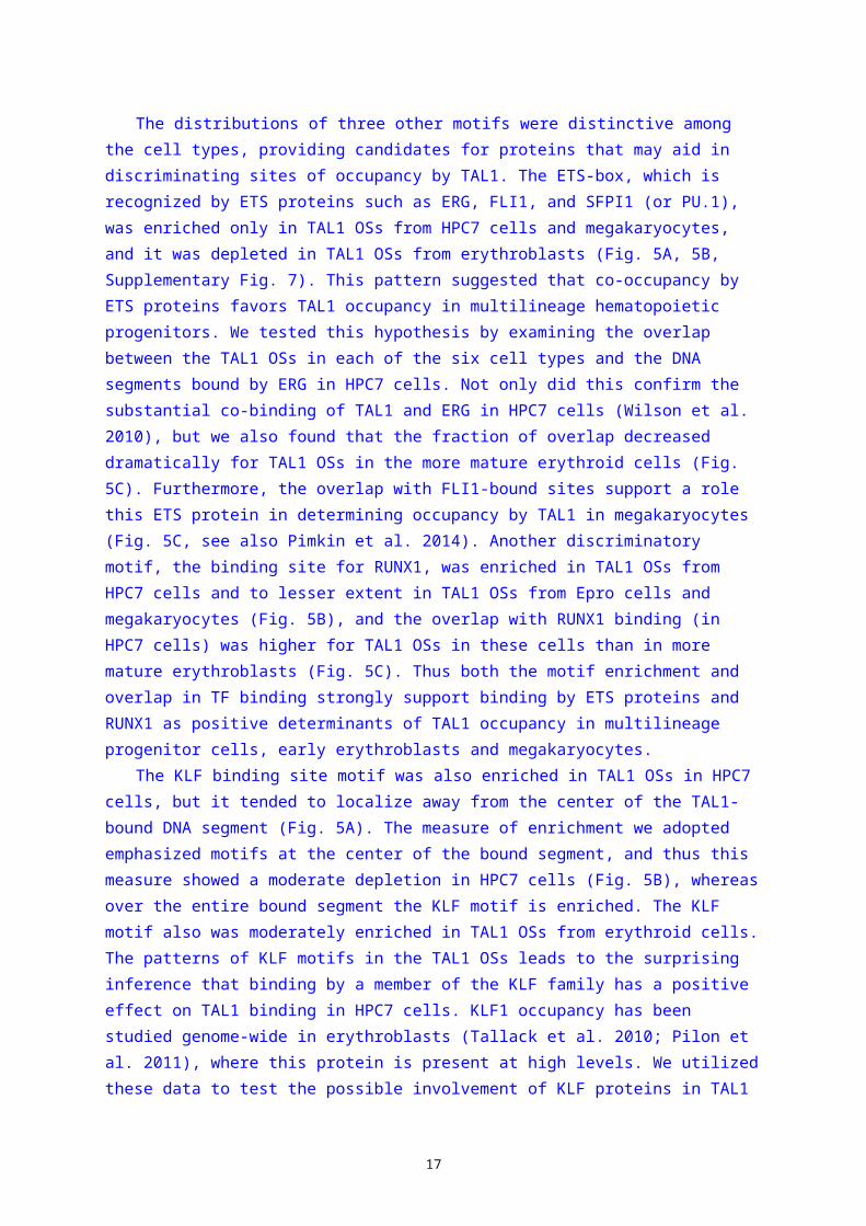
The distributions of three other motifs were distinctive among the cell types, providing
candidates for proteins that may aid in discriminating sites of occupancy by TAL1. The ETS-box, which
is recognized by ETS proteins such as ERG, FLI1, and SFPI1 (or PU.1), was enriched only in TAL1 OSs
from HPC7 cells and megakaryocytes, and it was depleted in TAL1 OSs from erythroblasts (Fig. 5A, 5B,
Supplementary Fig. 7). This pattern suggested that co-occupancy by ETS proteins favors TAL1
12
occupancy in multilineage hematopoietic progenitors. We tested this hypothesis by examining the
overlap between the TAL1 OSs in each of the six cell types and the DNA segments bound by ERG in
HPC7 cells. Not only did this confirm the substantial co-binding of TAL1 and ERG in HPC7 cells (Wilson
et al. 2010), but we also found that the fraction of overlap decreased dramatically for TAL1 OSs in the
more mature erythroid cells (Fig. 5C). Furthermore, the overlap with FLI1-bound sites support a role
this ETS protein in determining occupancy by TAL1 in megakaryocytes (Fig. 5C, see also Pimkin et al.
2014). Another discriminatory motif, the binding site for RUNX1, was enriched in TAL1 OSs from
HPC7 cells and to lesser extent in TAL1 OSs from Epro cells and megakaryocytes (Fig. 5B), and the
overlap with RUNX1 binding (in HPC7 cells) was higher for TAL1 OSs in these cells than in more
mature erythroblasts (Fig. 5C). Thus both the motif enrichment and overlap in TF binding strongly
support binding by ETS proteins and RUNX1 as positive determinants of TAL1 occupancy in
multilineage progenitor cells, early erythroblasts and megakaryocytes.
The KLF binding site motif was also enriched in TAL1 OSs in HPC7 cells, but it tended to localize
away from the center of the TAL1-bound DNA segment (Fig. 5A). The measure of enrichment we
adopted emphasized motifs at the center of the bound segment, and thus this measure showed a
moderate depletion in HPC7 cells (Fig. 5B), whereas over the entire bound segment the KLF motif is
enriched. The KLF motif also was moderately enriched in TAL1 OSs from erythroid cells. The patterns
of KLF motifs in the TAL1 OSs leads to the surprising inference that binding by a member of the KLF
family has a positive effect on TAL1 binding in HPC7 cells. KLF1 occupancy has been studied genome-
wide in erythroblasts (Tallack et al. 2010; Pilon et al. 2011), where this protein is present at high
levels. We utilized these data to test the possible involvement of KLF proteins in TAL1 binding,
assuming that some sites bound by KLF1 in erythroid cells were previously bound by a paralogous
member of the KLF family in progenitor cells. This assumption is analogous to the overlap seen in
GATA1 (erythroid cells) and GATA2 (earlier progenitor cells) binding (Anguita et al. 2004; Bresnick et
al. 2010; Wu et al. 2011; Suzuki et al. 2013). As predicted from the motif enrichment, KLF binding
(Pilon et al. 2011) overlapped TAL1 occupancy in HPC7 cells (Fig. 5C). Thus both lines of evidence
implicate binding by a KLF family member as a contributor to TAL1 occupancy in multilineage
progenitors cells.
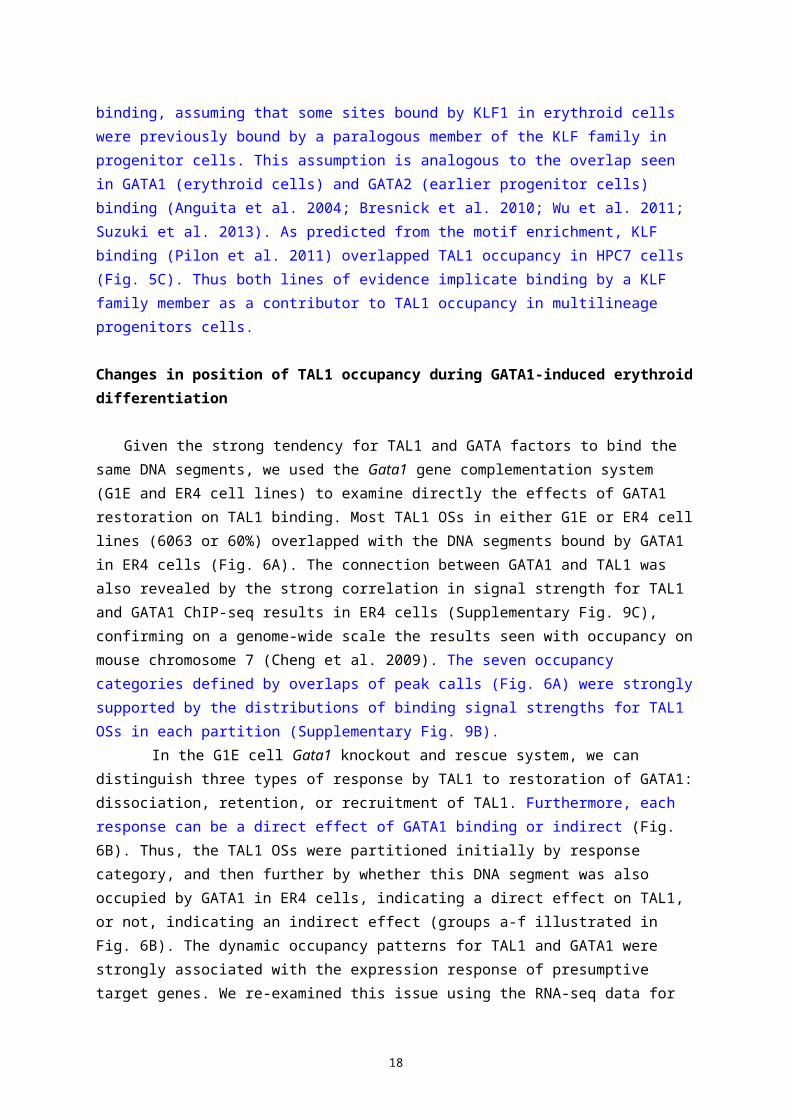
Changes in position of TAL1 occupancy during GATA1-induced erythroid differentiation
Given the strong tendency for TAL1 and GATA factors to bind the same DNA segments, we used
the Gata1 gene complementation system (G1E and ER4 cell lines) to examine directly the effects of
GATA1 restoration on TAL1 binding. Most TAL1 OSs in either G1E or ER4 cell lines (6063 or 60%)
overlapped with the DNA segments bound by GATA1 in ER4 cells (Fig. 6A). The connection between
GATA1 and TAL1 was also revealed by the strong correlation in signal strength for TAL1 and GATA1
ChIP-seq results in ER4 cells (Supplementary Fig. 9C), confirming on a genome-wide scale the results
seen with occupancy on mouse chromosome 7 (Cheng et al. 2009). The seven occupancy categories
13
defined by overlaps of peak calls (Fig. 6A) were strongly supported by the distributions of binding
signal strengths for TAL1 OSs in each partition (Supplementary Fig. 9B).
In the G1E cell Gata1 knockout and rescue system, we can distinguish three types of
response by TAL1 to restoration of GATA1: dissociation, retention, or recruitment of TAL1.
Furthermore, each response can be a direct effect of GATA1 binding or indirect (Fig. 6B). Thus, the
TAL1 OSs were partitioned initially by response category, and then further by whether this DNA
segment was also occupied by GATA1 in ER4 cells, indicating a direct effect on TAL1, or not, indicating
an indirect effect (groups a-f illustrated in Fig. 6B). The dynamic occupancy patterns for TAL1 and
GATA1 were strongly associated with the expression response of presumptive target genes. We re-
examined this issue using the RNA-seq data for G1E and G1E-ER4+E2 cells (Wu et al. 2011; Paralkar et
al. 2014) and employing EPUs for assigning presumptive gene targets. For each TAL1 OS, all genes
with a TSS within the same EPU were considered as potential targets, and the fraction of those genes
that were induced or repressed was computed. The data for all the TAL1 OSs in each response
category were combined to generate an average (shown as the percent of differentially expressed
genes), which allows a comparison of whether the presumptive targets tend to be induced or
repressed (bar plot in Fig. 6B). To assess whether these averages represent enrichment for the
response relative to that of all the presumptive TAL1 targets, a shuffling strategy was employed to
generate the distribution of enrichment (or depletion) values compared to permutations (boxplots in
Fig. 6B).
The genes that are presumptive targets of the 4192 DNA segments co-occupied by GATA1 and
TAL1 in ER4 cells were strongly associated with induction, regardless of the presence or absence of
TAL1 on the DNA segment in G1E cells. Group d contains the 2714 instances of TAL1 retention
whereas group f contains the 1478 cases of direct recruitment in response to GATA1 restoration.
Both groups show more frequent induction than repression, and these results represent enrichment
for induction and depletion for repression (Fig. 6B). In contrast, the presumptive gene targets of the
5021 DNA segments at which GATA1 led to dissociation of TAL1 (i.e. bound by TAL1 in G1E but not
ER4 cells) were enriched for repression and depleted for induction. This was the case whether the
dissociation of TAL1 was inferred to be a direct (1871 TAL1 OSs in group b) or indirect (3150 TAL1 OSs
in group a) effect (Fig. 6B). Two additional occupancy categories were comprised of DNA segments at
which TAL1 was either retained (210 TAL1 OSs in group c) or recruited (457 TAL1 OSs in group e) after
GATA1 restoration, but inferred to be indirect effects because GATA1 was absent at those DNA
segments. The presumptive gene targets of these two categories of TAL1 OSs were depleted for both
induction and repression. These results strongly confirm and extend the positive association of
induction with co-occupancy by GATA1 and TAL1 (Cheng et al. 2009; Tripic et al. 2009; Soler et al.
2010). Furthermore, they show that loss of TAL1 occupancy is negatively associated with induction
but positively associated with repression.
Surprisingly, the sharp differences in dynamics of occupancy by TAL1 and GATA1 in these six
groups of TAL1 OSs were actuated on rather similar distributions of transcription factor binding site
14
motifs. We employed dot-plots to show the positions of all motif instances in each TAL1 OS (1 kb
intervals centered on the peak mid-point). Motifs that contribute strongly to occupancy are expected
to generate dense concentrations of motif instances close to peak centers, and to do so throughout
most of a dataset (Ozdemir et al. 2011). Consistent with recent reports (Kassouf et al. 2010), and the
strong enrichment of GATA motifs seen for the entire set of TAL1 OSs (Fig. 5), we found that the TAL1
OSs that were also bound by GATA1 had a strong enrichment for the GATA binding site motif around
the peak centers (Fig. 7, groups b, d, and f). However, the TAL1 OSs that were not co-occupied by
GATA1 also had a strong enrichment for the GATA binding site motif (Fig. 7, groups a, c, and e). The
canonical TAL1 binding site motif was also enriched toward the peak centers of TAL1 OSs in each
category, albeit considerably less strongly than the GATA binding site motif.
The presence of GATA binding site motifs in the centers of TAL1 OSs that were not bound by
GATA1 suggests that GATA2 could also direct TAL1 binding in G1E cells. GATA2 bound at specific sites
in erythroid progenitor cells can be replaced by GATA1 in maturing erythroblasts; these GATA switch
sites have been implicated in repression during maturation (Jing et al. 2008; Bresnick et al. 2010;
Dore et al. 2012; Suzuki et al. 2013). To examine the role of GATA2 and GATA switch sites in TAL1
occupancy, we examined the approximately 4000 DNA segments bound by GATA2 in G1E cells (a
lower-bound estimate, Wu et al. 2011), and found that 1140 TAL1 OSs overlapped with the GATA2
OSs. These comprised from 2% to 22% of the TAL1 OSs in the categories defined in Fig. 6A
(Supplementary Figure 10). Of these TAL1 OSs that were also bound by GATA2, most were at GATA
switch sites that retained TAL1 after the switch (Fig. 6C). These were part of group d, which was
enriched for induction of the presumptive target genes (Fig. 6B). Another 27% were at switch sites
that lost TAL1 after GATA2 was replaced by GATA1, which is a mechanism for repression (Jing et al.
2008; Bresnick et al. 2010) (group b in Fig. 6B). Some of the GATA2 OSs ascertained in G1E cells were
not bound by GATA1 in ER4 cells; we refer to these as GATA2 loss sites. These were most frequent in
group a, in which TAL1 dissociated upon GATA1 restoration (Fig. 6B, C). Again, the presumptive
targets for this group were enriched for repression.
DISCUSSION
TAL1 is a major regulator at multiple stages of hematopoiesis. Our comparative study of TAL1
occupancy shows that it contributes to the regulatory regimen in markedly different cell types by
binding to different genomic DNA sites at progressive stages of hematopoietic differentiation into
specific lineages and their subsequent maturation. Using data from the mouse ENCODE project, we
analyzed globally the binding of TAL1 and expression profiles in six mouse cell types representing
distinct stages of hematopoiesis. The sites of occupancy changed dramatically during the shift from
HPC7 cells, a model for a multilineage hematopoietic precursor cell, to cells committed to either the
erythroid or megakaryocytic lineage. These changes in sites occupied by TAL1 were linked to
15
alterations in the gene expression profiles of the different cell types. Genes that are likely targets for
regulation by TAL1 in HPC7 cells were enriched in functions associated not only with hematopoiesis
but also with the specific differentiated progeny derived from these multilineage precursors. Thus, a
subset of the sites occupied by TAL1 in multilineage progenitors remains bound in megakaryocytes,
and the presumptive targets genes are expressed at high levels in megakaryocytes. This represents a
clear example of lineage priming that confirms other recent work (Sanjuan-Pla et al. 2013; Pimkin et
al. 2014). In contrast, genes that are likely targets for regulation by TAL1 in cells committed to the
erythroid lineage are enriched for erythroid-specific functions. Furthermore, the sites of occupancy
continue to change during erythroblast maturation, leading to induction of erythroid genes and
repression alternate lineage and progenitor expressed genes. These data support a model in which
TAL1 regulates distinct cohorts of genes in different cell types by binding to different cis-regulatory
modules (Fig. 8).
Features that determine cell-specific binding by TAL1 are becoming defined, but we are still far
from understanding them completely. TAL1 binds to DNA segments that are in permissive chromatin,
i.e. having histone modifications associated with active expression or regulation (Fig. 8), as has been
seen for GATA1 (Zhang et al. 2009) and other TFs (Arvey et al. 2012; Kundaje et al. 2012). We have
proposed that the permissive chromatin states are established no later than lineage commitment
(Wu et al. 2011), based on the very limited changes observed in chromatin states during the
substantial changes in gene expression during maturation after erythroid commitment (Wong et al.
2011; Wu et al. 2011). In our current study, we examined earlier stages of differentiation to infer
some changes in chromatin states during lineage commitment. By assuming that TAL1 binding in
multipotential HPC7 cells is in permissive chromatin (as it is in the daughter lineages), we deduced
that almost 40% of the sites bound by TAL1 only in HPC7 cells (i.e. no longer bound in erythroid cells)
shift to inactive chromatin in erythroid cells. Thus shifting to a nonpermissive chromatin state
accompanies some of changes in TAL1 occupancy that occur during commitment to the erythroid
lineage. Another half of the DNA segments bound by TAL1 only in HPC7 cells show the opposite
trend. They stay in active chromatin in erythroid cells, which may reflect continued occupancy by
other factors or a cellular memory of occupancy.
TAL1 binds to appropriate sites within permissive chromatin largely by its interactions with other
TFs such as GATA factors. While a subset of E-boxes (CAGVTG) contributes to the specific binding by
TAL1 (as a heterodimer with E47 or E12), we find that the GATA binding site motif is a much stronger
determinant of specific occupancy. Previous studies revealed a role for the GATA motif and GATA
factors in binding of TAL1 to some sites (Ono et al. 1998; Cheng et al. 2009; Tripic et al. 2009; Kassouf
et al. 2010), and our current results emphasized that this motif is the dominant one in almost all
categories of TAL1 binding. Genetic rescue experiments showed that the DNA-binding domain was
not required for many TAL1 functions (Porcher et al. 1999). Recent structural analysis demonstrated
that the bHLH domains of TAL1:E47 do not make extensive contacts with the E-box in DNA, and the
interactions with GATA factors, via LMO2, were inferred to be major determinants of specific
16
occupancy (El Omari et al. 2013). These multiple lines of evidence show that TAL1 is directed to most
specific binding sites not through a high affinity interaction with an E-box, but rather by interactions
with other proteins. GATA2 and GATA1 are implicated at many (up to 90%) of the TAL1-bound sites in
normal hematopoietic cells, and GATA3 (Ono et al. 1998) along with ETS1 and RUNX1 (Palii et al.
2011) can direct TAL1 binding when it is aberrantly expressed in T-cell leukemias. Presumably other
TFs facilitate TAL1 binding in various other cell types where it exerts essential functions, such as
neurons (Achim et al. 2013) and endothelial cells (Visvader et al. 1998). While the DNA binding
domain of TAL1 is not needed for its function at some bound sites, at others the DNA binding domain
does interact with the E-box (El Omari et al. 2013). This latter subset of sites could contribute to the
observed enrichment for the canonical TAL1 binding site motif; these may have functions distinct
from those TAL1 OSs without the canonical motif.
The co-binding of TAL1:47 and GATA factors can occur by the binding of large multiprotein
complexes. A pentameric complex comprised of the TAL1:47 heterodimer and GATA1 (or GATA2)
connected by the bridge proteins LMO2 and LDB1 (Wadman et al. 1997) is present in erythroid cells
(Rodriguez et al. 2005). When bound to DNA, this complex can account for much of the TAL1-GATA
factor co-occupancy observed in all the cell types examined, i.e. HPC7 cells (Wilson et al. 2010),
megakaryoblasts (Tijssen et al. 2011; Dore et al. 2012; Pimkin et al. 2014) and erythroblasts (Cheng
et al. 2009; Soler et al. 2010; Wu et al. 2011) (Fig. 8).
The differential occupancy at progressive stages of differentiation and in distinct lineages is
further directed by the binding of additional, lineage-specific TFs. We confirmed that TAL1-bound
sites in HPC7 cells were frequently co-occupied by the ETS proteins ERG and FLI1 along with RUNX1
and GATA2 (Wilson et al. 2010). Our current study suggests that one or more KLF family members
also contribute to the DNA binding specificity of TAL1. FLI1 and RUNX1 continued to co-bind with
TAL1 (along with GATA1 and GATA2) in megakaryocytes (Tijssen et al. 2011; Pimkin et al. 2014). Thus,
binding of the ETS proteins and RUNX1 help direct TAL1 to regulatory sites in multipotential
progenitor cells and in megakaryocytes (Fig. 8). In contrast, ETS proteins and RUNX1 rarely co-
occupied TAL1 OSs in erythroid cells, and in fact the amount of co-occupancy declined with greater
erythroid differentiation. These results are consistent with the critical role of RUNX1 in
hematopoiesis (Okuda et al. 1996; Dowdy et al. 2010) and its down-regulation in erythroid cells
(Lorsbach et al. 2004; North et al. 2004). Other TFs that co-bind frequently with TAL1 in erythroid
cells are members of the pentameric complex. In addition, KLF1 co-binds with TAL1 and GATA1 at a
small number of key erythroid regulatory modules (Tallack et al. 2010; Tallack et al. 2012) (Fig. 8).
The major roles played by GATA factors and ETS proteins in directing binding of TAL1:E47 to
specific sites is reminiscent of observations on the binding of the TFs SMAD and TCF, which are the
targets of the BMP and Wnt signaling pathways, respectively. These two TFs co-occupy DNA
segments bound by lineage master regulators such as GATA1 and C/EBPalpha, thereby directing
SMAD and TCF to target genes (Trompouki et al. 2011). In a similar way, our data support a model of
TAL1 being directed to a distinctive set of binding sites by cell type specific combinations of TFs (Fig.
17
8).
The redistribution of TAL1 binding profoundly affects gene expression during hematopoietic
differentiation, leading to dramatic changes in cell morphology and function. The new genomic sites
to which TAL1 binds with each wave of redistribution place TAL1 in positions to regulate new sets of
target genes. The co-binding master regulatory TFs not only direct TAL1 binding to the appropriate
locations, but they also help to determine the impact of TAL1 on the target genes. In the cases
illustrated in Fig. 8, the impact is usually positive, i.e., associated with gene induction. Enhancement
is associated with co-binding of TAL1 with a GATA factor in all three cell types: TAL1 with GATA2 in
progenitor cells (Jing et al. 2008; Wozniak et al. 2008; Wilson et al. 2010), TAL1 with GATA1 and FLI1
in megakaryocytes (Wang et al. 2002; Tijssen et al. 2011; Dore et al. 2012; Pimkin et al. 2014), and
TAL1 with GATA1 and sometimes KLF1 in erythroid cells (Cheng et al. 2009; Tripic et al. 2009; Soler et
al. 2010; Tallack et al. 2010; Wu et al. 2011). The effects on gene expression are not exerted solely by
the DNA-bound TFs, but rather by additional proteins and complexes recruited, including co-
activators (Blobel et al. 1998) and regulators of chromatin-related processes such as BRD3 (Lamonica
et al. 2011) (Fig. 8). Conversely, TAL1 can play a role in repression of some genes by its association
with co-repressors SIN3A, HDAC1 (Huang and Brandt 2000) or CBFA2T3 (ETO2) (Schuh et al. 2005).
Additional work is needed to discover the features that favor binding of TAL1 with proteins that
promote either positive or negative regulation.
Our current study examined more closely particular aspects of the changes in regulation
associated with the GATA switch, i.e. the replacement of GATA2 by GATA1 during maturation after
erythroid commitment (Bresnick et al. 2010). We confirmed the previously described loss of TAL1 at
many GATA switch sites and the repression of likely targets (Grass et al. 2006; Jing et al. 2008;
Wozniak et al. 2008), but we also saw more frequently a retention of TAL1 after the GATA switch,
which was associated with target gene induction. Another class of sites bound both by GATA2 and
TAL1 simply lost TAL1 after the GATA switch, and this was associated with target gene repression.
Thus the effects of the GATA switch are strongly correlated with the effect on TAL1 at bound sites.
These results re-emphasize the importance of co-binding of GATA1 and TAL1, presumably within the
pentameric complex, in activating erythroid genes.
As is the case for almost all genome-wide mappings of TF occupancy, we found TAL1 occupying a
very large number of genomic DNA segments. The 18,595 OSs in the combined data set were largely
differentially bound in specific cell types, but each cell type still had thousands of bound segments.
Consequently, each predicted target gene was associated with multiple TAL1 OSs, each of which was
placed into categories based on binding across cell types or association with other TFs. These several
multiplicities added complexity to the functional analysis of the predicted gene targets for the
various categories of TAL1 OSs. We adopted two different methods for assigning gene targets to
focus on the more robust results. It is important to realize that virtually every gene associated with
TAL1 occupancy actually has several TAL1 OSs in its vicinity. Some of this multiplicity of binding likely
reflects complex regulatory interactions that insure correct timing and amount of expression, and
18
some may reflect redundancy to achieve more robust regulation. Future work uncovering consistent
trends in these networks of interactions should help illuminate the associated regulatory
mechanisms. On the other hand, we cannot rule out the possibility that some of the binding sites are
non-functional, e.g. they could be DNA segments with favorable motifs located in accessible
chromatin, and TAL1 (or other TFs) not actively engaged in regulation could be bound there. Such
“opportunistic” binding (John et al. 2011; Zhu et al. 2012) may explain some of the TAL1 OSs that
show no obvious effect on presumptive target genes, such as those in categories with depletion
(relative to all TAL1-associated genes) for both induction and repression (Fig. 6B). Future research,
e.g. utilizing higher throughput experimental assessment of function and/or quantitative modeling of
dynamic expression patterns based on genome-wide factor occupancy, should help answer the
question of why there are so many binding sites.
The highly dynamic nature of TAL1 occupancy during hematopoiesis helps explain some
apparent discrepancies in the literature. Studies with mouse erythroid progenitors (Kassouf et al.
2010) and in the G1E model system (Wu et al. 2011) led to the conclusion that TAL1 occupancy
precedes that of GATA1, whereas studies of human CD36+ erythroid precursor cells led to a model of
GATA1 binding before TAL1 (Hu et al. 2011). Clearly, TAL1 binds specifically to many genomic
locations in multilineage precursor and erythroid progenitor cells, before GATA1 is produced
abundantly. Once GATA1 is produced during erythroid differentiation, however, its binding appears to
lead to a redistribution of TAL1 to locations containing GATA1.
METHODS
TF occupancy datasetsTF occupancy was measured by chromatin immunoprecipitation (ChIP) followed by sequencing
of the ChIP DNA on the Illumina Genome Analyzer IIx or HiSeq2000 for a minimum of two biological
replicates. ChIP was done for TAL1 in G1E cells, G1E-ER4+E2 cells, Ter119+ erythroblasts and
megakaryocytes, for GATA1 in G1E-ER4+E2 and Ter119+ erythroblasts, and for GATA2 in G1E and G1E-
ER4+E2, using antibodies sc-12984, sc-265, and sc-9008, respectively, from Santa Cruz Biotechnology
(Wu et al. 2011; Mouse_ENCODE_Consortium 2014; Pimkin et al. 2014). The reads were mapped to
mouse genome assembly mm9 using Bowtie (Langmead et al. 2009). Following the methods
developed by the ENCODE Consortium (Landt et al. 2012), quality metrics were determined for each
individual replicate and reproducibility was estimated by IDR (Li et al. 2011) or other measures.
Mapped reads from the replicates were pooled, and a single set of peaks was called by the program
MACS (Zhang et al. 2008), with thresholds described in Wu et al. (2011) and Pimkin et al. (2014). The
peak sets and signal files of ChIP-seq data in HPC7 and Ter119- cells were downloaded from
publications (Kassouf et al. 2010; Wilson et al. 2010) or from the UCSC Genome Browser (Kent et al.
2002). Certain genomic regions in mm9 were “blacklisted” because they had high signal in the tracks
19
for input DNA that was not from immunoprecipitated chromatin (Pimkin et al. 2014); peaks from any
source falling in the blacklist regions were removed. An annotated list of all the TAL1 OSs is furnished
as Supplementary Table 4.
Transcriptome analysis in G1E and G1E-ER4+E2 cells by RNA-seqTotal RNA was extracted from 5 million to 10 million G1E and G1E-ER4 (treated with estradiol for
30 hr.) cells using Invitrogen’s TRIzol reagent. Subsequent steps including polyA selection,
fragmentation and cDNA synthesis were performed as previously described (Mortazavi et al. 2008),
with two changes to confer strand-specificity (Parkhomchuk et al. 2009). Second-strand synthesis
used dUTP rather than dTTP, followed by digestion of the uracil-containing second-strand cDNA using
uracil D-glycosylase during Illumina library preparation (prior to PCR amplification), thereby
selectively amplifying first-strand cDNA. Libraries were prepared using the Illumina ChIP-seq kit and
were sequenced on the Illumina HiSeq 2000 to obtain 2 x 99 nucleotide paired-end reads. All
samples were determined as biological replicates. RNA-seq reads were mapped using TopHat2 in a
reference-assisted manner (Trapnell et al. 2009; Kim et al. 2013; Paralkar et al. 2014). We used
Cuffdiff2 (Trapnell et al. 2013) to identify differentially expressed genes, using the options dispersion-
method = per-condition, library-type = fr-firststrand, max-bundle-frags = 20000000, min-reps-for-js-
test = 2, -b for bias correction and –M to mask globin transcripts. Transcript abundance levels pooled
across replicates were expressed in terms of log2-transformed FPKMs (Fragments Per Kilobase of
exon model per Million mapped fragments). Genes whose expression level exceeded our threshold
for active expression (log2 FPKM > 3) in both cell types and whose differential expression passed a
threshold of FDR=0.05 were declared as differentially expressed.
Comparison of TAL1 OSs among multiple cell typesThe TAL1 OSs from the six cell types were concatenated and merged. The mean TAL1 ChIP-seq
read counts were calculated for each segment in the merged set. K-means, hierarchical and model-
based clustering were performed on the quantile-normalized values. Heatmaps were generated with
segments sorted by the clustering order. For better visualization, the signals that exceeded the 0.5%
or 99.5% quantile in each cell type were forced to be the same color as the 0.5% or 99.5% quantile,
respectively (Fig. 2A). Pearson correlation was calculated for each pair-wise comparison of the raw
ChIP-seq read counts between each cell type. The correlation matrix was converted into a
dissimilarity matrix by subtracting the coefficients from 1. A hierarchical clustering was performed on
the dissimilarity matrix to give a dendrogram (Fig. 2B).
Functional enrichment for presumptive target genes of TAL1 OSsTo find the genes potentially regulated by each TAL1 OS, we first found the enhancer-promoter
unit (EPU) (Shen et al. 2012) that contained this OS and then identified all the genes whose TSSs
were within the same EPU. We submitted the coordinates of the TSSs to the server for GREAT
20

(McLean et al. 2010) to find the function-related terms enriched for the corresponding genes. We
collected all the terms that GREAT found to be significantly associated with the potentially regulated
gene sets for each category of TAL1 OSs. There are eleven gene sets based on the occupancy
dynamics of their associated TAL1 OSs in the six cell types, plus one set that is not associated with
any TAL1 OSs. Only the terms whose FDR q-values passed 0.05 for both the binomial test and the
hypergeometric test were examined further. In a second approach, the gene with the nearest TSS
was assigned as the likely target gene for each TAL1 OSs. The TSS had to be within 1 Mb of the TAL1
OS to be assigned as a target.
Comparison of enrichment of five TF binding motifs on TAL1 OSs in five hematopoietic cell typesWe used MEME-ChIP (Machanick and Bailey 2011) to find enrichment of known TF binding
motifs in the TAL1 OSs of each cell type. Nucleotide sequences of the OSs were extracted through
Galaxy (Goecks et al. 2010) and the sequences were masked by RepeatMasker (Smit and Green
1999) before being sent to MEME-ChIP. Locations of all occurrences of selected top ranking motifs
were determined with FIMO (p-value set to 1e-3). These selected motifs were also shown to be
overrepresented in a comprehensive TF OS set from previous literature (Wilson et al. 2010). Binding
site motifs (as position weight matrices, or PWMs) for GATA factors, the ETS factor SFPI1 (PU.1),
RUNX1 and KLF factors were downloaded from JASPAR database (Sandelin et al. 2004). In order to
find a strong TAL1 binding E-box motif, we first gathered the set of OSs that are occupied by TAL1 in
G1E but not by either TAL1 or GATA1 in induced G1E-ER4, under the assumption that these should
show enrichment for TAL1 binding directed primarily by its E-box motif, rather than indirect binding
through GATA1 co-occupancy. Analysis of this set of OSs by the program DREME in the MEME-ChIP
suite (Machanick and Bailey 2011) revealed the enriched E-box PWM that we used as the TAL1
binding motif. For each set of OSs, a histogram and a density curve were plotted to show frequency
of motif locations versus the positions relative to the OSs centers. A dot plot was also used to show
all the motif occurrences for each set of OSs that were sorted based on TAL1 occupancy strength
(Ozdemir et al. 2011).
To quantitatively measure the motif enrichment in each panel, we first counted the number
of motif occurrences (a) within 50bp on each side of the center of each OSs and (b) in the remainder
of the OS. Then we counted motif occurrences in comparably sized random DNA segments, again (c)
within 50bp on each side of the center and (d) in the remainder of the OS. Enrichment (or depletion)
was computed as log2 of the odds ratio of normalized counts in the center vs. the remainder of the
OS. Specifically, the odds ratio is [(average of a/average of c)/(average of b/average of d)].
Gene expression responses associated with changes in TAL1 occupancy during GATA1-induced
erythroid maturationCo-location within an EPU was used to assign potential gene targets for each TAL1 OS. Each
gene with a transcription start site (TSS) in the same EPU as the TAL1 OS was assigned as a potential
21

target. The expression response for each gene during maturation induced by restoration and
activation of GATA1 in ER4 cells was determined from the RNA-seq data. Thus each TAL1 OS was
associated with a specific number of genes that are induced, repressed or not responding, and the
percentage of differentially expressed (DE) genes that were induced or repressed was computed.
Data for all the TAL1 OSs in each of the groups defined by overlaps in binding patterns (Fig. 6A) were
combined by computing the mean of the percentages that were induced or repressed. To estimate a
background frequency of induction or repression based on the TAL1 occupancy patterns, the TAL1 OS
group designation (from Fig. 6A) was shuffled within the data matrix, and the (pseudo) average
percentage of differentially expressed target genes that are induced and repressed was computed for
each (pseudo) group. This shuffling and recomputation was repeated 1000 times. The mean of the
(pseudo) averages gave the estimate of the background frequency of induction or repression, and
these are shown as dotted lines in the boxplot of Fig. 6B. We used the results for each of the
shuffling and recomputations to produce a distribution of enrichment or depletion estimates. For
each of the 1000 permutations, the true percentage of DE genes that were induced was divided by
the percentage from the shuffled dataset, and likewise for the percentage of DE genes that were
repressed. The log2 (true percentage/ percentage after shuffling) gives an empirical estimate of the
enrichment or depletion for induction or repression of the presumptive gene targets in each TAL1 OS
category. Since the permutations were done 1000 times, we could evaluate a distribution of these
estimates for each TAL1 OS category.
DATA ACCESS
Data from this lab as part of the mouse ENCODE project are available at Gene Expression Omnibus
(Accession numbers GSE30142 and GSE36029 for transcription factor binding sites and GSE36028 for
histone modifications), the UCSC Genome Browser (tracks ENCODE/PSU on the mouse mm9
assembly), and a customized browser maintained at Penn State (http://main.genome-
browser.bx.psu.edu).
ACKNOWLEDGMENTS
This work was supported by the National Institutes of Health grants R01DK065806 (RCH, MJW, GAB),
U01HL099656 and P30DK090969 (MJW), RC2HG005573 and U54HG006998 (RCH), R01DK54937,
R01DK58044, and R37DK058044 (GAB). This work was supported in part through instrumentation
funded by the National Science Foundation through grant OCI-0821527 (the Penn State CyberSTAR
and BioSTAR computers).
DISCLOSURE DECLARATIONThe authors declare no conflict of interest.
22
23

FIGURE LEGENDS
Figure 1. Erythropoiesis, megakaryopoiesis, and relocation of TAL1 occupancy. (A) The diagram
shows differentiation from hematopoietic precursors to erythroblasts and megakaryocytes, including
the corresponding cell types or lines used in this study. (B) The numbers of merged TAL1 OSs that are
occupied by TAL1 in one to six of the assayed cell types or lines. (C) Venn diagram showing cell-type
specific and shared TAL1 OSs in G1E and G1E-ER4+E2 (ER4) cell lines. (D) Scatter plot showing the
normalized ChIP-seq read counts of TAL1 on the TAL1 OSs in G1E versus ER4 cells. The TAL1 OSs
identified only in G1E, only in ER4, or both are represented by green, pink, or red dots, respectively.
Figure 2. Comparison of TAL1 OSs among multiple cell types. TAL1 peaks were called individually
from ChIP-seq reads in each cell type, and then concatenated and merged into a union set. (A) The
segments were clustered (k-means) based on the TAL1 occupancy signals in the six cell types. The
clusters are dominated by binding in the following cell types: 1, HPC7; 2, Epro (Ter119- erythroid
progenitors); 3 and 4, G1E; 5, ER4; 6, Ebl (Ter119+ erythroblasts); 7, Meg (megakaryocytes); 8, HPC7
+ Epro; 9 and 10, HPC7 + Meg; 11, Epro + ER4 + Ebl; 12-14, all erythroid cells, Epro+G1E+ER4+Ebl; 15,
all six cell types; 16, HPC7+Epro+ER4+Meg. The numbers of genes in each cluster are given in
parentheses. (B) The correlation coefficients of the TAL1 binding signals between cell types were
computed and clustered by hierarchical clustering, shown as a dendrogram.
Figure 3. Loci illustrating shifts in patterns of TAL1 occupancy and co-occupancy by other
transcription factors among the six differentiation stages. Each panel shows on the left the signal
track for each indicated transcription factor binding in the designated cell type. Signal for TAL1
binding is blue, for GATA factors is red, and those for other factors are distinctive. Peak calls for KLF1
in erythroblasts are shown from two different sources; the upper boxes are from Tallack et al. (2010)
and the lower boxes are from Pilon et al. (2011). Direction of transcription for each gene is left to
right. The graphs on the right show levels of expression in hematopoietic stem progenitor cells
(HSPC), which are a proxy for expression in HPC7 cells, erythroblasts and megakaryocytes (Pimkin et
al. 2014). (A) The Pf4 gene (encoding platelet factor 4) illustrates TAL1 binding in megakaryocytes and
in HPC7, not in erythroid cells. (B) The Cpox gene (encoding coproporphyrinogen oxidase) is bound
almost exclusively in erythroid cells. (C) The Cbfa2t3 gene is bound at multiple locations, which show
a diversity of patterns across differentiation.
Figure 4. Histone modifications and chromatin states on TAL1 OSs. (A) Levels of five histone
modification ChIP-seq signals (shown by box plots) in G1E, G1E-ER4+E2, Ter119+ erythroblasts and
megakaryocytes were computed on the DNA segments occupied by TAL1 OSs in each cell type. (B)
Chromatin states were determined using chromHMM (Ernst and Kellis 2010; Ernst et al. 2011) after
learning the model from the five histone modifications in multiple cell types (Cheng et al. 2013). The
24
pie chart shows the number of HPC7-only TAL1 OSs that carry the designated pattern of chromatin
states in the three erythroid cell types.
Figure 5. Comparison of enrichment of transcription factor binding site motifs and co-occupancy on
TAL1 OSs in six hematopoietic cell types. (A) The locations of five motifs (logos at the top of the
panel) on 1kb intervals (only 800bp is displayed here) centered on TAL1 OS peak centers were found
by FIMO in each of the six cell types. The distribution of the locations is plotted by both histograms
(colored bars) and density plots (black curves on top of histograms). (B) Enrichment or depletion of
each motif in the TAL1 OSs for each cell type. (C) Fraction of TAL1 OSs (cell types indicated by color
key) bound by the transcription factor indicated on the x-axis. Cell types for the transcription factor
binding are abbreviated: h=HPC7, g=G1E, r=G1E-ER4+E2, b=erythroblast, p=progenitor cells,
m=megakaryocytes.
Figure 6. Changes of TAL1 occupancy in GATA1-induced erythroid differentiation. (A) Venn diagram
showing TF-occupied DNA segments partitioned by occupancy by TAL1 in G1E and ER4 cells and by
GATA1 in ER4 cells. (B) Expression response of presumptive target genes. Genes were associated with
TAL1 OSs based on their co-localization in EPUs. For each of the six TAL1 occupancy patterns, the
percentages of the differentially expressed (DE) presumptive targets that are induced or repressed
are shown as bar plots. The average percentages obtained after 1000 shufflings of the TAL1 OS
category (groups a-f) within the matrix are shown by the dotted lines in the bar plot (left for
repressed, right for induced). For each of the 1000 permutations, the true percentage of DE genes
was divided by the percentage from the shuffled dataset. The distributions of the log2 (true
percentage/shuffled percentage), shown as box plots, give an estimate of the relative enrichment or
depletion for induction or repression of the presumptive gene targets in each TAL1 OS category. (C)
Role of GATA2 in TAL1 occupancy. For the subset of TAL1 OSs that are also bound by GATA2 in G1E
cells, the percentages that fall into each of the TAL1 OS partitions defined in panel A are plotted. The
DNA segments bound by both TAL1 and GATA2 in groups a, c, and e in panel A are GATA2 loss sites,
whereas those in groups b, d and f are GATA switch sites.
Figure 7. Distribution of GATA and TAL1 binding site motif instances in different categories of
TAL1OSs. The location of DNA sequences matching the binding site motif for GATA factors or TAL1
within 500bp from the center of each OS is indicated by a red dot. TAL1 OSs were separated into six
categories based the cell types in which they are bound and their co-occupancy by GATA1, shown by
diagrams on the left, with green and red disks representing TAL1 and GATA1 occupancy, respectively.
In each panel, the OSs were sorted from left to right by increasing occupancy level of TAL1 in the
corresponding cell line.
Figure 8. Model for differential occupancy by TAL1 in multilineage versus erythroid cells. The
25
groups of proteins that co-occupy DNA in the two cell types are shown, along with cognate binding
site motifs (yellow boxes, identity of each motif is listed at the bottom) along the DNA (long brown
rectangle). The different arrangements of motifs signify the diversity of motifs seen in TAL1-occupied
segments, and emphasize the predominance of the GATA motif. The proteins TAL1:E47, LMO2, LDB1,
and GATA1 form a multiprotein complex (Rodriguez et al. 2005); the other proteins shown are in
proximity when bound to DNA. Additional proteins recognizing the same motif as other members of
the TF family and bound at some sites (e.g. GATA2 and ERG in megakaryocytes) are also shown.
Specific binding by TFs occurs within accessible DNA in chromatin that itself has histone
modifications associated with gene activity (green and yellow circles representing methylation of
lysines on H3 and acetylation of H4). The complex of proteins including TAL1 can exert positive
effects on expression (curved arrow as shown) on recruitment and release of RNA polymerase for
active transcription on induced genes. Other TAL1-containing complexes can exert negative effects
(not shown). Cell-specific binding of TAL1 and associated proteins target different cohorts of genes.
26

Table 1. Mapping DNA segments occupied by TAL1 in different hematopoietic cell types
Cells Model Number of TAL1 peaks
Number of mapped reads
Replicates Reference
HPC7 multilineage hematopoietic
precursor
6,885 19,484,806 no Wilson et al., 2010
Ter119- erythroid cells, from fetal
liver
erythroid progenitor
2,943 4,020,226 no Kassouf et al., 2010
G1E erythroid progenitor
7,945 36,547,806 yes Wu et al., 2011
G1E-ER4+E2, 24 hr differentiating erythroblasts
4,859 19,757,552 yes Wu et al., 2011
Ter119+ erythroid cells, from fetal
liver
differentiated erythroblasts
3,086 224,101,706 yes Wu et al., 2011
Megakaryocytes from fetal liver
Mature megakaryocytes
3,505 299,936,409 yes Pimkin et al., 2014
27
Table 2. Enriched functional categories for presumptive gene targets of TAL1 OSs with distinctive cell type distributions.
Cate-gory
Source
Cell type distribution for TAL1 occupancy
Examples
HPC7 + - - - - - + + - +
Misc
-
Epro - + - - - - + - + + -
G1E - - + - - - - - + + -
ER4 - - - + - - - - + + -
Ebl - - - - + - - - + + -
MEG - - - - - + - + - + -
Term Binomial FDR q-value
Hema-topoie-sis
GO Biological Process
regulation of myeloid cell differentiation
0 1 E-85
Kit, Cbfa2t3, Gata2, Klf10, Zbtb7a
Mouse Pheno-type
decreased hematopoietic cell number
0 0 7 E-291
0 1 E-216
0
abnormal bone marrow cell morphology/development
0 0 0 0 3 E-243
0
Proliferation
MSigDB Pathway
signaling by EGFR 9 E-28
Vegfa, Vegfc, Flt1, Tgfb1, Csf1, Csf1r, Map2k3, Myb,Itga2
Pathway Commons
VEGF and VEGFR signaling network
0 0 0 4 E-184
0
EGF receptor signaling pathway
0 0 0 5 E-184
0
Apoptosis
GO Biological Process
positive regulation of apoptosis
0 0 Casp3, Bax, Icam1, Psmc6Pathway
CommonsTRAIL signaling pathway
0 0 0 8 E-184
0
MSigDB Pathway
apoptosis 0 0 0
Innate immu-nity
GO Biological Process
defense response to bacterium
0 0 0 Plcg1, Nfatc3, Zap70, Il9r, MpoMouse
Pheno-type
abnormal immune cell physiology
0 0 0 0 3 E-226
0
abnormal innate immunity
0 0 5 E-174
0
28
MSigDB Pathway
signaling in immune system
0
Megakaryo-cyte
Mouse Pheno-type
abnormal megakaryocyte morphology
0 0 1 E-78
0 Pdgfb, Fli1, Pf4, Itga2b, Ttc7, Gp1ba
abnormal megakaryocyte progenitor cell morphology
0 0 6 E-88
1 E-76
0
abnormal platelet physiology
5 E-89
MSigDB Pathway
platelet activation triggers
1 E-188
4 E-56
Eryth-roid
GO Molecular Function
oxygen binding 4 E-50
5 E-167
Hbb-b1, Hba-a2, Cpox, Fech, Ppox, Urod, Epb4.2, Slc4a1
Mouse Pheno-type
reticulocytosis 9 E-172
abnormal erythrocyte physiology
5 E-116
BioCyc Pathway
heme biosynthesis 3 E-43
Table notes: TAL1 occupied segments with a pattern other than those in the other columns (e.g. positive in HPC7 and G1E) are combined in the “Misc” category. A set of genes with no TAL1 occupancy was used as a negative control (penultimate column). Categories with no significant enrichment have no entry for the FDR q-value. A value of 0 means the q-value is smaller than the limit computed.
29
REFERENCES
Achim K, Peltopuro P, Lahti L, Tsai HH, Zachariah A, Astrand M, Salminen M, Rowitch D, Partanen J. 2013. The role of Tal2 and Tal1 in the differentiation of midbrain GABAergic neuron precursors. Biology open 2: 990-997.
Anguita E, Hughes J, Heyworth C, Blobel GA, Wood WG, Higgs DR. 2004. Globin gene activation during haemopoiesis is driven by protein complexes nucleated by GATA-1 and GATA-2. Embo J 23: 2841-2852.
Aplan PD, Nakahara K, Orkin SH, Kirsch IR. 1992. The SCL gene product: a positive regulator of erythroid differentiation. EMBO J 11: 4073-4081.
Arvey A, Agius P, Noble WS, Leslie C. 2012. Sequence and chromatin determinants of cell-type-specific transcription factor binding. Genome Res 22: 1723-1734.
Blobel GA, Nakajima T, Eckner R, Montminy M, Orkin SH. 1998. CREB-binding protein cooperates with transcription factor GATA-1 and is required for erythroid differentiation. Proc Natl Acad Sci U S A 95: 2061-2066.
Bresnick EH, Lee HY, Fujiwara T, Johnson KD, Keles S. 2010. GATA switches as developmental drivers. J Biol Chem 285: 31087-31093.
Cheng Y, Ma Z, Kim B-H, Cayting P, Boyle AP, Wu W, Sundaram V, Xing X, Dogan N, Li J et al. 2013. Principles of Regulatory Information Conservation Revealed by Comparing Mouse and Human Transcription Factor Binding Profiles. Nature: submitted.
Cheng Y, Wu W, Kumar SA, Yu D, Deng W, Tripic T, King DC, Chen KB, Zhang Y, Drautz D et al. 2009. Erythroid GATA1 function revealed by genome-wide analysis of transcription factor occupancy, histone modifications, and mRNA expression. Genome Res 19: 2172-2184.
Church GM, Ephrussi A, Gilbert W, Tonegawa S. 1985. Cell-type-specific contacts to immunoglobulin enhancers in nuclei. Nature 313: 798-801.
Davidson EH, Erwin DH. 2006. Gene regulatory networks and the evolution of animal body plans. Science 311: 796-800.
Dore LC, Chlon TM, Brown CD, White KP, Crispino JD. 2012. Chromatin occupancy analysis reveals genome-wide GATA factor switching during hematopoiesis. Blood 119: 3724-3733.
Dowdy CR, Xie R, Frederick D, Hussain S, Zaidi SK, Vradii D, Javed A, Li X, Jones SN, Lian JB et al. 2010. Definitive hematopoiesis requires Runx1 C-terminal-mediated subnuclear targeting and transactivation. Hum Mol Genet 19: 1048-1057.
El Omari K, Hoosdally SJ, Tuladhar K, Karia D, Hall-Ponsele E, Platonova O, Vyas P, Patient R, Porcher C, Mancini EJ. 2013. Structural basis for LMO2-driven recruitment of the SCL:E47bHLH heterodimer to hematopoietic-specific transcriptional targets. Cell reports 4: 135-147.
Elnitski L, Li J, Noguchi CT, Miller W, Hardison R. 2001. A negative cis-element regulates the level of enhancement of hypersensitive site 2 of the beta-globin locus control region. J Biol Chem 276: 6289-6298.
Elnitski L, Miller W, Hardison R. 1997. Conserved E boxes function as part of the enhancer in hypersensitive site 2 of the beta-globin locus control region: Role of basic helix-loop-helix proteins. J Biol Chem 272: 369-378.
Ernst J, Kellis M. 2010. Discovery and characterization of chromatin states for systematic annotation of the human genome. Nat Biotechnol 28: 817-825.
30

Ernst J, Kheradpour P, Mikkelsen TS, Shoresh N, Ward LD, Epstein CB, Zhang X, Wang L, Issner R, Coyne M et al. 2011. Mapping and analysis of chromatin state dynamics in nine human cell types. Nature: 43-49.
Goecks J, Nekrutenko A, Taylor J. 2010. Galaxy: a comprehensive approach for supporting accessible, reproducible, and transparent computational research in the life sciences. Genome Biol 11: R86.
Grant CE, Bailey TL, Noble WS. 2011. FIMO: scanning for occurrences of a given motif. Bioinformatics 27: 1017-1018.
Grass JA, Jing H, Kim SI, Martowicz ML, Pal S, Blobel GA, Bresnick EH. 2006. Distinct functions of dispersed GATA factor complexes at an endogenous gene locus. Mol Cell Biol 26: 7056-7067.
Gregory T, Yu C, Ma A, Orkin SH, Blobel GA, Weiss MJ. 1999. GATA-1 and erythropoietin cooperate to promote erythroid cell survival by regulating bcl-xL expression. Blood 94: 87-96.
Hsu H-L, Huang L, Tsan JT, Funk W, Wright WE, Hu J-S, Kingston RE, Baer R. 1994. Preferred sequences for DNA recognition by the TAL1 helix-loop-helix proteins. Mol Cell Biol 14: 1256-1265.
Hu G, Schones DE, Cui K, Ybarra R, Northrup D, Tang Q, Gattinoni L, Restifo NP, Huang S, Zhao K. 2011. Regulation of nucleosome landscape and transcription factor targeting at tissue-specific enhancers by BRG1. Genome Res 21: 1650-1658.
Huang S, Brandt SJ. 2000. mSin3A regulates murine erythroleukemia cell differentiation through association with the TAL1 (or SCL) transcription factor. Mol Cell Biol 20: 2248-2259.
Huang S, Qiu Y, Stein RW, Brandt SJ. 1999. p300 functions as a transcriptional coactivator for the TAL1/SCL oncoprotein. Oncogene 18: 4958-4967.
Hug BA, Lazar MA. 2004. ETO interacting proteins. Oncogene 23: 4270-4274.Jing H, Vakoc CR, Ying L, Mandat S, Wang H, Zheng X, Blobel GA. 2008. Exchange of GATA factors
mediates transitions in looped chromatin organization at a developmentally regulated gene locus. Molecular Cell 29: 232-242.
John S, Sabo PJ, Thurman RE, Sung MH, Biddie SC, Johnson TA, Hager GL, Stamatoyannopoulos JA. 2011. Chromatin accessibility pre-determines glucocorticoid receptor binding patterns. Nat Genet 43: 264-268.
Johnson DS, Mortazavi A, Myers RM, Wold B. 2007. Genome-wide mapping of in vivo protein-DNA interactions. Science 316: 1497-1502.
Kassouf MT, Chagraoui H, Vyas P, Porcher C. 2008. Differential use of SCL/TAL-1 DNA-binding domain in developmental hematopoiesis. Blood 112: 1056-1067.
Kassouf MT, Hughes JR, Taylor S, McGowan SJ, Soneji S, Green AL, Vyas P, Porcher C. 2010. Genome-wide identification of TAL1's functional targets: insights into its mechanisms of action in primary erythroid cells. Genome Res 20: 1064-1083.
Kent WJ, Sugnet CW, Furey TS, Roskin KM, Pringle TH, Zahler AM, Haussler D. 2002. The human genome browser at UCSC. Genome Res 12: 996-1006.
Kim D, Pertea G, Trapnell C, Pimentel H, Kelley R, Salzberg SL. 2013. TopHat2: accurate alignment of transcriptomes in the presence of insertions, deletions and gene fusions. Genome Biol 14: R36.
Kowalczyk MS, Hughes JR, Garrick D, Lynch MD, Sharpe JA, Sloane-Stanley JA, McGowan SJ, De Gobbi M, Hosseini M, Vernimmen D et al. 2012. Intragenic enhancers act as alternative promoters. Mol Cell 45: 447-458.
Kundaje A, Kyriazopoulou-Panagiotopoulou S, Libbrecht M, Smith CL, Raha D, Winters EE, Johnson
31
SM, Snyder M, Batzoglou S, Sidow A. 2012. Ubiquitous heterogeneity and asymmetry of the chromatin environment at regulatory elements. Genome Res 22: 1735-1747.
Lamonica JM, Deng W, Kadauke S, Campbell AE, Gamsjaeger R, Wang H, Cheng Y, Billin AN, Hardison RC, Mackay JP et al. 2011. Bromodomain protein Brd3 associates with acetylated GATA1 to promote its chromatin occupancy at erythroid target genes. Proc Natl Acad Sci U S A 108: E159-168.
Landt SG, Marinov GK, Kundaje A, Kheradpour P, Pauli F, Batzoglou S, Bernstein BE, Bickel P, Brown JB, Cayting P et al. 2012. ChIP-seq guidelines and practices of the ENCODE and modENCODE consortia. Genome Res 22: 1813-1831.
Langmead B, Trapnell C, Pop M, Salzberg SL. 2009. Ultrafast and memory-efficient alignment of short DNA sequences to the human genome. Genome Biol 10: R25.
Li Q, Brown JB, Huang H, Bickel PJ. 2011. Measuring reproducibility of high-throughput experiments. Annals of Applied Statistics 5: 1752-1779.
Lorsbach RB, Moore J, Ang SO, Sun W, Lenny N, Downing JR. 2004. Role of RUNX1 in adult hematopoiesis: analysis of RUNX1-IRES-GFP knock-in mice reveals differential lineage expression. Blood 103: 2522-2529.
Machanick P, Bailey TL. 2011. MEME-ChIP: motif analysis of large DNA datasets. Bioinformatics 27: 1696-1697.
Mansson R, Hultquist A, Luc S, Yang L, Anderson K, Kharazi S, Al-Hashmi S, Liuba K, Thoren L, Adolfsson J et al. 2007. Molecular evidence for hierarchical transcriptional lineage priming in fetal and adult stem cells and multipotent progenitors. Immunity 26: 407-419.
McLean CY, Bristor D, Hiller M, Clarke SL, Schaar BT, Lowe CB, Wenger AM, Bejerano G. 2010. GREAT improves functional interpretation of cis-regulatory regions. Nat Biotechnol 28: 495-501.
Mortazavi A, Williams BA, McCue K, Schaeffer L, Wold B. 2008. Mapping and quantifying mammalian transcriptomes by RNA-Seq. Nat Methods 5: 621-628.
Mouse_ENCODE_Consortium. 2014. An integrated and comparative encyclopedia of DNA elements in the mouse genome. Nature: submitted.
North TE, Stacy T, Matheny CJ, Speck NA, de Bruijn MF. 2004. Runx1 is expressed in adult mouse hematopoietic stem cells and differentiating myeloid and lymphoid cells, but not in maturing erythroid cells. Stem Cells 22: 158-168.
Okuda T, van Deursen J, Hiebert SW, Grosveld G, Downing JR. 1996. AML1, the target of multiple chromosomal translocations in human leukemia, is essential for normal fetal liver hematopoiesis. Cell 84: 321-330.
Ono Y, Fukuhara N, Yoshie O. 1998. TAL1 and LIM-only proteins synergistically induce retinaldehyde dehydrogenase 2 expression in T-cell acute lymphoblastic leukemia by acting as cofactors for GATA3. Mol Cell Biol 18: 6939-6950.
Ozdemir A, Fisher-Aylor KI, Pepke S, Samanta M, Dunipace L, McCue K, Zeng L, Ogawa N, Wold BJ, Stathopoulos A. 2011. High resolution mapping of Twist to DNA in Drosophila embryos: Efficient functional analysis and evolutionary conservation. Genome Res 21: 566-577.
Palii CG, Perez-Iratxeta C, Yao Z, Cao Y, Dai F, Davison J, Atkins H, Allan D, Dilworth FJ, Gentleman R et al. 2011. Differential genomic targeting of the transcription factor TAL1 in alternate haematopoietic lineages. Embo J 30: 494-509.
Paralkar VR, Mishra T, Luan J, Yao Y, Kossenkov A, Sun D, Konuthula N, An X, Mohandas N, Bodine DM et al. 2014. Species-specific functions for long noncoding RNAs during erythroipoiesis.
32

Blood: submitted.Parkhomchuk D, Borodina T, Amstislavskiy V, Banaru M, Hallen L, Krobitsch S, Lehrach H, Soldatov A.
2009. Transcriptome analysis by strand-specific sequencing of complementary DNA. Nucleic Acids Res 37: e123.
Pilon AM, Ajay SS, Kumar SA, Steiner LA, Cherukuri PF, Wincovitch S, Anderson SM, Mullikin JC, Gallagher PG, Hardison RC et al. 2011. Genome-wide ChIP-Seq reveals a dramatic shift in the binding of the transcription factor erythroid Kruppel-like factor during erythrocyte differentiation. Blood 118: e139-148.
Pimkin M, Kossenkov AV, Mishra T, Morrissey CS, Wu W, Keller CA, Blobel GA, Lee D, Beer MA, Hardison RC et al. 2014. Divergent functions of hematopoietic transcription factors in lineage priming and differentiation during erythro-megakaryopoiesis Genome Res: submitted.
Pina C, Fugazza C, Tipping AJ, Brown J, Soneji S, Teles J, Peterson C, Enver T. 2012. Inferring rules of lineage commitment in haematopoiesis. Nat Cell Biol 14: 287-294.
Pinto do OP, Kolterud A, Carlsson L. 1998. Expression of the LIM-homeobox gene LH2 generates immortalized steel factor-dependent multipotent hematopoietic precursors. Embo J 17: 5744-5756.
Porcher C, Liao EC, Fujiwara Y, Zon LI, Orkin SH. 1999. Specification of hematopoietic and vascular development by the bHLH transcription factor SCL without direct DNA binding. Development 126: 4603-4615.
Porcher C, Swat W, Rockwell K, Fujiwara Y, Alt FW, Orkin SH. 1996. The T cell leukemia oncoprotein SCL/tal-1 is essential for development of all hematopoietic lineages. Cell 86: 47-57.
Robb L, Lyons I, Li R, Hartley L, Kontgen F, Harvey RP, Metcalf D, Begley CG. 1995. Absence of yolk sac hematopoiesis from mice with a targeted disruption of the scl gene. Proc Natl Acad Sci U S A 92: 7075-7079.
Robertson G, Hirst M, Bainbridge M, Bilenky M, Zhao Y, Zeng T, Euskirchen G, Bernier B, Varhol R, Delaney A et al. 2007. Genome-wide profiles of STAT1 DNA association using chromatin immunoprecipitation and massively parallel sequencing. Nat Methods 4: 651-657.
Rodriguez P, Bonte E, Krijgsveld J, Kolodziej KE, Guyot B, Heck AJ, Vyas P, de Boer E, Grosveld F, Strouboulis J. 2005. GATA-1 forms distinct activating and repressive complexes in erythroid cells. EMBO J 24: 2354-2366.
Sandelin A, Alkema W, Engstrom P, Wasserman WW, Lenhard B. 2004. JASPAR: an open-access database for eukaryotic transcription factor binding profiles. Nucleic Acids Res 32 Database issue: D91-D94.
Sanjuan-Pla A, Macaulay IC, Jensen CT, Woll PS, Luis TC, Mead A, Moore S, Carella C, Matsuoka S, Jones TB et al. 2013. Platelet-biased stem cells reside at the apex of the haematopoietic stem-cell hierarchy. Nature 502: 232-236.
Schlaeger TM, Mikkola HK, Gekas C, Helgadottir HB, Orkin SH. 2005. Tie2Cre-mediated gene ablation defines the stem-cell leukemia gene (SCL/tal1)-dependent window during hematopoietic stem-cell development. Blood 105: 3871-3874.
Schuh AH, Tipping AJ, Clark AJ, Hamlett I, Guyot B, Iborra FJ, Rodriguez P, Strouboulis J, Enver T, Vyas P et al. 2005. ETO-2 associates with SCL in erythroid cells and megakaryocytes and provides repressor functions in erythropoiesis. Mol Cell Biol 25: 10235-10250.
Shen Y, Yue F, McCleary DF, Ye Z, Edsall L, Kuan S, Wagner U, Dixon J, Lee L, Lobanenkov VV et al. 2012. A map of the cis-regulatory sequences in the mouse genome. Nature 488: 116-120.
33
Shivdasani RA, Mayer EL, Orkin SH. 1995. Absence of blood formation in mice lacking the T-cell leukaemia oncoprotein tal-1/SCL. Nature 373: 432-434.
Smit A, Green P. 1999. RepeatMasker at http://ftp.genome.washington.edu/RM/RepeatMasker.html.Soler E, Andrieu-Soler C, de Boer E, Bryne JC, Thongjuea S, Stadhouders R, Palstra RJ, Stevens M,
Kockx C, van Ijcken W et al. 2010. The genome-wide dynamics of the binding of Ldb1 complexes during erythroid differentiation. Genes Dev 24: 277-289.
Stamatoyannopoulos JA, Snyder M, Hardison R, Ren B, Gingeras T, Gilbert DM, Groudine M, Bender M, Kaul R, Canfield T et al. 2012. An encyclopedia of mouse DNA elements (Mouse ENCODE). Genome Biol 13: 418.
Suzuki M, Kobayashi-Osaki M, Tsutsumi S, Pan X, Ohmori S, Takai J, Moriguchi T, Ohneda O, Ohneda K, Shimizu R et al. 2013. GATA factor switching from GATA2 to GATA1 contributes to erythroid differentiation. Genes Cells 18: 921-933.
Tallack MR, Magor GW, Dartigues B, Sun L, Huang S, Fittock JM, Fry SV, Glazov EA, Bailey TL, Perkins AC. 2012. Novel roles for KLF1 in erythropoiesis revealed by mRNA-seq. Genome Res 22: 2385-2398.
Tallack MR, Whitington T, Yuen WS, Wainwright EN, Keys JR, Gardiner BB, Nourbakhsh E, Cloonan N, Grimmond SM, Bailey TL et al. 2010. A global role for KLF1 in erythropoiesis revealed by ChIP-seq in primary erythroid cells. Genome Res 20: 1052-1063.
Tijssen MR, Cvejic A, Joshi A, Hannah RL, Ferreira R, Forrai A, Bellissimo DC, Oram SH, Smethurst PA, Wilson NK et al. 2011. Genome-wide analysis of simultaneous GATA1/2, RUNX1, FLI1, and SCL binding in megakaryocytes identifies hematopoietic regulators. Dev Cell 20: 597-609.
Trapnell C, Hendrickson DG, Sauvageau M, Goff L, Rinn JL, Pachter L. 2013. Differential analysis of gene regulation at transcript resolution with RNA-seq. Nat Biotechnol 31: 46-53.
Trapnell C, Pachter L, Salzberg SL. 2009. TopHat: discovering splice junctions with RNA-Seq. Bioinformatics 25: 1105-1111.
Tripic T, Deng W, Cheng Y, Zhang Y, Vakoc CR, Gregory GD, Hardison RC, Blobel GA. 2009. SCL and associated proteins distinguish active from repressive GATA transcription factor complexes. Blood 113: 2191-2201.
Trompouki E, Bowman TV, Lawton LN, Fan ZP, Wu DC, DiBiase A, Martin CS, Cech JN, Sessa AK, Leblanc JL et al. 2011. Lineage regulators direct BMP and Wnt pathways to cell-specific programs during differentiation and regeneration. Cell 147: 577-589.
Visvader JE, Fujiwara Y, Orkin SH. 1998. Unsuspected role for the T-cell leukemia protein SCL/tal-1 in vascular development. Genes Dev 12: 473-479.
Wadman IA, Osada H, Grutz G, Agulnick AD, Westphal H, Forster A, Rabbitts TH. 1997. The LIM-only protein Lmo2 is a bridging molecule assembling an erythroid, DNA-binding complex which includes the TAL1, E47, GATA-1 and Ldb1/NL1 proteins. EMBO J 16: 3145-3157.
Wang X, Crispino J, Letting D, Nakazawa M, Poncz M, Blobel G. 2002. Control of megakaryocyte-specific gene expression by GATA-1 and FOG-1: role of Ets transcription factors. EMBO J 21: 5225-5234.
Weiss MJ, Yu C, Orkin SH. 1997. Erythroid-cell-specific properties of transcription factor GATA-1 revealed by phenotypic rescue of a gene-targeted cell line. Mol Cell Biol 17: 1642-1651.
Welch JJ, Watts JA, Vakoc CR, Yao Y, Wang H, Hardison RC, Blobel GA, Chodosh LA, Weiss MJ. 2004. Global regulation of erythroid gene expression by transcription factor GATA-1. Blood 104: 3136-3147.
34

Wilson NK, Foster SD, Wang X, Knezevic K, Schutte J, Kaimakis P, Chilarska PM, Kinston S, Ouwehand WH, Dzierzak E et al. 2010. Combinatorial transcriptional control in blood stem/progenitor cells: genome-wide analysis of ten major transcriptional regulators. Cell Stem Cell 7: 532-544.
Wilson NK, Miranda-Saavedra D, Kinston S, Bonadies N, Foster SD, Calero-Nieto F, Dawson MA, Donaldson IJ, Dumon S, Frampton J et al. 2009. The transcriptional program controlled by the stem cell leukemia gene Scl/Tal1 during early embryonic hematopoietic development. Blood 113: 5456-5465.
Wong P, Hattangadi SM, Cheng AW, Frampton GM, Young RA, Lodish HF. 2011. Gene induction and repression during terminal erythropoiesis are mediated by distinct epigenetic changes. Blood 118: e128-138.
Wozniak RJ, Keles S, Lugus JJ, Young KH, Boyer ME, Tran TM, Choi K, Bresnick EH. 2008. Molecular hallmarks of endogenous chromatin complexes containing master regulators of hematopoiesis. Mol Cell Biol 28: 6681-6694.
Wu W, Cheng Y, Keller CA, Ernst J, Kumar SA, Mishra T, Morrissey C, Dorman CM, Chen KB, Drautz D et al. 2011. Dynamics of the epigenetic landscape during erythroid differentiation after GATA1 restoration. Genome Res 21: 1659-1671.
Xu J, Shao Z, Glass K, Bauer DE, Pinello L, Van Handel B, Hou S, Stamatoyannopoulos JA, Mikkola HK, Yuan GC et al. 2012. Combinatorial assembly of developmental stage-specific enhancers controls gene expression programs during human erythropoiesis. Dev Cell 23: 796-811.
Zhang Y, Liu T, Meyer CA, Eeckhoute J, Johnson DS, Bernstein BE, Nussbaum C, Myers RM, Brown M, Li W et al. 2008. Model-based analysis of ChIP-Seq (MACS). Genome Biol 9: R137.
Zhang Y, Wu W, Cheng Y, King DC, Harris RS, Taylor J, Chiaromonte F, Hardison RC. 2009. Primary sequence and epigenetic determinants of in vivo occupancy of genomic DNA by GATA1. Nucleic Acids Res 37: 7024-7038.
Zhu BM, Kang K, Yu JH, Chen W, Smith HE, Lee D, Sun HW, Wei L, Hennighausen L. 2012. Genome-wide analyses reveal the extent of opportunistic STAT5 binding that does not yield transcriptional activation of neighboring genes. Nucleic Acids Res 40: 4461-4472.
35