Embed Size (px)
Citation preview
Objectives
Overview of Muscle Tissues
1. Compare and contrast the three basic types of muscle tissue.
2. List four important functions of muscle tissue.
Skeletal Muscle
3. Describe the gross structure of a skeletal muscle.
4. Describe the microscopic structure and functional roles of the myofibrils, sarcoplasmic reticulum, and T tubules of skeletal muscle fibers.
5. Describe the sliding filament model of muscle contraction.
6. Explain how muscle fibers are stimulated to contract by describing events that occur at the neuromuscular junction.
7. Describe how an action potential is generated.
8. Follow the events of excitation-contraction coupling that lead to cross bridge activity.
9. Define motor unit and muscle twitch, and describe the events occurring during the three phases of a muscle twitch.
10. Explain how smooth, graded contractions of a skeletal muscle are produced.
11. Differentiate between isometric and isotonic contractions.
12. Describe three ways in which ATP is regenerated during skeletal muscle contraction.
13. Define EPOC and muscle fatigue. List possible causes of muscle fatigue.
14. Describe factors that influence the force, velocity, and duration of skeletal muscle contraction.
15. Describe three types of skeletal muscle fibers and explain the relative value of each type.
16. Compare and contrast the effects of aerobic and resistance exercise on skeletal muscles and on other body systems.
Smooth Muscle
17. Compare the gross and microscopic anatomy of smooth muscle cells to that of skeletal muscle cells.
18. Compare and contrast the contractile mechanisms and the means of activation of skeletal and smooth muscles.
1
19. Distinguish between unitary and multi unit smooth muscle structurally and functionally.
Developmental Aspects of Muscles
20. Describe embryonic development of muscle tissues and the changes that occur in skeletal muscles with age.
Overview of Muscle Tissues (pp. 276–278; Table 9.1)
2

Muscles are distinguished by their ability to transform chemical energy (____________)
into directed __________________________ energy. In doing so they become capable of
exerting ________________________.
Types of Muscle Tissue
Important Terminology:
Muscle fiber –
“Myo” –
“Sarco” –
The three major types of muscle tissue are:
1. __________________________________________________________________
2. __________________________________________________________________
3. __________________________________________________________________
________________________________ muscle is associated with the bony skeleton and consists of large cells that bear striations and are under voluntary control.
_________________________________muscle occurs only in the heart and consists of small cells that are striated and under involuntary control.
__________________________________muscle is found in the walls of hollow organs and consists of small, elongated cells that are not striated and are under involuntary control.
Special Characteristics of Muscle Tissue
3

The four special characteristics of muscle tissue are:
1. ______________________________________________________
2. ______________________________________________________
3. ______________________________________________________
4. ______________________________________________________
______________________________, or responsiveness, is the ability to receive and respond to a stimulus.
_______________________________ is the ability to contract forcibly when stimulated.
_______________________________ is the ability to be stretched.
_________________________________ is the ability to resume the cells’ original length once stretched.
Muscle Functions
The four special functions that muscles perform are:
1. ______________________________________________________
2. ______________________________________________________
3. ______________________________________________________
4. ______________________________________________________
Muscles produce movement by acting on the bones of the skeleton, pumping blood, or propelling substances throughout hollow organ systems.
Muscles aid in maintaining posture by adjusting the position of the body with respect to gravity.
Muscles stabilize joints by exerting tension around the joint.
4

Muscles generate heat as a function of their cellular metabolic processes.
Muscles enclose and protect internal organs, form valves that regulate passage of substances in the body, control the size of the pupil of the eye, and attach to hair follicles as arrector pili muscles.
Skeletal Muscle (pp. 278–305; Figs. 9.1–9.24; Tables 9.1–9.3)
Gross Anatomy of a Skeletal Muscle
Skeletal muscle =
Nerve and Blood SupplyEach muscle has a nerve and blood supply that allows neural control and ensures adequate nutrient delivery and waste removal.
Connective Tissue SheathsConnective tissue sheaths are found at various structural levels of each muscle:
_______________________________surrounds each muscle fiber, ________________________________
surrounds groups of muscle fibers, and _____________________________________ surrounds whole muscles.
Figure 9.1 Connective tissue sheaths of skeletal muscle: epimysium, perimysium, and endomysium.
Attachments
5

Skeletal muscles span joints and cause movement to occur from the movable attachment (the muscle’s _________________________________) toward the less movable attachment (the muscle’s ________________________________).
Muscle attachments may be ___________________, in which the epimysium fuses with the periosteum or perichondrium; or ____________________________, in which the connective tissue wrappings of the muscle extend into a ropelike or sheetlike structure that attaches to the bone, cartilage, or fascia.
Indirect attachments are the most common because they are durable and are small in size, conserving space across joints.
Microscopic Anatomy of a Skeletal Muscle Fiber
Skeletal muscle fibers are large, cylindrical cells with multiple nuclei beneath the________________________________________, or plasma membrane.
______________________________________, the cytoplasm of a muscle cell, is similar to other types of cells, except it has large amounts of glycosomes, for glycogen storage, and______________________________________, an oxygen binding pigment similar to hemoglobin.
Myofibrils
Myofibrils account for roughly 80% of cellular volume and contain the contractile elements of the muscle cell.
6
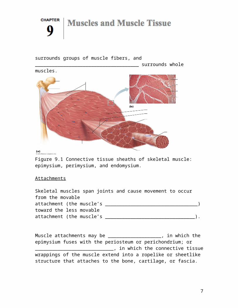
Figure 9.2 Microscopic anatomy of a skeletal muscle fiber.
7

Striations, Sarcomeres, and Myofilaments Striations
8

Striations are due to a repeating series of dark ______ bands and light _______ bands.
___________________________ make up the myofibrils and consist of thick and thin filaments.
Striations, alternating dark A bands and light I bands, extend the length of each myofibril.
Each A band has a lighter central region, the __________ zone, which is bisected vertically by an ___________ line.
Each I band is bisected vertically by a __________ disc, and the region extending from one Z disc to the next forms a ____________________________, the smallest contractile unit of a muscle cell.
There are two types of myofilaments in muscle cells:
1. ______________________ filaments composed of bundles of myosin
2. ______________________filaments composed of strands of actin.
Molecular Composition of Myofilaments
Each __________________________ filament consists of myosin molecules that have a rod-like tail attached to two globular heads that form cross bridges with actin during contraction.
_____________________________ filaments consist of polymerized G actin subunits that have active sites that bind myosin heads during contraction.
Thin filaments also have a set of regulatory proteins: _______________________________, that wrap around actin filaments, stabilizing it and blocking myosin binding sites; and ___________________________, which binds to both actin and tropomyosin, and binds calcium ions.
9

Figure 9.3 Composition of thick and thin filaments.
Sarcoplasmic reticulum and T TubulesThe sarcoplasmic reticulum, a smooth endoplasmic reticulum that regulates the availability of ______________________________ ions, surrounds each myofibril, and forms terminal cisterns at the A band–I band junction.
T tubules are infoldings of the sarcolemma that run between the terminal cisterns, forming triads, that conduct electrical impulses into the cell to cause release of calcium ions from the terminal cisterns.
Sliding Filament Model of Contraction
10

Contraction, according to physiologist is a term that refers to the activation of ___________________________ cross bridges, which are the ______________________________ sites.
Shortening occurs if and when the cross bridges generate enough tension on the thin filaments to exceed forces that oppose ______________________.
Contraction ends when the cross bridges become ________________________, the tension declines, and then the muscle fiber ___________________________.
In a relaxed muscle, the thin and thick filaments _________________________ only at ends of the A band.
Sliding Filament of Model Contraction:
1. During contraction the thin filaments ______________________ past the thick ones so that the _____________________ and ________________________ filaments overlap to a greater degree.
a. When the nervous system stimulates muscle fibers…
________________________________________________________________________________
________________________________________________________________________________
________________________________________________________________________________
b. The cross bridge attachments form…
________________________________________________________________________________
________________________________________________________________________________
________________________________________________________________________________
c. As this event occurs simultaneously…________________________________________________________________________________
________________________________________________________________________________
________________________________________________________________________________
11

2. As a muscle cell shortens: (1) the _____ bands shorten, (2) the distance between successive _____ discs shortens, (3) the _______ zones disappear, and (4) the contiguous _________ bands move closer together but their __________________ does not change.
Figure 9.6 Sliding filament model of contraction
Physiology of Skeletal Muscle Fibers
12

For a skeletal muscle fiber to contract:
1.
2.
3.
4.
13
Figure 9.7 The phases leading to muscle fiber contraction
14

The Nerve Stimulus and Events at the Neuromuscular Junction
Somatic motor neurons =
The _______________________________ junction is a connection between an axon terminal of a somatic motor neuron and a muscle fiber that is the route of electrical stimulation of the muscle cell.
A nerve impulse causes the release of ______________________________________ (ACh) from the axon terminal to the ________________________________ cleft, which binds to receptors on the _____________________________ folds of the muscle cell, triggering a series of electrical events on the sarcolemma.
After acetylcholine binds to ACh receptors, an enzyme in the synaptic cleft, _______________________________________________, breaks down acetylcholine to acetic acid and choline, to prevent continued contraction in the absence of stimulation.
Figure 9.8 Events at the Neuromuscular Junction
15

Generation of an Action Potential Across the Sarcolemma
A resting sarcolemma is _________________________________.
The outside of the membrane is positive and the inside is ______________________________.
An action potential (AP) is the result of a predictable sequence of ____________________ changes. Once initiated they occur along the entire surface of the ___________________________.
Generation of an Action Potential Steps:
1. Generation of an end plate potential.
2. Depolarization: Generation and propagation of an action potential.
16

3. Repolarization: Restoring the sarcolemma to its initial polarized state.
Figure 9.9 Summary of events in the generation and propagation of an action potential in a skeletal muscle fiber.
17

Figure 9.10 Action potential tracing indicates changes in Na+ and K+ ion channels.
Refractory Period =
18

Excitation-Contraction Coupling
Excitation-contraction coupling is the sequence of events by which an action potential on the sarcolemma results in the sliding of the myofilaments,
Summary: Channels Involved in Initiating Muscle Contraction.
1.
19

2.
3.
4.
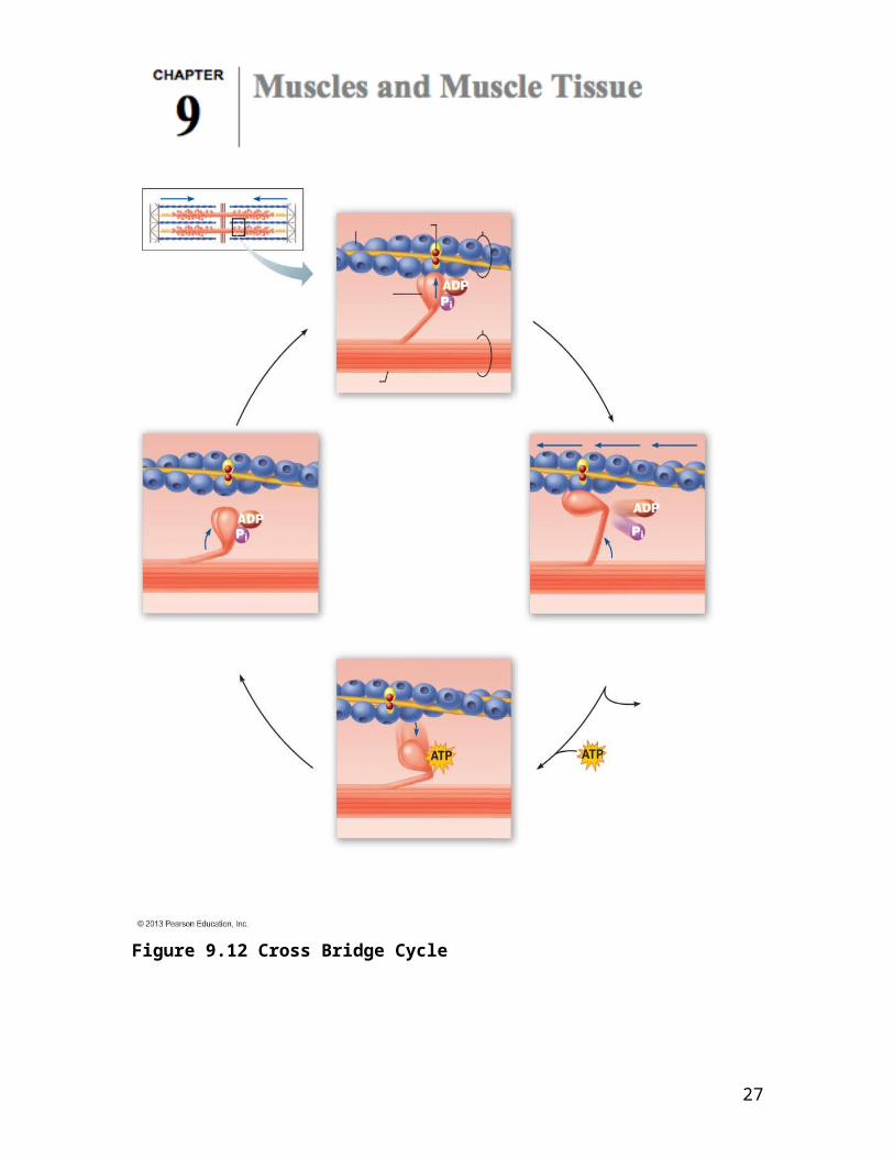
Muscle Fiber Contraction: Cross Bridge Cycling
Cross bridge formation =
As calcium levels in the cytosol increase, calcium binds to troponin, which causes tropomyosin to slide away from the binding sites for myosin on the actin filaments.
Energized myosin heads bind to actin and perform a power stroke, causing actin to slide over myosin.
20

Figure 9.12 Cross Bridge Cycle
21

Contraction of a Skeletal Muscle
Principles of muscle mechanics:
The principles governing contraction of a single muscle fiber and a skeletal muscle consisting of a large number of fibers are pretty much the same.
The force exerted by a contracting muscle on an object is called _______________________________. The opposing force exerted on the muscle by the weight of the object to be moved is called the ____________________________.
A contracting muscle does not always __________________ and move the load.
o If muscle tension develops, but the load is not moved the contraction is called __________________________ (“________________________________”).
o If the muscle tension developed overcomes the load and muscle shortening occurs, the contraction is __________________________ (“________________________________”).
o _____________________ muscle tension is measured for isometric contractions, whereas the ___________________________ of muscle shortening is measured for isotonic contractions.
A skeletal muscle contracts with varying force and for different periods of time in response to stimuli of varying frequencies and intensities.
The Motor Unit
A motor unit consists of a motor _____________________ and all the muscle fibers it innervates.
Muscles that exert fine control (such as those controlling the fingers and eyes) have _________________ motor units.
Large, weight-bearing muscles, whose movements are less precise, have ____________ motor units.
22
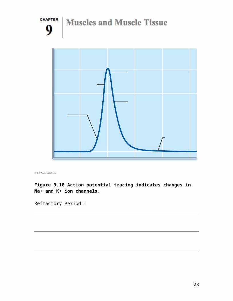
Figure 9.13 A motor unit consists of one motor neuron and all the muscle fibers it innervates.
The Muscle Twitch
The muscle twitch is the response of a muscle to a single action potential on its motor neuron, and has three phases:
Latent period:
Period of contraction:
Period of relaxation:
23

Figure 9.14 The muscle twitch.
24

Graded Muscle Responses
Muscle contractions are smooth and vary in strength, leading to different kinds of graded muscle responses.
Muscle contractions can be graded in two ways:
Changing the frequency of stimulation Changing the strength of stimulation
Muscle Response to Changes in Stimulus Frequency
Wave (Temporal) summation:
Unfused (incomplete) tetanus:
Fused (complete) tetanus:
25

Figure 9.15 A muscle’s response to changes in stimulation frequency.
26
Muscle Response to Changes in Stimuli Strength
Wave summation contributes to contractile force, but its primary function is to produce smooth, continuous muscle contractions by rapidly stimulating a specific number of muscle cells.
Recruitment, also called _________________________________________________ summation, controls the force of contraction more precisely.
Multiple motor unit summation (recruitment) involves the response of a muscle to increasing stimulus voltage: smaller stimuli result in contraction of the smallest motor units, and as voltage increases, larger, more forceful motor units respond, leading to progressively greater contractile force.
Sub threshold stimuli:
Threshold stimuli:
Maximal stimulus:
27

Figure 9.16 Relationship between stimulus intensity (graph at top) and muscle tension (tracing below).
28

Increasing the stimulus intensity beyond the maximal stimulus does not produce a strong contraction. The same phenomenon is caused by neural activation of an increasingly large number of ______________________________serving the muscle.
Size Principle of Recruitment
29

Figure 9.17 The size principle of recruitment
Isotonic and Isometric Contraction
Isotonic contractions produce uniform tension in a muscle, once a load has been overcome, and result in movement occurring at the joint and a change of length of muscles.
Concentric isotonic contractions result when muscle generates force when it shortens, while in eccentric isotonic contractions, the muscle generates force as it lengthens.
Isometric contractions result in increases in muscle tension, but no lengthening or shortening of the muscle occurs, and often are used to maintain posture or joint stability while movement occurs at other joints.
30

Figure 9.18 Isotonic (concentric) and isometric contractions.Muscle ToneMuscle tone is due to reflexive neural stimulation, resulting in muscles exhibiting slight contraction, even when at rest, which keeps muscles firm, healthy, and ready to respond.
Muscle Metabolism
Providing Energy for ContractionMuscles contain very little stored ATP, and consumed ATP is replenished rapidly through phosphorylation by creatine phosphate, anaerobic glycolysis, and aerobic respiration.
Direct Phosphorylation of ADP by Creatine Phosphate:
31

As muscle metabolism transitions to meet higher demand during vigorous exercise, consumed ATP is regenerated by transferring a phosphate to consumed ATP from creatine phosphate, a molecule unique to muscle tissue.
Anaerobic Pathway: Glycolysis and Lactic Acid Formation:As stored ATP and creatine phosphate are consumed, ATP is produced by breaking down blood glucose or stored glycogen in glycolysis, an anaerobic pathway that precedes both aerobic and anaerobic respiration. If adequate oxygen is not available to support aerobic respiration, anaerobic glycolysis converts the pyruvate formed from glycolysis into lactic acid.
This pathway produces only about 5% the ATP from each glucose compared to the aerobic pathway, but ATP production occurs 2½ times faster.
Most of the lactic acid produced is released to the bloodstream and taken to the liver, heart, or kidneys for use, but the lactic acid that remains in the muscle contributes to muscle soreness following exercise.
Aerobic Respiration:Aerobic respiration provides most of the ATP during light to moderate activity, includes glycolysis, along with reactions that occur within the mitochondria, and produces 32 ATP per glucose, as well as water, and CO2, which will be lost from the body in the lungs.
32

Figure 9.19 Pathways for regenerating ATP during muscle activity.
Energy Systems Used During Sports:Muscles function aerobically as long as there is adequate oxygen and nutrient delivery to support it, but when exercise demands for ATP exceed the production ability of aerobic reactions, the cell will switch to anaerobic pathways.
Figure 9.20 Comparison of energy sources used during short-duration exercise and prolonged-duration exercise.
Muscle Fatigue
33
Muscle fatigue is the physiological inability to contract, and results from ionic imbalances that interfere with normal excitation-contraction coupling.
Excess Postexercise Oxygen Consumption (EPOC)Excess postexercise oxygen consumption (EPOC) is the extra oxygen the body requires following exercise to replenish oxygen on myoglobin, reconvert lactic acid to pyruvic acid, replace stored glycogen, and restore ATP and creatine phosphate reserves.
Heat Production During Muscle ActivityMuscle activity produces excess energy that is lost from the body as heat: excess body heat can be lost through sweating and radiant heat loss from skin, while heat production through shivering can be used to warm the body when it is too cold.
Forces of Muscle Contraction
Factors that influence the number of cross bridges that are attached:
1.
2.
3.
4.
Number of Muscle Fibers RecruitedAs the number of muscle fibers stimulated increases, force of contractionincreases.
Size of Muscle FibersLarge muscle fibers generate more force than smaller muscle fibers.
Frequency of StimulationAs the rate of stimulation increases, contractions sum up, ultimately producing tetanus, allowing the external tension generated by the connective tissue elements to approach internal tension generated by the muscle fibers, increasing contractile force.
34

Degree of Muscle StretchThe length-tension relationship optimizes the overlap between the thick andthin filaments that produces optimal contraction.
Figure 9.21 Factors that influence the force of skeletal muscle contraction.
35
Figure 9.22 Length-tension relationships of sarcomeres in skeletal muscles.
Velocity and Duration of Contraction
Muscles vary in how fast they can contract and how long they can continue to contract before they fatigue.
These characteristics are influenced by:
Muscle Fiber Type
Two functional characteristics:
Speed of contraction:
Major pathways for forming ATP:
Skeletal muscle fiber types:
1.
36

2.
3.
Slow oxidative fibers contract slowly, rely mostly on aerobic respiration, and are highly fatigue resistant.
Fast glycolyic fibers contract rapidly, use anaerobic respiration, depend heavily on glycogen, but fatigue quickly.
Fast oxidative fibers are a less common, intermediate type of fiber that provide rapid contraction, but have excellent capillary penetration for oxygen and nutrient delivery, and rely on aerobic respiration.
37

All muscles have varying amounts of all fiber types and, while the proportion of each type is a genetically influenced trait, that proportion can be modified by specific types of exercise.
Load
As the load on a muscle increases, velocity and duration of contraction decreases.
Recruitment
Recruitment of additional motor units increases velocity and duration of contraction.
38
Adaptations to Exercise
Aerobic (Endurance) Exercise
Aerobic exercise promotes an increase in capillary penetration, the number of mitochondria, and synthesis of myoglobin, leading to higher efficiency and endurance, while possibly converting fast glycolytic fibers to fast oxidative fibers.
Resistance Exercise
Resistance exercise, such as weight lifting or isometric exercise, promotes an increase in the number of mitochondria, myofilaments and myofibrils, and glycogen storage, producing hypertrophied cells that may change from fast oxidative to fast glycolytic fibers.
Smooth Muscle (pp. 305–311; Figs. 9.25–9.28; Table 9.3)
Microscopic Structure of Smooth Muscle
Smooth muscle cells are small, _______________________-shaped cells with one central nucleus, and lack the coarse connective tissue coverings of skeletal muscle.
Smooth muscle cells are usually arranged into sheets of opposing fibers, forming a longitudinal layer and a circular layer.
39

Figure 9.25 Arrangement of smooth muscle in the walls of hollow organs.
Contraction of the opposing layers of muscle leads to a rhythmic form of contraction, called _________________________________, which propels substances through the organs.
Smooth muscle lacks neuromuscular junctions, but has ________________________________: numerous bulbous swellings that release neurotransmitters to a wide synaptic cleft.
Figure 9.26 Innervation of smooth muscle
Smooth muscle cells have a less developed sarcoplasmic reticulum, sequestering large amounts of calcium in extracellular fluid within caveolae in the cell membrane.
Smooth muscle has no striations, no sarcomeres, a lower ratio of thick to thin filaments compared with skeletal muscle, and has tropomyosin but no troponin.
Smooth muscle fibers contain longitudinal bundles of noncontractile intermediate filaments anchored to the sarcolemma and surrounding tissues via dense bodies.
40

Figure 9.27 Intermediate filaments and dense bodies of smooth muscle fibers harness the pull generated by myosin cross bridges.
Contraction of Smooth Muscle
Mechanism of ContractionSmooth muscle fibers exhibit slow, synchronized contractions due to electrical coupling by gap junctions.
Like skeletal muscle, actin and myosin interact by the sliding filament mechanism; contraction is triggered by a rise in intracellular calcium level, and the process is energized by ATP.
41

During excitation-contraction coupling, calcium ions enter the cell from the extracellular space, bind to calmodulin, and activate an enzyme, myosin light chain kinase, powering the cross bridging cycle.
Figure 9.28 Sequence of events in excitation-contraction coupling of smooth muscle.
Energy Efficiency of Smooth Muscle ContractionSmooth muscle contracts more slowly and consumes less ATP than skeletal muscle.
Regulation of Contraction
Neural Regulation:Autonomic nerve endings release either acetylcholine or norepinephrine, which may result in excitation of certain groups of smooth muscle cells, and inhibition of others.
42

Hormones and Local Chemical Factors:Hormones and local factors, such as lack of oxygen, histamine, excess carbon dioxide, or low pH, act as signals for contraction.
Special Features of Smooth Muscle Contraction
Response to Stretch:Smooth muscle initially contracts when stretched, but contraction is brief, and then the cells relax to accommodate the stretch.
Length and Tension Changes:Because the muscle filaments have an irregular overlapping pattern, smooth muscle stretches more and generates more tension when stretched than skeletal muscle.
Hyperplasia:Hyperplasia, an increase in cell number through division, is possible in addition to hypertrophy, an increase in individual cell size.
Types of Smooth Muscle
Unitary Smooth MuscleUnitary smooth muscle, called visceral muscle, is the most common type of smooth muscle. It contracts rhythmically as a unit, is electrically coupled by gap junctions, and exhibits spontaneous action potentials.
Multi Unit Smooth MuscleMulti unit smooth muscle is located in large airways to the lungs, large arteries, arrector pili muscles in hair follicles, and the iris of the eye. It consists of cells that are structurally independent of each other, has motor units, and is capable of graded
contractions.
43

44

45

Developmental Aspects of Muscles (pp. 312–313, 315; Fig. 9.29)
Nearly all muscle tissue develops from specialized mesodermal cells called _______________________________.
Skeletal muscle fibers form through the fusion of several myoblasts, and are actively contracting by week 7 of fetal development
Figure 9.29 Myoblasts fuse to form multinucleate skeletal muscle fiber
Myoblasts of cardiac and smooth muscle do not fuse but form gap junctions at a very
early stage.
Muscular development in infants is mostly reflexive at birth, and progresses in a head-to-toe and proximal-to-distal direction.
Women have relatively less muscle mass than men due to the effects of the male sex hormone testosterone, which accounts for the difference in strength between the sexes.
Muscular dystrophy is characterized by atrophy and degeneration of muscle tissue. Enlargement of muscles is due to fat and connective tissue deposit.
46
![[PPT]PowerPoint Presentation - nchu.edu.twweb.nchu.edu.tw/~jodytsao/Makrting2/Expanded PowerPoints... · Web viewObjectives Identify the essential components of a market. Outline](https://img.pdfslide.net/doc/110x75/5aea57997f8b9a66258be8bc/pptpowerpoint-presentation-nchuedutwwebnchuedutwjodytsaomakrting2expanded.jpg)


![[PPT]Introduction to the Oscilloscope - GW Blogsblogs.gwu.edu/ece2110/files/2016/11/Tutorial3_Oscope... · Web viewOBJECTIVES Lab Safety Review Electrical Signals – Quick Overview](https://img.pdfslide.net/doc/110x75/5adb42907f8b9afc0f8d9674/pptintroduction-to-the-oscilloscope-gw-viewobjectives-lab-safety-review-electrical.jpg)