Embed Size (px)
Citation preview
Thesis for doctoral degree (Ph.D.)2008
Gun Faager
Thesis for doctoral degree (Ph.D.) 2008
Gun Faager
Adjuvant strategies in exerciseperformance for patients with
chronic obstructive pulmonarydisease - COPDA
djuvant strategies in
exercise performan
ce for patients w
ith chronic obstru
ctive pulm
onary disease - C
OP
D
1
From Department of Neurobiology, Care sciences and Society, Division of Physiotherapy,
Karolinska Institutet, Stockholm, Sweden
Adjuvant strategies in exercise performance for patients with
chronic obstructive pulmonary disease - COPD
Gun Faager
Stockholm 2008

2008
Gårdsvägen 4, 169 70 Solna
Printed by
2
All previously published papers were reproduced with permission from the publisher. Published by Karolinska Institutet. Printed by [name of printer]
© Gun Faager, 2008ISBN 978-91-7409-177-9

3
When I do not move I feel cured, if I move I feel diseased,
but how can I exist without moving?
Expressed by a patient with COPD

4
5
ABSTRACTChronic obstructive pulmonary disease (COPD) is a multicomponent disease which affects both the lungs and organs outside the lungs. Patients with moderate to severe COPD are restricted by dyspnoea, especially during physical activities. This results in the patient avoiding such activities only to further impair physical capacity and to exert a negative effect on quality of life. It is, therefore, of great importance to establish strategies that can optimise the effect of physical exercise and enhance physical activity among these patients. This thesis is based on four studies that investigate the effects of a physiotherapy programme on patients requiring long-term oxygen therapy (LTOT) as a result of COPD and chronic hypoxia. In addition, it explores the effects of creatine supplementation in combination with physical training and the effects of the oral intake of glucose on arterial oxygen tension, exercise endurance and oxygen consumption in patients with moderate to severe COPD. It also looks at the influence of spontaneous pursed lips breathing (PLB) on oxygen saturation and walking endurance. To evaluate the effects of a physiotherapy programme (n=20) and the combination of creatine supplementation with physical training (n=23) two different walking tests were used. Activity of daily living and health related quality of life were also assessed. To determine whether or not pursed lips breathing influences exercise endurance (n=32), an endurance shuttle walking test and transcutaneous oxygen saturation were conducted. The effect of oral glucose intake (n=13) was evaluated in respect of arterial blood gas analysis, oxygen uptake, carbon dioxide production, ventilation and endurance time on a bicycle ergometer. Patients with chronic obstructive pulmonary disease receiving long-term oxygen treatment may improve their walking distance, experience less dyspnoea and improve their ability to perform daily activities after a physiotherapy programme. Creatine supplementation in combination with physical training showed no significant improvement in physical performance, muscle strength, pulmonary function and health related quality of life in patients with severe to moderate COPD when compared with physical training alone. However, the creatine group showed significant increased walking time after the eight-week training programme. When spontaneous pursed lips breathing was used the patients walked longer, with a significant difference in oxygen saturation in favour of spontaneous PLB. The technique can be useful to increase walking endurance and reduce oxygen desaturation during walking in patients with moderate to severe COPD. Oral intake of glucose may increase the arterial oxygen tension in COPD patients with slight to moderate hypoxia at rest, paralleled with increased blood lactate. When an oral glucose solution is taken before a bicycle exercise test there appears to be no increase in endurance or improved oxygen saturation. On the contrary, glucose intake may be associated with reduced ventilatory reserves and higher ratings of dyspnoea.
Key words: Chronic obstructive pulmonary disease, Long-term oxygen therapy, Physiotherapy, Creatine supplementation, Pursed lips breathing, Arterial oxygen tension, Glucose, Exercise endurance
6
SAMMANFATTNINGKroniskt obstruktiv lungsjukdom (KOL) är en systemsjukdom som leder till konsekvenser i både lungor och andra organ i kroppen. Patienter med måttlig till svår grad av KOL är begränsade av andfåddhet särskilt vid fysisk aktivitet. Till följd av detta undviker patienten fysiska aktiviteter som leder till andfåddhet, vilket i sin tur försämrar patientens fysiska kapacitet och allt sammantaget leder detta till en sänkt hälsorelaterad livskvalitet. Det är därför av stor vikt för patienten att finna strategier som kan optimera effekter av fysisk träning och på detta sätt förbättra förmågan till fysisk aktivitet hos patienter med måttlig till svår KOL. Denna avhandling är baserad på fyra studier och har undersökt effekten av ett sjukgymnastiskt program för patienter med svår KOL och kronisk syrebrist och med kontinuerlig syrgasbehandling i hemmet; effekten av ett tillägg av kreatin kombinerat med fysisk träning för patienter med medelsvår till svår KOL; effekten av spontan sluten läppandning på transkutan syremättnad och gångsträcka och effekten av ett intag av glukos på arteriellt syrgastryck, uthållighet på cykel och syreupptag hos patienter med medelsvår till svår KOL. För att utvärdera effekterna av ett sjukgymnastiskt program (n=20) och kombinationen av kreatinintag och fysisk träning (n=23) gjordes två olika gångtester. Daglig aktivitet och hälsorelaterad livskvalitet mättes också. Påverkan av sluten läppandning eller inte på fysisk uthållighet (n=32) mättes med gångtest och transkutan syremättnad. Effekten av glukosintag (n=13) mättes med arteriella blodgaser, syreupptag, koldioxidproduktion, ventilation och uthållighet på en ergometercykel. Patienter med svår KOL och kontinuerlig syrgas behandling i hemmet kan förbättra sin gångsträcka, minska andfåddhet och förbättra sin dagliga aktivitet. Ett tillägg av kreatin i kombination av fysisk träning ledde inte till någon säkerställd skillnad i fysisk förmåga, muskelstyrka, lungfunktion eller hälsorelaterad livskvalitet jämfört med enbart fysisk träning. Dock, i jämförelsen inom gruppen som fick kreatin så ökade gångtiden signifikant efter åtta veckors träningsprogram. När spontan sluten läppandning kunde användas så ökade patientens gångsträcka och det blev också en säkerställd skillnad i syremättnad till fördel för spontan sluten läppandning. Detta kan vara en användbar andningsteknik vid gång för patienter med medelsvår till svår KOL. Ett intag av glukos kan höja det arteriella syrgastrycket, parallellt med ett förhöjt laktatvärde i blodet hos patienter med lätt till måttlig syrebrist. Slutligen, när patienterna intog en glukoslösning före ett cykeltest kunde ingen ökad uthållighet eller syremättnad visas. I motsats till detta verkar glukosintag ha samband med en minskad ventilatorisk reserv och högre skattad andfåddhet.
Key words: Kroniskt obstruktiv lungsjukdom, kontinuerlig syrgasbehandling, kreatin, sjukgymnastik, sluten läppandning, arteriellt syrgastryck, glukos, fysisk uthållighet
7
LIST OF PUBLICATIONS I Faager G, Larsen F. Performance changes for patients with chronic obstructive
pulmonary disease on long-term oxygen therapy after physiotherapy. J Rehabil Med, 2004; 36:153-8.
II. Faager G, Söderlund K, Rundgren S, Tollbäck A, Sköld CM, Jakobsson P. Creatine supplementation and physical training in patients with COPD. A double blind, placebo-controlled study. Int J of Chron Obstruct Pulmon Dis. 2006;1(4) 445-53.
III. Faager G, Ståhle A, Larsen FF. Effects of Pursed Lips Breathing on walking endurance and oxygen saturation in patients with moderate and severe Chronic Obstructive Pulmonary Disease. Clin Rehabil, 2008; 22(8):675-83
IV. Faager G, Ståhle A, Broman L, Söderlund K, Larsen FF. Increased arterial oxygen tension after oral glucose intake in patients with COPD and moderate hypoxia - influence on oxygen consumption and exercise endurance (Submitted).
8
CONTENTS1 INTRODUCTION.................................................................................... 11
1.1 Chronic Obstructive Pulmonary Disease - COPD.......................... 11 1.2 COPD and intermittent hypoxia ..................................................... 11 1.3 COPD and chronic hypoxia ............................................................ 12 1.4 Peripheral skeletal muscles dysfunction in COPD ......................... 12 1.5 Treatment ........................................................................................ 12 1.6 Pulmonary Rehabilitation ............................................................... 13 1.7 Measure of physical capacity in patients with COPD .................... 13 1.8 Exercise training in COPD ............................................................. 13 1.9 Pursed Lips Breathing..................................................................... 15 1.10 Pharmacological interventions combined with exercise training 15 1.11 Rationale for the thesis ................................................................ 16
2 AIMS ........................................................................................................ 17 3 METHODS............................................................................................... 18
3.1 Patients............................................................................................ 18 3.2 Study design.................................................................................... 19 3.3 Procedure ........................................................................................ 20
3.3.1 Physiotherapy programme (Study I)................................... 20 3.3.2 Pulmonary rehabilitation and creatine supplementation ........ (Study II)............................................................................. 20 3.3.3 Endurance shuttle walking test with and without pursed ...... lips breathing (Study III)..................................................... 21 3.3.4 Endurance bicycle test with or without oral intake of ........... glucose (Study IV).............................................................. 21
3.4 Measurements ................................................................................. 22 3.4.1 Six-minute walking test ...................................................... 22 3.4.2 Incremental shuttle walking test (ISWT)............................ 22 3.4.3 Endurance shuttle walking test (ESWT)............................. 22 3.4.4 Knee muscle strength and fatigue....................................... 23 3.4.5 Grip test............................................................................... 23 3.4.6 Health- related quality of life.............................................. 24 3.4.7 Anxiety and depression....................................................... 24 3.4.8 Activity of daily living ability (ADL)................................. 24 3.4.9 Symptom limited exercise bicycle test ............................... 25 3.4.10 Metabolic stress test............................................................ 25 3.4.11 Spirometry .......................................................................... 25 3.4.12 Blood analyses .................................................................... 26 3.4.13 Transcutaneous oxygen saturation and respiratory rate...... 26 3.4.14 Perceived dyspnoea, leg fatigue and exertion..................... 26 3.4.15 Statistical methods .............................................................. 27 3.4.16 Ethical approval .................................................................. 27
4 RESULTS................................................................................................. 28 4.1 Study I............................................................................................. 28 4.2 Study II ........................................................................................... 29 4.3 Study III .......................................................................................... 32
9
4.4 Study IV ..........................................................................................36 5 DISCUSSION...........................................................................................39
5.1 General discussion...........................................................................39 5.2 Exercise testing in patients with COPD ..........................................40
5.2.1 Walking tests .......................................................................40 5.2.2 Symptom limited exercise test on a bicycle ........................41 5.2.3 Muscle strength and fatigue ................................................42
5.3 Health related quality of life............................................................42 5.4 Pulse oximetry.................................................................................43 5.5 Results .............................................................................................43 5.6 Future studies...................................................................................47
6 CONCLUSIONS.......................................................................................48 7 Acknowledgements...................................................................................49 8 References.................................................................................................52
10
LIST OF ABBREVIATIONS
A-a diff ADL ATP BE COPD CrP CR-10 CRDQ ECG ESWT FEV 1.0
GOLD H2O cm HADHAQHRQL ISWT kPa LTOT NPPV PAO2
PaO2
PaCO2
PEF PEP PLB RPE RM RQ SaO2
SD SGRQ SpO2
Stb VC VE
VT
VCO2
VO2
WWHO
Calculated alveolar-arterial oxygen difference Activity of daily living Adenosine triphosphate Base excess Chronic obstructive pulmonary disease Creatine phosphate Category ratio scale Chronic respiratory disease questionnaire Electrocardiogram Endurance shuttle walking test Forced expired volume during one second Global initiative for chronic obstructive lung disease water pressure Hospital anxiety and depression scale Stanford health assessment questionnaire Health related quality of life Incremental shuttle walking test Kilo Pascal Long-term oxygen therapy Non-invasive positive-pressure ventilation Estimated alveolar oxygen tension Arterial oxygen tension Arterial carbon dioxide tension Peak expiratory flow Positive expiratory pressure Pursed lips breathing Ratings of perceived exertion Repetition maximum Respiratory exchange ratio Arterial oxygen saturation Standard deviation St Georges respiratory questionnaire Transcutaneous oxygen tension Standard bicarbonate Vital capacity Minute ventilation Tidal volume Carbon dioxide production Oxygen uptake Watt World health organisation
11
1 INTRODUCTION Chronic obstructive pulmonary disease (COPD) is a condition with increasing
prevalence, closely associated with smoking and air pollution. According to the WHO´s
Global Burden of Disease Study, COPD will be in the third place as a cause of death by
2020 (1, 2).
1.1 CHRONIC OBSTRUCTIVE PULMONARY DISEASE - COPD Chronic bronchitis, bronchiolitis and emphysema of varying degrees are included in the
condition, which gradually impairs pulmonary function. The clinical picture is one of
productive cough, dyspnoea, general fatigue and for some, weight loss. With
progression and time, respiratory tract infections will be associated with brief
respiratory insufficiency which may become permanent later on (3). COPD is a
multicomponent disease and will consequently affect not only the lungs, but also
organs outside the lungs. The most common systemic effects of COPD are skeletal
muscle dysfunction, weight loss, cardiovascular and nervous system abnormalities and
osteoporosis (4).
Patients with COPD are limited by dyspnoea, especially during physical activities. As a
result the patient avoids physical activities that induce dyspnoea, thus further impairing
his/her physical capacity (5).
Progression of COPD is associated with increased dyspnoea, decreased physical
performance, skeletal muscular weakness and high hospitalisation rates - all with a
negative impact on quality of life (6).
1.2 COPD AND INTERMITTENT HYPOXIA Oxygen desaturation is often observed during walking tests in patients with COPD or
during other exercise tests (7). It has been described that hypoxemia is associated with
reduction in oxygen delivery from blood to muscles in patients with moderate to severe
COPD and it worsens during exercise (8). In combination with other multiple
mechanisms of dyspnoea, hypoxia reduces the daily physical activity for persons with
COPD (9).
Intermittent hypoxia during sleep and sleep disturbance in persons with COPD are
common and an important condition to treat, as for other groups of patients (10).
Hypoxia is probably one of the most important factors for developing pulmonary
hypertension in patients with COPD (11).
12
1.3 COPD AND CHRONIC HYPOXIA Brief respiratory insufficiency may become permanent in some patients (12).
Respiratory insufficiency is defined as arterial oxygen tension (PaO2 )< 8 kPa with or
without arterial carbon dioxide tension (PaCO2)> 6,7 kPa during air breathing. The
prognosis is worse when persons with COPD have developed chronic hypoxia and
hypercapnia (13).
1.4 PERIPHERAL SKELETAL MUSCLES DYSFUNCTION IN COPD Peripheral muscle strength has been shown to decrease in individuals with COPD (14).
The weakness seems to be more concentrated in arm and leg muscles than in other
muscle groups (15). Patients with COPD have been shown to have impaired skeletal
muscle endurance during physical activity and Huges et al (16) demonstrated that
patients with moderate COPD suffered type II fibre atrophy in quadriceps muscles and
experienced a decrease in body weight. Another study showed that patients with severe
COPD had an increased proportion of type II b fibres in their quadriceps, while type I
fibres were reduced (17).
An early onset of lactate production during submaximal physical activity in persons
with moderate to severe COPD leads to decreased pH in the muscles which may
contribute to earlier exhaustion during physical activity (18, 19).
Patients with COPD have a lower content of high energetic phosphates in their skeletal
and respiratory muscles than healthy individuals (20). The concentrations of adenosine
triphosphate (ATP) and creatine phosphate (CrP), lactate and glycogen in the
quadriceps are related to arterial blood gases - the lower the arterial PaO2 and the higher
the arterial PaCO2, the lower the energy status (17).
1.5 TREATMENT Several guidelines present recommendations for the standard care of patients with
COPD, including non-pharmacological treatment such as pulmonary rehabilitation and
non-invasive ventilation and pharmacological treatment such as like short/long-acting
inhaled bronchodilators, inhaled/oral steroids and supplemental oxygen (21-23). Long-
term oxygen therapy (LTOT) has been proven to prolong survival but, in order to be
successful, oxygen must be administrated at least 16 hours/day (24).
13
1.6 PULMONARY REHABILITATION It is of great importance that therapy for patients with COPD treats not only the
symptoms experienced by the lungs, such as dyspnoea, but also the multi-component
nature of the disease (4). Rehabilitation forms an important component of the
management of patients with COPD and includes education in respiratory training,
physical training, nutrition, energy-saving techniques and psychosocial support.
Several meta-analysis and review articles conclude that rehabilitation relieves dyspnoea
and fatigue, improves emotional function and enhances patients' sense of control over
their condition (25-27).
1.7 MEASURE OF PHYSICAL CAPACITY IN PATIENTS WITH COPD Measures to detect changes in physical capacity for patients with COPD are often based
on walking tests on the floor or the treadmill (23, 28).
Exercise training and self-management education are core components of pulmonary
rehabilitation and have been shown to be beneficial in improving health-related quality
of life (HRQL) in patients with chronic respiratory disease (25) . Exercise training can
also help to reduce the deconditioning of the muscles that follows when a patient’s
physical activity is restricted by his/her dyspnoea and fatigue. This is often associated
with an increase in the patient’s HRQL. As Health Related Quality of Life is closely
linked to ability to perform physical activity in patients with COPD, this demonstrates
how important activity is to human beings. One way to illustrate the effects of physical
training may therefore be to measure HRQL.
As far as dynamic muscle strength is concerned, ordinary measuring techniques like a
grip test or determining one repetition of maximum (1 RM) can be used in this group of
patients. For elderly persons with COPD there are recommendations to modify 1 RM to
6 RM for example. If the patient can perform 6 RM with heavier weights after an
exercise programme, this shows improvements (29).
1.8 EXERCISE TRAINING IN COPD Patients with COPD gradually suffer lower physical capacity and muscle strength (30).
Exercise training is, therefore, a cornerstone in pulmonary rehabilitation and has been
shown to decrease dyspnoea, increase physical capacity, muscle strength and health-
related quality of life (22, 31).
The optimal duration of exercise training has not yet been defined (28) but programmes
lasting from four to twelve weeks have shown improvements (32, 33). However, the
14
more sessions the programme includes and the longer the duration of the programme,
the better the improvements (34-38). To achieve optimal physiological effects from a
training programme, studies have shown that patients must exercise more than one, and
preferably three sessions, per week and that it is possible to combine supervised and
unsupervised sessions to attain improvements (39-43). Research shows that the higher
the intensity of the training programmes, the higher the physiological effects, but
disease severity and motivation must also be taken into account (44). Patients with
COPD cannot be exposed to the same training intensity that works for healthy adults as
they are already limited in respect of respiration capacity before reaching the anaerobic
threshold. An intensity that achieves approximately 60 percent of peak exercise
capacity is therefore recommended (45, 46). Clinical symptom scores can be used to
adjust the workload and an estimated dyspnoea in the range of “4 to 6” according to
Borg’s Category Ratio Scale (CR-10) (47)during exercise is recommended (47-50).
In order to be effective, training programmes must include both endurance and strength
training, but walking or cycling are still the most used forms of exercise training among
this group of patients (46, 51, 52).
The optimal duration of training is reported to be at least 30 minutes, but not all patients
with COPD can achieve this duration (31, 53). Interval training may, therefore, be an
alternative to endurance training (41, 54, 55).
Resistance or strength training is reported to be valuable for patients with COPD as it
improves muscle mass and power (56-59). The recommended number of repetitions per
session are two to four sets of 6 to 12 repetitions at an intensity of 50 to 85 percent of
one repetition maximum (1 RM), i.e. the heaviest weight that can be lifted once with a
good technique (56). Strength training is often better tolerated with regard to dyspnoea
and therefore an alternative for patients who do not tolerate aerobic training (60).
Clinical guidelines mostly recommend a combination of endurance and strength
training as this is aimed to increase both muscle strength and whole body endurance
(28, 58, 61).
Patients with COPD and LTOT are advised to use their prescribed dose of oxygen flow
during exercise and to increase the flow if needed (28). Studies in exercise training with
supplemental oxygen amongst patients with COPD with or without exercise induced
hypoxemia have shown different results (62-64). One study of non-hypoxemic patients
with COPD showed higher exercise intensity and increased exercise capacity with
oxygen supplementation than without and another study showed no improvement in
exercise capacity with or without oxygen supplementation (65, 66). No current clinical
15
guidelines recommend oxygen supplementation as it is unclear if this has any positive
clinical outcome (22, 28, 37).
Non-invasive positive pressure ventilation (NPPV), which has been shown to increase
minute ventilation and tidal volume and to reduce inspiratory effort and the sensation of
dyspnoea in addition to reducing respiratory acidosis, can assist during exercise (67-
70). NPPV is, however, not an easy intervention and present clinical guidelines
recommend it only for patients who really enjoy a true benefit from non-invasive
positive pressure ventilation (28).
It is evident from some studies among patients with decreased inspiratory muscle
strength that inspiratory muscle training as an adjunct to exercise training improves
inspiratory muscle strength and exercise capacity more than exercise alone (28, 71, 72).
Despite divergent study results in this field, it is recommended in practice guidelines
for patients with respiratory muscle weakness (28).
1.9 PURSED LIPS BREATHING Pursed Lips Breathing (PLB), one of several breathing strategies, is often used
spontaneously by patients with COPD. The technique prevents dynamic airway
compression and has been shown to increase tidal volume, decrease respiratory rate,
increase oxygen tension and saturation, and decrease carbon dioxide level at rest (73-
76).
Spahija et al (77) studied the effects of PLB on respiratory mechanics and dyspnoea
during exercise and concluded that PLB has a variable effect on dyspnoea during
exercise. Even if the technique has not yet been proven to increase exercise capacity, it
is listed in most pulmonary rehabilitation programmes, and patients are instructed on
the use of PLB technique at rest, during physical activity and in situations that may
cause panic.
1.10 PHARMACOLOGICAL INTERVENTIONS COMBINED WITH EXERCISE TRAINING
Pharmacological supplementation such as anabolic steroids and creatine
supplementation may have an addictive effect on exercise training in patients with
COPD (78-82). Some studies using anabolic steroids show positive results in respect of
increased fat mass and fat-free mass, which may improve the outcome of exercise
training. Among athletes, oral supplementation with creatine has been associated with
an increase in exercise capacity during brief periods of physical training (83).
16
Moreover, creatine supplementation has been shown to increase muscular strength and
volume. However, recent reports have shown that if combined with physical training,
creatine supplementation does not have an addictive effect on exercise capacity in
patients with COPD (80, 84). Nevertheless, existing guidelines inform that there are
ongoing studies that investigate safety and indications of anabolic and nutritional
treatments (28).
1.11 RATIONALE FOR THE THESIS COPD is a chronic and progressive disease strongly associated with dyspnoea and
exercise limitation in activities of daily life. So far there is no cure for the disease, only
efforts to alleviate the symptoms. It is, therefore, of great importance to define effective
training models and breathing techniques to relieve symptoms and to motivate patients
to exercise. It is also crucial to optimise different training models and to investigate
what can really help this group of patients to increase their level of physical activity,
capacity and quality of life.
17
2 AIMS The general aim of this thesis was to study the effects of different strategies to enhance
physical activity in patients with moderate to severe chronic obstructive pulmonary
disease (COPD).
The specific aims were
To evaluate if a special eight-week physiotherapeutic training programme
improved walking distance, health related quality of life and activity of daily
living in patients with COPD who have recently been put on long-term oxygen
therapy (LTOT).
To evaluate if oral creatine supplementation in combination with physical
training over a period of eight weeks may increase physical performance as
opposed to physical training on its own among patients with COPD.
To evaluate if spontaneous pursed lips breathing (PLB) would influence
walking endurance and oxygen saturation in patients with moderate and severe
COPD.
To evaluate if the oral intake of glucose would influence arterial oxygen tension
at rest and, if given before a bicycle test, would influence the oxygen saturation
and exercise endurance in patients with moderate/severe COPD.
18
3 METHODS 3.1 PATIENTS
For Study I a succession of patients with severe COPD were recruited from the
Department of Pulmonary Medicine, Karolinska University Hospital in Solna when a
need for LTOT was established. The criterion for oxygen therapy was a PaO2 <7.3 kPa
on repeated examinations at rest during three infection-free weeks. The inclusion
criteria constituted a diagnosis of COPD, established need for LTOT, ability to move
about with or without a walking frame and willingness to participate in the study.
Criteria for exclusion were symptomatic cardiac disease, or neurological orthopaedic
mobility impairments.
For Study II patients with moderate to severe COPD participating in the pulmonary
rehabilitation programme including exercise training at the Karolinska University
Hospital, Solna or at the University Hospital in Linköping were recruited. All patients
had COPD according to the British Thoracic Society’s guidelines (22) (FEV 1.0 < 80 %
of predicted, FEV1.0/VC < 70 %) and were in a clinically stable phase. Exclusion
criteria were symptomatic cardiac disease and neurological or orthopedic disability
with mobility impairments.
For Study III patients participating in the pulmonary rehabilitation programme at the
Karolinska University Hospital in Solna were recruited. As a routine, all patients
participating in the rehabilitation programme performed an incremental shuttle walking
test (ISWT) one week before starting the programme. The inclusion criteria were
clinically stable COPD, physical performance limited by dyspnoea and oxygen
desaturation to less than 95% at the end of the ISWT. Exclusion criteria were
symptomatic cardiac disease and neurological or orthopaedic mobility impairments.
For Study IV patients with moderate to severe COPD participating in a rehabilitation
programme or in a maintaining rehabilitation phase at the Karolinska University
Hospital in Solna were recruited. Inclusion criteria were clinically stable COPD,
oxygen saturation (SpO2) after 10 minutes’ rest between 89 to 95 %, no symptomatic
cardiac disease, neurological or orthopaedic mobility impairments and no diabetes
mellitus. Exclusion criteria were symptomatic cardiac disease and neurological or
orthopaedic mobility impairments.
Baseline characteristics for the patients included in Studies I-IV are presented in Table
I.
19
Table I. Characteristics of patients with chronic obstructive pulmonary disease (COPD) included in Studies I-IV. Values are presented as mean with one standard deviation (SD). Study I
(n=20)
Study II
(n=23)
Study III
(n=32)
Study IV
(n=17)
males/females
age
VC ( l )
VC (% of pred)
FEV 1.0 ( l )
FEV 1.0 (% of pred)
PaO2, kPa
6/14
71±8
1.9±0.5
63±13
0.6±0.1
27±7
19.4±1.6
10/13
66±6
2.6±0.8
73±17
1.2±0.6
43±18
9.6±1.5
12/20
66±5
2.7±0.7
83±18
0.9±0.5
37±18
9.8±1.7
3/14
71±6
2.4±0.8
71±15
0.9±0.4
38±12
10.1±1.3
VC= Vital Capacity; FEV 1.0 =Forced expired volume during one second; PaO2= arterial oxygen tension, 1With oxygen supplementation.
3.2 STUDY DESIGN Study I was a randomised controlled study where patients with recent onset of LTOT
either completed an eight-week physiotherapy programme (Group A) or engaged in no
exercise training at all (Group B). A six-minute walking test, activity of daily living and
health related quality of life were the objectives.
Study II was a double-blind placebo-controlled study and patients were randomised to
either a training group with creatine supplementation (the creatine group) or without
(the placebo group). Both test leaders and patients were blinded to which group the
patients belonged to. Objective: Endurance walking time, hand grip and quadriceps
muscle strength and health related quality of life.
Study III had a crossover design, with patients randomly alternating two walking tests
with pursed lips breathing (PLB) and without (no PLB). Objectives: Endurance
walking time and oxygen saturation (SpO2) at end of exercise.
During the first phase of Study IV patients’ arterial blood gas analysis was determined
during a glucose tolerance test at rest. This was designed as before-after. The second
phase had a crossover design where two exercise tests on a bicycle with glucose (G)
and without glucose (no G) supplementation were performed at random orders.
20
3.3 PROCEDURE 3.3.1 Physiotherapy programme (Study I) On discharge from the hospital, patients were randomised to the physiotherapy
programme or simply informed about the importance of daily physical activity. The
physiotherapy programme consisted of one session of exercise training per week over a
period of eight weeks. Three of the eight training sessions also included a 60-minute
lesson in the anatomy and physiology of the respiratory organs, breathing-cough/huff
techniques, methods for freeing secretions, functional rest postures and energy-saving
techniques.
During the exercise training each patient was given his or her permanent oxygen
prescription. The duration of exercise training was 90-120 minutes and consisted of
eight different components: ergometer cycling, arm muscle training with dumbbells,
rising from a stool, Theraband exercises for the shoulder girdle, thigh muscle training
with weight cuffs, getting up onto a low stool, stomach muscle training and movement
exercises for the thorax and adjacent joints. Each part was performed with individually
tested repetitions and loads. The aim of the cycling component was to succeed at
cycling without a break for 15 minutes and the load was based on the degree of
estimated dyspnoea and leg fatigue according to Borg’s Category Ratio scale (CR-10
scale) (47). The maximum permitted degree of dyspnoea and leg fatigue during this
part of the programme was set at 7. The load was increased when the patient had
achieved the aim of cycling, i.e. being able to cycle without interruption for 15 minutes.
The remaining components of the exercise programme were set to be 70% of one 1
RM, i.e. the heaviest weight that could be lifted once with good technique.
Transcutaneous oxygen saturation and heart rate were recorded before, during and at
the end of cycling, rising from a stool and getting up onto a low stool.
In addition to the supervised training session, patients performed an exercise home-
training programme at least three times a week, documenting the training in a training
diary.
3.3.2 Pulmonary rehabilitation and creatine supplementation (Study II) In Study II patients were randomised to either an exercise training programme with oral
creatine supplementation or with oral placebo supplementation. All involved
investigators were blinded to which patient that was receiving creatine
supplementation. The eight-week rehabilitation programme was designed as described
above (3.3.1), but with two supervised training sessions per week and ergometer
21
cycling for 30 minutes. Each patient also performed the exercises in 15 repetitions
repeated 3 times. In the exercises, which required different loads such as weight cuffs,
the physiotherapist guided the patients to identify the weight that after 15 repetitions
was too heavy to be lifted one more time. The educational programme was more
extensive than described above, and the patients participated in 8 sessions about lung
physiology, pulmonary disease, breathing techniques, secretion clearance techniques,
psychosocial issues, medications and nutrition.
The patients received the supplementation powder (placebo or creatine) in small tubes
and were informed before the programme started how to dissolve the powder in hot
liquid. Patients were scheduled to take the powder in individual doses during eight
weeks.
3.3.3 Endurance shuttle walking test with and without pursed lips breathing (Study III)
In Study III the patients performed two endurance shuttle walking tests (ESWT) which
either allowed Pursed Lips Breathing (PLB) or prevented it (no PLB) at random orders.
In the walking test with PLB, patients started to use the technique spontaneously and
kept on doing so until the end of the test. A mouthpiece that prevented patients from
using PLB was placed between the teeth and close to the lips. Walking time, oxygen
saturation and pulse were recorded and dyspnoea and leg fatigue were estimated
according to Borg CR-10 (47).
3.3.4 Endurance bicycle test with or without oral intake of glucose (Study IV)
In Study IV patients were scheduled to visit the hospital on four occasions for the
following: an arterial blood analysis during an oral glucose tolerance test, an
incremental bicycle exercise test and two endurance bicycle tests with or without oral
glucose intake before cycling.
During the glucose tolerance test arterial blood gases, blood glucose, lactate and s-
insulin were analysed. The incremental bicycle exercise test was symptom limited and
all patients started at a workload of 10 W which was increased by 10 W every minute.
Patients’ blood pressure, SpO2, heart rate and respiratory rate were recorded and
dyspnoea, leg fatigue and perceived exertion were rated according to Borg’s CR-10
scale and RPE (47, 85).
22
The patients performed two endurance bicycle tests with oral glucose or water taken at
random orders 60 minutes before the start The period between the two tests varied from
1-3 weeks. The test leaders were blinded to what solution the patients had taken before
cycling. The work load was set at 60% of the maximum load during the incremental
bicycle test. The patient’s working time, blood pressure, SpO2 and ratings of dyspnoea,
leg fatigue and perceived exertion, oxygen uptake (VO2), carbon dioxide production
(VCO2) and ventilation (VE) were recorded.
3.4 MEASUREMENTS 3.4.1 Six-minute walking test The six-minute walking test is a reproducible field test adapted from the 12-minute
walking test (86, 87). In Study I patients walked along a 30-metre corridor and were
instructed to go as far as possible in six minutes without encouragement (88). Distance
in metres was recorded. All patients used a walking frame (Staffan model, LEBER,
AB, Sweden) with the oxygen tube in a basket. The test was completed twice on the
same day before the physiotherapy programme started and with a recovery time of 10
minutes in between. Oxygen saturation and heart rate were measured with a pulse
oximeter before, during and directly after the walking test. (Model 8500, Nonin
Medical Inc, MN, USA). Dyspnoea and leg fatigue were estimated on Borg’s CR-10
scale (47).
3.4.2 Incremental shuttle walking test (ISWT) This test is a standardised, paced field walking test with an incremental and progressive
structure to assess functional capacity in patients with chronic airways obstruction (89).
The patient walks around two cones placed 10 metres apart in a corridor and the speed
is dictated by a timed signal played back by a cassette recorder. The test ends if the
patient is unable to continue due to breathlessness or any other reason. In Study II and
III patients performed the ISWT before they were included in order to calculate their
walking speed levels in the Endurance Shuttle Walking tests (ESWT). Oxygen
saturation (SpO2), heart rate and rated dyspnoea and leg fatigue (Borg’s CR-10 scale)
were recorded before and directly after the test (47).
3.4.3 Endurance shuttle walking test (ESWT) In the endurance shuttle walking test patients walk in the same way as for ISWT, but
the speed is constant and calculated at 85% of the maximum performance in ISWT
23
(90). In Study II the patients performed ESWT before the eight-week pulmonary
rehabilitation programme and directly after. Oxygen saturation and heart rate were
measured using pulse oximeters (Model 8500, Nonin Medical Inc, MN, USA/Model
512 and Novametrix Medical inc. Wallingford, CT, USA) (91). The patients rated their
dyspnoea and leg fatigue according to Borg’s CR-10 scale (47).
In Study III the participating patients performed two ESWTs, one with PLB and one
without. To prevent PLB in the one test, a plastic ring, 2,2 cm deep with an oval hole of
3 cm x 1,7 cm in diameter was placed between the teeth and close to the lips. In both
tests the patients also had a standard nose clip for spirometry investigations. The
recovery time between the walking tests was at least 15 minutes for all patients.
Oxygen saturation (SpO2) and heart rate were measured using a pulse oximeter with a
finger probe (Model 8500, Nonin Medical Inc, MN, USA). Heart rate and oxygen
saturation were recorded before the tests, every two minutes during the tests, directly
after the patient stopped walking and again after five and ten minutes. The patients
carried the pulse oximeter in a small bag with a window allowing the investigator to
read the values.
3.4.4 Knee muscle strength and fatigue In Study II maximal voluntary strength and fatigue in right knee extensors muscles
were measured with an isokinetic dynamic dynamometer, (Kin-Com 500H, Chattecx
Corp., Chattanooga TN, USA) before and after the rehabilitation programme (92). The
isokinetic dynamometer has been used for patients with COPD and is valid and reliable
for assessing strength and fatigue (59, 93).The patients were sitting with 90° hip and
knee flexion and the arms crossed in front of the chest. Maximal voluntary concentric
strength was measured at 30 /s angular velocity in a movement range from 90 to 30
knee flexion. After a five minutes rest, muscle fatigue was evaluated by 3 bouts of 30
maximal consecutive concentric repetitions at 180 /s angular velocity in a movement
range from 90 to 30 knee flexion (93). One learning-session was performed at least
two days or within one week before the actual baseline testing.
3.4.5 Grip test In Study II grip strength was measured with a Jamar Dynamometer/Grippit
(Jamardynamometer standard, BB44JR, WS Routband Comp LTD, Albionmille,
Helmshore, Rossendale, UK and Grippit type G100, serial number 197100, AB
24
detector, Gothenburg, Sweden) at baseline and after eight weeks. Measuring grip
strength has been used in several studies for patients with COPD and has been shown
to correlate with whole body strength (30, 94, 95).
The patients were told to squeeze at maximum strength for 5 seconds. After resting for
one minute, the patients repeated the test with the same instructions for the second and
third trial. The mean value of the three trials was recorded (96).
3.4.6 Health- related quality of life In Study I and II health related quality of life was assessed using the Chronic
Respiratory Disease Questionnaire (CRDQ) and the St. George’s Respiratory
Questionnaire (SGRQ) (97, 98). The Chronic Respiratory Disease Questionnaire is
based on interviews following the chronic respiratory disease and is divided into four
components: dyspnoea, fatigue, emotional disturbance and feeling of mastery over the
disease and its effects. The higher the component scores, the better the function. All
patients in Study I were interviewed before the physiotherapy programme started, eight
weeks after the programme ended and then six months after the programme started.
SGRQ is also a disease-specific questionnaire, but a self-administered form with 76
weighted responses and three component scores – Symptoms, Activity and Impacts and
one total score –are also calculated. A decrease in score means better health related
quality of life and a change of four units is considered clinically relevant (99). In Study
II the patients answered the questionnaire at baseline after eight weeks of training.
3.4.7 Anxiety and depression Anxiety and depression were measured by interview using the hospital anxiety and
depression scale (HAD) in Study I (100). The HAD consists of 12 statements each with
four answer alternatives. It is intended to reflect how the patient has been feeling during
the previous week. The higher the points are the more anxiety and depression. The
patients answered the questionnaire before starting the programme, at 8 weeks at the
end of the programme and 6 months from the start of the programme.
3.4.8 Activity of daily living ability (ADL) The Stanford Health Assessment Questionnaire (HAQ) was used before the programme
started, at the end of the programme, at eight weeks and after six months for Study I
(101). The questions are categorised in eight areas: dressing, standing up, eating,
walking, hygiene, reaching things, opening various things, and managing activities
25
outside the home. The patient answers questions regarding degree of difficulty, i.e. how
their dyspnoea is affected when they do different activities in their daily lives. The
lower the points, the better ADL ability.
3.4.9 Symptom limited exercise bicycle test In Study IV patients performed one symptom limited incremental exercise test and two
symptom limited endurance tests on a bicycle model Rodby ergometer/RE 820/830.
The ECG used was a model Siemens Electra MegaCarc/R/E and the workload, which
started with 10 W for the endurance tests and was constantly at 60 % of the maximum
work load reached during the incremental test. The patient’s blood pressure, SpO2,
heart rate and respiratory rate were recorded with Respons Optovent (Optovent AB,
Täby, Sweden) and the patient’s rated dyspnoea, leg fatigue and perceived exertion
according to Borg’s CR-10 scale and RPE (85, 102). The patients were instructed to
exercise for as long as possible until they reached a level of dyspnoea, leg fatigue or
exertion that prevented them from continuing in both the incremental and the
endurance test. The period between the two endurance tests varied from 1-3 weeks.
3.4.10 Metabolic stress test During the endurance tests in Study IV the patient’s oxygen uptake (VO2), carbon
dioxide production (VCO2) and ventilation (VE) were measured with a metabolic stress
test system (Metamax® II, Cortex, Biophysik GmbH, Leipzig, Germany). It is a
portable cardiopulmonary exercise system for pulmonary gas exchange measurements
based on a mixing chamber technology. Throughout the exercise test patients breathed
through a face mask and the exhaled air was measured every10th second. The baseline
values were calculated as an average of the patient’s ventilation one minute before the
start and the final value as an average of the last half minute before they ended the test.
3.4.11 Spirometry In Studies I and III the forced exhaled volume during one second (FEV1.0) was
determined on the basis of the best of three flow volume curves. In Study II spirometry
was performed by a Vitalograph Compact C (Förbandsmaterial AB, Gothenburg,
Sweden). In all three studies normal values were calculated according to European Coal
and Steel Union guidelines (103, 104).
26
In Study IV spirometry was performed by a Vitalograph Alfa (GP Supplies Limited,
Units 1 and 2, AMC Business Centre 12, Cumberland Avenue, London - NW10 7QL,
United Kingdom) and normal values were calculated according to Berglund (105).
3.4.12 Blood analyses In Studies I and II the arterial blood samples were taken with the patient seated and
spontaneously breathing and the samples were analysed within 15 minutes (IRMA,
Blood analysis system, Series, Diametrics Medical, MN, USA).
In order to establish whether oral glucose influences arterial oxygen tension, a blood
gas analysis at rest was performed as a first phase in Study IV. Arterial blood samples
were taken from a short catheter placed in the radial artery while the patient was seated
and breathing spontaneously. This intervention was done by a specially trained research
nurse, assisted by the physiotherapist responsible for the study. Blood gases were
analysed using an ABL 800 FLEX (Radiometer, Denmark).
The arterial glucose concentration (mmol/l) was analysed by a Glucosanalyser 2®
(Beckman Instruments Inc., Brea, Ca, USA).
The arterial plasma lactate was analysed according to Beckman LX manual and the
normal range established in our laboratory for arterial P-lactate was 0,3-2,3 mmol/l.
Arterial serum insulin was analysed using an immunometric (“sandwich”) method with
two monoclonal antibodies and detection with ECLIA (Electrochemiluminescence
Immunoassay).
3.4.13 Transcutaneous oxygen saturation and respiratory rate In Studies I, II and III oxygen saturation and heart rate were measured with a pulse
oximeter (Model 8500, Nonin Medical Inc, MN, USA). In study II the research group
in Linköping used a Model 512, Novametrix Medical inc. Wallingford, CT, USA. In
Study IV transcutaneous SpO2, heart rate and respiratory rate were measured with
Respons Optovent (Optovent AB, Täby, Sweden).
3.4.14 Perceived dyspnoea, leg fatigue and exertion Studies I-IV rated patients’dyspnoea and leg fatigue according to Borg’s CR-10 scale
during training and exercise capacity tests (102). In Study IV patients rated their
perceived exertion according to Borg’s RPE scale (85) during the endurance exercise
bicycle test.
27
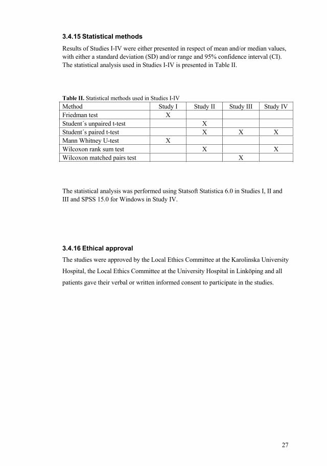
3.4.15 Statistical methods
Results of Studies I-IV were either presented in respect of mean and/or median values, with either a standard deviation (SD) and/or range and 95% confidence interval (CI). The statistical analysis used in Studies I-IV is presented in Table II.
Table II. Statistical methods used in Studies I-IV Method Study I Study II Study III Study IV Friedman test X Student´s unpaired t-test X Student´s paired t-test X X X Mann Whitney U-test X Wilcoxon rank sum test X X Wilcoxon matched pairs test X
The statistical analysis was performed using Statsoft Statistica 6.0 in Studies I, II and III and SPSS 15.0 for Windows in Study IV.
3.4.16 Ethical approval The studies were approved by the Local Ethics Committee at the Karolinska University
Hospital, the Local Ethics Committee at the University Hospital in Linköping and all
patients gave their verbal or written informed consent to participate in the studies.
28
4 RESULTS In Study I, three patients were excluded from the group that performed the
physiotherapy programme and three patients from the control group due to lack of
strength and serious infections of the airways. At the six-month follow-up five patients
in the intervention group and three in the control group were able to perform the tests.
There were no dropouts in Study II or III. In Study IV, four patients were excluded: one
male who developed a brain tumour that was discovered between tests III and IV, one
female who did not tolerate the face mask during the exercise endurance test, and two
females who developed ECG changes during the first exercise test.
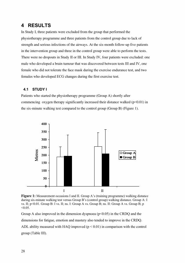
4.1 STUDY I Patients who started the physiotherapy programme (Group A) shortly after
commencing oxygen therapy significantly increased their distance walked (p<0.01) in
the six-minute walking test compared to the control group (Group B) (Figure 1).
Group AGroup B
0
50
100
150
200
250
300
350
400
Met
res
I II
Group AGroup B
0
50
100
150
200
250
300
350
400
Group AGroup B
0
50
100
150
200
250
300
350
400
Met
res
I IIFigure 1: Measurement occasions I and II. Group A’s (training programme) walking distance during six-minute walking test versus Group B’s (control group) walking distance. Group A: I vs. II; p<0.01. Group B: I vs. II; ns. I: Group A vs. Group B; ns. II: Group A vs. Group B; p <0.05.
Group A also improved in the dimension dyspnoea (p<0.05) in the CRDQ and the
dimensions for fatigue, emotion and mastery also tended to improve in the CRDQ.
ADL ability measured with HAQ improved (p < 0.01) in comparison with the control
group (Table III).
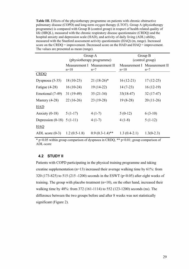
29
Table III. Effects of the physiotherapy programme on patients with chronic obstructive pulmonary disease (COPD) and long-term oxygen therapy (LTOT). Group A (physiotherapy programme) is compared with Group B (control group) in respect of health related quality of life (HRQL), measured with the chronic respiratory disease questionnaire (CRDQ) and the hospital anxiety and depression scale (HAD), and activity of daily living (ADL) ability, measured with the Stanford assessment activity questionnaire (HAQ) (m, range). Increased score on the CRDQ = improvement. Decreased score on the HAD and HAQ = improvement. The values are presented as mean (range).
Group A (physiotherapy programme)
Group B (control group)
Measurement I Measurement II Measurement I Measurement II n=10 n=7 n=10 n=7 CRDQ
Dyspnoea (5-35) 18 (10-23) 21 (18-26)* 16 (12-21) 17 (12-25)
Fatigue (4-28) 16 (10-24) 19 (14-22) 14 (7-23) 16 (12-19)
Emotional (7-49) 31 (19-49) 35 (21-34) 33(18-47) 32 (17-47)
Mastery (4-28) 22 (16-26) 23 (19-28) 19 (8-28) 20 (11-26)
HAD
Anxiety (0-18) 5 (1-17) 4 (1-7) 5 (0-12) 6 (3-10)
Depression (0-18) 5 (1-11) 4 (1-7) 4 (1-8) 5 (1-12)
HAQ
ADL score (0-3) 1.2 (0.5-1.8) 0.9 (0.3-1.4)** 1.3 (0.4-2.1) 1.3(0-2.3)
* p<0.05 within group comparison of dyspnoea in CRDQ, ** p<0.01; group comparison of ADL-score
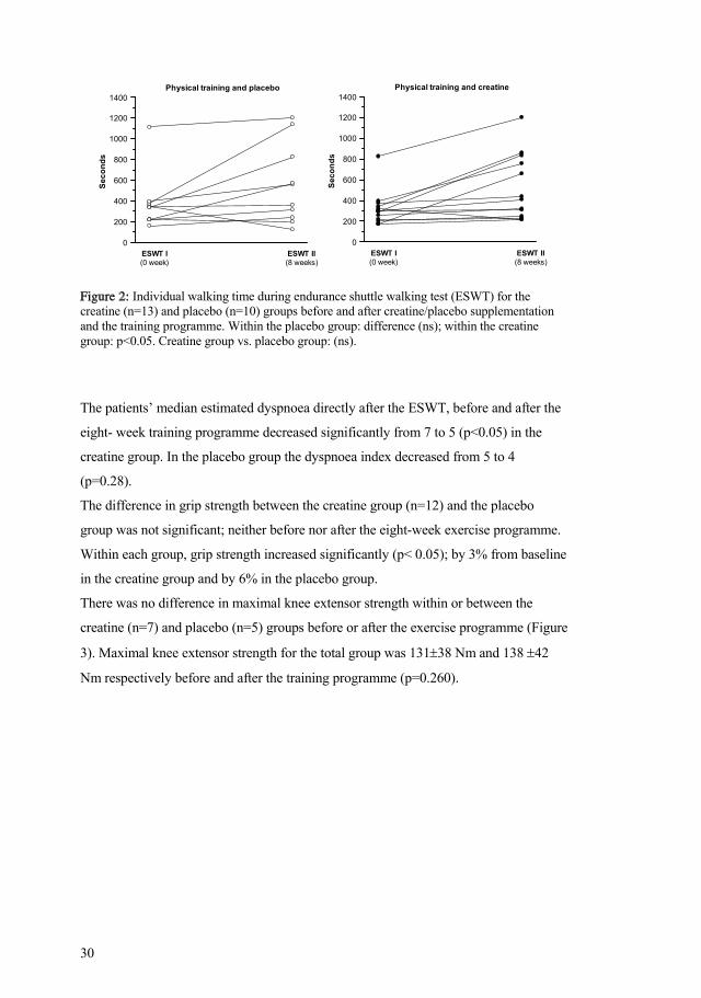
4.2 STUDY II Patients with COPD participating in the physical training programme and taking
creatine supplementation (n=13) increased their average walking time by 61%: from
320 (173-825) to 515 (215 -1200) seconds in the ESWT (p<0.05) after eight weeks of
training. The group with placebo treatment (n=10), on the other hand, increased their
walking time by 48%: from 372 (161-1114) to 552 (123-1200) seconds (ns). The
difference between the two groups before and after 8 weeks was not statistically
significant (Figure 2).
30
0
200
400
600
800
1000
1200
1400Physical training and creatine
Seco
nds
ESWT I(0 week)
ESWT II(8 weeks)
0
200
400
600
800
1000
1200
1400Physical training and placebo
Seco
nds
ESWT I(0 week)
ESWT II(8 weeks)
Figure 2: Individual walking time during endurance shuttle walking test (ESWT) for the creatine (n=13) and placebo (n=10) groups before and after creatine/placebo supplementation and the training programme. Within the placebo group: difference (ns); within the creatine group: p<0.05. Creatine group vs. placebo group: (ns).
The patients’ median estimated dyspnoea directly after the ESWT, before and after the
eight- week training programme decreased significantly from 7 to 5 (p<0.05) in the
creatine group. In the placebo group the dyspnoea index decreased from 5 to 4
(p=0.28).
The difference in grip strength between the creatine group (n=12) and the placebo
group was not significant; neither before nor after the eight-week exercise programme.
Within each group, grip strength increased significantly (p< 0.05); by 3% from baseline
in the creatine group and by 6% in the placebo group.
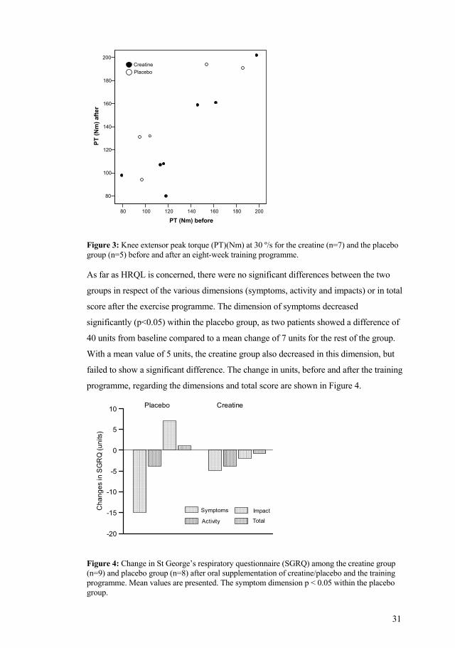
There was no difference in maximal knee extensor strength within or between the
creatine (n=7) and placebo (n=5) groups before or after the exercise programme (Figure
3). Maximal knee extensor strength for the total group was 131 38 Nm and 138 42
Nm respectively before and after the training programme (p=0.260).
31
20018016014012010080
PT (Nm) before
200
180
160
140
120
100
80
PT (N
m) a
fter
PlaceboCreatine
Figure 3: Knee extensor peak torque (PT)(Nm) at 30 º/s for the creatine (n=7) and the placebo group (n=5) before and after an eight-week training programme.
As far as HRQL is concerned, there were no significant differences between the two
groups in respect of the various dimensions (symptoms, activity and impacts) or in total
score after the exercise programme. The dimension of symptoms decreased
significantly (p<0.05) within the placebo group, as two patients showed a difference of
40 units from baseline compared to a mean change of 7 units for the rest of the group.
With a mean value of 5 units, the creatine group also decreased in this dimension, but
failed to show a significant difference. The change in units, before and after the training
programme, regarding the dimensions and total score are shown in Figure 4.
-20
-15
-10
-5
0
5
10
Cha
nges
in S
GR
Q (u
nits
)
Symptoms
Activity
Impact
Total
Placebo Creatine
Figure 4: Change in St George’s respiratory questionnaire (SGRQ) among the creatine group (n=9) and placebo group (n=8) after oral supplementation of creatine/placebo and the training programme. Mean values are presented. The symptom dimension p < 0.05 within the placebo group.
International Journal of COPD 2006:1(4) 449
Creatine supplimentation and physical training in patients with COPD
but did not reach significant difference. The change in units, atbaseline, and after the training programme regarding thedimensions and total score are shown in Figure 2.
Effect of creatine supplementationon muscle strength and fatigueThere were no significant differences in grip strengthbetween the creatine group (n = 12) and the placebo group(n = 10) before and after the training programme.
No differences in maximal knee extensor strength withinor between the creatine (n = 7, p = 0.74) and placebo (n = 5,p = 0.07) groups before or after the training programme(p = 0.08) (Figure 3). Maximal knee extensor strength for thetotal group was 131 Nm (95% CI 106 to 155) and 138 Nm(95% CI 111 to 165), before and after the training programme(p = 0.26).
The decline in mean and PT during the fatigue tests didnot differ within or between the groups before or after thetraining programme. The results from the leg muscle fatiguetest in the creatine group (n = 7) are shown in Figure 4.
All results are summarized in Table 2.
DiscussionThe aim of the present study was to investigate whether creatinesupplementation may have an additive effect on physicalperformance in COPD patients participating in a pulmonaryrehabilitation programme including exercise training. Wefailed to prove any differences in walking time (ESWT) after
the training period between the groups receiving creatinesupplementation or placebo. Our data did not show that exercisetraining and creatine supplementation improved walking timein ESWT in relative to exercise training and placebo.
All patients included in the study were referred from thePrimary Health Care in Stockholm and Linköping Countyand seemed to be representative for a group of patients withmoderate to severe COPD. Today the effect of exercise trainingin patients with COPD is well established and there are severalstudies including patients with moderate to severe stage ofCOPD showing benefits of pulmonary rehabilitation (Wijkstraet al 1994; Donner and Muir 1997; Tiep 1997). When weformed our programme we followed the evidence-basedguidelines as outlined in “Pulmonary rehabilitation” (ACCP/AACVPR 1997). There were no difficulties for the patients toaccomplish the exercise programme and reach the desiredlevels of dyspnea. The ESWT test is a submaximal test ofphysical performance and should therefore reflect a person’sdaily physical performance better than a maximal test. Thetest has also showed sensitivity to changes after exercisetraining in COPD patients (Revill et al 1999).
In the present study, ESWT improved in both groups.However, the improvement was significant only in the creatinegroup. There was no significant difference when comparingthe results of the ESWT between the groups. Hence, we failedto show any benefit of creatine supplementation incombination with exercise training.
In healthy subjects an increase of 25%–30% of totalcreatine concentration in the muscles was found after oralcreatine supplementation, as well as a significant increase inmaximal short-lasting muscle work after oral creatinesupplementation. The size of the increase in strength powerwas approximately 4%–6% (Balsom et al 1993).
Gordon and colleagues (1995) found significantlyincreased skeletal muscular performance in patients withchronic heart failure compared with a placebo group afteroral supplementation of creatine during ten days withoutany exercise training. They concluded that both patients withheart failure and healthy individuals with low levels ofcreatine in the muscles show better muscle function after oralsupplementation.
Studies of skeletal muscle metabolite concentrations inleg muscle of patients with COPD have shown derangedmuscle metabolism with decreased concentrations of ATPand creatine phosphate (Jakobsson and Jorfeldt 1995). Apositive effect in physical performance was thereforeexpected when creatine was supplemented to training.
-20
-15
-10
-5
0
5
10
Ch
an
ge
s in
SG
RQ
(u
nits)
Symptoms
Activity
Impact
Total
Placebo Creatine
Figure 2 Change in St George´s Respiratory Questionnaire (SGRQ) in theCreatine group (n = 9) and Placebo group (n = 8) after oral supplementation ofcreatine/placebo and the training programme. Mean values are presented.Note:*p < 0.05 within the group.
32
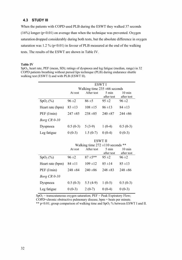
4.3 STUDY III When the patients with COPD used PLB during the ESWT they walked 37 seconds
(16%) longer (p<0.01) on average than when the technique was prevented. Oxygen
saturation dropped considerably during both tests, but the absolute difference in oxygen
saturation was 1.2 % (p<0.01) in favour of PLB measured at the end of the walking
tests. The results of the ESWT are shown in Table IV.
Table IV SpO2, heart rate, PEF (mean, SD); ratings of dyspnoea and leg fatigue (median, range) in 32 COPD patients breathing without pursed lips technique (PLB) during endurance shuttle walking test (ESWT I) and with PLB (ESWT II).
ESWT I Walking time 235 ±66 seconds
At rest After test 5 min after test
10 min after test
SpO2 (%) 96 ±2 86 ±5 95 ±2 96 ±2
Heart rate (bpm) 83 ±13 108 ±15 86 ±13 84 ±13
PEF (l/min) 247 ±85 238 ±85 240 ±87 244 ±86
Borg CR 0-10
Dyspnoea 0.5 (0-3) 5 (3-9) 1 (0-4) 0.5 (0-3)
Leg fatigue 0 (0-3) 1.5 (0-7) 0 (0-4) 0 (0-3)
ESWT II Walking time 272 ±110 seconds **
At rest After test 5 min after test
10 min after test
SpO2 (%) 96 ±2 87 ±5** 95 ±2 96 ±2
Heart rate (bpm) 84 ±11 109 ±12 85 ±14 85 ±13
PEF (l/min) 248 ±84 240 ±86 248 ±83 248 ±86
Borg CR 0-10
Dyspnoea 0.5 (0-3) 5.5 (4-9) 1 (0-5) 0.5 (0-3)
Leg fatigue 0 (0-3) 2 (0-7) 0 (0-4) 0 (0-3)
SpO2 = transcutaneous oxygen saturation; PEF = Peak Expiratory Flow; COPD=chronic obstructive pulmonary disease, bpm = beats per minute. ** p<0.01; group comparison of walking time and SpO2 % between ESWT I and II.
33
0
80
85
90
95
100
SpO
2 %
0 min 2 min 4 min
no PLB (n=32) PLB (n=32)
no PLB (n=11) PLB (n=11)
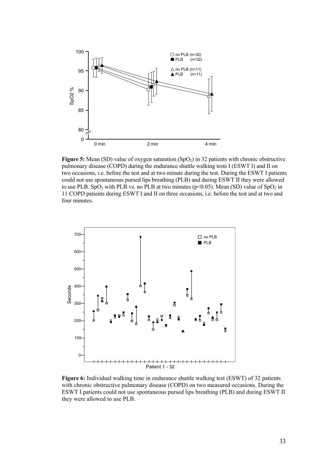
Figure 5: Mean (SD) value of oxygen saturation (SpO2) in 32 patients with chronic obstructive pulmonary disease (COPD) during the endurance shuttle walking tests I (ESWT I) and II on two occasions, i.e. before the test and at two minute during the test. During the ESWT I patients could not use spontaneous pursed lips breathing (PLB) and during ESWT II they were allowed to use PLB. SpO2 with PLB vs. no PLB at two minutes (p<0.05). Mean (SD) value of SpO2 in 11 COPD patients during ESWT I and II on three occasions, i.e. before the test and at two and four minutes.
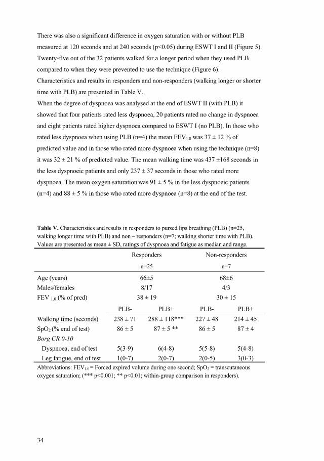
Patient 1 - 32
700
600
500
400
300
200
100
0
Seco
nds
PLBno PLB
Figure 6: Individual walking time in endurance shuttle walking test (ESWT) of 32 patients with chronic obstructive pulmonary disease (COPD) on two measured occasions. During the ESWT I patients could not use spontaneous pursed lips breathing (PLB) and during ESWT II they were allowed to use PLB.
34
There was also a significant difference in oxygen saturation with or without PLB
measured at 120 seconds and at 240 seconds (p<0.05) during ESWT I and II (Figure 5).
Twenty-five out of the 32 patients walked for a longer period when they used PLB
compared to when they were prevented to use the technique (Figure 6).
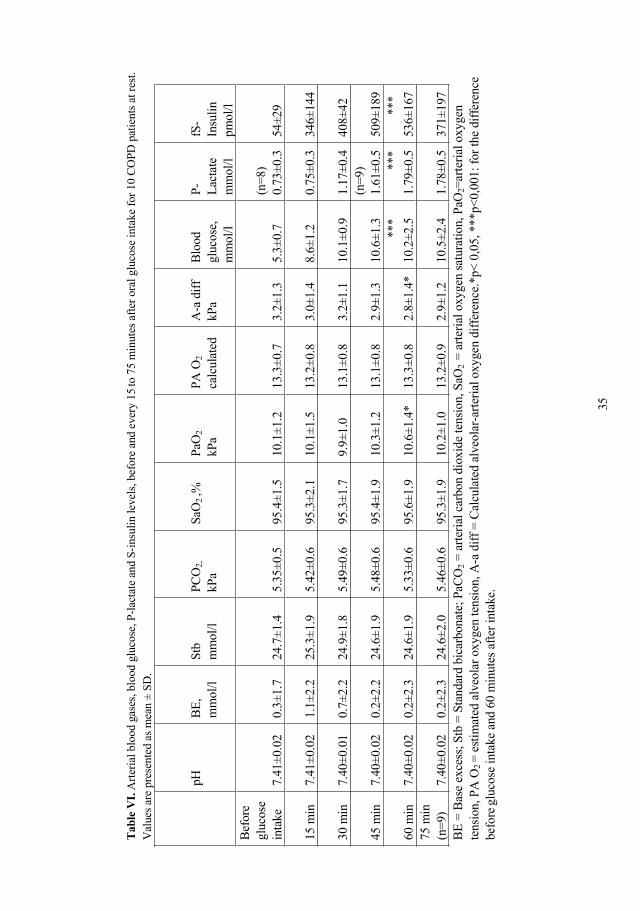
Characteristics and results in responders and non-responders (walking longer or shorter
time with PLB) are presented in Table V.
When the degree of dyspnoea was analysed at the end of ESWT II (with PLB) it
showed that four patients rated less dyspnoea, 20 patients rated no change in dyspnoea
and eight patients rated higher dyspnoea compared to ESWT I (no PLB). In those who
rated less dyspnoea when using PLB (n=4) the mean FEV1.0 was 37 ± 12 % of
predicted value and in those who rated more dyspnoea when using the technique (n=8)
it was 32 ± 21 % of predicted value. The mean walking time was 437 ±168 seconds in
the less dyspnoeic patients and only 237 ± 37 seconds in those who rated more
dyspnoea. The mean oxygen saturation was 91 ± 5 % in the less dyspnoeic patients
(n=4) and 88 ± 5 % in those who rated more dyspnoea (n=8) at the end of the test.
Table V. Characteristics and results in responders to pursed lips breathing (PLB) (n=25, walking longer time with PLB) and non – responders (n=7; walking shorter time with PLB). Values are presented as mean ± SD, ratings of dyspnoea and fatigue as median and range.
Responders
n=25
Non-responders
n=7
Age (years) 66±5 68±6 Males/females 8/17 4/3 FEV 1.0 (% of pred) 38 ± 19 30 ± 15
PLB- PLB+ PLB- PLB+ Walking time (seconds) 238 ± 71 288 ± 118*** 227 ± 48 214 ± 45 SpO2 (% end of test) 86 ± 5 87 ± 5 ** 86 ± 5 87 ± 4 Borg CR 0-10
Dyspnoea, end of test 5(3-9) 6(4-8) 5(5-8) 5(4-8) Leg fatigue, end of test 1(0-7) 2(0-7) 2(0-5) 3(0-3)
Abbreviations: FEV1.0 = Forced expired volume during one second; SpO2 = transcutaneous oxygen saturation; (*** p<0.001; ** p<0.01; within-group comparison in responders).
35
Tab
le V
I. A
rteria
l blo
od g
ases
, blo
od g
luco
se, P
-lact
ate
and
S-in
sulin
leve
ls, b
efor
e an
d ev
ery
15to
75
min
utes
afte
r ora
l glu
cose
inta
ke fo
r 10
COPD
pat
ient
s at r
est.
Val
ues a
repr
esen
ted
as m
ean
± SD
.
pH
BE,
mm
ol/l
Stb
mm
ol/l
PCO
2,
kPa
SaO
2,%
PaO
2
kPa
PA O
2
calc
ulat
ed
A-a
diff
kP
a Bl
ood
gluc
ose,
m
mol
/l
P- Lact
ate
mm
ol/l
fS-
Insu
lin
pmol
/l Be
fore
gl
ucos
e
inta
ke
7.41
±0.0
2 0.
3±1.
7 24
.7±1
.4
5.35
±0.5
95
.4±1
.5
10.1
±1.2
13
.3±0
.7
3.2±
1.3
5.3±
0.7
(n=8
) 0.
73±0
.3
54±2
9
15 m
in
7.41
±0.0
2 1.
1±2.
2 25
.3±1
.9
5.42
±0.6
95
.3±2
.1
10.1
±1.5
13
.2±0
.8
3.0±
1.4
8.6±
1.2
0.75
±0.3
34
6±14
4
30 m
in
7.40
±0.0
1 0.
7±2.
2 24
.9±1
.8
5.49
±0.6
95
.3±1
.7
9.9±
1.0
13.1
±0.8
3.
2±1.
1 10
.1±0
.9
1.17
±0.4
40
8±42
45 m
in
7.40
±0.0
2 0.
2±2.
2 24
.6±1
.9
5.48
±0.6
95
.4±1
.9
10.3
±1.2
13
.1±0
.8
2.9±
1.3
10.6
±1.3
(n
=9)
1.61
±0.5
50
9±18
9
60 m
in
7.40
±0.0
2 0.
2±2.
3 24
.6±1
.9
5.33
±0.6
95
.6±1
.9
10.6
±1.4
*13
.3±0
.8
2.8±
1.4*
*
**
10.2
±2.5
*
**
1.79
±0.5
*
**
536±
167
75 m
in
(n=9
) 7.
40±0
.02
0.2±
2.3
24.6
±2.0
5.
46±0
.6
95.3
±1.9
10
.2±1
.0
13.2
±0.9
2.
9±1.
2 10
.5±2
.4
1.78
±0.5
37
1±19
7 BE
= B
ase
exce
ss; S
tb =
Sta
ndar
d bi
carb
onat
e; P
aCO
2 = a
rteria
l car
bon
diox
ide
tens
ion,
SaO
2=
arte
rial o
xyge
n sa
tura
tion,
PaO
2=ar
teria
l oxy
gen
tens
ion,
PA
O2=
estim
ated
alv
eola
r oxy
gen
tens
ion,
A-a
diff
= C
alcu
late
d al
veol
ar-a
rteria
l oxy
gen
diffe
renc
e.*p
< 0,
05, *
**p<
0,00
1: fo
r the
diff
eren
ce
befo
regl
ucos
e in
take
and
60
min
utes
afte
r int
ake.
36
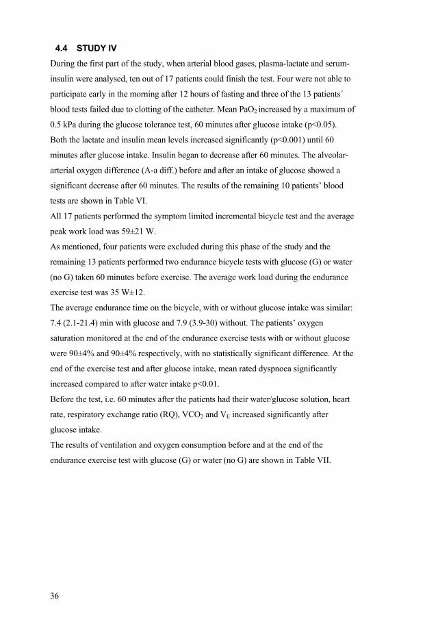
4.4 STUDY IV During the first part of the study, when arterial blood gases, plasma-lactate and serum-
insulin were analysed, ten out of 17 patients could finish the test. Four were not able to
participate early in the morning after 12 hours of fasting and three of the 13 patients´
blood tests failed due to clotting of the catheter. Mean PaO2 increased by a maximum of
0.5 kPa during the glucose tolerance test, 60 minutes after glucose intake (p<0.05).
Both the lactate and insulin mean levels increased significantly (p<0.001) until 60
minutes after glucose intake. Insulin began to decrease after 60 minutes. The alveolar-
arterial oxygen difference (A-a diff.) before and after an intake of glucose showed a
significant decrease after 60 minutes. The results of the remaining 10 patients’ blood
tests are shown in Table VI.
All 17 patients performed the symptom limited incremental bicycle test and the average
peak work load was 59±21 W.
As mentioned, four patients were excluded during this phase of the study and the
remaining 13 patients performed two endurance bicycle tests with glucose (G) or water
(no G) taken 60 minutes before exercise. The average work load during the endurance
exercise test was 35 W±12.
The average endurance time on the bicycle, with or without glucose intake was similar:
7.4 (2.1-21.4) min with glucose and 7.9 (3.9-30) without. The patients’ oxygen
saturation monitored at the end of the endurance exercise tests with or without glucose
were 90±4% and 90±4% respectively, with no statistically significant difference. At the
end of the exercise test and after glucose intake, mean rated dyspnoea significantly
increased compared to after water intake p<0.01.
Before the test, i.e. 60 minutes after the patients had their water/glucose solution, heart
rate, respiratory exchange ratio (RQ), VCO2 and VE increased significantly after
glucose intake.
The results of ventilation and oxygen consumption before and at the end of the
endurance exercise test with glucose (G) or water (no G) are shown in Table VII.

37
Table VII. Ventilation and oxygen consumption before and at the end of a symptom limited endurance bicycle test with oral intake of glucose (G) and without (no G) among 13 patients with COPD. The intake of glucose or water was 60 minutes before cycling. The values are presented as mean ±SD. Rated degree of exertion, dyspnoea and leg fatigue are presented as median and range.
Start/ no G Start/G End/ no G End/G
Heart rate, b/min 72±13 77±14* 109±16 111±18
SpO2, % 95±2 96±2 90±4 90±4
Respir. rate, 1b/min 20±5 20±5 30±6 31±7
RQ 0.85±0.05 0.92±0.05** 0.91±0.04 0.95±0.06*
VO2, l/min 0.32±0.03 0.36±0.07 0.90±0.15 0.94±0.17
VCO2, l/min 0.27±0.04 0.31±0.04* 0.83±0.16 0.9±0.2
VE, l/min 11.0±2 12.3±3* 28.0±7 29.7±8
VT, l 0.62±0.3 0.65±0.3 1.00±0.34 1.00±0.37
VE/ VO2, % 34±5 35±7 32±7 32±8
VE/ VCO2, % 40±6 40±5 35±7 34±9
Ventilation spare, % 67±12 63±13* 19±20 15±19 Rated dyspnoea (Borg CR-10) - - 7(0-10) 8.5(5-10)* Rated exertion (Borg 6-20) - - 17(15.5-19) 17(15-19) Rated leg fatigue (Borg CR-10) - - 4.5(0-10) 4(0-10) b/min= beats per minute, SpO2 = transcutaneous oxygen saturation, 1b/min = breaths/minute, RQ= respiratory exchange ratio, VO2 = Oxygen uptake, VCO2 = carbon dioxide production, VE= minute ventilation, VT = tidal volume, * p<0.05 ** p<0.01 for the difference between no Glucose (no G) and intake of Glucose (G).
38
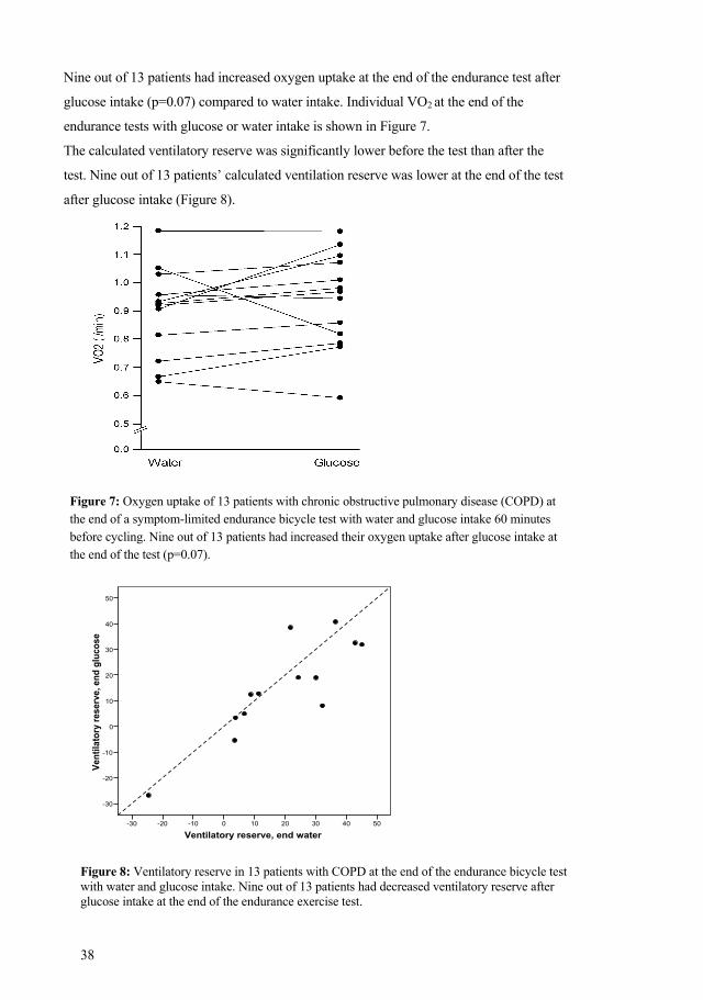
Nine out of 13 patients had increased oxygen uptake at the end of the endurance test after
glucose intake (p=0.07) compared to water intake. Individual VO2 at the end of the
endurance tests with glucose or water intake is shown in Figure 7.
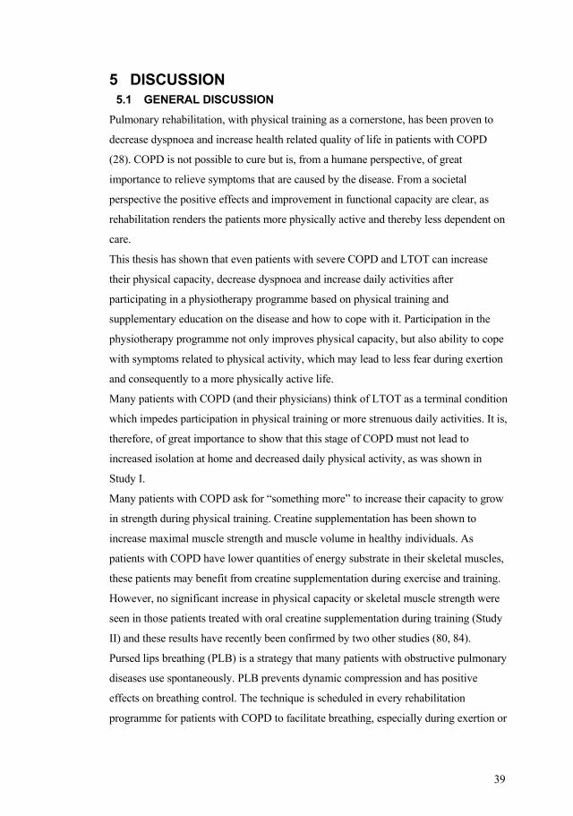
The calculated ventilatory reserve was significantly lower before the test than after the
test. Nine out of 13 patients’ calculated ventilation reserve was lower at the end of the test
after glucose intake (Figure 8).
Figure 7: Oxygen uptake of 13 patients with chronic obstructive pulmonary disease (COPD) at the end of a symptom-limited endurance bicycle test with water and glucose intake 60 minutes before cycling. Nine out of 13 patients had increased their oxygen uptake after glucose intake at the end of the test (p=0.07).
Ventilatory reserve, end water50403020100-10-20-30
Vent
ilato
ry re
serv
e, e
nd g
luco
se
50
40
30
20
10
0
-10
-20
-30
Figure 8: Ventilatory reserve in 13 patients with COPD at the end of the endurance bicycle test with water and glucose intake. Nine out of 13 patients had decreased ventilatory reserve after glucose intake at the end of the endurance exercise test.
39
5 DISCUSSION 5.1 GENERAL DISCUSSION
Pulmonary rehabilitation, with physical training as a cornerstone, has been proven to
decrease dyspnoea and increase health related quality of life in patients with COPD
(28). COPD is not possible to cure but is, from a humane perspective, of great
importance to relieve symptoms that are caused by the disease. From a societal
perspective the positive effects and improvement in functional capacity are clear, as
rehabilitation renders the patients more physically active and thereby less dependent on
care.
This thesis has shown that even patients with severe COPD and LTOT can increase
their physical capacity, decrease dyspnoea and increase daily activities after
participating in a physiotherapy programme based on physical training and
supplementary education on the disease and how to cope with it. Participation in the
physiotherapy programme not only improves physical capacity, but also ability to cope
with symptoms related to physical activity, which may lead to less fear during exertion
and consequently to a more physically active life.
Many patients with COPD (and their physicians) think of LTOT as a terminal condition
which impedes participation in physical training or more strenuous daily activities. It is,
therefore, of great importance to show that this stage of COPD must not lead to
increased isolation at home and decreased daily physical activity, as was shown in
Study I.
Many patients with COPD ask for “something more” to increase their capacity to grow
in strength during physical training. Creatine supplementation has been shown to
increase maximal muscle strength and muscle volume in healthy individuals. As
patients with COPD have lower quantities of energy substrate in their skeletal muscles,
these patients may benefit from creatine supplementation during exercise and training.
However, no significant increase in physical capacity or skeletal muscle strength were
seen in those patients treated with oral creatine supplementation during training (Study
II) and these results have recently been confirmed by two other studies (80, 84).
Pursed lips breathing (PLB) is a strategy that many patients with obstructive pulmonary
diseases use spontaneously. PLB prevents dynamic compression and has positive
effects on breathing control. The technique is scheduled in every rehabilitation
programme for patients with COPD to facilitate breathing, especially during exertion or
40
exacerbation, as PLB has been shown to have positive effects in these situations.
However, the technique not yet has been proven to increase physical capacity.
Once a patient with COPD has adopted the technique and incorporated it into his/her
breathing strategy, it is difficult to prevent the use of PLB. This was obvious in Study
III, and all patients spontaneously used PLB when walking. However, when a mouth
piece prevented them from using the technique, their walking capacity was lower than
when using PLB.
Patients with COPD often desaturate during physical activity and this, among others,
limits the patient’s ability to be physically active and to exercise. Glucose infusion has
been shown to increase blood oxygen tension in COPD patients at rest. If this is also
true after oral intake of glucose it may, most likely, positively influence exercise
capacity and, in the longer term, training intensity in patients with COPD. Study IV
showed no positive impact on exercise capacity after oral intake of glucose. On the
other hand, it is important to verify if oral intake of glucose may have negative effects
on physical capacity for this group of patients, as there is a demand for alternative
methods to improve physical capacity in COPD patients.
5.2 EXERCISE TESTING IN PATIENTS WITH COPD 5.2.1 Walking tests Exercise capacity was evaluated by the six-minute walking test in Study I. All patients
included in the study had severe COPD with LTOT and had to transport the oxygen
tubes when they moved outside their home. Therefore all patients used a walking frame
with a basket for their oxygen bottles. The six-minute walking test was chosen instead
of an incremental shuttle walking test (ISWT), as the six-minute walking test correlates
more closely with daily activities than an incremental test such as ISWT and may,
therefore, better reflect the patient’s normal capability (106). ISWT is more
standardised than the six-minute walking test, but in this test patients walk up and down
a 10-m course with many turns. This was not considered safe enough for the patients as
they all used their walking frame during the test situation.
The six-minute walk test is a simple field test with standardised instructions and good
reproducibility (106). In Study I no encouragement was used and all tests were
supervised by the same test leader (88). At the time the study was performed, the
recommendation was two learning tests (87) but, considering the patient’s status we
decided to perform only one learning test. However, the seven patients in Study I
increased their mean walking distance by 66 metres, an increase which cannot only be
41
due to learning (87), but may also be explained by a decreased fear to move, to dare to
be more active during dyspnoea after having participated in the training programme,
i.e. a better coping strategy with the symptoms during exertion.
The Endurance Shuttle Walking Test (ESWT) was used in Study II and III as this is a
more sensitive test to detect changes in physical capacity than the ISWT (89, 90).
ESWT is a standardised walking test which has developed from ISWT. In Study II
ESWT was used to detect a possible additional effect of creatine supplementation in
physical capacity linked to physical training. Within-group changes in physical
capacity, measured with ESWT, increased significantly in the creatine group, but not in
the placebo group. This result was unexpected, as many studies have shown the
positive effects of physical training and as all patients were exposed to training (23,
28). This may be due to a lack of power, as the number of participating patients and the
in-between-groups comparison was relatively low. However, two other recently
published studies have arrived at the same conclusion (80, 84) as we did and Deacon et
al (84) in particular showed, the same result as our Study II with a large number of
patients.
In Study II and III there was “no learning test” as the ISWT, which is always
performed before the ESWT, has been shown to be sufficient (107).
5.2.2 Symptom limited exercise test on a bicycle In Study IV we used a bicycle test to measure physical capacity after oral intake of
glucose in patients with exercised induced hypoxia. As patients with COPD are often
limited in their daily life, an endurance test was chosen to investigate if intake of
glucose positively influences endurance exercise capacity. We estimated that 60
percent of maximal capacity would be a suitable level for an endurance test for this
group of patients. To calculate the 60 percent level of peak exercise capacity, the
patients performed a symptom-limited incremental exercise test on a bicycle (108).
Considering COPD patients’ low level of capacity, patients started on 10 W with an
increase of 10 W every minute. The mean load in the bicycle test in Study IV was 59
±21 W. If the endurance exercise test had started at 30 W, which is the traditional way
of an incremental bicycle test, some patients would only have been capable of
exercising for a few seconds and, with this in mind, the starting load was set at an
adequate level.
During the endurance test oxygen uptake and ventilation were measured online and all
patients were wearing a face mask. Wearing a face mask may be uncomfortable and
42
could, therefore, impact the results. There was also the small risk that patients could
become used to the mask and would therefore be more comfortable with the mask in
the second test. However, when data were analysed there was no difference between
the first and second endurance tests.
5.2.3 Muscle strength and fatigue Grip strength was measured in Study II as it has been shown to correlate with whole
body strength (94, 95). Study II was performed in two places: Stockholm and
Linköping. Unfortunately, the only available instruments for measuring grip strength
were the Jamar Dynamometer in Stockholm and the Grippit in Linköping. Instruction
for the tests followed the recommendations of Mathiowetz et al (96) and as these two
instruments have been shown to be valid and reliable (96), we do not think that this has
influenced the results.
In Study II maximal voluntary strength and fatigue were measured with an isokinetic
dynamometer, Kin-Com, and with a protocol used in other studies with patients in the
same age group, but with coronary heart disease (92, 93). Although this test is rather
time-consuming and tiresome for patients, our patients could comfortably manage the
number of repetitions and follow the instructions. However, no significant difference
was found between the placebo or creatine group.
5.3 HEALTH RELATED QUALITY OF LIFE We used the Chronic Respiratory Disease Questionnaire (CRDQ) in Study I, as CRDQ
seems to be more responsive to rehabilitation than other health related quality of life
questionnaires (HRQL) among patients with COPD. Individuals with COPD have been
shown to suffer from depression to a higher degree than healthy individuals (109), and
being dependent on LTOT may have a negative impact on depression and anxiety. We
therefore completed HRQL assessments in Study I with the Hospital Anxiety and
Depression Scale (HAD) (100) to establish if our patients with very severe COPD were
suffering from depression and if this may influence the results. However, no increase in
depression or anxiety, whether within the respective groups or between the groups, was
found. We then concluded that the anxiety and depression grade of patients in Study I
may not have influenced the HRQL results. In Study II we used St George’s
Respiratory Questionnaire (SGRQ), also a disease specific HRQL questionnaire
available in Swedish which has been tested for validity (98). There were no significant
differences between the creatine or the placebo group after the exercise programme.
43
Within the placebo group, in the dimension of symptom, there was a significant
difference (p<0.05) as two patients had 40 units in difference from baseline compared
to a mean of 7 units change for the rest of the group. We speculate that these two
patients had exarcerbations shortly before the programme started and recovered
during the training programme.
5.4 PULSE OXIMETRY Transcutaneous oxygen saturation has inherent limitations, but valid trends can be
followed and even a small improvement in oxygen saturation may be of clinical
relevance in exercise capacity in patients with moderate to severe COPD (91).
5.5 RESULTS Participation in a pulmonary rehabilitation programme based on physical training and
supervision by a physiotherapist is beneficial, even for patients with severe COPD and
LTOT, This is evident from an increase in physical capacity, HRQL and ADL. These
results are encouraging not only for this group of severely ill patients, but also for other
patients with COPD. The fact that patients increased their walking distance, estimated a
higher grade of dyspnoea and higher heart rate at the end of the programme may be due
to the fact that they were more used to physical activity and less fearful of increased
exertion, as shown by a higher grade of dyspnoea and heart rate. The group that
participated in the physiotherapy programme showed a mean increase of 66 metres in
walking distance. To a healthy individual this may not seem like an impressive
improvement, but for these patients it is indeed clinically significant (110). The
increase in ADL ability is, from the patient’s perspective, very important as small
differences in daily activities for these severely ill patients are of great value.
Even if there were many dropouts in our study we still think that our results may have
been the same with a larger number of patients as other studies have confirmed our
results (111).
Even if the large number of dropouts in Study I is considered, the results are still
encouraging as other studies have confirmed our results (111).
When the patients recruited for Study II started, no other study on this subject had been
published to our knowledge, but while Study II was in progress Fuld et al (80)
concluded that exercise training and creatine supplementation resulted in an increase in
fat-free-mass, upper and lower limb muscle strength and endurance, but not in whole
body exercise capacity. This was confirmed by the results of Study II, showing no
44
significant improvement in walking time (measured by ESWT), grip strength, maximal
knee muscle strength or fatigue and HRQL in relation to exercise training and placebo.
As patients with moderate to severe COPD are limited in their daily activities, one of
the aims of pulmonary rehabilitation in conjunction with exercise training is to increase
endurance capacity. The exercise training in our programme was designed to reach that
goal and the ESWT was therefore chosen as the most appropriate and sensitive test to
study endurance capacity in Study II. However, no significant difference in walking
endurance was found when comparing the creatine and the placebo group. The reason
for this lack of improvement may be that creatine supplementation has been shown to
influence maximal muscle strength and not endurance exercise capacity after exercise
training in healthy individuals. This proves how important it is to choose the proper
method for the evaluation of trainings effects.
Knee maximal voluntary muscle strength and fatigue were only assessed at one of the
two study sites for Study II as the Kin-Com equipment was only available in
Stockholm. The sample size was therefore reduced to only a few patients. This could
explain the lack of effect on muscular strength and fatigue in Study II. On the other
hand Deacon et al, working with a much larger sample size, showed that creatine
supplementation does not enhance the effect of training; neither in respect of exercise
capacity - measured with the ISWT - nor muscular strength (84).
The exercise training programme followed the present guidelines and seems to have the
same level of intensity and duration as programmes in other studies with creatine
supplementation (80, 84).
The St. George’s respiratory questionnaire, a disease-specific instrument, was chosen to
measure any difference in HRQL in Study II (98). Within-group comparison in the
creatine group showed a significant increase in exercise endurance in the ESWT, but
this was not reflected in the SGRQ, although all dimensions showed a positive change
in direction. On analysis of the data it was found that the placebo group showed a
substantial decrease in the symptom dimension, with two patients displaying a change
of 40 units on completion of the programme. This may be due to exacerbations
unknown to the study leaders prior to the programme and recovery on completion of
the programme. It could also be that these two patients did not really understand the
questions the first time around. The SGRQ is a self-completed questionnaire and if
patients answer all questions without asking for help, there is no support from the
leaders.

45
As patients with COPD often desaturate during physical activity, PLB as a strategy
for breathing may decrease oxygen desaturation and therefore positively influence
physical ability. The most important finding of Study III was that most patients could
walk for a longer time during the endurance shuttle walking test, experiencing a
reduced decrease in oxygen saturation with spontaneous PLB when compared with
using open-mouth breathing as a result of a mouthpiece.
The focus in Study III was the effect of PLB on walking endurance and not on maximal
capacity. The ESWT was therefore chosen to assess walking endurance. All patients
performed an ISWT before they were registered at the department of Pulmonary
Rehabilitation and we also used this test to recruit patients for our study. Since an
ISWT needs to be done before an ESWT we considered the ISWT as a learning test
(107). As all our patients used PLB spontaneously, it was impossible to prevent them
from applying the technique. A mouthpiece was therefore included which, although
not a natural way of breathing, is as close as possible to a natural breathing pattern.
The patients walked longer when using PLB and experienced a reduced decrease in
oxygen saturation, but felt no relief in respect of graded dyspnoea. This may be due to
the fact that they walked a longer distance and reached a higher grade of exhaustion
using PLB or that the mouthpiece or nose clip influenced the estimated dyspnoea.
However, to our knowledge no other study has showed significant decreased dyspnoea
in patients with COPD using PLB during exercise. Dyspnoea is a very individual
experience and perhaps there is a need for more sensitive measurements than those
available at present.
Oxygen saturation decreased during the ESWT, both with and without PLB, but the
decrease at the end of the tests was lower with PLB. Even a slight improvement in
oxygen saturation may be of clinical relevance in this group of patients with COPD.
During the later stage of the walking tests the recorded oxygen saturation was well
below 90%, which indicates that the patients were operating on the steep part of the
dissociation curve for oxygen.
Patients in Study III increased their walking time with 37 seconds (approximately 44
meters). This increase may have a significant clinical benefit for the group of patients
that suffers from severe exercise limitations. However, to our knowledge there is no
study that shows how many seconds/metres the ESWT needs to be improved to become
clinically significant. Reidelmeier et al (110) found that the smallest clinically
significant increase in the six-minute walking test was 54 m and Singh et al (112)
46
recently identified two important minimum clinical levels in ISWT: 47.5 m and 78.7,
if patients were able to distinguish an additional benefit.
Glucose and insulin infusion in patients with COPD and hypoxia has been found to
increase the oxygen tension at rest (113). It has been suggested that intravenous
infusion of glucose and insulin under euglycemic conditions increases carbon dioxide
production and therefore alveolar ventilation and arterial oxygen tension in hypoxic
COPD patients (114). Patients with COPD often decrease their oxygen saturation and if
oral glucose intake - not infusion - can increase the PaO2 in these patients, this may lead
to increased physical capacity. This may therefore be viable for use during physical
training and result in COPD patients enjoying more benefits from physical training.
This hypothesis was investigated in Study IV. During the oral glucose tolerance test in
Study IV mean PaO2 increased significantly by 0.5 kPa with parallel increased lactate
concentrations in patients with COPD and moderate hypoxia. Another finding was that
intake of glucose 60 minutes before a symptom-limited endurance bicycle test did not
improve endurance time or oxygen saturation when compared to drinking ordinary
water.
Patients in Study IV had light hypoxia at rest, but became hypoxic during exercise.
Apparently increased oxygen tension and glucose supply before exercise was not
enough to overcome the undesired effects of CO2 production. Early lactic acidosis has
been reported in patients with mild to severe COPD during sub-maximal exercise when
compared to healthy individuals (115, 116). The increased lactate leads to a higher
level of pH in the muscles, and this may contribute to earlier muscle fatigue and
exercise limitation in patients with COPD. This early release of lactate may be due to
decreased oxidative metabolism in the muscles (17).
The reason for the unexpected increase in lactate levels at rest during the oral glucose
intolerance test could be that some glucose will still descend down the glycolytic
pathway even though most of the glucose absorbed by the muscle is converted to
glycogen and the muscle does not really require ATP. However, activity in the
mitochondria of the muscle is low when at rest and the overload of glucose will
probably accumulate and produce some lactate that will be released into the blood.
As patients already had a higher lactate level after the intake of glucose as opposed to
only water when they started their exercise, this may have contributed to the absence
of increased exercise endurance.
Before the start of the exercise test with glucose there was a significant increase in
mean minute ventilation and carbon dioxide elimination compared to water intake,
47
which may have been as a result of the glucose combustion, but on completion of the
tests these differences were less obvious. However, when the patients started to
exercise after their glucose intake they experienced a higher degree of breathing related
to minute ventilation. This, in combination with the other results, may also explain the
unchanged endurance time after oral glucose intake compared to no glucose.
All patients in Study IV estimated their level of dyspnoea, exertion and leg fatigue at
the end of all exercise tests with or without glucose and reported a significant increase
in dyspnoea with glucose. The calculated ventilatory reserve after an intake of oral
glucose was significantly lower at the start of the endurance test compared to when the
patients had only water. This may also explain the higher grade of dyspnoea at the end
of the exercise test with glucose.
Oxygen uptake changed at the end of the exercise test with glucose, but did not reach
the previously set significant level or improve the exercise capacity in time or in
oxygen saturation. It could be that the patients needed more oxygen for the same level
of work when they had also taken glucose orally.
The already higher level of lactate in comparison with healthy individuals before the
start of the exercise endurance test may also have contributed to the default in respect
increased exercise capacity, as we had expected.
Based on these findings, we cannot yet recommend a high intake of fast-acting
carbohydrates for patients with COPD.
5.6 FUTURE STUDIES It would be of great interest to follow a larger group of patients with COPD and LTOT
for a longer period and to study the persistence of the effects after a supervised exercise
programme. It would also be of great interest to establish if the exercise training has
any long-term effects on quality of life, morbidity and mortality.
Why some of the patients naturally chose the Pursed Lips Breathing strategy is another
question for future research; so too the underlying reasons why some patients
experience less and others more dyspnoea when using the technique. Another
interesting matter that could be pursued would be to establish which kind of
supplementation could have addictive effects when patients with COPD exercise and
which type of patient that would benefit most from the supplementation in question.
48
6 CONCLUSIONS
Supervised training during eight weeks increases walking distance improves health
related quality of life and improves activities of daily living in severely ill patients with
COPD and those recently instituted for long-term oxygen therapy.
Oral creatine supplementation in combination with physical training shows no
significant improvement in physical performance measured as endurance shuttle
waking test in patients with COPD when compared to physical training alone.
Spontaneous pursed lips breathing can be a useful technique to increase walking
endurance and reduce oxygen desaturation during walking in patients with moderate to
severe COPD.
Oral intake of glucose may increase the arterial oxygen tension in COPD patients, even
with slight to moderate hypoxia at rest. When a glucose solution is taken before a
bicycle exercise test there appears to be no increase in endurance capacity or improved
oxygen saturation. On the contrary, glucose intake may be associated with reduced
ventilatory spare and higher ratings of dyspnoea during exercise.
49
7 ACKNOWLEDGEMENTS
First of all I want to thank all patients who, with great patience, performed all the tests and never complained. Without you there would have been no thesis! There are many people who have contributed to this thesis and I am very grateful to all of you. However, I would especially like to express my sincere gratitude to the following: Flemming Larsen, my principal supervisor and co-author, whose endless energy guided me through all the steps of my clinical intervention research whilst sharing his profound knowledge of research, physiology, cardiology and pulmonary medicine. I also thank you for our fruitful discussions and for talks; not only on our research, but also about cats, among other things. Not to forget some great moments when our hypotheses were verified and you said, “The Nobel prize, Gunsan”. Agneta Ståhle, my co-supervisor and co-author, for good advice, encouragement, enthusiasm and unfailing support, guiding me as a physiotherapist to achieve more from my research. Thank you for the support when reaching the decision that December 5th would be the day to present my thesis. Magnus Sköld, my co-author, for your support and invaluable support with Study II. Per Jakobsson, my co-author, for your support and for conceiving the idea for Study II. Karin Söderlund, my co-author, for sharing your profound knowledge of muscle physiology. Anna Tollbäck, my co-author, for valuable support and excellent muscle testing in Study II.Lisbet Broman, research assistant and co-author, for excellent professional technical assistance, for expert help with figures and tables and for nice talks about our grandchildren. Siw Rundgren, my co-author, for valuable support in Study II Karin Malmberg and her collegues at University Hospital in Linköping, for exercising with the patients and collecting data for Study II. Gunnel de Forest and Margita Dahl, research nurses, for all the spirometry and for taking care of the patients for Study II. Professor Eva Mattsson, head of the “light blue” research group in the Physiotherapy Section, for sharing your profound knowledge in the field of research, constructive critique and interesting discussions. You are irreplaceable with your outstanding energy and a true asset for all research physiotherapists. Professor Karin Harms-Ringdahl, for asking unexpected questions which made me think further.Britta Eriksson de Franco Cereceda, head of the Department of Physiotherapy when I registered as a PhD student, for your enthusiasm and support to begin my education. Raija Tyni-Lenné, head of the Department of Physiotherapy, for giving me the opportunity to finish my education and for your interest in my research. Pia Bergendahl, head of the Medicine/Thoracic Unit of the Department of Physiotherapy, for always giving me support and for finding time to listen during this journey.
50
Pernilla Sönnerfors and Evelina Härenstam, physiotherpists in the Thoracic unit, for being so helpful with the walking tests in Study II. Lennart Jorfeldt, for valuable opinions in Study IV. Marie Boström, for her endless generosity, profound knowledge, enthusiasm and valuable discussions.Mikael Meijer, for first-rate glucose “fluff up” for the patients in Study IV. Gunilla Barr and her staff, for professional assistance with the arterial blood gases in Study IV. Susanne Hjelm, for professional assistance with the blood analysis, and for always being so positive. All my colleagues in the “light-blue” research group, for stimulating discussions, constructive feedback and endless support. Brita Klevbeck, for valuable input and for sharing the happiness of having grandchildren. The so-called “doctoral lunch group” including Margareta Börjesson, Louise Martinsson, Helene Alexandersson, Susanne Heiwe and Camilla Sköld, for sharing your experiences in the research world when I became a PhD student. The “doctoral Lidingö lunch group” including Marie Kierkegaard and Ylva Dalén, for fruitful and interesting discussions about our research, and for superb lunches. Charlotta Wredmark, my colleague and friend, for endless support, genuine interest in my research, valuable advice and fruitful discussions in our fields. Thank you for always listening and for lovely dinners throughout the years. All my current and previous colleagues at the Department of Physiotherapy, especially Kerstin Huss, Marianne Berg, Carolina Halén, Sofia Broman, Evelina Härenstam, Pernilla Sönnerfors, Tove Helgeström, Henrik Pettersson and Ulrika Thunström, for your endless support and for always believing in me. Susanne Karlsson, Eva Europe and Lotta Gustavsson, my colleagues in the field of pulmonary rehabilitation on the “south site”, for always supporting and showing an interest in my research. Michael Runold, pulmonary physician, for sending me and my colleagues articles that could be useful for a PT and for your continuous positive support. Hans, my beloved husband, for your endless support, encouragement and love; for being so tolerant while I wrote this thesis and for the delicious “pannbiffar” you made to cheer me up now and then and last but not least, for helping me calculate the percentages. Our children, Mia and Niklas, for your inexhaustible support, interest in my research and encouragements during these years. You two have a huge place in my heart! Niklas, you have been my very personal IT support. THANK YOU!!!! Jessica, my lovely daughter-in-law. Thanks for cheering me up now and then and for all the help with the English language. Andreas, my very nice son-in-law, thanks for your interest and endless support. Maja, Ebba, Pontus and Elliot, my dearly beloved grandchildren. Being with you makes it obvious what really counts in life. My mother, Ulla-Britta, for your concern and support and boundless faith in me. My brothers Ulf and Lars and their families, for your support and interest in my work on this thesis. My sister and brother in USA, Chris and Jim, for your interest. What a pity we have an ocean between us!

51
Irene and Anders, my sister-in-law and brother-in-law, for always asking and offering support during my work on the thesis.My oldest friend, Anna-Lena, for always asking and showing an interested in my work. For all the good laughs throughout the years and all the talks about our lives and families. All my other friends and relatives, no one mentioned, no one forgotten, for alwaysbeing interested and encouraging during my work on the thesis.
This work has been supported by grants from Karolinska University Hospital, the Karolinska Institutet, the National Association for Heart and Lung Patients, the Swedish Heart and Lung Foundation, Uppsala Hemsysterskola Foundation, Karolinska University Hospital Departments of Physiotherapy.
52
8 REFERENCES
1. Murray CJ, Lopez AD. Global mortality, disability, and the contribution of risk factors: Global Burden of Disease Study. Lancet 1997;349(9063):1436-42.
2. Lopez AD, Shibuya K, Rao C, Mathers CD, Hansell AL, Held LS, et al. Chronic obstructive pulmonary disease: current burden and future projections. Eur Respir J 2006;27(2):397-412.
3. Chen JC, Mannino DM. Worldwide epidemiology of chronic obstructive pulmonary disease. Curr Opin Pulm Med 1999;5(2):93-9.
4. Agusti AG. COPD, a multicomponent disease: implications for management. Respir Med 2005;99(6):670-82.
5. Folgering H, von Herwaarden C. Exercise limitations in patients with pulmonary diseases. Int J Sports Med 1994;15(3):107-11.
6. Jones PW. Issues concerning health-related quality of life in COPD. Chest 1995;107(5 Suppl):187S-193S.
7. Turner SE, Eastwood PR, Cecins NM, Hillman DR, Jenkins SC. Physiologic responses to incremental and self-paced exercise in COPD: a comparison of three tests. Chest 2004;126(3):766-73.
8. Maltais F, Simon M, Jobin J, Desmeules M, Sullivan MJ, Belanger M, et al. Effects of oxygen on lower limb blood flow and O2 uptake during exercise in COPD. Med Sci Sports Exerc 2001;33(6):916-22.
9. Antonucci R, Berton E, Huertas A, Laveneziana P, Palange P. Exercise physiology in COPD. Monaldi Arch Chest Dis 2003;59(2):134-9.
10. Gay PC. Chronic obstructive pulmonary disease and sleep. Respir Care 2004;49(1):39-51; discussion 51-2.
11. Magee F, Wright JL, Wiggs BR, Pare PD, Hogg JC. Pulmonary vascular structure and function in chronic obstructive pulmonary disease. Thorax 1988;43(3):183-9.
12. Tuder RM, Yun JH, Bhunia A, Fijalkowska I. Hypoxia and chronic lung disease. J Mol Med 2007;85(12):1317-24.
13. Renzetti AD, Jr., McClement JH, Litt BD. The Veterans Administration cooperative study of pulmonary function. 3. Mortality in relation to respiratory function in chronic obstructive pulmonary disease. Am J Med 1966;41(1):115-29.
14. Gosselink R, Decramer M. Peripheral skeletal muscles and exercise performance in patients with chronic obstructive pulmonary disease. Monaldi Arch Chest Dis 1998;53(4):419-23.
15. Gosker HR, Wouters EF, van der Vusse GJ, Schols AM. Skeletal muscle dysfunction in chronic obstructive pulmonary disease and chronic heart failure: underlying mechanisms and therapy perspectives. Am J Clin Nutr 2000;71(5):1033-47.
16. Hughes RL, Katz H, Sahgal V, Campbell JA, Hartz R, Shields TW. Fiber size and energy metabolites in five separate muscles from patients with chronic obstructive lung diseases. Respiration 1983;44(5):321-8.
17. Jakobsson P, Jorfeldt L, Brundin A. Skeletal muscle metabolites and fibre types in patients with advanced chronic obstructive pulmonary disease (COPD), with and without chronic respiratory failure. Eur Respir J 1990;3(2):192-6.
18. Casaburi R, Patessio A, Ioli F, Zanaboni S, Donner CF, Wasserman K. Reductions in exercise lactic acidosis and ventilation as a result of exercise training in patients with obstructive lung disease. Am Rev Respir Dis 1991;143(1):9-18.
19. Maltais F, LeBlanc P, Simard C, Jobin J, Berube C, Bruneau J, et al. Skeletal muscle adaptation to endurance training in patients with chronic obstructive pulmonary disease. Am J Respir Crit Care Med 1996;154(2 Pt 1):442-7.
20. Gertz I, Hedenstierna G, Hellers G, Wahren J. Muscle metabolism in patients with chronic obstructive lung disease and acute respiratory failure. Clin Sci Mol Med 1977;52(4):396-403.
53
21. Pauwels RA, Buist AS, Ma P, Jenkins CR, Hurd SS. Global strategy for the diagnosis, management, and prevention of chronic obstructive pulmonary disease: National Heart, Lung, and Blood Institute and World Health Organization Global Initiative for Chronic Obstructive Lung Disease (GOLD): executive summary. Respir Care 2001;46(8):798-825.
22. BTS guidelines for the management of chronic obstructive pulmonary disease. The COPD Guidelines Group of the Standards of Care Committee of the BTS. Thorax 1997;52 Suppl 5:S1-28.
23. Standards for the diagnosis and care of patients with chronic obstructive pulmonary disease. American Thoracic Society. Am J Respir Crit Care Med 1995;152(5 Pt 2):S77-121.
24. Long term domiciliary oxygen therapy in chronic hypoxic cor pulmonale complicating chronic bronchitis and emphysema. Report of the Medical Research Council Working Party. Lancet 1981;1(8222):681-6.
25. Lacasse Y, Goldstein R, Lasserson TJ, Martin S. Pulmonary rehabilitation for chronic obstructive pulmonary disease. Cochrane Database Syst Rev 2006(4):CD003793.
26. Lacasse Y, Martin S, Lasserson TJ, Goldstein RS. Meta-analysis of respiratory rehabilitation in chronic obstructive pulmonary disease. A Cochrane systematic review. Eura Medicophys 2007;43(4):475-85.
27. Reardon J, Casaburi R, Morgan M, Nici L, Rochester C. Pulmonary rehabilitation for COPD. Respir Med 2005;99 Suppl B:S19-27.
28. Nici L, Donner C, Wouters E, Zuwallack R, Ambrosino N, Bourbeau J, et al. American Thoracic Society/European Respiratory Society statement on pulmonary rehabilitation. Am J Respir Crit Care Med 2006;173(12):1390-413.
29. Emtner M OL. KOL- Kroniskt obstruktiv lungsjukdom. Stockholm: Boerhinger Ingelheim; 2006.
30. Gosselink R, Troosters T, Decramer M. Peripheral muscle weakness contributes to exercise limitation in COPD. Am J Respir Crit Care Med 1996;153(3):976-80.
31. American College of Sports Medicine Position Stand. Exercise and physical activity for older adults. Med Sci Sports Exerc 1998;30(6):992-1008.
32. Green RH, Singh SJ, Williams J, Morgan MD. A randomised controlled trial of four weeks versus seven weeks of pulmonary rehabilitation in chronic obstructive pulmonary disease. Thorax 2001;56(2):143-5.
33. Plankeel JF, McMullen B, MacIntyre NR. Exercise outcomes after pulmonary rehabilitation depend on the initial mechanism of exercise limitation among non-oxygen-dependent COPD patients. Chest 2005;127(1):110-6.
34. Rossi G, Florini F, Romagnoli M, Bellantone T, Lucic S, Lugli D, et al. Length and clinical effectiveness of pulmonary rehabilitation in outpatients with chronic airway obstruction. Chest 2005;127(1):105-9.
35. Fuchs-Climent D, Le Gallais D, Varray A, Desplan J, Cadopi M, Prefaut C. Quality of life and exercise tolerance in chronic obstructive pulmonary disease: effects of a short and intensive inpatient rehabilitation program. Am J Phys Med Rehabil 1999;78(4):330-5.
36. Salman GF, Mosier MC, Beasley BW, Calkins DR. Rehabilitation for patients with chronic obstructive pulmonary disease: meta-analysis of randomized controlled trials. J Gen Intern Med 2003;18(3):213-21.
37. Pauwels RA, Buist AS, Calverley PM, Jenkins CR, Hurd SS. Global strategy for the diagnosis, management, and prevention of chronic obstructive pulmonary disease. NHLBI/WHO Global Initiative for Chronic Obstructive Lung Disease (GOLD) Workshop summary. Am J Respir Crit Care Med 2001;163(5):1256-76.
38. Lacasse Y, Brosseau L, Milne S, Martin S, Wong E, Guyatt GH, et al. Pulmonary rehabilitation for chronic obstructive pulmonary disease. Cochrane Database Syst Rev 2002(3):CD003793.
39. Ringbaek TJ, Broendum E, Hemmingsen L, Lybeck K, Nielsen D, Andersen C, et al. Rehabilitation of patients with chronic obstructive pulmonary disease. Exercise twice a week is not sufficient! Respir Med 2000;94(2):150-4.
54
40. Puente-Maestu L, Sanz ML, Sanz P, Cubillo JM, Mayol J, Casaburi R. Comparison of effects of supervised versus self-monitored training programmes in patients with chronic obstructive pulmonary disease. Eur Respir J 2000;15(3):517-25.
41. Vogiatzis I, Nanas S, Roussos C. Interval training as an alternative modality to continuous exercise in patients with COPD. Eur Respir J 2002;20(1):12-9.
42. Engstrom CP, Persson LO, Larsson S, Sullivan M. Long-term effects of a pulmonary rehabilitation programme in outpatients with chronic obstructive pulmonary disease: a randomized controlled study. Scand J Rehabil Med 1999;31(4):207-13.
43. O'Neill B, McKevitt A, Rafferty S, Bradley JM, Johnston D, Bradbury I, et al. A comparison of twice- versus once-weekly supervision during pulmonary rehabilitation in chronic obstructive pulmonary disease. Arch Phys Med Rehabil 2007;88(2):167-72.
44. Vallet G, Ahmaidi S, Serres I, Fabre C, Bourgouin D, Desplan J, et al. Comparison of two training programmes in chronic airway limitation patients: standardized versus individualized protocols. Eur Respir J 1997;10(1):114-22.
45. Punzal PA, Ries AL, Kaplan RM, Prewitt LM. Maximum intensity exercise training in patients with chronic obstructive pulmonary disease. Chest 1991;100(3):618-23.
46. Maltais F, LeBlanc P, Jobin J, Berube C, Bruneau J, Carrier L, et al. Intensity of training and physiologic adaptation in patients with chronic obstructive pulmonary disease. Am J Respir Crit Care Med 1997;155(2):555-61.
47. Borg. A category scale with ratio properties for intermodel and interindividual comparisons. Amsterdam: North-Holland Publ Co; 1982.
48. Horowitz MB, Littenberg B, Mahler DA. Dyspnea ratings for prescribing exercise intensity in patients with COPD. Chest 1996;109(5):1169-75.
49. Chida M, Inase N, Ichioka M, Miyazato I, Marumo F. Ratings of perceived exertion in chronic obstructive pulmonary disease--a possible indicator for exercise training in patients with this disease. Eur J Appl Physiol Occup Physiol 1991;62(6):390-3.
50. Mahler DA, Ward J, Mejia-Alfaro R. Stability of dyspnea ratings after exercise training in patients with COPD. Med Sci Sports Exerc 2003;35(7):1083-7.
51. Casaburi R, Porszasz J, Burns MR, Carithers ER, Chang RS, Cooper CB. Physiologic benefits of exercise training in rehabilitation of patients with severe chronic obstructive pulmonary disease. Am J Respir Crit Care Med 1997;155(5):1541-51.
52. O'Donnell DE, McGuire M, Samis L, Webb KA. General exercise training improves ventilatory and peripheral muscle strength and endurance in chronic airflow limitation. Am J Respir Crit Care Med 1998;157(5 Pt 1):1489-97.
53. Folkhälsoinstitut YffaYoS. FYSS. Stockholm; 2008. 54. Coppoolse R, Schols AM, Baarends EM, Mostert R, Akkermans MA, Janssen
PP, et al. Interval versus continuous training in patients with severe COPD: a randomized clinical trial. Eur Respir J 1999;14(2):258-63.
55. Arnardottir RH, Boman G, Larsson K, Hedenstrom H, Emtner M. Interval training compared with continuous training in patients with COPD. Respir Med 2007;101(6):1196-204.
56. Simpson K, Killian K, McCartney N, Stubbing DG, Jones NL. Randomised controlled trial of weightlifting exercise in patients with chronic airflow limitation. Thorax 1992;47(2):70-5.
57. Spruit MA, Gosselink R, Troosters T, De Paepe K, Decramer M. Resistance versus endurance training in patients with COPD and peripheral muscle weakness. Eur Respir J 2002;19(6):1072-8.
58. Ortega F, Toral J, Cejudo P, Villagomez R, Sanchez H, Castillo J, et al. Comparison of effects of strength and endurance training in patients with chronic obstructive pulmonary disease. Am J Respir Crit Care Med 2002;166(5):669-74.
59. Clark CJ, Cochrane LM, Mackay E, Paton B. Skeletal muscle strength and endurance in patients with mild COPD and the effects of weight training. Eur Respir J 2000;15(1):92-7.
55
60. O'Shea SD, Taylor NF, Paratz J. Peripheral muscle strength training in COPD: a systematic review. Chest 2004;126(3):903-14.
61. Bernard S, Whittom F, Leblanc P, Jobin J, Belleau R, Berube C, et al. Aerobic and strength training in patients with chronic obstructive pulmonary disease. Am J Respir Crit Care Med 1999;159(3):896-901.
62. O'Donnell DE, D'Arsigny C, Webb KA. Effects of hyperoxia on ventilatory limitation during exercise in advanced chronic obstructive pulmonary disease. Am J Respir Crit Care Med 2001;163(4):892-8.
63. Wadell K, Henriksson-Larsen K, Lundgren R. Physical training with and without oxygen in patients with chronic obstructive pulmonary disease and exercise-induced hypoxaemia. J Rehabil Med 2001;33(5):200-5.
64. Garrod R, Paul EA, Wedzicha JA. Supplemental oxygen during pulmonary rehabilitation in patients with COPD with exercise hypoxaemia. Thorax 2000;55(7):539-43.
65. Emtner M, Porszasz J, Burns M, Somfay A, Casaburi R. Benefits of supplemental oxygen in exercise training in nonhypoxemic chronic obstructive pulmonary disease patients. Am J Respir Crit Care Med 2003;168(9):1034-42.
66. McDonald CF, Blyth CM, Lazarus MD, Marschner I, Barter CE. Exertional oxygen of limited benefit in patients with chronic obstructive pulmonary disease and mild hypoxemia. Am J Respir Crit Care Med 1995;152(5 Pt 1):1616-9.
67. Hawkins P, Johnson LC, Nikoletou D, Hamnegard CH, Sherwood R, Polkey MI, et al. Proportional assist ventilation as an aid to exercise training in severe chronic obstructive pulmonary disease. Thorax 2002;57(10):853-9.
68. Johnson JE, Gavin DJ, Adams-Dramiga S. Effects of training with heliox and noninvasive positive pressure ventilation on exercise ability in patients with severe COPD. Chest 2002;122(2):464-72.
69. Dreher M, Storre JH, Windisch W. Noninvasive ventilation during walking in patients with severe COPD: a randomised cross-over trial. Eur Respir J 2007;29(5):930-6.
70. Barakat S, Michele G, Nesme P, Nicole V, Guy A. Effect of a noninvasive ventilatory support during exercise of a program in pulmonary rehabilitation in patients with COPD. Int J Chron Obstruct Pulmon Dis 2007;2(4):585-91.
71. Lotters F, van Tol B, Kwakkel G, Gosselink R. Effects of controlled inspiratory muscle training in patients with COPD: a meta-analysis. Eur Respir J 2002;20(3):570-6.
72. O'Brien K, Geddes EL, Reid WD, Brooks D, Crowe J. Inspiratory Muscle Training Compared With Other Rehabilitation Interventions in Chronic Obstructive Pulmonary Disease: A SYSTEMATIC REVIEW UPDATE. J Cardiopulm Rehabil Prev 2008;28(2):128-141.
73. Janssens JP, de Muralt B, Titelion V. Management of dyspnea in severe chronic obstructive pulmonary disease. J Pain Symptom Manage 2000;19(5):378-92.
74. Mueller RE, Petty TL, Filley GF. Ventilation and arterial blood gas changes induced by pursed lips breathing. J Appl Physiol 1970;28(6):784-9.
75. Thoman RL, Stoker GL, Ross JC. The efficacy of pursed-lips breathing in patients with chronic obstructive pulmonary disease. Am Rev Respir Dis 1966;93(1):100-6.
76. Gosselink R. Controlled breathing and dyspnea in patients with chronic obstructive pulmonary disease (COPD). J Rehabil Res Dev 2003;40(5 Suppl 2):25-33.
77. Spahija J, de Marchie M, Grassino A. Effects of imposed pursed-lips breathing on respiratory mechanics and dyspnea at rest and during exercise in COPD. Chest 2005;128(2):640-50.
78. Ferreira IM, Brooks D, Lacasse Y, Goldstein RS, White J. Nutritional supplementation for stable chronic obstructive pulmonary disease. Cochrane Database Syst Rev 2005(2):CD000998.
79. Schols AM, Soeters PB, Mostert R, Pluymers RJ, Wouters EF. Physiologic effects of nutritional support and anabolic steroids in patients with chronic obstructive pulmonary disease. A placebo-controlled randomized trial. Am J Respir Crit Care Med 1995;152(4 Pt 1):1268-74.
56
80. Fuld JP, Kilduff LP, Neder JA, Pitsiladis Y, Lean ME, Ward SA, et al. Creatine supplementation during pulmonary rehabilitation in chronic obstructive pulmonary disease. Thorax 2005;60(7):531-7.
81. Creutzberg EC, Wouters EF, Mostert R, Pluymers RJ, Schols AM. A role for anabolic steroids in the rehabilitation of patients with COPD? A double-blind, placebo-controlled, randomized trial. Chest 2003;124(5):1733-42.
82. Vermeeren MA, Wouters EF, Nelissen LH, van Lier A, Hofman Z, Schols AM. Acute effects of different nutritional supplements on symptoms and functional capacity in patients with chronic obstructive pulmonary disease. Am J Clin Nutr 2001;73(2):295-301.
83. Harris RC, Viru M, Greenhaff PL, Hultman E. The effect of oral creatine supplementation on running performance during maximal short term exercise in man. Journal of Physiology. Vol. 467(pp 74P), 1993.
84. Deacon SJ, Vincent EE, Greenhaff PL, Fox J, Steiner MC, Singh SJ, et al. Randomised Controlled Trial of Dietary Creatine as an Adjunct Therapy to Physical Training in COPD. Am J Respir Crit Care Med 2008;17:17.
85. Borg G. Perceived exertion as an indicator of somatic stress. Scand J Rehabil Med 1970;2(2):92-8.
86. Butland RJ, Pang J, Gross ER, Woodcock AA, Geddes DM. Two-, six-, and 12-minute walking tests in respiratory disease. Br Med J (Clin Res Ed) 1982;284(6329):1607-8.
87. Guyatt GH, Sullivan MJ, Thompson PJ, Fallen EL, Pugsley SO, Taylor DW, et al. The 6-minute walk: a new measure of exercise capacity in patients with chronic heart failure. Can Med Assoc J 1985;132(8):919-23.
88. Guyatt GH, Pugsley SO, Sullivan MJ, Thompson PJ, Berman L, Jones NL, et al. Effect of encouragement on walking test performance. Thorax 1984;39(11):818-22.
89. Singh SJ, Morgan MD, Scott S, Walters D, Hardman AE. Development of a shuttle walking test of disability in patients with chronic airways obstruction. Thorax 1992;47(12):1019-24.
90. Revill SM, Morgan MD, Singh SJ, Williams J, Hardman AE. The endurance shuttle walk: a new field test for the assessment of endurance capacity in chronic obstructive pulmonary disease. Thorax 1999;54(3):213-22.
91. Wahr JA, Tremper KK, Diab M. Pulse oximetry. Respir Care Clin N Am 1995;1(1):77-105.
92. Stahle A, Tollback A. Effects of aerobic group training on exercise capacity, muscular endurance and recovery in elderly patients with recent coronary events: a randomized, controlled study. Advances in Physiotherapy 2001; 3(1): 29-37 (34 ref).
93. Colliander EB, Dudley GA, Tesch PA. Skeletal muscle fiber type composition and performance during repeated bouts of maximal, concentric contractions. Eur J Appl Physiol Occup Physiol 1988;58(1-2):81-6.
94. Troosters T, Gosselink R, Decramer M. Exercise training in COPD: how to distinguish responders from nonresponders. J Cardiopulm Rehabil 2001;21(1):10-7.
95. Rantanen T EP, Kauppinen M, Heikkinen E. Maximal isometric muscle strength and socio-economic status, health and physical activity in 75-year-old persons. J Aging Phys Activity 1994a;2:206- 220.
96. Mathiowetz V, Vizenor L, Melander D. Comparison of Baseline instruments to the Jamar dynamometer and the B and L Engineering pinch gauge. Occupational Therapy Journal of Research. Vol. 20(3)(pp 147-162), 2000.
97. Guyatt GH, Berman LB, Townsend M, Pugsley SO, Chambers LW. A measure of quality of life for clinical trials in chronic lung disease. Thorax 1987;42(10):773-8.
98. Jones PW, Quirk FH, Baveystock CM, Littlejohns P. A self-complete measure of health status for chronic airflow limitation. The St. George's Respiratory Questionnaire. Am Rev Respir Dis 1992;145(6):1321-7.
99. Jones PW. Interpreting thresholds for a clinically significant change in health status in asthma and COPD. Eur Respir J 2002;19(3):398-404.
57
100. Zigmond AS, Snaith RP. The hospital anxiety and depression scale. Acta Psychiatr Scand 1983;67(6):361-70.
101. Ekdahl C, Eberhardt K, Andersson SI, Svensson B. Assessing disability in patients with rheumatoid arthritis. Use of a Swedish version of the Stanford Health Assessment Questionnaire. Scand J Rheumatol 1988;17(4):263-71.
102. Borg G. A category scale with ratio properties for intermodel and interindividual comparisons. Amsterdam; 1982.
103. Quanjer P. Standardized lung function testing. Bull Europ Physiopath Resp 1983;19 (suppl 5):45-51.
104. Cotes JE CD, Quanjer PH, Roca J, Yernault JC. [Standardization of the measurement of transfer factor (diffusing capacity). Work Group on Standardization of Respiratory Function Tests. European Community for Coal and Steel. Official position of the European Respiratory Society]. Rev Mal Respir 1994;11(Suppl 3):41-52.
105. Berglund E, Birath G, Bjure J, Grimby G, Kjellmer I, Sandqvist L, et al. Spirometric studies in normal subjects. I. Forced expirograms in subjects between 7 and 70 years of age. Acta Med Scand 1963;173:185-92.
106. Brown CD, Wise RA. Field tests of exercise in COPD: the six-minute walk test and the shuttle walk test. Copd 2007;4(3):217-23.
107. Singh SJ CR, Williams J, Mason L, Morgan MDL. Is a practice endurance shuttle walking test necessary after performing the incremental shuttle walking test? Eur Respir J 1999(14):422.
108. Astrom H, Jonsson B. Design of exercise test, with special reference to heart patients. Br Heart J 1976;38(3):289-96.
109. Agusti A, Soriano JB. COPD as a systemic disease. Copd 2008;5(2):133-8. 110. Redelmeier DA, Bayoumi AM, Goldstein RS, Guyatt GH. Interpreting small
differences in functional status: the Six Minute Walk test in chronic lung disease patients. American Journal of Respiratory & Critical Care Medicine 1997;155(4):1278-82.
111. Cazzola M, Donner CF, Hanania NA. One hundred years of chronic obstructive pulmonary disease (COPD). Respir Med 2007;101(6):1049-65.
112. Singh SJ, Jones PW, Evans R, Morgan MD. Minimum clinically important improvement for the incremental shuttle walking test. Thorax 2008;63(9):775-7.
113. Jakobsson P, Jorfeldt L, von Schenck H. Insulin resistance is not exhibited by advanced chronic obstructive pulmonary disease patients. Clin Physiol 1995;15(6):547-55.
114. Walter H JP, Jorfeldt L, Larsen FF, Holmgren A. Arterial oxygen tension (PaO2) in COPD patients increased significantly by glucose and insulin infused intravenously. Chest 1995;108(Suppl):156.
115. Ames AC, Cobbold S, Maddock J. Lactic acidosis complicating treatment of ketosis of labour. Br Med J 1975;4(5997):611-3.
116. Maltais F, Jobin J, Sullivan MJ, Bernard S, Whittom F, Killian KJ, et al. Metabolic and hemodynamic responses of lower limb during exercise in patients with COPD. J Appl Physiol 1998;84(5):1573-80.