Embed Size (px)
Citation preview
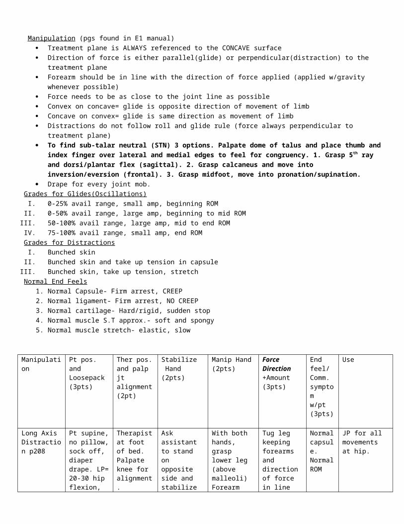
Manipulation (pgs found in E1 manual) Treatment plane is ALWAYS referenced to the CONCAVE surface Direction of force is either parallel(glide) or perpendicular(distraction) to the treatment plane Forearm should be in line with the direction of force applied (applied w/gravity whenever possible) Force needs to be as close to the joint line as possible Convex on concave= glide is opposite direction of movement of limb Concave on convex= glide is same direction as movement of limb Distractions do not follow roll and glide rule (force always perpendicular to treatment plane) To find sub-talar neutral (STN) 3 options. Palpate dome of talus and place thumb and index finger over lateral and medial edges
to feel for congruency. 1. Grasp 5th ray and dorsi/plantar flex (sagittal). 2. Grasp calcaneus and move into inversion/eversion (frontal). 3. Grasp midfoot, move into pronation/supination.
Drape for every joint mob. Grades for Glides(Oscillations)
I. 0-25% avail range, small amp, beginning ROMII. 0-50% avail range, large amp, beginning to mid ROM
III. 50-100% avail range, large amp, mid to end ROMIV. 75-100% avail range, small amp, end ROM
Grades for DistractionsI. Bunched skin
II. Bunched skin and take up tension in capsuleIII. Bunched skin, take up tension, stretch
Normal End Feels1. Normal Capsule- Firm arrest, CREEP2. Normal ligament- Firm arrest, NO CREEP3. Normal cartilage- Hard/rigid, sudden stop4. Normal muscle S.T approx.- soft and spongy5. Normal muscle stretch- elastic, slow
Manipulation Pt pos. and Loosepack (3pts)
Ther pos. and palp jt alignment(2pt)
Stabilize Hand(2pts)
Manip Hand(2pts)
Force Direction +Amount (3pts)
End feel/ Comm.symptomw/pt (3pts)
Use
Long Axis Distraction p208
Pt supine, no pillow, sock off, diaper drape. LP= 20-30 hip flexion, slight ABD
Therapist at foot of bed. Palpate knee for alignment.
Ask assistant to stand on opposite side and stabilize ASIS by first sliding skin down with palms and then push down and up.
With both hands, grasp lower leg (above malleoli) Forearm aligned with lower leg, tuck elbow and stagger stance (A/P)
Tug leg keeping forearms and direction of force in line with pt’s leg.
Normal capsule. Normal ROM
JP for all movements at hip.
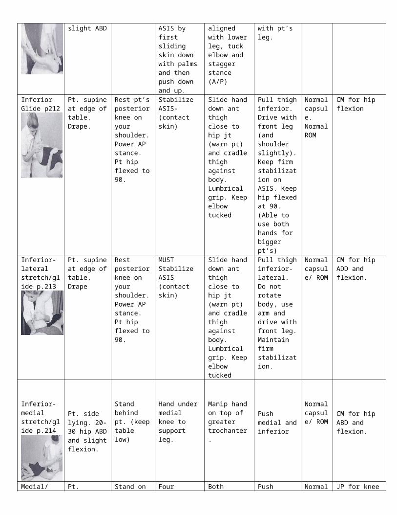
Inferior Glide p212
Pt. supine at edge of table. Drape.
Rest pt’s posterior knee on your shoulder. Power AP stance. Pt hip flexed to 90.
Stabilize ASIS-(contact skin)
Slide hand down ant thigh close to hip jt (warn pt) and cradle thigh against body. Lumbrical grip. Keep elbow tucked
Pull thigh inferior. Drive with front leg (and shoulder slightly). Keep firm stabilization on ASIS. Keep hip flexed at 90. (Able to use both hands for bigger pt’s)
Normal capsule. Normal ROM
CM for hip flexion
Inferior-lateral stretch/glide p.213
Pt. supine at edge of table. Drape
Rest posterior knee on your shoulder. Power AP stance. Pt hip flexed to 90.
MUST Stabilize ASIS (contact skin)
Slide hand down ant thigh close to hip jt (warn pt) and cradle thigh against body. Lumbrical grip. Keep elbow
Pull thigh inferior-lateral. Do not rotate body, use arm and drive with front leg. Maintain firm
Normal capsule/ ROM
CM for hip ADD and flexion.
axis distr.
tucked stabilization.
Inferior-medial stretch/glide p.214
Pt. side lying. 20-30 hip ABD and slight flexion.
Stand behind pt. (keep table low)
Hand under medial knee to support leg.
Manip hand on top of greater trochanter.
Push medial and inferior
Normal capsule/ ROM
CM for hip ABD and flexion.
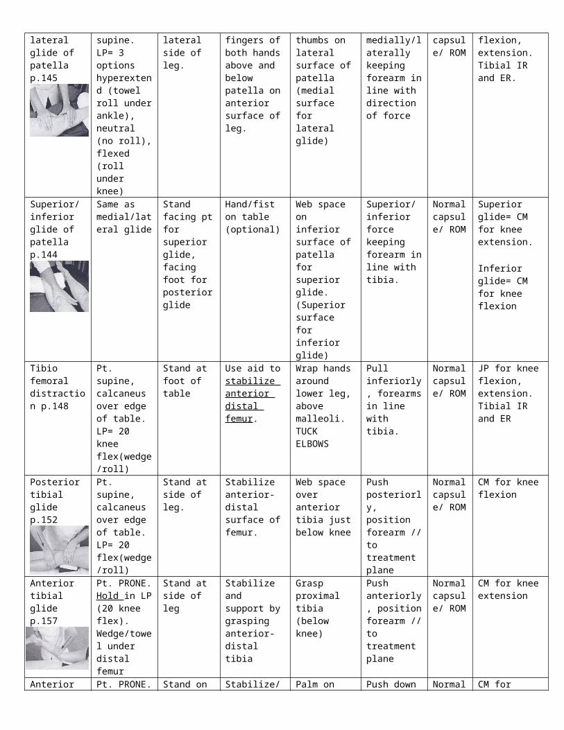
Medial/lateral glide of patella p.145
Pt. supine. LP= 3 options hyperextend (towel roll under ankle), neutral (no roll), flexed (roll under knee)
Stand on lateral side of leg.
Four fingers of both hands above and below patella on anterior surface of leg.
Both thumbs on lateral surface of patella (medial surface for lateral glide)
Push medially/laterally keeping forearm in line with direction of force
Normal capsule/ ROM
JP for knee flexion, extension. Tibial IR and ER.
Superior/inferior glide of patella p.144
Same as medial/lateral glide
Stand facing pt for superior glide, facing foot for posterior glide
Hand/fist on table (optional)
Web space on inferior surface of patella for superior glide. (Superior surface for inferior glide)
Superior/inferior force keeping forearm in line with tibia.
Normal capsule/ ROM
Superior glide= CM for knee extension.
Inferior glide= CM for knee flexion
Tibio femoral distraction p.148
Pt. supine, calcaneus over edge of table. LP= 20 knee flex(wedge/roll)
Stand at foot of table
Use aid to stabilize anterior distal femur.
Wrap hands around lower leg, above malleoli. TUCK ELBOWS
Pull inferiorly, forearms in line with tibia.
Normal capsule/ ROM
JP for knee flexion, extension. Tibial IR and ER
Posterior tibial glide p.152
Pt. supine, calcaneus over edge of table. LP= 20 flex(wedge/roll)
Stand at side of leg.
Stabilize anterior-distal surface of femur.
Web space over anterior tibia just below knee
Push posteriorly, position forearm // to treatment plane
Normal capsule/ ROM
CM for knee flexion
Anterior tibial glide p.157
Pt. PRONE. Hold in LP (20 knee flex). Wedge/towel under distal femur
Stand at side of leg
Stabilize and support by grasping anterior-distal tibia
Grasp proximal tibia (below knee)
Push anteriorly, position forearm // to treatment plane
Normal capsule/ ROM
CM for knee extension
Anterior glide of lateral tibial condyle p.158
Pt. PRONE. Thigh on wedge.Hold in LP (20 knee flex).
Stand on opposite side of table
Stabilize/support lower leg in 20 flex.
Palm on lateral surface of tibia.
Push down (anteriorly) forearm // to treatment plane
Normal cap, normal ROM
CM for tibial IR and knee flexion
Anterior glide of medial tibial condyle p.159
Pt. PRONE. Thigh on wedge. Hold in LP (20
Stand on same side of table
Stabilize/support lower leg in 20 flex.
Palm on medial surface of tibia.
Push down (anteriorly) forearm // to
Normal cap, normal
CM for tibial ER and knee extension
knee flex). treatment plane ROM
Posterior glide of lateral tibial condyle p.155
Pt. supine. Thigh under wedge. Heel off table
Stand on opposite side of table
Stabilize anterior distal thigh
Palm on lateral surface of tibia
Push down (posteriorly) forearm // to treatment plane
Normal cap, normal ROM
CM for tibial ER and knee extension
Posterior glide of medial tibial condyle p.154
Pt. supine. Thigh under wedge. Heel off table
Stand on same side of table
Stabilize anterior distal thigh
Palm on medial surface of tibia
Push down (posteriorly) forearm // to treatment plane
Normal cap, normal ROM
CM for tibial IR and knee flexion
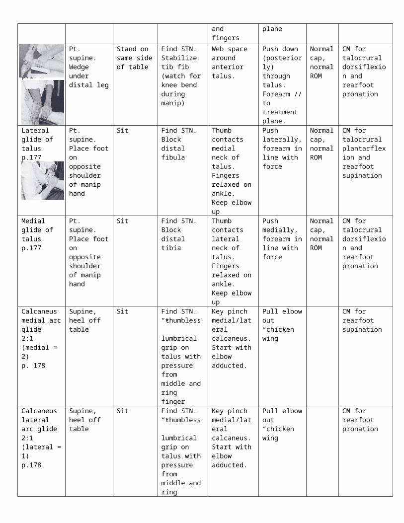
Anterior glide of talus p.175
Pt. PRONE. Wedge under distal leg
Stand on same side of table
Find STN. Stabilize tib-fib (watch for knee bend during manip)
Web space around post. talus. Contact ant talus with thumb and fingers
Push down (anteriorly) through talus. Forearm // to treatment plane
Normal cap, normal ROM
CM for talocrural plantarflexion and rearfoot supination
Posterior glide of talus p.174
Pt. supine. Wedge under distal leg
Stand on same side of table
Find STN. Stabilize tib fib (watch for knee bend during manip)
Web space around anterior talus.
Push down (posteriorly) through talus. Forearm // to treatment plane.
Normal cap, normal ROM
CM for talocrural dorsiflexion and rearfoot pronation
Lateral glide of talus p.177
Pt. supine. Place foot on opposite shoulder of manip hand
Sit Find STN. Block distal fibula
Thumb contacts medial neck of talus. Fingers relaxed on ankle. Keep elbow up
Push laterally, forearm in line with force
Normal cap, normal ROM
CM for talocrural plantarflexion and rearfoot supination
Medial glide of talus p.177
Pt. supine. Place foot on opposite shoulder of manip hand
Sit Find STN. Block distal tibia
Thumb contacts lateral neck of talus. Fingers relaxed on ankle. Keep elbow up
Push medially, forearm in line with force
Normal cap, normal ROM
CM for talocrural dorsiflexion and rearfoot pronation
Calcaneus medial arc glide2:1 (medial = 2)p. 178
Supine, heel off table
Sit Find STN. “thumbless” lumbrical grip on talus with pressure from middle and ring finger
Key pinch medial/lateral calcaneus. Start with elbow adducted.
Pull elbow out “chicken wing”
CM for rearfoot supination
Calcaneus lateral arc glide2:1 (lateral = 1)p.178
Supine, heel off table
Sit Find STN. “thumbless” lumbrical grip on talus with pressure from middle and ring
Key pinch medial/lateral calcaneus. Start with elbow adducted.
Pull elbow out “chicken wing”
CM for rearfoot pronation

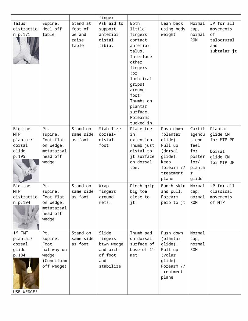
fingerTalus distraction p.171
Supine. Heel off table
Stand at foot of be and raise table
Ask aid to support anterior distal tibia.
Both little fingers contact anterior talus. Interlace other fingers (or lumbrical grips) around foot. Thumbs on plantar surface. Forearms tucked in.
Lean back using body weight
Normal cap, normal ROM
JP for all movements of talocrural and subtalar jt
Big toe MTP plantar/ dorsal glide p.195
Pt. supine. Foot flat on wedge, metatarsal head off wedge
Stand on same side as foot
Stabilize dorsal-distal foot
Place toe in extension. Thumb just distal to jt surface on dorsal toe.
Push down (plantar glide). Pull up (dorsal glide). Keep forearm // treatment plane
Cartilagenous end feel for posterior/ plantar glide
Plantar glide CM for MTP PF
Dorsal glide CM for MTP DF
Big toe MTP distraction p.194
Pt. supine. Foot flat on wedge, metatarsal head off wedge
Stand on same side as foot
Wrap fingers around mets.
Pinch grip big toe close to jt.
Bunch skin and pull. Forearm perp to jt
Normal cap, normal ROM
JP for all classical movements of MTP
1st TMT plantar/ dorsal glide p.184
USE WEDGE!
Pt. supine. Foot halfway on wedge (Cuneiform off wedge)
Stand on same side as foot
Slide fingers btwn wedge and arch of foot and stabilize
Thumb pad on dorsal surface of base of 1st met
Push down (plantar glide). Pull up (volar glide). Forearm // treatment plane
Normal cap, normal ROM
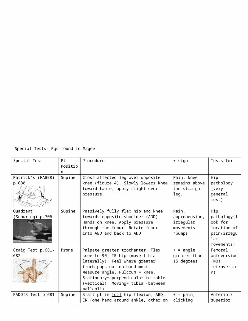
Special Tests- Pgs found in Magee
Special Test Pt Position Procedure + sign Tests forPatrick’s (FABER) p.680 Supine Cross affected leg over opposite knee (figure 4). Slowly
lowers knee toward table, apply slight over-pressure. Pain, knee remains above the straight leg.
Hip pathology (very general test)
Quadrant (Scouring) p.706 Supine Passively fully flex hip and knee towards opposite shoulder (ADD). Hands on knee. Apply pressure through the femur. Rotate femur into ABD and back to ADD
Pain, apprehension, irregular movements “bumps”
Hip pathology(look for location of pain/irregular movements)
Craig Test p.681-682 Prone Palpate greater trochanter. Flex knee to 90. IR hip (move tibia laterally). Feel where greater troch pops out on hand most. Measure angle. Fulcrum = knee. Stationary= perpendicular to table (vertical). Moving= tibia (between malleoli)
+ = angle greater than 15 degrees
Femoral anteversion (NOT retroversion)
FADDIR Test p.681 Supine Start pt in full hip flexion, ABD, ER (one hand around ankle, other on knee). Then take pt to full ADD and IR (maintain full hip flexion)
+ = pain, clicking Anterior/ superior impingement syndrome, anterior labral tear, iliopsoas tendinitis.
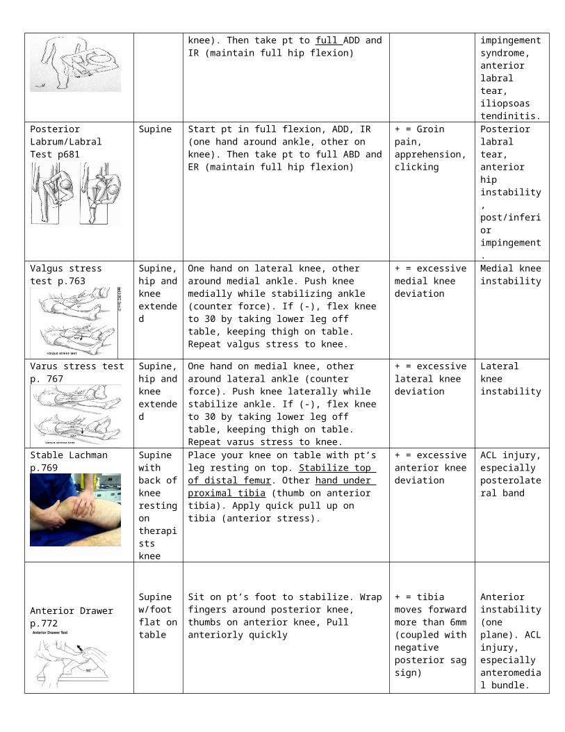
Posterior Labrum/Labral Test p681
Supine Start pt in full flexion, ADD, IR (one hand around ankle, other on knee). Then take pt to full ABD and ER (maintain full hip flexion)
+ = Groin pain, apprehension, clicking
Posterior labral tear, anterior hip instability, post/inferior impingement.
Valgus stress test p.763 Supine, hip and knee extended
One hand on lateral knee, other around medial ankle. Push knee medially while stabilizing ankle (counter force). If (-), flex knee to 30 by taking lower leg off table, keeping thigh on table. Repeat valgus stress to knee.
+ = excessive medial knee deviation
Medial knee instability
Varus stress test p. 767 Supine, hip and knee extended
One hand on medial knee, other around lateral ankle (counter force). Push knee laterally while stabilize ankle. If (-), flex knee to 30 by taking lower leg off table, keeping thigh on table. Repeat varus stress to knee.
+ = excessive lateral knee deviation
Lateral knee instability
Stable Lachman p.769 Supine with back of knee resting on therapists knee
Place your knee on table with pt’s leg resting on top. Stabilize top of distal femur. Other hand under proximal tibia (thumb on anterior tibia). Apply quick pull up on tibia (anterior stress).
+ = excessive anterior knee deviation
ACL injury, especially posterolateral band
Anterior Drawer p.772Supine w/foot flat on table
Sit on pt’s foot to stabilize. Wrap fingers around posterior knee, thumbs on anterior knee, Pull anteriorly quickly
+ = tibia moves forward more than 6mm (coupled with negative posterior sag
Anterior instability (one plane). ACL injury, especially anteromedial
sign) bundle. (Additional structures listed on pg 771)
Posterior Drawer p. 772 Supine w/foot flat on table
Same as anterior drawer except push tibia posteriorly. + = excessive posterior deviation
Posteriorly instability (one-plane). PCL (Additional structures listed on pg 772)
Posterior sag sign p. 773 Supine with both feet flat on table
Visual observation. + = tibia “drops back”/ sags on the femur
PCL instability, torn PCL
Godfrey test p.774 Supine Hold both legs, hip 90 knee 90 + = Posterior sag of the tibia
Posterior instability
Reverse Lachman p.774 PRONE, knee flexed to 30
Stabilize posterior-distal thigh. Grasp proximal tibia w/other hand and hold between arm and body. Pull up (posteriorly).
+ = excessive posterior deviation
PCL instability
Lateral pivot shift p.776 Supine w/hip flexed and ABD 30, slight IR. Knee flexed 90.
Hold pt in test position by supporting foot (under calcaneus) with one hand, other hand behind fibula (heel of hand under lateral gastroc head) causing IR of leg. Hold tibia in IR as you extend knee. Then apply valgus stress to knee and flex knee to 30-40
+ = subluxation at extension. Tibia reduces/”jogs” backward when flexed.
ACL ruptures (3rd degree sprain), anterolateral rotary instability
McMurray test p.791 Supine w/knee fully flexed. Hip flexed 90.
IR tibia, then extend knee to test lateral meniscus. Repeat with ER of tibia to test medial meniscus.
Pain, snapping, clicking
Meniscus lesions
Apley’s Test p.791 PRONE w/knee flexed to 90.
Place your knee on pt’s thigh. Wrap both hands around ankle, lift (distract) and rotate medially and laterally. Then push down (compress) and rotate med/lat.
+ = Pain Meniscus lesion of + with compression. Ligamentous if + with distraction
Plica “stutter” test p.795 Seated with leg off table
Place finger over medial patella. Instruct pt to slowly extend
+ = patella stutters/jumps btwn 45-60 flexion
Presence of plica remnants
Brush test p796
Seated, knee extended
Sweep medial side of patella upward 3 times. Then downward on lateral side of patella w/opposite hand.
+ = extra wave of fluid bulging on medial knee
Swelling in knee (minimal effusion)
Indentation test p.797 Supine, knee extended.
Passively flex knee, watch for indentation on the lateral side of the patellar tendon to disappear (should not disappear in normal knee). Observe good leg 1st
+ = indentation disappears (sooner indentation = greater the swelling
Swelling in knee
Patellar tap test p.798 Supine, knee extended
Tap patella to push fluid down. + = floating/dancing patella
Swelling in knee
Patellar grind/ compression test p.798
Supine, knee extended
GENTLY press patella down. Have pt contract quad Pain Patellofemoral dysfunction
Noble compression test p.803
Supine Passively flex knee to 90. With thumb, apply pressure 1 cm proximal to lateral femoral epicondyle and passively extend knee (maintaining pressure).
+ = severe pain at 30 flexion (pt states same pain occurs w/activity)
IT band syndrome
Anterior drawer (ankle) p.888 Pt supine, heels off table.
Stabilize anterior distal tib-fib with one hand. Cup calcaneus with other. Quickly pull up on heel
+ = pain, excessive anterior deviation
ATFL injury
Homan’s sign p.897 Supine, knee extended
Passively dorsiflex foot with hand under heel and bottom of foot against forearm. (make sure knee is extended) Stabilize distal tib fib.
+ = pain in calf DVT
Tinel’s sign p. 896 Supine Tap front of ankle (deep peroneal n.) Tap behind medial malleolus (posterior tibial n.)
+ = tingling/ paresthesia
Deep peroneal n./ posterior tibial n.
Thompson’s test p.894 PRONE Squeeze calf Absence of PF Achilles tendon tear (3rd degree)
Palpation Use one finger to identify
Structure Location ConfirmaitonGreater trochanter Pt prone. Flex knee and IR/ER leg to feel GT rotate (expose
skin)Moves with IR/ER of leg
Ischial tuberosity Pt prone. Drape. Take heel of hand to gluteal fold and apply pressure.
ASIS Supine. Drape. Have pt tuck towel and expose ASIS. Use thumbs and roll up in ASIS
Adductor tubercle Superior attachment to MCLMeniscusITB Insertion at Gerdy’s tube. Runs up btwn VL and hamsMed/lat jt lines Thin gap btwn tibia and femurPatellar lig Thick lig under patellaTibial tubercle Big bump just below patella femoral ligLateral tibial condyle Have pt knee 90. On either side below patellaMedial tibial condyle Have pt knee 90. Gurdy’s tubercle Bump on lateral tibial condyleFibular head On lateral proximal portion of lower leg IR/ER lower leg to feel fib head rotateBiceps femoris Only tendon on lateral side of posterior kneeLateral/medial femoral condyle
Have pt knee 90. On either side of middle patella
Lateral/medial femoral epicondyle
Have pt knee 90. On either side superior to patella
LCLMCL (stress at 0 and 30)Lateral meniscusMedial meniscus w/ IR/ERPes anserine tendon On medial knee around where knee bendsATFL Btwn lateral malleolus and talus Plantarflex and invert footCFL Below and posterior to lateral malleolusDorsalis pedis a. Btwn big toe and 2nd toe, just inferior to ankle jt
Neck of talus (med and lat) Have pt PF. Btwn both malleolisSinus taris Gap below lateral malleolusBase of 5th met (peroneus brevis attachment site)
Bump on 5th met
Navicular tuberosity High arch bone
MMT For knee flexors have pt seated, rest pt’s foot on your knee. LP= 20 knee flexion Same position for knee extensors, except move your leg before applying force
Loose packed position Therapist position
Contact Procedure
Knee flexors (medial/lateral hams, popliteus)
20 knee flexion Seated 2 finger contact to posterior mid/lower leg
Apply 3-5 lbs pressure/force toward extension
Lower leg ER (lateral hams) 20 knee flexion Seated 2 finger contact to lateral-posterior mid/lower leg
Apply 3-5 lbs pressure/force toward IR and
Lower leg IR (medial hams, popliteus)
20 knee flexion Seated 2 finger contact to medial-posterior mid/lower leg
Apply 3-5 lbs pressure/force toward ER and
Knee extensors (quads) 20 knee flexion Seated 2 finger contact to anterior mid/lower leg
Apply 3-5 lbs pressure/force toward flexion