Embed Size (px)
Citation preview

1
VILNIUS UNIVERSITY
DEPARTMENT OF ANALYTICAL AND ENVIRONMENTAL
CHEMISTRY, FACULTY OF CHEMISTRY
VIKTOR MAŽEIKO
POLYANILINE AND POLYPYRROLE, GOLD NANOPARTICLES
AND DIFFERENT ELECTRON TRANSFER MEDIATORS
APPLICATION IN GLUCOSE BIOSENSORS DESIGN
Summary of doctoral dissertation
Physical sciences, chemistry (03 P)
Vilnius, 2015

2
The research was carried out in Vilnius University, Faculty of Chemistry, Department of
Analytical and Environmental Chemistry in the period of 2010 – 2014.
Scientific supervisor - Doc. dr. Asta Kaušaitė - Minkštimienė (Vilnius University, Physical
Sciences, Chemistry – 03 P)
The defense council - Prof. dr. Stasys Tautkus (Vilnius university, Physical sciences,
Chemistry – 03P)
Members:
1. Prof. habil. dr. Albertas Malinauskas (Center for physical sciences and technology,
Physical sciences, Chemistry – 03P)
2. Prof. dr. Genė Biziulevičienė (Centre for Innovative Medicine, Biomedicine
sciences, biology – 01B)
3. dr. Germanas Peleckis (The Institute for Superconducting and Electronic Materials,
University of Wollongong)
4. Prof. dr. Vida Vičkačkaitė (Vilnius university, Physical sciences, Chemistry – 03P)
The defense of the dissertation will be held at 2 p.m. on 25th of September 2015 at the
public meeting of the council at the Faculty of Chemistry of Vilnius University. Address:
Naugarduko 24, LT-03225,Vilnius, Lithuania.
The summary of the dissertation was mailed on the 11th of August, 2015. The dissertation is
available at the Library of Vilnius University and at the libraries of Center for physical
sciences and technology and VU internet page adress: www.vu.lt/lt/naujienos/ivykiu-
kalendorius

3
VILNIAUS UNIVERSITETAS
FIZINIŲ IR TECHNOLOGIJOS MOKSLŲ CENTRO CHEMIJOS
INSTITUTAS
VIKTOR MAŽEIKO
POLIANILINO IR POLIPIROLO, AUKSO NANODALELIŲ BEI
ĮVAIRIŲ ELEKTRONŲ PERNAŠOS TARPININKŲ TAIKYMAS
GLIUKOZĖS BIOLOGINIUOSE JUTIKLIUOSE
Daktaro disertacijos santrauka
Fiziniai mokslai, chemija (03 P)
Vilnius, 2015 metai

4
Disertacija buvo ruošiama 2010 – 2014 metais Vilniaus universitete, Chemijos
fakultete, Analizinės ir aplinkos chemijos katedroje.
Mokslinė vadovė – doc. dr. Asta Kaušaitė–Minkštimienė (Vilniaus universitetas,
fiziniai mokslai, chemija – 03 P)
Disertacija ginama Vilniaus universiteto Chemijos mokslo krypties taryboje:
Pirmininkas – prof. dr. Stasys Tautkus (Vilniaus universitetas, fiziniai mokslai, chemija –
03P)
Nariai:
1. prof. habil. dr. Albertas Malinauskas (Fizinių ir technologijos mokslų centro
Chemijos institutas, fiziniai mokslai, chemija – 03P)
2. prof. dr. Genė Biziulevičienė (Valstybinis mokslinių tyrimų institutas Inovatyvios
medicinos centras, biomedicinos mokslai, biologija – 01B)
3. dr. Germanas Peleckis (Australijos Superlaidžių ir Elektroninių Medžiagų Institutas,
Volongongo universitetas, fiziniai mokslai, chemija – 03P)
4. prof. dr. Vida Vičkačkaitė (Vilniaus universitetas, fiziniai mokslai, chemija – 03P)
Disertacija bus ginama viešame Chemijos mokslo krypties tarybos posėdyje 2015
m.rugsėjo mėn. 25 d. 14 val. Vilniaus universiteto Chemijos fakulteto bendrosios ir
neorganinės chemijos auditorijoje.
Adresas: Naugarduko g. 24, LT-03225 Vilnius, Lietuva.
Disertacijos santrauka išsiuntinėta 2015 m. rugpjūčio mėn. 11 d.
Disertaciją galima peržiūrėti Vilniaus universiteto, Fizinių ir technologijos mokslų centro
bibliotekose ir VU interneto svetainėje adresu: www.vu.lt/lt/naujienos/ivykiu-kalendorius

5
INTRODUCTION
Diabetes mellitus, or simply diabetes, is a group of metabolic diseases characterized
by high blood glucose levels that result from defects in the body's ability to produce and/or
use insulin. Diabetes is a worldwide public health problem, because it is one of the leading
causes of death and disability in the world. The diagnosis of this disease requires a precise
detection of blood glucose concentration. Electrochemical biosensing systems are the most
suitable for the determination of analytes in complicated and complex samples including
blood, blood serum, etc. Among electrochemical biosensing systems enzymatic
amperometric glucose biosensors are the most common devices commercially available, and
have been widely studied over the last few decades. These biosensors are usually based on
the two enzyme families, glucose oxidase (GOx) and glucose-1-dehydrogenase (GDH).
Because of a relatively higher selectivity for glucose over other blood sugars and stability,
more simple purification procedure and lower price, better resistance in extremes pH, ionic
strength and temperature than many other enzymes], GOx is the most popular enzyme for
the modelling of biosensors. GOx is a dimmeric protein containing one tightly bound flavin
adenine dinucleotide (FAD) per monomer as cofactor. FAD functions as a coenzyme
because of its ability to undergo reversible redox reactions. The redox active centre of the
FAD is the isoalloxazine ring system. The basic concept of the glucose biosensor is based
on the fact that the immobilized flavoprotein GOx catalyses the oxidation of β-D-glucose to
D-glucono-δ-lactone, which is non-enzymatically hydrolyzed to β-D-gluconic acid, and
hydrogen peroxide using molecular oxygen as an electron acceptor.
Immobilization of enzymes and establishment of electron transfer (ET) are the most
challenging and important steps in the development of amperometric biosensors.
Immobilized enzymes have many operational advantages if compared with dissolved
enzymes including possible reusability, continuous operational mode, easy separation from
the reaction mixture, and possible modulation of the catalytic properties. Fast, simple and
low-cost detection of biologically active analytes is the major advantage of biosensing
systems. In some cases a combination of nanomaterials and nanotechnological approaches
resolve challenging bioanalytical problems, including specificity, stability and sensitivity.
6
Some conjugated polymers provide an effective immobilization patterning for enzymes on
surfaces of different electrodes and in some cases facilitate ET from enzymes to
electronically conductive electrodes and improve biosensor sensitivity. Composite
nanomaterials exhibit improved physical and chemical properties over their single-
component counterparts, and hence they are potentially useful in a wide range of
applications including analytical, bioanalytical systems and biofuel cells. Conducting
polymer-based layers on the electrode surface are traditionally synthesized by
electrochemical or chemical synthesis. Both, electrochemical and chemical synthesis,
require high concentrations of monomers and usually are performed at extremely low pH
values. In traditional chemical synthesis of conducting polymers, usually toxic catalysts or
strong oxidizing agents are used. Therefore enzymatic synthesis of conducting polymers is
applied as an alternative method suitable for the formation of composite materials based on
conducting polymers such as polyaniline (PANI) and polypyrrole (PPY).
Despite of numerous scientific papers and other informational issues related to
improvement of biosensor performance using advanced redox mediators, the most
significant part of this information is not showing clear comparison between different
mediators influence in the biosensor response to substrate. Consequently, one of the
objectives of this study was to evaluate the effect of few different mediators on the response
of GOx-based amperometric biosensor.
The aim of the work:
Apply π-π conjugated polymers polyaniline and polypyrrole, gold nanoparticles and
different electron transfer mediators in amperometric glucose biosensor
Main tasks of the work:
Investigate the formation of polyaniline and polypirole and encapsulate glucose
oxsidase immobilized on electrode surface within the polymer layer;
Determine and compare the influence of polyaniline and polypirole layer to the
characteristics of amperometric glucose biosensor;

7
Synthesize composite particles of glucose oxsidase encapsulated within polyaniline
layer and glucose oxsidase with gold nanoparticles encapsulated within polyaniline
layer and determine the influence of gold nanoparticles on polyaniline formation rate;
Compare the influence of synthesized composite structures on amperometric glucose
biosensor analytic characteristics;
Investigate the influence of different electron transfer mediators immobilized on carbon
electrode surface on amperometric glucose biosensor analytic characteristics;
Statements to be defended:
Polypyrrole and polyaniline layer formed on graphite electrode surface modified by
glucose oxidase, extends concentration range of linear dependence of analytical signal for
glucose biosensors, increases stability and improves the repeatability of analytical signal;
Gold nanoparticles not only increases the enzymatic aniline polymerization reaction
rate, but also being as a part of glucose oxidase and polyaniline nanocomposite structures,
during the enzymatic oxidation of glucose, ensure efficient transport of electrons from the
glucose oxidase redox center flavine adenine dinucleotide to the electrode, and thus
increases the biosensor analytical signal;
Electron transfer mediators tetrathiafulvalene, phenazine methosulfate, 5,6-diamin-
1,10-phenanthroline, tetrathiafulvalene - tetracyanoquinodimethane complex, methylene
blue, toluidine blue and potassium ferrocyanide immobilized on the surface of the graphite
electrode enables efficient transport of electrons from the glucose oxidase redox center to
the electrode surface . The most effective electron transfer has tetrathiafulvalene and
tetrathiafulvalene - tetracyanoquinodimethane complex.
EXPERIMENTAL
Electrode pre-treatment prior to modification
Graphite rod electrodes 3mm in diameter, 150mm in length, 99.999% pure and low
density were obtained from SIGMA–ALDRICH, Inc. (St. Louis, MO, USA). Graphite rod
electrodes were sealed into epoxy to prevent the contact of electrode side surface with the
solution. Working surface area of graphite electrodes was 0.7mm2. Before the formation

8
graphite electrodes were prepared as follows: first, rods of graphite were cut and polished on
a fine emery paper and then polished by Al2O3 slurry (grain size0.1µm), followed by rinsing
the electrode surface with ethanol and distilled water. Electrodes were dried at room
temperature before coating them with enzymes.
Electrode modification by GOx
During the preparation of GOx-modified electrodes (CR/GOx electrodes), 3 µl of 40
mg/ml of enzyme solution in water were deposited on the electrode and later water was
evaporated at room
temperature. Then electrodes were stored for 24 h over the 5% solution of glutar
aldehyde at 4 ◦C in the closed vessel. The lateral surface of the electrode was isolated with
silicone tube to prevent the contact of electrode side surface with the solution and working
surface area of modified electrodes was 7.065 mm2. Prior to electrochemical measurements
CR/GOx electrodes were thoroughly washed with distilled water to remove non-cross-
linked enzyme and stored at 4 ◦C in the closed vessel hanging over a drop of A-PBS buffer,
pH 6.0.
Modification of GOx coated electrode by PANI or PPY layer
For the covering of CR/GOx electrodes by polymer layer these electrodes were
immersed into A-PBS buffer, pH 6.0,containing 20 mM of glucose and 200 mM of aniline
(CR/GOx/PANI electrodes) or 200 mM of pyrrole (CR/GOx/PPY electrodes) at 4 ◦C, for a
defined period, lasting from 0 to 272 h. Prepared electrodes were thoroughly washed with
distilled water and were stored at 4 ◦C in a closed vessel hanging over a drop of A-PBS
buffer, pH 6.0, until they were used in experiments.
Investigation of enzymatic polymerisation of aniline and pyrrole in solution
Polymerisation was performed in A-PBS buffer, pH 6.0, containing 1 mg/ml of
glucose oxidase, 20 mM of glucose and 200 mM of suitable monomer. Synthesis was
carried out at room temperature in darkness. The UV–Vis spectra of solutions were recorded
after 48 h from the beginning of polymerization process. UV–vis spectrophotometer Perkin-
Elmer LAMBDA 25 (Shelton, USA) was usedfor monitoring of polymers formation.
Formation of GOx/PANI and GOx/Au-NPs/PANI nanoparticles

9
A number of different polymerization solutions were prepared for the selection of the
optimal polyaniline formation pHs. For this, A-PBS buffers were prepared at pH values of
4.0; 4.5; 5.0; 6.0; 7.0; 8.0. In order to get PANI-based (GOx)PANI and (GOx/AuNP)PANI
nanoparticles, enzymatic formation of PANI was applied. To obtainthe (GOx)PANI
nanoparticles, the polymerization bulk solution was consisted of 200 mM aniline, 1 mg/mL
GOx and 20 mM glucose. In order to form (GOx/AuNP)PANI nanocomposite, AuNP were
added into the polymerization solution. A fixed concentration of 7.5 mg/mL of different size
(3.5 nm; 6 nm; 13 nm) gold nanoparticles was used in the polymerization solution with a
total volume of 1.0 mL. The polymerization reaction was carried out at +4◦C in the dark.
After 24 h reaction time, the solution was centrifuged, and all formed nanocomposites were
separated from the polymerization solution and transferred into 100 µL volume of buffer for
additional washing procedures. After a second centrifugation procedure, the washing buffer
solution was removed and all synthesized particles were mixed thoroughly in 100 µL of
buffer. Prepared (GOx)PANI particle solution was applied in the design of the
CR/(GOx)PANI electrodes and (GOx/AuNP)PANI nanoparticle solution for the formation
of the (GOx/AuNP)PANI electrodes.
Electrode modification by GOx and mediators
Two methods were used for preparation of modified electrodes. During preparation
by the first method, 3.0 µL of mediator solution were dropped and distributed on the
electrode surface and solvent evaporated at room temperature by intensive ventilation. Then
3.0 µL of GOx solution were deposited and solvent evaporated, and finally, after complete
drying, the electrode was left in a closed vessel at 2 cm above the 5 % GA solution at +4°C
for 24 h. Then the modified electrode (CR/M/GOx) was thoroughly washed with distilled
water to remove non-cross-linked enzyme. Prepared electrodes were stored in closed test-
tubes over a drop of A-PBS, pH 6.0, at +4°C between electrochemical measurements. In
order to get modified electrodes by the second method (CR/M-GOx), solutions of enzyme
and mediator were mixed in a ratio or 1:1 and 3.0 µL of prepared solution were dropped and
distributed on the electrode surface twice with evaporating between each depositing. Then
the electrodes were incubated in glutar aldehyde vapor and stored as previously described.

10
Electrochemical detection of analytical signal
All electrochemical measurements were performed by potentiostat-galvanostat
PGSTAT 30 with GPES3 v3.2 software ECO-Chemie/Autolab (Utrech, Netherlands). For
the registration of amperometric signal three-electrode circuits were applied. The modified
graphite electrode was switched as the working one, Ag/AgCl electrode as reference and 2
cm2 Pt electrode was used as an auxiliary one. Electrochemical signals were detected in
0.05M sodium acetate and sodium/potassium phosphate (A-PBS) buffer solution, pH 6.0
containing 0.1M KCl. Solution in electrochemical cell was mixed at 120 rpm.
Electrochemical detection of analytical signal was performed at room temperature at
+300mV vs. Ag/AgCl in presence of 10mM of phenasine methoslulphate (PMS) and
different concentrations of glucose.
Special conditions used for determination of pH dependence
Modified electrodes were tested in A-PBS buffer solution contained 0.1M of KCl,
with fixed pH value. Electrochemical detection of analytical signal was performed similarly
as it is presented in previous section.
Special conditions in lifetime and stability test
Modified electrodes were stored between the measurements at 4 ◦C in closed vessel
hanging over A-PBS buffer solutions, pH 6.0. Electrochemical detection of analytical signal
was performed similarly as it is presented in previous section.
Special conditions in temperature influence test
For this experiment modified electrodes were prepared by polymerization lasting for
24, 48 and 72 h. The effect of temperature variations on the enzyme activity of modified
electrodes were tested by heating of these electrodes at particular temperature for 5 min in
laboratory incubator “incucell 55-standart” from “Blue Line” (MMM Medcenter
Einrichtungen GmbH, Germany) in air atmosphere and in A-PBS buffer solutions, pH 6.0.
The imaging by scanning electron microscopy
Modified graphite electrodes were examined using JEOL JSM-7600F scanning
electron microscope. Prior to investigations the modified electrodes were thoroughly
washed with distilled water and then dried at room temperature.

11
The imaging by atomic force microscopy
Tapping mode atomic force microscopy was used for the imaging of differently
modified graphite electrode surfaces. The BioScope II, Veeco Instruments Ltd. (Santa
Barbara, USA) was used for all AFM experiments. For all AFM Images 200 Images
200×200 pixels image resolution was applied, scanning rate 10 µm/s. Experimental data
were processed by diNanoScope 7.30 and Gwyddion 2.10 NT-MDT Nova programs. Sharp
silicon probes ideal for Tapping Mode were used for all measurements.
RESULTS AND DISCUSSION
Glucose oxidase (GOx) is a FAD-dependent enzyme that catalyzes oxidation of ˇ-d-
glucose by molecular oxygen to hydrogenperoxide and d-glucono-1,5-lactone (1) which
subsequently hydrolyzes spontaneously to gluconic acid. In this work section study catalytic
activity of GOx immobilized on graphite electrode by cross-linking with glutar aldehyde
was exploited for polymerisation of aniline. The main precursors for initiation of
polymerization reaction was hydrogen peroxide as an initiator of polymerization reaction (3)
and gluconic acid as a medium what reduced pH towards the acidic pH suitable for template
polymerization of aniline. All mentioned precursors were produced during the catalytic
action of immobilized GOx (1) and following hydrolization of reaction products. Since
aniline has a pKa of 4.63, whereas GOx has a pI of 4.2 at a pH lower than 4.63 aniline is
positively charged and GOx at a pH higher than 4.2 is negatively charged. In the presence of
glucose and dissolved oxygen immobilized on graphite electrodes GOx generated hydrogen
peroxide and gluconolactone, which hydrolyzes to gluconic acid (Fig. 1). In consequence of
gluconolactone hydrolysis it might be predicted that the pH decreased locally because of the
formation of gluconic acid, while hydrogen peroxide concentration increased close to the
immobilized GOx active site and the interfaces between electrode/enzyme/solution.

12
Fig. 1 Principle scheme illustrating modification of GOx coated electrode by PANI.
It is presumptive that locally lowered pH (4.3–4.5) and high concentration of
oxidator (H2O2) created optimal conditions for the polymerizations of aniline and increased
the probability of GOx encapsulation within formed PANI to form CR/GOx/PANI
electrode. Thus, our proposed GOx encapsulation method was at least partially based on
electrostatic alignment of aniline monomer onto anionic immobilized GOx template to
minimize branching and promote linear PANI chains growth.
22
GOx2 OHlactone-1,5-glucone-DOgliucose-D-β (1)
For confirmation of insight that under such favourable conditions the polymerizations
of aniline might be initiated a basic amperometric biosensor design was selected. In our
opinion it was the most appropriate way for this kind of estimation: (i) it needs just a very
small amount of materials; (ii) the same electrode might be applied for several
measurements; (iii) modification of electrodes might be performed at similar conditions; (iv)
free diffusing and/or not encapsulated enzyme might be washed out fromthe sensor surface.
The cross-linking GOx by glutaraldehyde was applied since it can stabilize the steric
structure of enzyme and to avoid the denaturation of it under relatively severe conditions.

13
Fig. 2. (A) Glucose calibration curves obtained with a GOx modified graphite electrode,
before treatment (curve 8) and after treatment with 200 mM aniline and 20 mM glucose
solution at 4 ◦C temperature (curve 7–10 min, 6–70 min, 5–18 h,4–43 h, 3–113 h, 2–181 h
and 1–272 h). Calculated KM(apparent) (B) and Imax (C) values for glucose vs. duration of
incubation in control solution 1 at 4 ◦C (curve 1), control solution 2 at 4 ◦C (curve 2) and
polymerization solution at 4 ◦C (curve 3) and at 18 ◦C (curve 4). Experiments presented in
plot ‘A’ were performed at +300mV vs. Ag/AgCl in A-PBS buffer solution, pH 6.0,
contained 100 mM of KCl and 10 mM of PMS
The peculiarity of the present work section is that formation of PANI layer on GOx
modified electrodes occurs only in the case if the GOx modified electrode is immersed in
polymerization solution containing both glucose and aniline. The main evidence of
polymerization process on GOx modified electrodes was an increase of apparent Michaelis–
Menten constant (KM(apparent)) and decrease in maximum current measured under saturated
analyte (glucose) conditions (Imax). Kinetic properties of GOx acting as a biocatalyst in the
GOx-electrode and GOx/PANI-electrode were analyzed at room temperature using different
concentrations of glucose(0.05–305 mM). The results obtained are shown in Fig. 2 and
Table 1. The amperometric signals showed hyperbolic dependence on glucose concentration
(Fig. 2A), this was in agreement with Michaelis–Menten kinetics. The kinetic parameters
Imax (correspond to Vmax) and KM(apparent) are correspondingly a and b parameters of

14
hyperbolic function y = ax/(b + x). According to culations presented in Fig. 2B and Table 1
CR/GOx and CR/GOx/PANI modified electrodes exhibit significantly different KM(apparent)
values. As seen from the Table 1 for electrode based on cross-linked GOx calculated values
of KM(apparent) and Imax were around 5.75mM and 51.72µA, respectively. While the
KM(apparent) for the CR/GOx/PANI modified electrode might be extended up to 134.90mM, if
polymerization was carried out at 4 ◦C temperature (Fig. 2B curve 3) and up to 356.00mM,
if polymerisation is carried out at 18 ◦C temperature (Fig. 2B curve 4). An opposite effect
was obtained for the Imax values. The results presented in Fig. 2C demonstrate than the Imax
can decrease from 51.72µA (CR/GOx) up to 3.06 µA or 0.42µA, if polymerization is
carried out at 4 ◦C or 18◦C temperature, respectively. The comparison of kinetic parameters
of cross-linked CR/GOx and CR/GOx/PANI based electrodes let us to state that in the case
of CR/GOx/PANI Imax decreased by 16.69 or 121.59 times and at the same time KM(apparent)
increased by 23.46 or 61.91 times (after 270 h of polymerization). Such significant increase
of KM(apparent) and decrease of Imax are demonstrating that diffusion limitations in this case
of CR/GOx/PANI are playing a significant role. An increment of KM(apparent) by over 10
times might be exploited as the evidence that the immobilized on graphite electrode surface
GOx was entrapped within formed PANI layer. Moreover, due to diffusion limitations
increased KM(apparent) caused significant increases in linear range of CR/GOx/PANI based
analytical system if it is compared with cross-linked GOx based system. Such significant
extensions of analyte detection intervals are especially relevant for detection of glucose
concentration in food and beverage samples. On the other hand, increased diffusion
limitations caused the decrease in maximal current generated by CR/GOx/PANI based
electrode because of hindered diffusion of both: glucose and redox mediator phenasine
methoslulphate (PMS). Detected thickness of the PANI layer was 0,4µm, 0.8µm and 1.1µm
after 18 h, 113 h and 181 h of polymerization period correspondingly. No linear dependence
between polymerization period and/or thickness of film and/or KM(apparent) as well as with
Imax was determined. It means that morphology of PANI film has changed and/or internal
density of formed polymer layer has increased during here reported formation of PANI film.
The results are in principle agreement with theoretical studies, which have predicted that
15
after embedment of enzyme into conducting polymer film, the thickness of enzymatically
active layer is typically 200–400 nm due to limitations in the diffusion of the analyte and
reaction products in/out from the film.
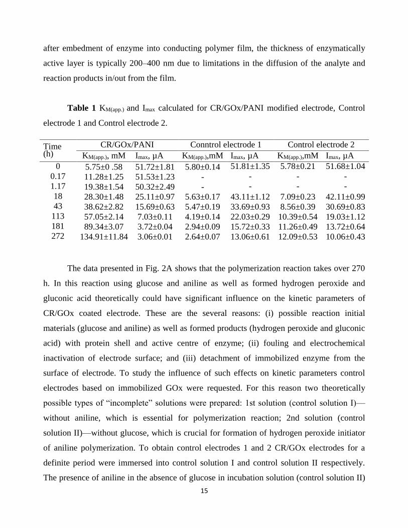
Table 1 KM(app.) and Imax calculated for CR/GOx/PANI modified electrode, Control
electrode 1 and Control electrode 2.
Time (h)
CR/GOx/PANI Conntrol electrode 1 Control electrode 2
KM(app.), mM Imax, µA KM(app.),mM Imax, µA KM(app.),mM Imax, µA
0 5.75±0 .58 51.72±1.81 5.80±0.14 51.81±1.35 5.78±0.21 51.68±1.04
0.17 11.28±1.25 51.53±1.23 - - - -
1.17 19.38±1.54 50.32±2.49 - - - -
18 28.30±1.48 25.11±0.97 5.63±0.17 43.11±1.12 7.09±0.23 42.11±0.99 43 38.62±2.82 15.69±0.63 5.47±0.19 33.69±0.93 8.56±0.39 30.69±0.83
113 57.05±2.14 7.03±0.11 4.19±0.14 22.03±0.29 10.39±0.54 19.03±1.12 181 89.34±3.07 3.72±0.04 2.94±0.09 15.72±0.33 11.26±0.49 13.72±0.64 272 134.91±11.84 3.06±0.01 2.64±0.07 13.06±0.61 12.09±0.53 10.06±0.43
The data presented in Fig. 2A shows that the polymerization reaction takes over 270
h. In this reaction using glucose and aniline as well as formed hydrogen peroxide and
gluconic acid theoretically could have significant influence on the kinetic parameters of
CR/GOx coated electrode. These are the several reasons: (i) possible reaction initial
materials (glucose and aniline) as well as formed products (hydrogen peroxide and gluconic
acid) with protein shell and active centre of enzyme; (ii) fouling and electrochemical
inactivation of electrode surface; and (iii) detachment of immobilized enzyme from the
surface of electrode. To study the influence of such effects on kinetic parameters control
electrodes based on immobilized GOx were requested. For this reason two theoretically
possible types of “incomplete” solutions were prepared: 1st solution (control solution I)—
without aniline, which is essential for polymerization reaction; 2nd solution (control
solution II)—without glucose, which is crucial for formation of hydrogen peroxide initiator
of aniline polymerization. To obtain control electrodes 1 and 2 CR/GOx electrodes for a
definite period were immersed into control solution I and control solution II respectively.
The presence of aniline in the absence of glucose in incubation solution (control solution II)
16
resulted in an increase of KM(apparent) for control electrode 2, but in this case the increase of
KM(apparent) was over 11 times (Fig. 2B curve 2, Table 1) lower at 4 ◦C (Fig. 2B curve 3 and
Table 1) and almost 30 times lower at 18 ◦C temperature (Fig. 2B curve 4) when compared
with that obtained using both components (glucose and aniline). The decrease of Imax for the
control electrode 2 was 3.29 times smaller (Fig. 2C curve 2 and Table 1) if compared with
that obtained for CR/GOx/PANI-electrode when polymerization was carried out at 4 ◦C
(Fig. 2C curve 3, Table 1). If glucose was present and aniline was absent in incubation
solution (control solution I) the KM(apparent) calculated for control electrode 1 after 272 h of
incubation period was smaller if compared with KM(apparent) calculated for control electrode 2
and CR/GOx/PANI electrode 5 and 51 times, respectively (Table 1). Additionally in
contrast to the results represented above for control electrode 2 and CR/GOx/PANI
electrode during incubation the KM(apparent) calculated for control electrode 1 has decreased
three-times. It can be explained by slow degradation of GOx layer, which became thinner
and diffusion through it become faster. Similar results were achieved when bare buffer
solution without any aniline or glucose was used. After 272 h of incubation the calculated
KM(apparent) value was 2.64 mM (Fig. 2C curve 1, Table 1) which is close to KM of native
GOx. It means that the GOx layer became so thin that sensor started to act in kinetically
controlled mode instead of previously detected diffusion controlled mode.
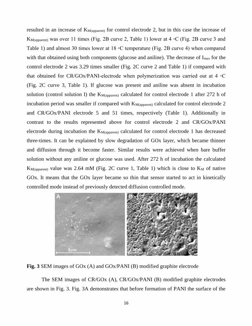
Fig. 3 SEM images of GOx (A) and GOx/PANI (B) modified graphite electrode
The SEM images of CR/GOx (A), CR/GOx/PANI (B) modified graphite electrodes
are shown in Fig. 3. Fig. 3A demonstrates that before formation of PANI the surface of the

17
electrode was doughy due to non-conducting GOx layer, which scattered the electrons. Fig.
3B shows that the morphology and conductivity of electrode surface has been changed
significantly after the formation of PANI layer. Different electron scattering in Fig. 3A and
B shows that surface of PANI/GOx-electrode is more conducting if compared with the
surface of GOx-electrode. The morphology PANI/GOx-electrode clearly shows that some
new polymeric structures appeared on the electrode surface. AFM images (Fig. 4A1–C1)
and from these images derived height distribution diagrams (Fig. 4A2–C2) clearly
demonstrates changes in morphology during immobilization of GOx (Fig. 4A and B) as well
as during formation of the PANI layer (Fig. 4B and C). Presented AFM figures as well as
height histograms (Fig. 4C) demonstrates increase of surface roughness what is in
agreement with previously presented SEM image (Fig. 3B) and can be attributed to the
formation of PANI layer, which is not smooth if compared with immobilized GOx layer
(Figs. 3A and 4B).
Fig. 4 AFM images and height distribution diagrams of: AFM image (A1) and height
distribution diagram (A2) of unmodified graphite electrode, AFM image (B1) and height
distribution diagram (B2) of CR/GOx modified graphite electrode, and AFM image (C1)
and height distribution diagram (C2) of CR/GOx/PANI modified graphite electrode

18
The value of pH of the polymerization reaction medium allows an efficient
entrapment of the enzyme. It also prevents the loss of the enzyme activity under
polymerization conditions. Therefore, biosensor response depends on the working pH of the
sample solution. The effect of pH on the catalytic activity of enzyme in CR/GOx and
CR/GOx/PANI modified electrodes was investigated in A-PBS buffer solutions contained
0.1M of KCl over the range of pH 3.0–10.0 in the presence of 20 mM glucose (Fig. 5). The
CR/GOx modified electrode exhibited pH optimum at pH= 6.0 (Fig. 5 dash and line), which
is in agreement with the pH value reported for the native GOx. As it can be seen from the
figure below pH4 and above pH8, the catalytic activity of GOx is rapidly lost. For example,
at pH 10 only about 10% of the initial GOx activity remains. At a low-pH, GOx is inhibited
by anions. At pH 3, even 0.1M KCl completely inhibits the enzyme activity. The pH
optimum was detected at pH= 6.5 (Fig. 5, solid line) for CR/GOx/PANI modified
electrodes. This indicates that the GOx entrapment into PANI layer procedure kept the
native characteristics of glucose oxidase. The pH profile of the GOx activity in case of
CR/GOx/PANI electrode became broader and shifted towards higher pH values than that of
CR/GOx electrode. This broader shift of 0.5 pH units pH profile was detected because of the
influence of PANI matrix.
Fig. 5 Amperometric signal vs. pH of solution: CR/GOx (dashed line) and
CR/GOx/PANI(solid line)modified graphite electrode. Detection of analytical signal was
performed in 50 mM sodium acetate and sodium/potassium phosphate buffer contained 100
mM of KCl at room temperature in presence of 10 mM of PMS and 20 mM of glucose;
Working electrode potential was +300mV vs. Ag/AgCl.

19
One of the most important characteristics is the stability of the electrode current
response over a period of time. The stability of CR/GOx and CR/GOx/PANI modified
electrodes were studied by measuring immobilized GOx activity during an 86-day period.
Between measurements electrodes were stored at 4 ◦C in the closed vessel above a drop of
A-PBS buffer solution,pH6.0. As seen from the results presented the current response of the
CR/GOx electrode to glucose fell below 65% of the initial its value after the first
measurement (Fig. 6, curve 1). However, after this downfall, significantly less decrement of
current response was registered. It has been found that CR/GOx electrode retained about
44% of GOx activity after 86 days. The current response downfall of CR/GOx/PANI
modified electrodes (Fig. 6, curves 2, 3 and 4) was much less than CR/GOx modified
electrode. Furthermore, this current response downfall decreased with elongation of
polymerization period. Such current response reduction of first derivative could be
attributed to decreased leaking of immobilized GOx due to encapsulation of GOx within
PANI layer. The results represented in Fig. 6 show that CR/GOx/PANI modified electrodes
retained 54.31%, 59.2% and 68.5% of GOx activity after an 86-day period in case of
polymerization period 22 h, 48 h and 69 h, respectively.
Fig. 6 Stability of GOx modified electrodes: electrochemical signal vs. time: CR/GOx
electrode (1), CR/GOx/PANI electrode (polymerization time: 22 h (2), 48 h (3) and 69 h
(4)). Experiments were performed at room temperature in 5A-PBS buffer, pH 6.0, and
containing 100 mM of KCl and 10 mM of PMS, 20 mM of glucose was added for each
measurement; working electrode potential was +300mV vs. Ag/AgCl

20
Enzymes are known to be sensitive to the changes of temperature and they display
maximal activity at a temperature known as“optimal-temperature”. In the range between 40
◦C and 70 ◦C the most of enzymes get denaturized and they lose their activity. Thermal
denaturation of the GOx is mainly influenced by the destabilization of ionic and
hydrophobic interactions and by the dissociation of hydrogen bonds, Van der Waal’s forces
and ionic interactions; since all here mentioned factors lead to a conformational change in
the tertiary structure of the enzyme and render it inactive. The effect of temperature on the
activity of immobilized GOx was examined (Fig. 7). Before amperometric measurements
the CR/GOx and CR/GOx/PANI electrodes (formed by polymerisation lasting for 24, 48
and 72 h) were heated at indicated temperature for 5 min in air atmosphere (Fig. 7A) or in
A-PBS buffer, pH 6.0 (Fig. 7B). As it is shown in Fig. 7, current responses of CR/GOx and
CR/GOx/PANI electrodes (formed by polymerization lasting for 24 and 48 h) to 20 mM of
glucose in both cases slowly decreases by the increase of temperature, but this decrease was
lower for CR/GOx/PANI electrodes. Quite different result was observed for the
CR/GOx/PANI-electrode formed by longest duration (72 h) of polymerization. The current-
response of this electrode increases gradually with the temperature while reaching a
maximal value at 40 ◦C, and then it decreases sharply as the temperature is further
increased. Studies on thermal stability of GOx show that the entrapment of GOx into PANI
matrix stabilizes the enzyme. The decrease of current-response at over 40 ◦C can be
attributed to the loss of enzymatic activity what is caused by the denaturation of GOx and/or
some degradation of polymer matrix.

21
Fig. 7 Effect of temperature variations on the activity of GOx obtained with a GOx modified
graphite electrode before (curve 1) and after treatment with 200 mM aniline and 20 mM
glucose solution (curve 2–24 h, 3–48 h and 4–72 h). Experiments presented in A plot were
carried out by heating electrodes at particular temperature for 5 min in incubator in air
atmosphere and presented in B plot in 0.05 M sodium acetate buffer solutions, pH 6.0.
Detection of analytical signal was performed in A-PBS buffer at room temperature at +300
mV vs. Ag/AgCl in presence of 10 mM of PMS and 20 mM of glucose.
The glucose detection may be interfered by some materials including ascorbic and
uric acid. To evaluate the effect of polymer film on these interfering substances, a series of
experiments were performed by GOx- and GOx/PANI-electrodes. In these experiments the
influence of ascorbic and uric acid were analyzed within the concentration range of 0.05–
1.33 mM, which is the most probable range of both compounds in real samples. The data
presented in Fig. 8 shows that the current-responses to 1.33 mM ascorbic and uric acid
registered by the CR/GOx electrode were as 14.29 µA (Fig. 8A, curve 1) and 0.76 µA (Fig.
8B, curve 1), respectively. The current responses to the same concentration of ascorbic and
uric acid registered by the CR/GOx/PANI electrode were as 12.47 µA (Fig. 8A, curve 2)
and 0.70 µA (Fig. 8B, curve 2), respectively. Experimental data shows that the presence of
PANI film on the CR/GOx/PANI electrode slows down the diffusion of ascorbic and uric
acid by 12.74 and 7.89%, respectively if compared to the GOx electrode. Thus the PANI
film on the electrode surface acts as a diffusional barrier, which limits the diffusion of some
electro active compounds.

22
Fig. 8 Response to ascorbic (A) and uric (B) acid for both GOx-electrode (1) and
GOx/PANI-electrode (2). Experiments presented in plots were performed at room
temperature at +300 mV vs. Ag/AgCl in A-PBS buffer containing 10 mM of PMS.
In this work section, we demonstrated a new way for the construction of
amperometric glucose biosensor based on glucose oxidase self-encapsulated within
polyaniline matrix. Proposed self-encapsulation opens a new venue for biosensor designing.
It is presumed that in this study proposed mild conditions for self-encapsulation of
immobilized GOx will provide a promising route for the fabrication of biosensors based on
other enzymes. Here reported glucose biosensor displayed a significantly wider linear
detection range, broader pH profile of the GOx activity and better stability in comparison
with conventional glucose biosensor based on cross-linked GOx. The most important point
in this study is that polymerization of PANI was induced by GOx catalyzed reaction. Our
continuing works will demonstrate that similar immobilization route is suitable for
development of biofuel cells based on oxidases.
In order to compare PANI and PPY application in newly created glucose biosensor,
CR/GOx/PANI and CR/GOx/PPY were prepared and thoroughly investigated. the kinetic
properties of GOx, which is acting as a biocatalyst in the CR/GOx, CR/GOx/PANI and
CR/GOx/PPY electrodes, were compared.

23
Fig. 9 Glucose calibration curves obtained with a CR/GOx modified graphite electrode: (i)
before treatment (curve 1) and (ii) after treatment (curve 2 after treatment for 18 h, curve 3
after 43 h) with solution containing 200 mM of aniline and 20 mM of glucose and (iii) after
treatment with 200 mM of pyrrole and 20 mM of glucose solution (curve 4 after treatment
for 18 h, curve 5 after treatment for 43 h)
For this the modified electrodes were analyzed at room temperature in the presence
of 0.05–300 mM of glucose. The amperometric signals after the addition of various glucose
concentrations showed hyperbolic dependence on the glucose concentration (Fig. 9A) and
this was in agreement with Michaelis–Menten kinetics. According to calculations presented
in Fig. 10A, CR/GOx/PANI and CR/GOx/PPY electrodes exhibit significantly different
KM(app.) values. Besides the KM(app.)) depends on the duration of polymerisation reaction. The
calculated KM(app.) value for the GOx-electrode was 5.75 mM. The data presented in Fig.
10A shows that after 272 h duration of polymerization the KM(app.) might be extended up to
134.90 and 241.96 mM that allows to increase the KM(app.) by 23.5 and 42.1 times for the
CR/GOx/PANI (Fig. 10A, curve 1) and GOx/PPY electrode (Fig. 10A, curve 2),
respectively. An opposite effect was obtained for the Imax values, what is in agreement with
the diffusion-based kinetics of enzymatic reactions. The results presented in Fig. 10B
demonstrate than the Imax can decrease down from nearly 52 µA for CR/GOx electrode up to
3.06 for CR/GOx/PANI electrode and 1.34 µA for CR/GOx/PPY electrode. Thus the
comparison of the Imax of CR/GOx, CR/GOx/PANI and CR/GOx/PPY electrodes illustrates
that the Imax of CR/GOx/PANI electrode and CR/GOx/PPY electrode has decreased by 16.9
and 38.9 times, respectively. From these results, it is evident that the CR/GOx electrode
behaves differently in comparison to CR/GOx/PANI and CR/GOx/PPY electrodes even if
the same substrate and redox mediator are used. In the case of CR/GOx/PANI and
CR/GOx/PPY electrodes the diffusional limitations are playing a significant role and
therefore they are operating in the diffusion-controlled mode, while the CR/GOx electrode
is operating in the kinetic-controlled mode since the diffusion limitations in this case are
minimal. Increased diffusional limitations caused the decrease of Imax, which is generated by
CR/GOx/polymer electrodes because of hindered diffusion of both glucose and redox

24
mediator phenasine methosulphate (PMS). An increment of KM(app.), which exceeded 10
times, was exploited as an evidence for the entrapment of GOx within formed polymer
layer. On the other hand, the KM(app.) has increased due to diffusional limitations and it
caused significant increase in the linear-range of CR/GOx/PANI and CR/GOx/PPY
electrodes based analytical systems when compared with CR/GOx electrode based system.
Moreover, the polymer layer can prevent the GOx molecules from leaking out from the
electrode surface.
Fig. 10 Calculated KM(app.) (A) and Imax (B) values for glucose vs. duration of incubation in
the polymerization solution, which contained aniline (curve 1) and pyrrole (curve 2).
The results presented in Fig. 10A demonstrate that the KM(app.) value of CR/GOx/PPY
electrode after polymerization period lasting for 272 h is almost 2 times higher if compared
with that for CR/GOx/PANI electrode after the same polymerization period. The difference
of KM(app.) for CR/GOx/PANI electrode and CR/GOx/PPY electrode might be related to
different pyrrole and aniline polymerization rates and different thickness and/or density of
formed polymer film. Analysis of polymerization rate indicates that pyrrole polymerization
rate is higher than aniline polymerization rate; therefore the formed PPY layer is thicker
and/or denser than PANI layer. Here presented research results are in principle agreement
with theoretical studies, which by the evaluation of limitations in the diffusion of the analyte
and reaction products in/out from the modified electrode have predicted that after

25
entrapment of enzyme into conducting polymer film, the thickness of enzymatically active
layer is typically 200–400 nm.
The influence of pH on the catalytic activity of glucose oxidase in CR/GOx,
CR/GOx/PANI and CR/GOx/PPY electrodes were investigated in A-PBS buffer within the
pH range of 3.0–10.0, at room temperature in the presence of 10 mM of PMS and 20 mM of
glucose. As it is illustrated in Fig. 11, the current response of modified electrodes vs. pH of
sample solution shows well-expressed “bell-shaped” curve. The maximal change of current-
response registered by CR/GOx electrode was observed at pH 6.0 (Fig. 11, curve 1), it is in
agreement with an optimal pH value reported for the native GOx. As it can be seen from the
figure below the pH 4 and above the pH 9, the catalytic activity of the GOx and current,
which is generated by the CR/GOx electrode, decreases significantly, e.g., at pH 10 only
about 10% of the initial GOx activity remains. The pH optimum was determined at pH 6.5
for CR/GOx/PANI electrode (Fig. 11, curve 2) and at pH 7 for CR/GOx/PPY electrode (Fig.
11, curve 3). This not significant shift of pH-optimum indicates that the GOx entrapped into
PANI and PPY layer retained the native structure and therefore major enzymatic
characteristics retained unchanged. The pH profile of the GOx activity in the case of
CR/GOx/PANI and CR/GOx/PPY electrodes became broader and it is shifted towards
higher pH-values than that of CR/GOx electrode. This not significant shift of pH-profile was
determined by some interactions of GOx with polymer matrix. It should be noted,that the
catalytic activity of enzymes depends on the presence of a native conformational structure in
the folded polypeptide chain and even minor alterations in the tertiary structure results in the
loss of their biocatalytic activity; these alterations and even denaturation can inspired by
several major factors such as temperature, pH, unsuitable solvent composition and long term
storage. Consequently, the response of CR/GOx modified electrodes depends on these
factors. Whereas the enzyme activity depends: (i) on the ionization state of amino acids in
the active site; (ii) on the pH value of the sample solution, which is usually regarded as the
most important factor in the performance of the enzyme and its sensitivity for substrate.

26
Fig. 11 Amperometric signal vs. pH of solution: CR/GOx (curve 1), CR/GOx/PANI
(curve2) and CR/GOx/PPY (curve 3) modified graphite electrode. Detection of analytical
signal was performed in A-PBS buffer at room temperature at +300 mV vs. Ag/AgCl in
presence of 10 mM of PMS and 20 mM of glucose.
Fig. 12 Electrochemical signal vs. time: CR/GOx electrode (1), CR/GOx/PANI-electrode
(2) and CR/GOx/PPY electrode, polymerization duration was 69 h. Experiments were
performed at room temperature at +300 mV vs. Ag/AgCl in A-PBS buffer containing 10
mM of PMS and 20 mM of glucose.
Other important characteristic of biosensor is the stability of the current-response
within actual time-frame. Material utilized for the fabrication of bioelectrode, the
morphology of fabricated film and the method of biomolecule immobilization are among the
most important factors that significantly affect the stability of the biosensor. The operational
stability of the CRGOx, CR/GOx/PANI and CR/GOx/PPY electrodes were investigated by

27
the repeated measurementsof analytical signals towards 20 mM of glucose within 86-days.
Three consecutive measurements of 20 mM of glucose were performed to get each point
indicated in Fig. 12. Between measurements electrodes were stored at 4 ◦C in the closed
vessel above a drop of A-PBS buffer solution, pH 6.0. The results obtained revealed that the
current response of the CR/GOx electrode to glucose decreased below 65% of the initial
value after the first measurement (this value is not shown in the graph). But after this
significant downfall the analytical signal appeared more stable and it was found that the
CR/GOx electrode retained about 72% of its initial activity even after 86 days lasting
investigations (Fig. 12, curve 1). The decrease of analytical signals generated by
CR/GOx/PANI and CR/GOx/PPY electrodes after the first measurement was much lower
than that for the CR/GOx electrode. Such stabilization of current-response could be
attributed to the decreased leakage of immobilized GOx due to efficient encapsulation of
GOx within polymer layer. The results represented in Fig. 12 show that the CR/GOx/PPY
electrode was more stable than other two types of electrodes while tested within 22-days.
However, after this time-frame an opposite effect was discovered. Other stability tests reveal
that the CR/GOx/PANI electrode retains almost 80% of initial activity after 86 days (Fig.
12, curve 2), while the CR/GOx/PPY electrode retains only about 2% of its initial activity
(Fig. 12, curve 3) after the same period.
In this study, we demonstrated a new way for the creation of amperometric
biosensors based on GOx, which was self-encapsulated within conducting polymers PANI
and PPY matrixes. The polymerization of PANI and PPY was induced by catalytic activity
of GOx cross-linked on electrode surface. Self-encapsulated GOx exhibited significantly
different kinetic constants of catalyzed reaction due to increased diffusional limitations if
compared with that of the GOx electrode. Major advantages of proposed biosensors are: (i)
the layer of PPy or PANI at some extent increase the selectivity of here proposed glucose
biosensors, because both polymeric layers are acting as discrimination membranes against
ascorbic acid and uric acid (both of them are usually present in actual glucose samples); (ii)
the layer of PPy or PANI at some extent are increasing stability of proposed biosensors; (iii)
the layer of PPy or PANI increase the KM(app.) it means that the linear range of proposed

28
biosensors is increasing and it allows determination of glucose concentrations in undiluted
samples, here mentioned effect is observed because self-encapsulated GOx exhibited
significantly different kinetic constants of catalyzed reaction due to increased diffusional
limitations if compared with that of the CR/GOx electrode. Despite worse performance of
CR/GOx/PPY electrodes in comparison to CR/GOx/PANI electrodes, here reported
GOx/polymer electrodes displayed significantly wider linear range of glucose detection,
much broader pH-profile of the GOx activity, better thermal and operational stabilities and
increased reproducibility in comparison with those parameters of CR/GOx electrode. The
proposed self-encapsulation of GOx opens a new avenue for the development of enzymatic
biosensors and biofuel cells due to mild polymerization reaction conditions, which are very
suitable for enzyme immobilization. It is expected that other oxidases could have been
immobilized and/or modified by here-evaluated approach.
In order to compare some parameters of two different nanocomposite materials,
enzymatic polymerization of polyaniline was performed in two types of polymerization
solution, which differed by the presence/absence of AuNPs. The first aniline polymerization
solution was made up of four main components (aniline, glucose oxidase, glucose, oxygen)
dissolved in a buffer. In such a solution the GOx-catalyzed the formation of the strong
oxidizing agent, hydrogen peroxide, which initiated the formation of polyaniline and
encapsulation, or at least partial coverage, of GOx with the formed polyaniline layer (Fig.
13A).

29
Fig. 13 Schemes of: (A) (GOx)PANI particle formation and carbon rod electrode
modification, (B) (GOx/AuNP)PANI ‘nanoparticle’ formation and modification of electrode
The next type of polymerization solution contained Au-NPs, and it was observed that
in the presence of AuNPs the formation of PANI (Fig. 13B) is taking place and is faster in
comparison with that in the absence of AuNPs (Fig. 14). In order to determine the influence
of gold nanoparticles on the kinetics of polyaniline formation, polymerization solutions
were supplemented by 7.5 mg/mL of different size (3.5, 6 and 13 nm) AuNPs. In all
evaluated polymerization solutions, absorption increased with time (Fig. 13 A), indicating
the formation of polyaniline. In Fig. 13 B polyaniline absorption spectra after
polymerization for 116 h are presented. Two absorption maxima were observed: one at 1=
360 nm and another at 2= 450 nm. The absorption peak, which was observed at around 1=
360 nm, indicates that small molecular weight PANI oligomers are formed in bulk of
polymerization solution. The other absorption peak observed around 2= 450 nm is related
to the formation of polyaniline with branched structure. The experimental data indicates that
the presence of gold nanoparticles in the polymerization solution increased the
concentration of the formed polyaniline after 300 h and the highest polyaniline
concentration was observed in the solution supplemented by 3.5 nm gold nanoparticles.
Therefore particles of this size were chosen for further research (Fig. 14A).
Fig. 14 (A) Polyaniline absorption at different periods of the polymerization process; the
measurement points are determined as changes in absorption (A) at2= 450 nm vs.
duration of polymerization (time), in the presence of 1 mg/ml GOx,20 mM glucose and 200

30
mM aniline and different size AuNP: 1 – without AuNP, 2 – with 3.5 nm AuNP, 3 – with 6
nm AuNP, 4 – with 13 nm AuNP; (B) Polymerization solution spectra after 116 h: 1 –
without AuNP; 2 – with 3.5 nm AuNP, 3– with 6 nm AuNP, 4 – with 13 nm AuNP.
In order to determine the effect of pH on polyaniline formation two sets of mixtures
of A-PBS buffers of different pHs (4.0–8.0.) were investigated. One of those sets contained
200 mM of aniline,1 mg/mL of GOx and 20 mM of glucose (Fig. 14A). The other
polymerization solution set was enriched by 7.5 mg/mL of 3.5 nm gold nanoparticles (Fig.
14B). The polymerization reaction course overtime was monitored spectrophotometrically
by recording the absorption over the wavelength range of 300–800 nm. Absorption peak (at
2= 450 nm) dependence on the duration of polymerization is presented in Fig. 15.
Fig. 15 Polyaniline absorption maxima (2= 450 nm) propagation within time: (A) without
AuNP (B) with 7.5 mg/mL of 3.5 nm AuNP. All polymerization reactions were performed
in polymerization bulk solution consisting of 1 mg/ml GOx, 20 mM glucose and 200 mM
aniline at different pH values: 1 – pH 4.0; 2 – pH 5.0; 3 – pH 6.0; 4 – pH7.0; 5 – pH 8.0.
The data illustrates that with or without the gold nanoparticles, faster polymerization
occurs in acidic media than in neutral or alkaline one. The fastest polymerization reaction is
observed at pH 4.0. More acidic and more alkali medias were not evaluated because
according to our previous experience the activity of GOx decreases significantly and
irreversibly outside of this pH region, thus leading to a reduction of formed PANI.
The aim of this part of the research was to evaluate the kinetic parameters of the
formed (GOx)PANI and (GOx/AuNP)PANI nanocomposite materials. Amperometric

31
biosensor design was chosen for the evaluation of enzymatic activity of GOx, which was
entrapped within (GOx)PANI and (GOx/AuNP)PANI nanocomposites, due to some
advantages of this technique: (i) the same electrode modified by (GOx)PANI and
(GOx/AuNP)PANI nanocomposites can be used for a number of measurements, therefore it
allows reuse of these materials; (ii) for such investigations, just a very small amount of
(GOx)PANI and (GOx/AuNP)PANI nanocomposites is required; (iii) electrode
modifications can be performed very easily; (iv) and experimental conditions are
reproducible. In order to get a reproducible system for evaluation carbon rod electrodes
were prepared as described and were modified by (GOx)PANI or (GOx/AuNP)PANI
nanocomposites synthesized at different pH. Investigations of the catalytic activity of
(GOx)PANI and (GOx/AuNP)PANI nanocomposites demonstrated that GOx retained its
catalytic activity (Fig. 16). The results revealed that the highest amperometric signal was
observed for the electrode modified by (GOx)PANI nanocomposites that were synthesized
in the range of pH 4.0 – 4.5 and for (GOx/AuNP)PANI nanocomposites synthesized at pH
4.5. The influence of pH on the efficiency of (GOx)PANI and (GOx/AuNP)PANI
nanocomposites formation is related to pKa value of aniline, which is 4.63, while the pI
value of GOx is 4.2. At pH values lower than 4.63 the aniline is positively charged, while
GOx at pH greater than 4.2 is negatively charged. Therefore at pH 4.5, electrostatic
interaction of positively charged aniline monomer with negatively charged GOx results in
PANI formation in close proximity to the GOx/solution interface.

32
Fig. 16 The dependence of amperometric signal on the pH of the polymerization solution: 1
– electrodes modified by (GOx/AuNP)PANI nanocomposites, 2 – electrodes modified by
(GOx)PANI nanocomposites
Fig. 17 Current dependence on the amount of the synthesized nanocomposite immobilized
on working electrode surface: (A) GOx control electrode; KM(app.) was 6.7 ± 1.1 mM.(B)
Electrodes modified by (GOx)PANI particles: 1 – 40 mg/cm2, 2 – 120 mg/cm2, 3 – 360
mg/cm2; KM(app.) for electrodes 1, 2 and 3 evaluated were 42.2, 43.7 and 40.7 mM,
respectively. (C) Electrodes modified by (GOx/AuNP)PANI particles: 1 – 40 mg/cm2, 2 –
120 mg/cm2, 3 – 360 mg/cm2; KM(app.) for electrodes 1, 2 and 3 evaluated were 36.6, 33.5
and 35.1 mM, respectively.
The herein proposed GOx encapsulation method is partially based on electrostatic
interaction between the positively charged aniline monomer and negatively charged GOx. It
reduces the formation of branched polymer structure and promotes linear growth of PANI
chains. In addition, the more enzyme was covered with a PANI layer to form nanoparticles,
the greater amount of the enzyme entrapped within PANI nanoparticles that was separated
from the polymerization solution by centrifugation. Furthermore, the data presented in Fig.
16 also shows that the working electrode modified by (GOx/AuNP)PANI nanoparticles
synthesized at pH 4.5 exhibits a current strength twice that of the electrode modified with
(GOx)PANI nanoparticles synthesized under the same conditions. This effect can be
explained by the fact that the gold nanoparticles not only enhance the GOx activity and

33
positively affect PANI formation, but they also may have the properties of an ET agent,
biomolecule protective agent or catalytic properties in an electrochemical reaction. It is in
agreement with previously publishedresearch where the influence of gold nanoparticles in
ET efficiency from GOx to some organic dyes and electrodes was evaluated. Amperometric
signals of electrodes modified by GOx and AuNP layer were investigated and it was found
that gold nanoparticles have a positive effect on the amperometric signals of such
electrodes. This might be explained by a significantly increased ET rate from GOx to the
graphite electrode, where AuNP increased the effective surface area of the electrodes and/or
played the role of a redox mediator. Prior to the present study, the best amperometric signals
were obtained when gold nanoparticles were deposited as an interlayer between the
electrode surface and GOx. This electrode design was used in the evaluation of the influence
of diameter of Au-NPs and it was found that highest ET rate was achieved if 3.5 nm AuNP
were applied in the system.
From increased amperometric signals it could be predicted that some AuNP were
close to the redox site of GOx (Fig. 16). Moreover it should be noted that AuNP containing
composites are more conductive. Hyperbolic dependencies of amperometric signals on the
concentration of glucose in the range from 0.1 to 150 mM were observed for both types
(GOx)PANI and (GOx/AuNP)PANI of electrodes (Fig. 17 B and C) and they obeyed
Michaelis-Menten kinetics, which is in agreement with research where CR/GOx modified
electrodes were enzymatically covered by Ppy [1,6,14] and PANI [1,14].From the data
presented in Fig. 17, the apparent Michaelis constants of the CR/GOx, CR/(GOx)PANI and
CR/(GOx/AuNP)PANI electrodes were calculated as 6.7 ± 1.1, 42.2 ± 1.7 and 35.1 ± 1.6
mM, respectively. The preservation of catalytic properties of GOx in (GOx)PANI and
(GOx/AuNP)PANI nanocomposites and a significant increase of KM(app.) is proof that GOx
was encapsulated within the polymer layer. Incrementally greater KM(app.) values of
(GOx)PANI (Fig. 17 B) and (GOx/AuNP)PANI (Fig. 17 C) based electrodes if com-pared
with that for a control electrode based only on bare GOx (Fig. 17 A) is in agreement with
previous studies reporting that KM of immobilized GOx significantly increases if a GOx
layer is coveredby PPY or PANI.

34
During more detailed examination of CR/(GOx)PANI and CR/(GOx/AuNP)PANI
modified electrodes, three different surface concentrations (40 mg/cm2; 120 mg/cm2; 360
mg/cm2) of (GOx)PANI or (GOx/AuNP)PANI nanocomposites synthesized at pH 4.5 were
deposited on the surface of the electrodes. The evaluation of amperometric signals (Fig. 17)
indicates that the increase of AuNP concentration positively influences the recorded
electrocatalytic currents. This can be explained by the higher enzyme concentration and
increased conductivity of nanocomposite material containing higher AuNP concentration. In
addition, the electrode modified by (GOx/AuNP)PANI nanocomposites generated current
twice as high compared to that obtained with electrodes modified by the same amount of
(GOx)PANI nanocomposites. AuNP in combination with the enzymatically formed PANI
(Fig. 13 B) layer offered some advantages for the design of electrochemical biosensors as
also reported by other researchers based on other conducting polymers e.g. PPY.
Fig. 18 Evaluation of amperometric signal with time: 1 – electrode modified by
(GOx)PANI; 2 – electrode modified by (GOx/AuNP)PANI. Both (GOx)PANI and
(GOx/AuNP)PANI nanocomposites were synthesized at pH 4.5
Stability of the amperometric signals of (GOx)PANI and (GOx/AuNP)PANI
modified electrodes was tested over 538 h (Fig. 18). It was determined that current
generated by the (GOx)PANI nanocomposites modified electrode sharply drops to 43.5% of
its initial value within the first 24 h while the (GOx/AuNP)PANI modified electrode was at
least slightly more stable because the signal from this electrode during the same time period
decreased to only 64% of its initial value. Such decrease of amperometric signal can be

35
explained by the partial disengagement of particles from the electrode surface during the
initial investigation phase. After the sharp initial decrease of amperometric signal, a
significant stabilization of amperometric signal was observed; even after 538 h, the
(GOx)PANI modified electrode retained about 23%, while the (GOx/AuNP)PANI modified
electrode retained 20% of its initial activity. This phase of decrease of amperometric signal
could be mostly related to the inactivation of encapsulated enzyme.
In this work section the applicability of (GOx)PANI and (GOx/AuNP)PANI
nanocomposites in the design of amperometric glucose sensors was demonstrated. It was
found that AuNP included in (GOx/AuNP)PANI nanocomposites significantly increases the
amperometric signal compared with that of (GOx)PANI.
Glucose oxidase catalyzes the oxidation of glucose to hydrogen peroxide and
gluconolactone using molecular oxygen as an electron acceptor. During this process
gluconolactone is non-enzymatically hydrolyzed to gluconic acid. GOx does not directly
transfer electrons to conventional electrodes because its redox-active centre flavin adenine
dinucleotide is enclosed by a thick protein layer, which determines a spatial separation of
the electron donor-acceptor pair and an intrinsic barrier to direct electron transfer. Therefore
the transfer of electrons between the FAD and the electrode surface is limiting factor in the
operation of amperometric glucose biosensors. In order to establish electrical contact
between the FAD and electrode surface it is useful to use artificial mediators that are able to
transfer electrons between the active site of GOx and the electrode surface. Such action of
mediators is displayed in Figure 1, where Mox and Mred are the oxidized and reduced forms
of the mediator. The Mred is reoxidized at the electrode, generating a current signal
proportional to the glucose concentration.

36
Fig. 19 Schematic representation of the amperometric glucose biosensor operating with
mediators.
In the recent study TTF, TTF-TCNQ, PMS, 5,6-DAP, K4[Fe(CN)6], MB and TB
were investigated in order to evaluate their ability to act as electron carriers between the
FAD and graphite rod electrode surface and to compare their influence in the designed
amperometric biosensor response to glucose. The selected mediator and GOx were adsorbed
at graphite electrode surface and then enzyme cross-linked by glutaraldehyde as described in
experimental section. Non-free-diffusionally mediated electrical communication between
redox centre of the GOx and an electrode surface provides the significant advantage of
eliminating the need for addition of soluble mediator before each measurement. Kinetic
properties of GOx acting as a biocatalyst in the CR/M/GOx and CR/M-GOx electrodes were
analyzed in A-PBS pH 6.0 at ambient temperature in the range of 0.6 – 290 mM
concentrations of glucose. The amperometric signals after addition of glucose showed
hyperbolic dependence on glucose concentration (Figure 2) and this was in agreement with
Michaelis–Menten kinetics. The kinetic parameters maximal current intensity (Imax)
(correspond to the maximal limiting rate velocity (Vmax)) and apparent Michaelis constant
(KM(app.)) are correspondingly a and b parameters of hyperbolic function y = ax/(b + x).
According to experimental results and calculations presented in Figure 20 and Table 2
respectively, CR/M/GOx and CR/M-GOx modified electrodes exhibit slightly different
KM(app.) and Imax values. Besides kinetic parameters depends on the used mediator. The
reported values for the Michaelis constant of the native Aspergillus niger GOx for β-D-
glucose varies from 33 to 110 mM at pH 5.6. Coinciding with our calculations, the
maximum KM(app.) (72.9 ± 8.2 mM) was calculated for CR/MB/GOx electrode, whereas the
least for CR/TB-GOx electrode (13.9 ± 1.8 mM). An opposite effect was obtained for the
Imax values. As seen from the Table 1, the highest Imax values were obtained in the case of
TTF (59.9 ± 1.0 and 55.9 ± 0.9 µA for CR/TTF/GOx and CR/TTF-GOx electrodes
respectively), whereas the least in the case of TB (2.7 ± 0.1 and 1.4 ± 0.0 µA for
CR/TB/GOx and CR/TB-GOx electrodes respectively). The next most intensive
amperometric signal was observed by using TTF-TCNQ as mediator (36.3 ± 0.8 and

37
34.3 ± 0.4 µA for CR/TTF-TCNQ/GOx and CR/TTF-TCNQ-GOx electrodes respectively).
High Michaelis constant value provides a broad linear range of analytical system. It is
particularly relevant for the detection of glucose concentrations in food and beverages
samples. But the magnitude of analytical signal is also highly important factor that
influences the accuracy of an analysis. As a result, CR/TTF/GOx and CR/TTF-TCNQ/GOx
electrodes were chosen for rest investigations, though the highest KM(app.) was calculated for
CR/MB/GOx electrode. These modified electrodes allowed us to have sufficient Michaelis
constant and significant anodic current values.
Fig. 20 Glucose calibration curves obtained in A-PBS, pH 6.0, with the CR/M/GOx (A) and
CR/M-GOx (B) electrodes where M is: TTF (1), TTF-TCNQ (2), PMS (3), 5,6-DAP (4),
K4[Fe(CN)6] (5), MB (6) and TB (7). Conditions: for TTF, TTF-TCNQ, 5,6-DAP or
K4[Fe(CN)6] working electrode potential was +300 mV, for FMS ‒ +200 mV, for MB ‒
+50 mV and for TB ‒ 0 mV. All potentials are referred vs Ag/AgCl/KCl.
cGlu, mM
0 60 120 180 240
I,
A
0
10
20
30
40
50
cGlu
, mM
0 60 120 180 240
I,
A0
10
20
30
40
501
2
3
4
5
6
7
1
2
3
4
5
67
A B
38
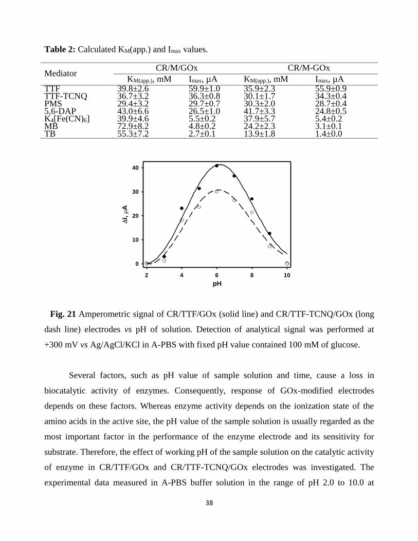
Table 2: Calculated KM(app.) and Imax values.
Mediator CR/M/GOx CR/M-GOx
KM(app.), mM Imax, µA KM(app.), mM Imax, µA TTF 39.8±2.6 59.9±1.0 35.9±2.3 55.9±0.9 TTF-TCNQ 36.7±3.2 36.3±0.8 30.1±1.7 34.3±0.4 PMS 29.4±3.2 29.7±0.7 30.3±2.0 28.7±0.4 5,6-DAP 43.0±6.6 26.5±1.0 41.7±3.3 24.8±0.5 K4[Fe(CN)6] 39.9±4.6 5.5±0.2 37.9±5.7 5.4±0.2 MB 72.9±8.2 4.8±0.2 24.2±2.3 3.1±0.1 TB 55.3±7.2 2.7±0.1 13.9±1.8 1.4±0.0
Fig. 21 Amperometric signal of CR/TTF/GOx (solid line) and CR/TTF-TCNQ/GOx (long
dash line) electrodes vs pH of solution. Detection of analytical signal was performed at
+300 mV vs Ag/AgCl/KCl in A-PBS with fixed pH value contained 100 mM of glucose.
Several factors, such as pH value of sample solution and time, cause a loss in
biocatalytic activity of enzymes. Consequently, response of GOx-modified electrodes
depends on these factors. Whereas enzyme activity depends on the ionization state of the
amino acids in the active site, the pH value of the sample solution is usually regarded as the
most important factor in the performance of the enzyme electrode and its sensitivity for
substrate. Therefore, the effect of working pH of the sample solution on the catalytic activity
of enzyme in CR/TTF/GOx and CR/TTF-TCNQ/GOx electrodes was investigated. The
experimental data measured in A-PBS buffer solution in the range of pH 2.0 to 10.0 at
pH
2 4 6 8 10
I,
A
0
10
20
30
40

39
ambient temperature in the presence of 100 mM glucose is shown in Figure 21. As can be
seen, the response of modified electrodes versus sample solution pH shows a bell-shaped
curve. More than 50 % of the maximum response current change value was observed
between pH 4.0 − 8.0 for both modified electrodes. An optimum was observed at pH 6.0,
which is in agreement with the pH value reported for the native GOx from Aspergilus Niger.
Below the pH 4.0 and above the pH 9.0, the catalytic activity of GOx and current generated
by the modified electrodes are rapidly decreasing and at pH 10 nearly 0 % of the initial GOx
activity remains. At a low-pH, GOx is inhibited by anions. At pH 3.0, even 0.1 M KCl
almost completely inhibits the enzyme activity.
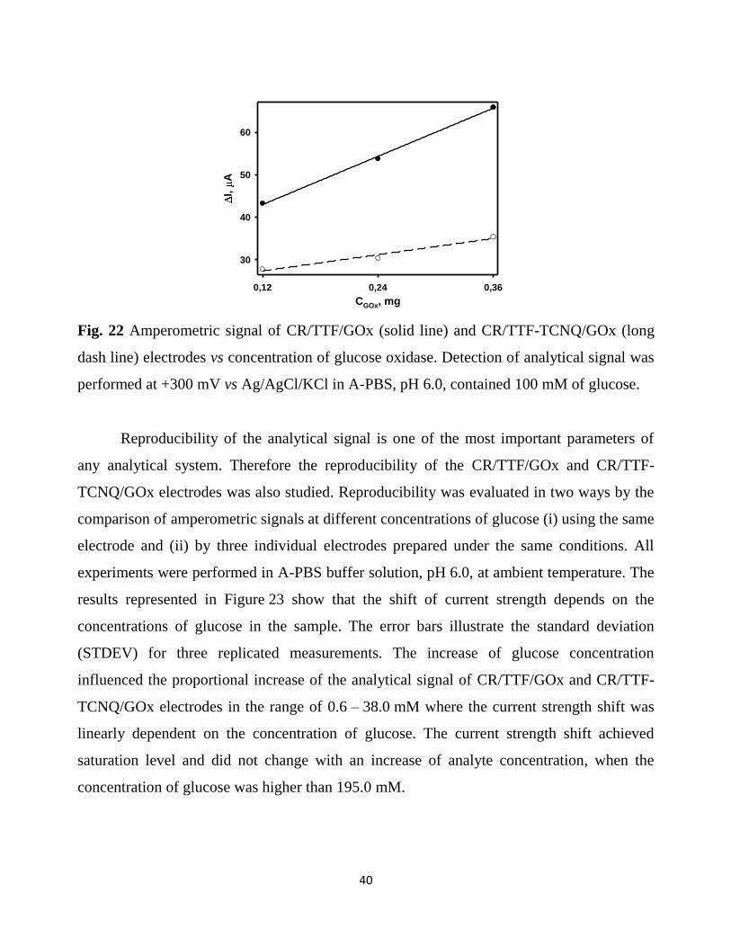
The influence of enzyme concentration on the magnitude of CR/TTF/GOx and
CR/TTF-TCNQ/GOx electrodes analytical signal was examined. The experimental data
measured in A-PBS buffer solution pH 6.0 at ambient temperature in the presence of
100 mM glucose is shown in Figure 22. The results illustrate that higher amount of GOx
produces higher biosensor signal. According to the presented results, the highest
amperometric signal (65.9 and 35.4 µA for CR/TTF/GOx and CR/TTF-TCNQ/GOx
electrode respectively) was detected in the case of the maximal evaluated concentration of
GOx (0.36 mg), whereas the least (43.3 and 27.8 µA for CR/TTF/GOx and CR/TTF-
TCNQ/GOx electrode respectively) was detected in the case of the minimal examined
concentration of GOx (0.12 mg). Thus, experimental results show that by using three times
higher surface concentration of enzyme 1.52 and 1.28 times higher amperometric signal of
the CR/TTF/GOx and CR/TTF-TCNQ/GOx electrode respectively was registered.
40
Fig. 22 Amperometric signal of CR/TTF/GOx (solid line) and CR/TTF-TCNQ/GOx (long
dash line) electrodes vs concentration of glucose oxidase. Detection of analytical signal was
performed at +300 mV vs Ag/AgCl/KCl in A-PBS, pH 6.0, contained 100 mM of glucose.
Reproducibility of the analytical signal is one of the most important parameters of
any analytical system. Therefore the reproducibility of the CR/TTF/GOx and CR/TTF-
TCNQ/GOx electrodes was also studied. Reproducibility was evaluated in two ways by the
comparison of amperometric signals at different concentrations of glucose (i) using the same
electrode and (ii) by three individual electrodes prepared under the same conditions. All
experiments were performed in A-PBS buffer solution, pH 6.0, at ambient temperature. The
results represented in Figure 23 show that the shift of current strength depends on the
concentrations of glucose in the sample. The error bars illustrate the standard deviation
(STDEV) for three replicated measurements. The increase of glucose concentration
influenced the proportional increase of the analytical signal of CR/TTF/GOx and CR/TTF-
TCNQ/GOx electrodes in the range of 0.6 – 38.0 mM where the current strength shift was
linearly dependent on the concentration of glucose. The current strength shift achieved
saturation level and did not change with an increase of analyte concentration, when the
concentration of glucose was higher than 195.0 mM.
CGOx, mg
0,12 0,24 0,36
I,
A
30
40
50
60

41
Fig. 23 Amperometric signal vs concentration of glucose: A – for the same CR/TTF/GOx
electrode; B – for three CR/TTF/GOx electrodes; C – for the same CR/TTF-TCNQ/GOx
electrode; D – for three CR/TTF-TCNQ/GOx electrodes. Detection of analytical signal was
performed at +300 mV vs Ag/AgCl/KCl in A-PBS, pH 6.0.
The results presented in Figure 23 revealed that CR/TTF/GOx and CR/TTF-
TCNQ/GOx electrodes have a sufficient reproducibility. In both cases the same electrode
and three electrodes prepared under the same conditions show similar current responses for
the same amount of glucose. Normal glucose level for adults is 3.3 – 5.5 mM in capillary
blood, 4.1 – 5.9 mM in venous blood and 4.25 – 6.4 mM in blood serum. The STDEV
calculated for the same CR/TTF/GOx and CR/TTF-TCNQ/GOx electrode amperometric
signals in 4.37 mM of glucose, was found to be 0.24 and 0.08 respectively. Relative
standard deviation (RSD) was found to be 3.68 and 2.92 %, yielding a measurement
repeatability of 96.32 and 97.08 %. The STDEV of the amperometric responses calculated
for three similarly prepared CR/TTF/GOx and CR/TTF-TCNQ/GOx electrodes in 4.37 mM
of glucose was a little higher. It was found to be 0.40 and 0.15 for CR/TTF/GOx and
CR/TTF-TCNQ/GOx electrodes respectively. The RSD was found to be 5.81 and 5.73 %,
CGlu, mM
0 80 160 240
I,
A
0
10
20
30
40
50
CGlu, mM
0 80 160 240
I,
A
0
10
20
30
40
50
CGlu, mM
0 80 160 240
I,
A
0
10
20
CGlu, mM
0 80 160 240
I,
A
0
10
20
A B
C D
42
yielding the repeatability of measurement of 94.19 and 94.27 %. Insignificant inaccuracy
between the different electrodes might be caused by slightly varying surface-concentration
of immobilized enzyme and mediator, loss of enzymatic activity during preparation of
electrodes, which differ for different electrodes, and unequal distribution of enzyme on the
carbon rod electrode surface.
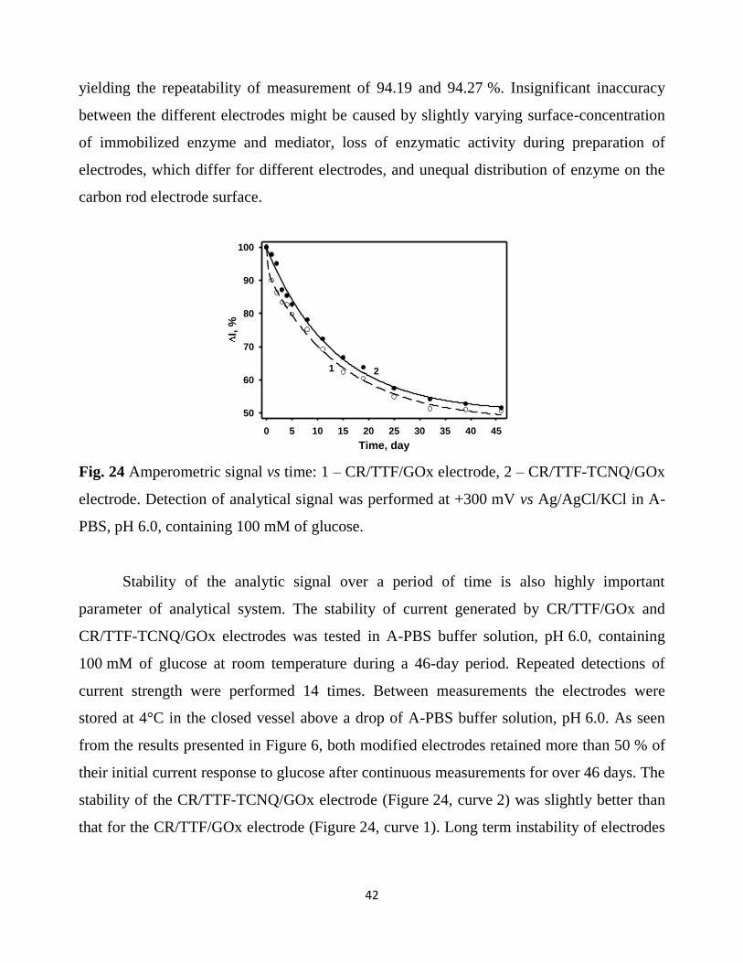
Fig. 24 Amperometric signal vs time: 1 – CR/TTF/GOx electrode, 2 – CR/TTF-TCNQ/GOx
electrode. Detection of analytical signal was performed at +300 mV vs Ag/AgCl/KCl in A-
PBS, pH 6.0, containing 100 mM of glucose.
Stability of the analytic signal over a period of time is also highly important
parameter of analytical system. The stability of current generated by CR/TTF/GOx and
CR/TTF-TCNQ/GOx electrodes was tested in A-PBS buffer solution, pH 6.0, containing
100 mM of glucose at room temperature during a 46-day period. Repeated detections of
current strength were performed 14 times. Between measurements the electrodes were
stored at 4°C in the closed vessel above a drop of A-PBS buffer solution, pH 6.0. As seen
from the results presented in Figure 6, both modified electrodes retained more than 50 % of
their initial current response to glucose after continuous measurements for over 46 days. The
stability of the CR/TTF-TCNQ/GOx electrode (Figure 24, curve 2) was slightly better than
that for the CR/TTF/GOx electrode (Figure 24, curve 1). Long term instability of electrodes
Time, day
0 5 10 15 20 25 30 35 40 45
I, %
50
60
70
80
90
100
1 2

43
could be attributed to dissociation of enzyme and mediator from the electrode surface and/or
inactivation of enzyme.
This work demonstrates the comparison of influence of immobilized redox mediators
TTF, TTF-TCNQ, PMS, MB, 5,6-DAP, K4[Fe(CN)6] and TB on the GOx-based biosensor
electrochemical response to the different concentrations of glucose. According to our
knowledge, this is the first scientific paper wherein is present such comparison. All
investigated mediators were capable to transfer electrons between the active site of GOx and
the graphite rod electrode surface, but the highest amperometric signals were observed by
using TTF and TTF-TCNQ. Here investigated biosensors have several advantages, such as:
(i) simple procedure for the preparation of the working electrode, (ii) basic application
because no addition of soluble mediator before each measurement was required, and (iii)
sufficient reproducibility of the analytic signal. Due to these advantages, the designed
biosensor is expected to be applicable for the determination of glucose.

44
Conclusions
Absorption spectra of synthesized polypyrrole and polyaniline are identical using
enzymatic and chemical oxidation synthesis methods. Appropriate pH value of the
polymerization solution is the main difference of these synthesis methods. In case of
chemical synthesis faster polymerization reaction takes place in more acidic media,
in an alkaline medium polymerization will not occur. Enzymatic synthesis is
preferably carried out in the pH range in which the enzyme is most active. In
addition, the synthesis will occur in alkaline conditions. The presence of gold
nanoparticles in polymerization solution increases rate of polyaniline formation. The
greatest impact has a 3.5 nm-sized gold nanoparticles.
Polymer layer formed on graphite electrode surface modified by glucose oxidase,
extends concentration range of linear dependence of analytical signal for glucose
biosensors, increases stability and improves the repeatability of analytical signal. The
electrodes modified by polypyrrole layer are less stable compared to the electrodes
modified by polyaniline layer.
Gold nanoparticles being as a part of glucose oxidase and polyaniline nanocomposite
structures ensure efficient transport of electrons from the glucose oxidase redox
center to the electrode. Best catalytic properties have nanocomposite structures
synthesized at pH 4.5 polymerization solution. These nanocomposite structures twice
increases registered analytical signal of biosensors
Electron transfer mediators tetrathiafulvalene, phenazine methosulfate, 5,6-diamin-
1,10-phenanthroline, tetrathiafulvalene - tetracyanoquinodimethane complex,
methylene blue, toluidine blue and potassium ferrocyanide immobilized on the
surface of the graphite electrode has an effective electron transfer. The kinetic
parameters of glucose oxidase depend not only on immobilized electron transfer
mediator in nature, but also on electrode preparation method. The most effective
electron transfer has tetrathiafulvalene and tetrathiafulvalene -
tetracyanoquinodimethane complex. Electrodes modified by these electron transfer
mediators have good analytical characteristics.

45
List of publications by the author included into dissertation thesis:
1. A. Kausaite – Minkstimiene, V. Mazeiko, A. Ramanaviciene, A.
Ramanavicius. Enzymatically synthesized polyaniline layer for extension of
linear detection region of amperometric glucose biosensor, Biosensors and
Bioelectronics 26 (2010) 790–797
2. A. Kausaite – Minkstimiene, V. Mazeiko, A. Ramanaviciene, A.
Ramanavicius. Evaluation of amperometric glucose biosensors based on
glucose oxidase encapsulated within enzymatically synthesized polyaniline and
polypyrrole, Sensors and Actuators 158 (2011) 278– 285 B
3. V. Mazeiko, A. Kausaite-Minkstimiene, A. Ramanaviciene, Z. Balevicius, A.
Ramanavicius, Gold Nanoparticle and Conducting Polymer – Polyaniline –
Based Nanocomposites for Glucose Biosensor Design, Sensors and Actuators
B 189 (2013) 187 - 193
4. A. Kausaite - Minkstimiene, V. Mazeiko, A. Ramanaviciene, Y. Oztekin, A.
O. Solak, A. Ramanavicius. Evaluation of Some Redox Mediators in the
Design of Reagentless Amperometric Glucose Biosensor, Electroanalysis 26 –
7 (2014) 1528–1535
5. A. Kausaite-Minkstimiene, V. Mazeiko, A. Ramanaviciene, A. Ramanavicius,
Evaluation of chemical synthesis of polypyrrole particles, Colloids and
Surfaces A: Physicochemical and Engineering Aspects (2015)
List of conference thesis:
1. V. Mazeiko, A Kausaite – Minkstimiene. Enzyme/Polymeric and
Enzyme/Gold/Polymeric nanoparticles for amperometric glucose biosensor
design. The 13-th International Conference-School Advanced Materials and
Technologies, August 27-31, 2011, Palanga, Lithuania.

46
2. V. Mazeiko, A Kausaite – Minkstimiene. Amperometric Glucose Biosensor
based on Glucose Oxidase encapsulated within Enzymatically Synthesized
Conducting Polymers. 5-th International Conference The Vital Nature Sign,
May 19-21, 2011, Kaunas, Lithuania.
3. A. Ramanavicius, A. Kausaite – Minkstimiene, V. Mazeiko, V. Ratautaite, A.
Vaitkuviene, Y. Oztekin, A. Ramanaviciene. Novel synthesis of conducting
polymers. PITCON 2011. CD-ROM abstracts 1480 – 11P, March 13 – 18,
2011, Atlanta, USA
4. A. Ramanavicius, A. Kausaite – Minkstimiene, V. Mazeiko, J. Voronovic, A.
Ramanaviciene, Y. Oztekin. Some analytical aplications of electrochemically
generated polymers and conducting polymers. PITCON 2012. CD-ROM
abstracts 2070 – 1, March 11 – 15, Florida, USA
5. V. Mazeiko, A Kausaite – Minkstimiene. Investigation of redox mediators for
reagentless amperometric glucose biosensors. 14-th International Conference-
School Advanced Materials and Technologies, August 27-31, 2012, Palanga,
Lithuania.
6. V. Mazeiko, A Kausaite – Minkstimiene. Polyaniline synthesis and
application in bioanalytical systems. 15-th International Conference-School
Advanced Materials and Technologies, August 27-31, 2013, Palanga,
Lithuania.

47
ACKNOWLEDGEMENTS
I sincerely thank my scientific supervisor Dr. Asta Kaušaitė-Minkštimienė for granted
knowledge, full support and assistance in the preparation of a dissertation work and
experiments during doctoral studies.
Thank you Doc. Dr. Almira Ramanaviciene and prof. Habil. Dr. Arunas Ramanavičius for
scientific advices, discussions and support both during experiments and publishing the
results.
Thank you, Dr. Lina Mikoliūnaitė for assistance in experiments with atomic force
microscopy and advice in interpreting findings.
Thank you, Dr. Jaroslav Voronovič for assistance in the synthesis of colloidal gold
compounds.
I sincerely thank all of Vilnius Universisty Faculty of Chemistry Department of Analytical
and Environmental Chemistry colleagues for assistance, patience, understanding and
working atmosphere.
Part of the research was carried out in the presence of Lithuanian Science Council sponsored
by Lithuania-Belarus bilateral cooperation in science and technology program in the project
"Improving glucose biosensors with immobilized nanomaterials“ with Belarusian Institute
of Microbiology. I thank my colleagues from Belarus for scientific discussions and
assistance in investigations and interpreting the data.
Also, I am grateful to the Lithuanian Science Council for financial support.
I thank to my wife and daughter, my mother, all the relatives for their support and great
patience.