Embed Size (px)
Citation preview
Vision in vertebrates
How do we see ?
Aims
To describe how sensory systems fulfil their need to transduce energy compare inputs lead to appropriate behaviour
using the vertebrate visual system
Main parts of visual system eye
lens, retina brain - primates
lateral geniculate nucleus visual cortex
brain - other vertebrates superior colliculus / optic tectum
Warning
Not all vertebrates work the same way differences in anatomy differences in retinal & CNS function
even between closely related species, e.g. frog and toad
always look and see what species the work was done on!
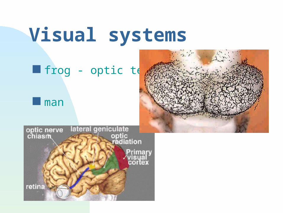
Visual systems
frog - optic tectum
man
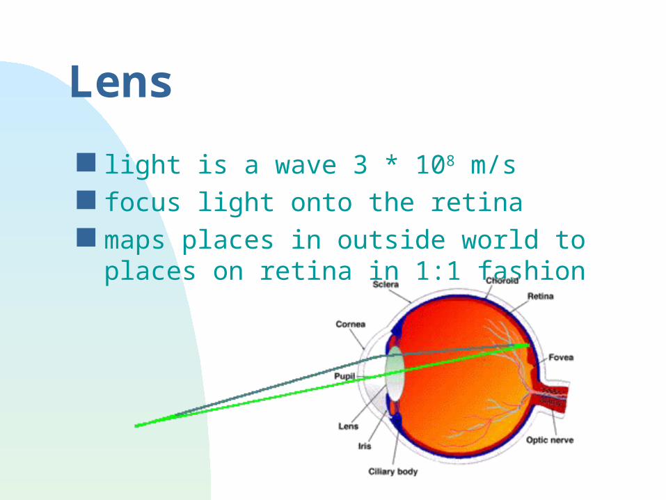
Lens
light is a wave 3 * 108 m/s focus light onto the retina maps places in outside world to places
on retina in 1:1 fashion
Retina
structure photoreceptors - at back of retina layers of cells output from ganglion cells - at front of
retina Blindspot
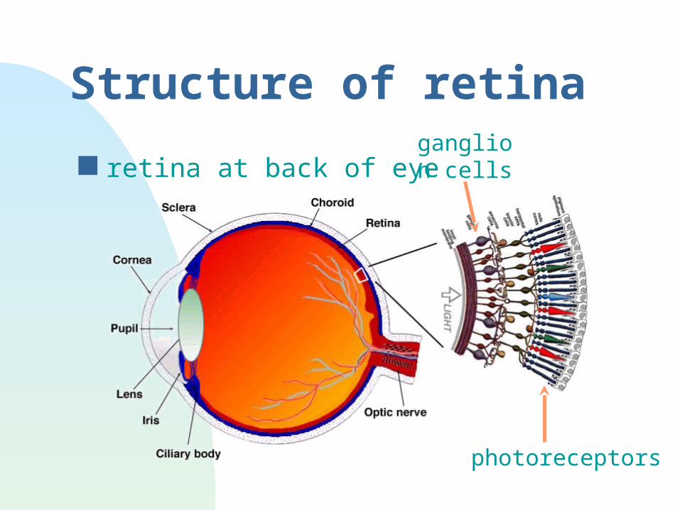
Structure of retina
retina at back of eye
photoreceptors
ganglion cells
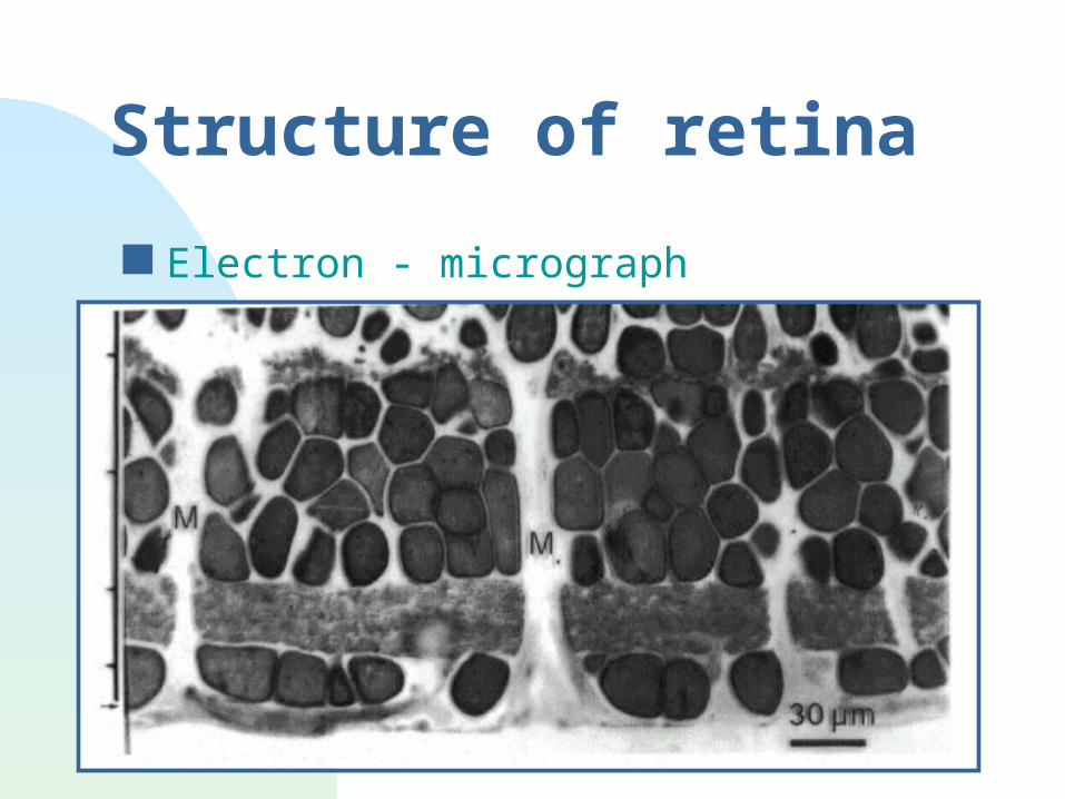
Structure of retina
Electron - micrograph
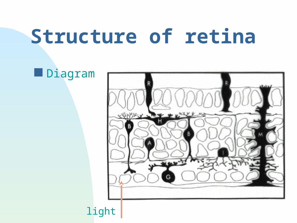
Structure of retina
Diagram
light
Photoreceptors
Rods and Cones Transduction Can we perceive photons? Colour vision
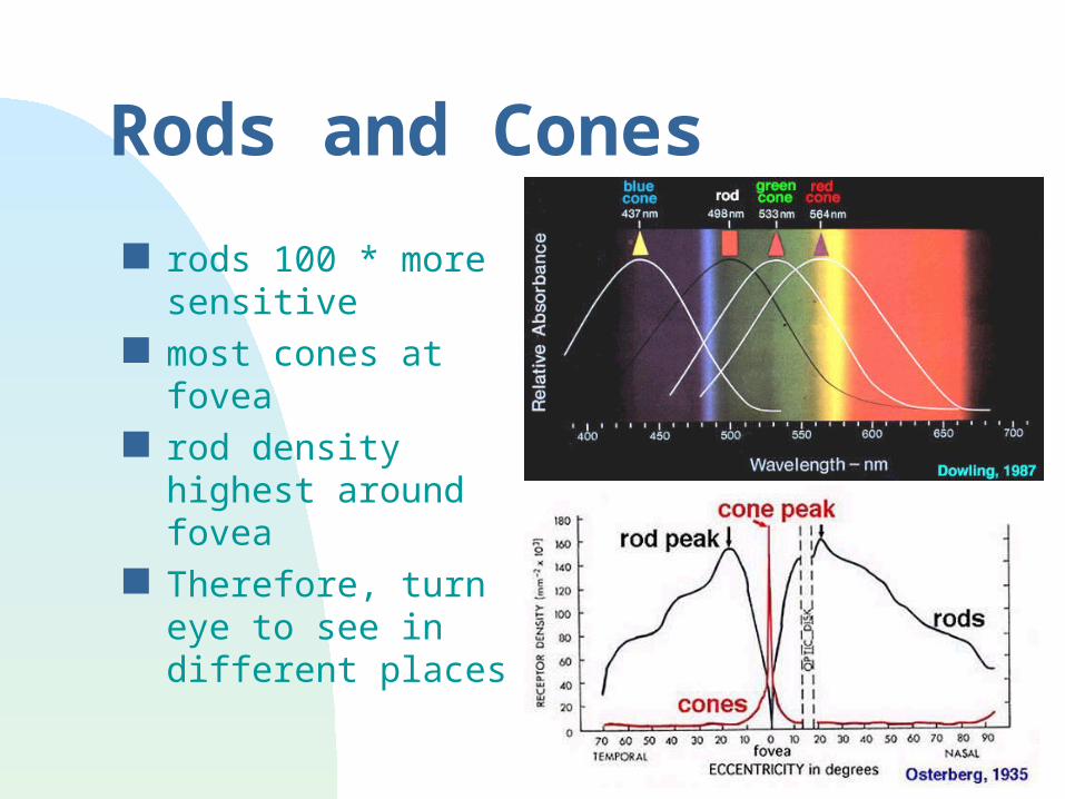
Rods and Cones
rods 100 * more sensitive
most cones at fovea
rod density highest around fovea
Therefore, turn eye to see in different places
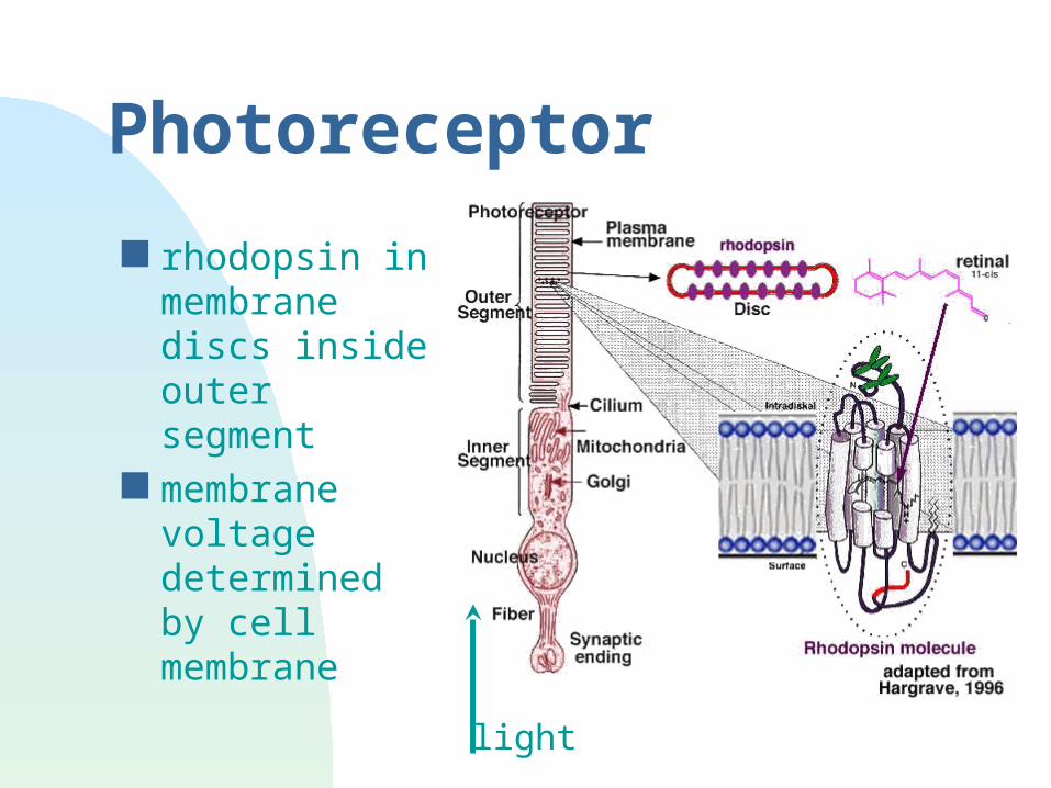
Photoreceptor
rhodopsin in membrane discs inside outer segment
membrane voltage determined by cell membrane
light
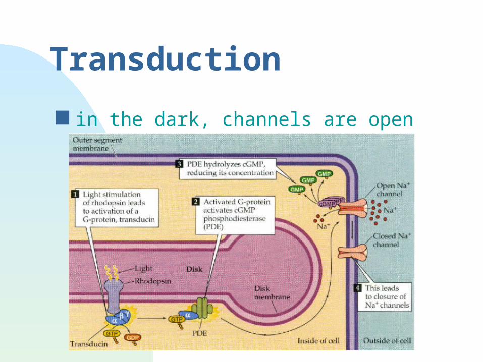
Transduction
in the dark, channels are open
Transduction
Transduction movie
Transduction
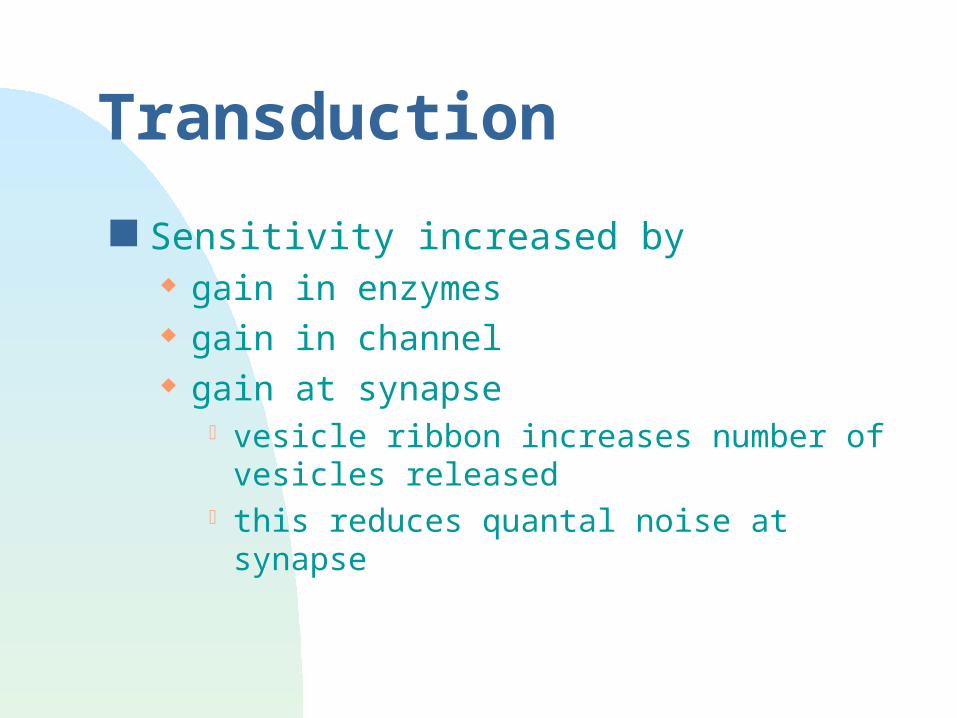
Sensitivity increased by gain in enzymes gain in channel gain at synapse
vesicle ribbon increases number of vesicles released
this reduces quantal noise at synapse
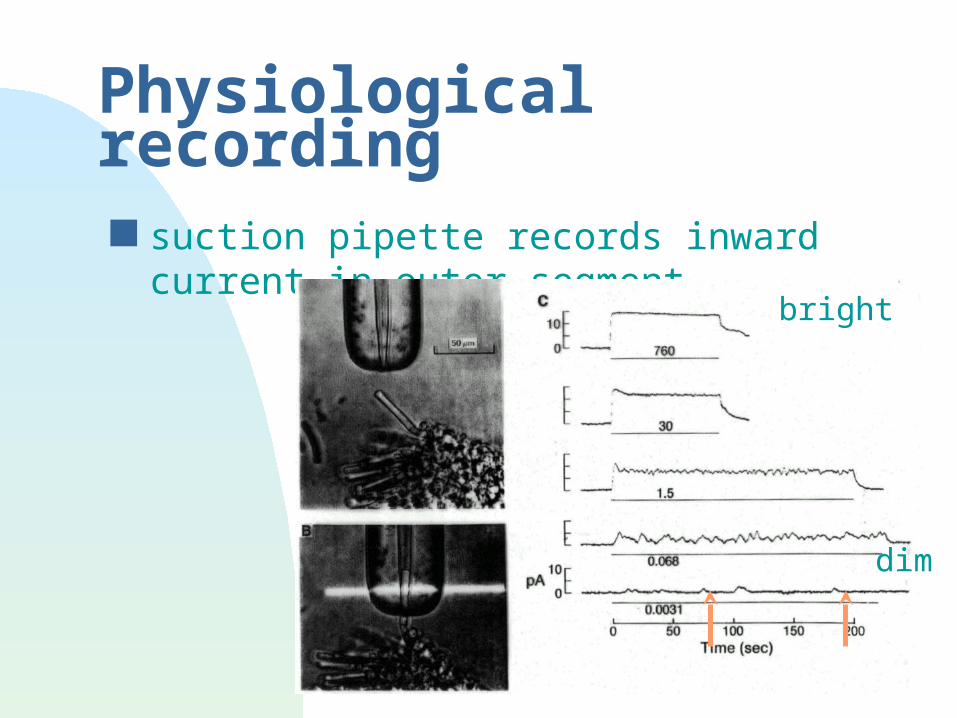
Physiological recording suction pipette records inward current in
outer segmentbright
dim
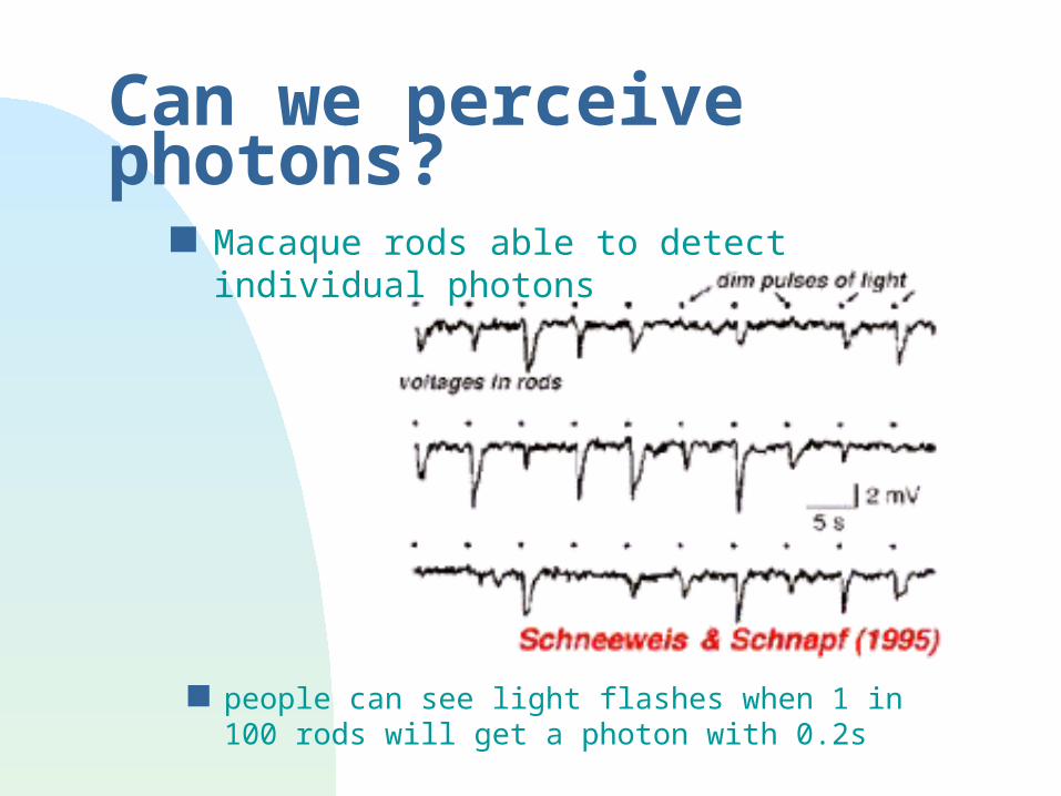
Can we perceive photons?
people can see light flashes when 1 in 100 rods will get a photon with 0.2s
Macaque rods able to detect individual photons
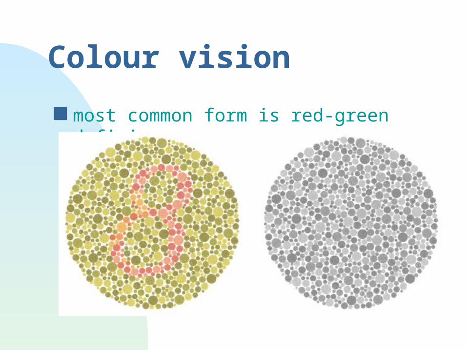
Colour vision
most common form is red-green deficiency
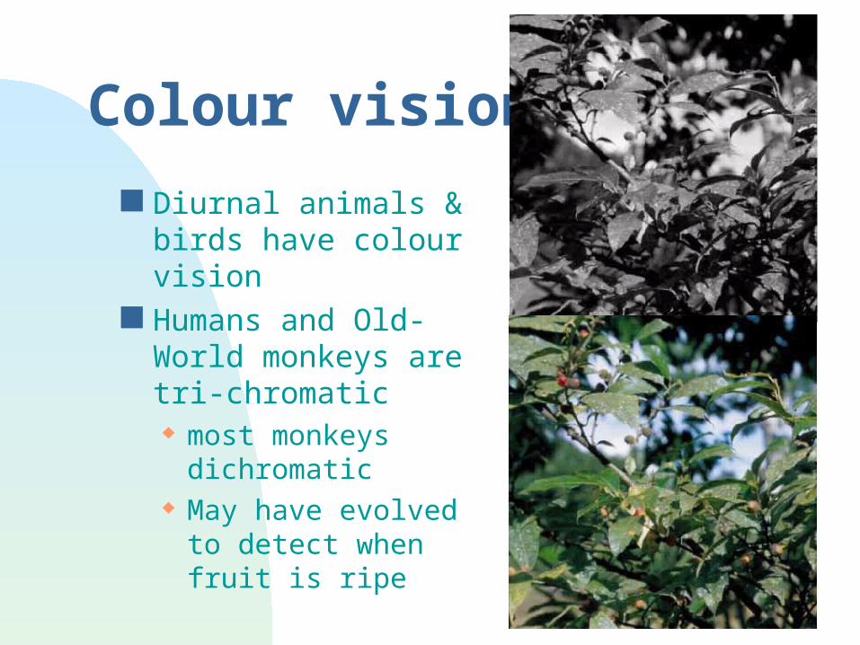
Colour vision
Diurnal animals & birds have colour vision
Humans and Old-World monkeys are tri-chromatic most monkeys
dichromatic May have evolved to
detect when fruit is ripe
Colour vision
Diurnal animals/birds have colour vision Humans and Old-World monkeys are
trichromatic most monkeys dichromatic May have evolved to detect when fruit is
ripe other mechanisms of color vision exist
oil droplets in amphibians, turtles gene homology
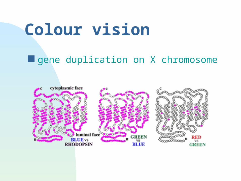
Colour vision
gene duplication on X chromosome
Summary so far
At retina, world is spatially mapped light level is encoded by current color is used (but not in all animals) very sensitive
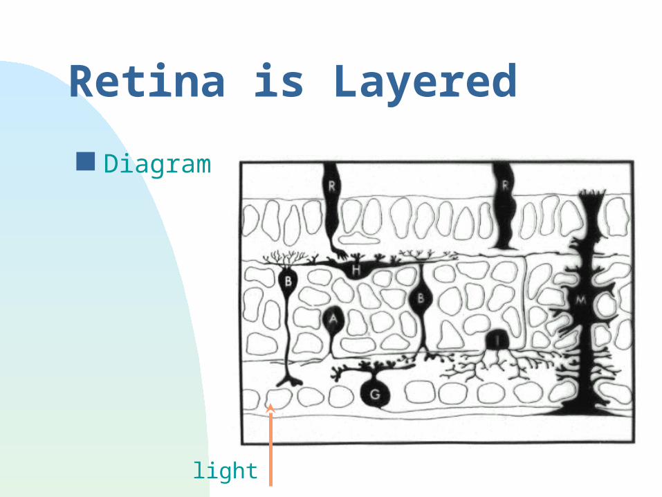
Retina is Layered
Diagram
light
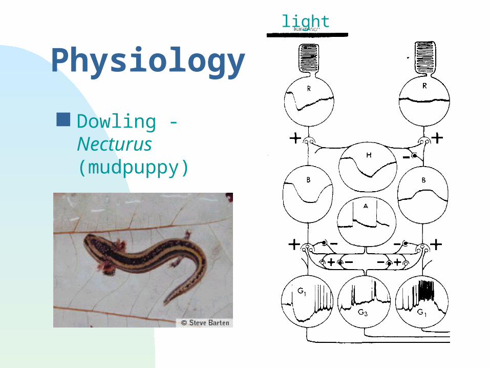
Physiology
Dowling - Necturus (mudpuppy)
light
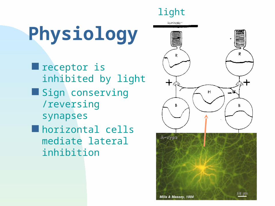
Physiology
receptor is inhibited by light
Sign conserving /reversing synapses
horizontal cells mediate lateral inhibition
light
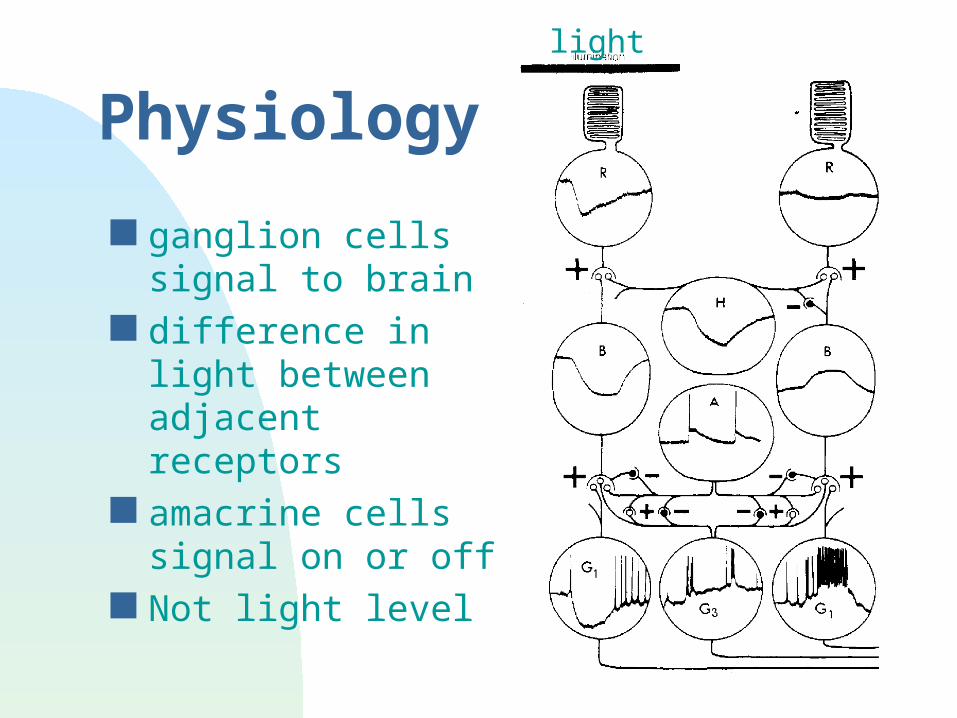
Physiology
ganglion cells signal to brain
difference in light between adjacent receptors
amacrine cells signal on or off
Not light level
light
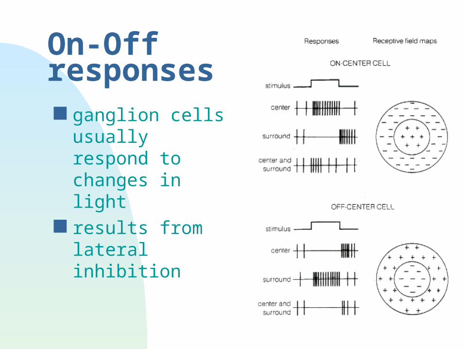
On-Off responses ganglion cells
usually respond to changes in light
results from lateral inhibition
Lateral inhibition
Hermann Grid
common to vision, touch, hearing...
Summary so far
At retina, world is spatially mapped light level is encoded by current color is used (but not in all animals) very sensitive
At ganglion cells on/off & surround /center not a 1:1 relation between light level and
signal this enhances dynamic range
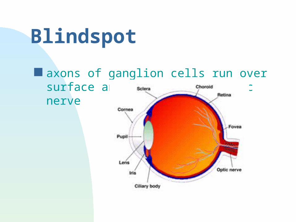
Blindspot
axons of ganglion cells run over surface and turn to give optic nerve
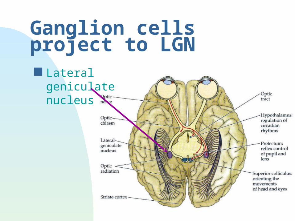
Ganglion cells project to LGN Lateral geniculate
nucleus
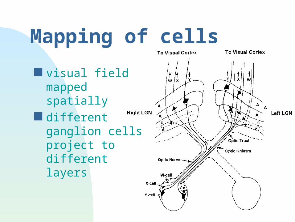
Mapping of cells
visual field mapped spatially
different ganglion cells project to different layers
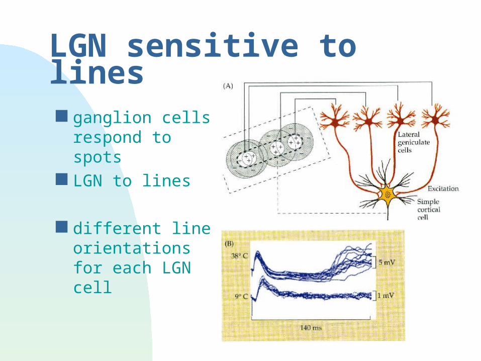
LGN sensitive to lines ganglion cells
respond to spots
LGN to lines
different line orientations for each LGN cell
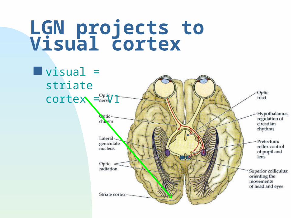
LGN projects to Visual cortex visual = striate
cortex = V1
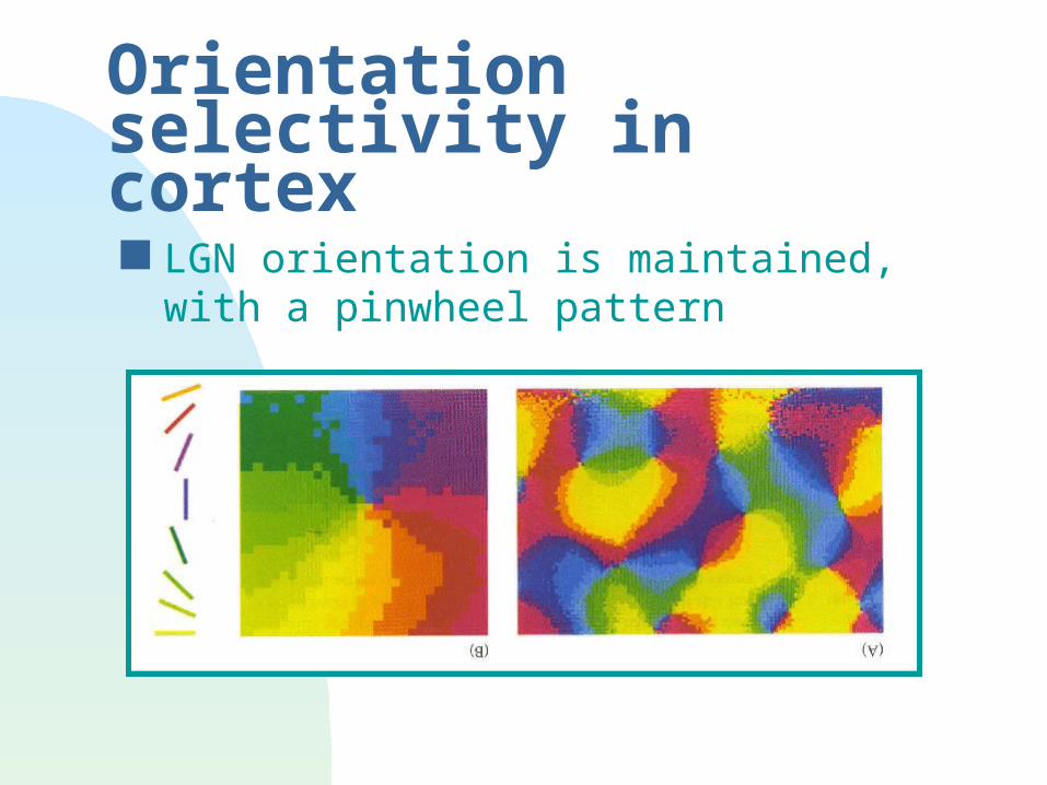
Orientation selectivity in cortex LGN orientation is maintained, with a
pinwheel pattern
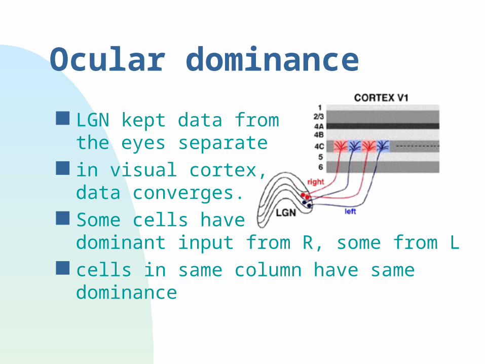
Ocular dominance
LGN kept data from the eyes separate
in visual cortex, data converges.
Some cells have dominant input from R, some from L
cells in same column have same dominance

V1 Cortex
orientation and ocular dominance work together grey is
contralateral eye
Depth perception
Use both eyes to calculate how far away objects are
Hypothesis 1: rangefinder Hypothesis 2: measure overlap of
images
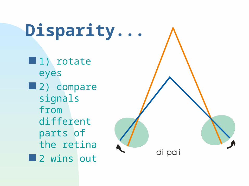
Disparity...
1) rotate eyes 2) compare
signals from different parts of the retina
2 wins out
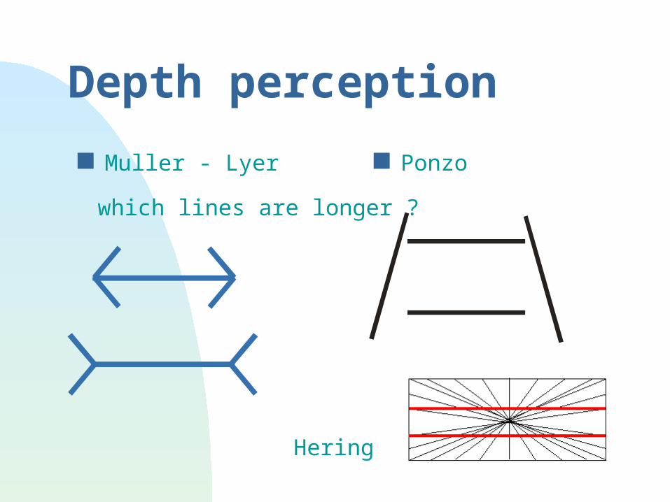
Depth perception
Muller - Lyer Ponzo
which lines are longer ?
Hering
Blindsight
loss of visual cortex may show evidence for blindsight
patient cannot “see” but can follow targets with their eyes
patient can discriminate words projection to superior colliculus may be
responsible
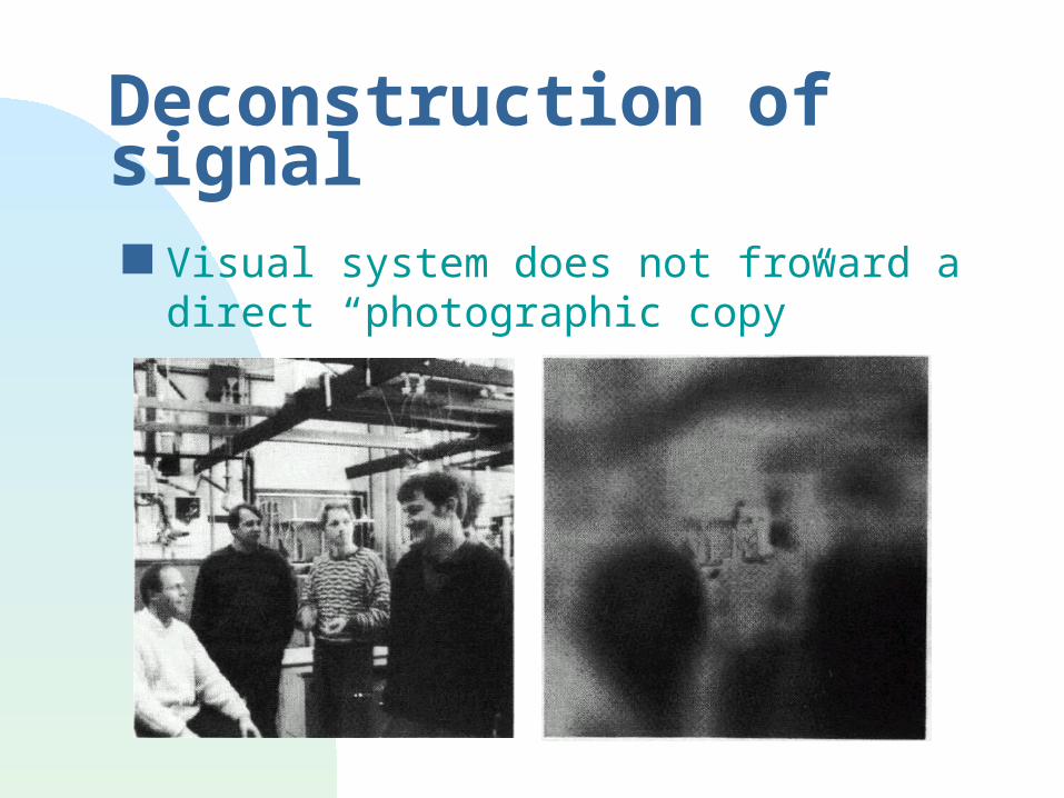
Deconstruction of signal Visual system does not froward a direct
“photographic copy”
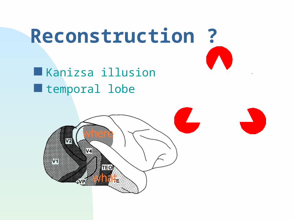
Reconstruction ?
Kanizsa illusion temporal lobe

Summary
transduction well understood; high gain system
retina well understood; lateral inhibitory mechanism
LGN and V1 cortex fairly well understood; lateral & temporal inhibition; binocular vision
further processing still to be elucidated