Embed Size (px)
Citation preview

7/24/2019 Visucam 500 Brochure
http://slidepdf.com/reader/full/visucam-500-brochure 1/8
VISUCAM 500
Have it all

7/24/2019 Visucam 500 Brochure
http://slidepdf.com/reader/full/visucam-500-brochure 2/8
22
VISUCAM 500
As versatile as your patients
Superior patient comfort, more efficient workflow and improved eye care: The advantages
of the all-in-one fundus platform VISUCAM 500 from ZEISS are obvious. The high-quality system
provides everything you need for detailed diagnoses of typical eye diseases like diabetic
retinopathy, glaucoma and AMD in a single workplace.
Advanced features such as fundus autofluorescence, easy stereo image handling and innovative
assessment of macular pigment density are combined with intelligent auto functions that enable
reproducible and intuitive imaging for every single patient eye.

7/24/2019 Visucam 500 Brochure
http://slidepdf.com/reader/full/visucam-500-brochure 3/8
333
7/24/2019 Visucam 500 Brochure
http://slidepdf.com/reader/full/visucam-500-brochure 4/8
4
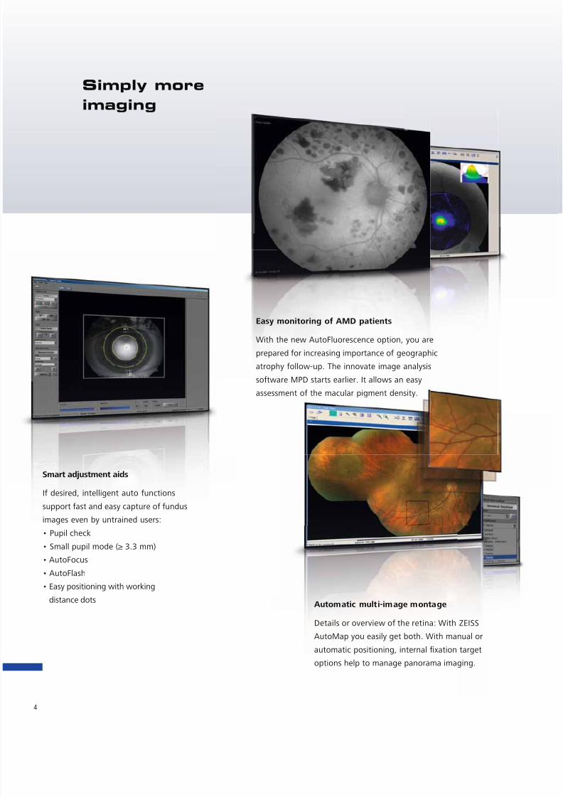
Simply more
imaging
Easy monitoring of AMD patients
With the new AutoFluorescence option, you are
prepared for increasing importance of geographic
atrophy follow-up. The innovate image analysis
software MPD starts earlier. It allows an easy
assessment of the macular pigment density.
Smart adjustment aids
If desired, intelligent auto functions
support fast and easy capture of fundus
images even by untrained users:
• Pupil check
• Small pupil mode (≥ 3.3 mm)
• AutoFocus
• AutoFlash
• Easy positioning with working
distance dots Automatic multi-image montage
Details or overview of the retina: With ZEISS
AutoMap you easily get both. With manual or
automatic positioning, internal fixation target
options help to manage panorama imaging.

7/24/2019 Visucam 500 Brochure
http://slidepdf.com/reader/full/visucam-500-brochure 5/8
55
More information in 3D quality
With the stereo mode, 3D images are easy
to capture and handle. Saved in a single file,
the VISUCAM 500 software remembers every
change of the JPS image pair.
With its excellent image quality, the 7-field
mode and stereo images, VISUCAM 500 is a
perfect fit for clinical studies.
Step-by-step support
From easy capture and view-ing to comprehensive patienteducation, VISUCAM 500
provides high-quality images ina natural, intuitive and continu-ous sequence.
Live visualization
Captured images are displayed on the
monitor immediately for fast assess-
ment. All processing functions are only
a double click away. The supporting
tools for your daily workflow include:
• Optional arterial venous ratio (AVR)
• Various magnifying features
• Simplified search for images
and patients
• Drag & drop functionality for
printout preparation

7/24/2019 Visucam 500 Brochure
http://slidepdf.com/reader/full/visucam-500-brochure 6/8
6
VISUCAM 500 can significantly improve practice workflow efficiency, allowing you to concen-
trate on what counts the most: providing optimal care for your patients.
All relevant data at your fi ngertips
FORUM allows efficient management, central storage and fast access of reports,data and images from virtually all ophthalmic systems. It is an ideal fit togetherwith VISUCAM 500, enabling seamless data flow throughout your practice.
All- in-one eye care workplace
VISUCAM 500 not only incorporates all capture
modes required for routine procedures; it also
integrates a computer and database for fast image
transfer.
Safe, consistent data storage
Providing multiple archive options, VISUCAM 500
enables heightened data security. Besides the
backup functionality of FORUM, data can be easily
stored on the Network Archive Storage (NAS),
hard disk or DVD.
Images wherever you need them
Images can be transferred throughout your local
network via USB or DVD. When combined with the
FORUM data management solution from ZEISS,
VISUCAM 500 enables you to view and compare
diagnostic data quickly and conveniently from
virtually anywhere.
With modality worklist patient demographic
information can be transferred from FORUM into
VISUCAM 500 without retyping thus reducing time
and charting errors.
Receptionarea OR
Receptionarea practice
Exam lane1
Exam lane2
Preparation for surgery
VisualField
OCT Biometry Fundus photogra-
phy
Back Office
SurgeonsOffice
Recovery room
OR1
OR2
FORUM
Archive
Diagnosticinstruments
FORUMOR Manager
FORUMViewer
EMR
CALLISTO eyeOR Cockpit

7/24/2019 Visucam 500 Brochure
http://slidepdf.com/reader/full/visucam-500-brochure 7/8
7
Clearly arranged control elements
Enabling fast and easy imaging
workflow
Sliding keyboard shelf (optional)
Offering more working space on the table
Network Archive Storage
Providing data security
independent from the EMR

7/24/2019 Visucam 500 Brochure
http://slidepdf.com/reader/full/visucam-500-brochure 8/8
Technical Data
P u b l i c a t i o n
N o : 0 0 0 0 0 0 - 1 8 4 3 - 1 2 0
I N T E R N A T I O N A L V E R S I O N
T h e c o n t e n t s o f t h e
b r o c h u r e m a y d i f f e r f r o m t h e c u r r e n t s t a t u s o f a p p r o v a l o f
t h e p r o d u c t i n y o u r c o u n t r y . P l e a s e c o n t a c t o u r r e g i o n a l r e
p r e s e n t a t i v e f o r m o r e i n f o r m a t i o n .
S u b j e c t t o c h a n g e i n d e s i g n a n d s c o p e o f d e l i v e r y a n d a s a r e s u l t o f o n g o i n g t e c h n i c a l d e v e l o p m e n t . P r i n t e d o n e l e m e n t a l c h l o r i n e - f r e e
b l e a c h e d p a p e r . P U
B L I C
I S
V / 2 0 1 0 .
© 2
0 1 0 b y C a r l Z e i s s M e d i t e c A G .
A l l c o p y r i g h t s r e s e r v e d
.
Not for sale in the US.
VISUCAM 500
Fundus camera system
Field angle 45° and 30°Capture modes Co lor, red- free, b lue and red images , and images of the
anterior segment, as well as fluorescein angiography.
Optional: fundus autofluorescence, ICG angiography, stereo, MPD
Filters Optical filter: FA + ICGA
exciter and barrier filters, filters for green and
blue images, filters for fundus autofluorescence and MPD,
UV / IR barrier filters
Compensation for Ametropia +35 ... –35 D, continuous
Capture sequence F rom 1.5 seconds (depends on f lash energy )
Pupil diameter ≥ 4.0 mm
≥ 3.3 mm (30° small pupil mode)
Working distance 40 mm (patient eye – front lens)
Capture sensor CCD 5.0 mega pixels
Monitor 19“ TFT (1280 x 1024) connected via isolating transformer
Fixation targets External and internal
Attention mode for internal fixation target
(magnified and blinking cross)
Various programmed sequences or freely positionable
Flash energy Xenon flash lamp, 22 flash levels (max 60 Ws)
Database Patient information and images with field angle,
FA time, R/L recognition and date of visit
Computer
Operating system Windows XP Professional
Hard drive Storage of over 150,000 images possible
(present size: 320 GB)
Interfaces USB ports and network connectors, DVI port
Export/import Image formats: DICOM-OP, BMP, TIFF, JPEG
Patient list, DICOM MWL, DICOM storage
Internal DVD burner UDF format (DVD, CD)
Instrument table Asymmetric, suitable for wheelchair
Accessories Network printer, USB memory stick,
FORUM / VISUPAC archiving and image analysis system
Network isolator
Dimensions
Basic device 410 mm x 480 mm x 650 mm (headrest)
(W 16.14 x D 18.90 x H 25.59 inches)
Monitor 405 x 65 x 335 mm (depends on model)
(W 15.95 x D 2.56 x H 13.19 inches)
Weight Basic device 30 kg (66.1 lbs)
Rated voltage 100 … 240 V ±10% (self-adjusting)
Frequency 50 / 60 Hz
Power consumption 400 VA
Carl Zeiss Meditec AG
Goeschwitzer Str. 51– 52
07745 Jena
GERMANY
Phone: +49 36 41 22 03 33
Fax: +49 36 41 22 01 12
www.meditec.zeiss.com