Embed Size (px)
Citation preview
Available online http://arthritis-research.com/content/10/3/R64
Open AccessVol 10 No 3Research articleIdentification of novel monosodium urate crystal regulated mRNAs by transcript profiling of dissected murine air pouch membranesFrank Pessler1,2, Christian T Mayer3,4, Sung Mun Jung4,5, Ed M Behrens1, Lie Dai4,6, Joseph P Menetski7 and H Ralph Schumacher4,8
1Klinik und Poliklinik für Kinder und Jugendmedizin, Technische Universität Dresden, Fetscherstraße, 01307 Dresden, Germany2Division of Rheumatology, The Children's Hospital of Philadelphia, Civic Center Blvd, Philadelphia, Pennsylvania 19104, USA3Institut für Medizinische Mikrobiologie, Immunologie und Hygiene, Technische Universität München, Trogerstraße, 81675 München, Germany4Division of Rheumatology, University of Pennsylvania, Spruce St, Philadelphia, Pennsylvania 19104, USA5Faculty of Oriental Medicine, Department of Herbal Pharmacology, Kyung Hee University College of Oriental Medicine, Hoekidong, Dongdaemoonku, Seoul 130-701, Korea6Division of Rheumatology, Second Affiliated Hospital, Sun Yat-sen University, Yan Jiang West Road, Guangzhou 510120, PR China7Merck Research Laboratories, E. Lincoln Avenue, PO Box 2000, Rahway, New Jersey 07065, USA8Division of Rheumatology, Veteran Affairs Medical Center, University and Woodland Avenues, Philadelphia, Pennsylvania 19104, USA
Corresponding author: Frank Pessler, [email protected]
Received: 9 Dec 2007 Revisions requested: 23 Jan 2008 Revisions received: 2 Mar 2008 Accepted: 3 Jun 2008 Published: 3 Jun 2008
Arthritis Research & Therapy 2008, 10:R64 (doi:10.1186/ar2435)This article is online at: http://arthritis-research.com/content/10/3/R64© 2008 Pessler et al.; licensee BioMed Central Ltd. This is an open access article distributed under the terms of the Creative Commons Attribution License (http://creativecommons.org/licenses/by/2.0), which permits unrestricted use, distribution, and reproduction in any medium, provided the original work is properly cited.
Abstract
Introduction The murine air pouch is a bursa-like space thatresembles the human synovial membrane. Injection ofmonosodium urate (MSU) crystals into the pouch elicits anacute inflammatory response similar to human gout. Weconducted the present study to identify mRNAs that were highlyregulated by MSU crystals in the pouch membrane.
Methods Air pouch membranes were meticulously dissectedaway from the overlying skin. Gene expression differencesbetween MSU crystal stimulated and control membranes weredetermined by oligonucleotide microarray analysis 9 hours afterinjection of MSU crystals or buffer only. Differential regulation ofselected targets was validated by relative quantitative PCR intime course experiments with dissected air pouch membranesand murine peritoneal macrophages.
Results Eleven of the 12 most highly upregulated mRNAs wererelated to innate immunity and inflammation. They includedmRNAs encoding histidine decarboxylase (the enzyme thatsynthesizes histamine), IL-6, the cell surface receptors PUMA-gand TREM-1, and the polypeptides Irg1 and PROK-2. IL-6mRNA rose 108-fold 1 hour after crystal injection, coinciding
with a surge in mRNAs encoding IL-1β, tumour necrosis factor-α and the immediate early transcription factor Egr-1. The othermRNAs rose up to 200-fold within the subsequent 3 to 8 hours.MSU crystals induced these mRNAs in a dose-dependentmanner in cultured macrophages, with similar kinetics but lowerfold changes. Among the downregulated mRNAs, quantitativePCR confirmed significant decreases in mRNAs encodingTREM-2 (an inhibitor of macrophage activation) and granzyme D(a constituent of natural killer and cytotoxic T cells) within 50hours after crystal injection.
Conclusion This analysis identified several genes that werepreviously not implicated in MSU crystal inflammation. Themarked rise of the upregulated mRNAs after the early surge incytokine and Egr-1 mRNAs suggests that they may be part of a'second wave' of factors that amplify or perpetuate inflammation.Transcript profiling of the isolated air pouch membranepromises to be a powerful tool for identifying genes that act atdifferent stages of inflammation.
Page 1 of 13(page number not for citation purposes)
CSF = colony-stimulating factor; Egr = early growth response; Hdc = histidine decarboxylase; IL = interleukin; Irg = immunoresponsive gene; LRF = leukaemia/lymphoma-related factor; MSU = monosodium urate; NK = natural killer; PBS = phosphate-buffered saline; PCR = polymerase chain reac-tion; PROK = prokineticin; PUMA-g = protein upregulated on macrophages activated with interferon-γ; qPCR = quantitative polymerase chain reac-tion; TLR = Toll-like receptor; TNF = tumour necrosis factor; TREM = triggering receptor expressed on myeloid cells.
Arthritis Research & Therapy Vol 10 No 3 Pessler et al.
IntroductionThe murine air pouch is an easily accessible bursa-like spacethat can be produced de novo in the dorsal subcutaneous tis-sue [1]. Within several days of injecting a small volume of air(2 to 3 ml), a membrane of several layers of cells, which con-sist mostly of fibroblasts, mononuclear cells and small bloodvessels, grows around this air-filled space [1]. This membraneresembles the synovial membrane histologically and hasimportant properties of the synovial lining, such as hyaluronicacid synthesis [2] and expression of the Ia antigen [1]. Inflam-matory substances or micro-organisms can be injected easilyinto the pouch, leading to different forms of inflammationdepending on the agent used. For instance, inflammationcaused by monosodium urate (MSU) [3] and calcium pyro-phosphate crystals [4], carrageenan [4], joint prosthesisdebris [5] and bacterial cell wall components [6] has beenstudied in this model.
Gene profiling of intact tissues is potentially hampered by thepresence of adjacent noninflamed tissue, which increases thecomplexity of the tissue and introduces background 'noise'.Upon incision of the overlying dorsal skin, the air pouch mem-brane appears relatively loosely attached to the overlying sub-cutaneous tissue. We thus reasoned that it should be possibleto dissect the membrane away from the overlying tissues anduse this isolated membrane to study tissue-wide gene expres-sion changes in an inflamed tissue of minimal complexity.Here, we report a dissection method leading to the isolation ofthe pouch membrane from the surrounding tissue. Microarrayanalysis of dissected inflamed and control membranes, cou-pled with validation of differential expression by relative quan-titative polymerase chain reaction (qPCR), revealed a highyield of genes involved in innate immune responses and iden-tified highly inducible mRNAs that were previously not impli-cated in crystal-induced inflammation.
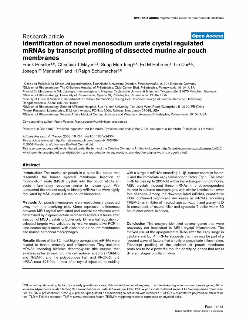
Materials and methodsAir pouchesFigure 1a outlines the sequence of events of the air pouchexperiments. Air pouches were raised on the backs of 6- to 8-week-old female BALB/c mice (Taconic, Tarrytown, NY, USA)by subcutaneous injection of 3 ml filtered air [1,7]. Poucheswere re-inflated on day 3 with an additional 2 ml filtered air.MSU crystals were prepared in accordance with the methodproposed by McCarty and Faires [8] and were determined tobe free from endotoxin using the Gelclot LAL reagent (CharlesRiver Labs, Wilmington, MA, USA). Aliquots from the samebatch were used for all experiments. A suspension of 2 mgMSU crystals in 1 ml sterile endotoxin-free phosphate-buff-ered saline (PBS) was injected into the pouch on day 6. Toverify the time points of peak and natural resolution of inflam-mation in the pouch, a 50-hour time course experiment wasperformed during which pouch exudate leukocyte counts weredetermined at several time points after injection of MSU crys-tals. In agreement with our previous findings [9], the leukocyte
count rose 56-fold from 0 to 9 hours and then subsided,returning close to baseline by 50 hours (Figure 1b). Negativecontrol pouches (n = 5) were injected with 1 ml PBS and har-vested at 9 hours. Their mean leukocyte count was similar tothat of the pouches at t = 0 hours (0.32 ± 0.18 × 10⁶ cells/pouch at 9 hours versus 0.18 ± 0.09 at 0 hours; p = 0.31, one-tailed t-test). All animal experiments followed internationallyrecognized guidelines and were approved by the InstitutionalAnimal Care and Research Committee of the Philadelphia VAMedical Center.
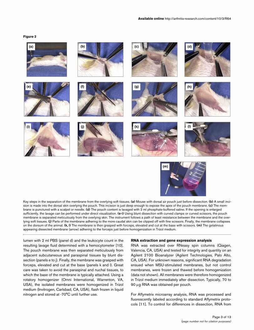
Dissection of the air pouch membraneFigure 2 shows key steps in the membrane dissection. Afterkilling the animals by carbon dioxide asphyxiation, the apex ofthe pouch membrane was exposed with a small skin incision(panel b). The membrane was then punctured with a scalpel(panel c). Typically, little free exudate accumulates within thepouch. Leukocytes were therefore lavaged out of the pouch
Figure 1
Outline of the air pouch experimentsOutline of the air pouch experiments. (a) Sequence of events. Air is injected subcutaneously on day 0 and again on day 3 to keep the pouch inflated. On day 6 the remaining air is aspirated, and the mono-sodium urate (MSU) crystal suspension (2 mg in 1 ml phosphate-buff-ered saline [PBS]) or 1 ml PBS only is injected into the pouch cavity. Pouch exudate and tissue are obtained for analysis up to 50 hours after crystal injection. (b) Determination of the time of maximal inflammation in the pouch lumen. MSU crystal suspension (2 mg in 1 ml PBS) was injected into the pouch at t = 0 hours. Pouch exudate leukocyte counts were determined by manual cell counting at the indicated time points (n = 4 mice for each time point).
Page 2 of 13(page number not for citation purposes)
Available online http://arthritis-research.com/content/10/3/R64
lumen with 2 ml PBS (panel d) and the leukocyte count in theresulting lavage fluid determined with a hemocytometer [10].The pouch membrane was then separated meticulously fromadjacent subcutaneous and paraspinal tissues by blunt dis-section (panels e to j). Finally, the membrane was grasped withforceps, elevated and cut at the base (panels k and l). Greatcare was taken to avoid the paraspinal and nuchal tissues, towhich the base of the membrane is typically attached. Using arotatory homogenizer (Omni International, Warrenton, VA,USA), the isolated membranes were homogenized in Trizolmedium (Invitrogen, Carlsbad, CA, USA), flash frozen in liquidnitrogen and stored at -70°C until further use.
RNA extraction and gene expression analysisRNA was extracted over RNeasy spin columns (Qiagen,Valencia, CA, USA) and tested for integrity and quantity on anAgilent 2100 Bioanalyzer (Agilent Technologies, Palo Alto,CA, USA). For unknown reasons, significant RNA degradationensued when MSU-stimulated membranes, but not controlmembranes, were frozen and thawed before homogenization(data not shown). All membranes were therefore homogenizedin Trizol medium immediately after dissection. Typically, 70 to90 μg RNA was obtained per pouch.
For Affymetrix microarray analysis, RNA was processed andfluorescently labeled according to standard Affymetrix proto-cols [11]. To control for differences in dissection, RNA from
Figure 2
Key steps in the separation of the membrane from the overlying soft tissuesKey steps in the separation of the membrane from the overlying soft tissues. (a) Mouse with dorsal air pouch just before dissection. (b) A small inci-sion is made into the dorsal skin overlying the pouch. This incision is just deep enough to expose the apex of the pouch membrane. (c) The mem-brane is punctured with a scalpel or needle. (d) The pouch content is lavaged with 2 ml phosphate-buffered saline. If the opening is enlarged sufficiently, the lavage can be performed under direct visualization. (e-i) Using blunt dissection with curved clamps or curved scissors, the pouch membrane is separated meticulously from the overlying skin. The instrument follows a path of least resistance between the membrane and the over-lying soft tissues. (j) Parts of the membrane adhering to the more caudal skin can be clipped off with fine scissors. Finally, the membrane collapses on the dorsum of the animal. (k, l) The membrane is then grasped with forceps, elevated and cut at the base with scissors. (m) The gelatinous appearing dissected membrane (arrow) adhering to the forceps just before homogenization in Trizol medium.
Page 3 of 13(page number not for citation purposes)
Arthritis Research & Therapy Vol 10 No 3 Pessler et al.
three control or three MSU-stimulated pouch membranes waspooled, processed, labeled and then hybridized to AffymetrixMo430_2 oligonucleotide microarrays (Affymetrix Inc., SantaClara, CA). Microarray signals were scanned and analyzedusing Affymetrix GCOS software and then imported into thesoftware program GeneSpring version 7.2 (Silicon Genetics,Redwood City, CA, USA) for visualization and filtering. RNAaliquots were also reverse transcribed into cDNA according tostandard protocols and analyzed further by qPCR using theTaqMan system and ABI Prism 7000 or 7900 HT sequencedetectors (Applied Biosystems, Foster City, CA, USA). Com-mercially available primer-probe sets (Applied Biosystems)were used.
IL-6 protein concentration in the pouch exudate was deter-mined by enzyme-linked immunosorbent assay (eBioscience,San Diego, CA, USA), after removal of cells and debris bycentrifugation.
MacrophagesMouse peritoneal macrophages were harvested by peritoneallavage 4 days after intraperitoneal injection of 2 ml aged 3%Brewer's thyoglycollate (Invitrogen Corporation, Carlsbad,CA, USA). Macrophages were allowed to rest in 3 ml tissueculture wells for 1 hour at 37°C (1 × 10⁶ cells/well). Afterremoval of nonadherent cells, cells were grown in medium withor without MSU crystals for the time periods indicated in thefigure legends. Previously frozen cells were used for the doseresponse experiment. Macrophage cultures were more than95% pure, as verified by flow cytometry for CD11b, major his-tocompatibility complex class II, and F4/80.
Histology and immunohistochemistryTissues were obtained 9 hours after injecting MSU crystalsinto the pouch, fixed in formalin for 24 to 48 hours, and embed-ded in paraffin blocks. Blocks were cut into 5 μm thin sectionson a rotatory microtome. Immunoperoxidase staining for IL-6was performed using a semi-automated immunostaining sys-tem (DAKO, Carpinteria, CA, USA) and commercially availablepolyclonal goat anti-mouse IL-6 IgG (Santa Cruz Biotechnol-ogy Inc., Santa Cruz, CA, USA) at 1:100 dilution. Nonspecificgoat IgG was used as negative control.
ResultsDissection of the pouch membraneCrucial steps in the dissection procedure are shown in Figure2 and are also described in the Materials and methods section(see above). Dissected pouch membranes had a gelatinousbut also somewhat fibrous consistency and usually weighed70 to 110 mg. Membranes from MSU-stimulated pouchestended to be firmer and to rupture somewhat less easily duringthe dissection, probably because of a mild increase in thick-ness from inflammation [12]. Figure 3a illustrates the plane ofdissection between the membrane and the overlying subcuta-neous tissue. According to our observations, the air pouch
membrane originates from longitudinal soft tissue ridges thatoverlie the paraspinal musculature and from a cape-like,thicker membrane in the nuchal area. Because pieces of thesetissues might contaminate the membrane during the dissec-tion and confound a gene expression analysis, we evaluatedthem histologically (Figure 3b). The nuchal structure was iden-tified as adipose tissue (Figure 3b, left image) and thus origi-nates from the nuchal fat pad. The paraspinal ridge tissue(shown macroscopically in Fig. 2, panel k) turned out to be richin blood vessels, striated muscle and fascia (Figure 3b, centre)and thus probably was contiguous with the paraspinal mus-cles. To test the histologic purity of the dissected membranes,haematoxylin and eosin stains were prepared from severalmembranes. Adipocytes, skeletal muscle and fascia were notobserved, confirming that the membranes had been dissectedrelatively free from surrounding tissue (Figure 3b, right).
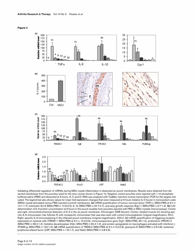
Expression of selected targets in isolated membranes versus the overlying soft tissuesAs expected, levels of mRNAs encoding resistin (a marker foradipocytes) and dystrophin (skeletal muscle) were by far high-est in RNA extracted from the soft tissues overlying the mem-branes (Figure 3c). In contrast, mRNAs of several mRNAsidentified by the microarray analysis of dissected membranesas inducible by MSU crystals (see below) were highest inmembrane RNA. Consistent with the observations that IL-6 isalso expressed in striated muscle (for instance, Figure 4b) andPUMA-g (protein upregulated on macrophages activated withinterferon-γ) in adipocytes [13], the relative soft tissue frac-tions of these two mRNAs were larger than those of TREM(triggering receptor expressed on myeloid cells)-1 and TREM-2, both of which are predominantly expressed on inflammatorycells.
Identification of differentially expressed genes by microarray analysisRelative quantitative PCR (qPCR) analysis for resistin and dys-trophin revealed that, despite the histologic absence of fat andskeletal muscle from the dissected membranes, individualnoninflamed (control) membranes varied in the amounts ofmRNAs encoding these markers. This suggested the pres-ence of small remnants of fat and muscle on the membranesthat persisted despite the histologically clean dissection. Tocorrect for this heterogeneity and other potential differencesresulting from the dissection, RNA aliquots from three controlor three MSU-stimulated membranes (obtained 9 hours afterinjection of MSU crystals in PBS or PBS alone) were pooled.An aliquot from each pool was then processed according tostandard Affymetrix protocols, and the resulting labelledcRNAs hybridized to separate Affymetrix Mo430_2 oligonu-cleotide microarrays. Of the 45,101 probe sets contained onthe microarrays, 21,009 and 19,941 were detected (anno-tated 'p' in the raw Affymetrix data) in the pooled RNAs fromcontrol and MSU membranes, respectively. A total of 5,988were differentially regulated (Affymetrix flag) in response to
Page 4 of 13(page number not for citation purposes)

Available online http://arthritis-research.com/content/10/3/R64
MSU crystals. These were filtered on the Affymetrix change Pvalues below 2 × 10⁻⁵ for upregulated and 1 for the downreg-ulated probe sets.
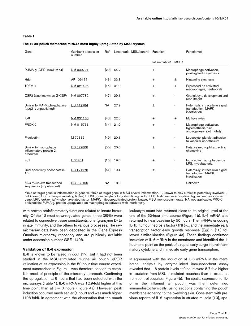
Of these, the 12 most highly over-expressed (linear ratio MSU/control >17) targets are listed in Table 1. Eight (75%) weregenes with known proinflammatory functions: histidine decar-boxylase (Hdc, the enzyme that catalyzes the conversion ofhistidine to histamine); the surface receptors PUMA-g (alsoknown as GPR109 or HM74) [14] and TREM-1 [15], which
are induced on activated macrophages and neutrophils; immu-noresponsive gene (Irg)1, a protein that is rapidly induced dur-ing monocyte activation with endotoxin [16]; prokineticin(PROK)-2, which is a small polypeptide that is involved in mac-rophage activation, hyperesthesia and other processes [14];IL-6; colony-stimulating factor (CSF)3 (also known as granulo-cyte colony-stimulating factor); and the adhesion molecule P-selectin. Three genes (25%) encoded less well characterizedfactors but had potential functions in inflammation. One genewas uncharacterized. Of note, all of the over-expressed genes
Figure 3
Histologic and molecular characterization of dissected membranes and adjacent tissues (a) Histologic cross-sections illustrating the plane of dissectionHistologic and molecular characterization of dissected membranes and adjacent tissues (a) Histologic cross-sections illustrating the plane of dis-section. Left image: cross-section through an entire monosodium urate (MSU) crystal inflamed pouch wall, showing membrane (short red arrow) and the overlying cutaneous soft tissue (long black arrow). Original magnification: 100×. Center image: cutaneous soft tissue of the air pouch wall after removal of the membrane by the dissection method outlined in Figure 2. Original magnification: 100×. Right image: normal dorsal skin. It is nearly identical in appearance to the cutaneous parts shown in part b, which are left after dissection of the membrane. Original magnification: 100×. (b) Tissues that will probably contaminate the dissected membrane if they are not avoided during the final steps of the dissection. Left image: tissue from the nuchal cape-like structure to which the most rostral parts of the membrane are often attached. Original magnification: 200×. Center image: tissue obtained from the paraspinal ridges from which the base of the membrane arises (for the macroscopic appearance see the tissue marked with the arrow in Figure 2k). Original magnification: 200×. Right image: dissected membrane obtained from an air pouch injected with MSU crystals (2 mg in 1 ml phosphate-buffered saline). It consists mostly of fibroblasts and inflammatory cells. Blood vessels can also be found but are not abundant. Original magnification: 100×. (c) Partitioning of selected mRNAs between pouch membrane and the overlying cutaneous soft tissues. Membranes (n = 4) were dissected from the soft tissues 9 hours after injecting MSU crystals into the air pouches. RNA was extracted from dissected mem-branes or the soft tissues and analyzed separately by quantitative PCR. Results were normalized to GAPDH and the data obtained from membrane RNA arbitrarily assigned the value 1.
Page 5 of 13(page number not for citation purposes)
Arthritis Research & Therapy Vol 10 No 3 Pessler et al.
Figure 4
Validating differential regulation of mRNAs during MSU crystal inflammation in dissected air pouch membranesValidating differential regulation of mRNAs during MSU crystal inflammation in dissected air pouch membranes. Results were obtained from dis-sected membranes from the pouches used for the time course shown in Figure 1b. Negative control pouches were injected with 1 ml phosphate-buffered saline (PBS) and dissected at 9 hours. A, C and D: RNA was analyzed with TaqMan real-time reverse transcription PCR for the targets indi-cated. The legend text also shows values for mean fold expression changes that were measured at 9 hours (relative to 0 hours) in monosodium urate (MSU) crystal stimulated versus PBS injected (control) membranes. (a) mRNA quantification of tumour necrosis factor (TNF)-α (MSU:PBS at 9 h = 14.1:1.7), interleukin (IL)-6 (MSU:PBS = 12.9:0.5), IL-1β (MSU:PBS = 34.7:0.7), and early growth response (Egr)-1 (MSU:PBS = 3.7:1.4). (b) Left: determination of IL-6 protein concentration at 9 hours in the pouch exudate from pouches injected with PBS or MSU crystals (immunoassay). Center and right: immunohistochemical detection of IL-6 in the air pouch membrane. Chromogen: DAB (brown). Center: striated muscle (m) showing spe-cific IL-6 immunostain; hair follicles (f) with nonspecific immunostain that was also seen with control immunoglobulin (original magnification, 50×). Right: specific IL-6 immunostaining in the inflamed pouch membrane (original magnification, 400×). (c) mRNA quantification of triggering receptor expressed on myeloid cells (TREM)-1 (MSU:PBS at 9 h = 15.3:0.6), immunoresponsive gene (Irg)1 (MSU:PBS, 65:1.0), prokineticin (PROK)-2 (MSU:PBS = 58.4:1.0), histidine decarboxylase (Hdc; MSU:PBS = 60.4:1.3), and protein upregulated on macrophages activated with interferon-γ (PUMA-g; MSU:PBS = 120:1.3). (d) mRNA quantification of TREM-2 (MSU:PBS at 9 h = 0.2:0.9), granzyme D (MSU:PBS = 0.5:0.8), leukemia/lymphoma-related factor (LRF; MSU:PBS = 1.8:1.7), and Nab2 (MSU:PBS = 0.8:0.9).
Page 6 of 13(page number not for citation purposes)
Available online http://arthritis-research.com/content/10/3/R64
with proven proinflammatory functions related to innate immu-nity. Of the 12 most downregulated genes, three (25%) wererelated to connective tissue constituents, one (granzyme D) toinnate immunity, and the others to various processes. The rawmicroarray data have been deposited in the Gene ExpressOmnibus microarray repository and are publically availableunder accession number GSE11498.
Validation of IL-6 expressionIL-6 is known to be raised in gout [17], but it had not beenstudied in the MSU-stimulated murine air pouch. qPCRvalidation of its expression in the 50-hour time course experi-ment summarized in Figure 1 was therefore chosen to estab-lish proof of principle of the microrray approach. Confirmingthe upregulation at 9 hours that had been detected with themicroarrays (Table 1), IL-6 mRNA was 12.9-fold higher at thistime point than at t = 0 hours (Figure 4a). However, peakinduction occurred much earlier (1 hour) and was much higher(108-fold). In agreement with the observation that the pouch
leukocyte count had returned close to its original level at theend of the 50-hour time course (Figure 1b), IL-6 mRNA alsoreturned to near baseline by 50 hours. The mRNAs encodingIL-1β, tumour necrosis factor (TNF)-α, and the immediate earlytranscription factor early growth response (Egr)-1 [18] fol-lowed similar kinetics (Figure 4a). These findings confirmedinduction of IL-6 mRNA in the membrane and identified the 1-hour time point as the peak of a rapid, early surge in proinflam-matory cytokine and immediate early gene transcription.
In agreement with the induction of IL-6 mRNA in the mem-brane, analysis by enzyme-linked immunosorbent assayrevealed that IL-6 protein levels at 9 hours were 8.7-fold higherin exudates from MSU-stimulated pouches than in exudatesfrom control pouches (Figure 4b). The spatial expression of IL-6 in the inflamed air pouch was then determinedimmunohistochemically, using sections containing the pouchmembrane adhering to the overlying skin. Consistent with pre-vious reports of IL-6 expression in striated muscle [19], spe-
Table 1
The 12 air pouch membrane mRNAs most highly upregulated by MSU crystals
Gene Genbank accession number
Ref. Linear ratio: MSU/control Function Function(s)
Inflammationa MSUb
PUMA-g (GPR 109/HM74) NM 030701 [29] 64.2 + - Macrophage activation, prostaglandin synthesis
Hdc AF 109137 [46] 33.8 + ± Histamine synthesis
TREM-1 NM 021406 [15] 31.9 + + Expressed on activated macrophages, neutrophils
CSF3 (also known as G-CSF) NM 007782 [47] 29.1 + - Granulocyte development and recruitment
Similar to MAPK phosphatase (cpg21; unpublished)
BB 442784 NA 27.9 ± - Potentially, intracellular signal transduction, MAPK inactivation
IL-6 NM 031168 [48] 22.5 + + Multiple roles
PROK-2 NM 015768 [14] 21.0 + - Macrophage activation, hyperesthesia/pain, angiogenesis, gut motility
P-selectin M 72332 [49] 20.1 + - Leucocyte, platelet adhesion to vascular endothelium
Similar to macrophage inflammatory protein 2 precursor
BB 829808 [50] 20.0 ± - Putative neutrophil attracting chemokine
Irg1 L 38281 [16] 19.8 + - Induced in macrophages by LPS, mycobacteria
Dual specificity phosphatase 16
BB 121278 [51] 19.4 ± - Potentially, intracellular signal transduction, MAPK inactivation
Mus musculus transcribed sequences (unpublished)
BB 993160 NA 18.0 - - Unknown
aRole of target gene in inflammation in general. bRole of target gene in MSU crystal inflammation. +, known to play a role; ±, potentially involved; -, not known; CSF, colony-stimulating factor; G-CSF, granulocyte colony stimulating factor; Hdc, histidine decarboxylase; Irg, immunoresponsive gene; LRF, leukaemia/lymphoma-related factor; MAPK, mitogen-activated protein kinase; MSU, monosodium urate; NA, not applicable; PROK, prokineticin; PUMA-g, protein upregulated on macrophages activated with interferon-γ.
Page 7 of 13(page number not for citation purposes)
Arthritis Research & Therapy Vol 10 No 3 Pessler et al.
cific staining was seen in muscle fibres of the laminamuscularis of the subcutaneous tissue (Figure 4b, centre;marked 'm'), whereas the signal in hair follicles was not spe-cific (marked 'f', arrows). In the inflamed membrane, specificIL-6 staining was seen in a multitude of cells, including mono-nuclear and polymorphonuclear cells and fibroblasts (Figure4b, right).
Validating induction of Hdc, TREM-1, PUMA-g, Irg1 and PROK-2 mRNAs by MSU crystals in the air pouch membraneqPCR demonstrated dramatic induction of these mRNAsthroughout the air pouch time course (Figure 4c). Confirmingthe microarray results, all of these mRNAs were elevated at 9hours. Interestingly, the extent and kinetics of their inductiondiffered; whereas maximal induction of Irg1 (177-fold), PROK-2 (136-fold), TREM-1 (39-fold) and Hdc (120-fold) occurredat 4 hours, PUMA-g mRNA was upregulated 120-fold andpeaked at 9 hours.
To validate the results of the microarray analysis further, wedetermined kinetics of two mRNAs that were downregulatedby MSU crystals, according to the array analysis (Figure 4d).TREM-2 is a homologue of TREM-1 that is downregulated dur-ing macrophage activation [20], and the microarray analysishad revealed an 81% decrease of its mRNA 9 hours afterMSU crystal injection. Indeed, the time course demonstrateda 79% decrease in TREM-2 mRNA, with a nadir between 9and 18 hours and subsequent recovery to near baseline levelat 50 hours. In contrast, mRNA encoding granzyme D mRNA,which was 96% downregulated at 9 hours according to themicroarrays, decreased steadily and reached 6% of its originallevel at 50 hours. As negative controls, we measured expres-sion of two mRNAs that were not among the differentially reg-ulated genes on the arrays (Figure 4d). First, leukaemia/lymphoma-related factor (LRF; also known as FBI-1 andOCZF), a transcription factor important in cellular transforma-tion [21], osteoclastogenesis [22] and regulation of the HIV-1promoter [23,24]. Levels of its mRNA demonstrated a statisti-cally insignificant twofold increase at 4 hours. Second, mRNAencoding Nab2, a co-regulator of Egr-1 [25], exhibited onlyminor fluctuations. To exclude their origin from remnant adi-pose or muscle tissue, qPCR results for all of the abovemRNAs at 9 hours were also correlated with resistin and dys-trophin mRNA expression, but no significant associationswere found.
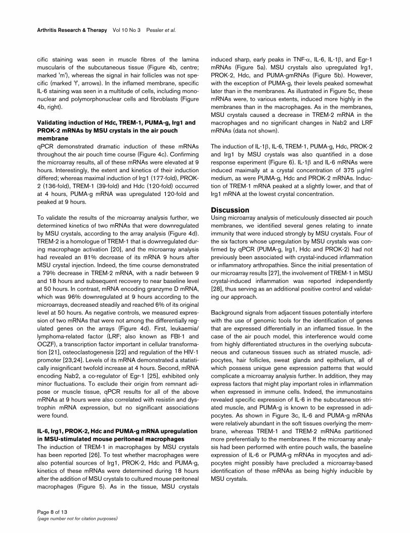
IL-6, Irg1, PROK-2, Hdc and PUMA-g mRNA upregulation in MSU-stimulated mouse peritoneal macrophagesThe induction of TREM-1 in macrophages by MSU crystalshas been reported [26]. To test whether macrophages werealso potential sources of Irg1, PROK-2, Hdc and PUMA-g,kinetics of these mRNAs were determined during 18 hoursafter the addition of MSU crystals to cultured mouse peritonealmacrophages (Figure 5). As in the tissue, MSU crystals
induced sharp, early peaks in TNF-α, IL-6, IL-1β, and Egr-1mRNAs (Figure 5a). MSU crystals also upregulated Irg1,PROK-2, Hdc, and PUMA-gmRNAs (Figure 5b). However,with the exception of PUMA-g, their levels peaked somewhatlater than in the membranes. As illustrated in Figure 5c, thesemRNAs were, to various extents, induced more highly in themembranes than in the macrophages. As in the membranes,MSU crystals caused a decrease in TREM-2 mRNA in themacrophages and no significant changes in Nab2 and LRFmRNAs (data not shown).
The induction of IL-1β, IL-6, TREM-1, PUMA-g, Hdc, PROK-2and Irg1 by MSU crystals was also quantified in a doseresponse experiment (Figure 6). IL-1β and IL-6 mRNAs wereinduced maximally at a crystal concentration of 375 μg/mlmedium, as were PUMA-g, Hdc and PROK-2 mRNAs. Induc-tion of TREM-1 mRNA peaked at a slightly lower, and that ofIrg1 mRNA at the lowest crystal concentration.
DiscussionUsing microarray analysis of meticulously dissected air pouchmembranes, we identified several genes relating to innateimmunity that were induced strongly by MSU crystals. Four ofthe six factors whose upregulation by MSU crystals was con-firmed by qPCR (PUMA-g, Irg1, Hdc and PROK-2) had notpreviously been associated with crystal-induced inflammationor inflammatory arthropathies. Since the initial presentation ofour microarray results [27], the involvement of TREM-1 in MSUcrystal-induced inflammation was reported independently[28], thus serving as an additional positive control and validat-ing our approach.
Background signals from adjacent tissues potentially interferewith the use of genomic tools for the identification of genesthat are expressed differentially in an inflamed tissue. In thecase of the air pouch model, this interference would comefrom highly differentiated structures in the overlying subcuta-neous and cutaneous tissues such as striated muscle, adi-pocytes, hair follicles, sweat glands and epithelium, all ofwhich possess unique gene expression patterns that wouldcomplicate a microarray analysis further. In addition, they mayexpress factors that might play important roles in inflammationwhen expressed in immune cells. Indeed, the immunostainsrevealed specific expression of IL-6 in the subcutaneous stri-ated muscle, and PUMA-g is known to be expressed in adi-pocytes. As shown in Figure 3c, IL-6 and PUMA-g mRNAswere relatively abundant in the soft tissues overlying the mem-brane, whereas TREM-1 and TREM-2 mRNAs partitionedmore preferentially to the membranes. If the microarray analy-sis had been performed with entire pouch walls, the baselineexpression of IL-6 or PUMA-g mRNAs in myocytes and adi-pocytes might possibly have precluded a microarray-basedidentification of these mRNAs as being highly inducible byMSU crystals.
Page 8 of 13(page number not for citation purposes)

Available online http://arthritis-research.com/content/10/3/R64
Potential roles of PUMA-g, TREM-1, Irg1, PROK-2 and Hdc in the pathogenesis of crystal inflammationWe validated the induction of six of the most highly over-expressed genes by qPCR. How might these function in thepathogenesis of crystal-induced inflammation? PUMA-g is aG-protein-coupled transmembrane receptor that was initiallyidentified in a microarray screen of macrophages activatedwith TNF-α and interferon-γ [29]. In addition to activated mac-rophages, it is also expressed on neutrophilic granulocytesand adipocytes. On the latter, it is a major receptor for the cho-lesterol-lowering agent nicotinic acid [13]. Binding of this lig-
and to PUMA-g is also responsible for the cutaneous flushingresponse that is frequently observed in patients taking nico-tinic acid [30], and it has been proposed that cutaneouslyexpressed PUMA-g leads to the release of prostaglandins D2and E2, which in turn cause local vasodilatation [30]. Theseresults suggest that in MSU crystal inflammation PUMA-gtransmits signals in macrophages, and perhaps also granulo-cytes, which potentiate release of prostaglandins and thusenhance the inflammatory process. Further studies withcultured cells or PUMA-g-/- mice will now be important in elu-
Figure 5
Validating differential regulation of mRNAs during MSU crystal inflammation in mouse peritoneal macrophagesValidating differential regulation of mRNAs during MSU crystal inflammation in mouse peritoneal macrophages. Cells were harvested at the indicated time points after the addition of medium containing monosodium urate (MSU) crystals (200 μg/ml) or medium alone. RNA was analyzed by TaqMan real-time reverse transcription PCR for expression of the targets indicated in the figure. Results represent the averages of two experiments. Induction of target mRNAs in negative control (medium only) cells was negligible in nearly all cases. Therefore, the curves corresponding to the negative con-trols are not shown. However, numeric values for mean fold expression changes in MSU stimulated and negative controls with respect to t = 0 hours are listed below for the time points of maximal induction by MSU crystals. (a) Tumour necrosis factor (TNF)-α (2 hours: MSU:medium = 28.1:1.2), IL-6 (1 hour: MSU:medium = 49.7:1.2), IL-1β (2 hours: MSU:medium = 13.1:0.5); and early growth response (Egr)-1 (1 hour: MSU:medium = 17.4:1). (b) Irg1 (6 hours: MSU:medium = 60.0:6.8); prokineticin (PROK)-2 (6 hours: MSU:medium, 11.0:1.0), histidine decarboxylase (Hdc; 9 hours: MSU:medium = 11.6:0.9) and protein upregulated on macrophages activated with interferon-γ (PUMA-g; 9 hours: MSU:medium = 73.5:2.5). (c) Comparison of fold induction by MSU crystals in dissected membranes versus macrophage culture.
Page 9 of 13(page number not for citation purposes)
Arthritis Research & Therapy Vol 10 No 3 Pessler et al.
cidating the function of PUMA-g in crystal-inducedinflammation.
TREM-1, too, is induced on the surface of activated macro-phages and neutrophils, and has been shown to play impor-tant roles in the systemic manifestations of sepsis [26].Interestingly, Murakami and coworkers [28] showed thatTREM-1 mRNA in the pouch exudate peaked before the max-imum accumulation of leukocytes, thus indicating activation ofTREM-1 transcription early on. It has been proposed that onefunction of TREM-1 is to amplify Toll-like receptor (TLR) sign-aling [26]. Considering that TREM-1 mRNA reached maximumlevels after the initial cytokine mRNA surge (Figure 4a) andthat recognition of MSU crystals via TLR signaling is believedto be among the earliest events in crystal inflammation [31],one may postulate that the role of TREM-1 in crystal inflamma-tion also relates to potentiation of signals transmitted by TLRs.
Irg1 was isolated originally from a cDNA library made fromendotoxin-stimulated macrophages [16], and was later foundto be expressed in macrophages infected with mycobacteria[32,33]. Its induction by MSU crystals in peritoneal macro-phages (Figures 5 and 6) suggests that it may play generalroles in macrophage activation.
Prokineticins are evolutionary highly conserved small secretedpolypeptides that play roles in various tissues, including the
central nervous system, the gastrointestinal tract, the haemat-opoietic system and the vasculature [14,34,35]. PROK-2 wasrecently shown to augment macrophage chemotaxis andproinflammatory cytokine synthesis [36]. Interestingly, it alsocauses hyperesthesia, including by local injection into rodentpaws, and mice lacking the PROK receptor PKR1 have dimin-ished pain responses [37]. It is therefore intriguing to specu-late that PROK-2 contributes to the symptoms of MSU crystalinflammation by enhancing both inflammation and pain inaffected joints.
The enzyme Hdc converts histidine to histamine. Histamine iselevated in the joint fluid of patients with gout [38] and in theMSU crystal stimulated rat air pouch, where it is believed to bethe result of mast cell degranulation [12]. Our results, how-ever, suggest that histamine may also arise from increasedHdc levels during MSU crystal inflammation. Consistent withour observation that MSU crystals induced Hdc mRNA synthe-sis in macrophage culture, with reports of Hdc mRNA expres-sion in neutrophils [39], and with the well documentedpresence of mast cells in the air pouch membrane [12], all ofthese three cell types are probably highly inducible sources ofHdc in the pouch membrane. Thus, Hdc and histamine maycontribute significantly to the evolution of MSU crystal inflam-mation in this model, but also in gouty synovitis in humans.
Figure 6
MSU crystal dose responseMSU crystal dose response. Mouse peritoneal macrophages were grown in medium overnight. After removal of nonadherent cells, medium contain-ing increasing concentrations of monosodium urate (MSU) crystals was added. RNA was harvested 4 hours after the addition of crystal-containing medium and analyzed for target gene expression by TaqMan real-time reverse transcription PCR. Results represent the averages of two experiments.
Page 10 of 13(page number not for citation purposes)
Available online http://arthritis-research.com/content/10/3/R64
Significance of the downregulated genesTREM-2 is a cell surface receptor similar to TREM-1. It pro-motes differentiation of macrophages into osteoclasts [40] butinhibits macrophage activation and is downregulated duringthis process [20]. The decrease in TREM-2 mRNA in bothmembranes and cultured macrophages suggests that macro-phage activation by MSU crystals leads to an increase inTREM-1 but to a decrease in TREM-2 mRNA levels. GranzymeD has been detected in murine cytotoxic T cells [41] andendometrial natural killer (NK) cells [42]. The roles of these celltypes in urate crystal inflammation have not been studied.However, it is known that uric acid can activate CD8+ T cellsunder some circumstances [43], and in preliminary studies wehave detected mRNA encoding the NK cell lectin-like receptorA2 in the air pouch membrane (Mayer CT, Schumacher HR,Pessler F, unpublished data). Thus, the persistent downregu-lation of granzyme D throughout the time course suggests thatMSU crystals may modulate cytotoxic T cell or NK cell activityin the membrane. Alternatively, one must consider that dilutionof the membrane RNA pool by mRNAs contained in immigrat-ing inflammatory cells or by otherwise highly upregulatedmRNAs might also cause – at least in part – artifactualdecreases in mRNAs expressed by resident membrane cells.
Kinetics of MSU crystal inflammation in the air pouch membraneThe results of the present study also shed new light on thekinetics of transcriptional regulation in the air pouch mem-brane. We observed an early, steep rise in proinflammatorycytokine mRNA synthesis that essentially paralleled the syn-thesis of mRNA encoding the immediate early transcriptionfactor Egr-1. Similarly rapid kinetics were observed in culturedmacrophages (Figure 5). Indeed, inclusion of the 30-minutetime point revealed that increasing levels of mRNAs encodingEgr-1 and all three cytokines could be detected this early. It iscurrently believed that TLR-mediated activation of the Nalp3containing inflammasome, leading to proteolytic cleavage ofpre-existing pro-IL-1β to IL-1β, is among the first steps in MSUcrystal inflammation [31,44]. Our findings suggest that activa-tion of the proinflammatory cytokine genes encoding IL-1β, IL-6 and TNF-α, as well as Egr-1 and probably other immediateearly transcription factors, is among the next, early events inreprogramming the transcriptional machinery in MSU crystalinflammation, both in an intact tissue and in cultured cells.TREM-1, Irg1, PROK-2, Hdc and PUMA-g mRNA levels allpeaked after this dramatic burst in early cytokine transcription.These factors may therefore play roles in perpetuating oramplifying this early inflammation. We had performed themicroarray analysis 9 hours after MSU crystal injection, thepeak of leukocyte accumulation in the pouch lumen. However,results of the time course analysis demonstrate that major tran-scriptional reprogramming in the air pouch membrane occursseveral hours earlier. Future RNA-based studies into eventsleading to the peak phase of inflammation in the membrane
should therefore focus on relatively early time points up to 4hours.
Caveats of the present studyGene expression results obtained from the dissected mem-branes do not allow one to determine whether a given overex-pressed gene was induced in the resident membrane cells orimported by infiltrating cells. It is therefore important to validatedifferential expression of a given gene in a purified cell popu-lation, as was done here with cultured macrophages. Eventhough we validated the induction of all six targets in culturedmacrophages, their fold induction was lower in these cellsthan in the membranes (Figure 5). This suggests that events inthe membranes due to cell flux, expression in cells other thanmacrophages (for example, neutrophils), or more efficientgene regulation due to cell-cell interactions contributesignificantly to regulation of these genes in the intact mem-branes. Another caveat is that the microarray analysis was notreplicated, which precluded an extended statistical analysis.However, the unusually high degrees of over-expression andunder-expression apparently compensated for this lack of rep-lication; they allowed us to choose a small number of genes forqPCR validation strictly by the high magnitudes of their expres-sion changes.
It is also important to remember that inflammation extendsbeyond the membrane into the loose, fibroblast-rich tissuebetween the membrane and the striated muscle layer (Figure3a and Schiltz and coworkers [12]). The air pouch membranetherefore does not contain the entire inflammation that devel-ops in the air pouch, and inflammatory cells may remain in theair pouch wall if the membrane is removed with the blunt dis-section method described here. Furthermore, MSU crystalscause swelling of the air pouch membrane [45]. This is mostlikely due to the influx of inflammatory cells and associatedinterstitial edema and must be considered when evaluating themembrane histologically.
Potential uses of the isolated air pouch membrane in gene discoveryOur findings suggest that a more extensive, replicated micro-array analysis involving higher numbers of air pouch mem-branes may be a powerful tool to identify genes that aredifferentially active in various aspects of inflammation andinnate immunity. In particular, it should lend itself well to theidentification of genes that act at different time points in theevolution or resolution of inflammation, genes that are regu-lated in response to treatment with anti-inflammatory agents,or genes that reflect changes in the resident connective tissuecells in response to inflammation.
ConclusionA dissection method was established that allowed for the sep-aration of the murine air pouch membrane from the overlyingcutaneous tissues. Transcript profiling of this minimal tissue
Page 11 of 13(page number not for citation purposes)
Arthritis Research & Therapy Vol 10 No 3 Pessler et al.
identified several mRNAs relating to innate immune responsesthat were previously not implicated in MSU crystal inflamma-tion. The appearance of these mRNAs after an initial surge inproinflammatory and immediate early gene transcription sug-gests that they may form part of a 'second wave' of factors thatamplify or perpetuate acute inflammation.
Competing interestsThe authors declare that they have no competing interests.
Authors' contributionsFP designed the study, performed the experiments and wrotethe manuscript. CTM, SMJ and LD participated in theexperiments. EMB provided the macrophages and discussion.JPM provided laboratory equipment, reagents and discussion.HRS oversaw the study, provided the MSU crystals and initialinstruction in the use of the air pouch model, and edited themanuscript. All authors read and approved the finalmanuscript.
AcknowledgementsWe thank G Clayburne, Nicole Chartrain and Marlin Camps-Ramirez for technical support; Grace Strazewski and Don Baldwin (University of Pennsylvania Microarray Core) for microarray hybridization; and John Tobias (University of Pennsylvania Bioinformatics Core) for help with analyzing the microarray data; Dan Martinez and the staff of the histopa-thology core of The Children's Hospital of Philadelphia for histology sup-port; and Peri DeRitis and the staff of the VA Medical Center Animal Facility for expert animal care. F Pessler was supported by National Insti-tutes of Health Training Grants T32-AR 007442 and T32-CA 09140 and a 2007-8 American College of Rheumatology Education and Research Foundation Amgen Pediatric Rheumatology Research Award, and CT Mayer by a 2006 American College of Rheumatology Health Professional Graduate Student Preceptorship Award.
References1. Edwards JC, Sedgwick AD, Willoughby DA: The formation of a
structure with the features of synovial lining by subcutaneousinjection of air: an in vivo tissue culture system. J Pathol 1981,134:147-156.
2. Generini S, Matucci-Cerinic M, Partsch G, Stancikova M, PignoneA, Konttinen YT, Rovensky J, Baker DJ, Schumacher HR Jr: Evi-dence for hyaluronan production in the air pouch model inrats. Clin Exp Rheumatol 2001, 19:271-276.
3. Gordon TP, Kowanko IC, James M, Roberts-Thomson PJ: Mono-sodium urate crystal-induced prostaglandin synthesis in therat subcutaneous air pouch. Clin Exp Rheumatol 1985,3:291-296.
4. Sedgwick AD, Sin YM, Edwards JC, Willoughby DA: Increasedinflammatory reactivity in newly formed lining tissue. J Pathol1983, 141:483-495.
5. Nagase M, Baker DG, Schumacher HR Jr: Prolonged inflamma-tory reactions induced by artificial ceramics in the rat air pouchmodel. J Rheumatol 1988, 15:1334-1338.
6. Yoshino S, Cromartie WJ, Schwab JH: Inflammation induced bybacterial cell wall fragments in the rat air pouch. Comparisonof rat strains and measurement of arachidonic acidmetabolites. Am J Pathol 1985, 121:327-336.
7. Nalbant S, Chen LX, Sieck MS, Clayburne G, Schumacher HR:Prophylactic effect of highly selective COX-2 inhibition inacute monosodium urate crystal induced inflammation in therat subcutaneous air pouch. J Rheumatol 2005, 32:1762-1764.
8. McCarty DJ Jr, Faires JS: A comparison of the duration of localanti-inflammatory effect of several adrenocorticosteroid
esters – a bioassay technique. Curr Ther Res Clin Exp 1963,5:284-290.
9. Jung SM, Schumacher HR, Kim H, Kim M, Lee SH, Pessler F:Reduction of urate crystal-induced inflammation by rootextracts from traditional oriental medicinal plants: elevation ofprostaglandin D2 levels. Arthritis Res Ther 2007, 9:R64.
10. Schumacher HR, Reginato AJ: Atlas of Synovial Fluid Analysisand Crystal Identification 1st edition. Philadelphia, PA: Lea andFiebiger; 1991.
11. Affymetrix [http://www.affymetrix.com/support/technical/manual/expression-manual.affx]
12. Schiltz C, Lioté F, Prudhommeaux F, Meunier A, Champy R, Calle-bert J, Bardin T: Monosodium urate monohydrate crystal-induced inflammation in vivo: quantitative histomorphometricanalysis of cellular events. Arthritis Rheum 2002,46:1643-1650.
13. Tunaru S, Kero J, Schaub A, Wufka C, Blaukat A, Pfeffer K, Offer-manns S: PUMA-G and HM74 are receptors for nicotinic acidand mediate its anti-lipolytic effect. Nat Med 2003, 9:352-355.
14. Li M, Bullock CM, Knauer DJ, Ehlert FJ, Zhou QY: Identification oftwo prokineticin cDNAs: recombinant proteins potently con-tract gastrointestinal smooth muscle. Mol Pharmacol 2001,59:692-698.
15. Bouchon A, Dietrich J, Colonna M: Cutting edge: inflammatoryresponses can be triggered by TREM-1, a novel receptorexpressed on neutrophils and monocytes. J Immunol 2000,164:4991-4995.
16. Lee CG, Jenkins NA, Gilbert DJ, Copeland NG, O'Brien WE: Clon-ing and analysis of gene regulation of a novel LPS-induciblecDNA. Immunogenetics 1995, 41:263-270.
17. Brozik M, Rosztoczy I, Meretey K, Balint G, Gaal M, Balogh Z, BartM, Mituszova M, Velics V, Falus A: Interleukin 6 levels in synovialfluids of patients with different arthritides: correlation withlocal IgM rheumatoid factor and systemic acute phase proteinproduction. J Rheumatol 1992, 19:63-68.
18. Sukhatme VP, Cao XM, Chang LC, Tsai-Morris CH, StamenkovichD, Ferreira PC, Cohen DR, Edwards SA, Shows TB, Curran T, etal.: A zinc finger-encoding gene coregulated with c-fos duringgrowth and differentiation, and after cellular depolarization.Cell 1988, 53:37-43.
19. Febbraio MA, Pedersen BK: Contraction-induced myokine pro-duction and release: is skeletal muscle an endocrine organ?Exerc Sport Sci Rev 2005, 33:114-119.
20. Turnbull IR, Gilfillan S, Cella M, Aoshi T, Miller M, Piccio L, Hernan-dez M, Colonna M: Cutting edge: TREM-2 attenuates macro-phage activation. J Immunol 2006, 177:3520-3524.
21. Maeda T, Hobbs RM, Merghoub T, Guernah I, Zelent A, Cordon-Cardo C, Teruya-Feldstein J, Pandolfi PP: Role of the proto-onco-gene Pokemon in cellular transformation and ARF repression.Nature 2005, 433:278-285.
22. Kukita A, Kukita T, Ouchida M, Maeda H, Yatsuki H, Kohashi O:Osteoclast-derived zinc finger (OCZF) protein with POZdomain, a possible transcriptional repressor, is involved inosteoclastogenesis. Blood 1999, 94:1987-1997.
23. Pessler F, Hernandez N: Flexible DNA binding of the BTB/POZ-domain protein FBI-1. J Biol Chem 2003, 278:29327-29335.
24. Pessler F, Pendergrast PS, Hernandez N: Purification and char-acterization of FBI-1, a cellular factor that binds to the humanimmunodeficiency virus type 1 inducer of short transcripts.Mol Cell Biol 1997, 17:3786-3798.
25. Svaren J, Sevetson BR, Apel ED, Zimonjic DB, Popescu NC, Mil-brandt J: NAB2, a corepressor of NGFI-A (Egr-1) and Krox20, isinduced by proliferative and differentiative stimuli. Mol CellBiol 1996, 16:3545-3553.
26. Bouchon A, Facchetti F, Weigand MA, Colonna M: TREM-1amplifies inflammation and is a crucial mediator of septicshock. Nature 2001, 410:1103-1107.
27. Jung S, Schumacher H, Dai L, Pessler F: Identification of pro-inflammatory genes by genomic analysis of the isolatedmonosodium urate-stimulated murine air pouch membrane(abstract). Arthritis Rheum 2005, 52:4074.
28. Murakami Y, Akahoshi T, Hayashi I, Endo H, Kawai S, Inoue M,Kondo H, Kitasato H: Induction of triggering receptorexpressed on myeloid cells 1 in murine resident peritonealmacrophages by monosodium urate monohydrate crystals.Arthritis Rheum 2006, 54:455-462.
Page 12 of 13(page number not for citation purposes)
Available online http://arthritis-research.com/content/10/3/R64
29. Schaub A, Futterer A, Pfeffer K: PUMA-G, an IFN-gamma-induc-ible gene in macrophages is a novel member of the seventransmembrane spanning receptor superfamily. Eur J Immunol2001, 31:3714-3725.
30. Benyo Z, Gille A, Kero J, Csiky M, Suchankova MC, Nusing RM,Moers A, Pfeffer K, Offermanns S: GPR109A (PUMA-G/HM74A)mediates nicotinic acid-induced flushing. J Clin Invest 2005,115:3634-3640.
31. Akahoshi T, Murakami Y, Kitasato H: Recent advances in crystal-induced acute inflammation. Curr Opin Rheumatol 2007,19:146-150.
32. Basler T, Jeckstadt S, Valentin-Weigand P, Goethe R: Mycobac-terium paratuberculosis, Mycobacterium smegmatis, andlipopolysaccharide induce different transcriptional and post-transcriptional regulation of the IRG1 gene in murinemacrophages. J Leukoc Biol 2006, 79:628-638.
33. Shi S, Blumenthal A, Hickey CM, Gandotra S, Levy D, Ehrt S:Expression of many immunologically important genes inMycobacterium tuberculosis-infected macrophages is inde-pendent of both TLR2 and TLR4 but dependent on IFN-alpha-beta receptor and STAT1. J Immunol 2005, 175:3318-3328.
34. Mollay C, Wechselberger C, Mignogna G, Negri L, Melchiorri P,Barra D, Kreil G: Bv8, a small protein from frog skin and itshomologue from snake venom induce hyperalgesia in rats.Eur J Pharmacol 1999, 374:189-196.
35. LeCouter J, Zlot C, Tejada M, Peale F, Ferrara F: Bv8 and endo-crine gland-derived vascular endothelial growth factorstimulate hematopoiesis and hematopoietic cell mobilization.Proc Natl Acad Sci USA 2004, 101:16813-16818.
36. Martucci C, Franchi S, Giannini E, Tian H, Melchiorri P, Negri L,Sacerdote P: Bv8, the amphibian homologue of the mamma-lian prokineticins, induces a proinflammatory phenotype ofmouse macrophages. Br J Pharmacol 2006, 147:225-234.
37. Negri L, Lattanzi R, Giannini E, Colucci M, Margheriti F, MelchiorriP, Vellani V, Tian H, De Felice M, Porreca F: Impaired nociceptionand inflammatory pain sensation in mice lacking the proki-neticin receptor PKR1: focus on interaction between PKR1 andthe capsaicin receptor TRPV1 in pain behavior. J Neurosci2006, 26:6716-6727.
38. Dalbeth N, Haskard D: Pathophysiology of crystal-inducedarthritis. In Crystal-Induced Arthropathies Edited by: WortmannR, Schumacher H, Becker M, Ryan L. New York, NY: Taylor &Francis; 2007:239-354.
39. Shiraishi M, Hirasawa N, Oikawa S, Kobayashi Y, Ohuchi K: Anal-ysis of histamine-producing cells at the late phase of allergicinflammation in rats. Immunology 2000, 99:600-606.
40. Cella M, Buonsanti C, Strader C, Kondo T, Salmaggi A, ColonnaM: Impaired differentiation of osteoclasts in TREM-2-deficientindividuals. J Exp Med 2003, 198:645-651.
41. Masson D, Tschopp J: A family of serine esterases in lytic gran-ules of cytolytic T lymphocytes. Cell 1987, 161:679-685.
42. Allen MP, Nilsen-Hamilton M: Granzymes D, E, F, and G are reg-ulated through pregnancy and by IL-2 and IL-15 in granulatedmetrial gland cells. J Immunol 1998, 161:2772-2779.
43. Shi Y, Galusha SA, Rock L: Cutting edge: elimination of anendogenous adjuvant reduces the activation of CD8 T lym-phocytes to transplanted cells and in an autoimmune diabetesmodel. J Immunol 2006, 176:3905-3908.
44. Martinon F, Petrilli V, Mayor A, Tardivel A, Tschopp J: Gout-asso-ciated uric acid crystals activate the NALP3 inflammasome.Nature 2006, 440:237-241.
45. Lioté F, Prudhommeaux F, Schiltz C, Champy R, Herbelin A, Ortiz-Bravo E, Bardin T: Inhibition and prevention of monosodiumurate monohydrate crystal-induced acute inflammation in vivoby transforming growth factor beta1. Arthritis Rheum 1996,39:1192-1198.
46. Joseph DR, Sullivan PM, Wang YM, Kozak C, Fenstermacher DA,Behrendsen ME, Zahnow CA: Characterization and expressionof the complementary DNA encoding rat histidinedecarboxylase. Proc Natl Acad Sci USA 1990, 87:733-737.
47. Demetri GD, Griffin JD: Granulocyte colony-stimulating factorand its receptor. Blood 1991, 78:2791-2808.
48. Chiu CP, Moulds C, Coffman RL, Rennick D, Lee F: Multiple bio-logical activities are expressed by a mouse interleukin 6 cDNAclone isolated from bone marrow stromal cells. Proc Natl AcadSci USA 1988, 85:7099-7103.
49. Johnston GI, Cook RG, McEver RP: Cloning of GMP-140, a gran-ule membrane protein of platelets and endothelium: sequencesimilarity to proteins involved in cell adhesion andinflammation. Cell 1989, 56:1033-1044.
50. Carninci P, Kasukawa T, Katayama S, Gough J, Frith MC, MaedaN, Oyama R, Ravasi T, Lenhard B, Wells C, Kodzius R, ShimokawaK, Bajic VB, Brenner SE, Batalov S, Forrest AR, Zavolan M, DavisMJ, Wilming LG, Aidinis V, Allen JE, Ambesi-Impiombato A,Apweiler R, Aturaliya RN, Bailey TL, Bansal M, Baxter L, Beisel KW,Bersano T, Bono H, et al.: The transcriptional landscape of themammalian genome. Science 2005, 309:1559-1563.
51. Ko MS, Kitchen JR, Wang X, Threat TA, Wang X, Hasegawa A,Sun T, Grahovac MJ, Kargul GJ, Lim MK, Cui Y, Sano Y, Tanaka T,Liang Y, Mason S, Paonessa PD, Sauls AD, DePalma GE, ShararaR, Rowe LB, Eppig J, Morrell C, Doi H: Large-scale cDNA analy-sis reveals phased gene expression patterns during preim-plantation mouse development. Development 2000,127:1737-1749.
Page 13 of 13(page number not for citation purposes)