Embed Size (px)
Citation preview
SC I ENCE S I GNAL ING | R E S EARCH ART I C L E
VASCULAR B IOLOGY
Department of Pharmacology, University of Oxford, Mansfield Road, Oxford OX13QT, UK.*Joint first authors.†Present address: Department of Medical Physiology, College of Medicine, TexasA&M University Health Science Center, 702 Southwest H.K. Dodgen Loop, Temple,TX 76504, USA.‡Corresponding author. Email: [email protected]
Garland et al., Sci. Signal. 10, eaal3806 (2017) 4 July 2017
Copyright © 2017
The Authors, some
rights reserved;
exclusive licensee
American Association
for the Advancement
of Science. No claim
to original U.S.
Government Works
Voltage-dependent Ca2+ entry into smooth muscleduring contraction promotes endothelium-mediatedfeedback vasodilation in arterioles
Christopher J. Garland,* Pooneh Bagher,*† Chloe Powell, Xi Ye, Hamish A.L. Lemmey,Lyudmyla Borysova, Kim A. Dora‡httpD
ownloaded from
Vascular smooth muscle contraction is suppressed by feedback dilation mediated by the endothelium. In skel-etal muscle arterioles, this feedback can be activated by Ca2+ signals passing from smooth muscle through gapjunctions to endothelial cells, which protrude through holes in the internal elastic lamina to make contact withvascular smooth muscle cells. Although hypothetically either Ca2+ or inositol trisphosphate (IP3) may providethe intercellular signal, it is generally thought that IP3 diffusion is responsible. We provide evidence that Ca2+
entry through L-type voltage-dependent Ca2+ channels (VDCCs) in vascular smooth muscle can pass to theendothelium through positions aligned with holes in the internal elastic lamina in amounts sufficient to activateendothelial cell Ca2+ signaling. In endothelial cells in which IP3 receptors (IP3Rs) were blocked, VDCC-driven Ca2+
events were transient and localized to the endothelium that protrudes through the internal elastic lamina tocontact vascular smooth muscle cells. In endothelial cells in which IP3Rs were not blocked, VDCC-driven Ca2+
events in endothelial cells were amplified to form propagating waves. These waves activated voltage-insensitive,intermediate-conductance, Ca2+-activated K+ (IKCa) channels, thereby providing feedback that effectively sup-pressed vasoconstriction and enabled cycles of constriction and dilation called vasomotion. Thus, agonists thatstimulate vascular smooth muscle depolarization provide Ca2+ to endothelial cells to activate a feedback circuitthat protects tissue blood flow.
://st
on August 22, 2018ke.sciencem
ag.org/
INTRODUCTIONAutoregulation of tissue blood flow is crucial for normal physiologicalfunction. This fundamental response depends on myogenic tone, anongoing pressure-induced vascular smooth muscle cell contraction insmall arteries that can be rapidly modulated by perivascular nerves, au-tacoids, the extracellular accumulation of metabolites, and the endothe-lium (1, 2). Myogenic tone is underpinned by increases in intraluminalpressure depolarizing vascular smooth muscle and increasing the openprobability of L-type voltage-dependent Ca2+ channels (VDCCs) (3, 4).The L-type VDCC in smooth muscle is the CaV1.2 isoform, which isactivated by Bay K8644 (BayK) and inhibited by nifedipine (5, 6).
In cerebral arteries, myogenic tone is continuously suppressed by anintracellular signaling pathway intrinsic to smooth muscle that helpsensure constant blood flow. Ca2+ sparks, generated by Ca2+ releasethrough ryanodine receptors (RyRs) adjacent to the plasmalemma, ac-tivate large-conductance voltage- and Ca2+-activated K+ (BKCa) chan-nels, reducing the open probability of VDCCs, limiting Ca2+ influx, andsuppressing contraction (7, 8). This negative feedback mechanism re-duces excessive cerebral artery constriction, thereby preserving bloodflow and protecting against neuronal hypoxia. However, opposing my-ogenic tone to this extent is not a physiological priority in skeletalmuscle,where high resistance to blood flow is necessary at rest yet increased flowmust be accommodated during exercise. Consistent with this difference,Ca2+ sparks are absent from smooth muscle cells of smaller, second-order pressurized skeletal muscle arterioles although myogenic tone ispresent. Furthermore, although smooth muscle BKCa channel activation
can suppress contraction, these channels do not seem to be subject to thesame tight intracellular regulation as cerebral smoothmuscle cells (9–11).
In addition to intracellular control, negative feedback also operatesbetween vascular cells. Specifically, intercellular Ca2+ signaling from ac-tivated vascular smooth muscle to endothelial cells (myoendothelialsignaling) suppresses vasoconstriction because increases in endothelialcell Ca2+ stimulate vasodilation. Thus, a1-adrenoceptor stimulation ordirect depolarization with high extracellular KCl leads to increased en-dothelial cell [Ca2+]i, causing nitric oxide release to limit vasoconstric-tion (12). Similar myoendothelial signaling and feedback in responseto adrenergic agonists operates in other vascular beds and can also in-volve endothelium-dependent hyperpolarization (EDH) and cycles ofcontraction and dilation termed vasomotion (13–18). In both cases,endothelial cells are activated by a signal passing from smooth musclethrough myoendothelial gap junctions. Both inositol trisphosphate(IP3) and Ca2+ can, in theory, pass through these gap junctions, andit has been argued that paracrine signaling is due solely to IP3 move-ment into the endothelium to activate Ca2+ wavelets (17, 19). TheseCa2+ events are termed pulsars if they align within or near holes inthe internal elastic lamina, the only possible sites of myoendothelialgap junctions (13, 14, 16). However, data supporting this assertionare based on an assumption of IP3movement and the use of nonspecificpharmacological tools. Themovement of IP3 may be spatially and tem-porally far more restricted than previously thought (20–22), reopeningthe debate as to whether Ca2+ itself might act as an effective signal be-tween cells by passing through gap junctions.
The possibility that Ca2+ may pass from smooth muscle in amountssufficient to activate endothelial cell Ca2+ events and initiate feedbackhas not been explored or seriously considered (17). We showed inskeletal muscle arterioles that Ca2+ in smooth muscle could pass to theendothelium through positions aligned with holes in the internal elasticlamina in amounts sufficient to activate endothelial cell Ca2+ signaling.
1 of 14
SC I ENCE S I GNAL ING | R E S EARCH ART I C L E
To demonstrate this phenomenon, we loaded endothelial cells in pres-surized arterioles with heparin to antagonize the effect of IP3 and thenopened L-type VDCCs in the smooth muscle with BayK to stimulateCa2+ influx. BayK evoked a new, elementary Ca2+ event, VDCC-dependent Endothelial cell Ca2+ Transients (we term VECTors), whichcould evolve into endothelial cell Ca2+ pulsars and “waves” through IP3receptor (IP3R)–dependent Ca
2+-induced Ca2+ release. These direc-tional Ca2+ signals from smooth muscle then stimulated voltage-insensitive, intermediate-conductance, Ca2+-activated K+ (IKCa) channelslocalized within the endothelial cell membrane projections to thesmooth muscle, leading to suppression of agonist-induced vasocon-striction and initiating the dilation phase of vasomotion.
Dow
nloaded fr
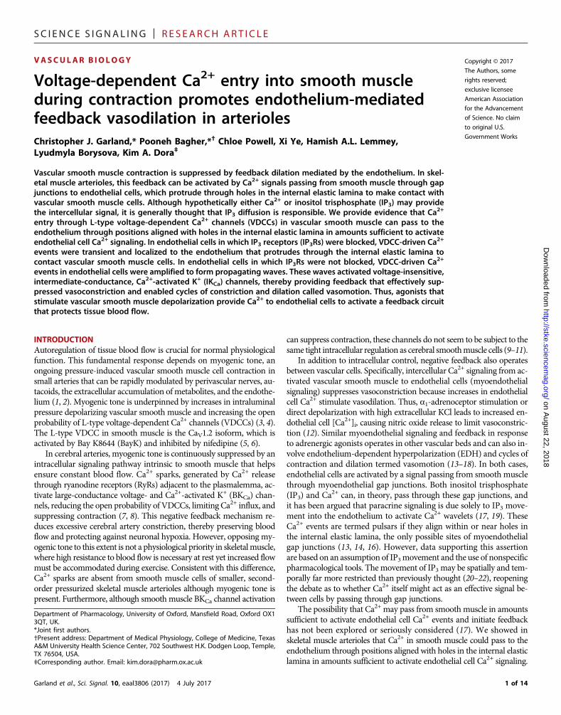
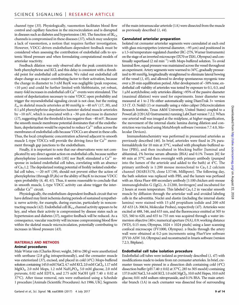
RESULTSDirect activation of arteriole L-type VDCCs with BayKtriggers myoendothelial Ca2+ signalingArterioles from rat cremaster had a maximum diameter of 163 ± 1 mmand developed ~50% pressure-inducedmyogenic tone, which was abol-ished by blocking L-type VDCCs with nifedipine (fig. S1). Endothelialcells loaded with a Ca2+ indicator (OGB-1) were visible at the bottomplane of pressurized arterioles with myogenic tone (Fig. 1A and fig.S2A).Ca2+ events were analyzedmanually (1), because automated anal-
Garland et al., Sci. Signal. 10, eaal3806 (2017) 4 July 2017
ysis did not provide complete or reliable data (fig. S2, B toD). To assesswhether directly opening L-type VDCCs influenced endothelial cell[Ca2+], we used two concentrations of BayK in the absence and presenceof nifedipine (Fig. 1, A andB).At 3 nM,BayK increased the frequency ofendothelial cell Ca2+ events fourfold without affecting arteriolar diam-eter (Fig. 1, A and C, fig. S3A, and movie S1), and recruited new activesites, doubling the percentage of active cells (Fig. 1D). Cumulative ad-dition of BayK to a final concentration 30 nM stimulated robust vaso-constriction and vasomotion (movie S1), and further increased Ca2+
event frequency to 10-fold above baseline (Fig. 1, A and C). Nearly allcells were now active, threefold more than at baseline (Fig. 1D andmovie S1). As expected, blocking L-type VDCCs with nifedipine pre-vented vasoconstriction and the activation of endothelial cell Ca2+
events in response to either concentration of BayK (Fig. 1, B to D).Inhibition of contraction with blebbistatin prevented the develop-ment of myogenic tone (contraction) without affecting the abilityof BayK to activate endothelial cell [Ca2+] (fig. S3, A to C), suggestingthat the diameter of arterioles per se did not influence endothelialcell [Ca2+].
As we have previously shown (1), spontaneous (in the absence ofBayK) endothelial cell Ca2+ events occurred mainly as localized Ca2+
increases, radiating less than ~10 mm from the center of an event, andwere insensitive to nifedipine (Fig. 1, C and D, and fig. S4, A and B).
on August 22, 2018
http://stke.sciencemag.org/
om
1 µM Nif
3
3
2
2
1
1
Nif + 30 nM BayK
Nif + 3 nM BayK
30 nM BayK3 nM BayKBaseline
Nif +30 nM BayKNif + 3 nM BayK1 µM Nif
A
10 s0.5 F
/F0
B
C
0
5
10
15
20
25 Baseline3 nM BayK30 nM BayK
Control Nif
EC
Ca2+
eve
ntfre
quen
cy (m
in−
1 )
*
§*
# #
1
1 2
2
3
3
D
Control Nif0
20
40
60
80
100
Act
ive
cells
(%) *
*
30 nM BayK
3 nM BayK
Baseline
Fig. 1. Direct activation of L-type VDCCs in arterioles with BayK increases Ca2+ events in endothelial cells. (A and B) Confocal fluorescence images of endothelialcells (ECs) loaded with the Ca2+ indicator OGB-1 viewed at the bottom plane in pressurized arterioles with myogenic tone in the absence (A) and presence (B) ofnifedipine (Nif), at baseline and with the indicated concentrations of the L-type VDCC agonist BayK (movie S1); scale bar, 30 mm. Representative fluorescence intensitydata shown as line scans [corresponding to white lines 1 to 3 in (A) or (B)] and fluorescence traces [F/F0; corresponding to colored subcellular regions of interest (ROIs) inthe images]. (C and D) Bar graphs summarize the effect of BayK on the frequency of EC Ca2+ events in active cells (C) and the overall percentage of active cells (D) in theabsence and presence of Nif. Data are means ± SEM (≥10 ECs in each field of view from n = 3 to 6 arterioles from different animals); *P < 0.05 compared with baseline;#P < 0.05 compared to control; §P < 0.05 compared to 3 nM BayK.
2 of 14
SC I ENCE S I GNAL ING | R E S EARCH ART I C L E
About a quarter of spontaneous Ca2+ events were propagating waves,often covering over half a cell length. Endothelial cell Ca2+ eventsstimulated by 3 nM BayK remained localized, whereas those stimu-lated by 30 nM BayK were sufficient to form propagating waves (fig.S4, A and B).
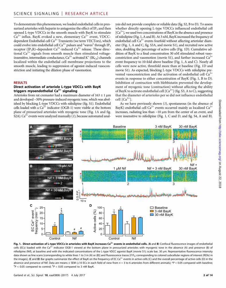
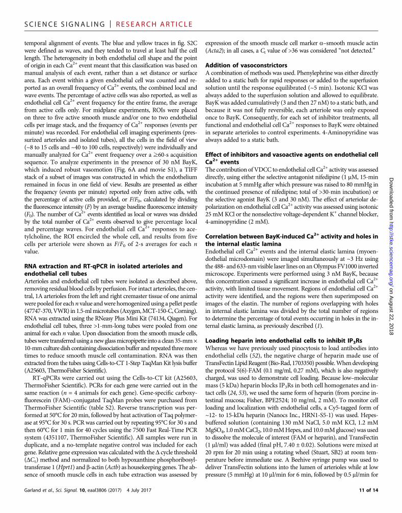
Elementary Ca2+ events occur in the myoendothelialsignaling domainRat cremaster arteriolar endothelial cells expressed the genes encodingall three isoforms of IP3R (Itpr1 to Itpr3), but not those encoding RyRs(Ryr1 to Ryr3) (fig. S5, A and B).We used the pan IP3R antagonist hep-arin to assess the role of these IP3Rs inmyoendothelial Ca2+ signaling in
Garland et al., Sci. Signal. 10, eaal3806 (2017) 4 July 2017
arterioles (fig. S6, A to D). In endothelial cells, heparin-Cy5 closelyalignedwith IP3R1 (Fig. 2A). By simultaneously imaging endothelial cellCa2+ and elastin (Fig. 2B), we found that discrete heparin-insensitiveendothelial cell Ca2+ events rapidly terminated both temporally (dura-tion, ~1 to 2 s) and radially (total width, <15 mm, often 5 to 10 mm) (Fig.2C) andwere localized to themyoendothelialmicrodomain foundwith-in holes in the internal elastic lamina (Fig. 2D). We defined these local-ized events as VECTors. Compared to untreated cells, heparin halvedthe frequency of endothelial cell Ca2+ events to 30 nM BayK (Fig. 2E),did not affect the percentage of active cells (Fig. 2F), and halved the in-cidence of Ca2+ waves (Fig. 2G and fig. S4A). In the same cells, theTRPV4 channel agonist GSK1016790A increased endothelial cell Ca2+
on August 22, 2018
http://stke.sciencemag.org/
Dow
nloaded from
A
B
IP3R1
Heparin-Cy5
Nuclei
ECs
Day 1
Overlay
Day 2
5-kDa heparin+ 3 nM BayK
Cell2
Cell1
E
†#
Heparin Heparin0
2
4
6
8
EC
Ca2+
eve
ntfre
quen
cy (m
in1 )
Baseline30 nM GSK100 nM GSK
†
†
HG
#
0
20
40
60
80
100
Ca2+
wav
es (%
)
Heparin
OG
B-1
W
Cell1
Cell2
10 s
0.2 F
/F0
10 s
0.2 F
/F0
C
D
Ca2+
eve
nts
inM
E m
icro
dom
ain
(%)
Baseline3 nM BayK30 nM BayK
*#
40
60
80
100
HeparinControl0
20
40
60
80
100
Act
ive
cells
(%)
Heparin
†
†
F
5-kDa heparin + 3 nM BayK
15 µ
m15
µm
EC
Ca2+
eve
ntfre
quen
cy (m
in1 )
0
5
10
15
20
25
Hep
-Cy5
IP3R
1 +
nucl
eiH
ep-C
y5 +
IP3R
1A
F-63
3
Fig. 2. VECTors occur in myoendothelial microdomains and are amplified by IP3Rs in endothelial cells. (A) Confocal fluorescence images of endothelial cells (ECs)loaded with heparin-Cy5 (yellow) and the corresponding IP3R1 fluorescence in the same arteriole (magenta). Nuclear staining (cyan) is indicated by dashed lines.Representative of three experiments; scale bar, 15 mm. (B) Simultaneous confocal fluorescence images of the internal elastic lamina labeled with AF-633 and ECs loadedwith the Ca2+ indicator OGB-1 in a pressurized arteriole with myogenic tone during treatment with heparin and BayK; scale bar, 15 mm. (C) Representative fluorescenceintensity data shown as line scans [corresponding to white lines Cell1 and Cell2 in (B)] and fluorescence traces [F/F0; corresponding to colored subcellular ROIs in (B)].Examples of VECTors are indicated by arrowheads. W, wave. (D) Bar graph summarizes the effects of BayK on the percentage of EC Ca2+ events in the myoendothelial(ME) microdomain visible as holes through the internal elastic lamina. (E to H) Bar graphs summarize the effect of heparin on the frequency of EC Ca2+ events in activecells during BayK (E), the overall percentage of active cells (F), the percentage of these events that propagated along cells and were considered to be waves (G), andsubsequent responses to the endothelium-dependent TRPV4 agonist GSK1016790A (GSK) (H). Data are means ± SEM (n = 4 arterioles from different animals); *P < 0.05compared with baseline; †P < 0.05 compared to baseline in the presence of heparin; #P < 0.05 compared to control. (E to G) Control data used for statistical comparisonsfor BayK are shown in Fig. 1; see (1) for control GSK data. Experiments using 5-kDa heparin were only included if the EC Ca2+ response to acetylcholine was markedlyreduced (fig. S6D).
3 of 14
SC I ENCE S I GNAL ING | R E S EARCH ART I C L E
on August 22, 2018
http://stke.sciencemag.org/
Dow
nloaded from
(Fig. 2H) in a manner similar to control conditions (1). Therefore,BayK-induced VECTors are normally amplified by endothelial cellIP3Rs, first to form puffs and then to propagating cell-wide Ca2+ waves.
Attempts to block IP3Rs with 2-aminoethoxydiphenylborate (2-APB)was complicated by actions at gap junctions, a previously reportedeffect of this inhibitor (23). 2-APB caused arterioles to dilate andcontract in an asynchronousmanner, supporting an effect on gap junc-tions (fig. S7A). Furthermore, 2-APB inhibited acetylcholine-mediateddilation (fig. S7B) and feedback dilation induced by BayK application(fig. S7, C and D). In addition, 2-APB blocked dilation mediated byNS309, an allosteric modulator of both isoforms of KCa channels in en-dothelial cells, SKCa (small-conductance, Ca2+-activated K+) and IKCa
channels, further supporting an effect of 2-APB on myoendothelialgap junctions (fig. S7B). Despite this action at gap junctions, 2-APBblocked the increase in endothelial cell Ca2+ normally stimulated by ace-tylcholine (fig. S7E), supporting an effect on IP3Rs. Therefore, the abilityto block the endothelial cell response to BayK could not reliably be as-signed to a single action of 2-APB, yet the block was more completethan with heparin (fig. S7F and Fig. 2E). Overall, the profile of 2-APBblock resembled that of heparin, including the ability to confine Ca2+
events to the myoendothelial microdomain (fig. S7, G andH).We wereunable to use the phospholipase C inhibitor U-73122 against BayK, be-cause it not only blocked dilation to acetylcholine but also preventedcontraction to KCl and damaged arterioles (fig. S8A). Similarly, wedid not consider the IP3R1 antagonist xestospongin C (24) effectiveenough to use against BayK (fig. S8B).
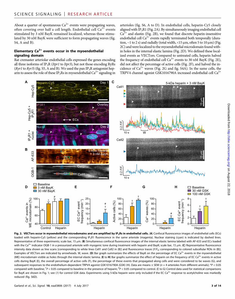
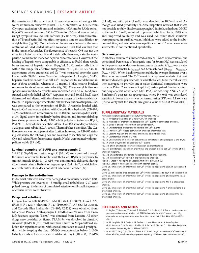
BayK does not trigger direct entry of Ca2+ intoendothelial cellsFreshly isolated endothelial cell tubes were pinned (Fig. 3A) and loadedwith Ca2+ indicator dye (Fig. 3, B andC). SpontaneousCa2+ events wererare (Fig. 3, D and E) and not altered by either BayK, increased KClconcentration, or the selective a1-adrenoceptor agonist phenylephrine(Fig. 3, B to E, andmovies S2 and S3).However, themuscarinic receptoragonist acetylcholine evoked an increase in Ca2+ activity in the sametubes (Fig. 3, B to E). Reverse transcription quantitative polymerasechain reaction (RT-qPCR) analysis showed that the endothelial cell tubemRNA samples were not contaminated with smooth muscle cellmRNA (Fig. 3F) and that mRNAs encoding the CaV1.2, CaV3.1, andCaV3.2 isoforms of L- and T-type VDCCs were detected in whole arte-rioles, but not endothelial cell tubes (Fig. 3, F and G). Immunolabelingfor the smooth muscle isoform of L-type VDCCs, CaV1.2, revealedpunctate labeling in smooth muscle cells but not the endothelium(Fig. 3, H and I), consistent with the mRNA results.
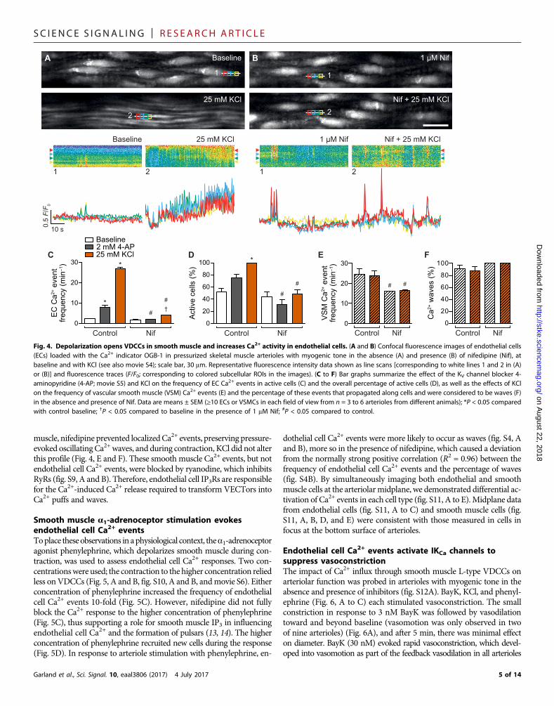
Depolarization stimulates endothelial cell Ca2+ eventsOther approaches were used to open L-type VDCCs and assess endo-thelial cell Ca2+ activity. Endothelial cell Ca2+ activity was increased bydepolarization with KCl at a concentration predicted to clamp themembrane potential at −40 mV or the application of the voltage-dependent K+ (Kv) channel blocker 4-aminopyridine. A clear thresholdfor the initiation of increased endothelial cell Ca2+ activity (Ca2+ triggerpoint) was evident during the time course of vasoconstriction to KCl(movie S4). The frequency of Ca2+ events increased 11-fold, and Ca2+
events were observed in twice as many endothelial cells, and as pre-dicted, nifedipine blocked the increase in Ca2+ events to KCl (Fig. 4,A to D). 4-Aminopyridine also increased the frequency of endothelialcell Ca2+ events (a greater than threefold increase), which remained lo-calized and relied onVDCCs (Fig. 4, C andD, andmovie S5). In smooth
Garland et al., Sci. Signal. 10, eaal3806 (2017) 4 July 2017
PEACh
Acta2
Pecam1
Baseline
30 nM BayK
1 µM ACh
1
1
1
Baseline
B
A
D
KCl
0
4
8
12 BayKBaseline
EC
Ca2+
eve
ntfre
quen
cy (m
in–1
) *E *
0
20
40
60
80
100
Act
ive
cells
(%)
12
2
2
2C
H
ECs
LuminalAbluminal
VSMCsCaV1.2ElastinNuclei
I
Exp
ress
ion
( 10
–3)
Exp
ress
ion
( 10
–3)F
Arterioles EC tubes0
100200300400500
10 s1.0 F
/F0
3 µM PE
1 µM ACh
Arterioles EC tubes0
2
4
6G
Cacna1h
Cacna1g
Cacna1c
Fig. 3. Lack of L-type VDCCs in arteriolar endothelium. (A) Bright-field images of afreshly isolated (left) and pinned (right) endothelial cell (EC) tube isolated from a skel-etal muscle arteriole; scale bar, 100 mm. (B and C) Confocal fluorescence images of twotubes loaded with Fluo-4 to detect Ca2+; scale bar, 30 mm. Representative fluorescenceintensity data showing both line scans [corresponding to white lines 1 and 2 in (B) and(C)] and fluorescence traces [F/F0; corresponding to subcellular ROIs on 1and2 (coloredsquares)] in response to either BayK (B; movie S2) or phenylephrine (PE) (C, movie S3)and the muscarinic agonist acetylcholine (ACh). (D and E) Bar graphs summarize theeffects of BayK, KCl, phenylephrine, and ACh on the frequency of EC Ca2+ events (D)and the percentage of active cells (E). Data are means ± SEM (n = 3 to 9 EC tubes fromdifferent animals); *P < 0.05 compared with baseline. (F and G) Bar graphs summarizethe gene expression for (F) EC (PECAM-1, Pecam1) and smooth muscle cell (a–smoothmuscle actin, Acta2) markers; and (G) L- and T-type CaV channel isoforms (CaV1.2,Cacna1c; Cav3.1, Cacna1g; and CaV3.2, Cacna1h) in arterioles and EC tubes. Data aremeans ± SEM; n = 4 sets of pooled mRNA samples from four animals. (H and I) Immu-nofluorescence for CaV1.2 (yellow) in a pressurized arteriole; scale bar, 20 mm. Vascularsmooth muscle cells (VSMCs) have vertically aligned nuclei (blue), and ECs have hori-zontally aligned nuclei (blue). Representative of three arterioles from different animals.
4 of 14
SC I ENCE S I GNAL ING | R E S EARCH ART I C L E
on August 22, 2018
http://stke.sciencemag.org/
Dow
nloaded from
muscle, nifedipine prevented localizedCa2+ events, preserving pressure-evoked oscillatingCa2+waves, and during contraction, KCl did not alterthis profile (Fig. 4, E and F). These smooth muscle Ca2+ events, but notendothelial cell Ca2+ events, were blocked by ryanodine, which inhibitsRyRs (fig. S9, A and B). Therefore, endothelial cell IP3Rs are responsiblefor the Ca2+-induced Ca2+ release required to transform VECTors intoCa2+ puffs and waves.
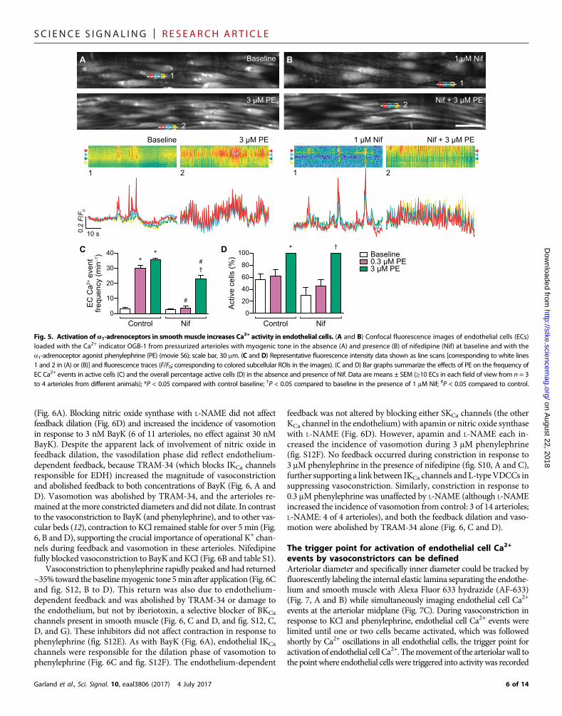
Smooth muscle a1-adrenoceptor stimulation evokesendothelial cell Ca2+ eventsToplace theseobservations in aphysiological context, thea1-adrenoceptoragonist phenylephrine, which depolarizes smooth muscle during con-traction, was used to assess endothelial cell Ca2+ responses. Two con-centrationswere used; the contraction to the higher concentration reliedless onVDCCs (Fig. 5, A and B, fig. S10, A and B, andmovie S6). Eitherconcentration of phenylephrine increased the frequency of endothelialcell Ca2+ events 10-fold (Fig. 5C). However, nifedipine did not fullyblock the Ca2+ response to the higher concentration of phenylephrine(Fig. 5C), thus supporting a role for smooth muscle IP3 in influencingendothelial cell Ca2+ and the formation of pulsars (13, 14). The higherconcentration of phenylephrine recruited new cells during the response(Fig. 5D). In response to arteriole stimulation with phenylephrine, en-
Garland et al., Sci. Signal. 10, eaal3806 (2017) 4 July 2017
dothelial cell Ca2+ events were more likely to occur as waves (fig. S4, Aand B), more so in the presence of nifedipine, which caused a deviationfrom the normally strong positive correlation (R2 = 0.96) between thefrequency of endothelial cell Ca2+ events and the percentage of waves(fig. S4B). By simultaneously imaging both endothelial and smoothmuscle cells at the arteriolarmidplane, we demonstrated differential ac-tivation of Ca2+ events in each cell type (fig. S11, A to E).Midplane datafrom endothelial cells (fig. S11, A to C) and smooth muscle cells (fig.S11, A, B, D, and E) were consistent with those measured in cells infocus at the bottom surface of arterioles.
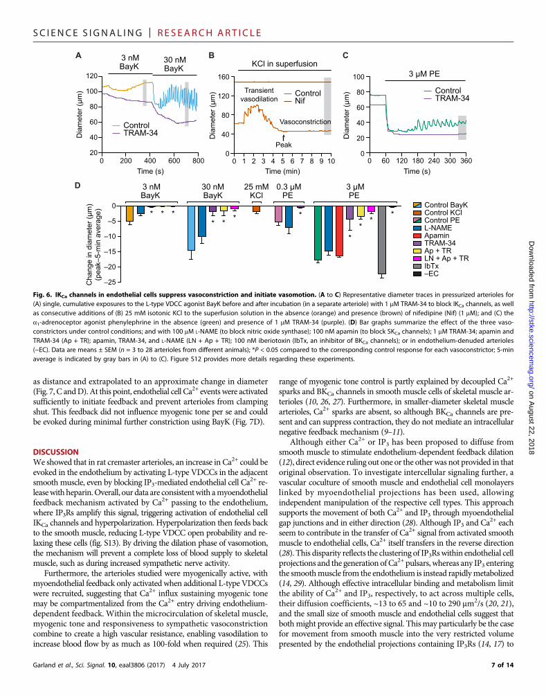
Endothelial cell Ca2+ events activate IKCa channels tosuppress vasoconstrictionThe impact of Ca2+ influx through smooth muscle L-type VDCCs onarteriolar function was probed in arterioles with myogenic tone in theabsence and presence of inhibitors (fig. S12A). BayK, KCl, and phenyl-ephrine (Fig. 6, A to C) each stimulated vasoconstriction. The smallconstriction in response to 3 nM BayK was followed by vasodilationtoward and beyond baseline (vasomotion was only observed in twoof nine arterioles) (Fig. 6A), and after 5 min, there was minimal effecton diameter. BayK (30 nM) evoked rapid vasoconstriction, which devel-oped into vasomotion as part of the feedback vasodilation in all arterioles
0
10
20
30
EC
Ca2+
eve
ntfre
quen
cy (m
in–1
)
Baseline2 mM 4-AP25 mM KCl
*
*
#†
Control Nif0
20
40
60
80
100
Act
ive
cells
(%)
NifControl
#
*
1 µM Nif
1
Baseline
1
25 mM KCl
2
Nif + 25 mM KCl
2
Nif + 25 mM KCl2
25 mM KCl
2
1 µM Nif
1
Baseline
1
10 s0.5 F
/F0
DC
BA
0
20
40
60
80
100
Ca2+
wav
es (%
)
Control Nif
F
0
10
20
30
Control NifV
SM
Ca2+
eve
ntfre
quen
cy (m
in–1
)
E
#
# # #
Fig. 4. Depolarization opens VDCCs in smooth muscle and increases Ca2+ activity in endothelial cells. (A and B) Confocal fluorescence images of endothelial cells(ECs) loaded with the Ca2+ indicator OGB-1 in pressurized skeletal muscle arterioles with myogenic tone in the absence (A) and presence (B) of nifedipine (Nif), atbaseline and with KCl (see also movie S4); scale bar, 30 mm. Representative fluorescence intensity data shown as line scans [corresponding to white lines 1 and 2 in (A)or (B)] and fluorescence traces (F/F0; corresponding to colored subcellular ROIs in the images). (C to F) Bar graphs summarize the effect of the KV channel blocker 4-aminopyridine (4-AP; movie S5) and KCl on the frequency of EC Ca2+ events in active cells (C) and the overall percentage of active cells (D), as well as the effects of KClon the frequency of vascular smooth muscle (VSM) Ca2+ events (E) and the percentage of these events that propagated along cells and were considered to be waves (F)in the absence and presence of Nif. Data are means ± SEM (≥10 ECs or VSMCs in each field of view from n = 3 to 6 arterioles from different animals); *P < 0.05 comparedwith control baseline; †P < 0.05 compared to baseline in the presence of 1 mM Nif; #P < 0.05 compared to control.
5 of 14
SC I ENCE S I GNAL ING | R E S EARCH ART I C L E
on August 22, 2018
http://stke.sciencemag.org/
Dow
nloaded from
(Fig. 6A). Blocking nitric oxide synthase with L-NAME did not affectfeedback dilation (Fig. 6D) and increased the incidence of vasomotionin response to 3 nM BayK (6 of 11 arterioles, no effect against 30 nMBayK). Despite the apparent lack of involvement of nitric oxide infeedback dilation, the vasodilation phase did reflect endothelium-dependent feedback, because TRAM-34 (which blocks IKCa channelsresponsible for EDH) increased the magnitude of vasoconstrictionand abolished feedback to both concentrations of BayK (Fig. 6, A andD). Vasomotion was abolished by TRAM-34, and the arterioles re-mained at themore constricted diameters and did not dilate. In contrastto the vasoconstriction to BayK (and phenylephrine), and to other vas-cular beds (12), contraction to KCl remained stable for over 5 min (Fig.6, B and D), supporting the crucial importance of operational K+ chan-nels during feedback and vasomotion in these arterioles. Nifedipinefully blocked vasoconstriction to BayK and KCl (Fig. 6B and table S1).
Vasoconstriction to phenylephrine rapidly peaked and had returned~35% toward the baselinemyogenic tone 5min after application (Fig. 6Cand fig. S12, B to D). This return was also due to endothelium-dependent feedback and was abolished by TRAM-34 or damage tothe endothelium, but not by iberiotoxin, a selective blocker of BKCa
channels present in smooth muscle (Fig. 6, C and D, and fig. S12, C,D, and G). These inhibitors did not affect contraction in response tophenylephrine (fig. S12E). As with BayK (Fig. 6A), endothelial IKCa
channels were responsible for the dilation phase of vasomotion tophenylephrine (Fig. 6C and fig. S12F). The endothelium-dependent
Garland et al., Sci. Signal. 10, eaal3806 (2017) 4 July 2017
feedback was not altered by blocking either SKCa channels (the otherKCa channel in the endothelium) with apamin or nitric oxide synthasewith L-NAME (Fig. 6D). However, apamin and L-NAME each in-creased the incidence of vasomotion during 3 mM phenylephrine(fig. S12F). No feedback occurred during constriction in response to3 mM phenylephrine in the presence of nifedipine (fig. S10, A and C),further supporting a link between IKCa channels and L-typeVDCCs insuppressing vasoconstriction. Similarly, constriction in response to0.3 mM phenylephrine was unaffected by L-NAME (although L-NAMEincreased the incidence of vasomotion from control: 3 of 14 arterioles;L-NAME: 4 of 4 arterioles), and both the feedback dilation and vaso-motion were abolished by TRAM-34 alone (Fig. 6, C and D).
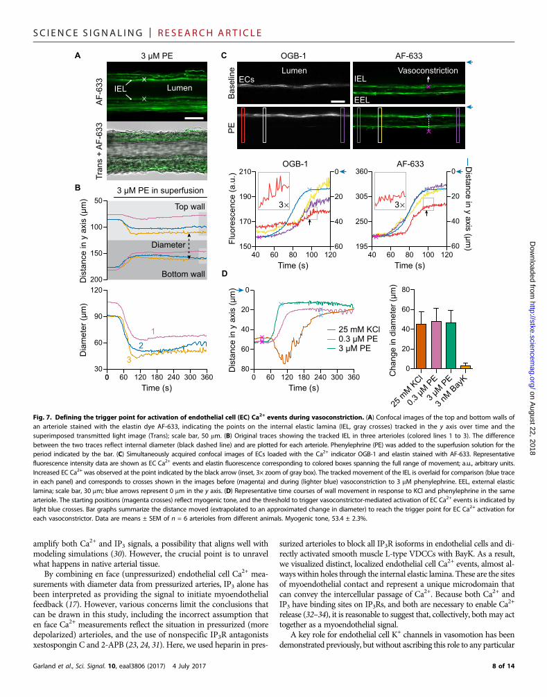
The trigger point for activation of endothelial cell Ca2+
events by vasoconstrictors can be definedArteriolar diameter and specifically inner diameter could be tracked byfluorescently labeling the internal elastic lamina separating the endothe-lium and smooth muscle with Alexa Fluor 633 hydrazide (AF-633)(Fig. 7, A and B) while simultaneously imaging endothelial cell Ca2+
events at the arteriolar midplane (Fig. 7C). During vasoconstriction inresponse to KCl and phenylephrine, endothelial cell Ca2+ events werelimited until one or two cells became activated, which was followedshortly by Ca2+ oscillations in all endothelial cells, the trigger point foractivationof endothelial cell Ca2+. Themovement of the arteriolarwall tothe point where endothelial cells were triggered into activity was recorded
**
†
#
#
NifControl0
10
20
30
40
EC
Ca2+
eve
ntfre
quen
cy (m
in–1
)
0
20
40
60
80
100
Act
ive
cells
(%)
*
NifControl
Baseline0.3 µM PE3 µM PE
1 µM Nif
1
BBaseline
1
3 µM PE
2
Nif + 3 µM PE2
A
Baseline
1
1 µM Nif
1
3 µM PE
2
Nif + 3 µM PE
2
10 s0.2 F
/F0
C D †
Fig. 5. Activation of a1-adrenoceptors in smoothmuscle increases Ca2+ activity in endothelial cells. (A and B) Confocal fluorescence images of endothelial cells (ECs)loaded with the Ca2+ indicator OGB-1 from pressurized arterioles with myogenic tone in the absence (A) and presence (B) of nifedipine (Nif) at baseline and with thea1-adrenoceptor agonist phenylephrine (PE) (movie S6); scale bar, 30 mm. (C and D) Representative fluorescence intensity data shown as line scans [corresponding to white lines1 and 2 in (A) or (B)] and fluorescence traces (F/F0; corresponding to colored subcellular ROIs in the images). (C and D) Bar graphs summarize the effects of PE on the frequency ofEC Ca2+ events in active cells (C) and the overall percentage active cells (D) in the absence and presence of Nif. Data are means ± SEM (≥10 ECs in each field of view from n = 3to 4 arterioles from different animals); *P < 0.05 compared with control baseline; †P < 0.05 compared to baseline in the presence of 1 mM Nif; #P < 0.05 compared to control.
6 of 14
SC I ENCE S I GNAL ING | R E S EARCH ART I C L E
on August 2
http://stke.sciencemag.org/
Dow
nloaded from
as distance and extrapolated to an approximate change in diameter(Fig. 7, C andD). At this point, endothelial cell Ca2+ events were activatedsufficiently to initiate feedback and prevent arterioles from clampingshut. This feedback did not influence myogenic tone per se and couldbe evoked during minimal further constriction using BayK (Fig. 7D).
2, 2018
DISCUSSIONWe showed that in rat cremaster arterioles, an increase in Ca2+ could beevoked in the endothelium by activating L-type VDCCs in the adjacentsmooth muscle, even by blocking IP3-mediated endothelial cell Ca2+ re-leasewithheparin.Overall, our data are consistentwith amyoendothelialfeedback mechanism activated by Ca2+ passing to the endothelium,where IP3Rs amplify this signal, triggering activation of endothelial cellIKCa channels and hyperpolarization. Hyperpolarization then feeds backto the smooth muscle, reducing L-type VDCC open probability and re-laxing these cells (fig. S13). By driving the dilation phase of vasomotion,the mechanism will prevent a complete loss of blood supply to skeletalmuscle, such as during increased sympathetic nerve activity.Furthermore, the arterioles studied were myogenically active, withmyoendothelial feedback only activated when additional L-type VDCCswere recruited, suggesting that Ca2+ influx sustaining myogenic tonemay be compartmentalized from the Ca2+ entry driving endothelium-dependent feedback. Within the microcirculation of skeletal muscle,myogenic tone and responsiveness to sympathetic vasoconstrictioncombine to create a high vascular resistance, enabling vasodilation toincrease blood flow by as much as 100-fold when required (25). This
Garland et al., Sci. Signal. 10, eaal3806 (2017) 4 July 2017
range of myogenic tone control is partly explained by decoupled Ca2+
sparks and BKCa channels in smooth muscle cells of skeletal muscle ar-terioles (10, 26, 27). Furthermore, in smaller-diameter skeletal musclearterioles, Ca2+ sparks are absent, so although BKCa channels are pre-sent and can suppress contraction, they do not mediate an intracellularnegative feedback mechanism (9–11).
Although either Ca2+ or IP3 has been proposed to diffuse fromsmooth muscle to stimulate endothelium-dependent feedback dilation(12), direct evidence ruling out one or the otherwas not provided in thatoriginal observation. To investigate intercellular signaling further, avascular coculture of smooth muscle and endothelial cell monolayerslinked by myoendothelial projections has been used, allowingindependent manipulation of the respective cell types. This approachsupports the movement of both Ca2+ and IP3 through myoendothelialgap junctions and in either direction (28). Although IP3 and Ca
2+ eachseem to contribute in the transfer of Ca2+ signal from activated smoothmuscle to endothelial cells, Ca2+ itself transfers in the reverse direction(28). This disparity reflects the clustering of IP3Rswithin endothelial cellprojections and the generation ofCa2+ pulsars, whereas any IP3 enteringthe smoothmuscle from the endothelium is instead rapidlymetabolized(14, 29). Although effective intracellular binding and metabolism limitthe ability of Ca2+ and IP3, respectively, to act across multiple cells,their diffusion coefficients, ~13 to 65 and ~10 to 290 mm2/s (20, 21),and the small size of smooth muscle and endothelial cells suggest thatbothmight provide an effective signal. This may particularly be the casefor movement from smooth muscle into the very restricted volumepresented by the endothelial projections containing IP3Rs (14, 17) to
Cha
nge
in d
iam
eter
(µm
)(p
eak–
5-m
in a
vera
ge)
D
–25
–20
–15
–10
–5
0
30 nMBayK
3 nMBayK
L-NAMEApaminTRAM-34Ap + TRLN + Ap + TRIbTx–EC
Control PEControl KClControl BayK
**
**
3 µMPE
25 mMKCl
A
Dia
met
er (µ
m)
0 200 400 600 80020
40
60
80
100
120
ControlTRAM-34
3 nMBayK
30 nMBayK
Time (s)
B C
Dia
met
er (µ
m)
Time (s)0 60 120 300240180
ControlTRAM-34
3 µM PE
0
20
40
60
80
100
360Time (min)
Dia
met
er (µ
m)
0
80
120
KCl in superfusion
Peak
Transientvasodilation
0 1 2 3 4 5 6 7 8 9 10
ControlNif
Vasoconstriction
0.3 µMPE
***
40
160
* * **
Fig. 6. IKCa channels in endothelial cells suppress vasoconstriction and initiate vasomotion. (A to C) Representative diameter traces in pressurized arterioles for(A) single, cumulative exposures to the L-type VDCC agonist BayK before and after incubation (in a separate arteriole) with 1 mM TRAM-34 to block IKCa channels, as wellas consecutive additions of (B) 25 mM isotonic KCl to the superfusion solution in the absence (orange) and presence (brown) of nifedipine (Nif) (1 mM); and (C) thea1-adrenoceptor agonist phenylephrine in the absence (green) and presence of 1 mM TRAM-34 (purple). (D) Bar graphs summarize the effect of the three vaso-constrictors under control conditions; and with 100 mM L-NAME (to block nitric oxide synthase); 100 nM apamin (to block SKCa channels); 1 mM TRAM-34; apamin andTRAM-34 (Ap + TR); apamin, TRAM-34, and L-NAME (LN + Ap + TR); 100 nM iberiotoxin (IbTx, an inhibitor of BKCa channels); or in endothelium-denuded arterioles(−EC). Data are means ± SEM (n = 3 to 28 arterioles from different animals); *P < 0.05 compared to the corresponding control response for each vasoconstrictor; 5-minaverage is indicated by gray bars in (A) to (C). Figure S12 provides more details regarding these experiments.
7 of 14
SC I ENCE S I GNAL ING | R E S EARCH ART I C L E
on August 22, 2018
http://stke.sciencemag.org/
Dow
nloaded from
amplify both Ca2+ and IP3 signals, a possibility that aligns well withmodeling simulations (30). However, the crucial point is to unravelwhat happens in native arterial tissue.
By combining en face (unpressurized) endothelial cell Ca2+ mea-surements with diameter data from pressurized arteries, IP3 alone hasbeen interpreted as providing the signal to initiate myoendothelialfeedback (17). However, various concerns limit the conclusions thatcan be drawn in this study, including the incorrect assumption thaten face Ca2+ measurements reflect the situation in pressurized (moredepolarized) arterioles, and the use of nonspecific IP3R antagonistsxestospongin C and 2-APB (23, 24, 31). Here, we used heparin in pres-
Garland et al., Sci. Signal. 10, eaal3806 (2017) 4 July 2017
surized arterioles to block all IP3R isoforms in endothelial cells and di-rectly activated smooth muscle L-type VDCCs with BayK. As a result,we visualized distinct, localized endothelial cell Ca2+ events, almost al-wayswithin holes through the internal elastic lamina. These are the sitesof myoendothelial contact and represent a unique microdomain thatcan convey the intercellular passage of Ca2+. Because both Ca2+ andIP3 have binding sites on IP3Rs, and both are necessary to enable Ca2+
release (32–34), it is reasonable to suggest that, collectively, both may acttogether as a myoendothelial signal.
A key role for endothelial cell K+ channels in vasomotion has beendemonstrated previously, but without ascribing this role to any particular
40 60 80 100 120
C
D
Bas
elin
eP
E
Lumen
OGB-1
ECs
AF-633
IEL
EEL
Vasoconstriction
AF-633 Distance in y axis (µm
)195
250
305
360OGB-1
3
Time (s) Time (s)40 60 80 100 120
150
170
190
210 0
20
40
60Fluo
resc
ence
(a.u
.) 0
20
40
60
0
3 µM PE0.3 µM PE25 mM KCl
20
40
60
800 60 120 180 240 300 360
Time (s)
Dis
tanc
e in
y a
xis
(µm
)
A
B
3 µM PE
LumenA
F-63
3Tr
ans
+ A
F-63
3IEL
50
100
150
200
3 µM PE in superfusion
Top wall
Bottom wall
Diameter
Dis
tanc
e in
y a
xis
(µm
)
1
2
3
0 60 120 180 240 300 360Time (s)
0
120
90
60
30
Dia
met
er (µ
m)
0
20
40
60
80
Cha
nge
in d
iam
eter
(µm
)25
mM K
Cl
0.3 µM
PE
3 µM P
E
3 nM B
ayK
3
Fig. 7. Defining the trigger point for activation of endothelial cell (EC) Ca2+ events during vasoconstriction. (A) Confocal images of the top and bottom walls ofan arteriole stained with the elastin dye AF-633, indicating the points on the internal elastic lamina (IEL, gray crosses) tracked in the y axis over time and thesuperimposed transmitted light image (Trans); scale bar, 50 mm. (B) Original traces showing the tracked IEL in three arterioles (colored lines 1 to 3). The differencebetween the two traces reflect internal diameter (black dashed line) and are plotted for each arteriole. Phenylephrine (PE) was added to the superfusion solution for theperiod indicated by the bar. (C) Simultaneously acquired confocal images of ECs loaded with the Ca2+ indicator OGB-1 and elastin stained with AF-633. Representativefluorescence intensity data are shown as EC Ca2+ events and elastin fluorescence corresponding to colored boxes spanning the full range of movement; a.u., arbitrary units.Increased EC Ca2+ was observed at the point indicated by the black arrow (inset, 3× zoom of gray box). The tracked movement of the IEL is overlaid for comparison (blue tracein each panel) and corresponds to crosses shown in the images before (magenta) and during (lighter blue) vasoconstriction to 3 mM phenylephrine. EEL, external elasticlamina; scale bar, 30 mm; blue arrows represent 0 mm in the y axis. (D) Representative time courses of wall movement in response to KCl and phenylephrine in the samearteriole. The starting positions (magenta crosses) reflect myogenic tone, and the threshold to trigger vasoconstrictor-mediated activation of EC Ca2+ events is indicated bylight blue crosses. Bar graphs summarize the distance moved (extrapolated to an approximated change in diameter) to reach the trigger point for EC Ca2+ activation foreach vasoconstrictor. Data are means ± SEM of n = 6 arterioles from different animals. Myogenic tone, 53.4 ± 2.3%.
8 of 14
SC I ENCE S I GNAL ING | R E S EARCH ART I C L E
on August 22, 2018
http://stke.sciencemag.org/
Dow
nloaded from
channel type (35). Physiologically, vasomotion facilitates blood flowcontrol and capillary function in the microcirculation and is disruptedin diseases such as diabetes and hypertension (36). The function of IKCa
channels is compromised in these diseases (37), whichmay underpina lack of vasomotion, a notion that requires further investigation.However, VDCC-driven endothelium-dependent feedback must beconsidered when assessing the contribution of endothelial cells to sys-temic blood pressure and when formulating computational models ofarteriolar reactivity.
Feedback dilation was only observed after the peak constriction toboth phenylephrine and KCl, and this delay is consistent with a thresh-old point for endothelial cell activation. We ruled out endothelial cellshape change as a major contributing factor to their activation, becausethe change in diameter to 3 nM BayK was negligible (peak response,<10 mm) and could be further limited with blebbistatin, yet robust,many-fold increases in endothelial cell Ca2+ eventswere stimulated. Theextent of depolarization necessary to raise VDCC open probability andtrigger the myoendothelial signaling circuit is not clear, but the restingEm in skeletal muscle arterioles at 80 mmHg is −40 mV (17, 38), and0.1 mMphenylephrine depolarizes hamster skeletal muscle arteriolesby ~10 mV, which is associated with a ~30-mm decrease in diameter(17), suggesting that the threshold is less negative than−40mV. Becausethe smoothmusclemembrane potential dominates that of the endothe-lium, depolarization would be expected to reduce Ca2+ influx across themembranes of endothelial cells becauseVDCCs are absent in these cells.Thus, the local cytoplasmic concentration achieved adjacent to smoothmuscle L-type VDCCs will provide the driving force for Ca2+ move-ment through gap junctions to the endothelium.
Finally, it is important to note that our observations were not com-plicated by any direct agonist action on endothelial cells, because neitherphenylephrine [consistent with (18)] nor BayK stimulated a Ca2+ re-sponse in isolated endothelial cell tubes, correlating with an absenceof CaV1.2. The depolarized resting potential of freshly isolated endothe-lial cell tubes, ~−20 mV (39), should not prevent either the action ofphenylephrine (through IP3Rs) or the ability of BayK to increaseVDCCopen probability (6, 40, 41). Thus, it seems reasonable to conclude thatin smooth muscle, L-type VDCC activity can alone trigger the inter-cellular Ca2+ circuit.
Physiologically, the endothelium-dependent feedback circuit thatwehave definedmay limit ischemia during periods of sustained sympathet-ic nerve activity, for example, during exercise, particularly in noncon-tractingmuscle (42). Endothelial cell IKCa channel activity appears to bekey, and when their activity is compromised by disease states such ashypertension and diabetes (37), negative feedback will be reduced. As aconsequence, vascular reactivity will increase compromising blood flowwithin the skeletal muscle microcirculation, potentially contributing toincreases in blood pressure (43).
MATERIALS AND METHODSAnimal proceduresMaleWistar rats (Charles River; weight, 240 to 280 g) were anesthetizedwith urethane (2.8 g/kg intraperitoneally), and the cremaster musclewas exteriorized (37), excised, and placed in cold (4°C) Mops-bufferedsolution containing 145.0mMNaCl, 4.7mMKCl, 2.0mMCaCl2, 1.17mMMgSO4, 2.0 mM Mops, 1.2 mM NaH2PO4, 5.0 mM glucose, 2.0 mMpyruvate, 0.02 mM EDTA, and 2.75 mM NaOH (pH 7.40 ± 0.02 at37°C). After tissue removal, rats were euthanized using a Schedule1 procedure [Animals (Scientific Procedures) Act 1986, UK]. Segments
Garland et al., Sci. Signal. 10, eaal3806 (2017) 4 July 2017
of themain intramuscular arteriole (1A)were dissected from themuscleas previously described (1, 44).
Cannulated arteriolar preparationIsolated rat cremaster arteriolar segments were cannulated at each endwith glass micropipettes (external diameter, ~95 mm) and positioned ina 1.5-ml temperature-regulated chamber (RC-27N,Warner Instruments)on the stage of an invertedmicroscope (IX70or IX81,Olympus) and con-tinually superfused (2 ml min−1) with Mops-buffered solution. To avoidluminal flow, equal pressurewasmaintained across the vessel throughoutan experiment. Artery segmentswerewarmed to 34°C, gradually pressur-ized to 80mmHg, longitudinally straightened to eliminate lateral bowingof the vessel (1, 45), and allowed to develop spontaneous myogenic toneover a 20-min equilibration period. After development of ~50% tone, en-dothelial cell viability of arterioles was tested by exposure to 0.1, 0.3, and1 mM acetylcholine; only arterioles dilating >95% of the passive diameter(maximal dilation) were used for experiments. Inner diameter wasmeasured at 1 to 2 Hz either automatically using DiamTrak 3+ version3.5 (T. O. Neild) (1) or manually using a video caliper (MicrocirculationResearch Institute, Texas A&M University, United States) coupled to aPowerLab 2/20 (AD Instruments) running LabChart version 7.2.2.Whenone arterial wall was imaged at the midplane, at higher magnification,the movement of the internal elastic lamina during dilation and con-striction was tracked usingMetaMorph software (version 7.7.4.0, Mo-lecular Devices).
Immunohistochemistry was performed in pressurized arterioles aspreviously described (46). In brief, arterioles were fixed in 2% para-formaldehyde for 10 min at 37°C, washed with phosphate-buffered sa-line (PBS), and then incubated in blocking buffer [luminal andabluminal, 1% bovine serum albumin (BSA) and 0.1% Tween 20] for60 min at 37°C and then overnight with primary antibody (pumpedinto the lumen of the arteriole and added to the bath) at 4°C. Theprimary antibody is 1:200 mouse monoclonal to CaV1.2 calciumchannel (MAB13170, clone L57/46, Millipore). The following day,the bath solution was replaced with PBS, and the lumen was perfusedwith an Alexa Fluor 488 secondary antibody [1:100 chicken anti-mouseimmunoglobulin G (IgG), A-21200, Invitrogen] and incubated for2 hours at room temperature. This labeled CaV1.2 in vascular smoothmuscle by diffusion through the arteriolar wall and avoided labelingcells in the adventitia. Nuclei and elastin (including the internal elasticlamina) were stained with 15 mM propidium iodide and 200 nMAF-633 (A-30634, Molecular Probes), respectively (47). Arterioles wereexcited at 488, 546, and 633 nm, and the fluorescence emitted at 505 to525, 560 to 620, and 655 to 755 nm was acquired through a water im-mersion objective [40×; numerical aperture (NA), 0.9; working distance(WD), 0.15 mm; Olympus, 1024 × 1024 pixels] using a laser scanningconfocal microscope (FV1000, Olympus). z-Stacks through the arterywall were obtained at 0.2-mm increments using FluoView software(FV10-ASW3.0, Olympus) and reconstructed in Imaris software (version7.2.3, Bitplane).
Endothelial cell tube isolation procedureEndothelial cell tubes were isolated as previously described (1, 47) withmodificationsmade to isolate from rat cremaster arterioles. In brief, cre-master tissues were pinned in a dissection dish containing cold (4°C)dissection buffer (pH 7.40 ± 0.02 at 37°C, 285 to 305mosM) containing137.0mMNaCl, 5.6mMKCl, 1.0mMMgCl2, 10.0mMHepes, 10.0mMglucose, 0.01 mM sodium nitroprusside, and 0.1% BSA. The main arteri-olar branch (1A) in each cremaster was dissected free of surrounding
9 of 14

SC I ENCE S I GNAL ING | R E S EARCH ART I C L E
on August 22, 2018
http://stke.sciencemag.org/
Dow
nloaded from
tissue, one end cannulated by a glass micropipette (~100 mmouter diam-eter) and the lumen flushed with cold dissection buffer to remove residualblood components. The arterioles were then cut into two to three smallersegments (~1 to 1.5 mm in length) and transferred into a 1.5-ml AxygenMaxyClear snaplockmicrotube (MCT-150-C, Corning) on ice containing1 ml of dissection buffer.
To obtain endothelial cell tubes, the collected arteriole segmentswere then allowed to warm to room temperature (~10 min), and thesolution was aspirated and gently replaced with enzyme-free dissocia-tion buffer (pH 7.40 ± 0.02 at 37°C, 285 to 305 mosM) containing137.0mMNaCl, 5.6mMKCl, 1.0mMMgCl2, 10.0mMHepes, 10.0mMglucose, 2.0 mM CaCl2, and 0.1% BSA to remove residual dissectionbuffer. This solution was aspirated and replaced with preheated (to37°C) dissociation buffer containing the following enzymes: papain(0.62 mg ml−1) (P4762, Sigma), dithioerythritol (1.0 mg ml−1) (D8255,Sigma), and collagenase (1.5 mg ml−1) (C8051, Sigma). Arteriolesegments were incubated for 25 min at 37°C. The buffer was then aspi-rated and replaced with room temperature enzyme-free dissociationbuffer to terminate the digestion. Arteriole segments were transferred toa 35-mm × 10-mm culture dish (430165, Corning) containing enzyme-free dissociation buffer for trituration. Endothelial cell tubes weredissociated from surrounding smoothmuscle cells by gentle triturationusing a glassmicropipette (4878,World Precision Instruments)with aninner diameter of ~90 to 110 mm. An injector (Nanoliter 2010, ~300 to500nlmin−1) coupledwithaMicro4controller (WorldPrecisionInstruments)wasmounted on an upright Olympus BX51WImicroscope to allow forreal-time visualization of the trituration procedure (48).
Freshly isolated endothelial cell tubes were transferred to a 1.5-mltemperature-regulated chamber containing perfusion buffer (pH 7.40 ±0.02 at 37°C, 285 to 305 mosM) containing 137.0 mM NaCl, 5.0 mMKCl, 1.0 mM MgCl2, 10.0 mM Hepes, 10.0 mM glucose, and 2.0 mMCaCl2. Endothelial cell tubeswere pinned on the bottomof the chamberusing two round-tippedpinning pipettes (~140- to 160-mmtipdiameter)positioned using micromanipulators. Tubes were then stretched to theirapproximate physiological lengths by adjusting the horizontal tension onthe tissue using the micromanipulators. Experiments on endothelial celltubes were performed at room temperature, because the integrity of thepreparation is reduced at higher temperatures (48).
Measurement of [Ca2+]i in pressurized arterioles andendothelial cell tubesLoading and imaging protocols were performed as previously described(49). Pressurized vessels were perfused intraluminally (20 to 30min) forselective loading of the endotheliumor bathed (120min) at 34°C to loadthe smooth muscle cells, in both cases with Mops-buffered solutioncontaining filtered (0.2-mm pore) Oregon Green 488 BAPTA-1 AM(OGB-1, 10 or 20 mM, respectively) and 0.0025% pluronic F-127. Forsimultaneous imaging of both cell types by multiphoton microscopy,endothelial cells were loaded for the last 30 min of the smooth muscleincubation period. In some experiments, arterioles were incubated with0.1 mMAF-633 (A-30634,Molecular Probes) for 10min to label elastin.The objective was heated (custom-built Peltier heater, Department ofPharmacology, University of Oxford, UK) to prevent heat loss fromthe chamber. Endothelial cell tubes were loaded at room temperaturewith perfusion buffer containing Fluo-4 AM (20 mM) and 0.005%pluronic F-127 for 30 to 35 min. In all instances, excess indicatorwas washed from the preparation, and the loaded AM form of thedye was allowed to de-esterify and equilibrate. Pressurized arterioleswere de-esterified for 30 min, whereas tubes were de-esterified for 10
Garland et al., Sci. Signal. 10, eaal3806 (2017) 4 July 2017
to 30 min. Only cells responsive to 1 mM acetylcholine were used foranalysis.
In most experiments, the fluorescence intensity from loaded endo-thelial or smoothmuscle cells was visualized by lowering the focal planeto cells at the bottomof the pressurized arteriole or endothelial cell tube.Images were obtained using a 40× water immersion objective (40×/1.15-NA objective; WD, 0.25 mm, Olympus; excitation, 488 nm; emis-sion, >505 nm) and were acquired using an Olympus FV500 orFV1000MPE (FluoView version 5.0 or FV10-ASW software, respec-tively) at ~3 Hz. In two sets of experiments, the focal plane was raisedto the midplane of the arteriolar wall. Endothelial cell Ca2+ responsesand elastin were simultaneously visualized with a Mai Tai DeepSeeTi:Sapphiremultiphoton laser (excitation, 790 nm; 2.5%; 1.96W; pulsewidth, <80 fs; emission split to capture 495 to 540 nm: OGB-1; emis-sion split to capture 575 to 630 nm: AF-633). To ensure that the field ofview included the range of wall movement during exposure to vasocon-strictors, the image size was 512 × 192 pixels, equating to 234 × 88 mm;images were acquired at 2.2 Hz. In these experiments, because only onearterial wall was imaged, the distancemovedby the internal elastic laminaduring vasoconstriction was doubled to approximate changes in diame-ter. Using this approach, we could not obtain a value for the frequency ofendothelial cell Ca2+ events due to arterialmotion during vasoconstrictionand the lower frequency of acquisition. Instead, multiple ROIs largeenough to include the endothelial cells throughout the range of wallpositions were used, and the time and therefore y-axis position at whichoscillations in endothelial cell [Ca2+]i occurred weremonitored both vi-sually and by analysis of average-intensity plots. To establish whetherarterialmotion per se altered fluorescence intensity, the sameROIswereplaced over the elastin channel. Constriction increased fluorescenceintensity, which may be due to the concentration of fluorescence inthe z axis or a relatively thinner distance for laser and/or emitted photonpenetration in a smaller-diameter arteriole. Therefore, in these ex-periments, measurements of OGB-1 fluorescence were limited toobserving the onset of endothelial cell Ca2+ events (waves). In the sec-ond set of experiments, endothelial and smoothmuscle cell Ca2+ activitywere imaged simultaneously by using themultiphoton laser (excitation,790 nm) and raising the focal plane to the midplane of one side ofthe arteriolar wall as previously described (1); images were acquiredat ~3 Hz.
Data were analyzed using MetaMorph software (version 7.7.4.0,Molecular Devices) as previously described using subcellular ROIs(diameter, ~5 mm) (1) (fig. S2, A to D). This approach was directlycompared to an automated analysis software plugin for ImageJ, usedfor detection of endothelial cell Ca2+ events in intact, unpressurizedarteries (LC_Pro) (50, 51). The same TIFF (tagged image file format)stack was used for both automated and manual analyses to compareoutputs to visual inspection, and for pressurized arterioles, manualanalysis was more accurate (fig. S2, A to D). A major advantage ofMetaMorph is the ability to place and move an ROI within a cell ina TIFF image stack and see traces of intensity over time (Region Mea-surements) on the screen, allowing the user to move the ROI aroundthe entire surface of each individual cell and watch for events. When adeflection was detected, the frames were advanced through that eventto confirm that it was not due to an artifact. The point of origin of theevent was then marked with an ROI. Once complete, the events werethen classified as local if they radiated from a single point and termi-nated within ~10 mm (value not fixed due to heterogeneous points oforigin in varied-shaped endothelial cells) or propagating waves, inwhich case a second ROI was placed along the cell to establish the
10 of 14
SC I ENCE S I GNAL ING | R E S EARCH ART I C L E
on August 22, 2018
http://stke.sciencemag.org/
Dow
nloaded from
temporal alignment of events. The blue and yellow traces in fig. S2Cwere defined as waves, and they tended to travel at least half the celllength. The heterogeneity in both endothelial cell shape and the pointof origin in each Ca2+ event meant that this classification was based onmanual analysis of each event, rather than a set distance or surfacearea. Each event within a given endothelial cell was counted and re-ported as an overall frequency of Ca2+ events, the combined local andwave events. The percentage of active cells was also reported, as well asendothelial cell Ca2+ event frequency for the entire frame, the averagefrom active cells only. For midplane experiments, ROIs were placedon three to five active smooth muscle and/or one to two endothelialcells per image stack, and the frequency of Ca2+ responses (events perminute) was recorded. For endothelial cell imaging experiments (pres-surized arterioles and isolated tubes), all the cells in the field of view(~8 to 15 cells and ~40 to 100 cells, respectively) were individually andmanually analyzed for Ca2+ event frequency over a ≥60-s acquisitionsequence. To analyze experiments in the presence of 30 nM BayK,which induced robust vasomotion (Fig. 6A and movie S1), a TIFFstack of a subset of images was constructed in which the endotheliumremained in focus in one field of view. Results are presented as eitherthe frequency (events per minute) reported only from active cells, withthe percentage of active cells provided, or F/F0, calculated by dividingthe fluorescence intensity (F) by an average baseline fluorescence intensity(F0). The number of Ca2+ events identified as local or waves was dividedby the total number of Ca2+ events observed to give percentage localand percentage waves. For endothelial cell Ca2+ responses to ace-tylcholine, the ROI encircled the whole cell, and results from fivecells per arteriole were shown as F/F0 of 2-s averages for each nvalue.
RNA extraction and RT-qPCR in isolated arterioles andendothelial cell tubesArterioles and endothelial cell tubes were isolated as described above,removing residual blood cells by perfusion. For intact arterioles, the cen-tral, 1A arterioles from the left and right cremaster tissue of one animalwere pooled for eachn value andwere homogenized using a pellet pestle(47747-370,VWR) in 1.5-mlmicrotubes (Axygen,MCT-150-C,Corning).RNA was extracted using the RNeasy Plus Mini Kit (74134, Qiagen). Forendothelial cell tubes, three >1-mm-long tubes were pooled from oneanimal for each n value. Upon dissociation from the smooth muscle cells,tubeswere transferredusinganewglassmicropipette into a clean35-mm×10-mmculturedish containingdissociationbuffer and repeated threemoretimes to reduce smooth muscle cell contamination. RNA was thenextracted from the tubes using Cells-to-CT 1-Step TaqMan Kit lysis buffer(A25603, ThermoFisher Scientific).
RT-qPCRs were carried out using the Cells-to-CT kit (A25603,ThermoFisher Scientific). PCRs for each gene were carried out in thesame reaction (n = 4 animals for each gene). Gene-specific carboxy-fluorescein (FAM)–conjugated TaqMan probes were purchased fromThermoFisher Scientific (table S2). Reverse transcription was per-formed at 50°C for 20min, followed by heat activation of Taq polymer-ase at 95°C for 30 s. PCRwas carried out by repeating 95°C for 30 s andthen 60°C for 1 min for 40 cycles using the 7500 Fast Real-Time PCRsystem (4351107, ThermoFisher Scientific). All samples were run induplicate, and a no-template negative control was included for eachgene. Relative gene expression was calculated with theD cycle threshold(DCt) method and normalized to both hypoxanthine phosphoribosyl-transferase 1 (Hprt1) and b-actin (Actb) as housekeeping genes. The ab-sence of smooth muscle cells in each tube extraction was assessed by
Garland et al., Sci. Signal. 10, eaal3806 (2017) 4 July 2017
expression of the smooth muscle cell marker a–smooth muscle actin(Acta2); in all cases, a Ct value of >36 was considered “not detected.”
Addition of vasoconstrictorsA combination of methods was used. Phenylephrine was either directlyadded to a static bath for rapid responses or added to the superfusionsolution until the response equilibrated (~5 min). Isotonic KCl wasalways added to the superfusion solution and allowed to equilibrate.BayKwas added cumulatively (3 and then 27 nM) to a static bath, andbecause it was not fully reversible, each arteriole was only exposedonce to BayK. Consequently, for each set of inhibitor treatments, allfunctional and endothelial cell Ca2+ responses to BayK were obtainedin separate arterioles to control experiments. 4-Aminopyridine wasalways added to a static bath.
Effect of inhibitors and vasoactive agents on endothelial cellCa2+ eventsThe contribution of VDCC to endothelial cell Ca2+ activity was assesseddirectly, using either the selective antagonist nifedipine (1 mM, 15-minincubation at 5 mmHg after which pressure was raised to 80 mmHg inthe continued presence of nifedipine; total of >30-min incubation) orthe selective agonist BayK (3 and 30 nM). The effect of arteriolar de-polarization on endothelial cell Ca2+ activity was assessed using isotonic25 mMKCl or the nonselective voltage-dependent K+ channel blocker,4-aminopyridine (2 mM).
Correlation between BayK-induced Ca2+ activity and holes inthe internal elastic laminaEndothelial cell Ca2+ events and the internal elastic lamina (myoen-dothelial microdomain) were imaged simultaneously at ~3 Hz usingthe 488- and 633-nmvisible laser lines on anOlympus FV1000 invertedmicroscope. Experiments were performed using 3 nM BayK, becausethis concentration caused a significant increase in endothelial cell Ca2+
activity, with limited tissue movement. Regions of endothelial cell Ca2+
activity were identified, and the regions were then superimposed onimages of the elastin. The number of regions overlapping with holesin internal elastic lamina was divided by the total number of regionsto determine the percentage of total events occurring in holes in the in-ternal elastic lamina, as previously described (1).
Loading heparin into endothelial cells to inhibit IP3RsWhereas we have previously used pinocytosis to load antibodies intoendothelial cells (52), the negative charge of heparin made use ofTransFectinLipidReagent (Bio-Rad, 1703350) possible.Whendevelopingthe protocol 5(6)-FAM (0.1 mg/ml, 0.27 mM), which is also negativelycharged, was used to demonstrate cell loading. Because low–molecularmass (5 kDa) heparin blocks IP3Rs in both cell homogenates and in-tact cells (24, 53), we used the same form of heparin (from porcine in-testinal mucosa; Fisher, BPE2524; 10 mg/ml, 2 mM). To monitor cellloading and localization with endothelial cells, a Cy5-tagged form of~12- to 15-kDa heparin (Nanocs Inc., HRN1-S5-1) was used. Hepes-buffered solution (containing 130 mM NaCl, 5.0 mM KCl, 1.2 mMMgSO4, 1.0mMCaCl2, 10.0mMHepes, and 10.0mMglucose)was usedto dissolve the molecule of interest (FAM or heparin), and TransFectin(1 ml/ml) was added (final pH, 7.40 ± 0.02). Solutions were mixed at20 rpm for 20 min using a rotating wheel (Stuart, SB2) at room tem-perature before immediate use. A Beehive syringe pump was used todeliver TransFectin solutions into the lumen of arterioles while at lowpressure (5 mmHg) at 10 ml/min for 6 min, followed by 0.5 ml/min for
11 of 14
SC I ENCE S I GNAL ING | R E S EARCH ART I C L E
on August 22, 2018
http://stke.sciencemag.org/
Dow
nloaded from
the remainder of the experiment. Images were obtained using a 40×water immersion objective (40×/1.15 NA objective; WD, 0.25 mm,Olympus; excitation, 488 nm and emission, >505 nm for FAM; excita-tion, 635 nm and emission, 655 to 755 nm for Cy5) and were acquiredusing Olympus FluoView 1000 software (FV10-ASW). This concentra-tion of TransFectin did not affect myogenic tone or vasodilation toacetylcholine (fig. S4). On the basis of fluorescence intensity, the con-centration of FAM loaded into cells was about 1000-fold less than thatin the lumen of arterioles. The fluorescence of heparin-Cy5 was not thesame in solution as when bound inside cells; therefore, a similar com-parison could not be made for heparin concentrations. However, if theloading of heparin were comparable in efficiency to FAM, there wouldbe an amount of heparin (about 10 mg/ml, 2 mM) inside cells that iswithin the range for effective antagonism of IP3Rs (24, 53, 54). Inexperiments where endothelial cell Ca2+ was measured, arterioles wereloaded with OGB-1 before TransFectin-heparin. At 5 mg/ml, 5-kDaheparin blocked endothelial cell Ca2+ responses to acetylcholine intwo of three arterioles, whereas at 10 mg/ml, 5-kDa heparin blockedresponses in six of seven arterioles (fig. S4). Once acetylcholine re-sponseswere inhibited, arterioleswere incubatedwithAF-633 andpres-surized, and endothelial cell Ca2+ responses to 3 and 30 nM BayK weredetermined and alignedwith simultaneous images of the internal elasticlamina. In separate experiments, the cellular localization of heparin-Cy5was compared to the expression of IP3R1. Arterioles loaded withheparin-Cy5 and elastin stained with Cascade Blue hydrazide (CB-405,1mM;excitation, 405nm; emission, 430 to 460nm)were imaged (z-stack)at 3× digital zoom immediately before fixation and immunolabeling(see above; primary antibody: 1:200 rabbit polyclonal to human IP3R1,PA1-901, ThermoFisher Scientific; Alexa Fluor 488 secondary antibody:1:200 goat anti-rabbit IgG, A-11008, ThermoFisher Scientific). The Cy5fluorescence was not apparent after fixation; however, the CB-405 stain-ing was visible the following day and was used to identify and align theCy5 and Alexa Fluor fluorescence signals. Nuclei were stained with pro-pidium iodide (15 mM).
Luminal pumping of 2-APB and xestospongin C2-APB (100 mM) and xestospongin C (10 mM) were pumped throughthe lumen of arterioles to inhibit endothelial cell IP3Rs in preference tosmooth muscle IP3Rs (1). 2-APB was continuously delivered duringexperiments using a Beehive syringe pump at 2 ml min−1, at which flowrate with buffer alone does not affect arteriolar diameter (17).
Damage to the endotheliumEndothelial cells were selectively damaged as previously described (24).While pressurewas lowered to~5mmHg, small air bubbles (~2ml) werepulsed through the lumen of cannulated arterioles until small fragmentsof cellular debris were observed.
Drugs and solutionsOregon Green 488 BAPTA-1 AM (OGB-1; O-6807), Fluo-4 AM(Fluo-4; F-14201), pluronic F-127 (P3000MP), AF-633 (A-30634),and Cascade Blue hydrazide (CB-405; C3221) were obtained fromMolecular Probes. Xestospongin C (BML-CA409) was from EnzoLife Sciences; apamin (L8407) was obtained from Latoxan. All otherdrugs were provided by Sigma. TRAM-34 was dissolved in dimethylsulfoxide (DMSO) (to 1 mM) and then diluted in Mops-buffered so-lution for experimentation, with special care taken to avoid precipita-tion while keeping the final DMSO concentration below 1:1000(which avoids vehicle-associated artifacts). BayK (10 mM), 2-APB
Garland et al., Sci. Signal. 10, eaal3806 (2017) 4 July 2017
(0.1 M), and nifedipine (1 mM) were dissolved in 100% ethanol. Al-though also used previously (1), close inspection revealed that it wasnot possible to fully dissolve xestospongin C in fresh, anhydrous DMSOto the stock (10 mM) required to prevent vehicle artifacts; 100% eth-anol improved solubility and was used. All other stock solutionswere prepared in purified water. Inhibitors were added to the incuba-tion solution, and arterioles were equilibrated for >15 min before mea-surements, if not mentioned specifically.
Data analysisIn all cases, results are summarized as means ± SEM of n arterioles, oneper animal. Percentage of myogenic tone (at 80 mmHg) was calculatedas the percentage of decrease in maximum diameter (DMax) once a sta-ble baseline diameter (DBaseline) had been achieved [(DMax − DBaseline)/DMax × 100].When baseline was not stable, the average diameter over a10-s period was used. The Ca2+ event data represent analysis of at least10 individual cells per arteriole or endothelial cell tube; the values werethen averaged to provide one n value. Statistical comparisons weremade in Prism 7 software (GraphPad) using paired Student’s t test,one-way analysis of variance (ANOVA), or two-way ANOVA withBonferroni’s post-test as appropriate, where P < 0.05 was consideredsignificant. Power analysis was performed using G*Power 3.1 software(55) to verify that the sample size gave a value of >0.8 if P was >0.05.
SUPPLEMENTARY MATERIALSwww.sciencesignaling.org/cgi/content/full/10/486/eaal3806/DC1Fig. S1. Myogenic tone relies on L-type VDCCs in arterioles.Fig. S2. Methods for analyzing endothelial cell Ca2+ events in cannulated arterioles.Fig. S3. Block of arteriole constriction using blebbistatin.Fig. S4. Characteristics of arteriolar endothelial cell Ca2+ events.Fig. S5. Profile of Ca2+ release pathways in arteriolar endothelial cells.Fig. S6. Loading heparin into arteriolar endothelial cells inhibits IP3Rs.Fig. S7. Dichotomous effects of 2-APB.Fig. S8. Observations on the use of cell-permeant inhibitors of phospholipase C and IP3Rs.Fig. S9. Effect of ryanodine on arteriolar Ca2+ events.Fig. S10. Effect of nifedipine on vasoconstriction to phenylephrine.Fig. S11. Simultaneous imaging of endothelial and smooth muscle cell Ca2+ events at thearteriolar midplane.Fig. S12. Characteristics of arteriole vasoconstriction to phenylephrine.Fig. S13. Intercellular Ca2+ circuit in skeletal muscle arterioles.Table S1. Effect of nifedipine on vasoconstriction to BayK and KCl.Table S2. Details of rat genes detected with TaqMan probes.Movie S1. Time course of endothelial cell Ca2+ events in response to BayK in a pressurizedarteriole.Movie S2. Time course of endothelial cell Ca2+ events in response to BayK in an isolated tube.Movie S3. Time course of endothelial cell Ca2+ events in response to phenylephrine in anisolated tube.Movie S4. Time course of endothelial cell Ca2+ events in response to KCl in a pressurizedarteriole.Movie S5. Time course of endothelial cell Ca2+ events in response to 4-aminopyridine in apressurized arteriole.Movie S6. Time course of endothelial cell Ca2+ events in response to phenylephrine in apressurized arteriole.
REFERENCES AND NOTES1. P. Bagher, T. Beleznai, Y. Kansui, R. Mitchell, C. J. Garland, K. A. Dora, Low intravascular
pressure activates endothelial cell TRPV4 channels, local Ca2+ events, and IKCachannels, reducing arteriolar tone. Proc. Natl. Acad. Sci. U.S.A. 109, 18174–18179(2012).
2. M. H. Laughlin, M. J. Davis, N. H. Secher, J. J. van Lieshout, A. A. Arce-Esquivel,G. H. Simmons, S. B. Bender, J. Padilla, R. J. Bache, D. Merkus, D. J. Duncker, Peripheralcirculation. Compr. Physiol. 2, 321–447 (2012).
3. M. A. Hill, Y. Yang, S. R. Ella, M. J. Davis, A. P. Braun, Large conductance, Ca2+-activated K+
channels (BKCa) and arteriolar myogenic signaling. FEBS Lett. 584, 2033–2042 (2010).
12 of 14
SC I ENCE S I GNAL ING | R E S EARCH ART I C L E
on August 22, 2018
http://stke.sciencemag.org/
Dow
nloaded from
4. M. A. Hill, G. A. Meininger, Calcium entry and myogenic phenomena in skeletal musclearterioles. Am. J. Physiol. Heart Circ. Physiol. 267, H1085–H1092 (1994).
5. W. A. Catterall, Structure and regulation of voltage-gated Ca2+ channels. Annu. Rev. CellDev. Biol. 16, 521–555 (2000).
6. G. Thomas, M. Chung, C. J. Cohen, A dihydropyridine (Bay k 8644) that enhances calciumcurrents in guinea pig and calf myocardial cells. A new type of positive inotropic agent.Circ. Res. 56, 87–96 (1985).
7. M. T. Nelson, H. Cheng, M. Rubart, L. F. Santana, A. D. Bonev, H. J. Knot, W. J. Lederer,Relaxation of arterial smooth muscle by calcium sparks. Science 270, 633–637 (1995).
8. O. F. Harraz, R. R. Abd El-Rahman, K. Bigdely-Shamloo, S. M. Wilson, S. E. Brett, M. Romero,A. L. Gonzales, S. Earley, E. J. Vigmond, A. Nygren, B. K. Menon, R. E. Mufti, T. Watson,Y. Starreveld, T. Furstenhaupt, P. R. Muellerleile, D. T. Kurjiaka, B. D. Kyle, A. P. Braun,D. G. Welsh, CaV3.2 channels and the induction of negative feedback in cerebral arteries.Circ. Res. 115, 650–661 (2014).
9. E. B. Westcott, W. F. Jackson, Heterogeneous function of ryanodine receptors,but not IP3 receptors, in hamster cremaster muscle feed arteries and arterioles.Am. J. Physiol. Heart Circ. Physiol. 300, H1616–H1630 (2011).
10. E. B. Westcott, E. L. Goodwin, S. S. Segal, W. F. Jackson, Function and expression ofryanodine receptors and inositol 1,4,5-trisphosphate receptors in smooth muscle cells ofmurine feed arteries and arterioles. J. Physiol. 590, 1849–1869 (2012).
11. Y. Yang, T. V. Murphy, S. R. Ella, T. H. Grayson, R. Haddock, Y. T. Hwang, A. P. Braun,G. Peichun, R. J. Korthuis, M. J. Davis, M. A. Hill, Heterogeneity in function ofsmall artery smooth muscle BKCa: Involvement of the b1-subunit. J. Physiol. 587,3025–3044 (2009).
12. K. A. Dora, M. P. Doyle, B. R. Duling, Elevation of intracellular calcium in smooth musclecauses endothelial cell generation of NO in arterioles. Proc. Natl. Acad. Sci. U.S.A. 94,6529–6534 (1997).
13. L. W. M. Nausch, A. D. Bonev, T. J. Heppner, Y. Tallini, M. I. Kotlikoff, M. T. Nelson,Sympathetic nerve stimulation induces local endothelial Ca2+ signals to opposevasoconstriction of mouse mesenteric arteries. Am. J. Physiol. Heart Circ. Physiol. 302,H594–H602 (2012).
14. J. Ledoux, M. S. Taylor, A. D. Bonev, R. M. Hannah, V. Solodushko, B. Shui, Y. Tallini,M. I. Kotlikoff, M. T. Nelson, Functional architecture of inositol 1,4,5-trisphosphatesignaling in restricted spaces of myoendothelial projections. Proc. Natl. Acad. Sci. U.S.A.105, 9627–9632 (2008).
15. K. A. Dora, J. M. Hinton, S. D. Walker, C. J. Garland, An indirect influence of phenylephrineon the release of endothelium-derived vasodilators in rat small mesenteric artery.Br. J. Pharmacol. 129, 381–387 (2000).
16. Y. Kansui, C. J. Garland, K. A. Dora, Enhanced spontaneous Ca2+ events in endothelial cellsreflects signalling through myoendothelial gap junctions in pressurized mesentericarteries. Cell Calcium 44, 135–146 (2008).
17. C. H. Tran, M. S. Taylor, F. Plane, S. Nagaraja, N. M. Tsoukias, V. Solodushko, E. J. Vigmond,T. Furstenhaupt, M. Brigdan, D. G. Welsh, Endothelial Ca2+ wavelets and the induction ofmyoendothelial feedback. Am. J. Physiol. Cell Physiol. 302, C1226–C1242 (2012).
18. W. F. Jackson, E. M. Boerman, E. J. Lange, S. S. Lundback, K. D. Cohen, Smooth musclea1D-adrenoceptors mediate phenylephrine-induced vasoconstriction and increases inendothelial cell Ca2+ in hamster cremaster arterioles. Br. J. Pharmacol. 155, 514–524(2008).
19. A. C. Straub, A. C. Zeigler, B. E. Isakson, The myoendothelial junction: Connections thatdeliver the message. Physiology (Bethesda) 29, 242–249 (2014).
20. N. L. Allbritton, T. Meyer, L. Stryer, Range of messenger action of calcium ion and inositol1,4,5- trisphosphate. Science 258, 1812–1815 (1992).
21. G. D. Dickinson, K. L. Ellefsen, S. P. Dawson, J. E. Pearson, I. Parker, Hindered cytoplasmicdiffusion of inositol trisphosphate restricts its cellular range of action. Sci. Signal. 9, ra108(2016).
22. L. Leybaert, IP3, still on the move but now in the slow lane. Sci. Signal. 9, fs17 (2016).23. D. Bai, C. del Corsso, M. Srinivas, D. C. Spray, Block of specific gap junction channel
subtypes by 2-aminoethoxydiphenyl borate (2-APB). J. Pharmacol. Exp. Ther. 319,1452–1458 (2006).
24. H. Saleem, S. C. Tovey, T. F. Molinski, C. W. Taylor, Interactions of antagonists withsubtypes of inositol 1,4,5-trisphosphate (IP3) receptor. Br. J. Pharmacol. 171, 3298–3312(2014).
25. A. W. Moore, S. E. Bearden, S. S. Segal, Regional activation of rapid onset vasodilatation inmouse skeletal muscle: Regulation through a-adrenoreceptors. J. Physiol. 588, 3321–3331(2010).
26. Y. Yang, Y. Sohma, Z. Nourian, S. R. Ella, M. Li, A. Stupica, R. J. Korthuis, M. J. Davis,A. P. Braun, M. A. Hill, Mechanisms underlying regional differences in the Ca2+ sensitivityof BKCa current in arteriolar smooth muscle. J. Physiol. 591, 1277–1293 (2013).
27. W. F. Jackson, K. L. Blair, Characterization and function of Ca2+-activated K+ channels inarteriolar muscle cells. Am. J. Physiol. 274, H27–H34 (1998).
28. B. E. Isakson, S. I. Ramos, B. R. Duling, Ca2+ and inositol 1,4,5-trisphosphate–mediatedsignaling across the myoendothelial junction. Circ. Res. 100, 246–254 (2007).
Garland et al., Sci. Signal. 10, eaal3806 (2017) 4 July 2017
29. B. E. Isakson, Localized expression of an Ins(1,4,5)P3 receptor at the myoendothelialjunction selectively regulates heterocellular Ca2+ communication. J. Cell Sci. 121,3664–3673 (2008).
30. S. Nagaraja, A. Kapela, N. M. Tsoukias, Intercellular communication in the vascular wall: Amodeling perspective. Microcirculation 19, 391–402 (2012).
31. H. Ozaki, M. Hori, Y.-S. Kim, S.-C. Kwon, D.-S. Ahn, H. Nakazawa, M. Kobayashi, H. Karaki,Inhibitory mechanism of xestospongin-C on contraction and ion channels in theintestinal smooth muscle. Br. J. Pharmacol. 137, 1207–1212 (2002).
32. I. Bezprozvanny, J. Watras, B. E. Ehrlich, Bell-shaped calcium-response curves of Ins(1,4,5)P3- and calcium-gated channels from endoplasmic reticulum of cerebellum. Nature 351,751–754 (1991).
33. T. Michikawa, J. Hirota, S. Kawano, M. Hiraoka, M. Yamada, T. Furuichi, K. Mikoshiba,Calmodulin mediates calcium-dependent inactivation of the cerebellar type 1 inositol1,4,5-trisphosphate receptor. Neuron 23, 799–808 (1999).
34. E. C. Thrower, E. J. Lea, A. P. Dawson, The effects of free [Ca2+] on the cytosolic face of theinositol (1,4,5)-trisphosphate receptor at the single channel level. Biochem. J. 330,559–564 (1998).
35. J. R. H. Mauban, W. G. Wier, Essential role of EDHF in the initiation and maintenance ofadrenergic vasomotion in rat mesenteric arteries. Am. J. Physiol. Heart Circ. Physiol. 287,H608–H616 (2004).
36. C. Aalkjær, D. Boedtkjer, V. Matchkov, Vasomotion—What is currently thought?Acta. Physiol. 202, 253–269 (2011).
37. M. Félétou, Endothelium-dependent hyperpolarization and endothelial dysfunction.J. Cardiovasc. Pharmacol. 67, 373–387 (2016).
38. N. Kotecha, M. A. Hill, Myogenic contraction in rat skeletal muscle arterioles: Smoothmuscle membrane potential and Ca2+ signaling. Am. J. Physiol. Heart Circ. Physiol. 289,H1326–H1334 (2005).
39. E. J. Behringer, S. S. Segal, Tuning electrical conduction along endothelial tubes of resistancearteries through Ca2+-activated K+ channels. Circ. Res. 110, 1311–1321 (2012).
40. M. C. Sanguinetti, D. S. Krafte, R. S. Kass, Voltage-dependent modulation of Ca channelcurrent in heart cells by Bay K8644. J. Gen. Physiol. 88, 369–392 (1986).
41. J. F. Worley, J. M. Quayle, N. B. Standen, M. T. Nelson, Regulation of single calciumchannels in cerebral arteries by voltage, serotonin, and dihydropyridines. Am. J. Physiol261, H1951–H1960 (1991).
42. M. J. Joyner, G. D. Thomas, Having it both ways? Vasoconstriction in contracting muscles.J. Physiol. 550, 333 (2003).
43. H. Si, W.-T. Heyken, S. E. Wölfle, M. Tysiac, R. Schubert, I. Grgic, L. Vilianovich,G. Giebing, T. Maier, V. Gross, M. Bader, C. de Wit, J. Hoyer, R. Köhler, Impairedendothelium-derived hyperpolarizing factor-mediated dilations and increased bloodpressure in mice deficient of the intermediate-conductance Ca2+-activated K+
channel. Circ. Res. 99, 537–544 (2006).44. P. Bagher, S. S. Segal, The mouse cremaster muscle preparation for intravital imaging of
the microcirculation. J. Vis. Exp. 2011, 2874 (2011).45. I. N. McSherry, S. L. Sandow, W. B. Campbell, J. R. Falck, M. A. Hill, K. A. Dora, A role
for heterocellular coupling and EETs in dilation of rat cremaster arteries.Microcirculation 13, 119–130 (2006).
46. T. O. Neild, Measurement of arteriole diameter changes by analysis of television images.Blood Vessels 26, 48–52 (1989).
47. K. A. Dora, N. T. Gallagher, A. McNeish, C. J. Garland, Modulation of endothelial cell KCa3.1channels during endothelium-derived hyperpolarizing factor signaling in mesentericresistance arteries. Circ. Res. 102, 1247–1255 (2008).
48. M. J. Socha, S. S. Segal, Isolation of microvascular endothelial tubes from mouseresistance arteries. J. Vis. Exp. 2013, e50759 (2013).
49. M. J. Socha, C. H. Hakim, W. F. Jackson, S. S. Segal, Temperature effects onmorphological integrity and Ca2+ signaling in freshly isolated murine feed arteryendothelial cell tubes. Am. J. Physiol. Heart Circ. Physiol. 301, H773–H783 (2011).
50. M. Francis, J. Waldrup, X. Qian, M. S. Taylor, Automated analysis of dynamic Ca2+ signalsin image sequences. J. Vis. Exp. 2014, e51560 (2014).
51. M. Francis, J. R. Waldrup, X. Qian, V. Solodushko, J. Meriwether, M. S. Taylor, Functionaltuning of intrinsic endothelial Ca2+ dynamics in swine coronary arteries. Circ. Res. 118,1078–1090 (2016).
52. S. Mather, K. A. Dora, S. L. Sandow, P. Winter, C. J. Garland, Rapid endothelial cell–selective loading of connexin 40 antibody blocks endothelium-derived hyperpolarizingfactor dilation in rat small mesenteric arteries. Circ. Res. 97, 399–407 (2005).
53. P. F. Worley, J. M. Baraban, S. Supattapone, V. S. Wilson, S. H. Snyder, Characterizationof inositol trisphosphate receptor binding in brain. Regulation by pH and calcium.J. Biol. Chem. 262, 12132–12136 (1987).
54. S. Boitano, E. R. Dirksen, M. J. Sanderson, Intercellular propagation of calcium wavesmediated by inositol trisphosphate. Science 258, 292–295 (1992).
55. F. Faul, E. Erdfelder, A.-G. Lang, A. Buchner, G*Power 3: A flexible statistical power analysisprogram for the social, behavioral, and biomedical sciences. Behav. Res. Methods 39,175–191 (2007).
13 of 14
SC I ENCE S I GNAL ING | R E S EARCH ART I C L E
Acknowledgments: We thank R. Mitchell for help in preparing figures. Funding: C.J.G. holds aRoyal Society Wolfson Merit Award. This work was supported by the Wellcome Trust (089815/Z/09/Z), British Heart Foundation (BHF) (grant numbers FS/08/033/25111, FS/13/16/30199,and IG/13/5/30431), and the Oxford BHF Centre of Research Excellence (RE/13/1/30181). K.A.D.is a BHF-funded Senior Basic Science Research Fellow. Author contributions: C.J.G., P.B.,and K.A.D. designed the research; P.B., C.P., X.Y., H.A.L.L., L.B., and K.A.D. performed theexperiments and analyzed the data. C.J.G., P.B., and K.A.D. wrote and edited the manuscript.Competing interests: The authors declare that they have no competing interests.
Garland et al., Sci. Signal. 10, eaal3806 (2017) 4 July 2017
Submitted 10 November 2016Accepted 15 June 2017Published 4 July 201710.1126/scisignal.aal3806
Citation: C. J. Garland, P. Bagher, C. Powell, X. Ye, H. A.L. Lemmey, L. Borysova, K. A. Dora,Voltage-dependent Ca2+ entry into smooth muscle during contraction promotes endothelium-mediated feedback vasodilation in arterioles. Sci. Signal. 10, eaal3806 (2017).
14 of 14
on August 22, 2018
http://stke.sciencemag.org/
Dow
nloaded from
endothelium-mediated feedback vasodilation in arterioles entry into smooth muscle during contraction promotes2+Voltage-dependent Ca
Christopher J. Garland, Pooneh Bagher, Chloe Powell, Xi Ye, Hamish A. L. Lemmey, Lyudmyla Borysova and Kim A. Dora
DOI: 10.1126/scisignal.aal3806 (486), eaal3806.10Sci. Signal.
endothelial cells to trigger feedback vasodilation.receptors. These results resolve a long-standing controversy over how vascular smooth muscle cells communicate with
3 signals in endothelial cells depended on IP2+vasodilation mediated by endothelial cells. The magnitude of these Ca channels, subsequently passed through gap junctions into endothelial cells, and initiated2+voltage-gated Ca
, that entered vascular smooth muscle cells through3, rather than IP2+. showed that it was Caet alHowever, Garland has been thought to be a signal that passes through these gap junctions to endothelial cells to mediate vasodilation.
3muscle cells. Gap junctions are present at these sites where endothelial cells meet vascular smooth muscle cells. IPEndothelial cells protrude through holes in the internal elastic lamina in arterioles to make contact with vascular smooth
Vasoconstriction must be balanced with vasodilation, particularly in the arterioles that supply tissues with blood., the intercellular signal in arterioles2+Ca
ARTICLE TOOLS http://stke.sciencemag.org/content/10/486/eaal3806
MATERIALSSUPPLEMENTARY http://stke.sciencemag.org/content/suppl/2017/06/28/10.486.eaal3806.DC1
CONTENTRELATED
http://stke.sciencemag.org/content/sigtrans/10/497/eaan2694.fullhttp://science.sciencemag.org/content/sci/336/6081/597.fullhttp://stke.sciencemag.org/content/sigtrans/8/390/ra83.fullhttp://stke.sciencemag.org/content/sigtrans/9/435/ra68.fullhttp://stke.sciencemag.org/content/sigtrans/9/449/ra100.fullhttp://stke.sciencemag.org/content/sigtrans/10/478/eaah5417.full
REFERENCES
http://stke.sciencemag.org/content/10/486/eaal3806#BIBLThis article cites 55 articles, 21 of which you can access for free
PERMISSIONS http://www.sciencemag.org/help/reprints-and-permissions
Terms of ServiceUse of this article is subject to the
registered trademark of AAAS.is aScience Signaling Association for the Advancement of Science. No claim to original U.S. Government Works. The title
York Avenue NW, Washington, DC 20005. 2017 © The Authors, some rights reserved; exclusive licensee American (ISSN 1937-9145) is published by the American Association for the Advancement of Science, 1200 NewScience Signaling
on August 22, 2018
http://stke.sciencemag.org/
Dow
nloaded from