Embed Size (px)
Citation preview

www.gcriindia.org Official Journal of Gujarat Cancer Society, Ahmedabad, India
Research Journal G u j a r a t C a n c e r S o c i e t y
ISSN 2320-1150Volume 23 Number 1 April 2021
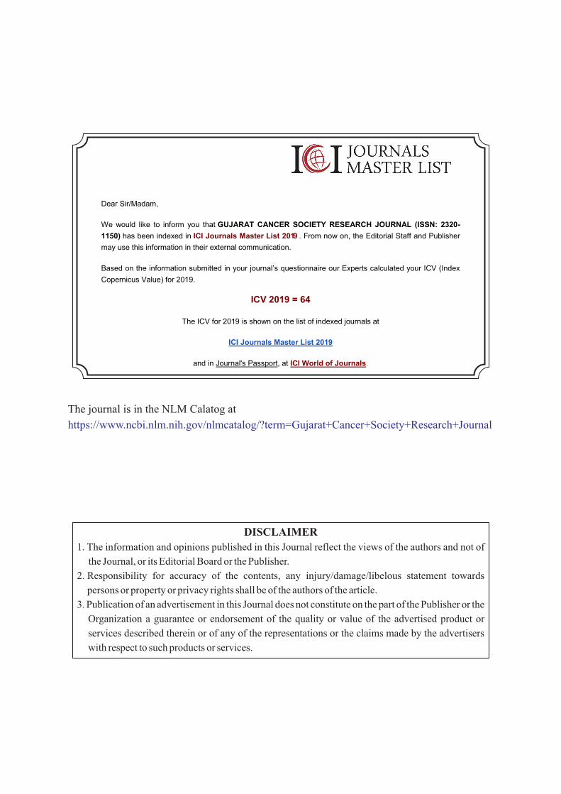
The journal is in the NLM Calatog at
https://www.ncbi.nlm.nih.gov/nlmcatalog/?term=Gujarat+Cancer+Society+Research+Journal
DISCLAIMER
1. The information and opinions published in this Journal reflect the views of the authors and not of
the Journal, or its Editorial Board or the Publisher.
2. Responsibility for accuracy of the contents, any injury/damage/libelous statement towards
persons or property or privacy rights shall be of the authors of the article.
3. Publication of an advertisement in this Journal does not constitute on the part of the Publisher or the
Organization a guarantee or endorsement of the quality or value of the advertised product or
services described therein or of any of the representations or the claims made by the advertisers
with respect to such products or services.
ISSN 2320-1150
Research Journal G u j a r a t C a n c e r S o c i e t y
(Formerly Published as GCS Research Bulletin)
Address for correspondence:
The Editors,Gujarat Cancer Society Research Journal The Gujarat Cancer and Research InstituteGCS Journal Office, Research Wing,Asarwa, Ahmedabad 380016Gujarat, [email protected]@gcriindia.org
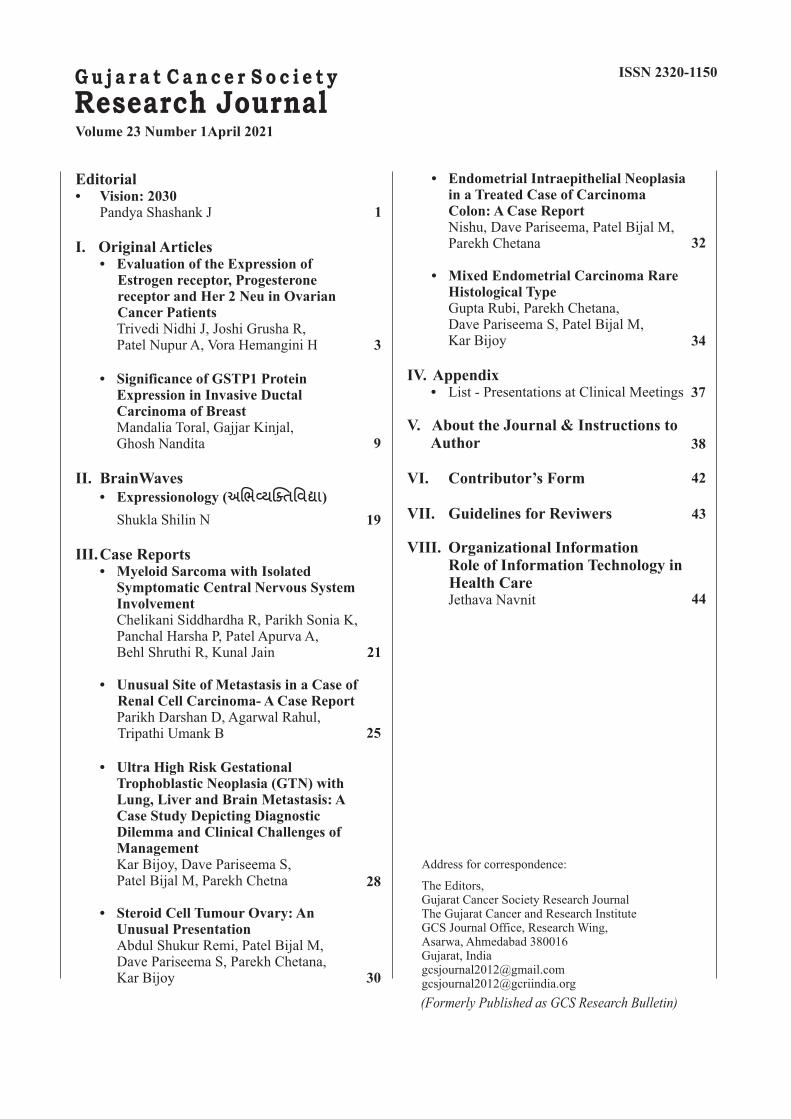
1
3
9
19
21
25
28
30
• Endometrial Intraepithelial Neoplasia in a Treated Case of Carcinoma Colon: A Case Report Nishu, Dave Pariseema, Patel Bijal M, Parekh Chetana
• Mixed Endometrial Carcinoma Rare Histological Type Gupta Rubi, Parekh Chetana, Dave Pariseema S, Patel Bijal M, Kar Bijoy
IV. Appendix • List - Presentations at Clinical Meetings V. About the Journal & Instructions to
Author
VI. Contributor’s Form
VII. Guidelines for Reviwers
VIII. Organizational Information Role of Information Technology in
Health Care Jethava Navnit
32
37
38
43
44
42
34
Volume 23 Number 1April 2021
Editorial • Vision: 2030 Pandya Shashank J
I. Original Articles • Evaluation of the Expression of
Estrogen receptor, Progesterone receptor and Her 2 Neu in Ovarian Cancer Patients
Trivedi Nidhi J, Joshi Grusha R, Patel Nupur A, Vora Hemangini H
• Significance of GSTP1 Protein Expression in Invasive Ductal Carcinoma of Breast Mandalia Toral, Gajjar Kinjal, Ghosh Nandita II. BrainWaves • Expressionology (અ�ભ�ય��તિવ�ા)
Shukla Shilin N
III. Case Reports • Myeloid Sarcoma with Isolated Symptomatic Central Nervous System Involvement Chelikani Siddhardha R, Parikh Sonia K, Panchal Harsha P, Patel Apurva A, Behl Shruthi R, Kunal Jain
• Unusual Site of Metastasis in a Case ofRenal Cell Carcinoma- A Case Report
Parikh Darshan D, Agarwal Rahul, Tripathi Umank B
• Ultra High Risk Gestational Trophoblastic Neoplasia (GTN) with Lung, Liver and Brain Metastasis: A Case Study Depicting Diagnostic Dilemma and Clinical Challenges of Management Kar Bijoy, Dave Pariseema S, Patel Bijal M, Parekh Chetna
• Steroid Cell Tumour Ovary: An Unusual Presentation Abdul Shukur Remi, Patel Bijal M, Dave Pariseema S, Parekh Chetana, Kar Bijoy
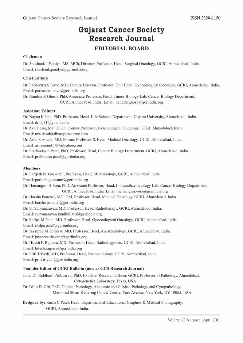
ISSN 2320-1150Gujarat Cancer Society Research Journal
Gujarat Cancer SocietyResearch Journal
EDITORIAL BOARD
ISSN 2320-1150
Designed by: Rushi C Patel, Head, Department of Educational Graphics & Medical Photography,
GCRI, Ahmedabad, India.
Chairman
Dr. Shashank J Pandya, MS, MCh, Director, Professor, Head, Surgical Oncology, GCRI, Ahmedabad, India.
Email: [email protected]
Chief Editors
Dr. Pariseema S Dave, MD, Deputy Director, Professor, Unit Head, Gynecological Oncology, GCRI, Ahmedabad, India.
Email: [email protected]
Dr. Nandita R Ghosh, PhD, Associate Professor, Head, Tumor Biology Lab, Cancer Biology Department,
GCRI, Ahmedabad, India. Email: [email protected]
Associate Editors
Dr. Nayan K Jain, PhD, Professor, Head, Life Science Department, Gujarat University, Ahmedabad, India
Email: [email protected]
Dr. Ava Desai, MD, DGO, Former Professor, Gynecological Oncology, GCRI, Ahmedabad, India.
Email: [email protected]
Dr. Asha S Anand, MD, Former Professor & Head, Medical Oncology, GCRI, Ahmedabad, India.
Email: [email protected]
Dr. Pradhudas S Patel, PhD, Professor, Head, Cancer Biology Department, GCRI, Ahmedabad, India.
Email: [email protected]
Members
Dr. Parijath N. Goswami, Professor, Head, Microbiology, GCRI, Ahmedabad, India.
Email: [email protected]
Dr. Hemangini H Vora, PhD, Associate Professor, Head, Immunohaematology Lab, Cancer Biology Department,
GCRI, Ahmedabad, India. Email: [email protected]
Dr. Harsha Panchal, MD, DM, Professor, Head, Medical Oncology, GCRI, Ahmedabad, India.
Email: [email protected]
Dr. U. Suryanarayan, MD, Professor, Head, Radiotherapy, GCRI, Ahmedabad, India.
Email: [email protected]
Dr. Shilpa M Patel, MD, Professor, Head, Gynecological Oncology, GCRI, Ahmedabad, India.
Email: [email protected]
Dr. Jayshree M Thakkar, MD, Professor, Head, Anesthesiology, GCRI, Ahmedabad, India.
Email: [email protected]
Dr. Hitesh K Rajpura, MD, Professor, Head, Radiodiagnosis, GCRI, Ahmedabad, India.
Email: [email protected]
Dr. Priti Trivedi, MD, Professor, Head, Oncopathology, GCRI, Ahmedabad, India.
Email: [email protected]
Founder Editor of GCRI Bulletin (now as GCS Research Journal)
Late, Dr. Siddharth Adhvaryu, PhD, Ex Chief Research Officer, GCRI, Professor of Pathology, Ahmedabad,
Cytogenetics Laboratory, Texas, USA
Dr. Dilip D. Giri, PhD, Clinical Pathology, Anatomic and Clinical Pathology and Cytopathology,
Memorial Sloan-Kettering Cancer Centre, York Avenue, New York, NY 10065, USA
Volume 23 Number 1April 2021
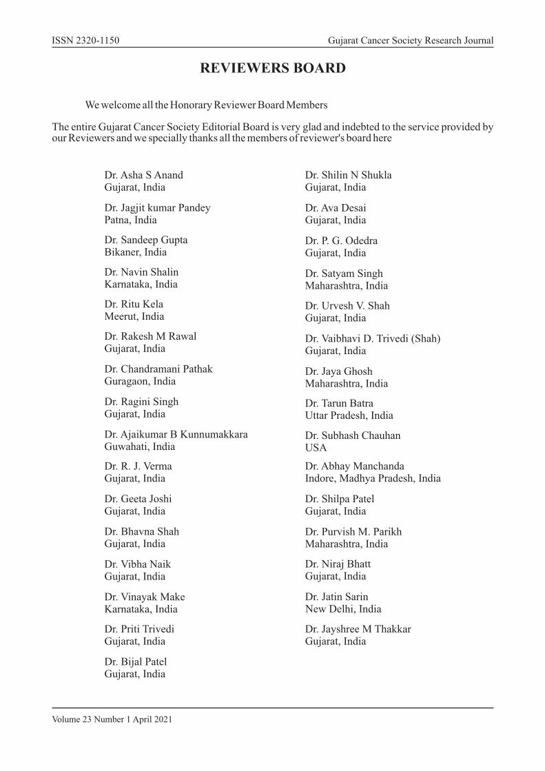
ISSN 2320-1150 Gujarat Cancer Society Research Journal
REVIEWERS BOARD
We welcome all the Honorary Reviewer Board Members
The entire Gujarat Cancer Society Editorial Board is very glad and indebted to the service provided by our Reviewers and we specially thanks all the members of reviewer's board here
Dr. Jagjit kumar PandeyPatna, India
Dr. Sandeep Gupta Bikaner, India
Dr. Navin ShalinKarnataka, India
Dr. Ritu Kela Meerut, India
Dr. Rakesh M RawalGujarat, India
Dr. Chandramani PathakGuragaon, India
Dr. Ragini SinghGujarat, India
Dr. Ajaikumar B Kunnumakkara Guwahati, India
Dr. R. J. Verma Gujarat, India
Dr. Geeta JoshiGujarat, India
Dr. Bhavna ShahGujarat, India
Dr. Vibha Naik Gujarat, India
Dr. Vinayak Make Karnataka, India
Dr. Priti TrivediGujarat, India
Dr. Bijal PatelGujarat, India
Dr. Asha S AnandGujarat, India
Dr. Shilin N ShuklaGujarat, India
Dr. Ava DesaiGujarat, India
Dr. P. G. OdedraGujarat, India
Dr. Satyam SinghMaharashtra, India
Dr. Urvesh V. Shah Gujarat, India
Dr. Vaibhavi D. Trivedi (Shah)Gujarat, India
Dr. Jaya Ghosh Maharashtra, India
Dr. Tarun BatraUttar Pradesh, India
Dr. Subhash Chauhan USA
Dr. Abhay ManchandaIndore, Madhya Pradesh, India
Dr. Shilpa PatelGujarat, India
Dr. Purvish M. ParikhMaharashtra, India
Dr. Niraj Bhatt Gujarat, India
Dr. Jatin Sarin New Delhi, India
Dr. Jayshree M ThakkarGujarat, India
Volume 23 Number 1 April 2021
About GCRI: Foundation of M.P. Shah Cancer Hospital was done as a 50 bedded hospital by Gujarat Cancer Society in 1962. Thereafter in the year 1972, Govt. of Gujarat converted this hospital into an Autonomous Body through a tripartite agreement between Govt. of Gujarat, Gujarat Cancer Society and a new body called 'The Gujarat Cancer & Research Institute (GCRI)' with 100% Grant-in-Aid from Govt. of Gujarat. In view of the availability of comprehensive cancer facilities in the Western Part of India and progress made by The Gujarat Cancer & Research Institute, Ministry of Health & Family Welfare Govt. of India has recognized this Institute as 'Regional Cancer Centre' in the year 1981 and finally promoted to “State Cancer Institute” in the year 2015.
Cancer Statistics: Cancers is among the leading causes of morbidity and mortality worldwide, as per GLOBOCAN report approximately 19.3 million new cases were reported in 2020 and this number is expected to reach 21.5 million by 2030. In India, cancer prevalence is 70-90 cases per one lakh population. As per reports, number of new cancer cases in India will rise to 25 lakhs by 2030. Cancer has become one of the ten leading causes of death in India and approximately 6.8 lakh deaths occur annually due to cancer. Data from Ahmedabad urban cancer registry indicates that the prevalence of cancer among male and female is 116 and 85 cases per one lakh population respectively. By 2030, cancer burden will rise extensively, and therefore there is need to formulate and organise to overcome the forthcoming problem. With this vision GCRI have started to upgrade our institute on following points:
• Manpower: We have started many new medical and paramedical courses like DM Oncopathology, MD Palliative Medicine, increase in seats of MCH Gynec Oncology, Msc in Medical Physics, Postgraduate Diploma Medical Laboratory Technician (DMLT), Certificate Course in Medical Radiotherapy Technology (CMRT). We are also planning to add many courses like DM
Paediatric Oncology, MCH Head & Neck oncology, DM in Haematology, MD/DNB Nuclear Medicine and many more. Along with this our efforts are continued to increase our hospital manpower to cater rising demand of cancer care.
• Beds: We are already in process of increasing our bed strength from 650 beds to 1000 beds.
• Technologies: Shortly, “New Operation Theatre (OT) Complex”, comprising of 19 Modular high-end OTs will be operational at GCRI. These OTs will be having all high end and state of art technologies and in future newer facilities like robotic surgery, Intra operative radiotherapy and Hyperthermic Intraperitoneal Chemotherapy (HIPEC) facilities will be introduced.
• New technologies will change the way doctors treat and interact with patients over the next decade. Experts predict artificial intelligence (AI) will soon be used to help inform clinicians on the best treatment plans for each individual patient, instead of “waiting a few months” to see how a patient responds to a treatment - especially when “some patients don't have that time.
• Radiation Oncology has seen 120 years of development from deep X-Ray therapy to High End machines (with special techniques like IMRT, IGRT etc.), mainly to treat target tumour and spare normal tissues. As Cancer survivors are increasing all over the world and with that patients are increasing with years lived with disability due to cancer. Looking to Proton therapy treatment – which have been accepted worldwide with main advantage of its physical and radiobiological properties like NO EXIT DOSE, and reduction of clinically observable undesirable side effects. Its main use is in paediatric tumors and for the targets which are nearby critical structures. Currently we have state of art equipment’s like Cyber Knife, Tomotherapy which only few institutes have at national level. We will continue to procure new technologies and machine like Proton Therapy, Cyclotron machines and many more to provide recent cancer care at GCRI.
EditorialGujarat Cancer Society Research Journal
1 Volume 23 Number 1 April 2021
Vision: 2030
Pandya Shashank J
Director, Professor & Head of Surgical Oncology
The Gujarat Cancer & Research Institute, Asarwa, Ahmedabad, Gujarat, India.
Corresponding Author: [email protected]
https://orcid.org/0000-0002-8432-4293
• Screening activities: From decades, GCRI is doing cancer screening activities throughout the Gujarat state as community outreach activity, however in next decade we will priorities our in-house cancer screening and cancer awareness activities. Oral, breast and cervical cancer forms almost 50% of cancer load of GCRI cancer cases. Moreover, all these cancers can be easily screened and identify in their early stage which will make our efforts more effective. We are also planning to start liquid-based cytology with HPV DNA testing for cervical cancer screening, which will increase sensitivity of the screening method.
• Targeted treatment: Traditional chemotherapy has long been a standard treatment in cancer care. But it's increasingly taking a back seat to a more precise and personalized approach, called targeted therapy. GCRI will also incorporate personalised and targeted approach as more and more such treatment will be available.
• Molecular Diagnostic Testing: Currently the molecular diagnostic services at GCRI is being managed by using PCR and RT-PCR technology and are being performed in several solid and liquid malignancies such as lung cancer, breast cancer, brain malignancies, hereditary breast and ovarian cancer and blood cancers along with HLA typing for bone marrow transplantation. Since the current era demands a need to stratify individuals who are at a higher risk for development of cancer, and for personalized m e d i c i n e o f d i a g n o s e d c a n c e r , t h e
Gujarat Cancer Society Research Journal
2Volume 23 Number 1 April 2021
implementation of “Next Generation Sequencer platform” will contribute remarkably with the clinical demand in identification of actionable molecular diagnostic, prognostic, and therapeutic targets at gene level and provide meaningful knowledge to unravel the genetics of disease, diagnostic and treatment strategies to a new level.
• Accreditation: GCRI hospital is accredited with entry level NABH and all laboratories of GCRI are NABL certified. We are working towards full NABH certification which will enable the organisation in demonstrating commitment to quality care.
• Holistic and integrated approach: Cancer treatment is a multimodality treatment; our prime focus will be to provide integrated and holistic health care to cancer patients. Increased efforts will be given to have a team-based approach in managing cancer care.
• Research: More efforts will be given on research and academic activities. Staff and students will be encouraged to have newer research projects. In this digital era, we are also working to make GCRI digitalised and to strengthen telemedicine services which will promote research and academic activities manifold as well as will reduce patient’s follow-up visits respectively.
• Providing comprehensive cancer care at our satellite centres – Siddhpur Cancer Care Centre- Siddhpur, Saurasthra Cancer Care Canter - Rajkot and Bhavnagar Cancer Care and Research Centre - Bhavnagar.
I alone will not be able to complete this vision on my own,I will need help of each GCRI staff to achieve this vision.
I believe, if we work together as “Team GCRI”,We can represent our institute as one of the
Best Cancer Treating Institute on International level.
Dr. Shashank J Pandya
Director, GCRI
Summary Steroid hormone receptors expression in epithelial ovarian cancers has been proposed to have therapeutic and prognostic relevance. Steroid hormones, primarily estrogen, progesterone and HER 2 Neu have been implicated in ovarian carcinogenesis. The prognostic characterisation of ovarian cancer patients, based on clinicopathological parameters such as age, menopausal status, stage, histology, grade, CA 125 level and treatment. This study mainly used to evaluate the expression of Estrogen Receptor (ER), Progesterone Receptor (PR) and Her 2 Neu in ovar ian cancer pat ients and corre la te wi th clinicopathological parameters using immunohistochemistry technique. Nuclear ER expression was noted in tumor tissue of 60% (30/50) in ovarian cancer patients. Significatly higher ER expression was noted with pre-menopausal status. A trend of higher ER expression in Grade 2 tumors. Increased incidence of disease relapse and over death noted in ER positive patients than ER negative patients. Nuclear PR expression was found to be positive in 60% (31/50) cases. Significantly higher PR expression was noted in Grade 2 tumors. Similar incidence of disease relapse and death was noted in positive PR expression and negative PR expression. Membranous HER 2 Neu expression was found to be positive in 18% (09/50) cases. Significantly higher HER 2 Neu receptor expression was noted in CA 125 normal level and histological type of Mucinous Adenocarcinoma which was statistically significant. Higher incidence of disease relapse and death was noted in positive HER 2 Neu and negative HER 2 Neu patients.Keywords: ER, PR, Her-2-neu, Ovarian Cancer
Introduction Ovarian cancer is the second most common gynecologic malignancy, and in developed countries, in women it remains the fifth leading cause of cancer
1death. In India, it is the third leading cancer amongst women, after cervix, and breast cancer. It is about 1 in every 70 women have a lifetime risk of developing
2ovarian cancer. Age is considered as a significant risk of ovarian cancer. Ovulation, growth factors, cytokines, and environmental agents may play an important role in the initiation as well as progression
3of ovarian cancer. The majority of cases are sporadic while about 5-10% cases of ovarian cancers are familial. However, the risk for developing ovarian cancer increases four fold in women with affected first degree relative. Lack of knowledge about the etiology
and pathogenesis of the tumor leads to its late diagnosis at advanced stage which presents it with highest mortality rate. Therefore, new therapeutic strategies and reliable screening methods for diagnosis are urgently needed. Estrogen Receptor (ER) and Progesterone Receptor (PR) are main secreted hormones by ovary acting through specific
4receptors. It is known fact that these two hormones and their specific receptors are involved in the process of tumor genesis in ovarian cancer. In addition, evaluation of ER and PR by immunohistochemistry would have advantage in the understanding of the difference in distribution of the expression of the protein between tumor tissues as well as surrounding normal tissue. As well, the determination of hormone receptor in malignant ovarian neoplasms may probably aid in selection of patients for endocrine therapy in a manner similar to that has been already
5established for certain hormone dependent cancers. Human epidermal growth factor receptor type 2(Her 2 Neu) a proto-oncogene that encodes a transmembrane receptor protein involved in the development and progression of the majority of cancers. Studies have shown that Her2 Neu is overexpressed in
6approximately 15-30% of ovarian carcinomas. It has also been tested as potential biomarkers of individualized clinical behaviour of cancers, however, findings regarding the overexpression and
6prognosis are still conflicting. So, the present study aims at evaluation of the expression of ER, PR and Her 2 Neu Receptor in ovarian cancer patients. Furthermore to correlate their expression with various clinicopathological parameters.
Material and MethodPatients This retrospective study was approved by institutional scientific and ethics committees, included 50 ovarian cancer patients diagnosed and treated at The Gujarat Cancer & Research Institute.
Original ArticleGujarat Cancer Society Research Journal
3 Volume 23 Number 1 April 2021
Evaluation of the Expression of Estrogen Receptor,Progesterone Receptor and Her2 Neu in OvarianCancer Patients
1 2 3 4Trivedi Nidhi J , Joshi Grusha R , Patel Nupur A , Vora Hemangini H
1 2 3 4M.Sc Cancer Biology Student , Junior Research Fellow , Research Assistant , Professor and Head
Immunohematology Laboratory, Department of Cancer Biology
Corresponding author: [email protected] 3
https://orcid.org/0000-0001-8165-8696
https://orcid.org/0000-0003-3893-9999
Detailed clinical history such as age, menopausal status, histopathological type, grade, CA125 levels, treatment offered and stage of the disease were recorded from the case files maintained at the Medical Record Department of the Institute. Disease staging was done according to AJCC classification. Disease status was assessed by clinical examination, radiological investigations and biochemical investigations.
Immunohistochemical Localization Localization of markers Estrogen Receptor (ER), Progesterone Receptor (PR) and Her 2 Neu was p e r f o r m e d o n Ve n t a n a B e n c h m a r k X T autoimmunostainer using Ventana reagents (Ventana, USA) . Pr imary an t ibodies were procured commercially from Ventana, Roche Diagnostics. The primary antibodies and secondary antibody were incubated as follows: ER (SP1, RTU, Ventana) for 16 minutes, PR (1E2, RTU, Ventana) for 16 minutes, Her 2 Neu (4B5, RTU, Ventana) for 32 minutes, HRP multimer for 8 minutes.
Scoring Two individual observers scored the sections. Nuclear staining pattern was observed for ER and PR, while Her 2 Neu showed membranous staining pattern. The sections were scored positive and negative for statistical analysis.
Statistical Analysis Statistical analysis was carried out using SPSS statistical software version 20 (SPSS Inc., USA). Univariate survival analysis was carried out by Kaplan Meier and Log Rank statistics was used to assess the prognostic significance of disease free survival (DFS) and overall survival (OS). P values ≤ 0.05 were considered to be significant.
Results
Patient’s Characteristics and Outcome This retrospective study included 50 patients, 30% had age ≤ 53 years, whereas 70% patients had >53 years. Majority of the patients i.e. 80% had postmenopausal status. In relation to pathological characteristics more than 50% were of late stage, having grade 3 tumor with serous papillary adenocarcinoma and higher CA125 levels. (Table 1)The primary treatment offered to the patients was surgery followed by adjuvant chemotherapy (Paclitaxel + Carboplatin). The maximum follow-up period was 68 months with a median follow-up of 12 months.
Gujarat Cancer Society Research Journal
4Volume 23 Number 1 April 2021
ER Expression Nuclear expression of ER was noted in 60% of the tumors. A significant higher incidence of ER expression was noted with premenopausal women as compared to postmenopausal women (p=0.03) whereas similar incidence of ER expression was observed with age group (Table 1; Figure 1). A trend towards higher incidence of ER expression was observed in patients with Grade II (p=0.09) as compared with their counterparts. No significant correlation was observed with other clinical and pathological parameters. (Table 1)
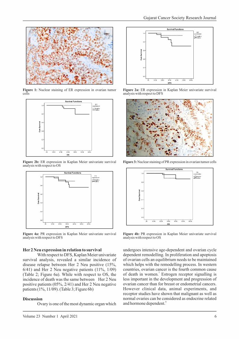
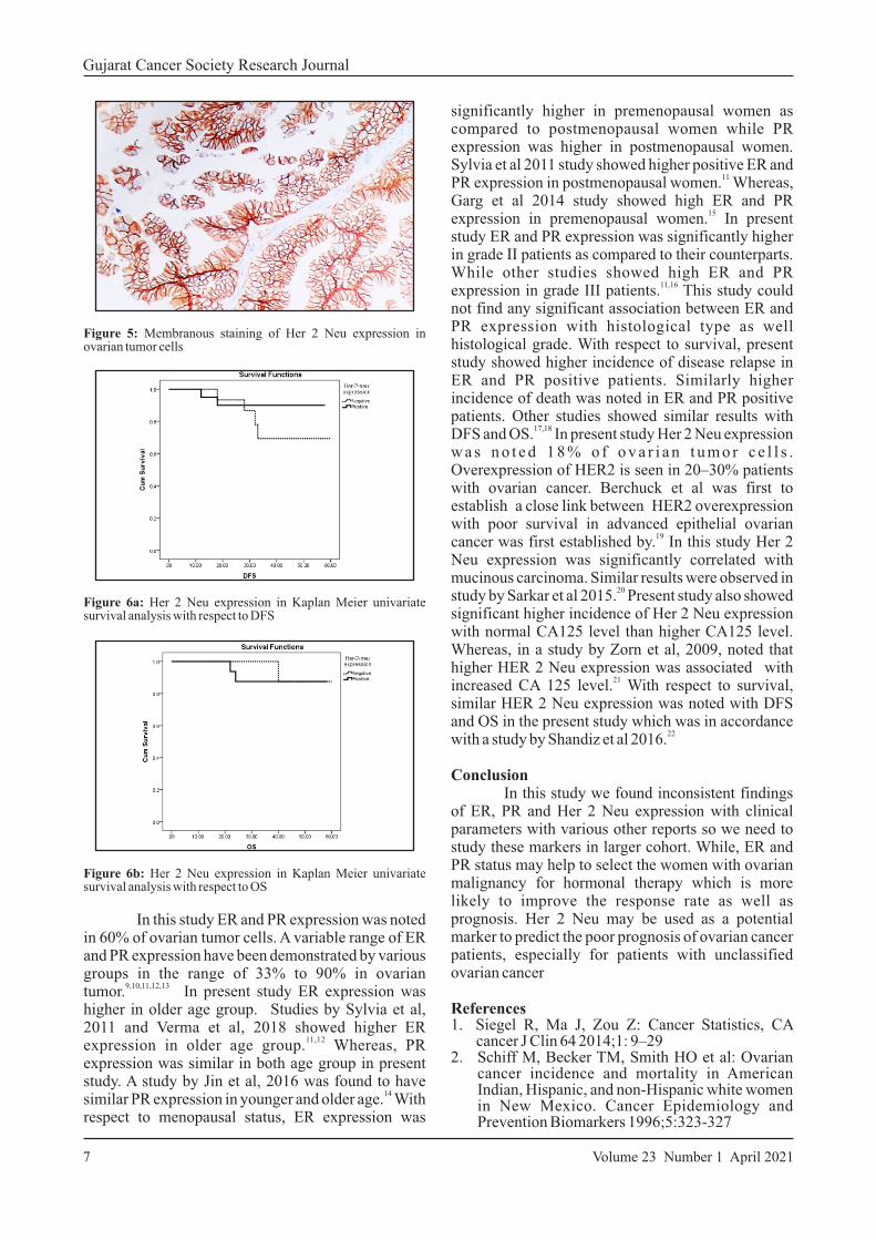
ER expression in relation to survival According to Kaplan Meier univariate survival analysis, with respect to DFS, higher incidence of disease relapse was noted in ER positive (20%, 4/30) than ER negative patients (5%, 1/20). (Table 2; Figure 2a) While with respect to OS, higher incidence of death was noted in ER positive patients (10%, 3/30) than ER negative patients (0%, 0/20). (Table 3; Figure 2b)
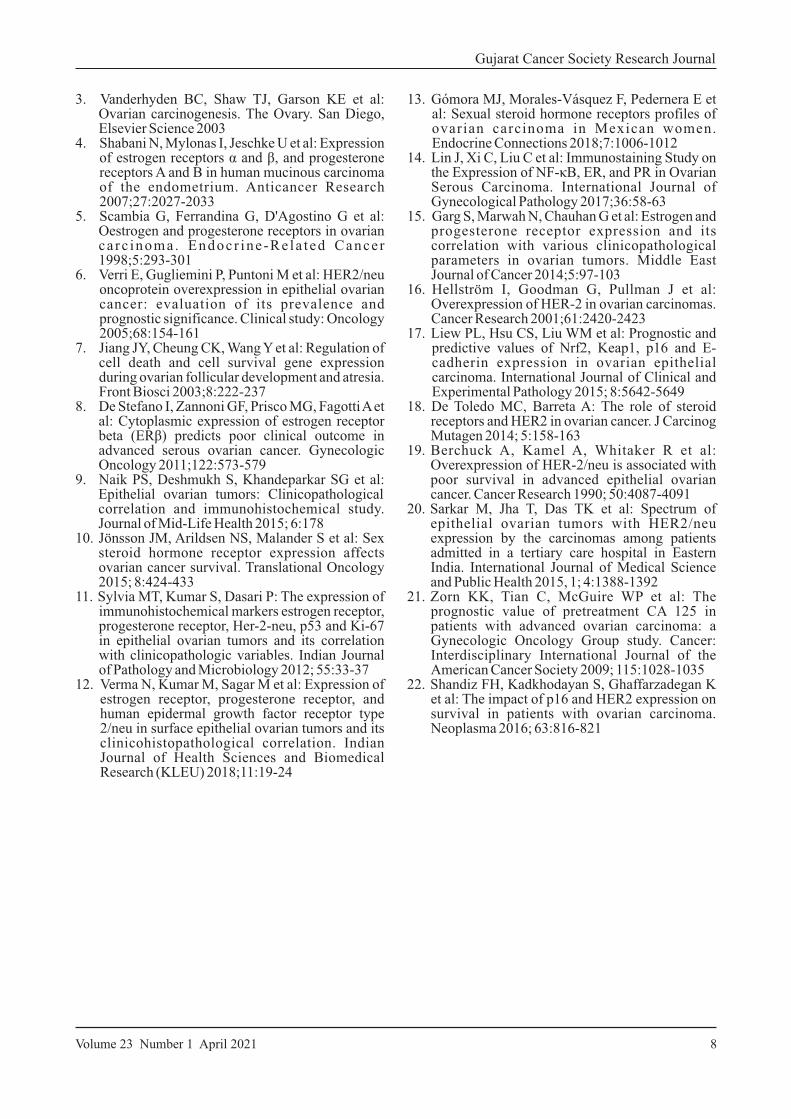
PR Expression Nuclear expression of PR was noted in 60% of the ovarian cancer cases. No significant correlation of PR expression was observed with clinical parameters. (Table 1; Figure 3) A significant higher incidence of PR expression was observed with Grade II patients (p=0.02) as compared to their counter parts. While no other pathological parameters were found to be significantly associated with PR expression. (Table 1)
PR expression in relation to survival According to Kaplan Meier univariate survival analysis, with respect to DFS, a trend higher incidence of disease relapse was noted in PR positive (23%, 7/30) than PR negative patients (0%, 0/20). (Table 2; Figure 4a) While with respect to OS, higher incidence of death was noted in PR positive patients (10%, 3/30) than PR negative patients (0%, 0/20). (Table 3; Figure 4b)
Her 2 Neu Expression Membranous Her 2 Neu expression was observed in 18% of the patients. No significant correlation of clinical parameters with Her2 Neu expression was observed. (Table 1, Figure 5) With pathological correlations, a significant higher incidence of Her 2 Neu expression was observed with mucinous adenocarcinoma as compared to other histologic type. Also, a significant higher incidence of Her 2 Neu expression was observed with normal CA125 level than higher CA125 level. (Table 1)
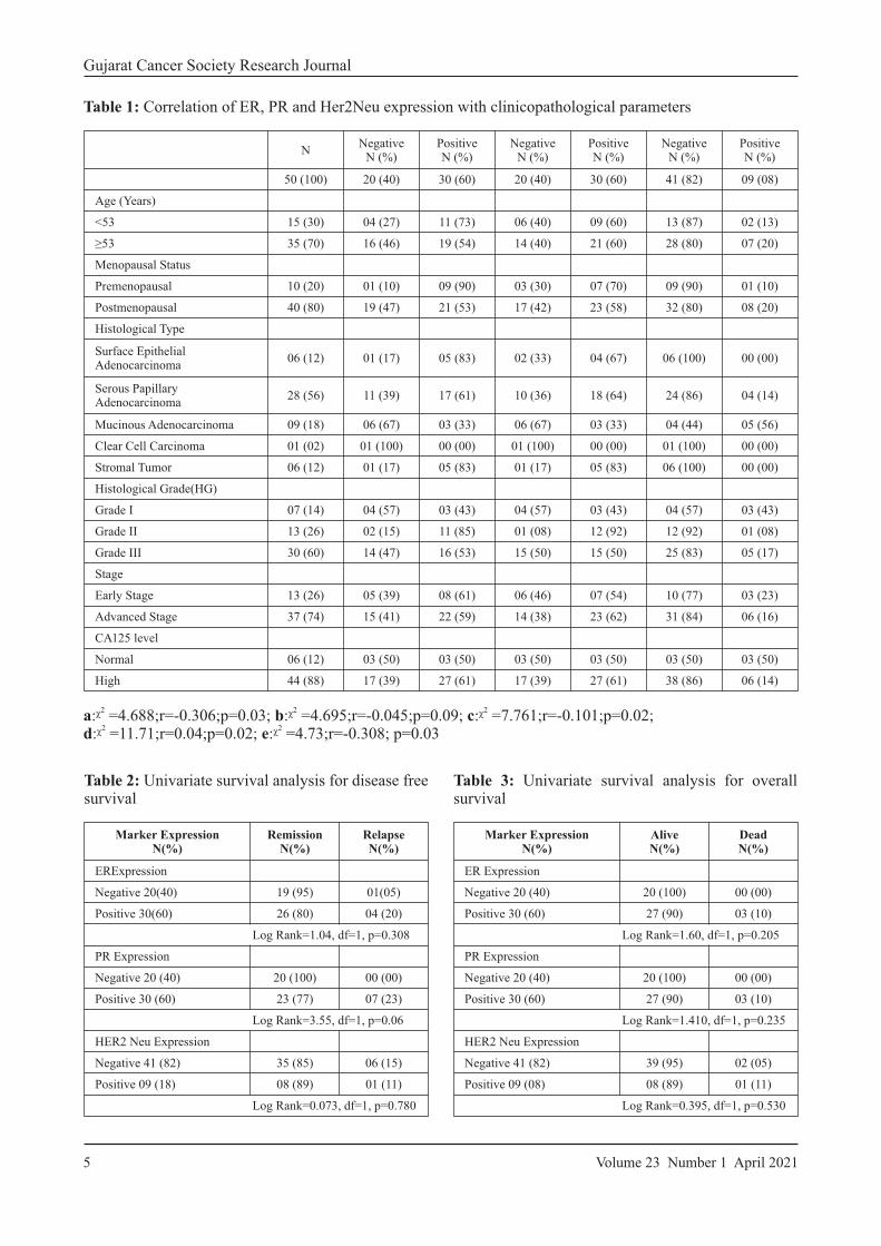
Table 3: Univariate survival analysis for overall survival
Marker ExpressionN (%)
AliveN (%)
DeadN (%)
ER Expression
Negative 20 (40) 20 (100) 00 (00)
Positive 30 (60) 27 (90) 03 (10)
Log Rank=1.60, df=1, p=0.205
PR Expression
Negative 20 (40) 20 (100) 00 (00)
Positive 30 (60) 27 (90) 03 (10)
Log Rank=1.410, df=1, p=0.235
HER2 Neu Expression
Negative 41 (82) 39 (95) 02 (05)
Positive 09 (08) 08 (89) 01 (11)
Log Rank=0.395, df=1, p=0.530
Table 2: Univariate survival analysis for disease free survival
Marker ExpressionN (%)
RemissionN (%)
RelapseN (%)
ER Expression
Negative 20 (40) 19 (95) 01 (05)
Positive 30 (60) 26 (80) 04 (20)
Log Rank=1.04, df=1, p=0.308
PR Expression
Negative 20 (40) 20 (100) 00 (00)
Positive 30 ( 60) 23 (77) 07 (23)
Log Rank=3.55, df=1, p=0.06
HER2 Neu Expression
Negative 41 (82) 35 (85) 06 (15)
Positive 09 (18) 08 (89) 01 (11)
Log Rank=0.073, df=1, p=0.780
Gujarat Cancer Society Research Journal
5 Volume 23 Number 1 April 2021
2 2 2a:ᵡ =4.688;r=-0.306;p=0.03; b:ᵡ =4.695;r=-0.045;p=0.09; c:ᵡ =7.761;r=-0.101;p=0.02; 2 2
d:ᵡ =11.71;r=0.04;p=0.02; e:ᵡ =4.73;r=-0.308; p=0.03
Table 1: Correlation of ER, PR and Her2Neu expression with clinicopathological parameters
NNegative
N (%)Positive N (%)
Negative N ( %)
Positive N (%)
Negative N (%)
Positive N (%)
50 (100) 20 ( 40) 30 (60) 20 (40) 30 (60) 41 (82) 09 (08)
Age (Years)
<53 15 (30) 04 (27) 11 (73) 06 (40) 09 (60) 13 (87) 02 (13)
≥53 35 (70) 16 (46) 19 (54) 14 (40) 21 (60) 28 (80) 07 (20)
Menopausal Status
Premenopausal 10 (20) 01 (10) 09 (90) 03 (30) 07 (70) 09 (90) 01 (10)
Postmenopausal 40 (80) 19 (47) 21 (53) 17 (42) 23 (58) 32 (80) 08 (20)
Histological T ype
Surface Epithelial Adenocarcinoma
06 (12) 01 (17) 05 (83) 02 (33) 04 (67) 06 (100) 00 (00)
Serous Papillary Adenocarcinoma
28 (56) 11 (39) 17 (61) 10 (36) 18 (64) 24 (86) 04 (14)
Mucinous A denocarcinoma 09 (18) 06 (67) 03 (33) 06 (67) 03 (33) 04 (44) 05 (56)
Clear Cell Carcinoma 01 (02) 01 (100) 00 (00) 01 (100) 00 (00) 01 (100) 00 (00)
Stromal T umor 06 (12) 01 (17) 05 (83) 01 (17) 05 (83) 06 (100) 00 (00)
Histological Grade (HG)
Grade I 07 (14) 04 (57) 03 (43) 04 (57) 03 (43) 04 (57) 03 (43)
Grade II 13 (26) 02 (15) 11 (85) 01 (08) 12 (92) 12 (92) 01 (08)
Grade III 30 (60) 14 (47) 16 (53) 15 (50) 15 (50) 25 (83) 05 (17)
Stage
Early Stage 13 (26) 05 (39) 08 (61) 06 (46) 07 (54) 10 (77) 03 (23)
Advanced Stage 37 (74) 15 (41) 22 (59) 14 (38) 23 (62) 31 (84) 06 (16)
CA 125 level
Normal 06 (12) 03 (50) 03 (50) 03 (50) 03 (50) 03 (50) 03 (50)
High 44 (88) 17 (39) 27 (61) 17 (39) 27 (61) 38 (86) 06 (14)
Gujarat Cancer Society Research Journal
6Volume 23 Number 1 April 2021
undergoes intensive age-dependent and ovarian cycle dependent remodelling. In proliferation and apoptosis of ovarian cells an equilibrium needs to be maintained which helps with the remodelling process. In western countries, ovarian cancer is the fourth common cause
7of death in women. Estrogen receptor signalling is less important in the development and progression of ovarian cancer than for breast or endometrial cancers. However clinical data, animal experiments, and receptor studies have shown that malignant as well as normal ovaries can be considered as endocrine related
8and hormone dependent.
Figure 2b: ER expression in Kaplan Meier univariate survival analysis with respect to OS
Figure 4a: PR expression in Kaplan Meier univariate survival analysis with respect to DFS
Figure 3: Nuclear staining of PR expression in ovarian tumor cells
Figure 4b: PR expression in Kaplan Meier univariate survival analysis with respect to OS
Figure 1: Nuclear staining of ER expression in ovarian tumor cells
Figure 2a: ER expression in Kaplan Meier univariate survival analysis with respect to DFS
Her 2 Neu expression in relation to survival With respect to DFS, Kaplan Meier univariate survival analysis, revealed a similar incidence of disease relapse between Her 2 Neu positive (15%, 6/41) and Her 2 Neu negative patients (11%, 1/09) (Table 2; Figure 6a). While with respect to OS, the incidence of death was the same between Her 2 Neu positive patients (05%, 2/41) and Her 2 Neu negative patients (1%, 11/09). (Table 3; Figure 6b)
Discussion Ovary is one of the most dynamic organ which
Gujarat Cancer Society Research Journal
7 Volume 23 Number 1 April 2021
Figure 5: Membranous staining of Her 2 Neu expression in ovarian tumor cells
Figure 6a: Her 2 Neu expression in Kaplan Meier univariate survival analysis with respect to DFS
Figure 6b: Her 2 Neu expression in Kaplan Meier univariate survival analysis with respect to OS
In this study ER and PR expression was noted in 60% of ovarian tumor cells. A variable range of ER and PR expression have been demonstrated by various groups in the range of 33% to 90% in ovarian
9,10,11,12,13tumor. In present study ER expression was higher in older age group. Studies by Sylvia et al, 2011 and Verma et al, 2018 showed higher ER
11,12expression in older age group. Whereas, PR expression was similar in both age group in present study. A study by Jin et al, 2016 was found to have
14similar PR expression in younger and older age. With respect to menopausal status, ER expression was
significantly higher in premenopausal women as compared to postmenopausal women while PR expression was higher in postmenopausal women. Sylvia et al 2011 study showed higher positive ER and
11PR expression in postmenopausal women. Whereas, Garg et al 2014 study showed high ER and PR
15expression in premenopausal women. In present study ER and PR expression was significantly higher in grade II patients as compared to their counterparts. While other studies showed high ER and PR
11,16expression in grade III patients. This study could not find any significant association between ER and PR expression with histological type as well histological grade. With respect to survival, present study showed higher incidence of disease relapse in ER and PR positive patients. Similarly higher incidence of death was noted in ER and PR positive patients. Other studies showed similar results with
17,18DFS and OS. In present study Her 2 Neu expression w a s n o t e d 1 8 % o f o v a r i a n t u m o r c e l l s . Overexpression of HER2 is seen in 20–30% patients with ovarian cancer. Berchuck et al was first to establish a close link between HER2 overexpression with poor survival in advanced epithelial ovarian
19cancer was first established by. In this study Her 2 Neu expression was significantly correlated with mucinous carcinoma. Similar results were observed in
20study by Sarkar et al 2015. Present study also showed significant higher incidence of Her 2 Neu expression with normal CA125 level than higher CA125 level. Whereas, in a study by Zorn et al, 2009, noted that higher HER 2 Neu expression was associated with
21increased CA 125 level. With respect to survival, similar HER 2 Neu expression was noted with DFS and OS in the present study which was in accordance
22with a study by Shandiz et al 2016.
Conclusion In this study we found inconsistent findings of ER, PR and Her 2 Neu expression with clinical parameters with various other reports so we need to study these markers in larger cohort. While, ER and PR status may help to select the women with ovarian malignancy for hormonal therapy which is more likely to improve the response rate as well as prognosis. Her 2 Neu may be used as a potential marker to predict the poor prognosis of ovarian cancer patients, especially for patients with unclassified ovarian cancer
References1. Siegel R, Ma J, Zou Z: Cancer Statistics, CA
cancer J Clin 64 2014;1: 9–292. Schiff M, Becker TM, Smith HO et al: Ovarian
cancer incidence and mortality in American Indian, Hispanic, and non-Hispanic white women in New Mexico. Cancer Epidemiology and Prevention Biomarkers 1996;5:323-327
13. Gómora MJ, Morales-Vásquez F, Pedernera E et al: Sexual steroid hormone receptors profiles of ovarian carcinoma in Mexican women. Endocrine Connections 2018;7:1006-1012
14. Lin J, Xi C, Liu C et al: Immunostaining Study on the Expression of NF-κB, ER, and PR in Ovarian Serous Carcinoma. International Journal of Gynecological Pathology 2017;36:58-63
15. Garg S, Marwah N, Chauhan G et al: Estrogen and progesterone receptor expression and its correlation with various clinicopathological parameters in ovarian tumors. Middle East Journal of Cancer 2014;5:97-103
16. Hellström I, Goodman G, Pullman J et al: Overexpression of HER-2 in ovarian carcinomas. Cancer Research 2001;61:2420-2423
17. Liew PL, Hsu CS, Liu WM et al: Prognostic and predictive values of Nrf2, Keap1, p16 and E-cadherin expression in ovarian epithelial carcinoma. International Journal of Clinical and Experimental Pathology 2015; 8:5642-5649
18. De Toledo MC, Barreta A: The role of steroid receptors and HER2 in ovarian cancer. J Carcinog Mutagen 2014; 5:158-163
19. Berchuck A, Kamel A, Whitaker R et al: Overexpression of HER-2/neu is associated with poor survival in advanced epithelial ovarian cancer. Cancer Research 1990; 50:4087-4091
20. Sarkar M, Jha T, Das TK et al: Spectrum of epithelial ovarian tumors with HER2/neu expression by the carcinomas among patients admitted in a tertiary care hospital in Eastern India. International Journal of Medical Science and Public Health 2015, 1; 4:1388-1392
21. Zorn KK, Tian C, McGuire WP et al: The prognostic value of pretreatment CA 125 in patients with advanced ovarian carcinoma: a Gynecologic Oncology Group study. Cancer: Interdisciplinary International Journal of the American Cancer Society 2009; 115:1028-1035
22. Shandiz FH, Kadkhodayan S, Ghaffarzadegan K et al: The impact of p16 and HER2 expression on survival in patients with ovarian carcinoma. Neoplasma 2016; 63:816-821
Gujarat Cancer Society Research Journal
8Volume 23 Number 1 April 2021
3. Vanderhyden BC, Shaw TJ, Garson KE et al: Ovarian carcinogenesis. The Ovary. San Diego, Elsevier Science 2003
4. Shabani N, Mylonas I, Jeschke U et al: Expression of estrogen receptors α and β, and progesterone receptors A and B in human mucinous carcinoma of the endometrium. Anticancer Research 2007;27:2027-2033
5. Scambia G, Ferrandina G, D'Agostino G et al: Oestrogen and progesterone receptors in ovarian ca rc inoma. Endocr ine -Re la ted Cancer 1998;5:293-301
6. Verri E, Gugliemini P, Puntoni M et al: HER2/neu oncoprotein overexpression in epithelial ovarian cancer: evaluation of its prevalence and prognostic significance. Clinical study: Oncology 2005;68:154-161
7. Jiang JY, Cheung CK, Wang Y et al: Regulation of cell death and cell survival gene expression during ovarian follicular development and atresia. Front Biosci 2003;8:222-237
8. De Stefano I, Zannoni GF, Prisco MG, Fagotti A et al: Cytoplasmic expression of estrogen receptor beta (ERβ) predicts poor clinical outcome in advanced serous ovarian cancer. Gynecologic Oncology 2011;122:573-579
9. Naik PS, Deshmukh S, Khandeparkar SG et al: Epithelial ovarian tumors: Clinicopathological correlation and immunohistochemical study. Journal of Mid-Life Health 2015; 6:178
10. Jönsson JM, Arildsen NS, Malander S et al: Sex steroid hormone receptor expression affects ovarian cancer survival. Translational Oncology 2015; 8:424-433
11. Sylvia MT, Kumar S, Dasari P: The expression of immunohistochemical markers estrogen receptor, progesterone receptor, Her-2-neu, p53 and Ki-67 in epithelial ovarian tumors and its correlation with clinicopathologic variables. Indian Journal of Pathology and Microbiology 2012; 55:33-37
12. Verma N, Kumar M, Sagar M et al: Expression of estrogen receptor, progesterone receptor, and human epidermal growth factor receptor type 2/neu in surface epithelial ovarian tumors and its clinicohistopathological correlation. Indian Journal of Health Sciences and Biomedical Research (KLEU) 2018;11:19-24
Summary Glutathione S-transferases (GSTs) are important isoenzymes that play an essential role in detoxification of carcinogens and acts as endogenous inhibitor of MAP kinase pathway. GST Pi 1 (GSTP1) isoform has been documented to contribute to drug resistance in breast cancer patients. Hence, present study aimed to investigate the prevalence of GSTP1 p r o t e i n e x p r e s s i o n i n b r e a s t c a n c e r p a t i e n t s b y immunohistochemistry method and further to examine its correlation with various clinicopathological parameters. Total 70 untreated patients with invasive ductal carcinoma of breast cancer (70 tumor tissues and 30 adjacent normal tissues) were included in the study. Statistical analysis was carried out using SPSS software. The results indicated that- cytoplasmic and/or nuclear GSTP1 immunoreactivity was observed in 76% tumors and 97% adjacent normal tissues of the breast cancer patients. Significant higher GSTP1 protein expression was observed in high BR score tumors (78%; P=0.007), ER-ve patients (68%; P=0.008), TNBC patients (78%; P=0.004) and patients having absence of perinodal extension (56%; P=0.050) as compared to their respective counter parts. Hence, there is loss of GSTP1 protective function during the transition of malignant transformation. Higher GSTP1 expression is associated with aggressive prognosticators of breast cancer. However, confirmation in larger set of patients and longer follow up details is needed to evaluate the potential of GSTP1 as a prognostic marker.Keywords: GSTP1, breast cancer, immunohistochemistry, TNBC, Glutathione S-transferases
Introduction Epidemiological studies suggest that breast cancer is the most common type of cancer among women with continuous prevalence throughout the
1world. Although its incidence is not the same in different countries and ethnic groups, breast cancer has become a significant public health challenge
2,3among women worldwide. It is a multifactorial and polygenic disease which may be influenced by both
4,5environmental and genetic factors. Although there are several comprehensive treatment options, such as surgery, chemotherapy, and endocrine therapy, many patients still have high rates of metastasis and recurrence, which remain the primary cause of death
6in patients with breast cancer. Patients with triple negative breast cancer (TNBC) account for about 15–20% of total breast cancer cases, which have
higher rates of metastasis and recurrence, and lower survival rates compared to other subtypes because these patients do not receive anti-receptor therapy. Therefore, other potential prognostic markers and
7new therapeutic targets for BC should be explored. In recent years, some genes have been confirmed as potential cancer susceptible genes. Glutathione S-transferases (GSTs) are overwhelmingly important genes, which play key role in the detoxification of toxic, potentially carcinogenic compounds, and a host
8–11of basic physiological processes of the human body. They are a super family of dimeric phase-II metabolic enzymes that have an irreplaceable role in the cellular
12, 13defense system. In human, classes of GST enzymes include alpha-α, mu-μ, pi-π, sigma-σ, omega- Ω and
14theta-θ. Louie S M. found that GST Pi 1 (GSTP1) was a new breast cancer oncogene that governed the pathogenicity of cancer by regulating glycolysis, and
15energy and fat metabolism. Although some reports had shown the association between GSTs and overall survival in breast cancer patients, the results were not
16-19consistent. Therefore, the aim of the present study was to investigate the relationship between the GSTP1protein expression and the clinicopathological characteristics of breast cancer patients.
Materials and MethodsPatients Seventy untreated and histopathologically confirmed invasive ductal breast carcinoma female patients diagnosed at Gujarat Cancer & Research Institute (GCRI) were included in this retrospective study. The study was approved by Institutional Scientific and Ethical Committees and informed consent was obtained from all subjects prior to treatment administration. Detailed clinical and pathological history i.e. age, menopause status, tumor size, diseases stage, histological grade, treatment given, disease status, were obtained from the case files maintained at the Medical Record Department of the institute.
Original ArticleGujarat Cancer Society Research Journal
9 Volume 23 Number 1 April 2021
Significance of GSTP1 Protein Expression in InvasiveDuctal Carcinoma of Breast a 1 b 1 2Mandalia Toral , Gajjar Kinjal , Ghosh Nandita
1 2Research Assistant , Associate Professor Tumor Biology Lab, Cancer Biology DepartmentThe Gujarat Cancer & Research Institute, Asarwa, Ahmedabad, Gujarat, IndiaCorresponding Author: [email protected],
a https://orcid.org/0000-0002-3495-1600 b https://orcid.org/0000-0002-1126-0217 https://orcid.org/0000-0002-0210-7317
Immunohistochemistry (IHC) Three to five micron thick sections were cut from the formalin fixed paraffin embedded tissue blocks of IDC patients using Leica microtome and mounted on APES coated glass slides. The protein e x p r e s s i o n o f G S T P 1 w a s s t u d i e d b y immunohistochemistry technique using HRP/DAB (ABC) detection IHC kit (Abcam). The instructions in the kit insert were followed for carrying out the procedure. Mouse monoclonal GSTP1 primary antibody (Cat#sc-66000, Santa Cruz) was used at 1:100 dilution. Antigenicity was retrieved by heating the sections in 10 mM sodium citrate buffer (pH, 6.0) for 15-20 minutes in a pressure cooker. The specific immune reaction was identified using 3,3′-Diaminobenzidine (DAB) chromogen and the sections were counterstained with haematoxylin. Finally, the stained sections were mounted with DPX and observed under a light microscope (Nikon, Japan).
Scoring by Modified H- Score method Scoring of the immunohistochemically stained sections was done by independently by two individual observers in a blinded manner. Semi quantitative H-score method based on staining positivity and staining intensity was used. The staining intensity was graded on a four-point scale from 0-3 (0- No staining, 1- weak staining intensity, 2- moderate staining intensity and 3- strong staining intensity). The percentage positivity of stained tumor cells (0-100%) was counted by 10% intervals. Final histoscore was calculated by multiplying the staining intensity and the staining positivity resulting in a range from 0 to 300.
Statistical analysis The data was analysed using Statistical package for Social Sciences-SPSS software (SPSS Inc. version 20). Two-tailed chi square test and Spearman’s correlation was used to determine the correlation between the GSTP1 protein expression and various clinicopathological parameters of breast cancer patients. P values ≤0.05 were considered to be significant.
Results T h e d e t a i l e d c l i n i c o p a t h o l o g i c a l characteristics of total 70 histologically confirmed breast cancer patients with invasive ductal carcinoma are shown in Table 1.
Incidence of GSTP1 protein expression in primary tumors and adjacent normal tissue of patients with breast cancer: Immunos ta in ing pa t t e rn o f GSTP1 expression in primary breast tumor cells was found to
Gujarat Cancer Society Research Journal
10Volume 23 Number 1 April 2021
Variables NPercentage
(%)
Age (Range: 33-85 years)(Median age: 50 years)
<50 38 54
>50 32 46
Family History Absent 60 86
Present 10 14
Site Left 34 49
Right 35 50
Bilateral 1 1
Menopausal Status
Pre-Menopausal 18 26
Post-Menopausal 52 74
Histological Type
Invasive Ductal Carcinoma 70 100
Invasive Lobular Carcinoma 0 0
Paget’s Diseases 0 0
BR Score Score-3-5 9 13
Score-6-7 43 61
Score-8-9 18 26
Unknown 0 0
Grade2 43 61
Grade3 18 26
Unknown 0 0
Tumor Size T1 13 18
T2 55 79
T3 2 3
T4 0 0
Lymph node Involvement
N0 28 40
N1 21 30
N2 14 20
N3 7 10
Metastasis M0 70 100
M1 0 0
Stage I 7 10
II 40 57
III 23 33
IV 0 0
Stromal Response
Positive 28 40
Negative 42 60
ER Status Positive 42 60
Negative 28 40
PR Status Positive 25 36
Negative 45 64
Her2 neu Status
Positive 21 30
Negative 49 70
Table 1: Patient and Tumor characteristics of Invasive Ductal Breast Carcinoma patients
GSTP1 protein expression
Primary tumors(N=70)
Adjacent normal tissues (N=30)
N % N %
Negative 17 24 1 3
Positive 53 76 29 97
+1 19 28 8 27
+2 17 24 9 30
+3 17 24 12 40
Median H-score (Range)
40 (0 to 300) 100 (40 - 300)
<Median score 36 51 16 53
>Median score 34 49 14 47
Table 2: Incidence of GSTP1 immunoreactivity in primary tumors and adjacent normal tissuesof breast cancer patients
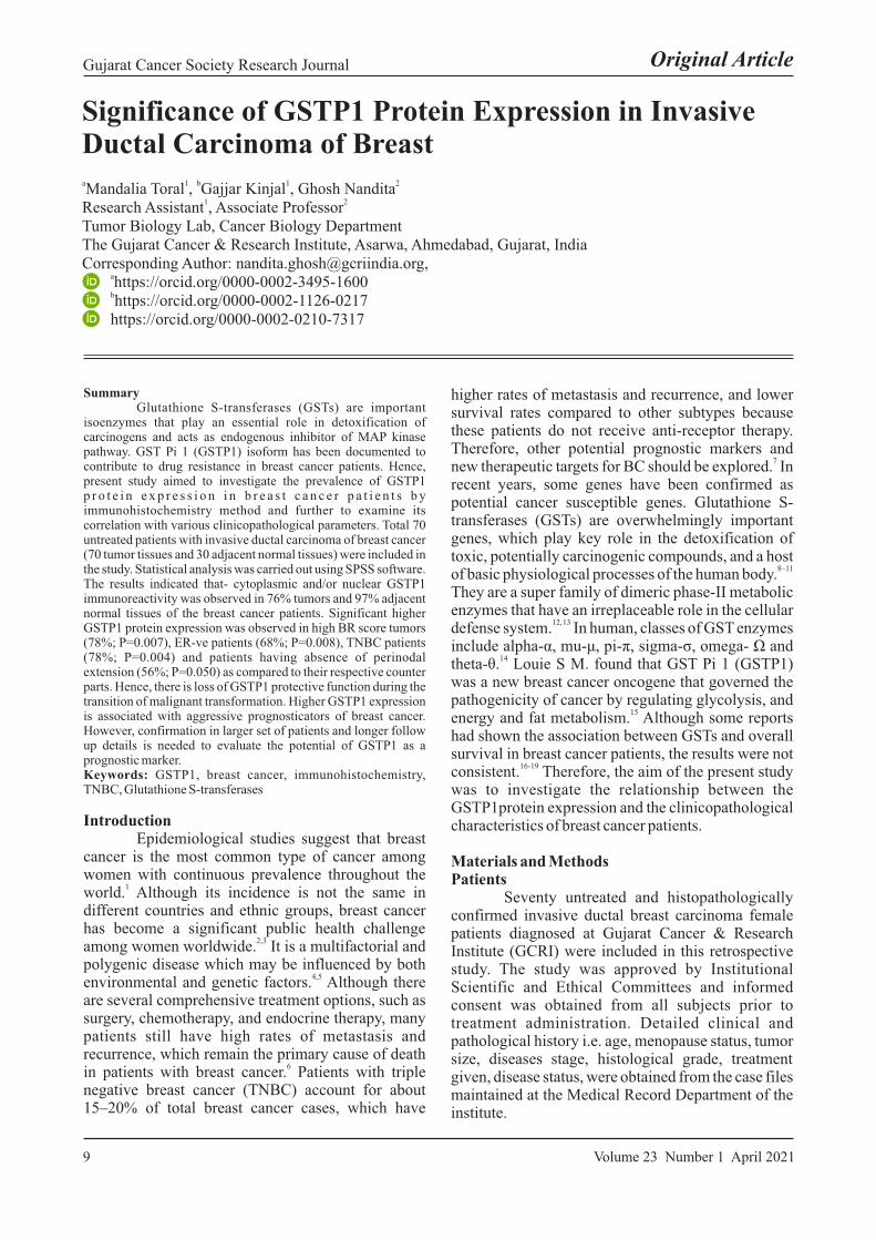
be heterogeneous and cytoplasmic and/or nuclear. GSTP1 immunoreactivity was detected in 76% (53/70) patients, while only 24% (17/70) of patient were negative for GSTP1 expression. The staining intensity was observed to be 28% (19/70) of +1, 24% (17/70) of +2 and 24% (17/70) of +3. The median H-score for GSTP1 immunoreactivity was 40 (Range 0 to 300) and this was used as a cut-off value to subgroup the patients into low (<40) and high (≥40) expression groups. Accordingly, 51% (36/70) patients displayed low (<40) and 49% (34/70) displayed high (>40) GSTP1 protein expression. (Table 2) In adjacent normal tissues the staining pattern of GSTP1 expression was intensely nuclear or/and cytoplasmic distributed throughout the epithelium. No membranous staining of GSTP1 was seen. Further, positive GSTP1 immunoreactivity in adjacent normal tissue was observed in 97% (29/30), with staining intensity of +1 in 27% (8/30), +2 in 30% (9/30) and +3 in 40% (12/30) in breast cancer patients (Table 2). The median H-score for immunoreactivity in adjacent normal adjacent tissue was 100 (Range 40 to 300).
Gujarat Cancer Society Research Journal
11 Volume 23 Number 1 April 2021
Molecular Subtype
LuminalA 31 44
Luminal B 11 16
Her2 amplification 10 14
TNBC 18 26
Lymphatic Permeation
Positive 30 43
Negative 40 57
Vascular Permeation
Positive 11 16
Negative 59 84
Perineural Invasion
Positive 7 10
Negative 63 90
Perinodal Extension
Positive 20 29
Negative 50 71
Necrosis Positive 16 23
Negative 54 77
Elastosis Positive 4 6
Negative 66 94
Treatment Surgery 4 6
S+CT 16 22
S+RT 2 3
S+HT 4 6
S+CT+HT 10 14
S+RT+HT 2 3
S+CT+RT 9 13
S+CT+RT+HT 23 33
Recurrence Presence 1 1
Absence 69 99
Survival Died 1 1
Alive 69 99
Figure 1: Representative photomicrographs of GSTP1 staining in primary tumors and adjacent normal tissue of breast cancer
GSTP1 staining inprimary tumor tissues
GSTP1 staining inadjacent normal tissues
This was used as a cut-off value to stratify the patients into low (<100) and high (≥100) expression group. Accordingly, 53% (16/30) patients displayed low (<100) and 47% (14/30) displayed high (>100) GSTP1 protein expression. (Table 2). Figure 1 shows the representative photomicrographs of GSTP1 immunoreactivity in primary tumor tissue and adjacent normal tissues.
Correlation of GSTP1 protein expression in tumor and adjacent normal tissues with clinical factors: A trend of decreased GSTP1 expression in both, the tumor (χ2=2.890, r=-0.203, P=0.091) and adjacent normal tissues (χ2=3.210, r=-0.320, P=0.070) was observed with increase in age of breast cancer patients. Similarly, a trend towards low GSTP1 protein expression was observed in primary tumor (χ2 =3.170, r=-0.210, P=0.070) and adjacent normal tissues (χ2=3.51, r=-0.342, P=0.064) in patients with post menopausal as compared to pre menopausal breast cancer patients. On the other hand, no significant difference was observed in the GSTP1 expression between the left and right sided breast tumor or adjacent normal tissues. (Table 3)
Primary T umor N = 70
Adjacent Normal T issue N = 30
GSTP1 Protein GSTP1 Protein
Low-expression
N (%)
High-expression
N (%)
Low-expression
N (%)
High-expression
N (%)
Age (years)
≤50 16(42) 22(58) 4(33) 8(67)
>50 20(62) 12 (38) 12(67) 6(33)
χ2=2.890, r=-0.203, P=0.091
χ2=3.210, r=-0.320, P=0.070
Menopausal status
Pre 6(33) 12(67) 2(25) 6(75)
Post 30(58) 22(42) 14(64) 8(36)
χ2=3.170, r=-0.210, P=0.070
χ2=3.510, r=-0.342, P=0.064
Site
Left 17(50) 17(50) 9(56) 7(44)
Right 19(51) 17(49) 7(50) 7(50)
χ2=0.972, r=-0.053, P=0.734
χ2=0.110, r=+0.063, P=0.743
Table 3: Correlation of GSTP1 protein expression in tumor and adjacent normal tissues with clinical factors of patients with breast cancer
Table 4: Correlation of GSTP1 protein expression in
tumor and adjacent normal tissues with pathological
characteristics of patients with breast cancer
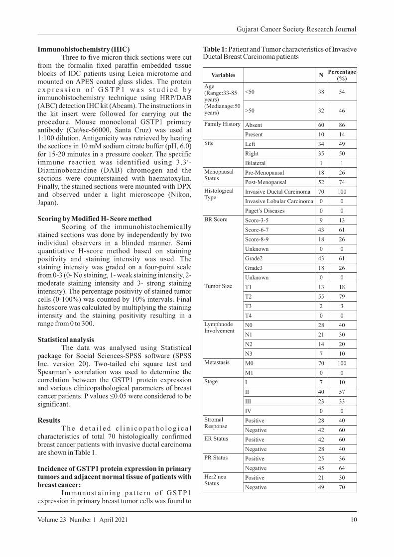
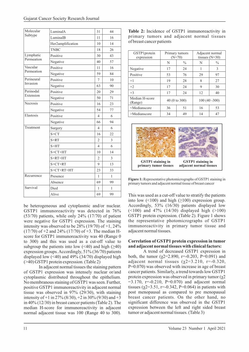
Correlation of GSTP1 protein expression in tumor and adjacent normal tissues with pathological characteristics When correlated with the pathological parameters, in primary tumors GSTP1 expression showed a significant positive correlation with increasing BR score. Furthermore, it was observed that GSTP1 expression was significantly higher in patients with high BR score (78%) as compared to low BR score (33%; χ2=5.082, r=+0.434, P=0.024) and intermediate BR score (40%; χ2=7.425, r=+0.349, P=0.006). (Table 4; Figure 2). Moreover, its expression significantly decreased in patients with perinodal extension (χ2=3.866, r=-0.235, P=0.050) indicating an inverse correlation of GSTP1 with perinodal extension of tumor. (Table 4; Figure 3). Apart from this, GSTP1 expression did not show any significant correlation with any of the pathological parameters in primary tumors or the adjacent normal tissues.
P=0.007
P=0.006
BR score
Figure 2: Correlation of GSTP1 expression in primary tumor with BR score
Figure 3: Correlation of GSTP1 expression in primary tumor with perinodal extension
Primary TumorN = 70
Adjacent Normal TissueN = 30
GSTP1 Protein GSTP1 Protein
Low-expression
N (%)
High-expression
N (%)
Low-expression
N (%)
High-expression
N (%)
Tumor Size
T1 6(46) 7(54) 3(60) 2(40)
T2 30(56) 24(44) 12(52) 11(48)
T3 0(0) 3(100) 1(50) 1(50)
χ2=3.68, r=+0.054, P=0.738
χ2=0.111, r=+0.057, P=0.763
Nodal Status
N0 14(50) 14(50) 6(37) 10(63)
N1 8(40) 12(60) 4(68) 2(32)
N2 10(67) 5(33) 4(80) 1(20)
N3 4(57) 3(43) 2(67) 1(33)
χ2=2.55, r=-0.090, P=0.450
χ2=3.683, r=-0.300, P=0.760
Stage
I 4(57) 3(43) 2(50) 2(50)
II 18(45) 22(55) 8(44) 10(56)
III 14(61) 9(39) 6(75) 2(25)
χ2=1.574, r=-0.095, P=0.436
χ2=2.098, r=-0.212, P=0.261
Early 22(47) 25(53) 10(45) 12(55)
Advanced 14(61) 9(39) 6(75) 2(25)
χ2=1.22, r=-0.130, P=0.276
χ2=2.058, r=-0.262, P=0.162
Gujarat Cancer Society Research Journal
12Volume 23 Number 1 April 2021
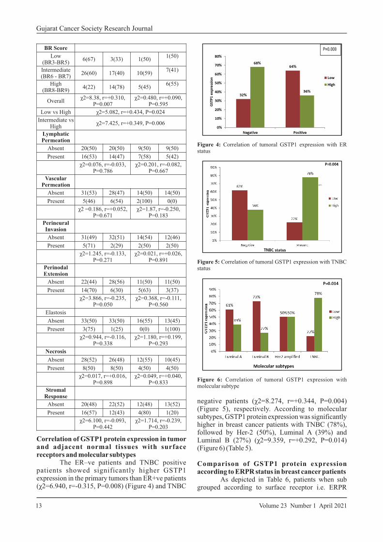
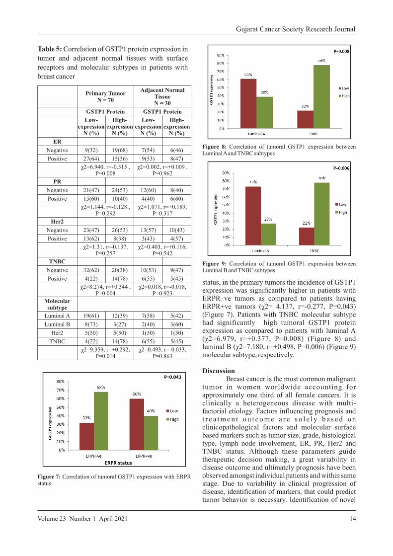
Correlation of GSTP1 protein expression in tumor and adjacent normal tissues with surface receptors and molecular subtypes The ER–ve patients and TNBC positive patients showed significantly higher GSTP1 expression in the primary tumors than ER+ve patients (χ2=6.940, r=-0.315, P=0.008) (Figure 4) and TNBC
BR Score
Low(BR3-BR5)
6(67) 3(33) 1(50)1(50)
Intermediate (BR6 - BR7)
26(60) 17(40) 10(59)7(41)
High(BR8-BR9)
4(22) 14(78) 5(45)6(55)
Overallχ2=8.38, r=+0.310,
P=0.007χ2=0.480, r=+0.090,
P=0.595
Low vs High χ2=5.082, r=+0.434, P=0.024
Intermediate vs High
χ2=7.425, r=+0.349, P=0.006
Lymphatic Permeation
Absent 20(50) 20(50) 9(50) 9(50)
Present 16(53) 14(47) 7(58) 5(42)
χ2=0.076, r=-0.033, P=0.786
χ2=0.201, r=-0.082, P=0.667
Vascular Permeation
Absent 31(53) 28(47) 14(50) 14(50)
Present 5(46) 6(54) 2(100) 0(0)
χ2 =0.186, r=+0.052, P=0.671
χ2=1.87, r=-0.250, P=0.183
Perineural Invasion
Absent 31(49) 32(51) 14(54) 12(46)
Present 5(71) 2(29) 2(50) 2(50)
χ2=1.245, r=-0.133, P=0.271
χ2=0.021, r=+0.026, P=0.891
Perinodal Extension
Absent 22(44) 28(56) 11(50) 11(50)
Present 14(70) 6(30) 5(63) 3(37)
χ2=3.866, r=-0.235, P=0.050
χ2=0.368, r=-0.111, P=0.560
Elastosis
Absent 33(50) 33(50) 16(55) 13(45)
Present 3(75) 1(25) 0(0) 1(100)
χ2=0.944, r=-0.116, P=0.338
χ2=1.180, r=+0.199, P=0.293
Necrosis
Absent 28(52) 26(48) 12(55) 10(45)
Present 8(50) 8(50) 4(50) 4(50)
χ2=0.017, r=+0.016, P=0.898
χ2=0.049, r=+0.040, P=0.833
Stromal Response
Absent 20(48) 22(52) 12(48) 13(52)
Present 16(57) 12(43) 4(80) 1(20)
χ2=6.100, r=-0.093, P=0.442
χ2=1.714, r=-0.239, P=0.203
Gujarat Cancer Society Research Journal
13 Volume 23 Number 1 April 2021
Figure 4: Correlation of tumoral GSTP1 expression with ER status
Figure 5: Correlation of tumoral GSTP1 expression with TNBC status
Figure 6: Correlation of tumoral GSTP1 expression with molecular subtype
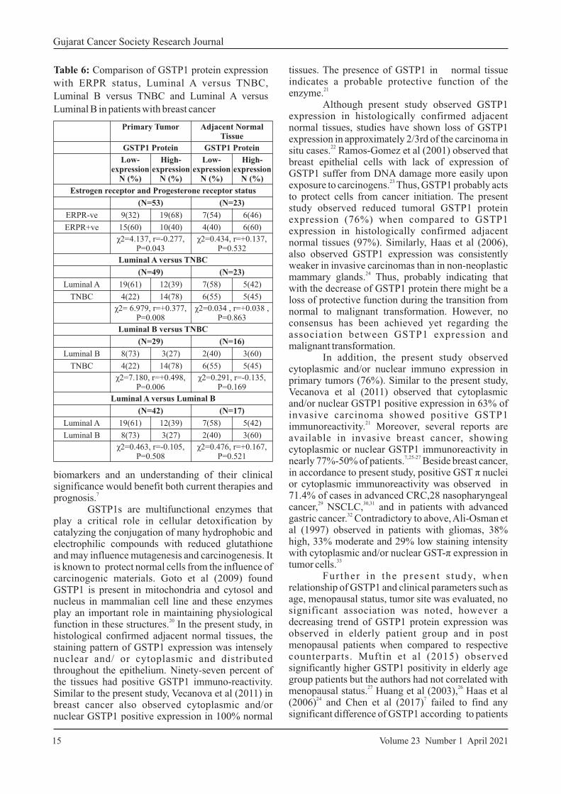
negative patients (χ2=8.274, r=+0.344, P=0.004) (Figure 5), respectively. According to molecular subtypes, GSTP1 protein expression was significantly higher in breast cancer patients with TNBC (78%), followed by Her-2 (50%), Luminal A (39%) and Luminal B (27%) (χ2=9.359, r=+0.292, P=0.014) (Figure 6) (Table 5).
Comparison of GSTP1 protein expression according to ERPR status in breast cancer patients As depicted in Table 6, patients when sub grouped according to surface receptor i.e. ERPR
Primary TumorN = 70
Adjacent Normal TissueN = 30
GSTP1 Protein GSTP1 Protein
Low-expression
N (%)
High-expression
N (%)
Low-expression
N (%)
High-expression
N (%)
ER
Negative 9(32) 19(68) 7(54) 6(46)
Positive 27(64) 15(36) 9(53) 8(47)
χ2=6.940, r=-0.315 , P=0.008
χ2=0.002, r=+0.009 , P=0.962
PR
Negative 21(47) 24(53) 12(60) 8(40)
Positive 15(60) 10(40) 4(40) 6(60)
χ2=1.144, r=-0.128 , P=0.292
χ2=1.071, r=+0.189,P=0.317
Her2
Negative 23(47) 26(53) 13(57) 10(43)
Positive 13(62) 8(38) 3(43) 4(57)
χ2=1.31, r=-0.137, P=0.257
χ2=0.403, r=+0.116,P=0.542
TNBC
Negative 32(62) 20(38) 10(53) 9(47)
Positive 4(22) 14(78) 6(55) 5(45)
χ2=8.274, r=+0.344 , P=0.004
χ2=0.018, r=-0.018,P=0.923
Molecular subtype
Luminal A 19(61) 12(39) 7(58) 5(42)
Luminal B 8(73) 3(27) 2(40) 3(60)
Her2 5(50) 5(50) 1(50) 1(50)
TNBC 4(22) 14(78) 6(55) 5(45)
χ2=9.359, r=+0.292, P=0.014
χ2=0.493, r=-0.033,P=0.863
Table 5: Correlation of GSTP1 protein expression in
tumor and adjacent normal tissues with surface
receptors and molecular subtypes in patients with
breast cancer
Figure 7: Correlation of tumoral GSTP1 expression with ERPR status
Figure 8: Correlation of tumoral GSTP1 expression between Luminal A and TNBC subtypes
Figure 9: Correlation of tumoral GSTP1 expression between Luminal B and TNBC subtypes
status, in the primary tumors the incidence of GSTP1 expression was significantly higher in patients with ERPR–ve tumors as compared to patients having ERPR+ve tumors (χ2= 4.137, r=-0.277, P=0.043) (Figure 7). Patients with TNBC molecular subtype had significantly high tumoral GSTP1 protein expression as compared to patients with luminal A (χ2=6.979, r=+0.377, P=0.008) (Figure 8) and luminal B (χ2=7.180, r=+0.498, P=0.006) (Figure 9) molecular subtype, respectively.
Discussion Breast cancer is the most common malignant tumor in women worldwide accounting for approximately one third of all female cancers. It is clinically a heterogeneous disease with multi-factorial etiology. Factors influencing prognosis and t r e a t m e n t o u t c o m e a r e s o l e l y b a s e d o n clinicopathological factors and molecular surface based markers such as tumor size, grade, histological type, lymph node involvement, ER, PR, Her2 and TNBC status. Although these parameters guide therapeutic decision making, a great variability in disease outcome and ultimately prognosis have been observed amongst individual patients and within same stage. Due to variability in clinical progression of disease, identification of markers, that could predict tumor behavior is necessary. Identification of novel
Gujarat Cancer Society Research Journal
14Volume 23 Number 1 April 2021
Gujarat Cancer Society Research Journal
15 Volume 23 Number 1 April 2021
Primary Tumor Adjacent Normal Tissue
GSTP1 Protein GSTP1 Protein
Low-expression
N (%)
High-expression
N (%)
Low-expression
N (%)
High-expression
N (%)
Estrogen receptor and Progesterone receptor status
(N=53) (N=23)
ERPR-ve 9(32) 19(68) 7(54) 6(46)
ERPR+ve 15(60) 10(40) 4(40) 6(60)
χ2=4.137, r=-0.277, P=0.043
χ2=0.434, r=+0.137, P=0.532
Luminal A versus TNBC
(N=49) (N=23)
Luminal A 19(61) 12(39) 7(58) 5(42)
TNBC 4(22) 14(78) 6(55) 5(45)
χ2= 6.979, r=+0.377, P=0.008
χ2=0.034 , r=+0.038 , P=0.863
Luminal B versus TNBC
(N=29) (N=16)
Luminal B 8(73) 3(27) 2(40) 3(60)
TNBC 4(22) 14(78) 6(55) 5(45)
χ2=7.180, r=+0.498, P=0.006
χ2=0.291, r=-0.135, P=0.169
Luminal A versus Luminal B
(N=42) (N=17)
Luminal A 19(61) 12(39) 7(58) 5(42)
Luminal B 8(73) 3(27) 2(40) 3(60)
χ2=0.463, r=-0.105, P=0.508
χ2=0.476, r=+0.167, P=0.521
Table 6: Comparison of GSTP1 protein expression
with ERPR status, Luminal A versus TNBC,
Luminal B versus TNBC and Luminal A versus
Luminal B in patients with breast cancer
biomarkers and an understanding of their clinical significance would benefit both current therapies and
7prognosis. GSTP1s are multifunctional enzymes that play a critical role in cellular detoxification by catalyzing the conjugation of many hydrophobic and electrophilic compounds with reduced glutathione and may influence mutagenesis and carcinogenesis. It is known to protect normal cells from the influence of carcinogenic materials. Goto et al (2009) found GSTP1 is present in mitochondria and cytosol and nucleus in mammalian cell line and these enzymes play an important role in maintaining physiological
20function in these structures. In the present study, in histological confirmed adjacent normal tissues, the staining pattern of GSTP1 expression was intensely nuclear and/ or cytoplasmic and distributed throughout the epithelium. Ninety-seven percent of the tissues had positive GSTP1 immuno-reactivity. Similar to the present study, Vecanova et al (2011) in breast cancer also observed cytoplasmic and/or nuclear GSTP1 positive expression in 100% normal
tissues. The presence of GSTP1 in normal tissue indicates a probable protective function of the
21enzyme. Although present study observed GSTP1 expression in histologically confirmed adjacent normal tissues, studies have shown loss of GSTP1 expression in approximately 2/3rd of the carcinoma in
22situ cases. Ramos-Gomez et al (2001) observed that breast epithelial cells with lack of expression of GSTP1 suffer from DNA damage more easily upon
23exposure to carcinogens. Thus, GSTP1 probably acts to protect cells from cancer initiation. The present study observed reduced tumoral GSTP1 protein expression (76%) when compared to GSTP1 expression in histologically confirmed adjacent normal tissues (97%). Similarly, Haas et al (2006), also observed GSTP1 expression was consistently weaker in invasive carcinomas than in non-neoplastic
24mammary glands. Thus, probably indicating that with the decrease of GSTP1 protein there might be a loss of protective function during the transition from normal to malignant transformation. However, no consensus has been achieved yet regarding the association between GSTP1 expression and malignant transformation. In addition, the present study observed cytoplasmic and/or nuclear immuno expression in primary tumors (76%). Similar to the present study, Vecanova et al (2011) observed that cytoplasmic and/or nuclear GSTP1 positive expression in 63% of invasive carcinoma showed positive GSTP1
21immunoreactivity. Moreover, several reports are available in invasive breast cancer, showing cytoplasmic or nuclear GSTP1 immunoreactivity in
7, 25-27nearly 77%-50% of patients. Beside breast cancer, in accordance to present study, positive GST π nuclei or cytoplasmic immunoreactivity was observed in 71.4% of cases in advanced CRC,28 nasopharyngeal
29 30,31cancer, NSCLC, and in patients with advanced 32
gastric cancer. Contradictory to above, Ali-Osman et al (1997) observed in patients with gliomas, 38% high, 33% moderate and 29% low staining intensity with cytoplasmic and/or nuclear GST-π expression in
33tumor cells. Fur ther in the present s tudy, when relationship of GSTP1 and clinical parameters such as age, menopausal status, tumor site was evaluated, no significant association was noted, however a decreasing trend of GSTP1 protein expression was observed in elderly patient group and in post menopausal patients when compared to respective counterparts. Muftin et al (2015) observed significantly higher GSTP1 positivity in elderly age group patients but the authors had not correlated with
27 26menopausal status. Huang et al (2003), Haas et al
24 7(2006) and Chen et al (2017) failed to find any significant difference of GSTP1 according to patients
to steroids and hormones, allowing it to act as an intracellular buffer to minimize short-term changes in steroid levels. The breast being an important organ of the body which is continuously exposed to these steroids and it is therefore estrogens act as endogenous tumor initiators in the breast tissue when GSTP1 is inactivated by promoter methylation. Therefore, expression of GSTP1 protein and surface receptor was evaluated, higher GSTP1 protein expression was observed in tumors with ER-ve patients (68%), PR-ve (53%) and TNBC patients (78%) as compared to their respective counter parts. Similar high GSTP1 protein expression was noted in patients with ERPR–ve tumors. Consistent with
34present study, Miyake et al (2012), Peters et al 39 40
(1993) and Gilbert L et al (1993) found that GSTP1 expression was significantly associated with ER negativity and PR negativity in patients with breast
23cancer. On the other hand, Huang et al (2003), and 24Haas et al (2006) failed to observe any significant
correlation between GSTP1 and ER, PR status. Additionally, when sub grouped according to molecular subtypes, GSTP1 protein expression was significantly higher in breast cancer patients with TNBC (78%), followed by Her-2 (50%), luminal A (39%) and luminal B (27%) (χ2 =9.359, r = 0.292, P=0.014). A recent study by Pakdeethai et al (2012), speculated a significant correlation of estrogen receptor negativity with high GSTP1 expression (p
250.001). The other parameters - tumor size, tumor grade, lymph node status, HER2- IHC score, Ki67 index did not correlate with high or low GSTP1 protein expression. It is evident that TNBC subtypes are considered more aggressive than the luminal A or B subtypes, or even those overexpressing HER-2/neu. Louie et al (2016) found that GSTP1 was a new TNBC oncogene that governed the pathogenicity of cancer by regulating glycolysis, and energy and fat
15metabolism. They believed that GSTP1, a new TNBC target, was a risk factor for breast cancer and promoted breast cancer. Chen et al (2017), found approximately 77% positive rate of GSTP1 protein
7expression in TNBC patients. Interestingly, the current study demonstrated significant high expression of tumoral GSTP1 protein expression in TNBC as compared to the other molecular subtypes (luminal A, luminal B and Her-2), indicating a useful target for TNBC patients.
Conclusion Our prel iminary data shows higher c y t o p l a s m i c a n d / o r n u c l e a r s t a i n i n g immunopositivity pattern of GSTP1 was observed in adjacent normal tissues as compared to tumor tissues, which was indicative of loss of GSTP1 protective function during the transition of malignant transformation. Observation of higher GSTP1 with
34 7age. Miyake et al (2012) and Chen et al (2017) could not find any significant difference of GSTP1 protein expression and menopausal status. To best of our knowledge, there exist very rare reports on association of GSTP1 protein expression and age, menopausal status, site in patients with invasive breast cancer. When relationship between GSTP1 and pathological variables were evaluated, it was observed that high tumoral GSTP1 protein expression was associated with breast cancer patients having N0 and N1 nodal status, T1 and T2 tumor size and in early disease stage when compared to their respective counterparts. Although, the difference was found to be statistically non significant but it confers a probable role of GSTP1 as an early event in breast carcinogenesis. Likewise, Buser et al (1997) showed that lower GSTs levels are associated with more
35advanced breast cancer. Haas et al (2006) linked
24smaller tumor sizes with high GSTP1 expression. Recently, Chen et al (2017) reported significantly higher GSTP in smaller tumors (P=0.023), early clinical stage of the tumor, but no significant association with the remaining clinicopathological characteristics, axillary lymph node status (P=0.071), pathological type (P=0.607), histological grade
7(P=0.750). Contrary to the present study, Muftin et al (2015) found high GSTP1 expression was significantly associated with stage III and large tumor
27size (>2cm), (p< 0.05). On the other hand, higher GSTP1 protein expression was significantly associated with aggressive prognostic factor such as high BR (8-9) score and presence of perinodal invasion. In accordance to the present results, Jardim
36 37et al (2012) and Li et al (2014), associated the highest GSTP1 expression with high histological levels of invasive ductal carcinomas. Nevertheless, other authors have demonstrated contrary results. Cairns et al (1992) associated an absence of GSTP1 in
38tumor tissue with the highest histological grade. According to Miyake et al (2012), GSTP1 positivity significantly varied according to histological grade (HG) that is, HG2 tumors showed a lower positivity (32/81, 39.5%) than HG1 tumors (9/19, 47.4%) and
34HG3 tumors (16/22, 72.7%). Muftin et al (2015) found high GSTP1 expression was significantly
27associated with grade III histology, whereas Haas et al (2006) linked GSTP1 with well differentiated
24tumors. Additionally, Huang et al observed GST-pi immunoreactivity was not significantly correlated with any of the traditional histological factors known
23to influence prognosis. The plausible reason for this difference between our results and those conflicting results may be due to the diversity of GSTP1 assessment methods and the difference in sample size. Since, GSTs isoenzyme facilitate clearance of endogenous hydrophobic compounds such as hormones, steroids, etc. GSTP1 binds non-covalently
Gujarat Cancer Society Research Journal
16Volume 23 Number 1 April 2021
Gujarat Cancer Society Research Journal
17 Volume 23 Number 1 April 2021
traditionally aggressive prognostic factors such as High BR score, presence of perinodal extension, ER PR negativity & TNBC, probably indicates that GSTP1 might be useful to identify patients with aggressive phenotype. In TNBC patients it may be a useful target. However, it needs to be confirmed by covering a larger number of patients.
References1. Farmohammadi A, Arab-Yarmohammadi V,
Ramzanpour R: Association analysis of rs1695 and rs1138272 variations in GSTP1 gene and breast cancer susceptibility. Asian Pac J Cancer Prev 2020;21:1167-1172
2. Miller JW, King JB, Joseph DA, Richardson LC: Centers for Disease Control and Prevention (CDC). Breast cancer screening among adult women-behavioral risk factor surveillance system, United States, 2010. MMWR Morb Mortal Wkly Rep 2012;61:46-50
3. Siegel R, DeSantis C, Virgo K et al: Cancer treatment and survivorship statistics, 2012. CA: A Cancer Journal for Clinicians 2012;62:220-241
4. Flores-Ramos LG, Escoto-De Dios A, Puebla-Pérez AM et al: Association of the tumor necrosis factor-alpha-308G> A polymorphism with breast cancer in Mexican women. Genet Mol Res 2013;12:5680-5693
5. Gallegos-Arreola MP, Figuera-Villanueva LE, Ramos-Silva A et al: The association between the 844ins68 polymorphism in the CBS gene and breast cancer. Archives of Medical Science: AMS 2014;10:1214-1224
6. Kinsella MD, Nassar A, Siddiqui MT, Cohen C: Estrogen receptor (ER), progesterone receptor (PR), and HER2 expression pre - and post- neoadjuvant chemotherapy in primary breast carcinoma: a single institutional experience. In t e rna t iona l Jou rna l o f C l in i ca l and Experimental Pathology 2012;5:530-536
7. Chen G, Zhang H, Sun L et al: Prognostic significance of GSTP1 in patients with triple negative breast cancer. Oncotarget 2017;8:68675-68680
8. Hayes JD, Pulford DJ: The glut athione S-transferase supergene family: regulation of GST and the contribution of the lsoenzymes to cancer chemoprotection and drug resistance part I. Critical Reviews in Biochemistry and Molecular Biology 1995;30:445-520
9. Hayes JD, Flanagan JU, Jowsey IR: Glutathione transferases. Annu. Rev. Pharmacol. Toxicol 2005;45:51-88
10. Atkinson HJ, Babbi t t PC: Gluta thione transferases are structural and functional outliers in the th ioredoxin fo ld . Biochemis t ry 2009;48:11108-11116
11. U d o m s i n p r a s e r t R , P o n g j a r o e n k i t S , Wongsant ichon J e t a l : Ident i f ica t ion, characterization and structure of a new Delta class glutathione transferase isoenzyme. Biochemical Journal 2005;388:763-771
12. Strange RC, Fryer AA: The glutathione S-transferases: influence of polymorphism on cancer suscept ib i l i ty. IARC sc ient i f ic publications 1999:231-249
13. Vijayakumar H, Thamilarasan SK, Shanmugam A et al: Glutathione transferases superfamily: cold-inducible expression of distinct GST genes in Brassica oleracea. International Journal of Molecular Sciences 2016;17:1211
14. Board PG, Baker RT, Chelvanayagam G, Jermiin LS: Zeta, a novel class of glutathione transferases in a range of species from plants to humans. Biochemical Journal 1997; 328:929-935
15. Louie SM, Grossman EA, Crawford LA et al: GSTP1 Is a Driver of Triple-Negative Breast Cancer Cell Metabolism and Pathogenicity. Cell Chemical Biology 2016;23:567-578
16. Bai YL, Zhou B, Jing XY et al: Predictive role of GSTs on the prognosis of breast cancer patients with neoadjuvant chemotherapy. APJCP 2012;13:5019-5022
17. Franco RL, Schenka NG, Schenka AA, Rezende LF, Gurgel MS: Glutathione S-transferase Pi expression in invasive breast cancer and its relation with the clinical outcome. Journal of BUON : Official Journal of the Balkan Union of Oncology 2012;17:259-264
18. Duggan C, Ballard-Barbash R, Baumgartner RN et al: Associations between null mutations in GSTT1 and GSTM1, the GSTP1 Ile 105 Val polymorphism, and mortality in breast cancer survivors. Springerplus 2013;2:1-9
19. Oliveira AL, Oliveira Rodrigues FF, Dos Santos RE, Rozenowicz RL, Barbosa de Melo M: GSTT1, GSTM1, and GSTP1 polymorphisms as a prognostic factor in women with breast cancer. GMR 2014;13:2521-2530
20. Goto S, Kawakatsu M, Izumi SI et al: Glutathione S-transferase π localizes in mitochondria and protects against oxidative stress. Free Radical Biology and Medicine 2009;46:1392-1403
21. Vecanova J, Hodorova I, Mihalik J et al: Immunohistochemical evaluation of Pi class glutathione S-transferase expression in invasive breast carcinoma. Bratislavske Lekarske Listy 2011;112:67-70
22. Bellamy CO, Harrison DJ: Evaluation of glutathione S-transferase Pi in non-invasive ductal carcinoma of breast. British Journal of Cancer 1994;69:183-185
23. Ramos-Gomez M, Kwak MK, Dolan PM et al: Sensitivity to carcinogenesis is increased and
32. Kwon HC, Roh MS, Oh SY et al: Prognostic value of expression of ERCC1, thymidylate synthase, and glutathione S-transferase P1 for 5-fluorouracil/oxaliplatin chemotherapy in advanced gastric cancer. Annals of Oncology 2007;18:504-509
33. Ali-Osman F, Brunner JM, Kutluk TM, Hess K: Prognostic significance of glutathione S-transferase pi expression and subcellular localization in human gliomas. Clinical Cancer Research 1997;3:2253-2261
34. Miyake T, Nakayama T, Naoi Y et al: GSTP 1 expression predicts poor pathological complete response to neoadjuvant chemotherapy in ER-negative breast cancer. Cancer Science 2012;103:913-920
35. Buser K, Joncourt F, Altermatt HJ et al: Breast cancer: pretreatment drug resistance parameters (GSH-system, ATase, P-glycoprotein) in tumor tissue and their correlation with clinical and prognostic characteristics. Annals of Oncology 1997;8:335-341
36. Jardim BV, Moschetta MG, Gelaleti GB et al: Glutathione transferase pi (GSTpi) expression in breast cancer: an immunohistochemical and m o l e c u l a r s t u d y. A c t a H i s t o c h e m i c a 2012;114:510-517
37. Li W, Song M: Expression of multidrug resistance proteins in invasive ductal carcinoma of the breast. Oncology Letters 2014;8:2103-2109
38. Ca i rns J , Wr igh t C , Ca t t an AR e t a l : Immunohistochemical demonstration of glutathione S-transferases in primary human breast carcinomas. The Journal of Pathology 1992;166:19-25
39. Peters WH, Roelofs HM, Van Putten WL et al: Response to adjuvant chemotherapy in primary breast cancer: no correlation with expression of glutathione S-transferases. British Journal of Cancer 1993;68:86-92
40. Gilbert L, Elwood LJ, Merino M et al: A pilot study of pi-class glutathione S-transferase expression in breast cancer: correlation with estrogen receptor expression and prognosis in node-negative breast cancer. Journal of Clinical Oncology 1993;11:49-58
chemoprotective efficacy of enzyme inducers is lost in nrf2 transcription factor-deficient mice. Proceedings of the National Academy of Sciences 2001;98:3410-3415
24. Haas S, Pierl C, Harth V et al: Expression of xenobiotic and steroid hormone metabolizing enzymes in human breas t carc inomas . International Journal of Cancer 2006;119:1785-1791
25. Pakdeethai S, Fongchaiya V, Pongtheerat T, Iampenkhae K, Sampatanukul P: Relationship between promoter methylation and protein expression of glutathione S - transferase gene class P1 in breast cancer. Asian Archives of Pathology 2012;8:45-53
26. Huang J, Tan PH, Thiyagarajan J, Bay BH: Prognostic significance of glutathione S-transferase-pi in invasive breast cancer. Modern Pathology 2003;16:558-565
27. Muftin NQ, AL-Rubaiꞌe SH, Yaseen NY, Aziz RS: Expression of glutathione S-transferase P1 in women with invasive ductal carcinoma. International Journal of Current Microbiology and Applied Sciences 2015;4:455-465
28. Kim M, Suh H, Cho EJ, Buratowski S: Phosphorylation of the yeast Rpb1 C-terminal domain at serines 2, 5, and 7. Journal of Biological Chemistry 2009;284:26421-26426
29. Jayasurya A, Yap WM, Tan NG, Tan BK, Bay BH: Glutathione S-transferase π expression in n a s o p h a r y n g e a l c a n c e r . A r c h i v e s o f Otolaryngology-Head & Neck Surgery 2002;128:1396-1399
30. Bai F, Nakanishi Y, Kawasaki M, Takayama K et al: Immunohistochemical expression of gluta thione S - t ransferase -π can predict chemotherapy response in patients with nonsmall cell lung carcinoma. Cancer: Interdisciplinary International Journal of the American Cancer Society 1996;78:416-421
31. Zhu WY, Hunag YY, Liu XG et al: Prognostic evaluation of CapG, gelsolin, P-gp, GSTP1, and Topo-II proteins in non-small cell lung cancer. The Anatomical Record: Advances in Integrative A n a t o m y a n d E v o l u t i o n a r y B i o l o g y 2012;295:208-214
Gujarat Cancer Society Research Journal
18Volume 23 Number 1 April 2021
There is no English word as ‘expressionology’. I have coined it to express what I feel and think as well as what I am. It means all that I do or do not do as well as the way I conduct myself and what I am or I am not. All living beings express themselves. A seed expresses its desire to grow as soon as it is in the soil and in contact with water. A plant grows and finds fulfilment in expressing as a flower. May there not be any beholder, its vivid colours and sweet smell make the surrounding pleasant, inviting and attractive for any bystander. Expression generates beauty and safety. Birds sing and call each other. The sound is a calling for mutual support. They express their happiness or fear by sounds they produce or when they hide for protection when they mimic the surroundings. All this is done beautifully. Beauty is always for some purpose. For the humans, plants and other creatures, it reflects interdependence. For plants, it may be cross-pollination. Sometimes their expression of beauty is just as a service to the Mother Nature. Why otherwise the honey-bees toil all day and collect minute drops of nectar and make and store honey? The Sun, the Moon, the Sky, the stars, the cool nights and bright and burning days, the showering or randomly floating and wandering clouds, the flowing and still water, the quiet and tranquil ocean or its roaring and rising seashore, the cool breeze and the sunshine and shadows, deserts and forests, lonely outskirts of a busy city - all these non-living things in the Nature do express themselves. What is the purpose? Just to make the world we live in heavenly? Not only the beautiful, but not so beautiful or less agreeable things and living beings in the world also express themselves. Our criteria for beauty, prettiness and loveliness has very little bearing on their expression of beauty as they see it best for them, be it a cockroach or a lizard. In Shrimad Bhagavad Geeta (2/28) it is said that
It means all life is not manifested in the beginning and in the end. They are manifested in the middle when we call them existing. What is true for the living being is also true for the non-living world. All started with a big bang or the desire of the Supreme to be manifested and become manifold. What is manifested is called �ય�ત. Hence, the person is �ય��ત
and his manifestation is �ય��ત�વ. When he/she expresses it and directs towards others, it is called અ�ભ�ય��ત.
Thus, expression is the manifestation or evidence of existence. It may be an action or a reaction. Genes express. Eyes and face express, hands and feet express, posture expresses, spoken words and silence express, movements and stillness express. Human expression is defined as making known one’s thoughts and emotions. But in a wider sense it is also an evidence of existence and activity. Human expressions take many forms - utterance, voicing, pronouncement, declaration, articulation, verbalization, statement, proclamation, assertion, announcement, setting forth, venting, mouthing, dissemination, broadcast, circulation, communication, spreading, promulgation, publicizing, publication, assertion, indication, intimation, demonstration, show, exhibition, manifestation, token, conveyance, illustration, revelation, disclosure, embodiment etc. Facial expressions, gestures, posture, and tone of voice are powerful communication tools. One of the most effective means of expression is non-verbal - body posture, gestures, eye contact and the space maintained during the process of communication. Touch, timings and place add a great deal to the force and effectiveness of the expressions and communications. It repeats and often strengthens the message, it can contradict the message, it can substitute for a verbal message and make it a far more vivid message than words ever can, it may add to or complement and it may accent or emphasize the message. Hearing and listening to the spoken and non-spoken words is important. Quiet and complete listening to understand is essential but should be sensitive enough to hear the unspoken emotions.
BrainWavesGujarat Cancer Society Research Journal
19 Volume 23 Number 1 April 2021
Expressionology (અ�ભ�ય��તિવ�ા)
Shukla Shilin Nandubhai Medical Oncologist and Haemato-Oncologist,Professor Emeritus and Former DirectorThe Gujarat Cancer & Research Institute, Asarwa, Ahmedabad, Gujarat, India.Corresponding Author: [email protected]
अ�य�ादीिन भतािन �य�म�यािन भारत।ूअ�य�िनधना�येव त� का प�रदेवना।।
Emotions are best expressed non-verbally. It is especially important when the expressions are taking place in or by a group of people. Self-control and stress-control keep the senses in order. Expression is also what is created. A substance, a situation, an innovation, a formation, a drama, a dance, a song, a picture or a movie, a poem, an essay, a story, an article, a research product or method etc. An architect builds a house is his expression of generating space that accommodates, comforts as well as tells a story. So is a dancer or an actor who enlivens a character. Expression takes many forms. Earliest narration that touches the subject is found in the treatise by Bharata Muni who wrote Natya Shastra. He coined two important words - Ras and Bhaav. Ras is the pleasure one derives when something is expressed. Bhaav is the process of expression. There is a Gujarati word Haav (HaavBhaav). Haav are the gestures. He has described various types of Bhaav - stable or constant Bhav is
Gujarat Cancer Society Research Journal
20Volume 23 Number 1 April 2021
called Sthaayee Bhaav; props or surroundings (stage, situation etc.) constitute the ViBhaav; Mental, Physical or Spirit-related Bhaav are called Anu Bhaav and those that undulate emotions in the mind and heart of the audience is called the Sancharee Bhaav or Vyabhichaaree Bhaav. There are 9 types of Ras (pleasures) that one derives from these expressions - decoration (attraction), unhappiness, laughter (happiness), valour, fear, anger, astonishment, peace and tranquility as well as extremely unpleasant. Devotion, Motherly Love and Pure Love are three more recently added Ras. Bharata Muni has described, in detail, various gestures that express Bhaav. He has called them Bhed. Gestures of head, eyebrows, eyes, nose, mouth and lips, face, neck, shoulder, waist, legs etc. are described in great details. Thus expressions (Bhaav) give meaning to and pleasure (Ras) of existence. Let us all master the skill of expression and derive and distribute pleasure.
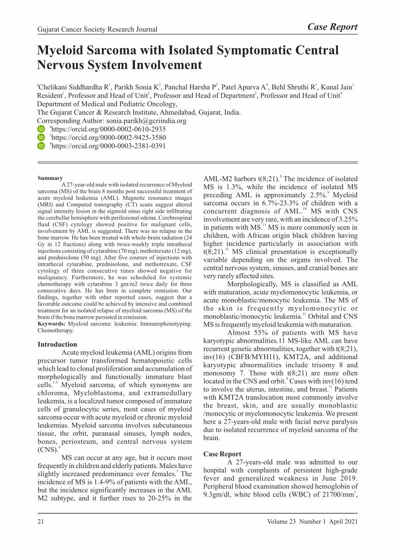
Summary A 27-year-old male with isolated recurrence of Myeloid sarcoma (MS) of the brain 8 months post successful treatment of acute myeloid leukemia (AML). Magnetic resonance images (MRI) and Computed tomography (CT) scans suggest altered signal intensity lesion in the sigmoid sinus right side infiltrating the cerebellar hemisphere with perilesional edema. Cerebrospinal fluid (CSF) cytology showed positive for malignant cells, involvement by AML is suggested. There was no relapse in the bone marrow. He has been treated with whole-brain radiation (24 Gy in 12 fractions) along with twice-weekly triple intrathecal injections consisting of cytarabine (70 mg), methotrexate (12 mg), and prednisolone (50 mg). After five courses of injections with intrathecal cytarabine, prednisolone, and methotrexate, CSF cytology of three consecutive times showed negative for malignancy. Furthermore, he was scheduled for systemic chemotherapy with cytarabine 3 gm/m2 twice daily for three consecutive days. He has been in complete remission. Our findings, together with other reported cases, suggest that a favorable outcome could be achieved by intensive and combined treatment for an isolated relapse of myeloid sarcoma (MS) of the brain if the bone marrow persisted in remission.Keywords: Myeloid sarcoma: leukemia: Immunophenotyping: Chemotherapy.
Introduction Acute myeloid leukemia (AML) origins from precursor tumor transformed hematopoietic cells which lead to clonal proliferation and accumulation of morphologically and functionally immature blast
1-5cells. Myeloid sarcoma, of which synonyms are chloroma, Myeloblastoma, and extramedullary leukemia, is a localized tumor composed of immature cells of granulocytic series, most cases of myeloid sarcoma occur with acute myeloid or chronic myeloid leukemias. Myeloid sarcoma involves subcutaneous tissue, the orbit, paranasal sinuses, lymph nodes, bones, periosteum, and central nervous system
6(CNS). MS can occur at any age, but it occurs most frequently in children and elderly patients. Males have
7slightly increased predominance over females. The incidence of MS is 1.4-9% of patients with the AML, but the incidence significantly increases in the AML M2 subtype, and it further rises to 20-25% in the
8AML-M2 harbors t(8;21). The incidence of isolated MS is 1.3%, while the incidence of isolated MS
9preceding AML is approximately 2.5%. Myeloid sarcoma occurs in 6.7%-23.3% of children with a
10concurrent diagnosis of AML. MS with CNS involvement are very rare, with an incidence of 3.25%
11in patients with MS. MS is more commonly seen in children, with African origin black children having higher incidence particularly in association with
12t(8;21). MS clinical presentation is exceptionally variable depending on the organs involved. The central nervous system, sinuses, and cranial bones are very rarely affected sites. Morphologically, MS is classified as AML with maturation, acute myelomonocytic leukemia, or acute monoblastic/monocytic leukemia. The MS of the skin is frequently myelomonocytic or
11monoblastic/monocytic leukemia. Orbital and CNS MS is frequently myeloid leukemia with maturation. Almost 55% of patients with MS have karyotypic abnormalities.11 MS-like AML can have recurrent genetic abnormalities, together with t(8;21), inv(16) (CBFB/MYH11), KMT2A, and additional karyotypic abnormalities include trisomy 8 and monosomy 7. Those with t(8;21) are more often
9located in the CNS and orbit. Cases with inv(16) tend
11to involve the uterus, intestine, and breast. Patients with KMT2A translocation most commonly involve the breast, skin, and are usually monoblastic /monocytic or myelomonocytic leukemia. We present here a 27-years-old male with facial nerve paralysis due to isolated recurrence of myeloid sarcoma of the brain.
Case Report A 27-years-old male was admitted to our hospital with complaints of persistent high-grade fever and generalized weakness in June 2019. Peripheral blood examination showed hemoglobin of
39.3gm/dl, white blood cells (WBC) of 21700/mm ,
Case ReportGujarat Cancer Society Research Journal
21 Volume 23 Number 1 April 2021
Myeloid Sarcoma with Isolated Symptomatic CentralNervous System Involvement
a 1 2 3 4 1 1Chelikani Siddhardha R , Parikh Sonia K , Panchal Harsha P , Patel Apurva A , Behl Shruthi R , Kunal Jain1 2 3 4
Resident , Professor and Head of Unit , Professor and Head of Department , Professor and Head of Unit Department of Medical and Pediatric Oncology,The Gujarat Cancer & Research Institute, Ahmedabad, Gujarat, India.Corresponding Author: [email protected]
a https://orcid.org/0000-0002-0610-2935
2 https://orcid.org/0000-0002-9425-3580
4 https://orcid.org/0000-0003-2381-0391
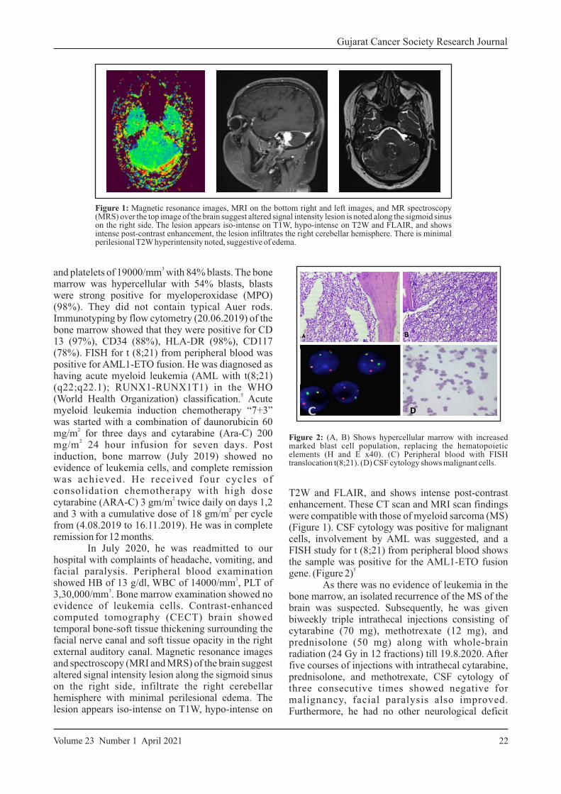
3and platelets of 19000/mm with 84% blasts. The bone marrow was hypercellular with 54% blasts, blasts were strong positive for myeloperoxidase (MPO) (98%). They did not contain typical Auer rods. Immunotyping by flow cytometry (20.06.2019) of the bone marrow showed that they were positive for CD 13 (97%), CD34 (88%), HLA-DR (98%), CD117 (78%). FISH for t (8;21) from peripheral blood was positive for AML1-ETO fusion. He was diagnosed as having acute myeloid leukemia (AML with t(8;21) (q22;q22.1); RUNX1-RUNX1T1) in the WHO
5(World Health Organization) classification. Acute myeloid leukemia induction chemotherapy “7+3” was started with a combination of daunorubicin 60
2mg/m for three days and cytarabine (Ara-C) 200 2
mg/m 24 hour infusion for seven days. Post induction, bone marrow (July 2019) showed no evidence of leukemia cells, and complete remission was achieved. He received four cycles of consolidation chemotherapy with high dose
2cytarabine (ARA-C) 3 gm/m twice daily on days 1,2
2and 3 with a cumulative dose of 18 gm/m per cycle from (4.08.2019 to 16.11.2019). He was in complete remission for 12 months. In July 2020, he was readmitted to our hospital with complaints of headache, vomiting, and facial paralysis. Peripheral blood examination
3showed HB of 13 g/dl, WBC of 14000/mm , PLT of
33,30,000/mm . Bone marrow examination showed no evidence of leukemia cells. Contrast-enhanced computed tomography (CECT) brain showed temporal bone-soft tissue thickening surrounding the facial nerve canal and soft tissue opacity in the right external auditory canal. Magnetic resonance images and spectroscopy (MRI and MRS) of the brain suggest altered signal intensity lesion along the sigmoid sinus on the right side, infiltrate the right cerebellar hemisphere with minimal perilesional edema. The lesion appears iso-intense on T1W, hypo-intense on
Gujarat Cancer Society Research Journal
22Volume 23 Number 1 April 2021
Figure 1: Magnetic resonance images, MRI on the bottom right and left images, and MR spectroscopy (MRS) over the top image of the brain suggest altered signal intensity lesion is noted along the sigmoid sinus on the right side. The lesion appears iso-intense on T1W, hypo-intense on T2W and FLAIR, and shows intense post-contrast enhancement, the lesion infiltrates the right cerebellar hemisphere. There is minimal perilesional T2W hyperintensity noted, suggestive of edema.
T2W and FLAIR, and shows intense post-contrast enhancement. These CT scan and MRI scan findings were compatible with those of myeloid sarcoma (MS) (Figure 1). CSF cytology was positive for malignant cells, involvement by AML was suggested, and a FISH study for t (8;21) from peripheral blood shows the sample was positive for the AML1-ETO fusion
5gene. (Figure 2) As there was no evidence of leukemia in the bone marrow, an isolated recurrence of the MS of the brain was suspected. Subsequently, he was given biweekly triple intrathecal injections consisting of cytarabine (70 mg), methotrexate (12 mg), and prednisolone (50 mg) along with whole-brain radiation (24 Gy in 12 fractions) till 19.8.2020. After five courses of injections with intrathecal cytarabine, prednisolone, and methotrexate, CSF cytology of three consecutive times showed negative for malignancy, facial paralysis also improved. Furthermore, he had no other neurological deficit
Figure 2: (A, B) Shows hypercellular marrow with increased marked blast cell population, replacing the hematopoietic elements (H and E x40). (C) Peripheral blood with FISH translocation t(8;21). (D) CSF cytology shows malignant cells.
except facial nerve palsy. Though there was no bone marrow relapse, systemic chemotherapy, including
2cytarabine 3 gm/m twice daily for three consecutive
2days with a cumulative dose of 18 gm/m was scheduled. Unfortunately, he was lost to follow up. Recently, he presented with respiratory distress and peripheral blood examination showed Hb of 7 gm/dl,
3 3WBC of 58000/mm , and platelets of 22000/mm with 21% blasts which suggest medullary relapse and succumbed to the disease.
Discussion The first case of AML with MS was described by Turk in 1903 and suggested the origin is the same
13for both the tumors. MS can occur in different sites such as bones, soft tissues, skin, lymph nodes, central
14nervous system, bladder, and breast. In the study by Pileri et al of 92 patients with newly diagnosed MS, 35% and 38% had a simultaneous or previous treated
11AML. The molecular and cytogenetic AML mutations might be associated with the development of MS. MS with translocation t(8;21)-positive cases commonly occur in the orbital, and CNS region in
12children, while patients with inv(16) have a high incidence of s tomach, intest ine, or breast
11involvement, specifically in adults. Our case lies in the rare location of the MS and its relationship with an AML with translocation t(8;21).
15 Byrd and Weiss reviewed 24 patients from various trials since 1973 with patients having isolated recurrences of MS following prior AML treatment. The isolated MS relapse generally develops bone marrow relapse. In these patients, the mean time interval to develop bone marrow relapse was 7 months, and the prognosis was poor. Only 3 of 24
16,17patients had MS of the brain. The mean time interval from diagnosis of AML to isolated MS relapse was 2 years. All patients were treated with irradiation, intrathecal injection, and/or operation. Systemic chemotherapy was administered in three patients
16during marrow remission. Six patients remained alive even though the follow-up periods were varied. In our study, the time interval from diagnosis of AML to isolated MS relapse was 13 months. The time interval to develop bone marrow relapse was 21 months. Gustavo et al reviewing the literature,
17identified 21 cases with intracranial MS. Fifty-four percent had intraparenchymal lesions of the brain, and 45% of the patients had lesions in the extra-axial brain compartment. MS appears even before the initial
18diagnosis of AML by years in 25% of the patients. Of the total patients, 91% showed a hyper-dense lesion on a non-contrast CT scan.
Gujarat Cancer Society Research Journal
23 Volume 23 Number 1 April 2021
Migration of leukemic cells from the bone marrow of periosteum and dura matter into the brain parenchyma can occur once there is disruption of the blood-brain barrier. Bone destructions are not commonly observed with MS. Out of 24 patients, 1 patient showed visible bone destruction of the temporal bone and simultaneous involvement of temporal lobe parenchyma.19 Seven patients were reviewed with brain MRI. MS showed either a hyper, iso-or hypo-intense signal on T2-weighted images. 4 patients showed T2 hyperintensity while 3 patients showed T2 iso or hypo-intensity.19 In our case, MS of the brain was diagnosed by MRI and CSF cytology. MRI brain of our patient showed hypo-intensity on T2-weighted images. The currently recommended treatment options for MS are the combination of chemotherapy and radiotherapy. There are no pathologic or clinical prognostic features, however, survival is better in patients who undergo allogenic bone marrow
11transplant. Tsimberidou et al assessed the outcome of 23 patients with AML was compared with MS, and they found that the event-free survival was longer in
20patients with isolated MS. Recently, Lee et al in a meta-analysis of 82 studies in which variables such as the extent of resection, treatment modality, and mortality were correlated and they found that surgical resection and extent of resection were not significantly associated with mortality. Patients who received chemotherapy or radiotherapy had lower rates of mortality versus patients who did not received chemotherapy or
21radiotherapy. In the present case study the patient received whole-brain radiation therapy with biweekly intrathecal chemotherapy. After these treatments, CSF cytology of three consecutive times showed no evidence of malignancy, and there was an improvement in facial paralysis. Even though there was no relapse in the bone marrow, prophylactic chemotherapy was planned. Unfortunately, he was lost to follow up. Recently, he presented with respiratory distress and peripheral blood examination
3showed Hb of 7 gm/dl, WBC of 58000/mm , and
3platelets of 22000/mm with 21% blasts which suggest medullary relapse and succumbed to the disease. From these findings, we advise that chemotherapy must be started after the completion of local therapy of the brain.
Conclusion Since randomized prospective studies are lacking, there is no proper consensus on the treatment of MS. The currently recommended treatment regimen in patients presenting with isolated MS of the brain is localized treatment with cranial irradiation and or operation with intrathecal chemotherapy
followed by prophylactic systemic chemotherapy. As from these findings, we advise that chemotherapy should be started after the completion of local treatment to the brain. Close follow-up of the patient is needed following AML treatment and any new onset of neurological symptoms should be thoroughly evaluated.
References1. Döhner H, Weisdorf DJ, Bloomfield CD et al:
Acute Myeloid Leukemia. N Engl J Med 2015; 373: 1136-1152
2. Weinberg OK, Sohani AR, Bhargava P et al: Diagnostic workup of acute myeloid leukemia. Am J Hematol 2017; 92: 317-321
3. Arber DA, Borowitz MJ, Cessna M et al: Initial Diagnostic Workup of Acute Leukemia. Guideline from the College of American Pathologists and the American Society of Hematology. Arch Pathol Lab Med 2017; 141: 1342-1393
4. Döhner H, Estey E, Grimwade D et al: Diagnosis and management of AML in adults. 2017 ELN recommendations from an international expert panel. Blood 2017; 129: 424-447
5. Arber DA, Orazi A, Hasserjian R et al: The 2016 revision to the World Health Organization classification of myeloid neoplasms and acute leukemia. Blood 2016; 127: 2391-2405
6. Byrd JC, Edenfield WJ, Shields OJ et al: Extramedullary myeloid cell tumors in acute nonlymphocytic leukemia. A clinical review. J Clin Oncol 1995; 13: 1800-1816
7. Kawamoto K, Miyoshi H, Yoshida N et al:Clinicopathological, cytogenetic, and prognostic analysis of 131 myeloid sarcoma patients. Am J Surg Pathol 2016; 40: 1473-1483
8. Lai Y-Y, Qiu J-Y, Jiang B et al: Characteristics and prognostic factors of acute myeloid leukemia with t(8;21) (q22; q22) Zhongguo Shi Yan Xue Ye Xue Za Zhi. 2005; 13: 733-740
9. Reinhardt D, Creutzig U: Isolated myelosarcoma in children-update and review. Leuk Lymphoma 2002; 43: 565-574
10. Stve HK, Sandahl JD, Abrahamsson J et al: Extramedullary leukemia in children with acute myeloid leukemia: A population-based cohort study from the Nordic Society of Pediatric Hematology and Oncology. Pediatr Blood Cancer 2017; 64: e26520
11. Pileri SA, Ascani S, Cox MC et al: Myeloid sarcoma: Clinico-pathologic, phenotypic, and cytogenetic analysis of 92 adult patients. Leukemia 2007; 21: 340-350
12. Bönig H, Göbel U, Nürnberger et al: Bilateral exophthalmus due to retro-orbital chloromas in a boy with t(8;21)-positive acute myeloblastic leukemia. Pediatric Hematology and Oncology 2002; 19 :597-600
13. Rappaport H: Tumors of the Hematopoietic System, Atlas of Tumor Pathology. Washington, DC, USA: Armed Forces Institutes of Pathology; 1966
14. Mangla D, Dewan M, Meyer DR. Adult orbital myeloid sarcoma (granulocytic sarcoma): two cases and review of the literature. Orbit 2012; 31:438-440
15. Byrd JC, Weiss RB: Recurrent granulocytic sarcoma: an unusual variation of acute myelogenous leukemia associated with 8;21 chromosomal translocation and blast expression of the neural cell adhesion molecule. Cancer 1994; 73: 2107-2712
16. Infante AJ, MacRae MA, Forbes GS et al: Intracranial granulocytic sarcoma complicating childhood acute myelomonocytic leukemia. Am J Pediatr Hematol Oncol 1981; 3: 173-176
17. Gustavo M, Cayci Z: Intracranial CNS manifestations of myeloid sarcoma in patients with acute myeloid leukemia: Review of the literature and three case reports from the author’s Institution. J Clin Med 2015; 4: 1102-1112
18. Avni B, Koren-Michowitz M: Myeloid sarcoma. Current approach and therapeutic options. Adv Hematol 2011; 2: 309-316
19. Murakami M, Uno T, Nakaguchi H et al: Isolated recurrence of intracranial and temporal bone myeloid sarcoma-Case report. Neurol Med Chir Tokyo 2011; 51: 850-854
20. Tsimberidou AM, Kantarjian HM, Wen S et al: Myeloid sarcoma is associated with superior event-free survival and overall survival compared with acute myeloid leukemia. Cancer 2008; 113: 1370-1378
21. Lee D, Omofoye OA, Nuño MA et al: Treatment Outcomes of Intracranial Myeloid Sarcomas: A Meta-Analysis. World Neurosurgery 2021; 148: 29-37
Gujarat Cancer Society Research Journal
24Volume 23 Number 1 April 2021
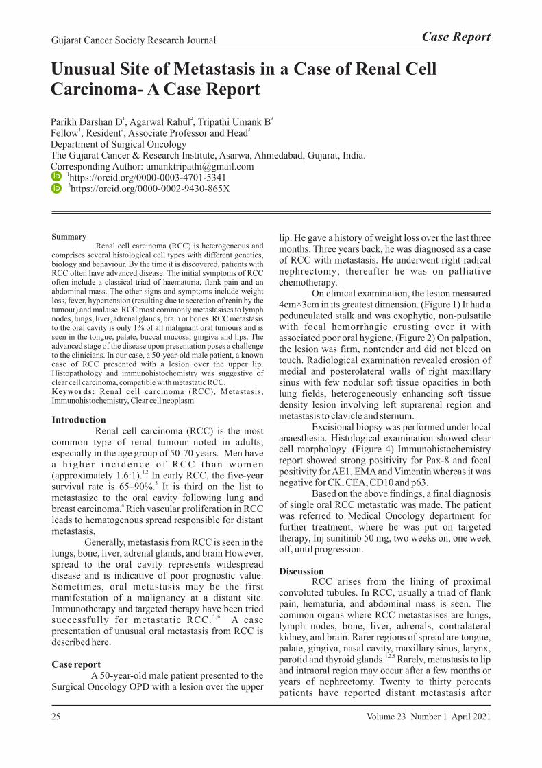
Summary Renal cell carcinoma (RCC) is heterogeneous and comprises several histological cell types with different genetics, biology and behaviour. By the time it is discovered, patients with RCC often have advanced disease. The initial symptoms of RCC often include a classical triad of haematuria, flank pain and an abdominal mass. The other signs and symptoms include weight loss, fever, hypertension (resulting due to secretion of renin by the tumour) and malaise. RCC most commonly metastasises to lymph nodes, lungs, liver, adrenal glands, brain or bones. RCC metastasis to the oral cavity is only 1% of all malignant oral tumours and is seen in the tongue, palate, buccal mucosa, gingiva and lips. The advanced stage of the disease upon presentation poses a challenge to the clinicians. In our case, a 50-year-old male patient, a known case of RCC presented with a lesion over the upper lip. Histopathology and immunohistochemistry was suggestive of clear cell carcinoma, compatible with metastatic RCC. Keywords: Renal cell carcinoma (RCC), Metastasis, Immunohistochemistry, Clear cell neoplasm
Introduction Renal cell carcinoma (RCC) is the most common type of renal tumour noted in adults, especially in the age group of 50-70 years. Men have a h i g h e r i n c i d e n c e o f R C C t h a n w o m e n
1,2(approximately 1.6:1). In early RCC, the five-year 3
survival rate is 65–90%. It is third on the list to metastasize to the oral cavity following lung and
4breast carcinoma. Rich vascular proliferation in RCC leads to hematogenous spread responsible for distant metastasis. Generally, metastasis from RCC is seen in the lungs, bone, liver, adrenal glands, and brain However, spread to the oral cavity represents widespread disease and is indicative of poor prognostic value. Sometimes, oral metastasis may be the first manifestation of a malignancy at a distant site. Immunotherapy and targeted therapy have been tried
5 , 6successfully for metastatic RCC. A case presentation of unusual oral metastasis from RCC is described here.
Case report A 50-year-old male patient presented to the Surgical Oncology OPD with a lesion over the upper
lip. He gave a history of weight loss over the last three months. Three years back, he was diagnosed as a case of RCC with metastasis. He underwent right radical nephrectomy; thereafter he was on palliative chemotherapy. On clinical examination, the lesion measured 4cm×3cm in its greatest dimension. (Figure 1) It had a pedunculated stalk and was exophytic, non-pulsatile with focal hemorrhagic crusting over it with associated poor oral hygiene. (Figure 2) On palpation, the lesion was firm, nontender and did not bleed on touch. Radiological examination revealed erosion of medial and posterolateral walls of right maxillary sinus with few nodular soft tissue opacities in both lung fields, heterogeneously enhancing soft tissue density lesion involving left suprarenal region and metastasis to clavicle and sternum. Excisional biopsy was performed under local anaesthesia. Histological examination showed clear cell morphology. (Figure 4) Immunohistochemistry report showed strong positivity for Pax-8 and focal positivity for AE1, EMA and Vimentin whereas it was negative for CK, CEA, CD10 and p63. Based on the above findings, a final diagnosis of single oral RCC metastatic was made. The patient was referred to Medical Oncology department for further treatment, where he was put on targeted therapy, Inj sunitinib 50 mg, two weeks on, one week off, until progression.
Discussion RCC arises from the lining of proximal convoluted tubules. In RCC, usually a triad of flank pain, hematuria, and abdominal mass is seen. The common organs where RCC metastasises are lungs, lymph nodes, bone, liver, adrenals, contralateral kidney, and brain. Rarer regions of spread are tongue, palate, gingiva, nasal cavity, maxillary sinus, larynx,
1,2,8parotid and thyroid glands. Rarely, metastasis to lip and intraoral region may occur after a few months or years of nephrectomy. Twenty to thirty percents patients have reported distant metastasis after
Case ReportGujarat Cancer Society Research Journal
25 Volume 23 Number 1 April 2021
Unusual Site of Metastasis in a Case of Renal CellCarcinoma- A Case Report
1 2 3Parikh Darshan D , Agarwal Rahul , Tripathi Umank B
1 2 3Fellow , Resident , Associate Professor and HeadDepartment of Surgical OncologyThe Gujarat Cancer & Research Institute, Asarwa, Ahmedabad, Gujarat, India.Corresponding Author: [email protected]
1 https://orcid.org/0000-0003-4701-5341
3 https://orcid.org/0000-0002-9430-865X
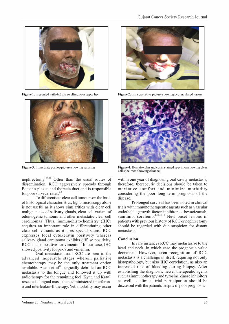
8,9,10nephrectomy. Other than the usual routes of dissemination, RCC aggressively spreads through Batson's plexus and thoracic duct and is responsible
2,8for poor survival rates. To differentiate clear cell tumours on the basis of histological characteristics, light microscopy alone is not useful as it shows similarities with clear cell malignancies of salivary glands, clear cell variant of odontogenic tumours and other metastatic clear cell
7carcinomas Thus, immunohistochemistry (IHC) acquires an important role in differentiating other clear cell variants as it uses special stains. RCC expresses focal cytokeratin positivity whereas salivary gland carcinoma exhibits diffuse positivity.
7RCC is also positive for vimentin. In our case, IHC showed positivity for pax 8 and vimentin. Oral metastasis from RCC are seen in the advanced inoperable stages wherein palliative chemotherapy may be the only treatment option
11available. Azam et al surgically debrided an RCC metastasis to the tongue and followed it up with
12radiotherapy for the remaining foci. Kyan and Kato resected a lingual mass, then administered interferon-α and interleukin-II therapy. Yet, mortality may occur
Gujarat Cancer Society Research Journal
26Volume 23 Number 1 April 2021
within one year of diagnosing oral cavity metastasis; therefore, therapeutic decisions should be taken to maximize comfort and minimize morbidity considering the poor long term prognosis of the disease. Prolonged survival has been noted in clinical trials with immunotherapeutic agents such as vascular endothelial growth factor inhibitors - bevacizumab,
2,10,11,12sunitinib, sorafenib. New onset lesions in patients with previous history of RCC or nephrectomy should be regarded with due suspicion for distant metastasis.
Conclusion In rare instances RCC may metastasise to the head and neck, in which case the prognostic value decreases. However, even recognition of RCC metastasis is a challenge in itself, requiring not only histopathology, but also IHC correlation, as also an increased risk of bleeding during biopsy. After establishing the diagnosis, newer therapeutic agents such as immunotherapy and tyrosine kinase inhibitors as well as clinical trial participation should be discussed with the patients in spite of poor prognosis.
Figure 4: Hematoxylin and eosin stained specimen showing clear cell specimen showing clear cell
Figure 2: Intra operative picture showing pedunculated lesion
Figure 3: Immediate post op picture showing suturing
Figure 1: Presented with 4x3 cm swelling over upper lip
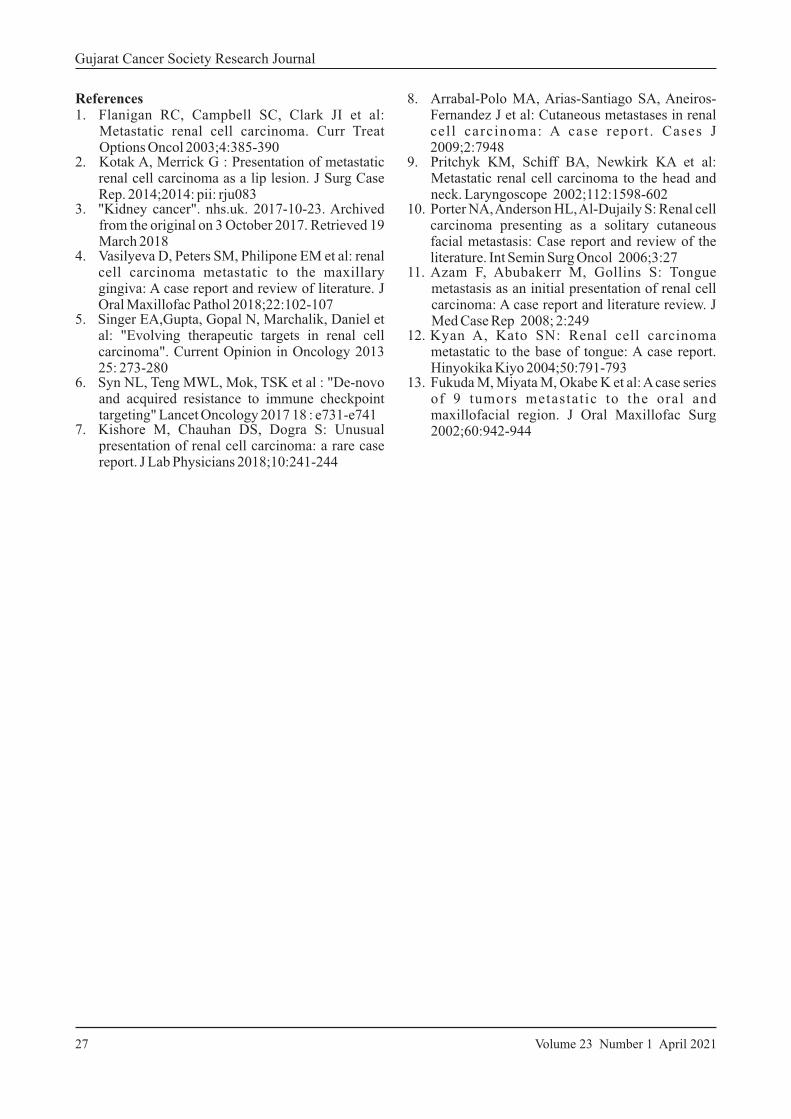
References1. Flanigan RC, Campbell SC, Clark JI et al:
Metastatic renal cell carcinoma. Curr Treat Options Oncol 2003;4:385-390
2. Kotak A, Merrick G : Presentation of metastatic renal cell carcinoma as a lip lesion. J Surg Case Rep. 2014;2014: pii: rju083
3. "Kidney cancer". nhs.uk. 2017-10-23. Archived from the original on 3 October 2017. Retrieved 19 March 2018
4. Vasilyeva D, Peters SM, Philipone EM et al: renal cell carcinoma metastatic to the maxillary gingiva: A case report and review of literature. J Oral Maxillofac Pathol 2018;22:102-107
5. Singer EA,Gupta, Gopal N, Marchalik, Daniel et al: "Evolving therapeutic targets in renal cell carcinoma". Current Opinion in Oncology 2013 25: 273-280
6. Syn NL, Teng MWL, Mok, TSK et al : "De-novo and acquired resistance to immune checkpoint targeting" Lancet Oncology 2017 18 : e731-e741
7. Kishore M, Chauhan DS, Dogra S: Unusual presentation of renal cell carcinoma: a rare case report. J Lab Physicians 2018;10:241-244
Gujarat Cancer Society Research Journal
27 Volume 23 Number 1 April 2021
8. Arrabal-Polo MA, Arias-Santiago SA, Aneiros-Fernandez J et al: Cutaneous metastases in renal cel l carcinoma: A case report . Cases J 2009;2:7948
9. Pritchyk KM, Schiff BA, Newkirk KA et al: Metastatic renal cell carcinoma to the head and neck. Laryngoscope 2002;112:1598-602
10. Porter NA, Anderson HL, Al-Dujaily S: Renal cell carcinoma presenting as a solitary cutaneous facial metastasis: Case report and review of the literature. Int Semin Surg Oncol 2006;3:27
11. Azam F, Abubakerr M, Gollins S: Tongue metastasis as an initial presentation of renal cell carcinoma: A case report and literature review. J Med Case Rep 2008; 2:249
12. Kyan A, Kato SN: Renal cell carcinoma metastatic to the base of tongue: A case report. Hinyokika Kiyo 2004;50:791-793
13. Fukuda M, Miyata M, Okabe K et al: A case series of 9 tumors metastatic to the oral and maxillofacial region. J Oral Maxillofac Surg 2002;60:942-944
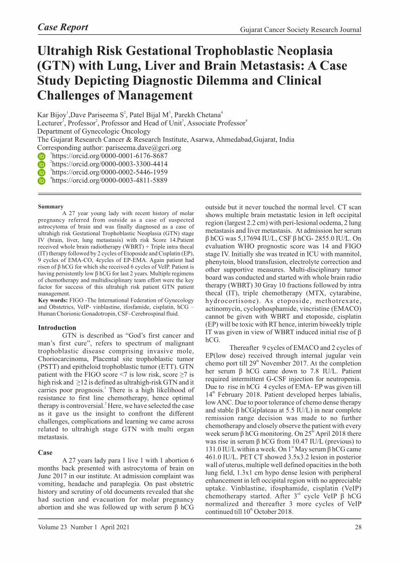
Summary A 27 year young lady with recent history of molar pregnancy referred from outside as a case of suspected astrocytoma of brain and was finally diagnosed as a case of ultrahigh risk Gestational Trophoblastic Neoplasia (GTN) stage IV (brain, liver, lung metastasis) with risk Score 14.Patient received whole brain radiotherapy (WBRT) + Triple intra thecal (IT) therapy followed by 2 cycles of Etoposide and Cisplatin (EP), 9 cycles of EMA-CO, 4cycles of EP-EMA. Again patient had risen of β hCG for which she received 6 cycles of VeIP. Patient is having persistently low β hCG for last 2 years. Multiple regimens of chemotherapy and multidisciplinary team effort were the key factor for success of this ultrahigh risk patient GTN patient management.Key words: FIGO -The International Federation of Gynecology and Obstetrics, VeIP- vinblastine, ifosfamide, cisplatin, hCG – Human Chorionic Gonadotropin, CSF- Cerebrospinal fluid.
Introduction GTN is described as “God’s first cancer and man’s first cure”, refers to spectrum of malignant trophoblastic disease comprising invasive mole, Choriocarcinoma, Placental site trophoblastic tumor (PSTT) and epitheloid trophoblastic tumor (ETT). GTN patient with the FIGO score <7 is low risk, score ≥7 is high risk and ≥12 is defined as ultrahigh-risk GTN and it
1carries poor prognosis. There is a high likelihood of resistance to first line chemotherapy, hence optimal
2therapy is controversial. Here, we have selected the case as it gave us the insight to confront the different challenges, complications and learning we came across related to ultrahigh stage GTN with multi organ metastasis.
Case A 27 years lady para 1 live 1 with 1 abortion 6 months back presented with astrocytoma of brain on June 2017 in our institute. At admission complaint was vomiting, headache and paraplegia. On past obstetric history and scrutiny of old documents revealed that she had suction and evacuation for molar pregnancy abortion and she was followed up with serum β hCG
outside but it never touched the normal level. CT scan shows multiple brain metastatic lesion in left occipital region (largest 2.2 cm) with peri-lesional oedema, 2 lung metastasis and liver metastasis. At admission her serum β hCG was 5,17694 IU/L, CSF β hCG- 2855.0 IU/L. On evaluation WHO prognostic score was 14 and FIGO stage IV. Initially she was treated in ICU with mannitol, phenytoin, blood transfusion, electrolyte correction and other supportive measures. Multi-disciplinary tumor board was conducted and started with whole brain radio therapy (WBRT) 30 Gray 10 fractions followed by intra thecal (IT), triple chemotherapy (MTX, cytarabine, hydrocortisone). As etoposide, methotrexate, actinomycin, cyclophosphamide, vincristine (EMACO) cannot be given with WBRT and etoposide, cisplatin (EP) will be toxic with RT hence, interim biweekly triple IT was given in view of WBRT induced initial rise of β hCG. Thereafter 9 cycles of EMACO and 2 cycles of EP(low dose) received through internal jugular vein
thchemo port till 29 November 2017. At the completion her serum β hCG came down to 7.8 IU/L. Patient required intermittent G-CSF injection for neutropenia. Due to rise in hCG 4 cycles of EMA- EP was given till
th14 February 2018. Patient developed herpes labialis, low ANC. Due to poor tolerance of chemo dense therapy and stable β hCG(plateau at 5.5 IU/L) in near complete remission range decision was made to no further chemotherapy and closely observe the patient with every
thweek serum β hCG monitoring. On 25 April 2018 there was rise in serum β hCG from 10.47 IU/L (previous) to
st131.0 IU/L within a week. On 1 May serum β hCG came 461.0 IU/L. PET CT showed 3.5x3.2 lesion in posterior wall of uterus, multiple well defined opacities in the both lung field, 1.3x1 cm hypo dense lesion with peripheral enhancement in left occipital region with no appreciable uptake. Vinblastine, ifosphamide, cisplatin (VeIP)
rdchemotherapy started. After 3 cycle VeIP β hCG normalized and thereafter 3 more cycles of VeIP
thcontinued till 10 October 2018.
Case Report Gujarat Cancer Society Research Journal
28Volume 23 Number 1 April 2021
Ultrahigh Risk Gestational Trophoblastic Neoplasia(GTN) with Lung, Liver and Brain Metastasis: A CaseStudy Depicting Diagnostic Dilemma and ClinicalChallenges of Management
1 2 3 4Kar Bijoy ,Dave Pariseema S , Patel Bijal M , Parekh Chetana1 2 3 4
Lecturer , Professor , Professor and Head of Unit , Associate ProfessorDepartment of Gynecologic OncologyThe Gujarat Research Cancer & Research Institute, Asarwa, Ahmedabad,Gujarat, IndiaCorresponding author: [email protected]
1 https://orcid.org/0000-0001-6176-8687 2 https://orcid.org/0000-0003-3300-4414 3 https://orcid.org/0000-0002-5446-1959 4 https://orcid.org/0000-0003-4811-5889
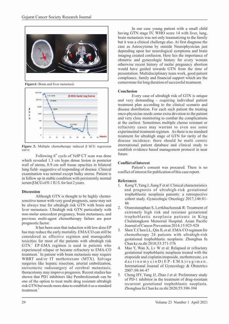
th Following 6 cycle of VeIP CT scan was done which revealed 1.3 cm hypo dense lesion in posterior wall of uterus, 0.9 cm soft tissue opacities in bilateral lung field- suggestive of responding of disease. Clinical examination was normal except bulky uterus. Patient is in follow up in stable condition with persistently normal serum β hCG of 0.1 IU/L for last 2 years.
Discussion Although GTN is thought to be highly chemo-sensitive tumor with very good prognosis, same may not be always true for ultrahigh risk GTN with brain and liver metastasis. Ultrahigh risk GTN particularly with non-molar antecedent pregnancy, brain metastases, and previous multi-agent chemotherapy failure are poor prognostic factor. It has been seen that induction with low dose EP has may reduce the early mortality. EMA/CO can still be considered as effective regimen and manageable toxicities for most of the patients with ultrahigh risk
3GTN. EP-EMA regimen is used in patients who experienced relapse or became refractory to EMA-CO
4treatment. In patient with brain metastasis may require WBRT and/or IT methotrexate (MTX). Salvage surgeries like hepatic resection, arterial embolization, stereotactic radiosurgery of cerebral metastasis, thoracotomy may improve prognosis. Recent studies has shown that PD1 inhibitors like Pembrolizumab can be one of the option to treat multi drug resistant ultrahigh risk GTN but needs more data to establish it as a standard
5treatment.
Gujarat Cancer Society Research Journal
29 Volume 23 Number 1 April 2021
In our case young patient with a small child having GTN stage IV, WHO score 14 with liver, lung, brain metastasis was not only traumatizing to the family but it was a clinical challenge also. At first diagnose the case as Astrocytoma by outside Neurophysician just depending upon her neurological symptoms and brain imaging created confusion. Here lies the importance of obstetric and gynecologic history for every woman otherwise recent history of molar pregnancy abortion would have guided towards GTN from the time of presentation. Multidisciplinary team work, good patient compliance, family and financial support which are the cornerstone for long duration of successful treatment.
Conclusion Every case of ultrahigh risk of GTN is unique and very demanding - requiring individual patient treatment plan according to the clinical scenario and disease distribution. For each such patient the treating onco-physician needs some extra devotion to the patient and very close monitoring to combat the complications at the earliest. Sometimes multiple chemo resistant or refractory cases may warrant to even use some experimental treatment regimen. As there is no standard treatment for ultrahigh stage of GTN for rarity of the disease incidence; there should be multi centric international patient database and clinical study to establish evidence based management protocol in near future.
Conflict of Interest Patient’s consent was procured. There is no conflict of interest for publication of this case report.
References1. Kong Y, Yang J, Jiang F et al: Clinical characteristics
and prognosis of ultrahigh-risk gestational trophoblastic neoplasia patients: a retrospective cohort study. Gynecologic Oncology 2017;146:81- 86
2. Oranratanaphan S, Lertkhachonsuk R: Treatment of extremely high risk and resistant gestational t rophoblast ic neoplasia pat ients in King Chulalongkorn Memorial Hospital. Asian Pacific Journal of Cancer Prevention 2014;15:925-928
3. Shen T, Chen LL, Qin JL et al: EMA/CO regimen for chemotherapy 24 patients with ultrahigh-risk gestational trophoblastic neoplasia. Zhonghua fu Chan ke za zhi 2018;53:371-376
4. Mao Y, Wan X, Lv W et al: Relapsed or refractory gestational trophoblastic neoplasia treated with the etoposide and cisplatin/etoposide, methotrexate, a n d a c t i n o m y c i n D ( E P - E M A ) r e g i m e n . International Journal of Gynecology & Obstetrics 2007 ;98:44-47
5. Cheng HY, Yang JJ, Zhao J et al: Preliminary study of PD-1 inhibitor in the treatment of drug-resistant recurrent gestational trophoblastic neoplasia. Zhonghua fu Chan ke za zhi 2020;55:390-394
Figure1: Brain and liver metastasis
Figure 2: Multiple chemotherapy induced β hCG regression curve
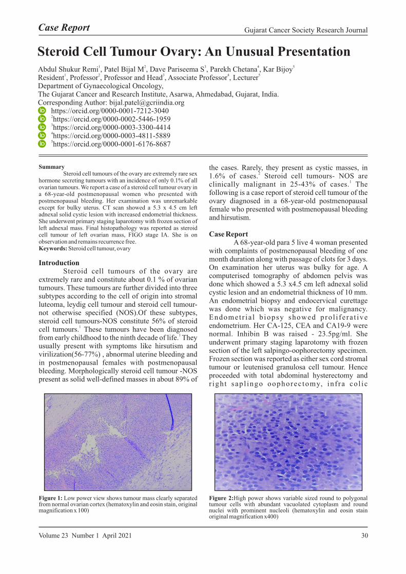
Summary Steroid cell tumours of the ovary are extremely rare sex hormone secreting tumours with an incidence of only 0.1% of all ovarian tumours. We report a case of a steroid cell tumour ovary in a 68-year-old postmenopausal women who presented with postmenopausal bleeding. Her examination was unremarkable except for bulky uterus. CT scan showed a 5.3 x 4.5 cm left adnexal solid cystic lesion with increased endometrial thickness. She underwent primary staging laparotomy with frozen section of left adnexal mass. Final histopathology was reported as steroid cell tumour of left ovarian mass, FIGO stage IA. She is on observation and remains recurrence free.Keywords: Steroid cell tumour, ovary
Introduction Steroid cell tumours of the ovary are extremely rare and constitute about 0.1 % of ovarian tumours. These tumours are further divided into three subtypes according to the cell of origin into stromal luteoma, leydig cell tumour and steroid cell tumour-not otherwise specified (NOS).Of these subtypes, steroid cell tumours-NOS constitute 56% of steroid
1cell tumours. These tumours have been diagnosed 1
from early childhood to the ninth decade of life. They usually present with symptoms like hirsutism and virilization(56-77%) , abnormal uterine bleeding and in postmenopausal females with postmenopausal bleeding. Morphologically steroid cell tumour -NOS present as solid well-defined masses in about 89% of
the cases. Rarely, they present as cystic masses, in 2
1.6% of cases. Steroid cell tumours- NOS are 1clinically malignant in 25-43% of cases. The
following is a case report of steroid cell tumour of the ovary diagnosed in a 68-year-old postmenopausal female who presented with postmenopausal bleeding and hirsutism.
Case Report A 68-year-old para 5 live 4 woman presented with complaints of postmenopausal bleeding of one month duration along with passage of clots for 3 days. On examination her uterus was bulky for age. A computerised tomography of abdomen pelvis was done which showed a 5.3 x4.5 cm left adnexal solid cystic lesion and an endometrial thickness of 10 mm. An endometrial biopsy and endocervical curettage was done which was negative for malignancy. Endomet r ia l b iopsy showed pro l i fe ra t ive endometrium. Her CA-125, CEA and CA19-9 were normal. Inhibin B was raised - 23.5pg/ml. She underwent primary staging laparotomy with frozen section of the left salpingo-oophorectomy specimen. Frozen section was reported as either sex cord stromal tumour or leutenised granulosa cell tumour. Hence proceeded with total abdominal hysterectomy and r igh t sap l ingo oophorec tomy, in f ra co l ic
Case Report Gujarat Cancer Society Research Journal
30Volume 23 Number 1 April 2021
1 2 3 4 5Abdul Shukur Remi , Patel Bijal M , Dave Pariseema S , Parekh Chetana , Kar Bijoy1 2 3 4 5Resident , Professor , Professor and Head , Associate Professor , Lecturer
Department of Gynaecological Oncology, The Gujarat Cancer and Research Institute, Asarwa, Ahmedabad, Gujarat, India.Corresponding Author: [email protected]
https://orcid.org/0000-0001-7212-3040 2 https://orcid.org/0000-0002-5446-1959
3 https://orcid.org/0000-0003-3300-4414
4 https://orcid.org/0000-0003-4811-5889 5 https://orcid.org/0000-0001-6176-8687
Steroid Cell Tumour Ovary: An Unusual Presentation
Figure 1: Low power view shows tumour mass clearly separated from normal ovarian cortex (hematoxylin and eosin stain, original magnification x 100)
Figure 2:High power shows variable sized round to polygonal tumour cells with abundant vacuolated cytoplasm and round nuclei with prominent nucleoli (hematoxylin and eosin stain original magnification x400)
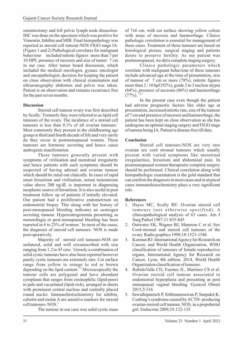
omentectomy and left pelvic lymph node dissection. IHC was done on the specimen which was positive for Vimentin, Inhibin and MIB. Final histopathology was reported as steroid cell tumour-NOS FIGO stage IA. (Figure 1 and 2) Pathological correlates for malignant behaviour included mitotic figures more than 7 per 10 HPF, presence of necrosis and size of tumor 7 cm in our case. After tumor board discussion, which included the medical oncologist, gynaec oncologist and oncopathologist, decision for keeping the patient on close observation with clinical examination and ultrasonography abdomen and pelvis was taken. Patient is on observation and remains recurrence free for the past seven months.
Discussion Steroid cell tumour ovary was first described
1by Scully. Formerly they were referred to as lipid cell tumours of the ovary. The incidence of a steroid cell
1tumours is less than 0.1% of all ovarian tumours. Most commonly they present in the childbearing age group or third and fourth decade of life and very rarely do they occur in postmenopausal women. These tumours are hormone secreting and hence cause androgenic manifestation. These tumours generally present with symptoms of virilisation and menstrual irregularity and hence patients with such symptoms should be suspected of having adrenal and ovarian tumour which should be ruled out clinically. In cases of rapid onset hirsuitism and virilisation, serum testosterone value above 200 ng/dL is important in diagnosing neoplastic source of hirsuitism. It is also useful in post treatment follow up of patients if initially elevated. Our patient had a proliferative endometrium on endometrial biopsy. This along with her history of post-menopausal bleeding indicates an oestrogen secreting tumour. Hyperestrogenemia presenting as menorrhagia or post-menopausal bleeding has been
1reported in 6 to 23% of women. In most of the cases, the diagnosis of steroid cell tumours- NOS is made post operatively. Majority of steroid cell tumours-NOS are unilateral, solid and well circumscribed with size
1ranging from 1.2 to 45 cms. Grossly a combination of solid cystic tumours have also been reported however purely cystic tumours are extremely rare. Cut surface range from yellow to orange to red or brown
1,2depending on the lipid content. Microscopically the tumour cells are polygonal and have abundant cytoplasm that ranges from eosinophilic (lipid-poor) to pale and vacuolated (lipid-rich), arranged in sheets with prominent central nucleus and centrally placed round nuclei. Immunohistochemistry for inhibin, calretin and melan A are sensitive markers for steroid
3cell tumours- NOS. The tumour in our case was solid cystic mass
Gujarat Cancer Society Research Journal
31 Volume 23 Number 1 April 2021
of 7x6 cm, with cut surface showing yellow colour with areas of necrosis and haemorrhage. Clinico pathologic correlation is essential for management of these cases. Treatment of these tumours are based on histological picture, surgical staging and patients desire to preserve fertility. As our patient was postmenopausal, we did a complete staging surgery. Clinico pathologic parameters which correlate with malignant behaviour of these tumours include advanced age at the time of presentation, size of tumour of 7 cm or more (78%), mitotic figures more than 2 / 10 hpf (92%), grade 2 to 3 nuclear atypia (64%), presence of necrosis (86%) and haemorrhage
1,4,5(77%). In the present case even though the patient had adverse prognostic factors like older age at presentation, increased mitotic rate, size of the tumour of 7 cm and presence of necrosis and haemorrhage, the patient has been kept on close observation as she has undergone an optimal staging surgery and FIGO stage of tumour being IA. Patient is disease free till date.
Conclusion Steroid cell tumours-NOS are very rare ovarian sex cord stromal tumours which usually present with varied symptoms like menstrual irregularities, hirsutism and abdominal pain. In postmenopausal women therapeutic complete surgery should be performed. Clinical correlation along with histopathologic examination is the gold standard that can confirm the diagnosis in most cases and in atypical cases immunohistochemistry plays a very significant role.
References1. Hayes MC, Scully RE: Ovarian steroid cell
t u m o u r s ( n o t o t h e r w i s e s p e c i f i e d ) . A clinicopathological analysis of 63 cases. Am J Surg Pathol 1987;11:835-845
2. Outwater EK, Wagner BJ, Mannion C et al: Sex Cord-stromal and steroid cell tumours of the ovary. Radio graphics 1998;18:1523-1546
3. Kurman RJ: International Agency for Research on Cancer, and World Health Organization, WHO classification of tumours of female reproductive organs, International Agency for Research on Cancer, Lyon, 4th edition, 2014, World Health Organization classification of tumours.
4. RubidoValle CD, Fuentes JL, Martinez CS et al: Ovarian steroid cell tumour associated to endometrial hyperplasia and presenting as post menopausal vaginal bleeding. Gynecol Obstet 2015;5:316
5. Sawathiparnich P, Sitthinamsuwan P, Sanpakit K: Cushing’s syndrome caused by ACTH- producing ovarian steroid cell tumour, NOS, in a prepubertal girl. Endocrine 2009;35:132-135
Summary Endometrial cancer is the third most common gynaecologic malignancy in India and is an important cause of postmenopausal bleeding. Hereditary predisposition as well as simultaneous occurrence of colon cancer and endometrial cancer have been studied and reported. Thus, any patient of diagnosed colon cancer presenting with abnormal/postmenopausal vaginal bleeding should be carefully investigated. This is a case of carcinoma ascending colon who presented with postmenopausal bleeding and careful workup could detect the endometrial pathology at preinvasive stage i.e endometrial intraepithelial neoplasia.Keywords: Endometrial carcinoma, Carcinoma colon, vaginal bleeding, salpingoopherectomy
Introduction Endometrial cancer is the most common gynaecologic malignancy in developed world and third most common gynaecologic malignancy in developing countries like India. A very peculiar presentation of endometrial cancer is with postmenopausal bleeding which is seen in 90% of
1patients as a presenting symptom. Endometrial cancer accounts for etiology of postmenopausal
2bleeding in 15% of pa t ien ts . Heredi ta ry predisposition to colorectal cancer and a wide range of other malignancies (e.g., endometrial, ovarian, and gastric cancer) has been studied over years. Also, simultaneous occurrence of endometrial cancer with
3colorectal cancer has been reported in literature. Thus, if a patient of colorectal cancer presents with abnormal vaginal bleeding, strict vigilance can help detect endometrial cancer at early/preinvasive stage. We hereby represent a case of carcinoma ascending colon with endometrial intraepithelial neoplasia.
Case A 40-year-old female, moderately built who was illiterate and a labourer by occupation presented in the outpatient department of our hospital, the Gujarat Cancer and Research Institute, Ahmedabad in February 2019 with complaints of postmenopausal bleeding from last 15days. She had attained
menopause 3 years back. She was a diagnosed case of poorly differentiated adenocarcinoma of ascending colon. She had undergone exploratory laparotomy with right hemicolectomy with ileotransverse anastomosis with double barrel stomy at a government hospital, Ahmedabad in 2016. She was then referred to our hospital for adjuvant treatment and had received 8 cycles of Capecitabine. After remaining disease free for 1 year, stoma closure surgery was done in 2017 at the same place of initial surgery. She did not have any medical comorbidity. She was a habituated tobacco chewer for 10 years. There was no significant history of cancers running in the family. Further on evaluat ing the cause of postmenopausal bleeding, on ultrasonography, she was found to have 18mm thickened endometrium. Endometrial aspiration showed complex hyperplasia without atypia. Endometrial biopsy was performed which favored precursor lesion naming endometrial intraepithelial neoplasia. A preoperative computed tomography of thorax, abdomen and pelvis was performed which did not show disease elsewhere. Bilateral mammography was normal. Patient was planned for staging laparotomy and before posting her, a clearance for surgical o n c o l o g i s t w a s s o u g h t t o l o o k f o r a n y recurrence/residual disease as well as need of any biopsy intraoperatively. A preoperative colonoscopy was advised which was normal. She was then posted for surgery with plan of intraoperative gross examination as well as frozen section examination of uterus. Peritoneal cytology was taken and total a b d o m i n a l h y s t e r e c t o m y w i t h b i l a t e r a l salpingoophorectomy, bilateral pelvic lymph node dissection and omental sampling were performed. Uterus was cut open in OT as planned which showed thick and irregular endometrium. Frozen report d e m o n s t r a t e d s u p e r f i c i a l E n d o m e t r i a l tumor/Neoplasia. No myometrial invasion was seen.
Case Report Gujarat Cancer Society Research Journal
32Volume 23 Number 1 April 2021
Endometrial Intraepithelial Neoplasia in a Treated Caseof Carcinoma Colon: A Case Report
1 2 3 4Nishu , Dave Pariseema , Patel Bijal M , Parekh Chetana
1 2 3 4Resident , Professor and Head of Unit , Professor , Associate Professor
Department of Gynaecological Oncology
The Gujarat Cancer & Research Institute, Asarwa Ahmedabad Gujarat
Corresponding Author: [email protected] 2 https://orcid.org/0000-0003-3300-4414 3 https://orcid.org/0000-0002-5446-1959 4 https://orcid.org/0000-0003-4811-5889
Peritoneal cytology came out to be negative for malignant cells. Final histopathology confirmed the endometrial intraepithelial neoplasia/endometrial carcinoma in situ (focally) without stromal invasion. Endometrial thickness was 1.5 cm, myometrium, cervix and both ovaries were unremarkable. Both fallopian tubes had normal histology. Omentum and bilateral pelvic lymph nodes were free of any tumor.
Discussion Abnormal genital bleeding is often attributed to the uterus, with postmenopausal women being described as having bleeding per vaginum after atleast 1 year of stoppage of menstruation. The various etiologies behind postmenopausal bleeding can be atrophy, either of the endometrium or the vaginal mucosa, endometrial hyperplasia/carcinoma, endometrial polyps, leiomyomas and cervical
2pathology. Endometrial cancer holds a significant burden of gynecologic malignancy in India with
426,514 patients in the year 2020. Surgery is the mainstay of treatment and early detection can reduce significant mortality with 5-year survival rates of 95%
2and 16% to 45% in early and late stages respectively. As the worldwide burden of endometrial cancer continues to rise, there is growing need in the early detection and prevention strategies among women at increased risk. Vaginal bleeding being a common symptom of endometrial cancer, a focused evaluation of this symptom may be a useful strategy. Universal screening has not been found to be effective hence, targeting high risk individuals and patients presenting with abnormal vaginal bleeding /postmenopausal bleeding is advocated. Thus, subjecting such patients to testing like transvaginal ultrasonography and endometrial aspiration/biopsy is advised. Association of colorectal cancer with endometrial cancer has been a topic of research. Rare finding of synchronous detection of different types of cancer at the pelvic level should also be kept in mind. Most colorectal cancer is sporadic, and approximately 3% to 5% of all cases of colorectal cancer and approximately 2% of all cases of endometrial cancer are to be due to hereditary syndromes like Lynch
5syndrome. Such patients may develop multiple cancers during their lifetime. The presentation of postmenopausal bleeding in a known case of colon
Gujarat Cancer Society Research Journal
33 Volume 23 Number 1 April 2021
carcinoma, should be thoroughly investigated as there are chances of finding an occult malignancy at the time of hysterectomy performed. A hysteroscopic guided endometrial biopsy should be performed, ideally by a gynecologic oncologist in such cases. In addition, the importance of communication with the pathologist is important. The pathologist should be aware that the patient has a colorectal cancer thus a potential high risk of harbouring a hereditary germline mutation and a candidate for endometrial cancer. A meticulous approach was followed in our case in properly investigating presenting signs and symptoms which could help in detecting such cancer in the preinvasive phase. Hysterectomy specimen was cut opened with careful examination of both the endometrial cavity and ovaries and was subjected to microscopic evaluation by frozen section in our case.
Conclusion Increased awareness of endometrial cancer wi th co lo rec ta l cancer, t imed workup of postmenopausal bleeding and accurate surgical intervention with frozen section examination could help in detecting endometrial cancer in the preinvasive phase. The patient was referred for genetic counseling. The patient is on follow-up and disease free for 48 months.
References1. Clarke and Nicolas Wentzensen: A closer look at
postmenopausal bleeding & endometrial cancer. Oncol times 2018; 40:33
2. Sung S, Abramovitz A: Postmenopausal Bleeding. In: StatPearls [online] Available at: https://www.ncbi.nlm.nih.gov/books/NBK562188. Accessed March 1, 2021
3. Álvarez JAP, Gonzàlez BC, López-Fé AAQ et al: Endometrial and Colorectal Synchronous Cancer: A Case Report. Obstet Gynaecol Cases Rev 2018; 5:133; DOI: 10.23937/2377-9004/1410133
4. Maheshwari A, Kumar N, Mahantshetty U: Gynecological cancers: A summary of published Indian data. South Asian J Cancer 2016; 5:112-120
5. Jasperson KW, Tuohy TM, Neklason DW et al: Heredi ta ry and fami l ia l co lon cancer. Gastroenterology 2010; 138:2044-2058
Summary WHO defines Mixed Endometrial Carcinoma (MEC) as a tumor composed of “two or more different histological types of endometrial carcinoma, at least one of which is of the type II category” The type II refers to non-endometroid (serous carcinoma or clear cell carcinoma) carcinoma. MEC comprises of about 5% of all endometrial cancers (EC), due to its rarity precise definition and exact data on occurrence is difficult to generate. Here we are reporting a case of sixty-one-year-old women presented with post-menopausal bleeding. Histopathology of endometrium revealed endometroid and clear cell components, consistent with mixed endometrial carcinoma. Patient underwent comprehensive staging laparotomy followed by adjuvant treatment with chemo-radiation. Patient is on close follow-up till date and continues to be disease free.Keywords: Mixed Endometrial Carcinoma, Clear Cell Carcinoma, Radiotherapy, Chemotherapy
Introduction Mixed endometrial carcinoma is composed of two or more distinct histologies in the same specimen and both the cell types must be recognisable distinctly on haematoxylin and eosin-stained pathological
1sections. According to WHO 2014 the minimum percentage of second component is arbitrarily set as 5 and can be confirmed on immunohistochemistry
1(IHC). MEC are almost clonal rather than being
2collision. Most common combination of MEC is the endometrioid with serous (endometroid with type II). Quantification of each component is very challenging and important in respect to management and
3prognosis. Incidence of Mixed endometrial carcinoma is rarity and is reported to be less than 5%. The report below gives the time line of events of one such case.
Case Report Sixty one year old postmenopausal, nulliparous women presented to our gynaecological oncology department (Gujarat Cancer and Research Institute) with a complaint of vaginal bleeding for one month. Her bleeding was sudden in onset with minimal flow and was not associated with passage of
clots. She was postmenopausal for 12 years. She was known case of hypertension and diabetes. She had no history suggestive of cancer in the family. On physical examination, she was overweight with body mass
2index 28.6 Kg/m On per-speculum examination cervical was pinpoint, looks healthy and vagina was normal. On bimanual examination uterus was bulky retroverted, bilateral fornixes were free. Her sonography revealed 44x49 mm heterogenous echotexture lesion in left lateral wall of uterus, with internal vascularity and loss of endo-myometrial junction. Lesion had myometrial invasion of more than 50%. Patient underwent dilatation and curettage (D & C). Histopathology of D&C material revealed endometroid carcinoma with clear cell change. IHC was positive for napsin, CK 7, ER, vimentin and negative for CK 20, PR, P40, CEA that indicated endometroid carcinoma with clear cell change. Further imaging with MRI was done to determine extent of disease. MRI revealed 33x53x35 mm altered intensity lesion in endometrium with endocervical gland involvement. Lesion invades more than half of myometrium. Few sub centimetric lymph nodes were noted along bilateral iliac vessels. Her baseline blood counts, liver and kidney function were with in normal range and her CA-125 was 22.6 IU/ml. Patient was diagnosed as carcinoma endometrium and underwent staging laparotomy with peritoneal cytology, total abdominal hysterectomy, bilateral salpoingo-ophorectomy, bilateral pelvic lymph node dissection, infra colic omentectomy. Final histopathology revealed tumor size 5 X 5 cm, mixed histology of clear cell and endometroid carcinoma. Clear cell accounting 70% of carcinoma while endometroid accounting to 30%. Myometrial invasion of about 80% (3/4 thickness of myometrial wall) along with lower uterine segment involvement. Uterine serosa, cervical stroma, bilateral tubes and ovaries, omentum and lymph nodes were free of tumor. IHC was positive for ER, PR confirmed
Case Report Gujarat Cancer Society Research Journal
34Volume 23 Number 1 April 2021
Mixed Endometrial Carcinoma Rare Histological Type
1 2 3 4 5Gupta Rubi , Parekh Chetana , Dave Pariseema S , Patel Bijal M , Kar Bijoy1 2 3 4 5Resident , Associate Professor , Professor and Head of Unit , Professor , Lecturer
Department of Gynaecological Oncology
The Gujarat Cancer & Research Institute, Asarwa Ahmedabad Gujarat
Corresponding Author: [email protected] 2 https://orcid.org/0000-0003-4811-5889
3 https://orcid.org/0000-0003-3300-4414 4 https://orcid.org/0000-0002-5446-1959 5 https://orcid.org/0000-0001-6176-8687
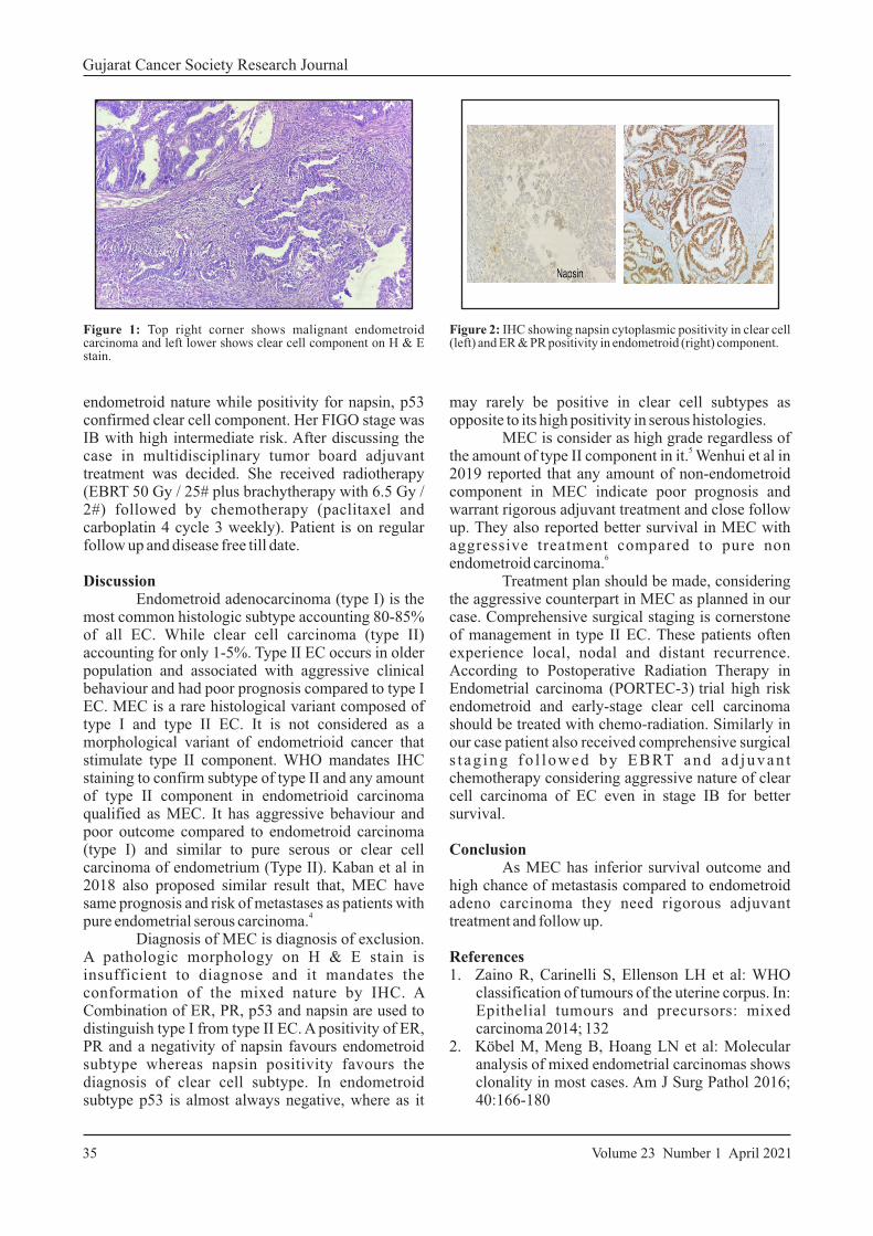
endometroid nature while positivity for napsin, p53 confirmed clear cell component. Her FIGO stage was IB with high intermediate risk. After discussing the case in multidisciplinary tumor board adjuvant treatment was decided. She received radiotherapy (EBRT 50 Gy / 25# plus brachytherapy with 6.5 Gy / 2#) followed by chemotherapy (paclitaxel and carboplatin 4 cycle 3 weekly). Patient is on regular follow up and disease free till date.
Discussion Endometroid adenocarcinoma (type I) is the most common histologic subtype accounting 80-85% of all EC. While clear cell carcinoma (type II) accounting for only 1-5%. Type II EC occurs in older population and associated with aggressive clinical behaviour and had poor prognosis compared to type I EC. MEC is a rare histological variant composed of type I and type II EC. It is not considered as a morphological variant of endometrioid cancer that stimulate type II component. WHO mandates IHC staining to confirm subtype of type II and any amount of type II component in endometrioid carcinoma qualified as MEC. It has aggressive behaviour and poor outcome compared to endometroid carcinoma (type I) and similar to pure serous or clear cell carcinoma of endometrium (Type II). Kaban et al in 2018 also proposed similar result that, MEC have same prognosis and risk of metastases as patients with
4pure endometrial serous carcinoma. Diagnosis of MEC is diagnosis of exclusion. A pathologic morphology on H & E stain is insufficient to diagnose and it mandates the conformation of the mixed nature by IHC. A Combination of ER, PR, p53 and napsin are used to distinguish type I from type II EC. A positivity of ER, PR and a negativity of napsin favours endometroid subtype whereas napsin positivity favours the diagnosis of clear cell subtype. In endometroid subtype p53 is almost always negative, where as it
Gujarat Cancer Society Research Journal
35 Volume 23 Number 1 April 2021
Figure 1: Top right corner shows malignant endometroid carcinoma and left lower shows clear cell component on H & E stain.
Figure 2: IHC showing napsin cytoplasmic positivity in clear cell (left) and ER & PR positivity in endometroid (right) component.
may rarely be positive in clear cell subtypes as opposite to its high positivity in serous histologies. MEC is consider as high grade regardless of
5the amount of type II component in it. Wenhui et al in 2019 reported that any amount of non-endometroid component in MEC indicate poor prognosis and warrant rigorous adjuvant treatment and close follow up. They also reported better survival in MEC with aggressive treatment compared to pure non
6endometroid carcinoma. Treatment plan should be made, considering the aggressive counterpart in MEC as planned in our case. Comprehensive surgical staging is cornerstone of management in type II EC. These patients often experience local, nodal and distant recurrence. According to Postoperative Radiation Therapy in
Endometrial carcinoma (PORTEC-3) trial high risk endometroid and early-stage clear cell carcinoma should be treated with chemo-radiation. Similarly in our case patient also received comprehensive surgical s t ag ing fo l lowed by EBRT and ad juvan t chemotherapy considering aggressive nature of clear cell carcinoma of EC even in stage IB for better survival.
Conclusion As MEC has inferior survival outcome and high chance of metastasis compared to endometroid adeno carcinoma they need rigorous adjuvant treatment and follow up.
References1. Zaino R, Carinelli S, Ellenson LH et al: WHO
classification of tumours of the uterine corpus. In: Epithelial tumours and precursors: mixed carcinoma 2014; 132
2. Köbel M, Meng B, Hoang LN et al: Molecular analysis of mixed endometrial carcinomas shows clonality in most cases. Am J Surg Pathol 2016; 40:166-180
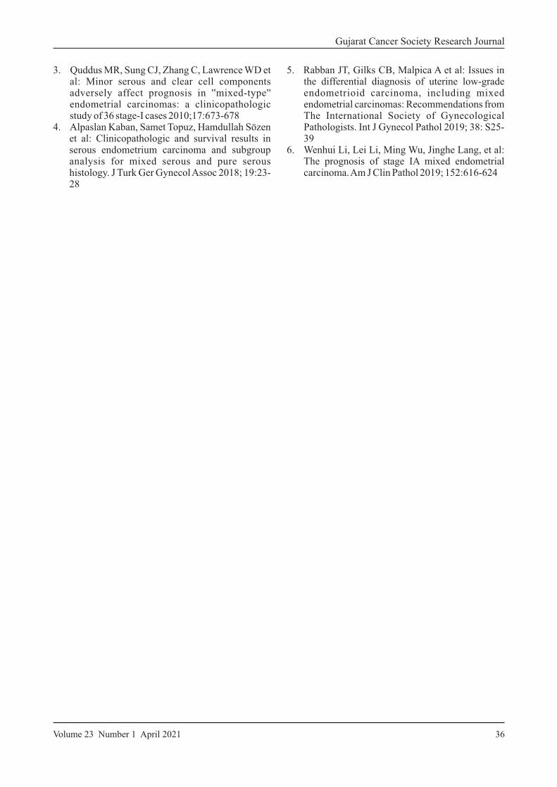
5. Rabban JT, Gilks CB, Malpica A et al: Issues in the differential diagnosis of uterine low-grade endometrioid carcinoma, including mixed endometrial carcinomas: Recommendations from The International Society of Gynecological Pathologists. Int J Gynecol Pathol 2019; 38: S25-39
6. Wenhui Li, Lei Li, Ming Wu, Jinghe Lang, et al: The prognosis of stage IA mixed endometrial carcinoma. Am J Clin Pathol 2019; 152:616-624
3. Quddus MR, Sung CJ, Zhang C, Lawrence WD et al: Minor serous and clear cell components adversely affect prognosis in ''mixed-type'' endometrial carcinomas: a clinicopathologic study of 36 stage-I cases 2010;17:673-678
4. Alpaslan Kaban, Samet Topuz, Hamdullah Sözen et al: Clinicopathologic and survival results in serous endometrium carcinoma and subgroup analysis for mixed serous and pure serous histology. J Turk Ger Gynecol Assoc 2018; 19:23-28
Gujarat Cancer Society Research Journal
36Volume 23 Number 1 April 2021
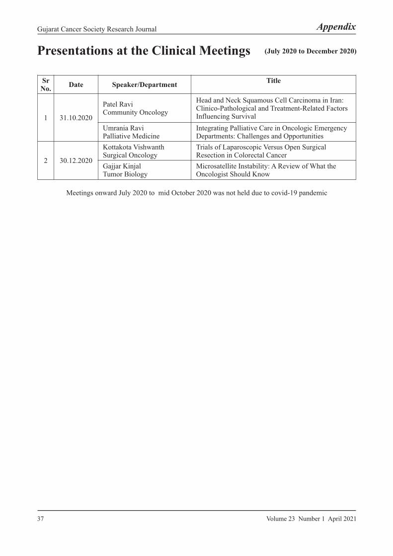
Gujarat Cancer Society Research Journal
37 Volume 23 Number 1 April 2021
(July 2020 to December 2020)Presentations at the Clinical Meetings
Sr No.
Date Speaker/DepartmentTitle
1 31.10.2020
Patel RaviCommunity Oncology
Head and Neck Squamous Cell Carcinoma in Iran: Clinico-Pathological and Treatment-Related Factors Influencing Survival
Umrania RaviPalliative Medicine
Integrating Palliative Care in Oncologic Emergency Departments: Challenges and Opportunities
2 30.12.2020
Kottakota VishwanthSurgical Oncology
Trials of Laparoscopic Versus Open Surgical Resection in Colorectal Cancer
Gajjar KinjalTumor Biology
Microsatellite Instability: A Review of What the Oncologist Should Know
Meetings onward July 2020 to mid October 2020 was not held due to covid-19 pandemic
Appendix
About the Journal Gujarat Cancer Society Research Journal is a biannually (April and October) peer-reviewed journal published by the Gujarat Cancer Society (formerly published as GCS Research Bulletin). The journal's full text is available online at http://www.cancerindia.org
Scope of the Journal The Journal intents to cover basic, clinical, clinico-basic research and medical education carried out by the staff of the Gujarat Cancer Society and Gujarat Cancer and Research Institute related to human well being including ethical and social issues in the field of Oncology. The Journal gives preferences to original scientific papers, case reports, anecdotal reports and minireviews. It may comprise invited review articles, publish oration speeches and work presented in the clinical meetings and the journal clubs. Hence it will continue to serve as an academic-research bridge between the basic sciences and the applied sciences, viz. various disciplines of medicine within and outside GCS-GCRI.
Authorship Criteria Authorship credit should be based only on contributions any of the following components mentioned below:— Concept and design of study or acquisition of data
or analysis and interpretation of data;— Drafting the article or revising it critically for
important intellectual content; and— Final approval of the version to be published. Each
c o n t r i b u t o r s h o u l d h a v e p a r t i c i p a t e d sufficiently in the work to take public responsibility for appropriate portions of the content of the manuscript.
The order of contributors should be based on the extent of contribution towards the study and writing the manuscript.
It is highly recommended that all authors provide their “Open Researcher and Contributor ID“ (ORCID) when submitting a paper. Visit https://orcid.org/ for further information and to register. Email id
Review Process The submitted manuscripts not meeting with the Instructions to Authors would be returned to the authors for technical correction, before they undergo editorial/ peer-review process. The editors will review all submitted manuscripts initially for suitability for formal review. Manuscripts with insufficient
originality, serious scientific or technical flaws, or lack of a significant message will be rejected before proceeding for formal peer-review. Manuscripts that are unlikely to be of interest to the Gujarat Cancer Society Research Journal readers are also liable to be rejected at this stage itself. Manuscripts that are found suitable for publication in Gujarat Cancer Society Research Journal are sent to expert reviewer/s. The journal follows a double-blind review process, therein the reviewer/s and authors are unaware of each other's identity. Every manuscript is also assigned to a member of the editorial team, who based on the comments from the reviewer/s takes a final decision on the manuscript.The comments and suggestions (acceptance/ rejection/ amendments in manuscript) received from reviewer/s are conveyed to the corresponding author. If requisite, the author is requested to provide a point by point response to reviewers' comments in a separate sheet and submit a revised version of the manuscript with the changes duly highlighted in different color. This process is repeated till reviewers and editors are satisfied with the manuscript. Manuscripts accepted for publication are copy edited for grammar, punctuation, print style, and format. Page proofs are sent to the corresponding author. The corresponding author is expected to return the corrected proofs within two days.
Copyright Contents of the Gujarat Cancer Society Research Journal are covered by copyright. Gujarat Cancer Society Research Journal does not accept any responsibility for the statements made by the authors. The Editorial Board has the right to introduce such changes in the articles as may be considered necessary for effectiveness of communication.
Plagiarism Plagiarism is considered by the Gujarat Cancer Society Research Journal as serious professional/scientific/publication misconduct. Each manuscript submitted to the Gujarat Cancer Society Research Journal shall be subjected to thorough plagiarism check with professional plagiarism detection 'iThenticate software as well as scrutiny by the editorial team before processing the manuscript. Authors are themselves responsible to ensure that a submitted manuscript is free from plagiarism. Authors and reviewers are advised to be careful to maintain high ethical standards as per existing international norms.
Gujarat Cancer Society Research Journal
38
About the Journal and Instructions to Authors
Volume 23 Number 1 April 2021
Ethics Do not use names and initials of patient or hospitals numbers, especially in illustrative material. When informed consent for the same has been taken from the patient, it should be mentioned in the manuscript. Any report of experimental investigation on human subjects must contain evidence of informed consents by the subjects and of approval by the institutional ethics committee.
Units and abbreviations Avoid abbreviations in the title and abstract. All unusual abbreviations should be fully explained at their first occurrence in the text. All measurements must be in metric units, preferably with corresponding SI units in parentheses. No periods, no plural form (eg. '10 cm' not '10 cms.').
Name of Drugs Use only generic names of drugs. In case trade names (Proprietary drugs) are used, the manufacture should be identified clearly.
Submission of Manuscripts All manuscripts must be submitted on [email protected] along with scanned IRC approval letter duly addressed to the editors. One hard copy of the same along with covering letter through the Head of the department should be submitted. By submitting the manuscript to Gujarat Cancer Society Research Journal, the authors agree that the work is original and free from plagiarism. It has not been submitted for publication/ is not under consideration for publication at another Journal. The journal expects that authors would authorize one of them to correspond with the journal for all matters related to the manuscript. The email address of corresponding author should be provided.
“Contributors Form” will be available on journal website www.gcriindia.org GCS Research Journal, Volume 22 Number 1 April 2020 should be downloaded duly signed by all contributors of manuscript/case report and submitted along with the hard copy to journal office.
Manuscript Format Manuscript submitted using Microsoft Word, Font Times Roman, Paper size A4, Margin 2.5 cm from all four sides for Windows is preferred. Images should be submitted as JPEG file. Manuscripts reporting clinical studies should, where appropriate, contain a statement that they have been carried out with ethical committee approval.
All manuscripts should include the following sections, in order. All sections are mandatory unless designated "optional":— Title page— Abstract— Main text— Abbreviations— Acknowledgements (optional)— Contributions (optional)— Competing interests— References— Tables— Figures
Types & size of Manuscripts
1. Original article: The text of original articles amounting to up to 3000 words (excluding summary, references and tables) should be divided into sections with the headings summary (unstructured - max. 200 words), Key-words, Introduction, Material and Methods, Results, Discussion, Conclusion, References (max. up to 25), Tables and Figure legends.
2. Case report: It should have maximum limit up to 1000 words (excluding summary and references) and should have the following headings: summary (unstructured - max. 200 words), Keywords, Introduction, Case report, Discussion, Reference (max. up to 10), Table and Figure legends.
3. Review article: It should have summary (max. 200 words), Introduction/Historical Background, Discussion, Conclusion, References, Tables and Figure Legends.
4. Short communication: The length of it should not exceed 1000 words and References 10.
Note: Discussion and conclusion can be combined in one section. Please do not add numbers before subtitles. Write subtitles and headings in sentence case.
Title Page Include in the title page the manuscript title, author’s name(s), affiliations, and corresponding author’s phone/fax number and/or email. The name of the department(s) and institution(s) to which the work should be attributed
Title: Use sentence format; only the first word and proper nouns should be capitalized.
Gujarat Cancer Society Research Journal
39 Volume 23 Number 1 April 2021
the only important observations of experimental results, their interpretation as well as the conclusions that can be drawn. Do not repeat in the text all the data in the Tables, and Figures. Restrict Tables and Figures to those needed to explain the argument of the paper and to assess its support. Avoid duplication and repetition of data in Figures and Tables. Specify the statistical methods used to analyze the data.
Discussion Authors should discuss the results and how they can be interpreted in perspective of previous studies and of the working hypotheses. The findings and their implications should be discussed in the broadest context possible and limitations of the work highlighted. Future research directions may also be mentioned. This section may be combined with Results.
Conclusions This section is not mandatory, but can be added to the discussion.
Tables (Font size: 12) Type each Table double-spaced on a separate sheet. Number Tables consecutively in Arabic numerals (e.g. 1, 2, 3) in the order of their first citation in the text and supply a brief title, which should be shown at the top of each table.
Figures and Legends All Figures must be submitted on separate sheet, in JPEG finished format that is ready for reproduction. Figures should be numbered consecutively in Arabic numerals (e.g. Figure 1, 2, 3) according to the order in which they have been first cited in the text. If photographs of persons are used, the subjects or patients must not be identifiable.
Legends Present the legends for Figures on separate sheet (Font size: 12) using double-spacing with Arabic numerals corresponding to the Figures.
Acknowledgements (Font size: 9) State contr ibut ions that need to be acknowledged.
References (Font size: 12): References on separate sheet and must be numbered in order of appearance in the text Identify references in the text in numerals in superscript and parenthesis. Omit month and issue number. List all authors, but if the number is six or more, list first three followed by et al. The references should be cited according to the Vancouver agreement (International Committee of Medical Journal Editors). Authors must
Authors: The list of authors and contributors should conform to the guidelines set out by the International Committee of Medical Journal Editors. Provide full names of all authors. Eg: Write surname before the first name and initials of middle name (Patel Rajesh K) with institutional affiliation.
Language and grammar- Uniformly American English- Abbreviations spelt out in full for the first time- Numerals from 1 to 10 spelt out- Numerals at the beginning of the sentence spelt outAbstract: A structured abstract must be included with each original scientific manuscript with 4 clearly identifiable elements of content: rationale (goals of the investigation), methods (description of study subjects, experiments, and observational and analytic techniques) , resul ts (major f indings) , and conclusions. Except for the rationale, these sections should be preceded by headings (i.e., Methods, Results, and Conclusion). Abstract should not contain citations to references, any images or math equations.
Keywords: Submit 5 keywords with the abstract.
Research manuscript sections (Font size: 12):This should comprise of Introduction (comprising of Aims and Objectives), Materials and Methods, Results, Discussion with Conclusions. Cite every Reference, Figures and Tables mentioned in the text in Arabic numerals (e.g. 1,2,3).
Introduction/Aims and Objective Briefly place the study in a broad context and highlight why it is important. Define the purpose of the study or observation and its significance, including specific hypotheses being tested. Review carefully current state of the study research field citing key publications. Finally, briefly mention the main aim of the work and highlight the main conclusions.
Materials and Methods Describe precisely your selection of the observational or experimental subjects (patients, including controls). Identify the methods, apparatus (including manufacturer’s name and address in parenthesis), and procedures in sufficient detail to allow others to reproduce the method. New methods and protocols should be described in detail while well-established methods can be briefly described and appropriately cited. Identify precisely all drugs and chemicals used, including their generic names, their manufacturer’s name, city and country in parenthesis, doses, and routes of administration.
Results Provide a concise and precise description of
Gujarat Cancer Society Research Journal
40Volume 23 Number 1 April 2021
check and ensure the accuracy of all references cited. Abbreviations of titles of medical periodicals should conform to the latest edition of Index Medicus. Some examples are shown below:
Standard JournalYou CH, Lee KY, Chey RY et al: Electrogastrographic study of patients with unexplained nausea, bloating, and vomiting. Gastroenterology 1980; 79:311-314
Online journal articleMiyamoto O, Auer RN: Hypoxia, hyperoxia, ischemia and brain necrosis. Neurology [serial online] 2000; 54:362-71. Available at: www.neurology.org. Accessed February 23, 2000.
Chapter in a bookWeinstein L, Swartz MN: Pathogenic properties of invading microorganisms. In: Sodeman WA Jr, Sodeman WA, eds. Pathologic Physiology: Mechanisms of Disease.Philadelphia: Saunders, 1974: 457-472
Online book or websiteGarrow A, Weinhouse GL: Anoxic brain injury: assessment and prognosis. In: Up To Date Cardiovascular Medicine [online] Available at: www.UpToDateInc.com/card. Accessed February 22, 2000.
In pressLillywhite HB, Donald JA: Pulmonary blood flow regulation in an aquatic snake. Science. In press.
Referees Generally, submitted manuscripts are sent to two experienced referee from our panel. The contributor’s may submit names of two to five qualified reviewers who have had experience in the subject of the submitted manuscript, but not associated with the same institution(s) as contributors nor have published manuscripts with the contributors in the past 10 years.
Gujarat Cancer Society Research Journal
41 Volume 23 Number 1 April 2021

Contributors' Form
( to be modified as applicable and one signed copy attached with the manuscript)
Journal Title:
Manuscript Title:
Manuscript type: Original article / Review article / case report / short communication / letter to editor
Manuscript Number:
I/we certify that
1. I/we have participated sufficiently in contributing to the intellectual content, concept and design of this work or the
analysis and interpretation of the data (when applicable), as well as preparation of the manuscript, to take public
responsibility for it and have agreed to have my/our name listed as a contributor.
2. We surrender the rights to the corresponding author to make necessary changes as per the request of the journal, do
the rest of the correspondence on our behalf and he/she will act as the guarantor for the manuscript on our behalf.
3. The manuscript is original work or compilation, without fabrication, plagiarism and fraud.
4. The manuscript neither is currently under consideration elsewhere nor will be submitted elsewhere for publication
unless a final decision is made by Editors of journal as it is not acceptable
Name Signature Date signed
1 ____________________________________ ________________ ____________________
2 ____________________________________ ________________ ____________________
3 ____________________________________ ________________ ____________________
4 ____________________________________ ________________ ____________________
(Up to 4 contributors for case report/ short communication / review)
5 ____________________________________ ________________ ____________________
6 ____________________________________ ________________ ____________________
(Up to 6 contributors for original studies)
Corresponding author: _________________________________________________________________________
Mailing address: ______________________________________________________________________________
___________________________________________________________________________________________
Phone: _________________________________ Email: _______________________________________________
Gujarat Cancer Society Research Journal
42Volume 23 Number 1 April 2021
The unpublished manuscript under peer review is a privileged document. Reviewers are expected not to cite a manuscript or refer to the work it describes before it has been published, and to refrain from using the information it contains for the advancement of their own research. Reviewers are requested to help authors improve their manuscript. The report should give constructive analysis to authors, particularly where revisions are recommended. Where reviewers do not wish authors to see certain comments, these can be added to the confidential comments to the Editor column in peer review sheet. Suggested revisions should be couched as such and not expressed as conditions for acceptance. Please distinguish between revisions considered essentials and those merely desirable. The editor gratefully receives a reviewer’s recommendations, nonetheless the editorial decisions are usually based on evolutions derived from several sources, a reviewer should not expect the editor to honors his or her every recommendation.
Reviewers should consider the following core aspects on the manuscript as far as applicable:
— Importance (clinical or otherwise) of the question or subject studied
— Scientific reliability?
—Originality (truly original or known to you through foreign or specialist publications or through the grapevine)
— Adequacy of abstract, key words.
— Patients (sample size) studied, adequately described and their condition defined?
—Appropriateness of approach or experimental design, adequacy of experimental techniques (including statistics where appropriate, need for statically assessment).
— Methods adequately described?
— Results relevant to problem posed? Credible? Well presented?
— Soundness of conclusions and interpretation. Interpretation and conclusions warranted by the data? Reasonable speculation? Is the message clear?
—Relevance of discussion
—References up to date and relevant? Any glaring omissions?
—Relevance of the figures and table, clarity of legends and titles.
—Suitabi l i ty for the journal and overal l recommendations. Appropriate for generel readerships or more appropriate for specialist journal?
—If not acceptable can the paper be made acceptable?
—Ethical aspects
—Overall presentation (including writing style, clarity of writing)
Gujarat Cancer Society Research Journal
Guidelines for Reviewers
43 Volume 23 Number 1 April 2021
Introduction Today, the utilisation of the resources of Information Technology Industry has become the building block of all organisations, companies and departments including health care. With the continuous evolution of health care organisations, information can no more be maintained manually. Hence, there becomes growing need for the information to become computerized so that it can be suitably stored and retrieved as and when required. The most convenient storage systems for this is the Databases System. The obvious area of database technology in health care is the maintenance of patient records. The Information Technology Department of The Gujarat Cancer & Research Institute started in the year 1993, initially with very few desktop computers for documentation work utilising WordStar 4 MS-DOS and simple data sheet creation in lotus software. In the year 1998, new patient registration was initiated with an in-house dos base customized application. Thereafter, in the year 2005, patient registration was done utilising window-based application. Then various web-based applications for patient services and administrative purposes was developed. All the above applications were developed in a phased manner, interlinked with a common database . Thus , exis t ing Hospital Information Management System (HIMS) i.e. GCRI Suite of Applications was implemented in the institute. Currently, up graded to SYNERGY SUITE OF APPLICATIONS.
Fur ther the “SYNERGY SUITE OF APPLICATIONS “comprises several modules as follows:
1) GCRI.NET - This is a GCRI Intranet Website, which is very useful for Employee Self Service Portal, Leave Management, it showing Payslip and Monthly Duty Hours Report, Patient Inquiry, Telephone Directory, Patient Information along with Billing Details, GCRI Tariff, and Notice Board etc.
2) InfoDIAGNOSTICA - Diagnostic Laboratory Data Management System like Patient Blood Collection and process for various reports by laboratory information system which allows effectively manage the flow of samples and patient data to improve lab efficiency. It improves access to
quality diagnostic testing and provides accurate, timely information for patient care.
3) InfoHAEMATICA - This module enabled with Core Blood Bank Management System for Blood Grouping, Investigation Keeping records of Blood donors, BT Demand, Issue and return etc.
4) InfoMEDICA - This module covers all the Surgical Procedure day to day done by Department of Surgery like , OT Entry & List, OT Performed Reports, PMJAY Software Package Entry, SMS Dispatch Module, Railway & ST Pass Module etc
5) HR and Payroll System - HR database, R e c r u i t m e n t , O n b o a r d i n g , Wo r k f o r c e management, Time and attendance Management, Absence and leave management
6) Billing - Patient billing integrates with all clinical and administrative modules: Outpatient, Inpatient, Laboratory, Radiology, Diet & Nutrition, Pharmacy, etc. TPA Authorisation for Various Govt Scheme like Ma Yojana, PMJAY, ESIS, School Health, SC, ST, LIG etc
7) Ward Management System - This module provides entire management for indoor patients like: Patient Admission, Transfer and Discharge, Item Indent and Demand for various department, Diet Register, Service Indent, etc.
8) Medical Records Department - Patient Registration, Patient Follow-up, Patient Visit & File Movement. It also manages and organize Patient health records; ICD-O code and classify diseases; store and retrieve health records; and collect, tabulate, analyse and interpret data for research, training, and administrative use. This Module is also connected with various projects are being run by ICMR like PBCR, HBCR, RCR etc
9) Radiotherapy - Radiotherapy Data Management System with Radiotherapy Appointment, Planning, Approval and Exposure of Patients on different Machines.
10) Health Check Up Programme - Health check-up for VIP and Diagnostic Camps for Oncology
11) S t o r e & P u r c h a s e - M A T E R I A L MANAGEMENT SYSTEM with Material Planning and Control, Purchasing, Stores Management, Stock Control or Management
12) Pharmacy - Pharmacy Management System supports the distribution and management of drugs,
Gujarat Cancer Society Research Journal
44Volume 23 Number 1 April 2021
Organisational Information
Jethava Navnit
Head - Information Technology Department (IT)
The Gujarat Cancer & Research Institute, Asarwa, Ahmedabad, Gujarat, India.
Corresponding author: [email protected]
Role of Information Technology in Health Care
shows drug and medical device inventory, and facilitates preparing needed reports.
13) InfoDiagnostica Report Viewer - Patient Laboratory Report Viewer& Printing Module
IT Department implements the governance for the use of IT facilities like network and operating systems, and it assists the operational units by providing them the functionality as per their need. This department is providing support for the IT infrastructure and automation facility by obeying the rules and regulations of IT SOP (Standard Operating Procedures). The Department of IT at GCRI is enabled to provide ITC enabled services for patients and administration. IT is facilitated with HELPLINE, Patient's Case File request system, complains registration systems for Patients, Maintenance request system for internal users.
Responsibility GCRI IT departments have an essential role to play in assisting hospital staff to manage and care for patients. This department is not only responsible for the smooth functioning of clinical software and the other processes that help administrative staff to keep patient records and admission systems, but, also have an important role to play in ensuring to run smooth daily activities of all wards, operating rooms, and emergency departments, etc which are working 24 X 7. Some of these functions include Patient billing, Patient registration, health information management, Patients Laboratory Reports, and special software for such things as Radiology Picture Archiving and Communication System (PACS) & Radiology Information System (RIS) , Interventional Therapy Centre (IVTC) & Modality Treatment Planning Software, Laboratory Information System (LIS) Software, etc. IT department also performs such tasks as providing IT Infrastructure, Providing support for online/offline meetings/conferences/academic programs, IT Network infrastructure, IT security, Server Maintenance, Software deployments, in-house customized Software Development, and so on. IT department is also responsible for desktop support and running the help desk. The support staff working in this particular area generally doesn't require clinical knowledge, but they are having sufficient knowledge to support operating hardware like desktops, laptops, printers, and other devices in the hospital. The GCRI IT helpdesk acts as a single point of contact to get requests and problems of users.IT Helpdesk always remains ready to receive phone calls from users to provide solutions when they have issues to get help. Normally, the staffs of the IT Helpdesk are not clinical experts, but they are having basic and
Gujarat Cancer Society Research Journal
45 Volume 23 Number 1 April 2021
sufficient knowledge of various applications used by different areas of the hospital. Apart from onsite support to GCRI, IT Department also providing remote support whenever needed by managing Community Oncology Centre -Vasna and Satellite Centres RCCC Siddhpur, SCCRI Rajkot and BCCRI Bhavnagar.
IT Department has also adopted the latest technologies and Business continuity planning (BCP), 1) Network Security through Firewall and Antivirus2) Data Security through Access Control3) Data Protection through Data Backups and Restore
Procedures, 4) License Compliance5) Cloud-Based E-Mail System Facility6) Hospital Information Management System7) Maintains Network Uptime and Monitoring of
Internet Bandwidth Utilization8) User Support Functions & Troubleshooting9) Provides Secure Connectivity to Remote Locations
through IP-Sec VPN and Secure Site to Site Tunnels with Auto Failover Facility
10) Centralized Helpdesk Support 11) Robust Internet Connectivity with Backup Line12) Front End Application Support13) Wi-Fi Access Point Monitoring & Support 14) Website Maintenance & Managing of Twitter
Account15) Data Management and Archival16) Data Centre with Hybrid Cloud17) Provides Support during Online/Offl ine
meetings/Conferences/Academic Programmes.18) Vendor Management
Online Training / Seminar— IT Department has undergone Online Training of
Sophos Firewall Zero Trust Network Access conducted by Netlogic.
— Online Seminar of Nutanix - Next Level Hypervisor with scalability conducted by Nutanix
— Online Webinar of Cybersecurity Incident Response Best Practices conducted by Sophos
Future Directions Our road map is to focus on the adoption latest technologies and move to the cloud-based computing system to minimize IT infrastructure with premises. IT department also takes initiative in selecting and implementing new applications.
To develop in-house IT Team To adop t l a t e s t t echno log ie s on IT Infrastructure side Like Hyperconverged Environment on Server, Integration of AD with Firewall for User-based Policies, More on Hybrid Cloud Computing, Maximum use of other software in Office 365 Portal.
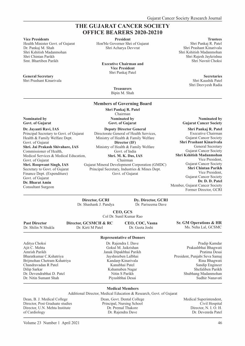
Director, GCRI�Dr. Shashank J. Pandya�
Dy. Director, GCRI�Dr. Pariseema Dave�
CEO, GCS�Col Dr. Sunil Kumar Rao�
Sr. GM Operations & HRMs. Neha Lal, GCSMC
Past Director�Dr. Shilin N Shukla�
Director, GCSMCH & RC Dr. Kirti M Patel
CEO, COC, VasnaDr. Geeta Joshi
Medical MembersAdditional Director, Medical Education & Research, Govt. of Gujarat
Dean, B. J. Medical College Director, Post Graduate studies Director, U.N. Mehta Institute of Cardiology
Medical Superintendent,Civil Hospital
Director, N. I. O. H.Dr. Devenrda Patel
Dean, Govt. Dental College Principal, Nursing School
Dr. Premal Thakore Dr. Rajendra Dave
THE GUJARAT CANCER SOCIETYOFFICE BEARERS 2020-20210
Executive Chairman andVice President
Shri Pankaj PatelGeneral Secretary Shri Prashant Kinarivala
SecretariesShri Kaushik Patel
Shri Deevyesh RadiaTreasurers
Bipin M. Shah
PresidentHon'ble Governer Shri of Gujarat
Shri Acharya Devvrat
TrusteesShri Pankaj R. Patel
Shri Prashant KinarivalaShri Kshitish Madanmohan
Shri Rajesh JaykrishnaShri Navnit Choksi
Vice PresidentsHealth Minister Govt. of GujaratDr. Pankaj M. ShahShri Kshitish MadanmohanShri Chintan ParikhSmt. Bhartiben Parikh
Representative of Donors
Aditya Choksi Ajit C. MehtaAmrish Parikh Bharatkumar C.KshatriyaBrijmohan Chetram KshatriyaChandravadan R PatelDilip SarkarDr. Devendrabhai D. PatelDr. Nitin Sumant Shah
Dr. Rajendra I. DaveGokul M. Jaikrishan
Janak Dipakbhai ParikhJayshreeben LalbhaiKandarp Kinarivala
Kanubhai PatelKshamaben Nagar
Nitin S ParikhPiyushbhai Desai
Pradip KamdarPrakashbhai Bhagwati
Pratima DesaiPresident, Punjabi Seva Samaj
Rina BhagwatiSandip Engineer
Shefaliben ParikhShubhang Madanmohan
Sudhir Nanavati
Shri Pankaj R. PatelChairman
Members of Governing Board
Nominated byGovt. of Gujarat
Nominated byGovt. of Gujarat
Nominated byGujarat Cancer Society
Dr. Jayanti Ravi, IASPrincipal Secretary to Govt. of Gujarat Health & Family Welfare Dept. Govt. of GujaratShri. Jai Prakash Shivahare, IASCommissioner of Health,Medical Services & Medical Education,Govt. of GujaratShri. Roopwant Singh, IASSecretary to Govt. of Gujarat Finance Dept. (Expenditure) Govt. of GujaratDr. Bharat AminConsultant Surgeon
Deputy Director General Directorate General of Health Services,Ministry of Health & Family Welfare
Director (IF) Ministry of Health & Family Welfare
Govt. of IndiaShri. M. K. Das, IAS
ChairmanGujarat Mineral Development Corporation (GMDC)
Principal Secretary, Industries & Mines Dept.Govt. of Gujarat
Shri Pankaj R. PatelExecutive Chairman
Gujarat Cancer SocietyShri Prashant Kinarivala
General SecretaryGujarat Cancer Society
Shri Kshitish MadanmohanVice President,
Gujarat Cancer SocietyShri Chintan Parikh
Vice President,Gujarat Cancer Society
Dr. D. D. PatelMember, Gujarat Cancer Society
Former Director, GCRI
Gujarat Cancer Society Research Journal
46Volume 23 Number 1 April 2021
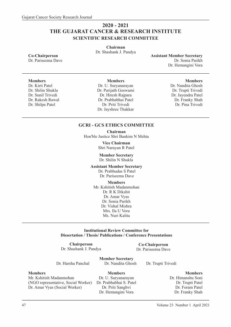
2020 - 2021THE GUJARAT CANCER & RESEARCH INSTITUTE
SCIENTIFIC RESEARCH COMMITTEE
ChairmanDr. Shashank J. Pandya
Assistant Member SecretaryDr. Sonia Parikh
Dr. Hemangini Vora�������������
Co-Chairperson�Dr. Pariseema Dave
MembersDr. Kirti PatelDr. Shilin ShuklaDr. Sunil TrivediDr. Rakesh RawalDr. Shilpa Patel
MembersDr. U. SuryanarayanDr. Parijath Goswami
Dr. Hitesh RajparaDr. Prabhabhai Patel
Dr. Priti TrivediDr. Jayshree Thakkar
MembersDr. Nandita GhoshDr. Trupti TrivediDr. Jayendra Patel
Dr. Franky ShahDr. Pina Trivedi
Institutional Review Committee forDissertation / Thesis/ Publications / Conference Presentations
Members Mr. Kshitish Madanmohan(NGO representative, Social Worker)Dr. Amar Vyas (Social Worker)
Members
Dr. U. SuryanarayanDr. Prabhabhai S. Patel
Dr. Priti SanghviDr. Hemangini Vora
Members Dr. Himanshu Soni
Dr. Trupti PatelDr. Foram PatelDr. Franky Shah
Co-Chairperson�Dr. Pariseema Dave
Chairperson Dr. Shashank J. Pandya
Member Secretary Dr. Harsha Panchal Dr. Nandita Ghosh Dr. Trupti Trivedi
GCRI - GCS ETHICS COMMITTEE
Assistant Member Secretary Dr. Prabhudas S Patel Dr. Pariseema Dave
Member Secretary�����Dr. Shilin N Shukla �
Vice ChairmanShri Narayan R Patel
ChairmanHon'ble Justice Shri Bankim N Mehta
MembersMr. Kshitish Madanmohan
Dr. R K DikshitDr. Amar Vyas
Dr. Sonia ParikhDr. Vishal MishraMrs. Ila U VoraMs. Nuri Kalita
Gujarat Cancer Society Research Journal
47 Volume 23 Number 1 April 2021
Information Technology Department
All Donations are exempted from Income Tax Under IT Act 35(i)(ii)(175%), 35AC(100%) & 80G(50%) Donations in Foreign Currencies Accepted approval
vide Reg. No.041910257 Dated 22-03-2001. Visit Us at on http://gcriindia.org E-mail:[email protected]
Maintenance Complain Attending by Engineer
IT Helpdesk Team
IT Department Room
Data Center Room, IT Department
Printed by: Shrinath Print Art