Embed Size (px)
Citation preview
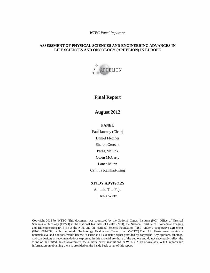
WTEC Panel Report on
ASSESSMENT OF PHYSICAL SCIENCES AND ENGINEERING ADVANCES IN LIFE SCIENCES AND ONCOLOGY (APHELION) IN EUROPE
Final Report
August 2012
PANEL
Paul Janmey (Chair)
Daniel Fletcher
Sharon Gerecht
Parag Mallick
Owen McCarty
Lance Munn
Cynthia Reinhart-King
STUDY ADVISORS
Antonio Tito Fojo
Denis Wirtz
Copyright 2012 by WTEC. This document was sponsored by the National Cancer Institute (NCI) Office of Physical Sciences – Oncology (OPSO) at the National Institutes of Health (NIH), the National Institute of Biomedical Imaging and Bioengineering (NIBIB) at the NIH, and the National Science Foundation (NSF) under a cooperative agreement (ENG 0844639) with the World Technology Evaluation Center, Inc. (WTEC).The U.S. Government retains a nonexclusive and nontransferable license to exercise all exclusive rights provided by copyright. Any opinions, findings, and conclusions or recommendations expressed in this material are those of the authors and do not necessarily reflect the views of the United States Government, the authors’ parent institutions, or WTEC. A list of available WTEC reports and information on obtaining them is provided on the inside back cover of this report.

WORLD TECHNOLOGY EVALUATION CENTER, INC. (WTEC) R.D. Shelton, President
Michael DeHaemer, Executive Vice President Geoffrey M. Holdridge, Vice President for Government Services
Frank Huband, Senior Vice President and General Counsel Patricia Foland, Vice President for Operations
Hassan Ali, Project Manager Hemant Sarin, International Policy Fellow
Sarah Michaud, Report Editor Grant Lewison, Advance Contractor
ACKNOWLEDGMENTS
We at WTEC wish to thank all the panelists for their valuable insights and their dedicated work in conducting this international assessment of physical sciences and engineering advances in life sciences and oncology (APHELION) research and development, and to thank all the site visit hosts for so generously sharing their time, expertise, and facilities with us. For their sponsorship of this important study, our sincere thanks go to the National Cancer Institute at the National Institutes of Health (NIH), the National Science Foundation, and the National Institute of Biomedical Imaging and Bioengineering at the NIH.
.
R.D. Shelton, President, WTEC
WTEC Mission WTEC provides assessments of international research and development in selected technologies under awards from the National Science Foundation (NSF), the Office of Naval Research, and other agencies. Formerly part of Loyola College, WTEC is now a separate nonprofit research institute. The Deputy Assistant Director for Engineering is NSF Program Director for WTEC. Sponsors interested in international technology assessments and related studies can provide support for the program through NSF or directly through separate grants or GSA task orders to WTEC.
WTEC’s mission is to inform U.S. scientists, engineers, and policy makers of global trends in science and technology. WTEC assessments cover basic research, advanced development, and applications. Panels of typically six technical experts conduct WTEC assessments. Panelists are leading authorities in their field, technically active, and knowledgeable about U.S. and foreign research programs. As part of the assessment process, panels visit and carry out extensive discussions with foreign scientists and engineers in their labs.
The WTEC staff helps select topics, recruits expert panelists, arranges study visits to foreign laboratories, organizes workshop presentations, and finally, edits and publishes the final reports. Dr. R.D. Shelton, President, is the WTEC point of contact: telephone 410-691-1579 or email [email protected].


Table of Contents Preface ............................................................................................................................................ xiii
Executive Summary ....................................................................................................................... xv
Introduction ...................................................................................................................................... 1
Overview ............................................................................................................................................... 2
Complexity and Information: Cancer as a Multi-Scale Complex Adaptive System ................. 5
Introduction ........................................................................................................................................... 5 Research ................................................................................................................................................ 7 Discussion ........................................................................................................................................... 19
Mimicking the Microenvironment ................................................................................................ 23
Introduction ......................................................................................................................................... 23 Research .............................................................................................................................................. 24 Discussion ........................................................................................................................................... 31
Cancer Cell Mechanics .................................................................................................................. 35
Introduction ......................................................................................................................................... 35 Research .............................................................................................................................................. 36 Discussion ........................................................................................................................................... 43
Fluid Mechanics and Transport in Tumors ................................................................................. 47
Introduction ......................................................................................................................................... 47 Research .............................................................................................................................................. 47 Discussion ........................................................................................................................................... 55
The Dynamics of Cell Motility ...................................................................................................... 59
Introduction ......................................................................................................................................... 59 Research .............................................................................................................................................. 60 Discussion ........................................................................................................................................... 71
Devices and New Diagnostic Principles ........................................................................................ 75
Introduction ......................................................................................................................................... 75 Research .............................................................................................................................................. 75 Discussion ........................................................................................................................................... 81
Appendix A. APHELION Study Panelists and Advisors ........................................................... 83
Panelists ............................................................................................................................................... 83 Advisors............................................................................................................................................... 85
Appendix B. Site Visit Reports ..................................................................................................... 87
École Polytechnique Fédérale de Lausanne (EPFL) ........................................................................... 88 European Institute of Oncology ........................................................................................................... 94 Hubrecht Institute ................................................................................................................................ 96 Institut Curie ...................................................................................................................................... 100 Max Planck Society, Dresden ............................................................................................................ 104 Max Planck Institute for Dynamics and Self-Organization, Göttingen ............................................. 106 Novocure Limited/Technion ............................................................................................................. 108 St Radboud University Nijmegen Medical Center ............................................................................ 110 The Royal Institute of Technology (KTH) ........................................................................................ 116
vi
Technical University of Munich ........................................................................................................ 120 University of Leipzig ......................................................................................................................... 128 University Medical Center Utrecht .................................................................................................... 131 University of Barcelona ..................................................................................................................... 133 University of Basel ............................................................................................................................ 141 University of the Basque Country ...................................................................................................... 143 University of Freiburg ........................................................................................................................ 146 University of Heidelberg .................................................................................................................... 151 Instituut-Lorentz ................................................................................................................................ 159 University of Milan ............................................................................................................................ 161 University of Mons ............................................................................................................................ 163 University of Nürnberg-Erlangen ...................................................................................................... 165 Venetian Institute of Molecular Medicine ......................................................................................... 168 University of Paris Diderot ................................................................................................................ 173 University of Rostock ........................................................................................................................ 179 Uppsala University and Science for Life Laboratory Uppsala .......................................................... 182 Weizmann Institute of Science .......................................................................................................... 186
vii
List of Figures Figure 1. Countries visited by APHELION panel members. .................................................. xxiii Figure 1.1. Two possible topologies of a regulatory network (Kollmann et al., 2005). ................... 9 Figure 1.2. The structure of the lac operon (Vilar and Leibler, 2003). ............................................ 9 Figure 1.3. Quantitative modeling of control gene expression by modulated self-assembly of the
retinoid X receptor (RXR). The model considers how intracellular signals are processed through modulated self-assembly into populations of different RXR oligomeric species that upon DNA binding engage in transcriptional control (J.M.G. Vilar and L. Saiz, 2011). ......... 10
Figure 1.4. Two computational approaches for determining the 3D structure of genomic domains and genomes (Marti-Renom and Mirny, 2011). ........................................................................ 10
Figure 1.5. A partially unwrapped nucleosome with exposed nucleosomal binding sites (stars). The nucleosome can lower its energy by closing those binding sites at the cost of bending the DNA (Prinsen and Schiessel, 2010). ......................................................................................... 11
Figure 1.6. A pendulum example can be used to explain a dynamical system (courtesy of Parag Mallick). .................................................................................................................................... 12
Figure 1.7. Nested model structure (Maus et al., 2011). ................................................................ 13 Figure 1.8. Schematic description of the example model (Bittig et al., 2011). .............................. 14 Figure 1.9. The long-term phenotypic response of a cell can be described as a state space (Huang
and Ingber 2006-2007). ............................................................................................................. 15 Figure 1.10. A 2D microwell molded in hydrogel (Gobaa et al., 2011)........................................... 15 Figure 1.11. An actin/myosin motility assay that shows that the motion is density dependent
(Schaller et al., 2010). ............................................................................................................... 16 Figure 1.12. High-resolution maps of stress components within an advancing monolayer sheet of
cells (Trepat and Fredberg 2011). ............................................................................................. 17 Figure 1.13. Spatial distribution of evolving cell populations (Hallatschek et al., 2007). ............... 18 Figure 1.14. Colon cancer model for understanding the growth of intestinal epithelial cells out of
the crypt (Martens et al., 2011). ................................................................................................ 18 Figure 1.15. Mathematical population dynamics model (La Porta et al., 2012). ............................. 19 Figure 2.1. Micellar block copolymer lithography and biofunctionalization. ................................ 25 Figure 2.2. Formation of a micro-epidermis on a collagen island. ................................................. 27 Figure 2.3. Microfluidic gas exchanger. ......................................................................................... 28 Figure 2.4. Compliance and nanoparticle decoration properties of PEG- diacrylate (DA)
hydrogels. .................................................................................................................................. 29 Figure 2.5. Engineered 3D microenvironment. .............................................................................. 31 Figure 3.1. The field of cell mechanics can be divided into three main subsections: cellular
mechanical properties, imposed forces, and cell-generated forces (courtesy of Reinhart-King).36 Figure 3.2. The metastatic cascade depicted through a cartoon and accompanying intravital
images (Beerling et al., 2011). .................................................................................................. 37 Figure 3.3 Methods to measure the mechanical properties of cells............................................... 38 Figure 3.4. Deformability of cells increases with metastatic potential, as measured using an
optical stretcher (Guck et al., 2005). ......................................................................................... 39 Figure 3.5. Methods to measure cell-generated traction stresses. .................................................. 40 Figure 3.6. Traction stresses increase with metastatic potential. ................................................... 41 Figure 3.7. Mechanical stress inhibits tumor growth. Spheroids were subjected to imposed
pressures and their growth monitored over two weeks (Montel et al., 2011). .......................... 42 Figure 4.1. The fluid microenvironment in and around tumors. .................................................... 48 Figure 4.2. Heterogeneous delivery of 90 nm liposomes in a mouse tumor model. Some regions
receive many particles, but many have no exposure. If this were a drug, treatment would be incomplete. Image from (Yuan et al., 1994a). .......................................................................... 48
viii
Figure 4.3. Cancer cells producing CCL21 chemokine detect the direction of interstitial flow because of the shift in the local chemical gradient. .................................................................. 50
Figure 4.4. Lymphangiogenesis is directed by fluid flow in the mouse tail. ................................. 50 Figure 4.5. Microfluidic device with localized 3D extracellular matrix for fluid force-mediated
angiogenic sprouting and morphogenesis. ............................................................................... 51 Figure 4.6. Angioadaptation in normal and tumor vasculatures (Pries et al., 2011). .................... 53 Figure 4.7. Upstream and downstream signals are critical for distributing flow correctly between
AV shunts and capillary beds. It is possible that these signals are absent in tumors (Pries et al., 2010). 54
Figure 4.8. Lymphatic vessel pumping dynamics. ........................................................................ 55 Figure 4.9. Valve structures in collecting lymphatic vessels in normal (left) and peritumor (right)
tissue. The valve leaflets in the peritumor lymphatic are malformed and unable to close to prevent back flow (Hagendoorn et al., 2006). .......................................................................... 55
Figure 4.10. Fluid pathways in the tumor microenvironment. ........................................................ 56 Figure 5.1. Rho, Rac, and Cdc42 control the assembly and organization of the actin cytoskeleton.60 Figure 5.2. Rho GTPase signaling to protrusion and adhesion. .................................................... 61 Figure 5.3. Integrins and extracellular matrix in mechanotransduction. ....................................... 62 Figure 5.4. Model of adhering membrane. The effective binding affinity of a membrane for a
surface is governed by the receptor density, (h) separation distance, and (l) receptor length (Reister-Gottfried et al., 2008). ................................................................................................ 63
Figure 5.5 Normalized total adhered area and number density of bonds. .................................... 63 Figure 5.6. Experimental design to characterize spatially and temporally coordinated migration.
(Cavalcanti-Adam et al., 2006) ................................................................................................ 64 Figure 5.7. Lateral spacing of integrin ligands regulates cell spreading and focal adhesion
assembly (Cavalcanti-Adam et al., 2006). ................................................................................ 64 Figure 5.8. Model of the rearrangement of the actin cytoskeleton. ............................................... 65 Figure 5.9. Experimental model of the collective dynamics of actin cytoskeletal networks
(courtesy of Andreas Baush of the Technical University of Munich). ..................................... 66 Figure 5.10. Structure and dynamics of actin/fascin/myosin networks. .......................................... 66 Figure 5.11. Actin cytoskeletal and focal adhesion organization in M.D.A-MB-231 cells invading
in modified Circular Invasion Assay (CIA). ............................................................................ 68 Figure 5.12. Collective cell migration. ............................................................................................ 69 Figure 5.13. Collective cell dynamics during dorsal closure (DC). ................................................ 70 Figure 6.1. Images of human dermatofibrosarcoma protuberans from (Friedl et al., 2007). ........ 76 Figure 6.2. The optical stretcher deforms cells with light, allowing non-contact measurement of
mechanical properties after exposure (Guck et al., 2000). ....................................................... 78 Figure 6.3. Magnetic particles and biological vesicles from (Lesieur et al., 2011). ...................... 78 Figure 6.4. Tumor treating fields from (Kirson et al., 2007). ........................................................ 80 Figure B.1. Fibronectin fragment including a factor XIIIa substrate fibrin-binding sequence
(courtesy of Jeffrey Hubbell, École Polytechnique Fédérale de Lausanne). ............................ 89 Figure B.2. VEGF-C expression in tumors interferes with the normal immune response to OVA
(courtesy of Jennifer Munson, École Polytechnique Fédérale de Lausanne). .......................... 90 Figure B.3. Creating artificial niches for stem cell culture using micropatterned assays (Lutolf et
al., 2009). .................................................................................................................................. 91 Figure B.4. Neuronal connections mapped by the Blue Brain Project (courtesy of BBP/ École
Polytechnique Fédérale de Lausanne 2012). ............................................................................ 91 Figure B.5. VE-cadherin regulation of cell-cell junctions. ............................................................ 97 Figure B.6. Intravital Microscopy (IVM) of individual steps of metastasis (from Beerling et al.,
2011). 98
ix
Figure B.7. Motor oscillations. Oscillations of molecular motor assemblies obtained from stochastic simulations. ............................................................................................................. 102
Figure B.8. How are cell and tissue shapes dictated by the generation of forces inside the cell at the molecular level? How are the original properties of active matter, driven out of equilibrium by ATP hydrolysis, related to biological processes? ........................................... 105
Figure B.9. Spatial structure increases the waiting time for cancer (Martens et al. 2011; www.evo.ds.mpg.de/). ............................................................................................................ 107
Figure B.10. AC field distribution in and around quiescent (A) and dividing (B) cells (Kirson et al., 2007). 109
Figure B.11. Examples of 3D tissue reconstructions obtained by intravital multiphoton microscopy and FLIM. ............................................................................................................................... 111
Figure B.12. Protease-dependent and non-proteolytic cancer cell migration. ................................ 112 Figure B.13. Cells can migrate individually or collectively as multicellular groups. ..................... 113 Figure B.14. Regulation of keratinocyte shape and differentiation on micropatterned substrates . 114 Figure B.15. Overview of the KTH Life Science Technologies Platform (Wouter van der
Wijngaart, courtesy of The Royal Institute of Technology). .................................................. 117 Figure B.16. Self-organizing actin filaments (Schaller et al., 2010). .............................................. 122 Figure B.17. Targeting early cancer and osteoporosis diagnostics (courtesy of Franz Pfeiffer,
Technical University of Munich). ........................................................................................... 123 Figure B.18. A conventional X-ray (left) and DPC image (right) of a fish (Pfeiffer et al., 2006). . 123 Figure B.19. MHC class 1 molecule (courtesy of Martin Zacharias, Technical University of
Munich). .................................................................................................................................. 123 Figure B.20. Scaffold and staple molecules (courtesy of Hendrik Dietz, Technical University of
Munich). .................................................................................................................................. 124 Figure B.21. Complex shapes obtained by using scaffold and staple molecules (Franco et al.,
2011). 124 Figure B.22. Synthetic transcriptional circuit (Franco et al., 2011). ............................................... 125 Figure B.23. Diagram of the rocking motion of yeast Hsp90 (Ratzke et al., 2010). ...................... 126 Figure B.24. As the tumor progresses, the number of cells that are soft under small deformations
increases (courtesy of Josef Käs, University of Leipzig). ....................................................... 129 Figure B.25. Pathways that inhibit formation of VWF-platelet aggregates (Lenting et al., 2010). 132 Figure B.26. Illustration of the 4-step protocol based on FE-AFM tips used to probe cell
mechanoresponses to compression and extension................................................................... 135 Figure B.27. Transmitted light image of the RPME cell monolayer (a) and the MDCK cell
monolayer (b). ......................................................................................................................... 136 Figure B.28. AS cell dynamics. ...................................................................................................... 138 Figure B.29. Experimental setup..................................................................................................... 139 Figure B.30. Quantitative modeling of control of gene expression by modulated self-assembly of
the Retinoid X Receptor (RXR). ............................................................................................. 144 Figure B.31. Experimental and computational approach to look at the very simple and measurable
system of Che proteins (adapted from Kollmann et al., 2005). ............................................... 148 Figure B.32. Definition of cell states (courtesy of Hauke Busch, University of Freiburg). ........... 149 Figure B.33. A schematic presentation of RGD and EGF surface functionalization. ..................... 153 Figure B.34. Traction force microscopy studies of cell movement. ............................................... 154 Figure B.35. Microscope based on light-sheet illumination that allows massively parallel
fluorescence correlation spectroscopy measurements. ............................................................ 156 Figure B.36. Nucleosome dynamics. .............................................................................................. 160 Figure B.37. Stochastic and cancer stem cell model for tumors (La Porta et al., 2012). ................ 162 Figure B.38. Cross talk between integrins and cadherins (courtesy of Sylvain Gabriele, University
of Mons). ................................................................................................................................. 164
x
Figure B.39. Model of adhering membrane. .................................................................................. 166 Figure B.40 Modeling the dynamics of cell adhesion. .................................................................. 166 Figure B.41. Effect of hypoxia on calcium transients. ................................................................... 170 Figure B.42. Atomic force microscopy nanografting was utilized to prepare DNA nanopatches of
different sizes (200 × 200 to 1000 × 1000 nm2) onto which DNA-protein conjugates can be anchored through DNA-directed immobilization. .................................................................. 171
Figure B.43. MCA-203 fibrosarcoma tumor samples from mice that received intratumoral injections of CCL2 (0.5 µg in hydrogel) were stained for CD3 by immunohistochemistry. The graphs represent the quantification of immunoreactive cells. Scale bar: 50 µm (courtesy of B. Molon). 172
Figure B.44. Gallet’s research addresses epithelial microanatomy and the cross-talk between cadherin and integrin adhesions. ............................................................................................ 174
Figure B.45. Applying high or low force (FH, FL, respectively) to a cell results in different dynamics of reciprocal force generation. This could explain why cells polarize on anisotropic substrates along the stiffest axis (Fouchard et al., 2011). ....................................................... 175
Figure B.46. Macromolecule translocation through nanopores. As temperature increases, thermal unfolding allows passage through the pores (courtesy of L. Auvray). ................................... 175
Figure B.47. Development of the chick vasculature. .................................................................... 176 Figure B.48. Applying cyclic forces to beads attached to integrins results in cell stiffening and
concentration of actin at the bead focal adhesions (courtesy of S. Hénon). ........................... 177 Figure B.49. Nested model structure (Maus et al., 2011). ............................................................. 180 Figure B.50. Schematic description of the example model. ........................................................... 181 Figure B.51. DDE uptake in a representative healthy control (left) and a whiplash patient (right).183 Figure B.52. The role of neural stem cells on brain development and brain cancer
(www.igp.uu.se/Research/Cancer_and_vascular_biology/karin_forsberg_nilsson/). ............ 184 Figure B.53. Two T lymphocytes sending invasive filopodia into the body of a cytokine stimulated
endothelial cell in the presence of shear forces. ..................................................................... 188 Figure B.54. Phase diagram of the system representing the different regimes of focal adhesion
aggregate assembly-disassembly corresponding to different ranges of three system parameters.189 Figure B.55. Schematic description of the model for actin polymerization-driven cell protrusion.190 Figure B.56. The synthetic DNA brush. ......................................................................................... 190 Figure B.57. Schematic diagram by which cell contractility generated by actin-myosin interactions
generates a fore dipole at the cell/substrate interface. Image from www.weizmann.ac.il/Biological_Physics/photos/gallery.html. ............................................. 191
Figure B.58. The effect of axial cell elongation on stress-fiber polarization and experimental values of the order parameter S for different elastic substrates. ........................................................ 191

xi
List of Tables Table 1. Sites Visited in Europe .................................................................................................... xxiv

xii
xiii
PREFACE
The National Cancer Institute (NCI) Office of Physical Sciences – Oncology (OPSO) has initiated a large-scale interdisciplinary research effort at the interface of the physical sciences and oncology through 12 centers located throughout the United States. The NCI Physical Sciences-Oncology Center (PS-OC) program brings together experts from physics/engineering and cancer biology/oncology to enable cross-disciplinary research that merges these fields and defines a new physics of cancer. Physicists strive to explain nature by precise mathematical equations, which could bring a new perspective to cancer research. From a molecular perspective, cancer is not a specific disease. Cancer arises as a result of a succession of randomly occurring mutations. Tumors are inherently molecularly diverse. This complexity might give the wrong impression that cancer is not accessible to physics, which strives to describe nature by precise quantitative laws. Nevertheless, statistical physics has proven to be able to find the laws behind the stochastic processes underlying thermodynamics, and nonlinear dynamics has even uncovered the principles that govern chaotic behavior in nature. Molecular background and pathogenesis of solid tumors may vary, but the pattern of tumor progression—uncontrolled proliferation, invasive growth, and metastasis—is the same. Defining and unifying physical laws that are rooted in soft matter physics are required to understand these three functions. The concept of functional modules developed in biological physics will greatly facilitate understanding the laws that govern tumor progression. In tumor cells, the modules that are responsible for division, tumor growth, and metastasis may not have identical molecular architecture, but the same physics is essential for their functions. All cells in a tissue can be motile and are viscous on long time scales, behaving very much like liquid droplets. Consequentially, tissue boundaries are comparable to fluid boundaries. Tissues can be described as a new form of fluid matter, which is a significant topic in the novel research area of active soft matter. The most common chemotherapy agents act by killing cells that divide rapidly. Newer anticancer drugs act directly against cancer-specific proteins or inhibit tumor angiogenesis. In all these cases the goal is to reduce the tumor. Yet, the primary tumor can often be removed by surgery and radiation. It is the residual tumor cells and their ability to transgress boundaries that have to be hindered. Inducing changes in tumor physical and material properties of tumor cells that disrupt the functional modules required for metastasis will provide a broad treatment option. The physics of cancer is substantially more than providing new techniques for oncology. Soft matter physics as a basis for the physics of cancer has been strong in Europe. Institutions such as the Institute Curie in Paris have traditionally demonstrated that a solid connection between physics and medicine is feasible. As well, the German strength in cell biophysics has provided a good foundation. The NCI PS-OC program, which is unfortunately not yet paralleled in Europe, will jump-start the physics of cancer throughout the United States and will serve to guide similar initiatives worldwide.
Prof. Josef A. Käs Principal Investigator and Head of Division University of Leipzig Faculty of Physics and Earth Science Institute for Experimental Physics I Soft Matter Physics Division.

xiv

EXECUTIVE SUMMARY
Paul Janmey More than 40 years ago, the U.S. Government declared a war on cancer and committed to investing in laboratory and clinical research in order to understand the causes of cancer and thereby aid its diagnosis, treatment, and cure. This research program in the cell biology, genetics, biochemistry, and animal models of cancer has led to enormous advances in many areas of science and important improvements in the diagnosis and treatment of many cancers. However, the “war” has in significant ways become a stalemate: In contrast to the enormous advances in the prevention, treatment, and cure of infectious disease, cardiovascular disease, and other major causes of death throughout the world—diagnosis of cancer is as devastating a reality today as it was decades ago. The more we learn about cancer biology, the more it has become apparent that: the relationship between a specific gene mutation and disease is often staggeringly complex; the interaction of cancer cells with their local environment is an essential but largely obscure aspect of the disease; and traditional methods of cancer biology research might not be sufficient to produce the results required for effective clinical improvements. To address these issues, the National Cancer Institute (NCI) held a series of three Physical Sciences in Oncology Workshops and Think Tanks between February and October 2008 (http://opso.cancer.gov/workshops/). The aim was to explore the opportunities to advance cancer research by integrating physical scientists and physical sciences approaches with the more traditional research effort in cancer biology. The ideas and discussions at these meetings helped guide an initiative within the NCI to establish an Office of Physical Sciences-Oncology (OPSO). The OPSO facilitates the development and implementation of physical science-based initiatives supporting cancer research for the NCI and integrates such efforts in other divisions of the National Institutes of Health (NIH) such as the National Institute of Biomedical Imaging and Bioengineering (NIBIB) and throughout the research community. The focus of this effort is to go beyond involving physicists and engineers in the development of new instrumentation or methods, and to engage in the methods and concepts from the physical sciences to foster discoveries and new fields of study related to cancer research. Broadly speaking, the goal is to study cancer from non-traditional perspectives, such as the physical sciences, and integrate those perspectives with existing knowledge about cancer in order to allow for a more comprehensive understanding of the disease. The National Science Foundation (NSF) has traditionally been involved in funding research in engineering and the physical sciences. Such efforts can have an important impact in biomedical research while maintaining a fundamentally basic research perspective. With this in mind, the NCI recently established a collaboration with the NSF for Physical and Life Sciences Early Research (PLIER) awards to establish stronger ties with the physical sciences and engineering communities. The NSF and NCI have collaborated on a funding opportunity, administered by the NSF, titled Physical and Engineering Sciences in Oncology (PESO) Awards that dovetail with the efforts of the OPSO. The main mission of the OPSO is to fund physical sciences-oncology research throughout the United States. As an initial thrust, 12 leading institutions were selected in 2009 to build a collaborative network of NCI Physical Science-Oncology Centers (PS-OCs) (http://opso.cancer.gov/centers/).
The PS-OCs are now midway into their initial assignments. To compare the missions of this and other initiatives with related research efforts abroad, the NCI, in co-sponsorship with the NSF and the NIBIB, commissioned the World Technology Evaluation Center (WTEC), Inc. to undertake an international Assessment of PHysical sciences and Engineering advances in LIfe sciences and ONcology (APHELION).
xvi Executive Summary
The initial phase of APHELION was to determine the status and trends of applying physical sciences and engineering principles to oncology research and development in leading laboratories and organizations in Europe via an on-site peer review process. More details on the study are available at: www.wtec.org/aphelion. On 18 January 2012, the sponsors/chair meeting of the WTEC APHELION study was held at NSF headquarters. The main goals of the sponsors meeting were to provide an overview of the plans for the study, to solicit interest and participation of other U.S. government agencies, and to coordinate the study design with WTEC. On 1 February 2012, the kickoff meeting of the WTEC APHELION study was held at NIH’s Bethesda, MD, campus where the scientific panel and advisors met with the sponsors.
On 12 June 2012 a final workshop presented by the panel members was held at the Cloisters Building lecture room on the NIH campus to report the findings of the study group and discuss these findings with the sponsors and the public. Details and documents presented at this workshop are available at www.tvworldwide.com/events/nih/120612/.
Scientific Panel Members
• Paul Janmey, Ph.D. (study chair). Professor of Physiology, Physics, and Bioengineering at the Institute of Medicine and Engineering at the University of Pennsylvania.
• Daniel Fletcher, Ph.D., D.Phil. Professor of Bioengineering and Biophysics at the University of California, Berkeley.
• Sharon Gerecht, Ph.D. Assistant Professor of Chemical and Biomolecular Engineering at Johns Hopkins University.
• Parag Mallick, Ph.D. Assistant Professor of Radiology, Bio-X Program, at the Canary Center for Cancer Early Detection, Stanford University.
• Owen McCarty, Ph.D. Associate Professor of Biomedical Engineering at the Oregon Health and Science University.
• Lance Munn, Ph.D. Associate Professor of Radiation Oncology at the Massachusetts General Hospital/Harvard Medical School.
• Cynthia Reinhart-King, Ph.D. Assistant Professor of Biomedical Engineering at Cornell University.
Expert Advisors to the Study Panel
• Antonio Tito Fojo, M.D., Ph.D. Head, Experimental Therapeutics Section Medical Oncology Branch and Affiliates, National Institutes of Health.
• Denis Wirtz, Ph.D. Theophilus H. Smoot Professor, Department of Chemical and Biomolecular Engineering, Johns Hopkins University.
Short biographies of the panel members and advisors are provided in Appendix A.
The Goals of the APHELION Study
1. Compare the United States research and development activities related to the interface between physics and oncology, or more generally between physical science and biomedicine, with similar work being done in Europe.
2. Identify the gaps and barriers for research groups and clinicians in the United States by working with leading European institutions.
3. Identify major innovations that are emerging abroad.
Paul Janmey xvii
4. Identify opportunities for cooperation and collaboration with research groups and industry in Europe.
5. Guide U.S. research investments at the physics/oncology interface. The initial meeting of the panel members and sponsors allowed for extensive discussion of the specific topic areas of interest. The panel identified areas of research and technology development with the greatest potential to advance understanding and treatment of cancer and other diseases. As a result of this exchange, six topic areas were identified. Each panel member took responsibility for one topic and analyzed information collected during the site visits and integrated their findings with the current state of understanding. The following topics form the basis for each of the chapters in this book:
1. Information and complexity: New methods for dealing with the enormous data sets generated by modern imaging methods and integrating methods developed by the physics community to understand complex, non-linear systems and emergent properties that cannot be predicted by traditional biological models.
2. Microenvironment: The influence of chemical composition, spatial patterning, nutrient supply, oxygen stress, and other features of the tissue and extracellular environment on the growth and homeostasis of normal tissues and tumors.
3. Cell and tissue mechanics: How the forces generated by cells and the viscoelasticity of the cell and extracellular matrix affect cell growth, survival, differentiation, and movement.
4. Transport: How the movement of cancer cells, nutrients, growth factors, drugs, and fluids affect cell survival and tissue mechanics. How the removal of metabolic waste products and cell debris are controlled and how they are altered in the tumor environment.
5. Dynamics: How the rates and patterns of cell shape change, migration, and division can be measured, understood, and integrated with biochemical and genetic information.
6. Devices and new diagnostic principles: New technologies based on physical principles, especially those in which the physical properties of tissues are exploited for cancer diagnosis or treatment.
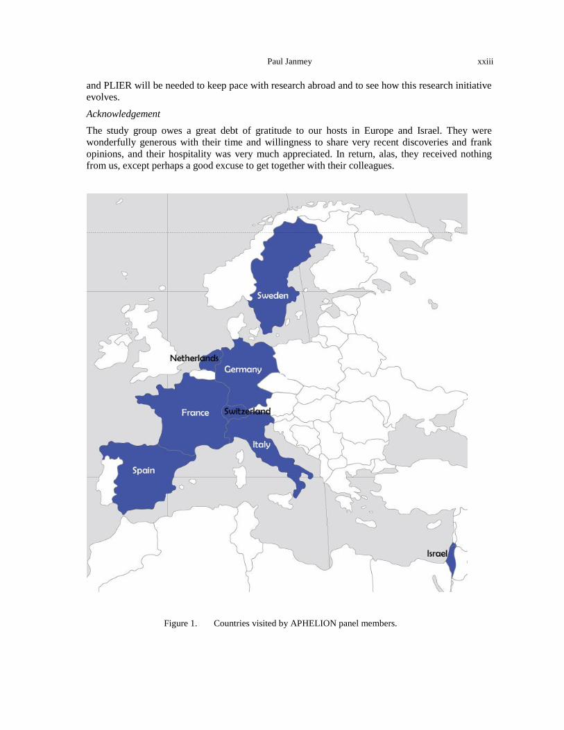
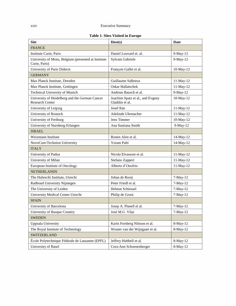
The panel members made visits to laboratories in France, Italy, Israel, Germany, the Netherlands, Spain, Sweden, and Switzerland, typically meeting with representatives of multiple institutions at each stop (Figure 1, Table 1). A complete list of the sites visited and assessments of their activities in the physics/biomedicine interface are provided in Appendix B.
Outcomes and Summary of Findings
The purpose of our visits was focused on learning about new scientific advances and plans for future studies. We also examined each institution’s facilities, traditions, advantages, and challenges related to performing transdisciplinary or multidisciplinary work. There was a clear perception among the investigators at every site that the interface of physical and biomedical sciences is a growth area with potential for both scientific discovery and medical applications. In a few institutions, such studies have become part of the established curriculum and research programs, such as the Institut Curie. At institutions in Heidelberg and Munich there was even a sense that a critical mass of researchers at this interface might already have been reached. In most institutions we visited, interdisciplinary studies at the physics/biomedicine interface were highly attractive to graduate students and young faculty and were often well supported by granting agencies. There was also a strong sense voiced by many of our hosts that interdisciplinarity cannot be optimized without a firm basic grounding in a specific physical or biological science in the education of students and young researchers.
xviii Executive Summary
Throughout Europe there is evidence that the vision of NCI and NSF to engage physics more deeply in cancer research coincided with initiatives based on similar beliefs that engagement of not only physicists, but the concepts and methods of physics research could benefit cancer research. One example of this type of initiative is the document, “Progress in the Domain of Physics Applications in Life Science with an Invention for Substantial Reduction of Premature Cancer Deaths: The Need for a Paradigm Change in Oncology Research” (www.crosettofoundation .org/uploads/371.pdf), which received nearly 1,000 signatures between 29-31 January 2010. The document argues for the need to engage new ways of thinking in cancer research, including integration of physical science and scientists to combat cancer. This study surveyed World Health Organization data to conclude that “Despite annual cancer costs of $741 billion/yr ($750/citizen), the 38 most industrialized nations had only a 5% reduction in cancer deaths over the past 50 yrs (heart disease was reduced by 64%).”
Such considerations have led to many new conferences and funding initiatives. For example, in 2012 the Cancer Multi-organization Thematic Institute (ITMO) and the Health Technologies ITMO of the French National Alliance for Life and Health Sciences (AVIESAN), in partnership with the French National Cancer Institute (INCa), initiated a call for research projects in physics, mathematics, or engineering sciences related to cancer (www.eva2.inserm.fr/EVA/jsp/). New laboratories of excellence (LABEX) have also been funded in France, including CELTISPHYBIO, initiated in 2012 at the Institut Curie to establish a center for physics in cell biology. In Sweden, the Science for Life Laboratory (SciLifeLab, www.scilifelab.se), which integrates research across multiple intuitions to enable collaborations between technical universities, medicals schools, and basic science research, is one of the largest scientific investments in Swedish history. New funding programs for interdisciplinary projects at the physical science/biomedicine interface funded by the German Science Foundation (DFG) and the Max-Planck-Society are almost too numerous to list. Overall, despite the many funding constraints for science throughout the world, this area of research appears to be robust and in some cases even expanding. The NCI sponsors who participated in the APHELION study visited with program officials at the French National Cancer Institute, the Health Directorate of the European Commission, and the European Research Council (ERC). Led by Director, Professor Fabien Calvo, the French National Cancer Institute (Institut National du Cancer; INCa), located in Paris, is a government-funded agency that was created by the France Public Health Law in August 2004. With a budget of about 114 million euro in 2011 (~$140 million)—2.8% of the total NCI budget—INCa funded 357 proposals (20% success rate) for 81 million euro (~$99 million) in 2010. Like the NCI, the INCa started an initiative to bring physical sciences perspectives to cancer. In response to a 2012 call for proposals for research projects in physics, mathematics or engineering sciences relating to cancer, INCa has funded (4 million euro; ~$5 million) 21 projects (of 57 eligible) that intersect these fields. Innovative, high-risk projects that were selected include: the evaluation of the impact of physical constraints on cancer stem cell resistance; detection of early skin cancer integrating physical/mathematical modeling and acousto-mechanical analysis; the use of gold nanocrescents for imaging and therapy of cancer cells; nanometric biocapsules for ultrasonic triggering of anti-cancer vectorized drug delivery, new concepts for real-time biodosimetry and pulsed cancer radiotherapy; 3D dose distribution delivered by radiotherapy beams using a new 3D chemical dosimeter; and development of high-resolution X-ray tomography for in vivo study of glioma vascularization. The ERC located in Brussels, Belgium, is a relatively young organization launched in 2007 that supports scientific research through a bottom-up, individual-based, pan-European competition. The ERC has an annual budget of roughly 1.1 billion euros per year (about $1.3 billion), and they support the individual scientist in all fields of science and humanities. Dr. José Labastida, leads the ERC’s Scientific Management Department and also assists the Scientific Council, the ERC’s
Paul Janmey xix
governing body. The Scientific Council has designed all of the granting schemes that have been implemented. The three main granting schemes are the starting grants for early career investigators, the consolidator grants for top researchers with 7-12 years of experience, and the advanced grants for excellent established researchers who are leaders in their field, all of which only support individual investigators. Recently the ERC initiated the proof-of concept grants that bridge the gap between research and the earliest stages of marketable innovation as well as the synergy grants, which notably support 2-4 principal investigators with up to 15 million euros for 6 years. Although not a requirement, the synergy grants will likely support interdisciplinary projects using multidisciplinary approaches as is done by the NCI OPSO. Clearly, funding mechanisms to support interdisciplinary work is sprouting throughout Europe. The Health Directorate housed in the Directorate-General for Research & Innovation of the European Commission is led by Director Dr. Ruxandra Draghia-Akli. The Health Directorate makes investments in medical research. The European Union (EU) allocated over 6 billion euros (about $7.5 billion) to the Health Directorate in the Seventh Framework Programme (FP7; 2007-2013) to support research and technological development for the prevention, diagnosis, treatment, and control of disease. Research activities and initiatives during FP7 focus on health biotechnology, translational research, the optimization of health care delivery to citizens, and support of EU policy needs. Generally speaking, EU-funded research projects include many scientists from different countries that collaborate together to achieve ambitious objectives that would be impossible to achieve by a single group or a single country. Not only funding interdisciplinary work, but also large-scale initiatives have proven to be a grand feat by the European Commission. Balance between the EU-funded research projects and the ERC investigator-initiated projects appears to help maintain the levels of top-down approaches while still nurturing bottom-up approaches that are vital towards some of the most noble advancements in science and technology. The vitality of this field in Europe is also evident in the number of innovative conferences on the physics of cancer. A sample of these conference and workshops is listed below:
Physics of Cancer
European Science Foundation Exploratory Workshop; Physical and Engineering Sciences and Medical Sciences
Convened by Stefano Zapperi and Caterina La Porta 13–15 September 2012 Varenna, Italy International Conference on Translational Research in Radio-Oncology and CERN’s Physics for Health in Europe
xx Executive Summary
European Organization for Nuclear Research 27 February–2 March 2012 Geneva, Switzerland Physics of Living Matter
University of Cambridge 13–14 September 2012 Cambridge, United Kingdom Computational Physics Methods for Cancer
Centre Européen de Calcul Atomique et Moléculaire 27–29 June 2012 Lausanne, Switzerland PhysCell 2012
Advanced School on Cellular Biophysics Conference Physics of Cell: From Soft to Living Matter
Fondation Pierre-Gilles de Gennes and the European Molecular Biology Organization 2–8 September 2012 Hyères, France Physics of Cancer
International Meeting of the German Society for Cell Biology
1–3 November 2012 Leipzig, Germany
Paul Janmey xxi
Comparative Patterns of Multidisciplinary Physics/Biomedical Research in Europe It is impossible to generate a comprehensive analysis of the relative strengths of research efforts throughout Europe from the limited sites that were visited and the personnel constraints of this project, but several consensus views emerged from the study group. These summaries are arranged somewhat arbitrarily by country, but it is important to emphasize that especially in multidisciplinary projects, national boundaries are blurred and nearly all large groups include partners from other European countries, and very often collaborators in North America or Asia.
France
• Outstanding history of combining physics and biology. • Strong influence of theory. • Often very strong support from leadership. • Long-term commitment to innovative projects. • Strong sense of community. • Physics/biology interface strongly represented in new excellence initiatives (LABEX). Germany
• Unparalleled resources and training. • Interdisciplinarity seen as a strength for academic career and funding. • Facilities, atmosphere, and tradition to think and plan on a large scale. Italy
• Creative and influential studies in complex systems and informatics integrated with cell biology.
• Challenges/uncertainties in support balanced by collaboration and outreach throughout Europe and elsewhere. Initiative to re-integrate scientists back to Italy.
Israel
• Decades of collaborations among physicists and biologists. • Strong emphasis on fundamental science as well as innovations. • Research programs are very integrated throughout the rest of the world and produce high-
impact results from both theoretical physics and experimental biology. Netherlands
• Very active interface among physics/biomedicine/engineering. • Exceptional coordination of instrumentation and biology. • Good relations with industry. • Cooperative approach and support for interdisciplinary work. Spain
• Unusually active physical biology program throughout Barcelona and elsewhere. • Innovative, creative projects initiating from relatively small, often junior research groups, as
well as integration from the top.
xxii Executive Summary
Sweden
• Unusually rich tradition and facilities for biobanking, access to clinical samples, integration of academic and industry research.
• Extensive coordination among different institutions, integrating technical, pure science, and clinical research in common programs.
• Significant research support from private foundations as well as government.
Switzerland
• Strong engineering involvement. • Very long-term, large-scale commitment to focused research areas such as bioengineering and
modeling neural function.
Conclusion
The clearest conclusion of this study is that research at the interface of physical and biological sciences, and the engagement of physical scientists in biomedical research directed at cancer, is a highly active and expanding undertaking throughout Europe. This research effort has attracted both senior professors at the most elite institutions and young scientists throughout the research community. It is driven both by policy makers and funding opportunities and by the students and young researchers who are attracted to new and promising areas of study. One point that was raised by several of the hosts as well as in the final workshop was that although scientific research is now a global enterprise where international collaboration is often essential, science funding remains an almost exclusively national undertaking. Creation or expansion of funding mechanisms that facilitate international research projects would help drive discovery and avoid unnecessary duplication of efforts. In this regard, Europe currently appears to have an advantage in facilitating multi-group and multi-national research efforts directed at physical/biomedical collaborations. The relative merits of large, long-term commitments to big labs or consortia vs. substantial but shorter-term support for research projects in individual labs were also frequently discussed and appear to vary substantially in different countries. There was a consensus that both elements are needed, and several of the hosts cautioned against what they perceived as too much consolidation of science in fewer elite laboratories both within Europe and the United States.
Overall, there is a clear trend at multiple sites in Europe toward increasing involvement of physical sciences in studies of biology in general and cancer in particular. This trend is driven in part by initiatives and policy decisions at funding organizations, but also by the interests of students and young researchers and by increased support of interdisciplinary studies in academic centers. Many of these same trends are evident in the United States, and some of our hosts mentioned that programs such as the NCI PS-OC have influenced research initiatives in Europe, but the interdisciplinary approach to cancer research in Europe and its engagement of physical sciences has a long history and its own roots.
As discussed in the report, there are numerous recent discoveries from European laboratories and conditions related to the intellectual property rights of academic scientists and the relation of academic labs with industry that might serve as guides to new research directions or science policies in the United States. Investment in both pure and applied science has traditionally been important in Europe, and the interface of physical and biological research is a relatively new field for which commitments of both funding and educational programs are substantially increasing. Increased collaboration between the United States and European/Israeli laboratories with similar interests and continued growth in the support of transdisciplinary programs such as the NCI PS-OC
Paul Janmey xxiii
and PLIER will be needed to keep pace with research abroad and to see how this research initiative evolves.
Acknowledgement The study group owes a great debt of gratitude to our hosts in Europe and Israel. They were wonderfully generous with their time and willingness to share very recent discoveries and frank opinions, and their hospitality was very much appreciated. In return, alas, they received nothing from us, except perhaps a good excuse to get together with their colleagues.
Figure 1. Countries visited by APHELION panel members.
xxiv Executive Summary
Table 1. Sites Visited in Europe Site Host(s) Date FRANCE Institute Curie, Paris Daniel Louvard et. al. 9-May-12 University of Mons, Belgium (presented at Institute Curie, Paris)
Sylvain Gabriele 9-May-12
University of Paris Diderot François Gallet et al. 10-May-12 GERMANY Max Planck Institute, Dresden Guillaume Salbreux 11-May-12 Max Planck Institute, Gottingen Oskar Hallatschek 11-May-12 Technical University of Munich Andreas Bausch et al. 9-May-12 University of Heidelberg and the German Cancer Research Center
Joachim Spatz et al., and Evgeny Gladilin et al.
10-May-12
University of Leipzig Josef Käs 11-May-12 University of Rostock Adelinde Uhrmacher 11-May-12 University of Freiburg Jens Timmer 10-May-12 University of Nurnberg-Erlangen Ana Sunčana Smith 9-May-12 ISRAEL Weizmann Institute Ronen Alon et al. 14-May-12 NovoCure/Technion University Yoram Palti 14-May-12 ITALY University of Padua Nicola Elvassore et al. 11-May-12 University of Milan Stefano Zapperi 11-May-12 European Institute of Oncology Alberto d’Onofrio 11-May-12 NETHERLANDS The Hubrecht Institute, Utrecht Johan de Rooij 7-May-12 Radboud University Nijmegen Peter Friedl et al. 7-May-12 The University of Leiden Helmut Schiessel 7-May-12 University Medical Center Utrecht Philip de Groot 7-May-12 SPAIN University of Barcelona Josep A. Planell et al. 7-May-12 University of Basque Country José M.G. Vilar 7-May-12 SWEDEN Uppsala University Karin Forsberg Nilsson et al. 8-May-12 The Royal Institute of Technology Wouter van der Wijngaart et al. 8-May-12 SWITZERLAND École Polytechnique Fédérale de Lausanne (EPFL) Jeffrey Hubbell et al. 8-May-12 University of Basel Cora-Ann Schoenenberger 8-May-12
INTRODUCTION
Paul Janmey
BACKGROUND
The idea that physical effects help determine biological structure and function has a long if often neglected history in cell biology and physiology. The classical work of D’Arcy Thompson explicitly emphasized the importance of incorporating the laws of physics into biological models (Thompson, 1942), and many experimental studies have revealed how important effects like force application, substrate stiffness, or surface topography are on cell growth in culture and tissue function in vivo. From the relations established by Kramers (Kramers, 1940) and Bell (Bell, 1978) that defined the effects of force on the dissociation rates of bonds at the molecular and cellular levels, respectively, to Wolff’s law which predicts how bones develop and are structured in response to imposed loads at the whole organism level (Wolff, 1892), the evidence that physical effects are important, quantifiable, and controllable in biology and medicine is compelling. New technologies and interest in mechanical effects enabled groundbreaking studies in the late 1990s that unambiguously showed how direct application of forces to cell adhesion sites or changes in the elastic modulus of the substrate altered cell function and structure. These advances have shown how specific, controllable, and in some cases reversible effects of mechanical stimuli on cell function can act in concert with or in some cases override or prevent chemical stimulation.
The influences of such physical effects on cancer are not unreasonable because tumor growth and metastasis are, from a macroscopic or cellular perspective, physical processes. Cells need to move through tissues and withstand the stresses of the bloodstream. Pressures build up in tumors and impinge on surrounding organs, and materials need to move into and out of tumors to facilitate their growth, spread, or metastasis. Diagnostic principles also often take advantage of physics. Clinical diagnoses are still routinely based on the size and shape of a cell or its nucleus, how a tissue feels when palpated, how it blocks radiation, and how it yields to a knife. The physical properties of cells and tissue that prove useful in diagnosis might also be informative about the differences in function and response of normal and cancerous cells. In the context of cancer, physics has had a long involvement. The essential interactions among the genetic, biochemical, and physical properties in cancer biology were long ago recognized at leading research institutes in Europe. Notably, the Institut Curie, established in 1909 by University of Paris and the Institut Pasteur as the Radium Institute to study the biological and medical effects of radiation, is one of the world’s leading cancer treatment and research centers. In 1995, it fused its physics and biology research enterprises into one unified research center. Many other European research centers have integrated biological and physical studies directed at cancer and other diseases both to develop new diagnostic and treatment methods and to understand the basic cell biology of cancer.
Pure and Applied Research
Scientific discovery is, by its nature, not possible to predict or dictate, but policy and funding decisions can do a lot to create environments that support or suppress it. The correct balance between pure and applied research needed to achieve a specific goal is an important issue. Many of the best institutions in Europe working at the interface of physical science and medicine integrate
2 Introduction
both basic and applied work, both in terms of funding mechanisms and interactions among different institutions and laboratories. In many settings, pure research is strongly supported and the results have often led to spectacularly useful applications. For example, a report in 2001 concluded that 30% of the gross national product of the United States stems from discoveries in quantum mechanics made nearly a century ago, and mostly in Europe (Tegmark and Wheeler, 2001). It seems unlikely that the originators of quantum mechanics had in mind any of its (peaceful) practical applications that are now routine. Much of this technology as well as other results of physics research, such as X-rays and proton beams, are used routinely in clinical medicine. These physical methods and the engineering it took to make them practical are all the more impressive in that they are used so routinely in western medicine that the physics behind them is almost invisible. X-ray and proton beams are widely used in treating tumors and their effectiveness depends not only on the physics that generates these radiation streams, but also on the biophysics of the cells and tissues with which they collide. Patients whose diagnosis and treatment are aided by PET imaging to detect possible metastasis are helped because the tumor can be seen as it accumulates tracer molecules that emit a positron (a piece of antimatter!) leading to emission of gamma rays that sensitive detectors can quantify.
Complexity and Emergence
Not only mechanics, but also other aspects of physics research have potential for fundamental contributions to cancer research. As more genomic information accrues to describe differences between normal and cancerous tissue, it is becoming increasingly clear that the number of mutations in tumors and the differences between mutations in similar tumors in different patients is far more complex than the simple dogma of one gene/one phenotype. It is possible that the deterministic paradigms that have traditionally proved useful for many aspects of cell biology research are inadequate for an understanding of cancer development, and that the formalism and methods developed in physics and engineering to study complex systems and emergent properties will provide new insights into cancer etiology. Similarly, the vast amount of data derived from modern sequencing and imaging methods presents great challenges even for data storage, let alone analysis. The physics and engineering communities have a long tradition of dealing with such challenges. As one example, the recent success of the search for the Higgs boson depended not only on the ability to generate enough energy so that collisions between protons could produce this particle, but also to the ability to track and analyze the trillions of events that resulted from such collisions.
OVERVIEW
In the following six chapters, experts in each of the topics of the WTEC APHELION study examine how different aspects of physics are being used to study problems in cancer biology, with an emphasis on recent results from European laboratories. In Chapter 1, Parag Mallick considers the potential for thinking about cancer as an emergent phenomenon and on the utility of its analysis as a complex system. In Chapter 2, Sharon Gerecht discusses how cancer cells react with the chemical, spatial, and physical features of their microenvironments. The mechanical properties of tissues and tumors are discussed in greater depth by Cynthia Reinhart-King in Chapter 3, where the effects of stiffness and forces on cancer cell biology are considered. Transport and fluid flow in tumors and the resulting physical effects are discussed by Lance Munn in Chapter 4. Owen McCarty discusses the dynamics of cancer cells in Chapter 5 and considers how aberrant movement and alteration of the cytoskeleton can arise and affect tumor growth and metastasis. Finally, recent advances in physics and especially methods that consider the physical properties of cells in development of diagnostic and treatment methods for cancer are discussed by Dan Fletcher in Chapter 6.
Paul Janmey 3
Studies at the interface between physics and oncology that have been initiated by the National Cancer Institute and its counterparts in Europe and elsewhere are at their very initial stages and have generated a great deal of activity and interest in the research community. The next few years are very likely to reveal new advances and surprises, and a hope of practical application to one of the most important unsolved medical challenges.
REFERENCES Bell, G.I. (1978). Models for the specific adhesion of cells to cells. Science 200, 618–627. Kramers, H.A. (1940). Brownian motion in a field of force and the diffusion model of chemical reactions. Physica
7, 284–230. Tegmark, M., and Wheeler, J.A. (2001). 100 years of quantum mysteries. Scientific American 284, 68-75. Thompson, D.W. (1942). On Growth and Form (Cambridge, Cambridge University Press). Wolff, J. (1892). Das Gesetz der Transformation der Knochen (Berlin, Verlag von August Hirschwald).

4 Introduction
CHAPTER 1
COMPLEXITY AND INFORMATION: CANCER AS A MULTI-SCALE COMPLEX ADAPTIVE SYSTEM
Parag Mallick
Life is a relationship among molecules and not a property of any molecule. —Linus Pauling Cancer is no more of a disease of cells than a traffic jam is a disease of cars. A lifetime of study of the internal combustion engine would not help anyone to understand our traffic problems. The causes of congestion can be many. A traffic jam is due to failure of the normal relationship between driven cars and their environment and can occur whether they themselves are running normally or not. —D.W. Smithers, Lancet, March 1962 (Smithers, 1962)
INTRODUCTION
Our current understanding of biology and cancer is an implicit model of cellular and organismic regulation with its roots in early biochemical genetics inquiries. The concept that a gene is responsible for a particular protein and can be responsible for a disease was first proposed in 1908 by Archibald Garrod, an English physician (Garrod, 1908). Garrod was interested in heritable diseases containing “inborn errors of metabolism.” He suggested (correctly) that alkaptonuria results from a single recessive gene, which causes a deficiency in the enzyme that normally breaks down alkapton. It is now known that alkaptonuria is caused by a defect in homogentisate 1,2-dioxygenase which impairs the degradation of tyrosine (La Du et al., 1958; Zatkova, 2011). Beadle and Tatum’s subsequent work demonstrated that single gene mutations could incapacitate specific enzymes, so that neurospora with these mutations had significantly altered physiology—they required an external supply of nutrients to generate something that endogenous enzyme normally produced (Beadle and Tatum, 1941). These results led them to the single-gene/single-enzyme hypothesis, which states that each gene is responsible for directing the construction of a single, specific enzyme. Many researchers, including Meyerhof (Meyerhof, 1945; Meyerhof and Junowicz-Kocholaty, 1943; Meyerhof and Oesper, 1947), have contributed to advancing the concept of “enzymatic pathways” through the elucidation of glycolysis. Taken together, these studies suggested that aberrant physiology (i.e., disease) could readily occur through the alteration of one or several genes that had immediate implications in “pathways.” Through the world view proposed by early biochemical geneticists, the relationship between genotype and phenotype was straightforward. Furthermore, the gene-centric approach was a robust, self-consistent model for biology and was able to readily explain a number of diseases and biological phenomena. A natural consequence of this single-gene/single-enzyme view of biology
6 1. Complexity and Information: Cancer as a Multi-scale Complex Adaptive System
has been that the major focus of cancer investigation has been identifying genes and gene products whose alteration leads to carcinogenesis or to changes in the “phenotype” of cancer cells. In this world view—the phenotype is a sum of its parts “genotype.”
In much the same way that Newtonian physics explains a lot, but not all of the behavior of objects in motion, the early views of biological regulation fail to fully explain or predict the biology. The largest hole in early models is a failure to account for the impact of context. By applying formalisms from systems and complexity theory we arrive at a very different view of the disease. We find that biology, in general, and cancer in particular can be viewed very naturally as a complex adaptive system (Deisboeck and Kresh, 2006; Schwab and Pienta, 1996). By altering our perception of cancer we may gain a deeper understanding of the disease, uncovering new ways to prevent it, diagnose it, and treat it.
Though systems-thinking can be traced back to early presocratics of the 6th century B.C.E., it is clearly articulated in an Aristotelian world view, which focuses on the holistic as summarized in his statement “the whole is more than the sum of its parts.” In modern times, systems approaches were significantly advanced in the late 1960s and 1970s by researchers such as Bertalanffy (Bertalanffy, 1973; Von Bertalanffy, 1972) and Laszlo (Laszlo, 1972). At a basic level, a system can be defined as a set of interacting, interdependent components. Systems theory provides a vocabulary and approach for modeling the behavior of any group of objects that work in concert to produce some result. Simple systems display superposition, scaling, and homogeneity, thus allowing one to readily explain behaviors driven purely by the components and not interactions amongst those components. However, interdependence is a critical feature of systems. Mathematically, if there were no interdependence and the result of a set of variables contained no cross-terms, by definition, the whole would be the sum of its parts. A system is considered complex if it displays emergence and self-organization. In other words, if the behavior of the whole is difficult to predict from the behavior of its parts the system is complex (e.g., water formation). A complex system is adaptive if the agents as well as the system are adaptive. Systems (simple, complex, or adaptive) may be composed of other systems. Importantly, “complicated” and complex are not the same. There are many systems with numerous interacting parts (e.g., your laptop) whose behavior is not “complex.” Typically, when studying complex systems we ask a set of questions:
• What are the components? • What are the connections between components? • What are the states of the components and the system as a whole? • How do those states evolve and transition? • What impacts the evolution of those states? • What are the emergent behaviors? • How does the system itself evolve?
Historically, much of “systems biology” has focused on the first two questions. However, there is a much wider set of questions affiliated with complex systems studies. Furthermore, complex adaptive systems display a variety of sophisticated properties, including:
Nonlinear behavior: The component parts do not act in linear ways. The superposition of the actions of the parts is not the output of the system. Small perturbations may lead to large effects (e.g., transitions in bi-stable systems).
Emergent behaviors: Properties are not obvious from the properties of the individual parts.
Parag Mallick 7
Self-organization: Order appears from the chaotic interactions of individuals and the rules they obey.
Adaptation (evolution): The environment becomes encoded in the rules governing the structure and/or behavior of the parts by a process of selection in which those that are better become more numerous than those that are not as “fit.”
Layers of description (nesting): A complex system may be composed of complex systems. Additionally, a rule may apply at some higher levels of description but not at lower layers. Sometimes systems exhibit fractal scale-independent behavior and can be represented by the same models at different scales. We use these properties as an organizing principle for this chapter showcasing diverse studies that provide examples of how these properties are widely prevalent throughout biology.
RESEARCH
Genome-Scale Models of Cellular Regulation: Nonlinearity
The torrential flood of data generated by -omics technologies has given us a fine-grained, detailed view of the world of genes, biomolecules, and cells—drowning us with data of immense complexity that we are just barely beginning to understand. Unfortunately, there is a deep chasm separating our knowledge of the molecular components of a cell and observations of cellular and organismic physiology—how these components interact and function together to enable cells to sense and respond to their environment, and to determine actions such as proliferation, migration, and apoptosis. We do not understand on a fundamental level how information is transferred and processed in a biological system. Through mysterious processes, cells are able to take signals from their environment, process those signals, and then act. Unfolding this mystery of information transfer in biological systems is a critical challenge to modern biology. To unfold this mystery, physical sciences researchers attempt to develop models of information transfer and communication. Importantly, these communication systems have been shaped by millions of years of evolution and are additionally shaped by evolutionary forces within tumors. This extensive history makes it extremely difficult to develop effective and accurate models of cellular behavior. Models of cellular regulation range from qualitative to quantitative, or from the conceptual to the mathematical. Biologists typically formulate their hypotheses (or models) in intuitive and conceptual ways, often through comparison amongst well-known systems. These biological models can be transformed into more quantitative models. In physics, mathematics is employed to describe physical phenomena. Similar approaches are required in biology to develop mechanistic and kinetic models of cellular phenomena.
Much of modern systems biology has focused on elucidating the components of a cell (which transcripts are present and in what abundance) and their connections to each other (which proteins interact or are co-regulated). These studies have led to increasingly complicated models of cellular regulation. These models often contain thousands or tens of thousands of components and are fundamentally rooted in the concept of “pathway.” There are currently hundreds of molecular interaction and pathways databases. In theory, these resources should enable building or validating models of how cells use their component machinery to achieve homeostasis and response; however, there is a significant lack of compact, principle-based models illustrating the ways in which biology self-regulates. Much like an architectural model is a replica of a building, models of cellular regulation are meant to be in silico replicas of the system. A biologic model conjoins a set of assumptions and
8 1. Complexity and Information: Cancer as a Multi-scale Complex Adaptive System
declarations to reproduce or illustrate the behavior of a system and, importantly, to offer predictions for testing the model’s validity. A clear definition of the system is the required first step in modeling. For example, the system of earth, its moon, and the sun is complicated. It is potentially very complicated if one includes details such as the composition of the earth and its atmosphere, as well as details about each crater on the moon. However, if the aim of the modeling is to plot the trajectories of earth around the sun and the moon around the earth, then it is sufficient to model the earth, sun, and moon as point masses and use Newton’s universal law of gravitation to calculate the trajectories from aphelion to perihelion. Sometimes such compressions are not possible, i.e., there are no details that can be abstracted away. All of the details available are necessary to accurately describe the behavior of the system. One of the key initiatives of our study was to examine work involving very compact models of regulatory mechanisms. This work has attempted to uncover fundamental properties of biological systems, asking why they are designed (or have evolved) to operate the way that they do and how it is that they are able to display non-linearity—a critical feature of biological regulation. Dr. Jens Timmer of the Freiburg Institute, Germany, (site report, Appendix B) is attempting to uncover the general principles governing regulatory processes (Bachmann et al., 2012; Becker et al., 2010; Becker et al., 2012). In one study, he looked at a bacterial signaling network to investigate the impact that diverse topologies might have on its function (Kollmann et al., 2005). In particular, he asserted that a network should have the following properties: 1) It should be robust to noise, stable under cell-to-cell fluctuations of protein concentration by factor of 10, have the ability to sense and respond to relative changes of attractant concentrations as small as 2% over a dynamic concentration range of five orders of magnitude and precise adaptation; and 2) It should be able to return to the same level of pathway activity under conditions of continuous stimulation. Given these design constraints, Dr. Jens Timmer, University of Freiburg, Germany (site report, Appendix B) evaluated a range of topologies for the impact they might have on regulatory behavior determining what the necessary complexity might be and the source of the non-linear regulation. He also has expanded this work to other systems, including cytokines. These principles of network design are likely to help interpret a wide variety of systems with larger numbers of components. Notably, even this compact model, which did not contain the rest of the regulatory circuitry, was able to match experimental data. Additional work in the role of noise in biological regulation is actively ongoing in the El-Samad lab (Stewart-Ornstein et al., 2012). Dr. van Oudenaarden discusses the issue of noise in biological systems extensively in two of his recent publications (Munsky et al., 2012; Balázsi et al., 2011). A common thread in biological models has been the role of cooperativity in DNA structure as a control element. Dr. José Vilar, from the University of the Basque Country, Spain, (site report, Appendix B) highlighted two examples of DNA proximity leading to nonlinear regulatory effects (Figure 1.1). In one example, Dr. Vilar presented early work on the lac repressor (Vilar et al., 2003; Vilar and Leibler, 2003), which binds to a primary operator O1 and prevents the RNA polymerase from transcribing the genes (Figure 1.2). If it is not bound, transcription proceeds at a given rate. In addition to O1, there are two sites outside the control region, the so-called auxiliary operators O2 and O3, which closely resemble O1 and where the repressor can also bind. However, they are much weaker than O1 (10 and 300 times weaker). Moreover, elimination of either one of them leaves the repression level practically unchanged. However, the role of O2 and O3 are actually quite significant: simultaneous elimination of both of these operators reduced the repression level about 100 times. Deeper investigations and computational modeling were able to detail how DNA looping could explain this result (Saiz and Vilar, 2007). Dr. Vilar then proceeded to demonstrate how general processes of self-assembly could lead to these sorts of emergent behaviors in regulation by mechanisms previously thought to be distinct, such as in retinoid X receptor regulation (Figure 1.3) (Vilar and Saiz, 2011).
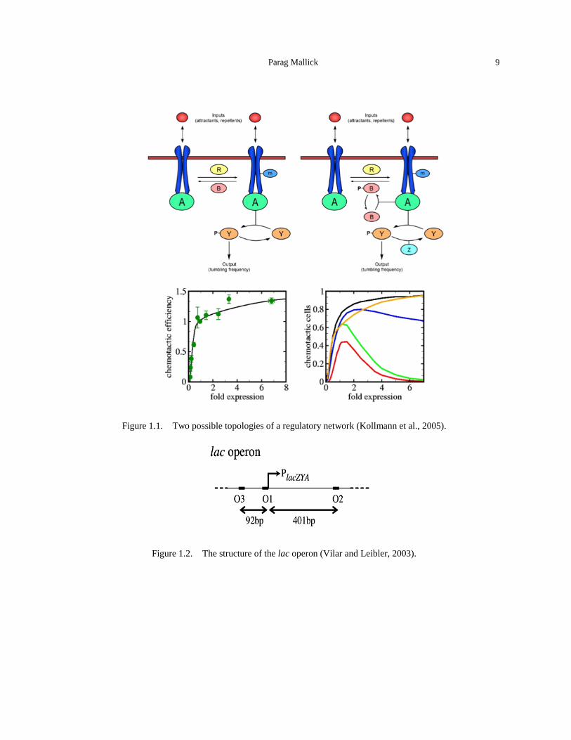
Parag Mallick 9
Figure 1.1. Two possible topologies of a regulatory network (Kollmann et al., 2005).
Figure 1.2. The structure of the lac operon (Vilar and Leibler, 2003).
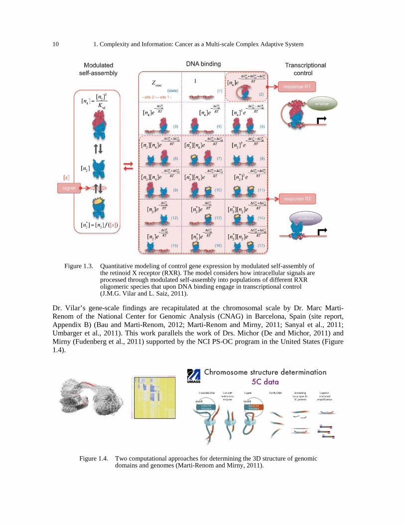
10 1. Complexity and Information: Cancer as a Multi-scale Complex Adaptive System
Figure 1.3. Quantitative modeling of control gene expression by modulated self-assembly of
the retinoid X receptor (RXR). The model considers how intracellular signals are processed through modulated self-assembly into populations of different RXR oligomeric species that upon DNA binding engage in transcriptional control (J.M.G. Vilar and L. Saiz, 2011).
Dr. Vilar’s gene-scale findings are recapitulated at the chromosomal scale by Dr. Marc Marti-Renom of the National Center for Genomic Analysis (CNAG) in Barcelona, Spain (site report, Appendix B) (Bau and Marti-Renom, 2012; Marti-Renom and Mirny, 2011; Sanyal et al., 2011; Umbarger et al., 2011). This work parallels the work of Drs. Michor (De and Michor, 2011) and Mirny (Fudenberg et al., 2011) supported by the NCI PS-OC program in the United States (Figure 1.4).
Figure 1.4. Two computational approaches for determining the 3D structure of genomic
domains and genomes (Marti-Renom and Mirny, 2011).
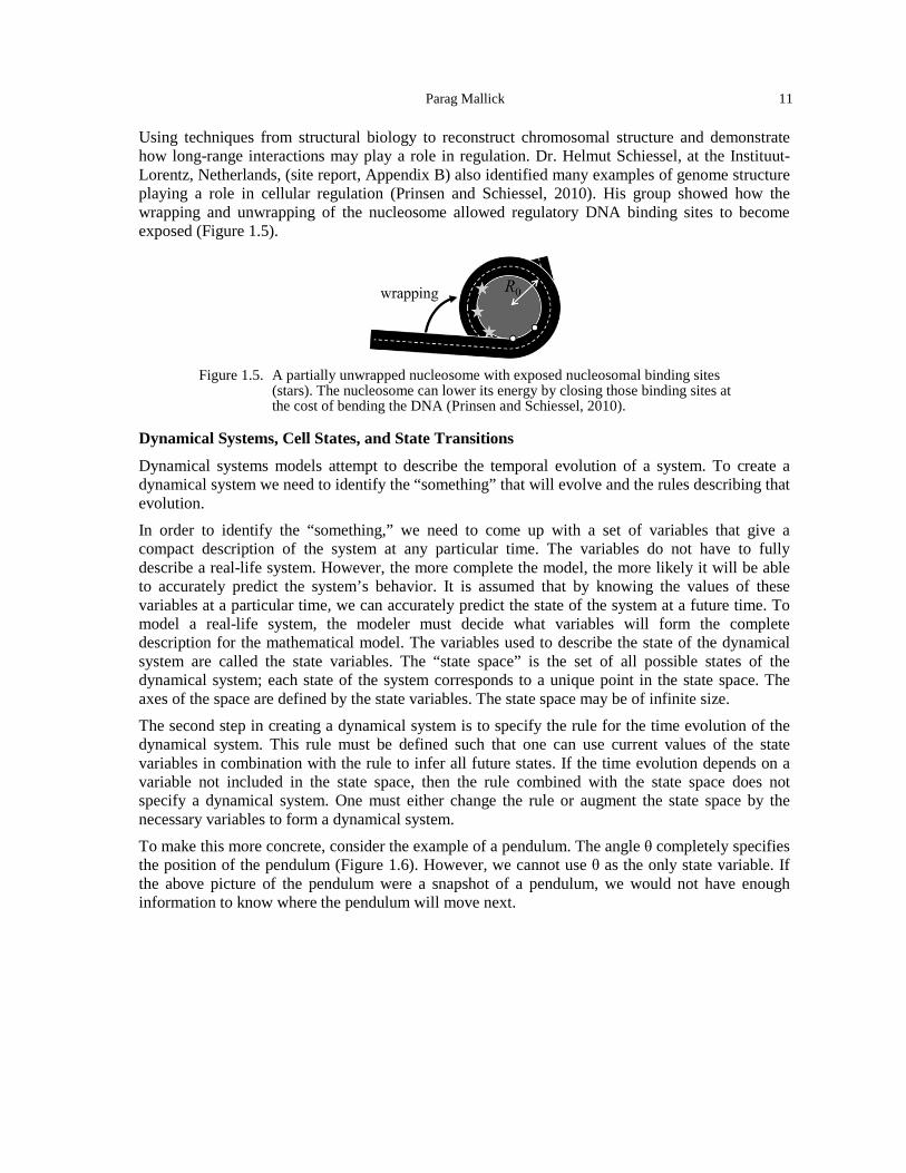
Parag Mallick 11
Using techniques from structural biology to reconstruct chromosomal structure and demonstrate how long-range interactions may play a role in regulation. Dr. Helmut Schiessel, at the Instituut-Lorentz, Netherlands, (site report, Appendix B) also identified many examples of genome structure playing a role in cellular regulation (Prinsen and Schiessel, 2010). His group showed how the wrapping and unwrapping of the nucleosome allowed regulatory DNA binding sites to become exposed (Figure 1.5).
Figure 1.5. A partially unwrapped nucleosome with exposed nucleosomal binding sites
(stars). The nucleosome can lower its energy by closing those binding sites at the cost of bending the DNA (Prinsen and Schiessel, 2010).
Dynamical Systems, Cell States, and State Transitions
Dynamical systems models attempt to describe the temporal evolution of a system. To create a dynamical system we need to identify the “something” that will evolve and the rules describing that evolution. In order to identify the “something,” we need to come up with a set of variables that give a compact description of the system at any particular time. The variables do not have to fully describe a real-life system. However, the more complete the model, the more likely it will be able to accurately predict the system’s behavior. It is assumed that by knowing the values of these variables at a particular time, we can accurately predict the state of the system at a future time. To model a real-life system, the modeler must decide what variables will form the complete description for the mathematical model. The variables used to describe the state of the dynamical system are called the state variables. The “state space” is the set of all possible states of the dynamical system; each state of the system corresponds to a unique point in the state space. The axes of the space are defined by the state variables. The state space may be of infinite size. The second step in creating a dynamical system is to specify the rule for the time evolution of the dynamical system. This rule must be defined such that one can use current values of the state variables in combination with the rule to infer all future states. If the time evolution depends on a variable not included in the state space, then the rule combined with the state space does not specify a dynamical system. One must either change the rule or augment the state space by the necessary variables to form a dynamical system. To make this more concrete, consider the example of a pendulum. The angle θ completely specifies the position of the pendulum (Figure 1.6). However, we cannot use θ as the only state variable. If the above picture of the pendulum were a snapshot of a pendulum, we would not have enough information to know where the pendulum will move next.
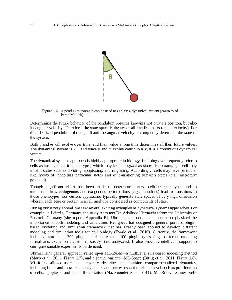
12 1. Complexity and Information: Cancer as a Multi-scale Complex Adaptive System
Figure 1.6. A pendulum example can be used to explain a dynamical system (courtesy of Parag Mallick).
Determining the future behavior of the pendulum requires knowing not only its position, but also its angular velocity. Therefore, the state space is the set of all possible pairs (angle, velocity). For this idealized pendulum, the angle θ and the angular velocity ω completely determine the state of the system. Both θ and ω will evolve over time, and their value at one time determines all their future values. The dynamical system is 2D, and since θ and ω evolve continuously, it is a continuous dynamical system. The dynamical systems approach is highly appropriate in biology. In biology we frequently refer to cells as having specific phenotypes, which may be analogized as states. For example, a cell may inhabit states such as dividing, apoptosing, and migrating. Accordingly, cells may have particular likelihoods of inhabiting particular states and of transitioning between states (e.g., metastatic potential). Though significant effort has been made to determine diverse cellular phenotypes and to understand how endogenous and exogenous perturbations (e.g., mutations) lead to transitions in those phenotypes, our current approaches typically generate state spaces of very high dimension wherein each gene or protein in a cell might be considered as components of state.
During our survey abroad, we saw several exciting examples of dynamical systems approaches. For example, in Leipzig, Germany, the study team met Dr. Adalinde Uhrmacher from the University of Rostock, Germany (site report, Appendix B). Uhrmacher, a computer scientist, emphasized the importance of both modeling and simulation. Her group has designed a general purpose plugin-based modeling and simulation framework that has already been applied to develop different modeling and simulation tools for cell biology (Ewald et al., 2010). Currently, the framework includes more than 700 plugins and more than 100 plugin types (e.g., different modeling formalisms, execution algorithms, steady state analyzers). It also provides intelligent support to configure suitable experiments on demand. Uhrmacher’s general approach relies upon ML-Rules—a multilevel rule-based modeling method (Maus et al., 2011; Figure 1.7), and a spatial variant—ML-Space (Bittig et al., 2011; Figure 1.8). ML-Rules allows users to compactly describe and combine compartmentalized dynamics, including inter- and intra-cellular dynamics and processes at the cellular level such as proliferation of cells, apoptosis, and cell differentiation (Mazemondet et al., 2011). ML-Rules assumes well-
θ
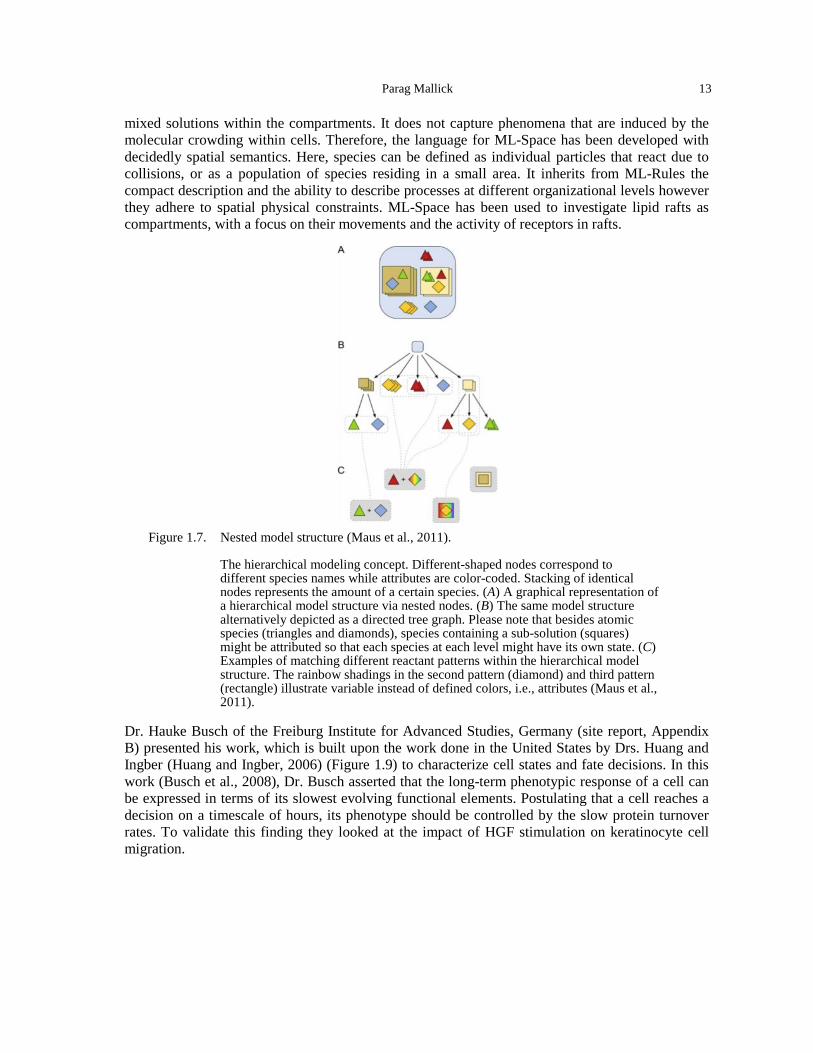
Parag Mallick 13
mixed solutions within the compartments. It does not capture phenomena that are induced by the molecular crowding within cells. Therefore, the language for ML-Space has been developed with decidedly spatial semantics. Here, species can be defined as individual particles that react due to collisions, or as a population of species residing in a small area. It inherits from ML-Rules the compact description and the ability to describe processes at different organizational levels however they adhere to spatial physical constraints. ML-Space has been used to investigate lipid rafts as compartments, with a focus on their movements and the activity of receptors in rafts.
Figure 1.7. Nested model structure (Maus et al., 2011).
The hierarchical modeling concept. Different-shaped nodes correspond to different species names while attributes are color-coded. Stacking of identical nodes represents the amount of a certain species. (A) A graphical representation of a hierarchical model structure via nested nodes. (B) The same model structure alternatively depicted as a directed tree graph. Please note that besides atomic species (triangles and diamonds), species containing a sub-solution (squares) might be attributed so that each species at each level might have its own state. (C) Examples of matching different reactant patterns within the hierarchical model structure. The rainbow shadings in the second pattern (diamond) and third pattern (rectangle) illustrate variable instead of defined colors, i.e., attributes (Maus et al., 2011).
Dr. Hauke Busch of the Freiburg Institute for Advanced Studies, Germany (site report, Appendix B) presented his work, which is built upon the work done in the United States by Drs. Huang and Ingber (Huang and Ingber, 2006) (Figure 1.9) to characterize cell states and fate decisions. In this work (Busch et al., 2008), Dr. Busch asserted that the long-term phenotypic response of a cell can be expressed in terms of its slowest evolving functional elements. Postulating that a cell reaches a decision on a timescale of hours, its phenotype should be controlled by the slow protein turnover rates. To validate this finding they looked at the impact of HGF stimulation on keratinocyte cell migration.
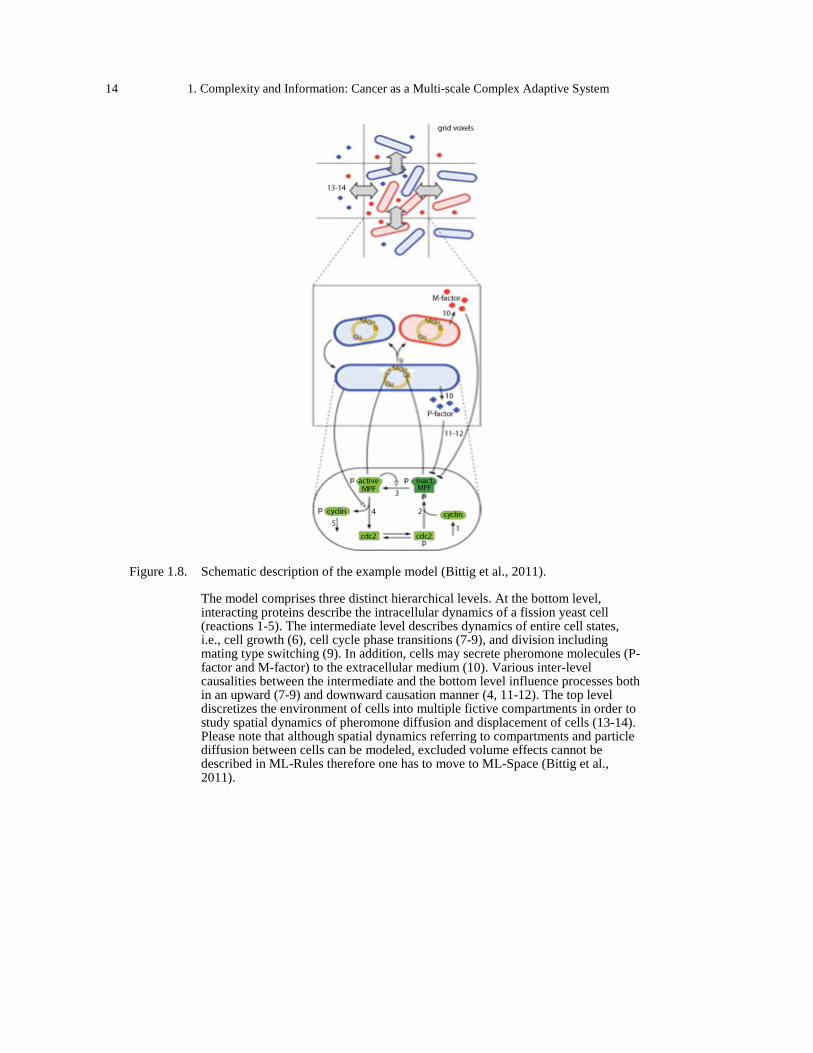
14 1. Complexity and Information: Cancer as a Multi-scale Complex Adaptive System
Figure 1.8. Schematic description of the example model (Bittig et al., 2011).
The model comprises three distinct hierarchical levels. At the bottom level, interacting proteins describe the intracellular dynamics of a fission yeast cell (reactions 1-5). The intermediate level describes dynamics of entire cell states, i.e., cell growth (6), cell cycle phase transitions (7-9), and division including mating type switching (9). In addition, cells may secrete pheromone molecules (P-factor and M-factor) to the extracellular medium (10). Various inter-level causalities between the intermediate and the bottom level influence processes both in an upward (7-9) and downward causation manner (4, 11-12). The top level discretizes the environment of cells into multiple fictive compartments in order to study spatial dynamics of pheromone diffusion and displacement of cells (13-14). Please note that although spatial dynamics referring to compartments and particle diffusion between cells can be modeled, excluded volume effects cannot be described in ML-Rules therefore one has to move to ML-Space (Bittig et al., 2011).
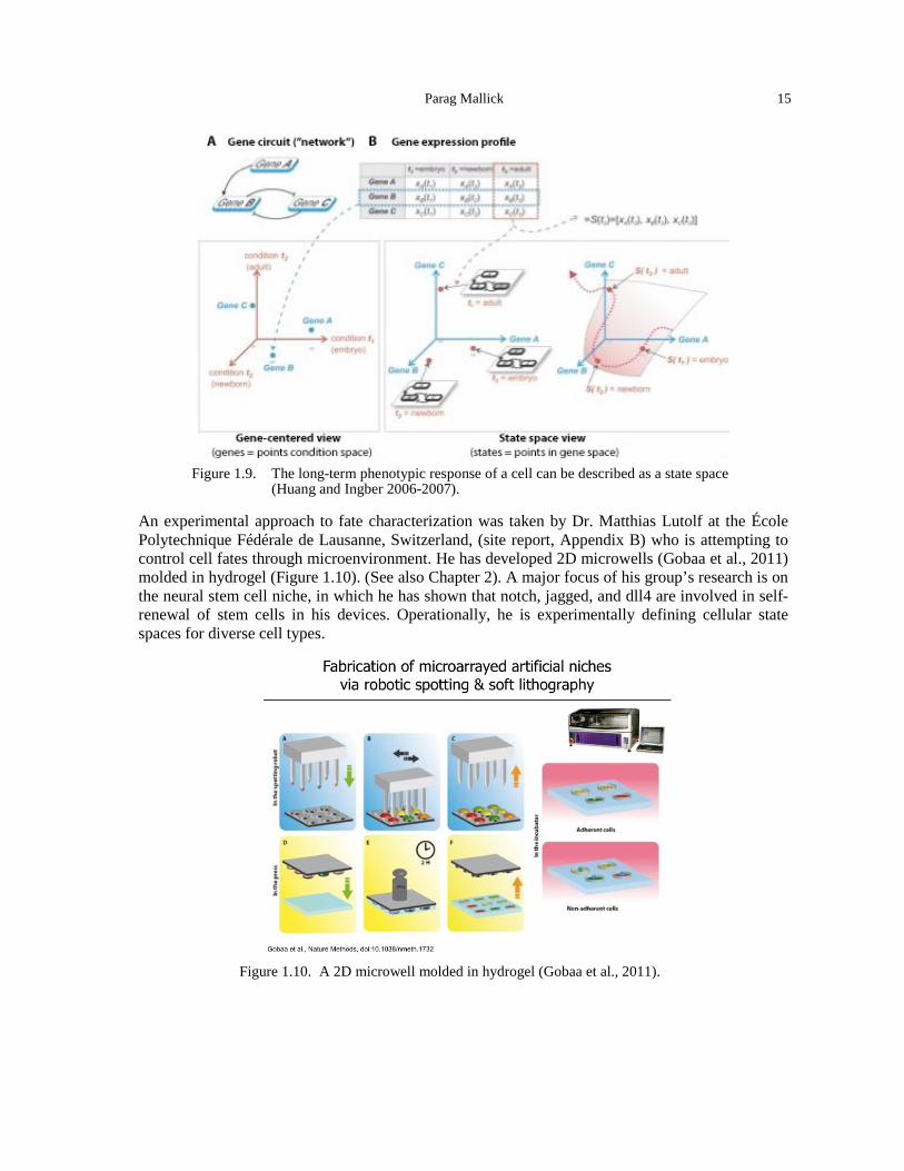
Parag Mallick 15
Figure 1.9. The long-term phenotypic response of a cell can be described as a state space
(Huang and Ingber 2006-2007).
An experimental approach to fate characterization was taken by Dr. Matthias Lutolf at the École Polytechnique Fédérale de Lausanne, Switzerland, (site report, Appendix B) who is attempting to control cell fates through microenvironment. He has developed 2D microwells (Gobaa et al., 2011) molded in hydrogel (Figure 1.10). (See also Chapter 2). A major focus of his group’s research is on the neural stem cell niche, in which he has shown that notch, jagged, and dll4 are involved in self-renewal of stem cells in his devices. Operationally, he is experimentally defining cellular state spaces for diverse cell types.
Figure 1.10. A 2D microwell molded in hydrogel (Gobaa et al., 2011).
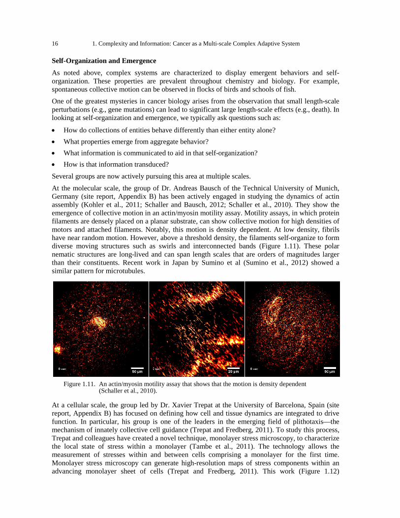
16 1. Complexity and Information: Cancer as a Multi-scale Complex Adaptive System
Self-Organization and Emergence
As noted above, complex systems are characterized to display emergent behaviors and self-organization. These properties are prevalent throughout chemistry and biology. For example, spontaneous collective motion can be observed in flocks of birds and schools of fish. One of the greatest mysteries in cancer biology arises from the observation that small length-scale perturbations (e.g., gene mutations) can lead to significant large length-scale effects (e.g., death). In looking at self-organization and emergence, we typically ask questions such as:
• How do collections of entities behave differently than either entity alone? • What properties emerge from aggregate behavior? • What information is communicated to aid in that self-organization? • How is that information transduced? Several groups are now actively pursuing this area at multiple scales.
At the molecular scale, the group of Dr. Andreas Bausch of the Technical University of Munich, Germany (site report, Appendix B) has been actively engaged in studying the dynamics of actin assembly (Kohler et al., 2011; Schaller and Bausch, 2012; Schaller et al., 2010). They show the emergence of collective motion in an actin/myosin motility assay. Motility assays, in which protein filaments are densely placed on a planar substrate, can show collective motion for high densities of motors and attached filaments. Notably, this motion is density dependent. At low density, fibrils have near random motion. However, above a threshold density, the filaments self-organize to form diverse moving structures such as swirls and interconnected bands (Figure 1.11). These polar nematic structures are long-lived and can span length scales that are orders of magnitudes larger than their constituents. Recent work in Japan by Sumino et al (Sumino et al., 2012) showed a similar pattern for microtubules.
Figure 1.11. An actin/myosin motility assay that shows that the motion is density dependent
(Schaller et al., 2010).
At a cellular scale, the group led by Dr. Xavier Trepat at the University of Barcelona, Spain (site report, Appendix B) has focused on defining how cell and tissue dynamics are integrated to drive function. In particular, his group is one of the leaders in the emerging field of plithotaxis—the mechanism of innately collective cell guidance (Trepat and Fredberg, 2011). To study this process, Trepat and colleagues have created a novel technique, monolayer stress microscopy, to characterize the local state of stress within a monolayer (Tambe et al., 2011). The technology allows the measurement of stresses within and between cells comprising a monolayer for the first time. Monolayer stress microscopy can generate high-resolution maps of stress components within an advancing monolayer sheet of cells (Trepat and Fredberg, 2011). This work (Figure 1.12)
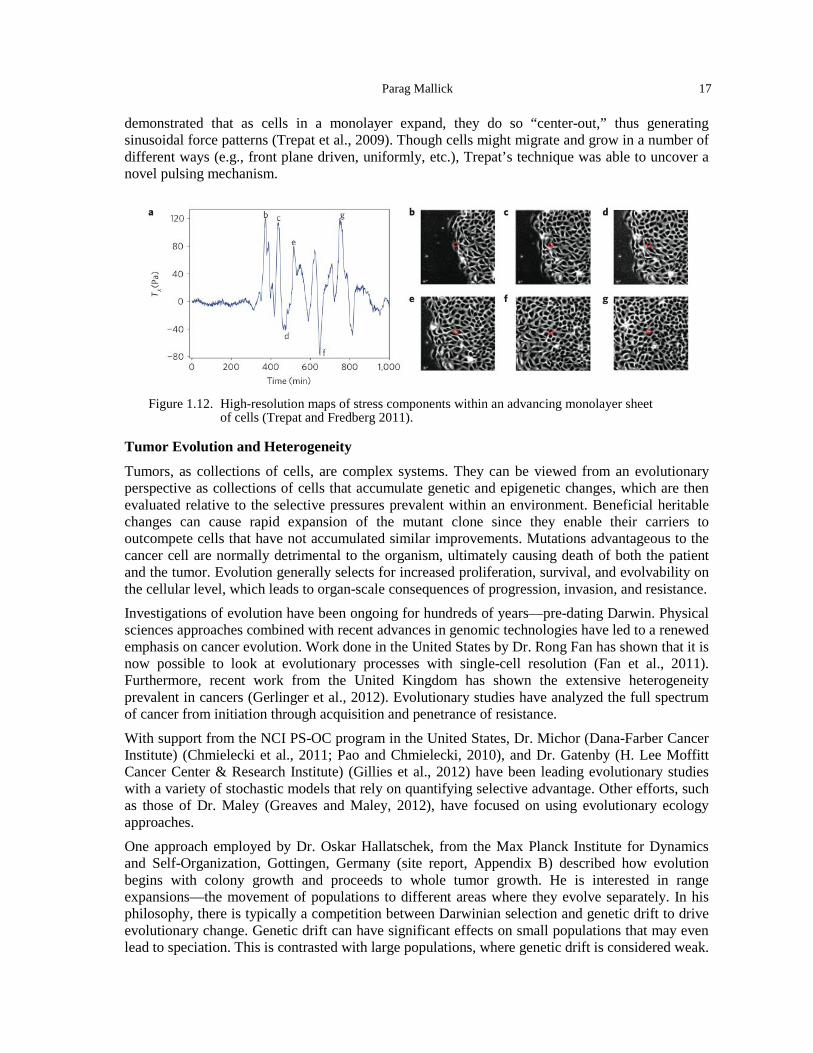
Parag Mallick 17
demonstrated that as cells in a monolayer expand, they do so “center-out,” thus generating sinusoidal force patterns (Trepat et al., 2009). Though cells might migrate and grow in a number of different ways (e.g., front plane driven, uniformly, etc.), Trepat’s technique was able to uncover a novel pulsing mechanism.
Figure 1.12. High-resolution maps of stress components within an advancing monolayer sheet
of cells (Trepat and Fredberg 2011).
Tumor Evolution and Heterogeneity
Tumors, as collections of cells, are complex systems. They can be viewed from an evolutionary perspective as collections of cells that accumulate genetic and epigenetic changes, which are then evaluated relative to the selective pressures prevalent within an environment. Beneficial heritable changes can cause rapid expansion of the mutant clone since they enable their carriers to outcompete cells that have not accumulated similar improvements. Mutations advantageous to the cancer cell are normally detrimental to the organism, ultimately causing death of both the patient and the tumor. Evolution generally selects for increased proliferation, survival, and evolvability on the cellular level, which leads to organ-scale consequences of progression, invasion, and resistance. Investigations of evolution have been ongoing for hundreds of years—pre-dating Darwin. Physical sciences approaches combined with recent advances in genomic technologies have led to a renewed emphasis on cancer evolution. Work done in the United States by Dr. Rong Fan has shown that it is now possible to look at evolutionary processes with single-cell resolution (Fan et al., 2011). Furthermore, recent work from the United Kingdom has shown the extensive heterogeneity prevalent in cancers (Gerlinger et al., 2012). Evolutionary studies have analyzed the full spectrum of cancer from initiation through acquisition and penetrance of resistance.
With support from the NCI PS-OC program in the United States, Dr. Michor (Dana-Farber Cancer Institute) (Chmielecki et al., 2011; Pao and Chmielecki, 2010), and Dr. Gatenby (H. Lee Moffitt Cancer Center & Research Institute) (Gillies et al., 2012) have been leading evolutionary studies with a variety of stochastic models that rely on quantifying selective advantage. Other efforts, such as those of Dr. Maley (Greaves and Maley, 2012), have focused on using evolutionary ecology approaches. One approach employed by Dr. Oskar Hallatschek, from the Max Planck Institute for Dynamics and Self-Organization, Gottingen, Germany (site report, Appendix B) described how evolution begins with colony growth and proceeds to whole tumor growth. He is interested in range expansions—the movement of populations to different areas where they evolve separately. In his philosophy, there is typically a competition between Darwinian selection and genetic drift to drive evolutionary change. Genetic drift can have significant effects on small populations that may even lead to speciation. This is contrasted with large populations, where genetic drift is considered weak.
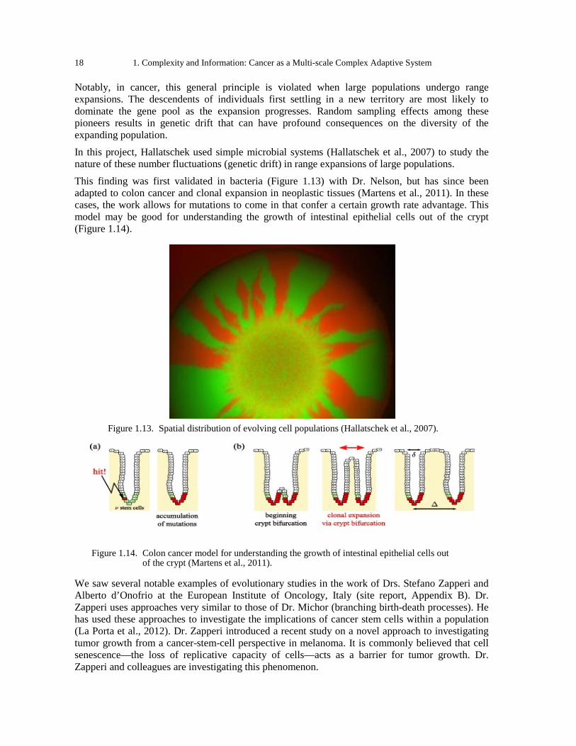
18 1. Complexity and Information: Cancer as a Multi-scale Complex Adaptive System
Notably, in cancer, this general principle is violated when large populations undergo range expansions. The descendents of individuals first settling in a new territory are most likely to dominate the gene pool as the expansion progresses. Random sampling effects among these pioneers results in genetic drift that can have profound consequences on the diversity of the expanding population. In this project, Hallatschek used simple microbial systems (Hallatschek et al., 2007) to study the nature of these number fluctuations (genetic drift) in range expansions of large populations.
This finding was first validated in bacteria (Figure 1.13) with Dr. Nelson, but has since been adapted to colon cancer and clonal expansion in neoplastic tissues (Martens et al., 2011). In these cases, the work allows for mutations to come in that confer a certain growth rate advantage. This model may be good for understanding the growth of intestinal epithelial cells out of the crypt (Figure 1.14).
Figure 1.13. Spatial distribution of evolving cell populations (Hallatschek et al., 2007).
Figure 1.14. Colon cancer model for understanding the growth of intestinal epithelial cells out
of the crypt (Martens et al., 2011).
We saw several notable examples of evolutionary studies in the work of Drs. Stefano Zapperi and Alberto d’Onofrio at the European Institute of Oncology, Italy (site report, Appendix B). Dr. Zapperi uses approaches very similar to those of Dr. Michor (branching birth-death processes). He has used these approaches to investigate the implications of cancer stem cells within a population (La Porta et al., 2012). Dr. Zapperi introduced a recent study on a novel approach to investigating tumor growth from a cancer-stem-cell perspective in melanoma. It is commonly believed that cell senescence—the loss of replicative capacity of cells—acts as a barrier for tumor growth. Dr. Zapperi and colleagues are investigating this phenomenon.
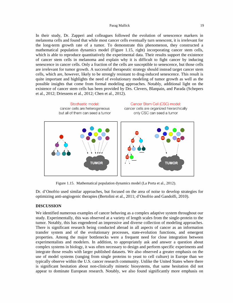
Parag Mallick 19
In their study, Dr. Zapperi and colleagues followed the evolution of senescence markers in melanoma cells and found that while most cancer cells eventually turn senescent, it is irrelevant for the long-term growth rate of a tumor. To demonstrate this phenomenon, they constructed a mathematical population dynamics model (Figure 1.15, right) incorporating cancer stem cells, which is able to reproduce quantitatively the experimental data. Their results support the existence of cancer stem cells in melanoma and explain why it is difficult to fight cancer by inducing senescence in cancer cells. Only a fraction of the cells are susceptible to senescence, but those cells are irrelevant for tumor growth. A successful therapeutic strategy should instead target cancer stem cells, which are, however, likely to be strongly resistant to drug-induced senescence. This result is quite important and highlights the need of evolutionary modeling of tumor growth as well as the possible insights that come from formal modeling approaches. Notably, additional light on the existence of cancer stem cells has been provided by Drs. Clevers, Blanpain, and Parada (Schepers et al., 2012; Driessens et al., 2012; Chen et al., 2012).
Figure 1.15. Mathematical population dynamics model (La Porta et al., 2012).
Dr. d’Onofrio used similar approaches, but focused on the area of noise to develop strategies for optimizing anti-angiogenic therapies (Bertolini et al., 2011; d’Onofrio and Gandolfi, 2010).
DISCUSSION
We identified numerous examples of cancer behaving as a complex adaptive system throughout our study. Experimentally, this was observed at a variety of length scales from the single-protein to the tumor. Notably, this has engendered an impressive and diverse collection of modeling approaches. There is significant research being conducted abroad in all aspects of cancer as an information transfer system and of the evolutionary processes, state-evolution functions, and emergent properties. Among the major bottlenecks were a frequent need for close integration between experimentalists and modelers. In addition, to appropriately ask and answer a question about complex systems in biology, it was often necessary to design and perform specific experiments and integrate those results with larger published datasets. We also observed a greater emphasis on the use of model systems (ranging from single proteins to yeast to cell culture) in Europe than we typically observe within the U.S. cancer research community. Unlike the United States where there is significant hesitation about non-clinically mimetic biosystems, that same hesitation did not appear to dominate European research. Notably, we also found significantly more emphasis on
20 1. Complexity and Information: Cancer as a Multi-scale Complex Adaptive System
compact biomodel systems in Europe than in the United States. There also was a significant emphasis on exploring specific types of effects and extracting principles that might be scaled up. This approach differed widely from the typical U.S. approach, which favors large-scale, global analyses. However, the advantage of global approaches is their ability to interrogate complex systems as a whole, rather than as a subset, such as in recent work to build total cell models (Karr et al., 2012). A major contributor to successful research endeavors was funding environment. Successful projects depended upon close collaboration amongst groups of researchers, particularly including a mix of experimentalists, bioinformaticians, and modelers. Consequently, multi-investigator funding mechanisms have been critical for pushing innovation at the frontier of information transfer and complex systems analysis of biology. Through programs, such as those engendered by the OPSO, and foundation awards at the interface between the physical and life sciences, investigators in the U.S. have been fortunate to have access to interdisciplinary funding opportunities. Generally, we conclude that the areas of information transfer, evolution, and complex adaptive systems research are rapidly progressing, and critically important for impacting cancer and more generally understanding biology.
REFERENCES Bachmann, J., Raue, A., Schilling, M., Becker, V., Timmer, J., and Klingmuller, U. (2012). Predictive
mathematical models of cancer signalling pathways. Journal of internal medicine 271, 155-165. Balázsi, G., van Oudenaarden, A., and Collins, J.J. (2011). Cellular decision making and biological noise: From
microbes to mammals. Cell 144, 910-925. Bau, D., and Marti-Renom, M.A. (2012). Genome structure determination via 3C-based data integration by the
Integrative Modeling Platform. Methods [Epub ahead of print, April 13]. Beadle, G.W., and Tatum, E.L. (1941). Genetic Control of Biochemical Reactions in Neurospora. Proceedings of
the National Academy of Sciences of the United States of America 27, 499-506. Becker, V., Schilling, M., Bachmann, J., Baumann, U., Raue, A., Maiwald, T., Timmer, J., and Klingmuller, U.
(2010). Covering a broad dynamic range: information processing at the erythropoietin receptor. Science 328, 1404-1408.
Becker, V., Timmer, J., and Klingmuller, U. (2012). Receptor dynamics in signaling. Advances in experimental medicine and biology 736, 313-323.
Bertalanffy, L.v. (1973). General system theory: foundations, development, applications (Harmondsworth: Penguin).
Bertolini, F., Marighetti, P., Martin-Padura, I., Mancuso, P., Hu-Lowe, D.D., Shaked, Y., and D’Onofrio, A. (2011). Anti-VEGF and beyond: shaping a new generation of anti-angiogenic therapies for cancer. Drug discovery today 16, 1052-1060.
Bittig, A.T., Haack, F., Maus, C., and Uhrmacher, A.M. (2011). Adapting rule-based model descriptions for simulating in continuous and hybrid space. In Proceedings of the 9th International Conference on Computational Methods in Systems Biology (Paris, France: ACM), pp. 161-170.
Busch, H., Camacho-Trullio, D., Rogon, Z., Breuhahn, K., Angel, P., Eils, R., and Szabowski, A. (2008). Gene network dynamics controlling keratinocyte migration. Molecular systems biology 4, 199.
Chen, J., Li, Y., Yu, T.-S., McKay, R., Burns, D.K., Kernie, S.G., and Parada, L.F. (2012). A restricted cell population propagates glioblastoma growth after chemotherapy. Nature doi:10.1038/nature11287.
Chmielecki, J., Foo, J., Oxnard, G.R., Hutchinson, K., Ohashi, K., Somwar, R., Wang, L., Amato, K.R., Arcila, M., Sos, M.L., et al. (2011). Optimization of dosing for EGFR-mutant non-small cell lung cancer with evolutionary cancer modeling. Science translational medicine 3, 90ra59.
d’Onofrio, A., and Gandolfi, A. (2010). Resistance to antitumor chemotherapy due to bounded-noise-induced transitions. Physical Review E 82, 061901.
De, S., and Michor, F. (2011). DNA replication timing and long-range DNA interactions predict mutational landscapes of cancer genomes. Nature biotechnology 29, 1103-1108.
Deisboeck, T.S., and Kresh, J.Y. (2006). Complex systems science in biomedicine (New York, NY: Springer). Driessens, G., Beck, B., Caauwe, A., Simons, B.D., and Blanpain, C. (2012). Defining the mode of tumour growth
by clonal analysis. Nature doi:10.1038/nature11344.
Parag Mallick 21
Ewald, R., Himmelspach, J., Jeschke, M., Leye, S., and Uhrmacher, A.M. (2010). Flexible experimentation in the modeling and simulation framework JAMES II--implications for computational systems biology. Briefings in bioinformatics 11, 290-300.
Fan, H.C., Wang, J., Potanina, A., and Quake, S.R. (2011). Whole-genome molecular haplotyping of single cells. Nature biotechnology 29, 51-57.
Fudenberg, G., Getz, G., Meyerson, M., and Mirny, L.A. (2011). High order chromatin architecture shapes the landscape of chromosomal alterations in cancer. Nature biotechnology 29, 1109-1113.
Garrod, A. (1908). The Croonian Lectures on Inborn Errors of Metabolism. The Lancet 172, 1-7. Gerlinger, M., Rowan, A.J., Horswell, S., Larkin, J., Endesfelder, D., Gronroos, E., Martinez, P., Matthews, N.,
Stewart, A., Tarpey, P., et al. (2012). Intratumor heterogeneity and branched evolution revealed by multiregion sequencing. The New England journal of medicine 366, 883-892.
Gillies, R.J., Verduzco, D., and Gatenby, R.A. (2012). Evolutionary dynamics of carcinogenesis and why targeted therapy does not work. Nature reviews Cancer 12, 487-493.
Gobaa, S., Hoehnel, S., Roccio, M., Negro, A., Kobel, S., and Lutolf, M.P. (2011). Artificial niche microarrays for probing single stem cell fate in high throughput. Nature methods 8, 949-955.
Greaves, M., and Maley, C.C. (2012). Clonal evolution in cancer. Nature 481, 306-313. Hallatschek, O., Hersen, P., Ramanathan, S., and Nelson, D.R. (2007). Genetic drift at expanding frontiers
promotes gene segregation. Proc Natl Acad Sci U S A 104, 19926-19930. Huang, S., and Ingber, D.E. (2006-2007). A non-genetic basis for cancer progression and metastasis: self-
organizing attractors in cell regulatory networks. Breast disease 26, 27-54. Karr, J.R., Sanghvi, J.C., Macklin, D.N., Gutschow, M.V., Jacobs, J.M., Bolival, B., Assad-Garcia, N., Glass, J.I.,
and Covert, M.W. (2012). Cell 150, 389-401. Kohler, S., Schaller, V., and Bausch, A.R. (2011). Collective dynamics of active cytoskeletal networks. PloS one 6,
e23798. Kollmann, M., Lovdok, L., Bartholome, K., Timmer, J., and Sourjik, V. (2005). Design principles of a bacterial
signalling network. Nature 438, 504-507. La Du, B.N., Zannoni, V.G., Laster, L., and Seegmiller, J.E. (1958). The nature of the defect in tyrosine
metabolism in alcaptonuria. The Journal of biological chemistry 230, 251-260. La Porta, C.A., Zapperi, S., and Sethna, J.P. (2012). Senescent cells in growing tumors: population dynamics and
cancer stem cells. PLoS computational biology 8, e1002316. Laszlo, E. (1972). The systems view of the world; the natural philosophy of the new developments in the sciences
(New York,: G. Braziller). Martens, E.A., Kostadinov, R., Maley, C.C., and Hallatschek, O. (2011). Spatial structure increases the waiting
time for cancer. New Journal of Physics 13, 115014. Marti-Renom, M.A., and Mirny, L.A. (2011). Bridging the resolution gap in structural modeling of 3D genome
organization. PLoS computational biology 7, e1002125. Maus, C., Rybacki, S., and Uhrmacher, A.M. (2011). Rule-based multi-level modeling of cell biological systems.
BMC systems biology 5, 166. Mazemondet, O., Hubner, R., Frahm, J., Koczan, D., Bader, B.M., Weiss, D.G., Uhrmacher, A.M., Frech, M.J.,
Rolfs, A., and Luo, J. (2011). Quantitative and kinetic profile of Wnt/beta-catenin signaling components during human neural progenitor cell differentiation. Cellular & molecular biology letters 16, 515-538.
Meyerhof, O. (1945). The origin of the reaction of Harden and Young in cell-free alcoholic fermentation. Journal of Biological Chemistry 157, 105-120.
Meyerhof, O., and Junowicz-Kocholaty, R. (1943). The equilibria of isomerase and aldolase, and the problem of the phosphorylation of glyceraldehyde phosphate. Journal of Biological Chemistry 149, 71.
Meyerhof, O., and Oesper, P. (1947). The Mechanism of the Oxidative Reaction in Fermentation. Journal of Biological Chemistry 170, 1.
Munsky, B., Neuert, G., and van Oudenaarden, A. (2012). Using gene expression noise to understand gene regulation. Science 336, 183-187.
Pao, W., and Chmielecki, J. (2010). Rational, biologically based treatment of EGFR-mutant non-small-cell lung cancer. Nature reviews Cancer 10, 760-774.
22 1. Complexity and Information: Cancer as a Multi-scale Complex Adaptive System
Prinsen, P., and Schiessel, H. (2010). Nucleosome stability and accessibility of its DNA to proteins. Biochimie 92, 1722-1728.
Saiz, L., and Vilar, J.M. (2007). Multilevel deconstruction of the In vivo behavior of looped DNA-protein complexes. PloS one 2, e355.
Sanyal, A., Bau, D., Marti-Renom, M.A., and Dekker, J. (2011). Chromatin globules: a common motif of higher order chromosome structure? Current opinion in cell biology 23, 325-331.
Schaller, V., and Bausch, A.R. (2012). Materials science: A fresh twist for self-assembly. Nature 481, 268-269. Schaller, V., Weber, C., Semmrich, C., Frey, E., and Bausch, A.R. (2010). Polar patterns of driven filaments.
Nature 467, 73-77. Schepers, A.G., Snippert, H.J., Stange, D.E., van den Born, M., van Es, J.H., van de Wetering, M., and Clevers, H.
(2012). Lineage tracing reveals Lgr5+ stem cell activity in mouse intestinal adenomas. Science 337, 730-735. Schwab, E.D., and Pienta, K.J. (1996). Cancer as a complex adaptive system. Medical hypotheses 47, 235-241. Smithers, D.W. (1962). An attack on cytologism. Lancet 1, 493-499. Stewart-Orstein, J., Weissman, J.S., and El-Samad, H. (2012). Cellular noise regulons underlie fluctuations in
Saccharomyces cerevisiae. Mol Cell 45, 483-493. Sumino, Y., Nagai, K.H., Shitaka, Y., Tanaka, D., Yoshikawa, K., Chate, H., and Oiwa, K. (2012). Large-scale
vortex lattice emerging from collectively moving microtubules. Nature 483, 448-452. Tambe, D.T., Hardin, C.C., Angelini, T.E., Rajendran, K., Park, C.Y., Serra-Picamal, X., Zhou, E.H., Zaman,
M.H., Butler, J.P., Weitz, D.A., et al. (2011). Collective cell guidance by cooperative intercellular forces. Nat Mater 10, 469-475.
Trepat, X., and Fredberg, J.J. (2011). Plithotaxis and emergent dynamics in collective cellular migration. Trends Cell Biol 21, 638-646.
Trepat, X., Wasserman, M.R., Angelini, T.E., Millet, E., Weitz, D.A., Butler, J.P., and Fredberg, J.J. (2009). Physical forces during collective cell migration. Nat Phys 5, 426-430.
Umbarger, M.A., Toro, E., Wright, M.A., Porreca, G.J., Bau, D., Hong, S.H., Fero, M.J., Zhu, L.J., Marti-Renom, M.A., McAdams, H.H., et al. (2011). The three-dimensional architecture of a bacterial genome and its alteration by genetic perturbation. Molecular cell 44, 252-264.
Vilar, J.M., Guet, C.C., and Leibler, S. (2003). Modeling network dynamics: the lac operon, a case study. The Journal of cell biology 161, 471-476.
Vilar, J.M., and Leibler, S. (2003). DNA looping and physical constraints on transcription regulation. Journal of molecular biology 331, 981-989.
Vilar, J.M., and Saiz, L. (2011). Control of gene expression by modulated self-assembly. Nucleic acids research 39, 6854-6863.
Von Bertalanffy, L. (1972). The history and status of general systems theory. Trends in general systems theory, 407-426.
Zatkova, A. (2011). An update on molecular genetics of Alkaptonuria (AKU). Journal of inherited metabolic disease 34, 1127-1136.
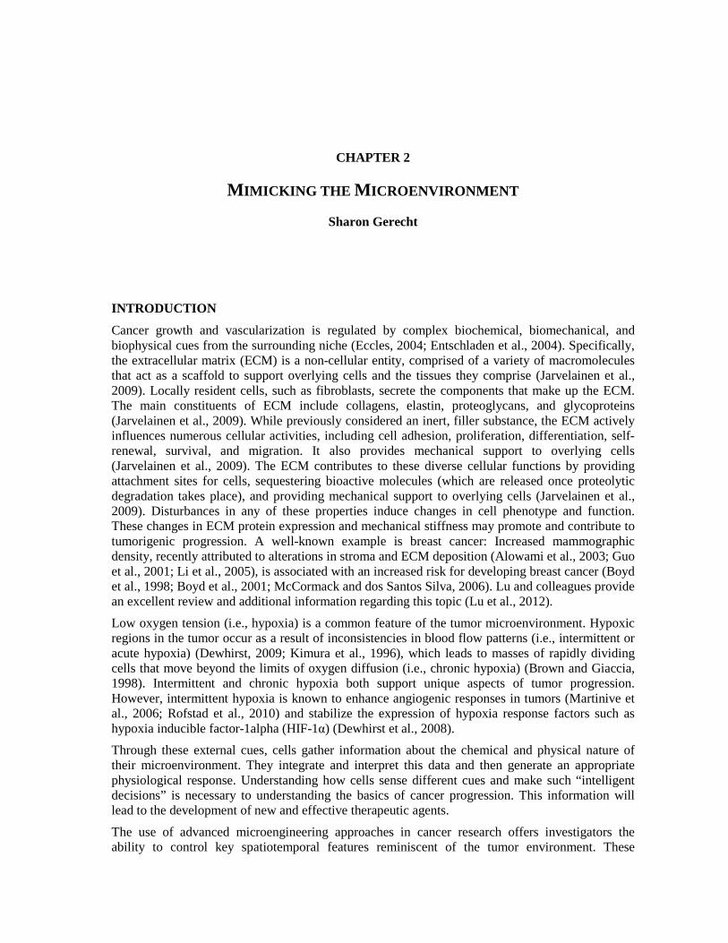
CHAPTER 2
MIMICKING THE MICROENVIRONMENT
Sharon Gerecht
INTRODUCTION
Cancer growth and vascularization is regulated by complex biochemical, biomechanical, and biophysical cues from the surrounding niche (Eccles, 2004; Entschladen et al., 2004). Specifically, the extracellular matrix (ECM) is a non-cellular entity, comprised of a variety of macromolecules that act as a scaffold to support overlying cells and the tissues they comprise (Jarvelainen et al., 2009). Locally resident cells, such as fibroblasts, secrete the components that make up the ECM. The main constituents of ECM include collagens, elastin, proteoglycans, and glycoproteins (Jarvelainen et al., 2009). While previously considered an inert, filler substance, the ECM actively influences numerous cellular activities, including cell adhesion, proliferation, differentiation, self-renewal, survival, and migration. It also provides mechanical support to overlying cells (Jarvelainen et al., 2009). The ECM contributes to these diverse cellular functions by providing attachment sites for cells, sequestering bioactive molecules (which are released once proteolytic degradation takes place), and providing mechanical support to overlying cells (Jarvelainen et al., 2009). Disturbances in any of these properties induce changes in cell phenotype and function. These changes in ECM protein expression and mechanical stiffness may promote and contribute to tumorigenic progression. A well-known example is breast cancer: Increased mammographic density, recently attributed to alterations in stroma and ECM deposition (Alowami et al., 2003; Guo et al., 2001; Li et al., 2005), is associated with an increased risk for developing breast cancer (Boyd et al., 1998; Boyd et al., 2001; McCormack and dos Santos Silva, 2006). Lu and colleagues provide an excellent review and additional information regarding this topic (Lu et al., 2012).
Low oxygen tension (i.e., hypoxia) is a common feature of the tumor microenvironment. Hypoxic regions in the tumor occur as a result of inconsistencies in blood flow patterns (i.e., intermittent or acute hypoxia) (Dewhirst, 2009; Kimura et al., 1996), which leads to masses of rapidly dividing cells that move beyond the limits of oxygen diffusion (i.e., chronic hypoxia) (Brown and Giaccia, 1998). Intermittent and chronic hypoxia both support unique aspects of tumor progression. However, intermittent hypoxia is known to enhance angiogenic responses in tumors (Martinive et al., 2006; Rofstad et al., 2010) and stabilize the expression of hypoxia response factors such as hypoxia inducible factor-1alpha (HIF-1α) (Dewhirst et al., 2008). Through these external cues, cells gather information about the chemical and physical nature of their microenvironment. They integrate and interpret this data and then generate an appropriate physiological response. Understanding how cells sense different cues and make such “intelligent decisions” is necessary to understanding the basics of cancer progression. This information will lead to the development of new and effective therapeutic agents.
The use of advanced microengineering approaches in cancer research offers investigators the ability to control key spatiotemporal features reminiscent of the tumor environment. These
24 2. Mimicking the Microenvironment
engineered technologies are allowing researchers to recapitulate conditions present within the tumor and to study the complex cell-cell and cell-ECM interactions that take place in the tumor environment. These systems are pivotal in accelerating research into the complex mechanisms regulating tumorigenesis in a relevant in vitro-mimetic environment. This chapter reviews several engineering approaches that are being used to investigate tumor growth in both the United States and Europe. Several of the sites we visited in Europe described the evolution of complex technologies that are able to detect single to multiple signals.
RESEARCH
Recapitulating Single Cues in the Microenvironment
Cell adhesion to the ECM and to neighboring cells is a complex, tightly regulated process that plays a crucial role in fundamental cellular functions, including cell migration, proliferation, differentiation, and apoptosis. Various approaches to control cell adhesion in vitro have been developed throughout the years both in the United States and Europe. Joachim Spatz’s laboratory at the University of Heidelberg, Germany (site report, Appendix B) pioneered a method to precisely defined spatial distribution of ligands on an otherwise inert substrate for enabling the understanding of how cell adhesion and signaling depend on the composition, size, and distribution of specific adhesion sites (Arnold et al., 2004). Utilizing micelle diblock copolymer lithography technology enabled the modification of substrates for the presentation of adhesive ligands in defined spacing at a length scale of 10-200 nm (Figure 2.1). This approach is based on the self-assembly of diblock copolymers of polystyrene-block-poly (2-vinylpyridine) (PS-b-P2VP) into reverse micelles in toluene. Using this approach, the size of the biofunctionalized nanoparticles may vary between 1 and 20 nm. The spacing between the nanoparticles may also be adjusted from 15 to 250 nm (Arnold et al., 2004; Spatz and Geiger, 2007). By using this platform Spatz and colleagues found that cell adhesion is receptor-space specific. Surfaces of nanoparticles functionalized with RGD—an ECM adhesive peptide that is recognized by αvβ3-integrin with high affinity—were generated with spacing of 28, 58, 73, and 85 nm. Different cell types, including MC3T3 osteoblasts, REF52 fibroblasts, 3T3 fibroblasts, and B16 melanocytes seeded on these surfaces spread very well on the 58-nm pattern. This was comparable to their spreading on uniform RGD- or fibronectin-coated surfaces. A separation of ≥73 nm between the adhesive dots results in limited cell attachment and spreading, and dramatically reduces the formation of focal adhesion and actin stress fibers. The researchers suggest that the range between 58 and 73 nm is a universal length scale for integrin clustering and activation. The lab created a spacing gradient surface to explore the role of adhesive spacing during migration (Hirschfeld-Warneken et al., 2008). It was found that MC3T3 osteoblasts’ morphology, adhesion area, actin, and vinculin distribution, as well as cell body polarization were influenced by the peptide patch spacing gradient. As a consequence, these adhesive ligand gradients induce MC3T3 osteoblasts orientation towards their optimal adhesion spacing (Hirschfeld-Warneken et al., 2008). Chapter 4 discusses these approaches to understanding cancer development and growth from the time domain perspective.
Sharon Gerecht 25
Figure 2.1. Micellar block copolymer lithography and biofunctionalization.
(A) Scheme of diblock copolymer micelle lithography. (B–E) Extended Au nanodot patterns are displayed by scanning electron microscope. Uniform nanodots (bright spots) of (B) 3 nm by PS(190)-b-P[2VP(HAuCl4)0.5](190), (C) 5 nm by PS(500)-b-P[2VP(HAuCl4)0.5](270), (D) 6 nm by PS(990)-b-P[2VP(HAuCl4)0.5](385), and (E) 8 nm by PS(1350)-b-P[2VP(HAuCl4)0.5](400) deposited onto Si-wafers. The number in brackets refers to the number of monomer units in each block that control the separation between Au dots. These varied between (B) 28, (C) 58, (D) 73, and (E) 85 nm. The nanodots form extended, nearly perfect, hexagonally close-packed patterns as indicated by the Fourier transform images (inset), which show second-order intensity spots. (F) Biofunctionalization of the nanodots pattern (Arnold et al., 2004). Since the nanodot is sufficiently small, it is likely that only one integrin transmembrane receptor directly interacts with one dot. The nanodots are presented as side-view micrographs taken with a high-resolution transmission electron microscope (Spatz and Geiger, 2007).
Micropatterning is a robust method of presenting micro and nanometer scale ECM molecules in distinct spatial patterns and allows for the control of various cell responses. Throughout the years, numerous approaches to generate micropatterns have been developed in the United States and Europe including direct printing techniques such as bioprinting or dip pen lithography. These approaches use inkjet/laser printers or atomic force microscope tips as well as micropattern fabrication methods like photolithography and soft lithography. An example of the role of cell adhesion in the formation of normal vs. abnormal tissue architecture was given by Dr. Wilhelm Huck at the University Nijmegen Medical Centre, Netherlands (site report, Appendix B) in collaboration with Dr. Fiona Watt (Cambridge, U.K.). Surfaces printed with collagen I in 10-µm rings containing non-adhesive disks (ranging from 0 to 60 mm) were generated with a polymer brush to examine the formation of the micro-epidermis. Human epidermal keratinocytes seeded on these surfaces were examined for cell differentiation vs. proliferation. For rings with a non-adhesive center of up to 40 mm diameter, cell-cell and cell-matrix adhesive interactions result in correct micro-epidermis assembly. Using this platform, cells isolated from oral cavity tumors (squamous cell carcinomas that have increased proliferation and reduced differentiation with disturbed tissue architecture) were cultured on these micropatterned islands and found to exhibit disturbed architecture that correlates with the original tumor characteristics (Figure 2.2). Tumors are “stiffer” than their normal tissue counterparts. The mechanical stiffness of breast carcinomas has been reported to be as high as 42.5 kilopascals (kPa) for high-grade invasive ductal carcinomas, as opposed to 3.25 kPa for non-malignant mammary tissues (Samani et al., 2007). Similarly, lymph nodes harboring metastatic tumor foci had a mechanical stiffness of 3.35 ± 1.57
26 2. Mimicking the Microenvironment
g/cm (i.e., 329 Pa) versus non-tumor bearing lymph nodes, which have a mechanical stiffness of 1.23 g/cm (i.e., 121 Pa) (Miyaji et al., 1997).
Engineering the mechanics of matrices for the 2D and 3D cell culture offers the opportunity to study cellular responses and the regulatory mechanism involved. Recent years’ increase in research of human stem cells towards their translational use has tremendously boosted studies focusing on the development and utilization of biomaterials to control cellular behaviors in both the United States and Europe. This has benefited the use of such biomaterials for the advancement of cancer research. In fact, the examples of approaches using biomaterials that were presented to us during our visits in Europe are mostly derived from studies aiming at guidance of stem cell fate. Overall, research focus and advancement in technology in this field seems to be comparable between the United States and Europe. Dr. Matthias Lutolf’s lab at the École Polytechnique Fédérale de Lausanne, Switzerland (site report, Appendix B) has been studying the effect of stiffness on stem cell behavior. In a landmark study, Lutolf and colleagues at Stanford University utilized tunable polyethylene glycol (PEG) hydrogels as substrates and showed that when cultured on soft hydrogel substrates that mimic the elasticity of muscle (12 kPa), muscle stem cells self-renew in vitro and contribute extensively to muscle regeneration when subsequently transplanted into mice (Gilbert et al., 2010). Continuing this line of research, The Lutolf laboratory developed microarrays for the high-throughput analysis of cellular responses to various biochemical and physical cues of the microenvironment (Gobaa et al., 2011). This microengineered platform consists of PEG hydrogel microwell arrays with individual microwells that are functionalized with combinations of proteins spotted by robotic technology. By varying the PEG precursor concentrations, they were able to generate microwells with a range of stiffness (shear moduli of 1-50 kPa) and the same adhesive functionality. This allowed the researchers to examine cellular responses to stiffness independent of adhesion. As proof of principle, they have tested the effect of substrate stiffness on osteogenic (bone) differentiation of human mesenchymal stem cells (MSCs). As expected, increasing the elastic modulus of the substrate resulted in increased osteogenic differentiation independent of the specific adhesion motif (Gobaa et al., 2011). This system can be used to deconstruct various parameters in the stem and cancer cell niche to examine their effect on differentiation vs. self-renewal. Similarly, the Nicola Elvassore group at the University of Padua, Italy (site report, Appendix B) used photo cross-linkable elastic poly-acrylamide (PAA) hydrogel to generate substrates with stiffness ranging from 12-21 kPa. They showed that sarcomere formation during myoblast differentiation occurs on a softer substrate (15 kPa) that enables the maturation and functionality of myotubes (Serena et al., 2010). This model is being used to study muscular dystrophy, where the soft surrounding of the diseased muscle results in dysfunctional myotubes with impaired calcium release and upregulation in dystrophin expression. Variations in oxygen concentrations lead to a range of cellular responses, depending on the cell type and microenvironment at every stage of embryogenesis, in different ischemic regions of diseased tissue (e.g., cardiac), and in developing tumors. Many cell types respond differently, but collectively to the changes in oxygen equilibrium through specialized sensing mechanisms and effectors to maintain homeostasis. Yet, to recreate in vitro ischemia pathological models, accurate gas concentration dynamics is one of the most difficult parameters to control. In fact, most of the in vitro studies that investigate the effect of hypoxia on cells have adopted one of the following two approaches: 1) cells are cultured in macroscopic environments maintained at low oxygen levels in order to activate signaling pathways involving HIFs (Ezashi et al., 2005; Niebruegge et al., 2008; Purpura et al., 2008), or 2) the expression of HIFs is genetically enforced in order to investigate the resulting molecular pathways (Jiang et al., 2008a; Jiang et al., 2008b). However, these two methods do not take into account the complex and dynamic feedback between the cells and their microenvironment.
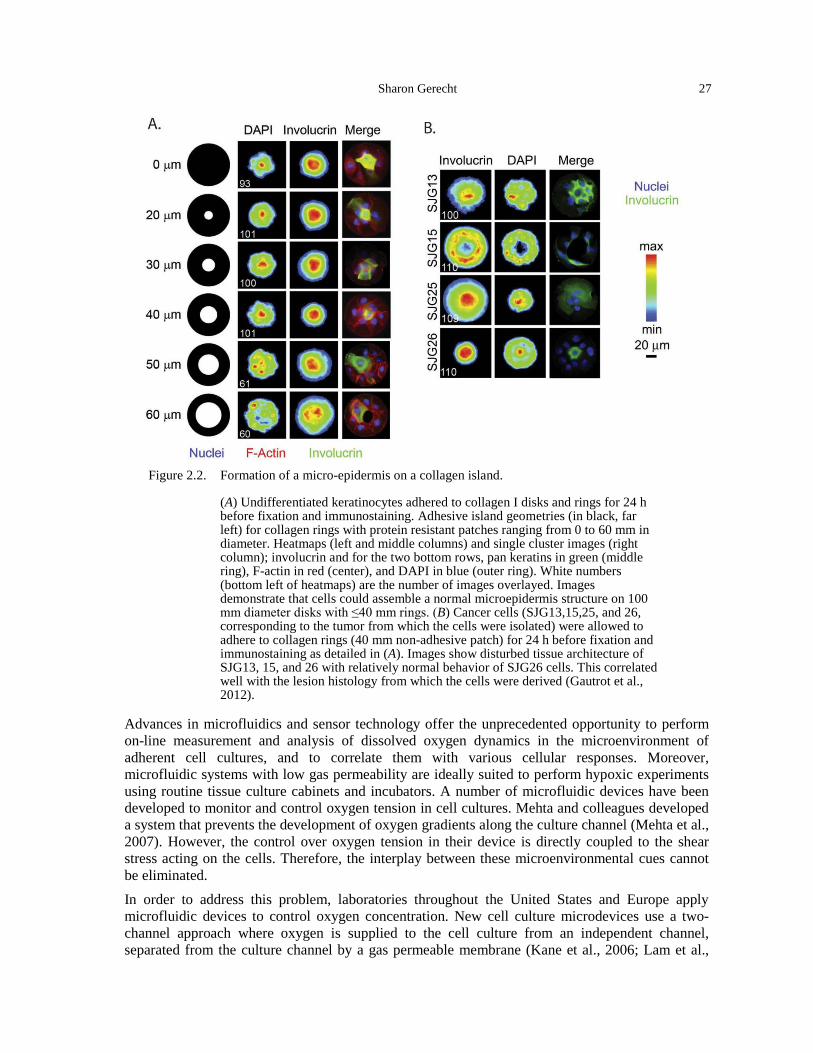
Sharon Gerecht 27
Figure 2.2. Formation of a micro-epidermis on a collagen island.
(A) Undifferentiated keratinocytes adhered to collagen I disks and rings for 24 h before fixation and immunostaining. Adhesive island geometries (in black, far left) for collagen rings with protein resistant patches ranging from 0 to 60 mm in diameter. Heatmaps (left and middle columns) and single cluster images (right column); involucrin and for the two bottom rows, pan keratins in green (middle ring), F-actin in red (center), and DAPI in blue (outer ring). White numbers (bottom left of heatmaps) are the number of images overlayed. Images demonstrate that cells could assemble a normal microepidermis structure on 100 mm diameter disks with ≤40 mm rings. (B) Cancer cells (SJG13,15,25, and 26, corresponding to the tumor from which the cells were isolated) were allowed to adhere to collagen rings (40 mm non-adhesive patch) for 24 h before fixation and immunostaining as detailed in (A). Images show disturbed tissue architecture of SJG13, 15, and 26 with relatively normal behavior of SJG26 cells. This correlated well with the lesion histology from which the cells were derived (Gautrot et al., 2012).
Advances in microfluidics and sensor technology offer the unprecedented opportunity to perform on-line measurement and analysis of dissolved oxygen dynamics in the microenvironment of adherent cell cultures, and to correlate them with various cellular responses. Moreover, microfluidic systems with low gas permeability are ideally suited to perform hypoxic experiments using routine tissue culture cabinets and incubators. A number of microfluidic devices have been developed to monitor and control oxygen tension in cell cultures. Mehta and colleagues developed a system that prevents the development of oxygen gradients along the culture channel (Mehta et al., 2007). However, the control over oxygen tension in their device is directly coupled to the shear stress acting on the cells. Therefore, the interplay between these microenvironmental cues cannot be eliminated. In order to address this problem, laboratories throughout the United States and Europe apply microfluidic devices to control oxygen concentration. New cell culture microdevices use a two-channel approach where oxygen is supplied to the cell culture from an independent channel, separated from the culture channel by a gas permeable membrane (Kane et al., 2006; Lam et al.,
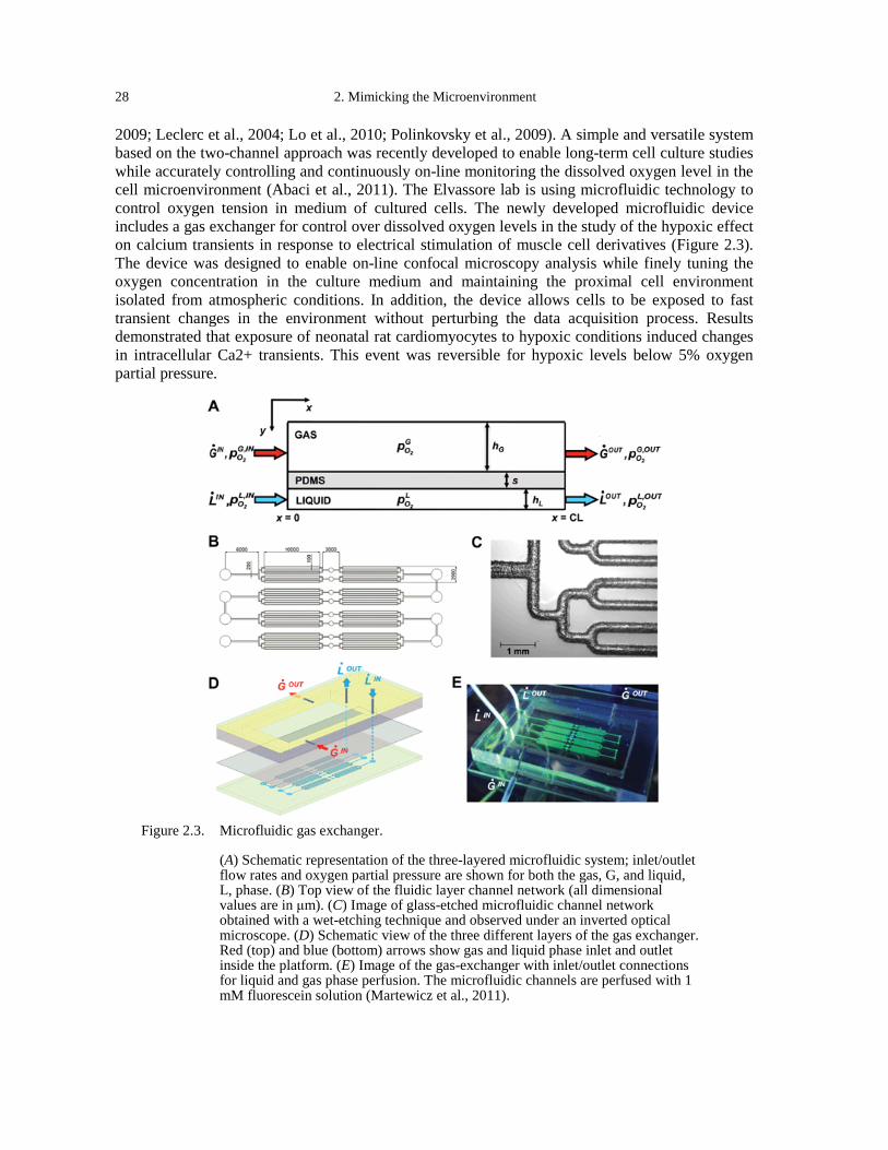
28 2. Mimicking the Microenvironment
2009; Leclerc et al., 2004; Lo et al., 2010; Polinkovsky et al., 2009). A simple and versatile system based on the two-channel approach was recently developed to enable long-term cell culture studies while accurately controlling and continuously on-line monitoring the dissolved oxygen level in the cell microenvironment (Abaci et al., 2011). The Elvassore lab is using microfluidic technology to control oxygen tension in medium of cultured cells. The newly developed microfluidic device includes a gas exchanger for control over dissolved oxygen levels in the study of the hypoxic effect on calcium transients in response to electrical stimulation of muscle cell derivatives (Figure 2.3). The device was designed to enable on-line confocal microscopy analysis while finely tuning the oxygen concentration in the culture medium and maintaining the proximal cell environment isolated from atmospheric conditions. In addition, the device allows cells to be exposed to fast transient changes in the environment without perturbing the data acquisition process. Results demonstrated that exposure of neonatal rat cardiomyocytes to hypoxic conditions induced changes in intracellular Ca2+ transients. This event was reversible for hypoxic levels below 5% oxygen partial pressure.
Figure 2.3. Microfluidic gas exchanger.
(A) Schematic representation of the three-layered microfluidic system; inlet/outlet flow rates and oxygen partial pressure are shown for both the gas, G, and liquid, L, phase. (B) Top view of the fluidic layer channel network (all dimensional values are in μm). (C) Image of glass-etched microfluidic channel network obtained with a wet-etching technique and observed under an inverted optical microscope. (D) Schematic view of the three different layers of the gas exchanger. Red (top) and blue (bottom) arrows show gas and liquid phase inlet and outlet inside the platform. (E) Image of the gas-exchanger with inlet/outlet connections for liquid and gas phase perfusion. The microfluidic channels are perfused with 1 mM fluorescein solution (Martewicz et al., 2011).
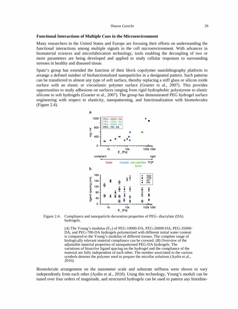
Sharon Gerecht 29
Functional Interactions of Multiple Cues in the Microenvironment
Many researchers in the United States and Europe are focusing their efforts on understanding the functional interactions among multiple signals in the cell microenvironment. With advances in biomaterial sciences and microfabrication technology, tools enabling the decoupling of two or more parameters are being developed and applied to study cellular responses to surrounding stresses in healthy and diseased tissue. Spatz’s group has extended the function of their block copolymer nanolithography platform to arrange a defined number of biofunctionalized nanoparticles in a designated pattern. Such patterns can be transferred to almost any type of soft surface, thereby replacing a stiff glass or silicon oxide surface with an elastic or viscoelastic polymer surface (Graeter et al., 2007). This provides opportunities to study adhesions on surfaces ranging from rigid hydrophobic polystyrene to elastic silicone to soft hydrogels (Graeter et al., 2007). The group has demonstrated PEG hydrogel surface engineering with respect to elasticity, nanopatterning, and functionalization with biomolecules (Figure 2.4).
Figure 2.4. Compliance and nanoparticle decoration properties of PEG- diacrylate (DA)
hydrogels.
(A) The Young’s modulus (EY) of PEG-10000-DA, PEG-20000-DA, PEG-35000-DA, and PEG-700-DA hydrogels polymerized with different initial water content is compared to the Young’s modulus of different tissues. The complete range of biologically relevant material compliance can be covered. (B) Overview of the adjustable material properties of nanopatterned PEG-DA hydrogels. The variations of bioactive ligand spacing on the hydrogel and the compliance of the material are fully independent of each other. The number associated to the various symbols denotes the polymer used to prepare the micellar solutions (Aydin et al., 2010).
Biomolecule arrangement on the nanometer scale and substrate stiffness were shown to vary independently from each other (Aydin et al., 2010). Using this technology, Young’s moduli can be tuned over four orders of magnitude, and structured hydrogels can be used to pattern any histidine-
30 2. Mimicking the Microenvironment
tagged protein as exemplified for his-protein A as an acceptor for immunoglobulin (Aydin et al., 2010). When the cell adhesion-promoting peptide RGD is used selectively, the PEG surfaces provide cues for cell-surface interaction and allow for the study of cellular adhesion modulation by the environmental mechanical properties. Therefore, these substrates represent a unique multipurpose platform for studying receptor/ligand interactions with adhering cells, mechanotransduction, and cell adhesion-dependent signaling. In recent work, Spatz and his team used their block copolymer nanolithography platform to arrange biofunctionalized nanoparticles with either biotinylated RGD, epidermal growth factor (EGF), or both on an inert surface (Shahal et al., 2012). With this surface, they investigated the coregulation of RGD- and immobilized/soluble EGF-mediated signaling in the adhesion of A431 epidermoid carcinoma cells. A synergism was found between integrin and the EGF receptor in an RGD and EGF density- or concentration-dependent manner. The effect of immobilized EGF differed from the effect of soluble EGF above a certain EGF density. The researchers speculate that the critical EGF density is most likely required for the induction of EGF receptor dimerization, and lies within the range of the RGD density. This suggests a role for EGFR-integrin cooperativity in EGF-mediated adhesion response (Shahal et al., 2012). It is thus becoming apparent that in addition to growth factor sequestration, ECM-growth factor interactions also directly modulate growth factor signaling through a co-association of integrins with growth factor receptors. In this context, complexes between ECM proteins and growth factors can mediate enhanced growth factor receptor-integrin signaling by the formation of clusters between growth factor receptors and integrins (Comoglio et al., 2003; Giancotti and Tarone, 2003; Guo and Giancotti, 2004). The Jeffrey Hubbell group at École Polytechnique Fédérale de Lausanne, Switzerland (site report, Appendix B) is moving towards a 3D scaffold platform. They recently explored whether growth factor-induced tissue response could be strongly enhanced when growth factors are delivered within a hydrogel with a well-defined microenvironment designed to trigger synergistic signaling between growth factor receptors and integrins (Martino et al., 2011). To achieve this, a multifunctional recombinant fibronectin (FN) fragment was engineered to display integrin-binding domains linked to growth factor binding domains. The sequence consisted of a coagulation transglutaminase enabling polymerization of fibrin (Figure 2.5). Such engineered microenvironments allow the sequestration of multiple growth factors while promoting joint integrin-growth factor receptor signaling. The group showed that proliferation and migration of endothelial cells, smooth muscle cells, and MSCs is enhanced by co-delivery of FnIII9-10/12-14 and VEGF, PDGF-BB and BMP-2, respectively. Moreover, the FN domains enhanced growth factor-induced morphogenesis in the fibrin matrices. Finally, the engineered fibrin scaffolds were shown to improve growth factor efficiency in tissue repair in vivo (i.e., diabetic wound healing and critical-size bone defect) (Martino et al., 2011). Chapter 4 discusses the use of such fibrin gels for the analysis of lymphatic drainage during tumor growth. A recent collaboration between Stefano Piccolo’s lab at the University of Padua, Italy (site report, Appendix B) and the Elvassore lab, identified the YAP/TAZ [Yorkie-homologues YAP (Yes-associated protein) and TAZ (transcriptional coactivator with PDZ-binding motif)] as nuclear relays of mechanical signals exerted by extracellular rigidity and cell shape, independently from Hippo pathways (Dupont et al., 2011). The groups used micropillar substrates with varying rigidity to decouple matrix stiffness and adhesion. First, following bioinformatic analysis that suggested YAP/TAZ involvement, they examined the expression levels and localization in mammary epithelial cells cultured on fibronectin-coated PAA hydrogel of varying stiffness. YAP/TAZ activity increased and was localized to the nucleus on stiff hydrogels. Next, they used micropatterning of fibronectin islands of different sizes to show that in large adhesion/cell
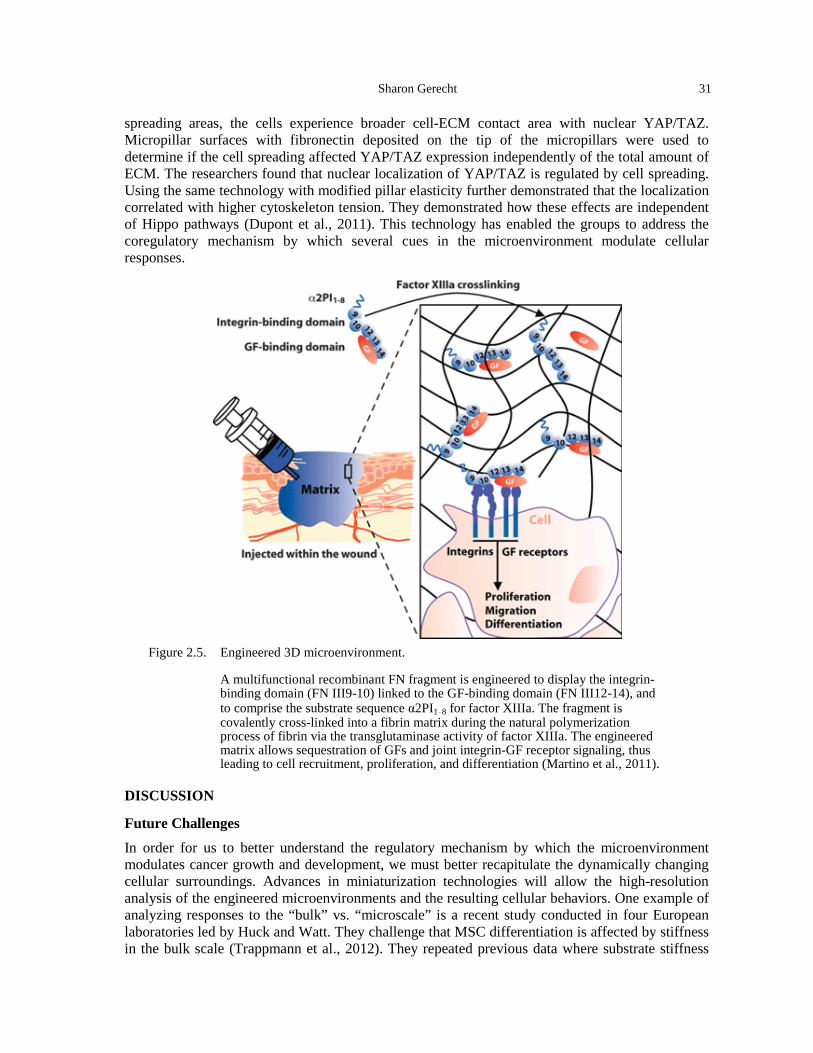
Sharon Gerecht 31
spreading areas, the cells experience broader cell-ECM contact area with nuclear YAP/TAZ. Micropillar surfaces with fibronectin deposited on the tip of the micropillars were used to determine if the cell spreading affected YAP/TAZ expression independently of the total amount of ECM. The researchers found that nuclear localization of YAP/TAZ is regulated by cell spreading. Using the same technology with modified pillar elasticity further demonstrated that the localization correlated with higher cytoskeleton tension. They demonstrated how these effects are independent of Hippo pathways (Dupont et al., 2011). This technology has enabled the groups to address the coregulatory mechanism by which several cues in the microenvironment modulate cellular responses.
Figure 2.5. Engineered 3D microenvironment.
A multifunctional recombinant FN fragment is engineered to display the integrin-binding domain (FN III9-10) linked to the GF-binding domain (FN III12-14), and to comprise the substrate sequence α2PI1–8 for factor XIIIa. The fragment is covalently cross-linked into a fibrin matrix during the natural polymerization process of fibrin via the transglutaminase activity of factor XIIIa. The engineered matrix allows sequestration of GFs and joint integrin-GF receptor signaling, thus leading to cell recruitment, proliferation, and differentiation (Martino et al., 2011).
DISCUSSION
Future Challenges
In order for us to better understand the regulatory mechanism by which the microenvironment modulates cancer growth and development, we must better recapitulate the dynamically changing cellular surroundings. Advances in miniaturization technologies will allow the high-resolution analysis of the engineered microenvironments and the resulting cellular behaviors. One example of analyzing responses to the “bulk” vs. “microscale” is a recent study conducted in four European laboratories led by Huck and Watt. They challenge that MSC differentiation is affected by stiffness in the bulk scale (Trappmann et al., 2012). They repeated previous data where substrate stiffness
32 2. Mimicking the Microenvironment
affects MSC spreading and adipocyte (fat) and osteoblast differentiation by using PAA. However, using polydimethylsiloxane (PDMS) substrate, the elastic modulus did not seem to affect cell spreading and differentiation. Therefore, they looked into the deposition of the coating protein, collagen I, on each of these surfaces. They found that it is differently deposited on PAA substrates with smaller pores in stiffer hydrogels. This suggests that adhesion to the collagen coating regulates cell response, not the bulk stiffness. They also controlled the density of adhesions using the same stiffness and demonstrated the different cellular responses by applying covalent binding (Trappmann et al., 2012). This approach utilizing technological capabilities in laboratories in different institutions and countries throughout Europe is a noticeable strength.
This work raises new questions and indicates the need for a more complex approach, which will take into account the presentation of adhesions at the microscale and how this can relate to mechanics. Advances in technology should also be made to enable better imitation of physiological conditions. For example, most technologies currently capture a specific time point in the microenvironment and allow the study of cellular behaviors in response to a given cue. The ability to generate dynamic environments that respond to feedback from the tissue/cells during long culture periods is envisioned to allow the analysis of cellular responses to the dynamically changing cancerous surroundings.
In a step toward this goal, a recent publication by Guvendiren and Burdick in the United States describes how new hydrogels were developed to enable dynamic stiffening along the culture period (Guvendiren and Burdick, 2012). Sequential crosslinking—first gelation by an addition reaction (of DTT) to methacrylated HA to generate soft environments in the presence of cells followed by radical polymerization (crosslinking of the remaining group by exposure to UV) created a stiffening niche for the encapsulated cells. The Burdick lab has shown the response of MSCs to the stiffening environment from non-spread to spreading morphology. They then demonstrated that differentiation of MSCs during a 14-day span can be controlled by temporal stiffening. While stiff hydrogels supported bone differentiation and soft hydrogels supported fat differentiation, the stiffening hydrogel induced mixed adipogenic/osteogenic population, suggesting that not all cells were responsive to changes in mechanical properties along the culture period (Guvendiren and Burdick, 2012). Again, new questions arise with the use of more “biologically” accurate biomaterials. Another venue is the generation of different cue gradients in vitro, similar to their presence within the growing tumor and its surroundings, such as cytokines and oxygen tension. Finally, decoupling parameters in 3D still presents a major challenge and worldwide advances in biomaterial synthesis are needed to enable progress.
REFERENCES Abaci, H.E., Devendra, R., Smith, Q., Gerecht, S., and Drazer, G. (2011). Design and development of
microbioreactors for long-term cell culture in controlled oxygen microenvironments. Biomed Microdevices 14, 145-152.
Alowami, S., Troup, S., Al-Haddad, S., Kirkpatrick, I., and Watson, P.H. (2003). Mammographic density is related to stroma and stromal proteoglycan expression. Breast Cancer Res 5, R129-135.
Arnold, M., Cavalcanti-Adam, E.A., Glass, R., Blummel, J., Eck, W., Kantlehner, M., Kessler, H., and Spatz, J.P. (2004). Activation of integrin function by nanopatterned adhesive interfaces. Chemphyschem 5, 383-388.
Aydin, D., Louban, I., Perschmann, N., Blummel, J., Lohmuller, T., Cavalcanti-Adam, E.A., Haas, T.L., Walczak, H., Kessler, H., Fiammengo, R., et al. (2010). Polymeric substrates with tunable elasticity and nanoscopically controlled biomolecule presentation. Langmuir 26, 15472-15480.
Boyd, N.F., Lockwood, G.A., Byng, J.W., Tritchler, D.L., and Yaffe, M.J. (1998). Mammographic densities and breast cancer risk. Cancer Epidemiol Biomarkers Prev 7, 1133-1144.
Sharon Gerecht 33
Boyd, N.F., Martin, L.J., Stone, J., Greenberg, C., Minkin, S., and Yaffe, M.J. (2001). Mammographic densities as a marker of human breast cancer risk and their use in chemoprevention. Curr Oncol Rep 3, 314-321.
Brown, J.M., and Giaccia, A.J. (1998). The unique physiology of solid tumors: opportunities (and problems) for cancer therapy. Cancer Res 58, 1408-1416.
Comoglio, P.M., Boccaccio, C., and Trusolino, L. (2003). Interactions between growth factor receptors and adhesion molecules: breaking the rules. Curr Opin Cell Biol 15, 565-571.
Dewhirst, M.W. (2009). Relationships between cycling hypoxia, HIF-1, angiogenesis and oxidative stress. Radiat Res 172, 653-665.
Dewhirst, M.W., Cao, Y., and Moeller, B. (2008). Cycling hypoxia and free radicals regulate angiogenesis and radiotherapy response. Nat Rev Cancer 8, 425-437.
Dupont, S., Morsut, L., Aragona, M., Enzo, E., Giulitti, S., Cordenonsi, M., Zanconato, F., Le Digabel, J., Forcato, M., Bicciato, S., et al. (2011). Role of YAP/TAZ in mechanotransduction. Nature 474, 179-183.
Eccles, S.A. (2004). Targeting key steps in metastic tumour progression. Current Opinion in Genetics and Development 15, 77-86.
Entschladen, F., Drell, T.L., Lang, K., Joseph, J., and Zaenker, K.S. (2004). Tumour-cell migration, invasion, and metastasis: navigation by neurotransmitters. The Lancet Oncology 5, 254-258.
Ezashi, T., Das, P., and Roberts, R.M. (2005). Low O2 tensions and the prevention of differentiation of hES cells. Proc Natl Acad Sci U S A 102, 4783-4788.
Gautrot, J.E., Wang, C., Liu, X., Goldie, S.J., Trappmann, B., Huck, W.T., and Watt, F.M. (2012). Mimicking normal tissue architecture and perturbation in cancer with engineered micro-epidermis. Biomaterials 33, 5221-5229.
Giancotti, F.G., and Tarone, G. (2003). Positional control of cell fate through joint integrin/receptor protein kinase signaling. Annu Rev Cell Dev Biol 19, 173-206.
Gilbert, P.M., Havenstrite, K.L., Magnusson, K.E., Sacco, A., Leonardi, N.A., Kraft, P., Nguyen, N.K., Thrun, S., Lutolf, M.P., and Blau, H.M. (2010). Substrate elasticity regulates skeletal muscle stem cell self-renewal in culture. Science 329, 1078-1081.
Gobaa, S., Hoehnel, S., Roccio, M., Negro, A., Kobel, S., and Lutolf, M.P. (2011). Artificial niche microarrays for probing single stem cell fate in high throughput. Nat Methods 8, 949-955.
Graeter, S.V., Huang, J., Perschmann, N., Lopez-Garcia, M., Kessler, H., Ding, J., and Spatz, J.P. (2007). Mimicking cellular environments by nanostructured soft interfaces. Nano Lett 7, 1413-1418.
Guo, W., and Giancotti, F.G. (2004). Integrin signalling during tumour progression. Nat Rev Mol Cell Biol 5, 816-826.
Guo, Y.P., Martin, L.J., Hanna, W., Banerjee, D., Miller, N., Fishell, E., Khokha, R., and Boyd, N.F. (2001). Growth factors and stromal matrix proteins associated with mammographic densities. Cancer Epidemiol Biomarkers Prev 10, 243-248.
Guvendiren, M., and Burdick, J.A. (2012). Stiffening hydrogels to probe short- and long-term cellular responses to dynamic mechanics. Nat Commun 3, 792.
Hirschfeld-Warneken, V.C., Arnold, M., Cavalcanti-Adam, A., Lopez-Garcia, M., Kessler, H., and Spatz, J.P. (2008). Cell adhesion and polarisation on molecularly defined spacing gradient surfaces of cyclic RGDfK peptide patches. Eur J Cell Biol 87, 743-750.
Jarvelainen, H., Sainio, A., Koulu, M., Wight, T.N., and Penttinen, R. (2009). Extracellular matrix molecules: potential targets in pharmacotherapy. Pharmacol Rev 61, 198-223.
Jiang, M., Wang, B., Wang, C., He, B., Fan, H., Guo, T.B., Shao, Q., Gao, L., and Liu, Y. (2008a). Angiogenesis by transplantation of HIF-1 alpha modified EPCs into ischemic limbs. J Cell Biochem 103, 321-334.
Jiang, M., Wang, B., Wang, C., He, B., Fan, H., Shao, Q., Gao, L., Liu, Y., Yan, G., and Pu, J. (2008b). In vivo enhancement of angiogenesis by adenoviral transfer of HIF-1alpha-modified endothelial progenitor cells (Ad-HIF-1alpha-modified EPC for angiogenesis). Int J Biochem Cell Biol 40, 2284-2295.
Kane, B.J., Zinner, M.J., Yarmush, M.L., and Toner, M. (2006). Liver-specific functional studies in a microfluidic array of primary mammalian hepatocytes. Anal Chem 78, 4291-4298.
Kimura, H., Braun, R.D., Ong, E.T., Hsu, R., Secomb, T.W., Papahadjopoulos, D., Hong, K., and Dewhirst, M.W. (1996). Fluctuations in red cell flux in tumor microvessels can lead to transient hypoxia and reoxygenation in tumor parenchyma. Cancer Res 56, 5522-5528.
34 2. Mimicking the Microenvironment
Lam, R.H., Kim, M.C., and Thorsen, T. (2009). Culturing aerobic and anaerobic bacteria and mammalian cells with a microfluidic differential oxygenator. Anal Chem 81, 5918-5924.
Leclerc, E., Sakai, Y., and Fujii, T. (2004). Microfluidic PDMSMS (polydimethylsiloxane) bioreactor for large-scale culture of hepatocytes. Biotechnol Prog 20, 750-755.
Li, T., Sun, L., Miller, N., Nicklee, T., Woo, J., Hulse-Smith, L., Tsao, M.S., Khokha, R., Martin, L., and Boyd, N. (2005). The association of measured breast tissue characteristics with mammographic density and other risk factors for breast cancer. Cancer Epidemiol Biomarkers Prev 14, 343-349.
Lo, J.F., Sinkala, E., and Eddington, D.T. (2010). Oxygen gradients for open well cellular cultures via microfluidic substrates. Lab Chip 10, 2394-2401.
Lu, P., Weaver, V.M., and Werb, Z. (2012). The extracellular matrix: a dynamic niche in cancer progression. J Cell Biol 196, 395-406.
Martewicz, S., Michielin, F., Serena, E., Zambon, A., Mongillo, M., and Elvassore, N. (2011). Reversible alteration of calcium dynamics in cardiomyocytes during acute hypoxia transient in a microfluidic platform. Integr Biol (Camb) 4, 153-164.
Martinive, P., Defresne, F., Bouzin, C., Saliez, J., Lair, F., Gregoire, V., Michiels, C., Dessy, C., and Feron, O. (2006). Preconditioning of the tumor vasculature and tumor cells by intermittent hypoxia: implications for anticancer therapies. Cancer Res 66, 11736-11744.
Martino, M.M., Tortelli, F., Mochizuki, M., Traub, S., Ben-David, D., Kuhn, G.A., Muller, R., Livne, E., Eming, S.A., and Hubbell, J.A. (2011). Engineering the growth factor microenvironment with fibronectin domains to promote wound and bone tissue healing. Sci Transl Med 3, 100ra189.
McCormack, V.A., and dos Santos Silva, I. (2006). Breast density and parenchymal patterns as markers of breast cancer risk: a meta-analysis. Cancer Epidemiol Biomarkers Prev 15, 1159-1169.
Mehta, G., Mehta, K., Sud, D., Song, J.W., Bersano-Begey, T., Futai, N., Heo, Y.S., Mycek, M.A., Linderman, J.J., and Takayama, S. (2007). Quantitative measurement and control of oxygen levels in microfluidic poly(dimethylsiloxane) bioreactors during cell culture. Biomed Microdevices 9, 123-134.
Miyaji, K., Furuse, A., Nakajima, J., Kohno, T., Ohtsuka, T., Yagyu, K., Oka, T., and Omata, S. (1997). The stiffness of lymph nodes containing lung carcinoma metastases: a new diagnostic parameter measured by a tactile sensor. Cancer 80, 1920-1925.
Niebruegge, S., Bauwens, C.L., Peerani, R., Thavandiran, N., Masse, S., Sevaptisidis, E., Nanthakumar, K., Woodhouse, K., Husain, M., Kumacheva, E., et al. (2008). Generation of human embryonic stem cell-derived mesoderm and cardiac cells using size-specified aggregates in an oxygen-controlled bioreactor. Biotechnol Bioeng 102, 493-507.
Polinkovsky, M., Gutierrez, E., Levchenko, A., and Groisman, A. (2009). Fine temporal control of the medium gas content and acidity and on-chip generation of series of oxygen concentrations for cell cultures. Lab Chip 9, 1073-1084.
Purpura, K.A., George, S.H., Dang, S.M., Choi, K., Nagy, A., and Zandstra, P.W. (2008). Soluble Flt-1 regulates Flk-1 activation to control hematopoietic and endothelial development in an oxygen-responsive manner. Stem Cells 26, 2832-2842.
Rofstad, E.K., Gaustad, J.V., Egeland, T.A., Mathiesen, B., and Galappathi, K. (2010). Tumors exposed to acute cyclic hypoxic stress show enhanced angiogenesis, perfusion and metastatic dissemination. Int J Cancer 127, 1535-1546.
Samani, A., Zubovits, J., and Plewes, D. (2007). Elastic moduli of normal and pathological human breast tissues: an inversion-technique-based investigation of 169 samples. Phys Med Biol 52, 1565-1576.
Serena, E., Zatti, S., Reghelin, E., Pasut, A., Cimetta, E., and Elvassore, N. (2010). Soft substrates drive optimal differentiation of human healthy and dystrophic myotubes. Integr Biol (Camb) 2, 193-201.
Shahal, T., Geiger, B., Dunlop, I.E., and Spatz, J.P. (2012). Regulation of integrin adhesions by varying the density of substrate-bound epidermal growth factor. Biointerphases 7, 23.
Spatz, J.P., and Geiger, B. (2007). Molecular engineering of cellular environments: cell adhesion to nano-digital surfaces. Methods Cell Biol 83, 89-111.
Trappmann, B., Gautrot, J.E., Connelly, J.T., Strange, D.G., Li, Y., Oyen, M.L., Cohen Stuart, M.A., Boehm, H., Li, B., Vogel, V., et al. (2012). Extracellular-matrix tethering regulates stem-cell fate. Nat Mater 11, 642-649.

CHAPTER 3
CANCER CELL MECHANICS
Cynthia A. Reinhart-King
INTRODUCTION
Cell mechanics is an expanding area of research within the cancer community. Its increasing importance in the cancer biology field likely stems from the fact that metastasis is an inherently physical process, involving the pushing/pulling of cells away from the primary tumor and through the surrounding stromal environment (Friedl and Alexander, 2011; Friedl and Wolf, 2003; Wirtz, Konstantopoulos and Searson, 2011). While cell mechanics has a long history, it has had a significant resurgence in the past couple of decades, largely enabled by new and better technologies to interrogate cells and molecules. Cell mechanics can be thought of as three separate subfields: 1) cellular mechanical properties; 2) mechanotransduction (the cellular response to forces imposed on cells by the external environment); and 3) cell-generated forces (Figure 3.1). Studies of cellular mechanical properties have largely focused on either the elastic modulus or deformability of the whole cell or the rheology of the cytoplasm. These measurements are important within the cancer field because they implicate if and how cells will migrate and squeeze through the matrix during metastatic invasion through the stromal matrix and into the vasculature. Mechanotransduction research in cancer has been centered on the response of cells to imposed pressures and fluid shear stresses within the tumor microenvironment. It is important to our understanding of the mechanical forces that influence tumor growth and metastasis. Lastly, studies of cell-generated force are critical to our understanding of how cells adhere, traverse, and sense their microenvironment. Each of these facets of cell mechanics (mechanical properties, mechanotransduction, and cell-generated forces) has been shown to influence each other, demonstrating the integration of inside and outside signals. This chapter will address all three of these subfields in oncology, as well as emerging themes in this area occurring in the United States and that were observed during the APHELION study tour in Europe. In many ways, the term “cell mechanics” is a misnomer for a field that encompasses much more than simply cell-scale behaviors and properties. It includes not only the mechanics of individual cells, but also the mechanical forces and mechanical properties at the molecular and tissue scales. In this chapter, the focus is primarily on mechanics at the cellular scale; however, it is important to note that the multiscale contributions of subcellular structures and supracellular tissue properties cannot be overlooked. At the subcellular scale, the cytoskeleton organizes to exert intracellular forces that translate into cell behaviors such as mitosis, intracellular transport, lamellipodial extension, and cell migration. Changes in various molecules within cells are closely tied to mechanical changes in the cell. For example, the mechanical properties and kinetics of cytoskeletal assembly at the molecular level contribute to changes in the mechanical properties of the cell (Kraning-Rush et al., 2011). Similarly, at the supracellular scale, the mechanical properties and architecture of a tissue contribute to cell function and dysfunction. In the cancer field, this is
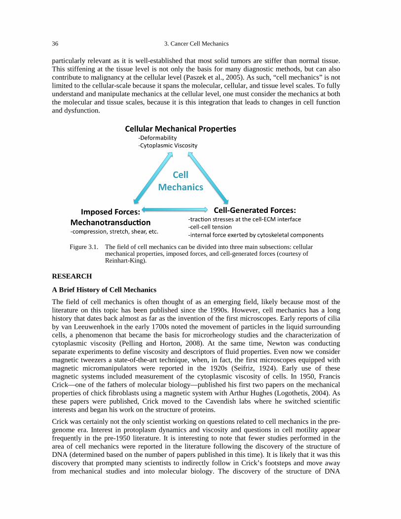
36 3. Cancer Cell Mechanics
particularly relevant as it is well-established that most solid tumors are stiffer than normal tissue. This stiffening at the tissue level is not only the basis for many diagnostic methods, but can also contribute to malignancy at the cellular level (Paszek et al., 2005). As such, “cell mechanics” is not limited to the cellular-scale because it spans the molecular, cellular, and tissue level scales. To fully understand and manipulate mechanics at the cellular level, one must consider the mechanics at both the molecular and tissue scales, because it is this integration that leads to changes in cell function and dysfunction.
Figure 3.1. The field of cell mechanics can be divided into three main subsections: cellular
mechanical properties, imposed forces, and cell-generated forces (courtesy of Reinhart-King).
RESEARCH
A Brief History of Cell Mechanics
The field of cell mechanics is often thought of as an emerging field, likely because most of the literature on this topic has been published since the 1990s. However, cell mechanics has a long history that dates back almost as far as the invention of the first microscopes. Early reports of cilia by van Leeuwenhoek in the early 1700s noted the movement of particles in the liquid surrounding cells, a phenomenon that became the basis for microrheology studies and the characterization of cytoplasmic viscosity (Pelling and Horton, 2008). At the same time, Newton was conducting separate experiments to define viscosity and descriptors of fluid properties. Even now we consider magnetic tweezers a state-of-the-art technique, when, in fact, the first microscopes equipped with magnetic micromanipulators were reported in the 1920s (Seifriz, 1924). Early use of these magnetic systems included measurement of the cytoplasmic viscosity of cells. In 1950, Francis Crick—one of the fathers of molecular biology—published his first two papers on the mechanical properties of chick fibroblasts using a magnetic system with Arthur Hughes (Logothetis, 2004). As these papers were published, Crick moved to the Cavendish labs where he switched scientific interests and began his work on the structure of proteins. Crick was certainly not the only scientist working on questions related to cell mechanics in the pre-genome era. Interest in protoplasm dynamics and viscosity and questions in cell motility appear frequently in the pre-1950 literature. It is interesting to note that fewer studies performed in the area of cell mechanics were reported in the literature following the discovery of the structure of DNA (determined based on the number of papers published in this time). It is likely that it was this discovery that prompted many scientists to indirectly follow in Crick’s footsteps and move away from mechanical studies and into molecular biology. The discovery of the structure of DNA
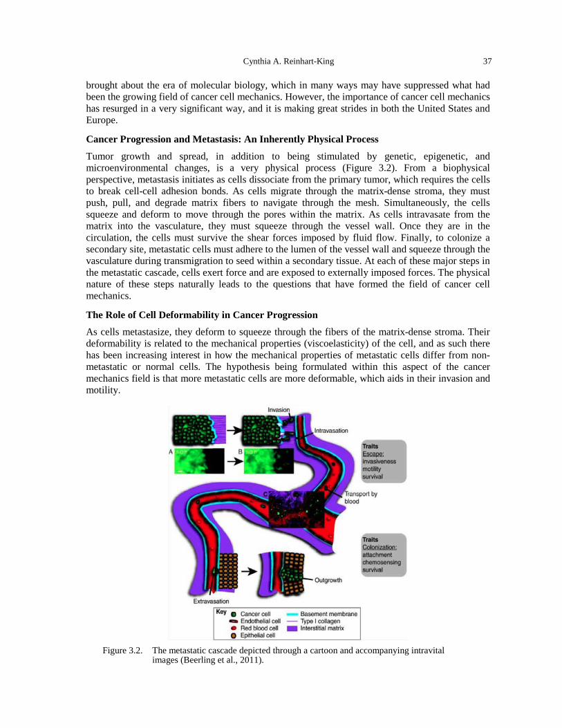
Cynthia A. Reinhart-King 37
brought about the era of molecular biology, which in many ways may have suppressed what had been the growing field of cancer cell mechanics. However, the importance of cancer cell mechanics has resurged in a very significant way, and it is making great strides in both the United States and Europe.
Cancer Progression and Metastasis: An Inherently Physical Process
Tumor growth and spread, in addition to being stimulated by genetic, epigenetic, and microenvironmental changes, is a very physical process (Figure 3.2). From a biophysical perspective, metastasis initiates as cells dissociate from the primary tumor, which requires the cells to break cell-cell adhesion bonds. As cells migrate through the matrix-dense stroma, they must push, pull, and degrade matrix fibers to navigate through the mesh. Simultaneously, the cells squeeze and deform to move through the pores within the matrix. As cells intravasate from the matrix into the vasculature, they must squeeze through the vessel wall. Once they are in the circulation, the cells must survive the shear forces imposed by fluid flow. Finally, to colonize a secondary site, metastatic cells must adhere to the lumen of the vessel wall and squeeze through the vasculature during transmigration to seed within a secondary tissue. At each of these major steps in the metastatic cascade, cells exert force and are exposed to externally imposed forces. The physical nature of these steps naturally leads to the questions that have formed the field of cancer cell mechanics.
The Role of Cell Deformability in Cancer Progression
As cells metastasize, they deform to squeeze through the fibers of the matrix-dense stroma. Their deformability is related to the mechanical properties (viscoelasticity) of the cell, and as such there has been increasing interest in how the mechanical properties of metastatic cells differ from non-metastatic or normal cells. The hypothesis being formulated within this aspect of the cancer mechanics field is that more metastatic cells are more deformable, which aids in their invasion and motility.
Figure 3.2. The metastatic cascade depicted through a cartoon and accompanying intravital
images (Beerling et al., 2011).
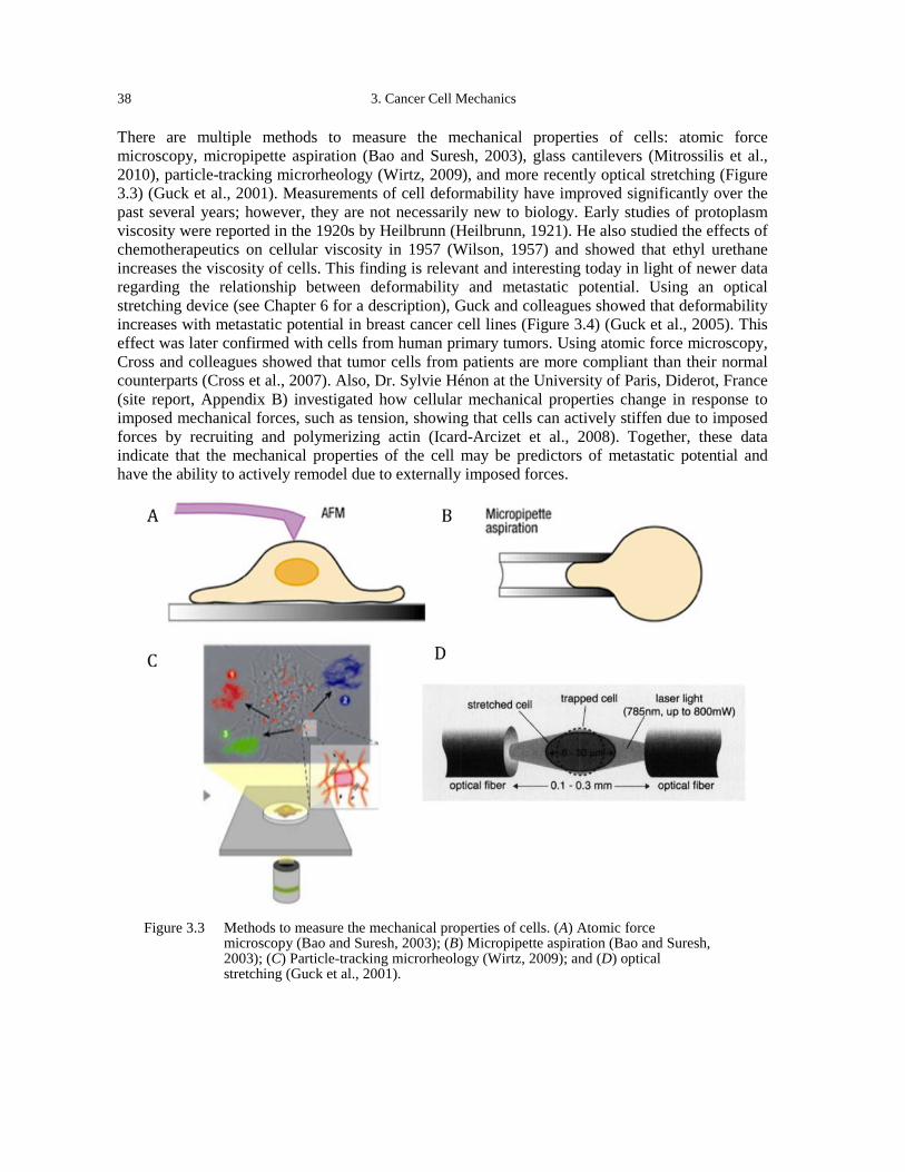
38 3. Cancer Cell Mechanics
There are multiple methods to measure the mechanical properties of cells: atomic force microscopy, micropipette aspiration (Bao and Suresh, 2003), glass cantilevers (Mitrossilis et al., 2010), particle-tracking microrheology (Wirtz, 2009), and more recently optical stretching (Figure 3.3) (Guck et al., 2001). Measurements of cell deformability have improved significantly over the past several years; however, they are not necessarily new to biology. Early studies of protoplasm viscosity were reported in the 1920s by Heilbrunn (Heilbrunn, 1921). He also studied the effects of chemotherapeutics on cellular viscosity in 1957 (Wilson, 1957) and showed that ethyl urethane increases the viscosity of cells. This finding is relevant and interesting today in light of newer data regarding the relationship between deformability and metastatic potential. Using an optical stretching device (see Chapter 6 for a description), Guck and colleagues showed that deformability increases with metastatic potential in breast cancer cell lines (Figure 3.4) (Guck et al., 2005). This effect was later confirmed with cells from human primary tumors. Using atomic force microscopy, Cross and colleagues showed that tumor cells from patients are more compliant than their normal counterparts (Cross et al., 2007). Also, Dr. Sylvie Hénon at the University of Paris, Diderot, France (site report, Appendix B) investigated how cellular mechanical properties change in response to imposed mechanical forces, such as tension, showing that cells can actively stiffen due to imposed forces by recruiting and polymerizing actin (Icard-Arcizet et al., 2008). Together, these data indicate that the mechanical properties of the cell may be predictors of metastatic potential and have the ability to actively remodel due to externally imposed forces.
Figure 3.3 Methods to measure the mechanical properties of cells. (A) Atomic force
microscopy (Bao and Suresh, 2003); (B) Micropipette aspiration (Bao and Suresh, 2003); (C) Particle-tracking microrheology (Wirtz, 2009); and (D) optical stretching (Guck et al., 2001).

Cynthia A. Reinhart-King 39
Figure 3.4. Deformability of cells increases with metastatic potential, as measured using an
optical stretcher (Guck et al., 2005).
The implications of cell stiffening may be both positive and negative with respect to cancer treatments. The data seem to suggest that stiffer cells are less metastatic and that chemotherapeutics stiffen cells, which is a positive effect. However, this stiffening may also have deleterious consequences. Recently, the Fletcher lab (author of Chapter 6) has shown that chemotherapeutic treatments can stiffen leukemic cells that then plug microfluidic channels (Lam et al., 2007). These data suggest that chemotherapy could result in vascular occlusion in capillaries by cancer cells. Therefore, it cannot yet be said that stiffening cells is necessarily a viable, universal method to prevent metastasis due to potential side effects. Work being done by Josef Käs’ lab at the University of Leipzig, Germany (site report, Appendix B) is continuing research in the area of cell deformability by testing clinical samples and working to translate the optical stretching methodology to the clinic. In order for it to be used in the clinic, the method must be both user-friendly and high throughput. While the method of optical stretching is technically complicated, significant strides are being made to make it tractable to clinical laboratory technicians as a potential mechanism to diagnose the likelihood of metastasis. Data are also emerging on the role of nuclear deformability in invasion. Nuclear mechanics could be a limiting factor in allowing cells to permeate through the pores of a matrix (Friedl et al., 2011). If the nucleus is unable to squeeze through a pore, then the cell cannot invade unless the matrix is degraded. Recent data from the Wirtz lab at Johns Hopkins University suggests that the nucleus is not only important due to its deformability, but its connection to the cytoskeleton through the LINC complex plays a critical role in pseudopodial extension during 3D migration (Khatau et al., 2012). Research on the role of nuclear mechanics in cancer progression is still in its infancy and requires further investigation.
Cell-Generated Forces in Adhesion and Migration
Cells generate traction stresses against their matrix to adhere and migrate. These forces aid in remodeling the matrix and propelling cells forward during migration. Because cell migration is necessary for metastatic invasion, there has been recent interest in characterizing and understanding cell-generated traction stresses.
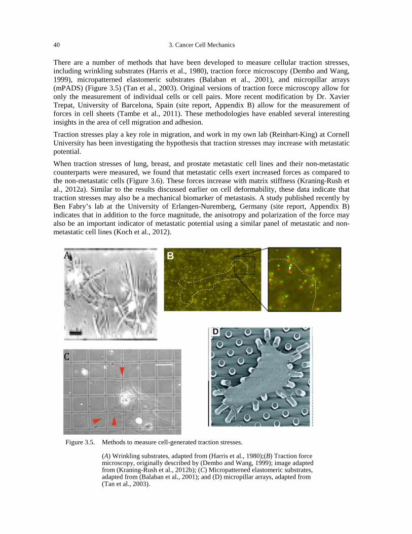
40 3. Cancer Cell Mechanics
There are a number of methods that have been developed to measure cellular traction stresses, including wrinkling substrates (Harris et al., 1980), traction force microscopy (Dembo and Wang, 1999), micropatterned elastomeric substrates (Balaban et al., 2001), and micropillar arrays (mPADS) (Figure 3.5) (Tan et al., 2003). Original versions of traction force microscopy allow for only the measurement of individual cells or cell pairs. More recent modification by Dr. Xavier Trepat, University of Barcelona, Spain (site report, Appendix B) allow for the measurement of forces in cell sheets (Tambe et al., 2011). These methodologies have enabled several interesting insights in the area of cell migration and adhesion. Traction stresses play a key role in migration, and work in my own lab (Reinhart-King) at Cornell University has been investigating the hypothesis that traction stresses may increase with metastatic potential. When traction stresses of lung, breast, and prostate metastatic cell lines and their non-metastatic counterparts were measured, we found that metastatic cells exert increased forces as compared to the non-metastatic cells (Figure 3.6). These forces increase with matrix stiffness (Kraning-Rush et al., 2012a). Similar to the results discussed earlier on cell deformability, these data indicate that traction stresses may also be a mechanical biomarker of metastasis. A study published recently by Ben Fabry’s lab at the University of Erlangen-Nuremberg, Germany (site report, Appendix B) indicates that in addition to the force magnitude, the anisotropy and polarization of the force may also be an important indicator of metastatic potential using a similar panel of metastatic and non-metastatic cell lines (Koch et al., 2012).
Figure 3.5. Methods to measure cell-generated traction stresses.
(A) Wrinkling substrates, adapted from (Harris et al., 1980);(B) Traction force microscopy, originally described by (Dembo and Wang, 1999); image adapted from (Kraning-Rush et al., 2012b); (C) Micropatterned elastomeric substrates, adapted from (Balaban et al., 2001); and (D) micropillar arrays, adapted from (Tan et al., 2003).
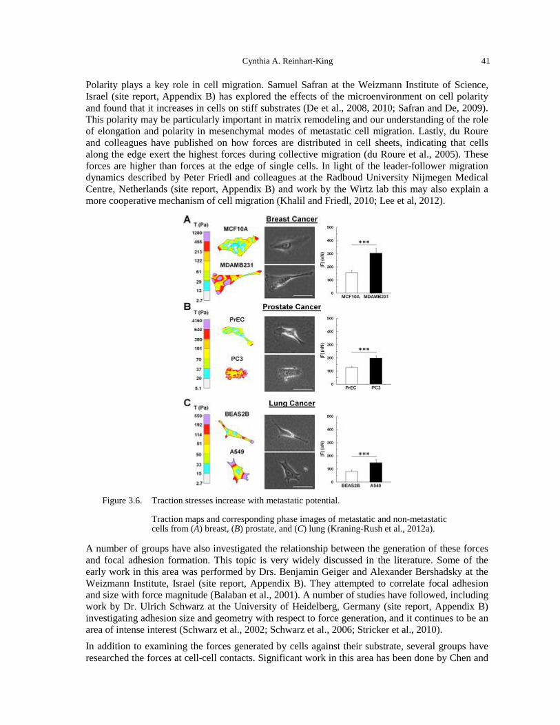
Cynthia A. Reinhart-King 41
Polarity plays a key role in cell migration. Samuel Safran at the Weizmann Institute of Science, Israel (site report, Appendix B) has explored the effects of the microenvironment on cell polarity and found that it increases in cells on stiff substrates (De et al., 2008, 2010; Safran and De, 2009). This polarity may be particularly important in matrix remodeling and our understanding of the role of elongation and polarity in mesenchymal modes of metastatic cell migration. Lastly, du Roure and colleagues have published on how forces are distributed in cell sheets, indicating that cells along the edge exert the highest forces during collective migration (du Roure et al., 2005). These forces are higher than forces at the edge of single cells. In light of the leader-follower migration dynamics described by Peter Friedl and colleagues at the Radboud University Nijmegen Medical Centre, Netherlands (site report, Appendix B) and work by the Wirtz lab this may also explain a more cooperative mechanism of cell migration (Khalil and Friedl, 2010; Lee et al, 2012).
Figure 3.6. Traction stresses increase with metastatic potential.
Traction maps and corresponding phase images of metastatic and non-metastatic cells from (A) breast, (B) prostate, and (C) lung (Kraning-Rush et al., 2012a).
A number of groups have also investigated the relationship between the generation of these forces and focal adhesion formation. This topic is very widely discussed in the literature. Some of the early work in this area was performed by Drs. Benjamin Geiger and Alexander Bershadsky at the Weizmann Institute, Israel (site report, Appendix B). They attempted to correlate focal adhesion and size with force magnitude (Balaban et al., 2001). A number of studies have followed, including work by Dr. Ulrich Schwarz at the University of Heidelberg, Germany (site report, Appendix B) investigating adhesion size and geometry with respect to force generation, and it continues to be an area of intense interest (Schwarz et al., 2002; Schwarz et al., 2006; Stricker et al., 2010). In addition to examining the forces generated by cells against their substrate, several groups have researched the forces at cell-cell contacts. Significant work in this area has been done by Chen and
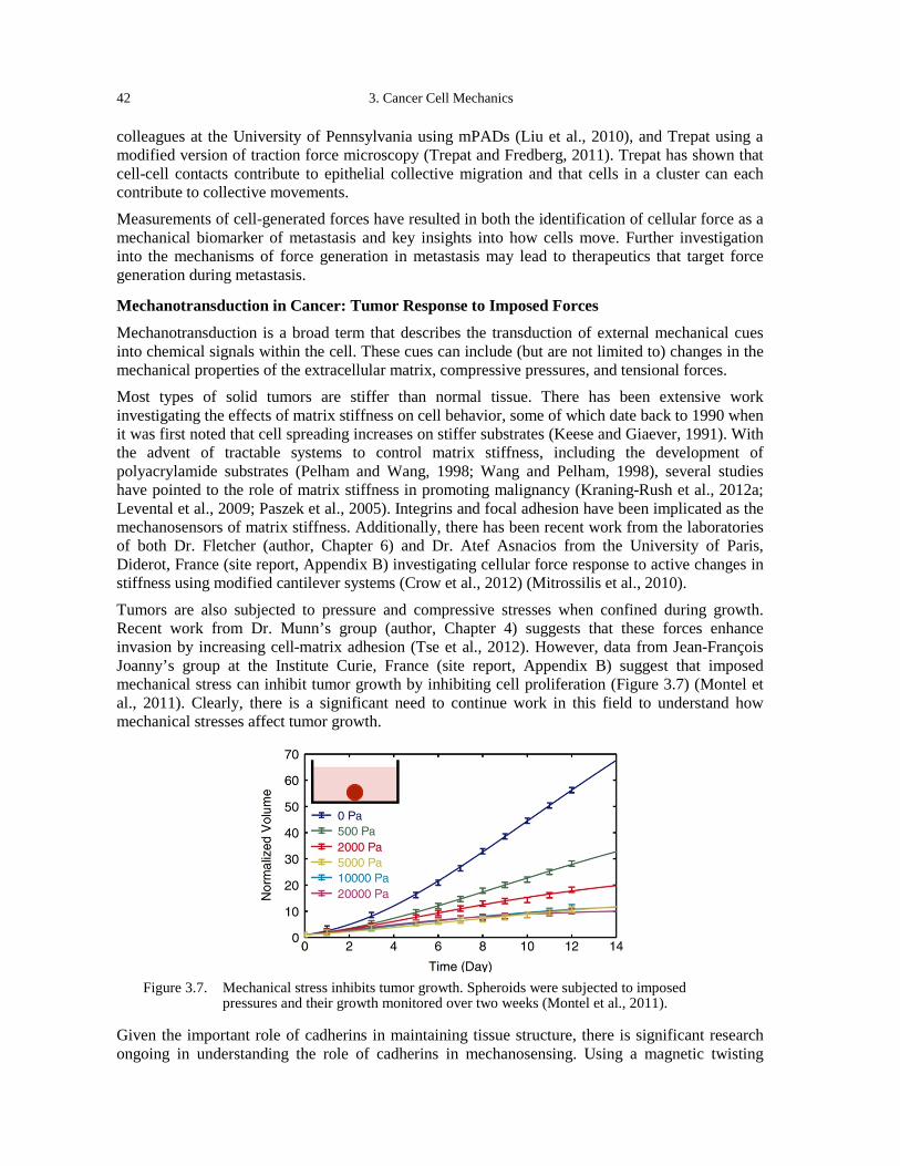
42 3. Cancer Cell Mechanics
colleagues at the University of Pennsylvania using mPADs (Liu et al., 2010), and Trepat using a modified version of traction force microscopy (Trepat and Fredberg, 2011). Trepat has shown that cell-cell contacts contribute to epithelial collective migration and that cells in a cluster can each contribute to collective movements. Measurements of cell-generated forces have resulted in both the identification of cellular force as a mechanical biomarker of metastasis and key insights into how cells move. Further investigation into the mechanisms of force generation in metastasis may lead to therapeutics that target force generation during metastasis.
Mechanotransduction in Cancer: Tumor Response to Imposed Forces
Mechanotransduction is a broad term that describes the transduction of external mechanical cues into chemical signals within the cell. These cues can include (but are not limited to) changes in the mechanical properties of the extracellular matrix, compressive pressures, and tensional forces. Most types of solid tumors are stiffer than normal tissue. There has been extensive work investigating the effects of matrix stiffness on cell behavior, some of which date back to 1990 when it was first noted that cell spreading increases on stiffer substrates (Keese and Giaever, 1991). With the advent of tractable systems to control matrix stiffness, including the development of polyacrylamide substrates (Pelham and Wang, 1998; Wang and Pelham, 1998), several studies have pointed to the role of matrix stiffness in promoting malignancy (Kraning-Rush et al., 2012a; Levental et al., 2009; Paszek et al., 2005). Integrins and focal adhesion have been implicated as the mechanosensors of matrix stiffness. Additionally, there has been recent work from the laboratories of both Dr. Fletcher (author, Chapter 6) and Dr. Atef Asnacios from the University of Paris, Diderot, France (site report, Appendix B) investigating cellular force response to active changes in stiffness using modified cantilever systems (Crow et al., 2012) (Mitrossilis et al., 2010).
Tumors are also subjected to pressure and compressive stresses when confined during growth. Recent work from Dr. Munn’s group (author, Chapter 4) suggests that these forces enhance invasion by increasing cell-matrix adhesion (Tse et al., 2012). However, data from Jean-François Joanny’s group at the Institute Curie, France (site report, Appendix B) suggest that imposed mechanical stress can inhibit tumor growth by inhibiting cell proliferation (Figure 3.7) (Montel et al., 2011). Clearly, there is a significant need to continue work in this field to understand how mechanical stresses affect tumor growth.
Figure 3.7. Mechanical stress inhibits tumor growth. Spheroids were subjected to imposed
pressures and their growth monitored over two weeks (Montel et al., 2011).
Given the important role of cadherins in maintaining tissue structure, there is significant research ongoing in understanding the role of cadherins in mechanosensing. Using a magnetic twisting
Cynthia A. Reinhart-King 43
cytometry system, Johan de Rooij’s lab at the Hubrecht Institute, Netherlands (site report, Appendix B) has found that E-cadherin is a mechanotransducer that causes cell stiffening when under tension (le Duc et al., 2010). This response is mediated by vinculin, a protein commonly associated with focal adhesions that is also known to localize to cell-cell junctions in structures termed focal adherens junctions. The François Gallet lab at the University of Paris, Diderot, France (site report, Appendix B) has also investigated cadherin mechanics and specifically the cross-talk between cadherins and integrins (Al-Kilani et al., 2011). Interestingly, their data suggest a negative feedback loop between cadherin binding and integrin binding (Bajpai et al, 2009). There is work being done in the United States that is also investigating cadherin-binding mechanics, including a collaboration between Deborak Leckband’s group at the University of Illinois at Urbana-Champaign with Johan de Rooij at the Hubrecht (site report, Appendix B) (Leckband et al, 2011; le Duc et al, 2010). These results are important in our understanding of the microenvironmental cues that stimulate metastasis because they indicate that increased matrix binding may actively lead to decreased cell-cell adhesion.
DISCUSSION
Challenges to the Field of Cell Mechanics
There are several primary challenges within the field of cell mechanics. While the importance of mechanics is increasingly appreciated, most of the current studies in the cancer field continue to focus exclusively on molecular biomarkers, signaling pathways, and small molecule inhibitors. Significant technology development has occurred in the molecular biomarker field, producing user-friendlier, high-throughput methods for analyzing the molecular signature of cells. These methodologies are readily adaptable by scientific labs and have found their way into clinical assays. In contrast, there has been less done to make measurements of cell mechanics tractable to scientists outside of the mechanics field. As a result, the impact of cell mechanics has been more limited as fewer research labs have adopted methods or become familiar with the conceptual framework of cell mechanics. Additionally, cell mechanical testing is traditionally done by probing cells on an individual basis and has not been translated into high throughput methods. These assays—both the actual experimental testing and the analysis required to convert the measurements into meaningful values—are typically very time consuming because each cell is tested and analyzed. Analysis of populations becomes difficult simply due to the time required to collect the amount of data necessary to analyze statistical differences. In contrast, many molecular techniques such as Western blotting and PCR are designed to test cell populations and are relatively high-throughput compared to mechanical testing techniques. Unlike molecular properties, mechanical properties are difficult to manipulate. There is no analogous technique to siRNA or knockdown in mechanics. Intervention in pathways related to mechanics often also alters multiple signaling pathways. Therefore, it is difficult to test whether a specific cell behavior (e.g., invasion, migration, or proliferation) is specifically due to mechanical changes. Another potential limitation in the field of cell mechanics is related to the way the larger biological community considers cell mechanical measurements relative to what is already known. Because molecular biology has dominated the literature, there is a tendency for researchers to try to link mechanical changes to genetic and molecular changes. While it is logical that physical changes have their roots in genetic changes, it is possible that this may not always be the case. For instance, a number of different genetic or signaling changes can result in the same mechanical phenotype. Therefore, screening a population could result in the identification of a certain mechanical phenotype without finding a unifying underlying molecular biomarker. Additionally, the mechanical traits of a cell are transient—changing in time as a function of migration and protrusive
44 3. Cancer Cell Mechanics
activity, cell cycle state, matrix properties, and other factors. Therefore, it may not be possible to capture individual molecular or signaling changes that are responsible for mechanical properties. Given the lack of success in identifying universal molecular biomarkers of cancer progression and the recent surge of data showing that metastatic cells are more deformable and exert stronger forces, it is possible that mechanical biomarkers may be a promising avenue for diagnosing and treating cancer. A greater understanding of cell behavior may come from viewing mechanics and molecular biology as two separate fields that may not always intersect.
ACKNOWLEDGEMENT
The author would like to thank both Denis Wirtz and Paul Janmey for helpful discussions.
REFERENCES Al-Kilani, A., de Freitas, O., Dufour, S., and Gallet, F. (2011). Negative feedback from integrins to cadherins: a
micromechanical study. Biophys J 101, 336-344. Balaban, N.Q., Schwarz, U.S., Riveline, D., Goichberg, P., Tzur, G., Sabanay, I., Mahalu, D., Safran, S.,
Bershadsky, A., Addadi, L., et al. (2001). Force and focal adhesion assembly: a close relationship studied using elastic micropatterned substrates. Nat Cell Biol 3, 466-472.
Bajpai, S., Feng, Y., Krishnamurthy, R., Longmore, G.D., and Wirtz, D. (2009). Loss of alpha-catenin decreases the strength of single E-cadherin bonds between human cancer cells. J Biol Chem 284, 18252-18259.
Bao, G., and Suresh, S. (2003). Cell and molecular mechanics of biological materials. Nat Mater 2, 715-725. Beerling, E., Ritsma, L., Vrisekoop, N., Derksen, P.W., and van Rheenen, J. (2011). Intravital microscopy: new
insights into metastasis of tumors. J Cell Sci 124, 299-310. Cross, S.E., Jin, Y.S., Rao, J., and Gimzewski, J.K. (2007). Nanomechanical analysis of cells from cancer patients.
Nat Nanotechnol 2, 780-783. Crow, A., Webster, K.D., Hohlfeld, E., Ng, W.P., Geissler, P., and Fletcher, D.A. (2012). Contractile equilibration
of single cells to step changes in extracellular stiffness. Biophys J 102, 443-451. De, R., Zemel, A., and Safran, S.A. (2008). Do cells sense stress or strain? Measurement of cellular orientation can
provide a clue. Biophys J 94, L29-31. De, R., Zemel, A., and Safran, S.A. (2010). Theoretical concepts and models of cellular mechanosensing. Methods
Cell Biol 98, 143-175. Dembo, M., and Wang, Y.L. (1999). Stresses at the cell-to-substrate interface during locomotion of fibroblasts.
Biophys J 76, 2307-2316. du Roure, O., Saez, A., Buguin, A., Austin, R.H., Chavrier, P., Silberzan, P., and Ladoux, B. (2005). Force
mapping in epithelial cell migration. Proc Natl Acad Sci U S A 102, 2390-2395. Friedl, P., and Alexander, S. (2011). Cancer invasion and the microenvironment: plasticity and reciprocity. Cell
147, 992-1009. Friedl, P., and Wolf, K. (2003). Tumour-cell invasion and migration: diversity and escape mechanisms. Nat Rev
Cancer 3, 362-374. Friedl, P., Wolf, K., and Lammerding, J. (2011). Nuclear mechanics during cell migration. Curr Opin Cell Biol 23,
55-64. Guck, J., Ananthakrishnan, R., Mahmood, H., Moon, T.J., Cunningham, C.C., and Kas, J. (2001). The optical
stretcher: a novel laser tool to micromanipulate cells. Biophys J 81, 767-784. Guck, J., Schinkinger, S., Lincoln, B., Wottawah, F., Ebert, S., Romeyke, M., Lenz, D., Erickson, H.M.,
Ananthakrishnan, R., Mitchell, D., et al. (2005). Optical deformability as an inherent cell marker for testing malignant transformation and metastatic competence. Biophys J 88, 3689-3698.
Harris, A.K., Wild, P., and Stopak, D. (1980). Silicone rubber substrata: a new wrinkle in the study of cell locomotion. Science 208, 177-179.
Heilbrunn, L.V. (1921). Protoplasmic viscosity changes during mitosis. J Exptl Zool 34:, 417-447. Icard-Arcizet, D., Cardoso, O., Richert, A., and Henon, S. (2008). Cell stiffening in response to external stress is
correlated to actin recruitment. Biophys J 94, 2906-2913. Keese, C.R., and Giaever, I. (1991). Substrate mechanics and cell spreading. Exp Cell Res 195, 528-532.

Cynthia A. Reinhart-King 45
Khalil, A.A., and Friedl, P. (2010). Determinants of leader cells in collective cell migration. Integr Biol (Camb) 2, 568-574.
Khatau, S.B., Bloom, R.J., Bajpai, S., Razafsky, D., Zang, S., Giri, A., Wu, P.H., Marchand, J., Celedon, A., Hale, C.M., et al. (2012). The distinct roles of the nucleus and nucleus-cytoskeleton connections in three-dimensional cell migration. Sci Rep 2, 488.
Koch, T.M., Munster, S., Bonakdar, N., Butler, J.P., and Fabry, B. (2012). 3D Traction forces in cancer cell invasion. PLoS One 7, e33476.
Kraning-Rush, C.M., Califano, J.P., and Reinhart-King, C.A. (2012a). Cellular traction stresses increase with increasing metastatic potential. PLoS One 7, e32572.
Kraning-Rush, C.M., Carey, S.P., Califano, J.P., and Reinhart-King, C.A. (2012b). Quantifying traction stresses in adherent cells. Methods Cell Biol 110, 139-178.
Kraning-Rush, C.M., Carey, S.P., Califano, J.P., Smith, B.N., and Reinhart-King, C.A. (2011). The role of the cytoskeleton in cellular force generation in 2D and 3D environments. Phys Biol 8, 015009.
Lam, W.A., Rosenbluth, M.J., and Fletcher, D.A. (2007). Chemotherapy exposure increases leukemia cell stiffness. Blood 109, 3505-3508.
le Duc, Q., Shi, Q., Blonk, I., Sonnenberg, A., Wang, N., Leckband, D., and de Rooij, J. (2010). Vinculin potentiates E-cadherin mechanosensing and is recruited to actin-anchored sites within adherens junctions in a myosin II-dependent manner. J Cell Biol 189, 1107-1115.
Leckband, D.E., le Duc, Q., Wang, N., and de Rooij, J. (2011). Mechanotransduction at cadherin-mediated adhesions. Curr Opin Cell Biol 23, 523-530.
Lee, M.H., Wu, P.H., Staunton, J.R., Ros, R., Longmore, G.D., and Wirtz, D. (2012). Mismatch in mechanical and adhesive properties induces pulsating cancer cell migration in epithelial monolayer. Biophys J 102, 2731-2741.
Levental, K.R., Yu, H., Kass, L., Lakins, J.N., Egeblad, M., Erler, J.T., Fong, S.F., Csiszar, K., Giaccia, A., Weninger, W., et al. (2009). Matrix crosslinking forces tumor progression by enhancing integrin signaling. Cell 139, 891-906.
Liu, Z., Tan, J.L., Cohen, D.M., Yang, M.T., Sniadecki, N.J., Ruiz, S.A., Nelson, C.M., and Chen, C.S. (2010). Mechanical tugging force regulates the size of cell-cell junctions. Proc Natl Acad Sci U S A 107, 9944-9949.
Logothetis, N.K. (2004). Francis crick 1916-2004. Nat Neurosci 7, 1027-1028. Mitrossilis, D., Fouchard, J., Pereira, D., Postic, F., Richert, A., Saint-Jean, M., and Asnacios, A. (2010). Real-time
single-cell response to stiffness. Proc Natl Acad Sci U S A 107, 16518-16523. Montel, F., Delarue, M., Elgeti, J., Malaquin, L., Basan, M., Risler, T., Cabane, B., Vignjevic, D., Prost, J.,
Cappello, G., et al. (2011). Stress clamp experiments on multicellular tumor spheroids. Phys Rev Lett 107, 188102.
Paszek, M.J., Zahir, N., Johnson, K.R., Lakins, J.N., Rozenberg, G.I., Gefen, A., Reinhart-King, C.A., Margulies, S.S., Dembo, M., Boettiger, D., et al. (2005). Tensional homeostasis and the malignant phenotype. Cancer Cell 8, 241-254.
Pelham, R.J., Jr., and Wang, Y.L. (1998). Cell locomotion and focal adhesions are regulated by the mechanical properties of the substrate. Biol Bull 194, 348-349; discussion 349-350.
Pelling, A.E., and Horton, M.A. (2008). An historical perspective on cell mechanics. Pflugers Arch 456, 3-12. Safran, S.A., and De, R. (2009). Nonlinear dynamics of cell orientation. Phys Rev E Stat Nonlin Soft Matter Phys
80, 060901. Schwarz, U.S., Balaban, N.Q., Riveline, D., Bershadsky, A., Geiger, B., and Safran, S.A. (2002). Calculation of
forces at focal adhesions from elastic substrate data: the effect of localized force and the need for regularization. Biophys J 83, 1380-1394.
Schwarz, U.S., Erdmann, T., and Bischofs, I.B. (2006). Focal adhesions as mechanosensors: the two-spring model. Biosystems 83, 225-232.
Seifriz, W. (1924). An elastic value of protoplasm, with further observations on the viscosity of protoplasm. J Exp Biol 2:1–11.
Stricker, J., Sabass, B., Schwarz, U.S., and Gardel, M.L. (2010). Optimization of traction force microscopy for micron-sized focal adhesions. J Phys Condens Matter 22, 194104.
46 3. Cancer Cell Mechanics
Tambe, D.T., Hardin, C.C., Angelini, T.E., Rajendran, K., Park, C.Y., Serra-Picamal, X., Zhou, E.H., Zaman, M.H., Butler, J.P., Weitz, D.A., et al. (2011). Collective cell guidance by cooperative intercellular forces. Nat Mater 10, 469-475.
Tan, J.L., Tien, J., Pirone, D.M., Gray, D.S., Bhadriraju, K., and Chen, C.S. (2003). Cells lying on a bed of microneedles: an approach to isolate mechanical force. Proc Natl Acad Sci U S A 100, 1484-1489.
Trepat, X., and Fredberg, J.J. (2011). Plithotaxis and emergent dynamics in collective cellular migration. Trends Cell Biol 21, 638-646.
Tse, J.M., Cheng, G., Tyrrell, J.A., Wilcox-Adelman, S.A., Boucher, Y., Jain, R.K., and Munn, L.L. (2012). Mechanical compression drives cancer cells toward invasive phenotype. Proc Natl Acad Sci U S A 109, 911-916.
Wang, Y.L., and Pelham, R.J., Jr. (1998). Preparation of a flexible, porous polyacrylamide substrate for mechanical studies of cultured cells. Methods Enzymol 298, 489-496.
Wilson, L.V.H.a.W.L. (1957). A rational appraoch to the problem of cancer chemotherapy. Biol Bull 113, 388-396.
Wirtz, D. (2009). Particle-tracking microrheology of living cells: principles and applications. Annu Rev Biophys 38, 301-326.
Wirtz, D., Konstantopoulos, K., and Searson, P.C. (2011). The physics of cancer: the role of physical interactions and mechanical forces in metastasis. Nat Rev Cancer 11, 512–522.
CHAPTER 4
FLUID MECHANICS AND TRANSPORT IN TUMORS
Lance L. Munn
INTRODUCTION
The physical structure of a tumor is determined by “solid,” structural components such as actin and collagen, but most of its volume is fluid. Fluid flows through the blood vessels and bathes the extravascular tissue. The structural microenvironment of a solid tumor plays a role in tumor physiology by compartmentalizing tissues and providing a dynamic substrate to support cell migration and differentiation (see Chapters 2 and 3), but the fluid phase is equally important. Within the scope of tumor fluid mechanics are a number of interesting processes relevant to progression and treatment (Figure 4.1). Blood flowing through the vasculature carries nutrients necessary for growth, or drugs designed to kill a tumor. Blood also carries circulating tumor cells—important for metastasis, and the recent Holy Grail for cancer diagnosis and biomarker development. Flowing blood also exerts forces on the cells lining the blood vessels. This can affect their behavior by mediating local diameter adjustments for flow optimization. Because vessels are permeable to water—and those in tumors are especially leaky—fluid can leave the blood vessels and flow past cells in the extravascular space before leaving via another blood or lymphatic vessel. This flow provides cues to stromal, immune, and cancer cells as well as cells in the blood vessel wall. In addition, this convecting fluid can skew gradients of growth factors or cytokines produced locally by cells in the tissue. By following the resulting asymmetric gradient, cells can sense flow direction during migration. This chapter provides an overview of the major issues in tumor fluid mechanics and dynamics, focusing on current research in the United States and Europe.
RESEARCH
Delivery of Nutrients and Drugs to Tumors
The primary function of blood vessels is to carry cells and biomolecules to tissues. In normal tissue, this is an optimized process; in tumors, on the other hand, there are many barriers that make delivery difficult (Jain, 1994).
Many of the problems stem from the fact that tumor vasculature is topologically abnormal and not well-regulated (Jain, 1996). This leads to non-uniform blood flow, with some vessels having fast flow, and others with little or no perfusion. Blood flow is also temporally non-uniform, so that different regions of the tumor are perfused at different times, again preventing uniform delivery.
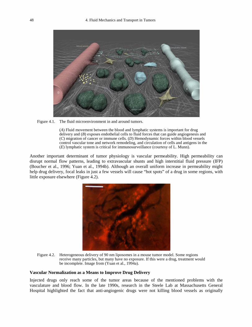
48 4. Fluid Mechanics and Transport in Tumors
Figure 4.1. The fluid microenvironment in and around tumors.
(A) Fluid movement between the blood and lymphatic systems is important for drug delivery and (B) exposes endothelial cells to fluid forces that can guide angiogenesis and (C) migration of cancer or immune cells. (D) Hemodynamic forces within blood vessels control vascular tone and network remodeling, and circulation of cells and antigens in the (E) lymphatic system is critical for immunosurveillance (courtesy of L. Munn).
Another important determinant of tumor physiology is vascular permeability. High permeability can disrupt normal flow patterns, leading to extravascular shunts and high interstitial fluid pressure (IFP) (Boucher et al., 1996; Yuan et al., 1994b). Although an overall uniform increase in permeability might help drug delivery, focal leaks in just a few vessels will cause “hot spots” of a drug in some regions, with little exposure elsewhere (Figure 4.2).
Figure 4.2. Heterogeneous delivery of 90 nm liposomes in a mouse tumor model. Some regions
receive many particles, but many have no exposure. If this were a drug, treatment would be incomplete. Image from (Yuan et al., 1994a).
Vascular Normalization as a Means to Improve Drug Delivery
Injected drugs only reach some of the tumor areas because of the mentioned problems with the vasculature and blood flow. In the late 1990s, research in the Steele Lab at Massachusetts General Hospital highlighted the fact that anti-angiogenic drugs were not killing blood vessels as originally

Lance L. Munn 49
intended; instead, they were decreasing vessel diameters, reducing leaks, pruning inefficient segments, and increasing overall blood flow. This led Dr. Rakesh Jain to hypothesize that anti-angiogenic therapies might be used to normalize tumor blood vessels—increasing blood flow and restoring uniformity—to enhance delivery of subsequently-injected chemotherapeutics (Jain, 2001). A lot of preclinical work and recent clinical trial data support this strategy. For example, in a recent trial for newly-diagnosed glioblastoma patients, those with increased blood flow after treatment with Cediranib (a VEGF tyrosine kinase inhibitor) had the best response to chemotherapy and radiation (Pinho et al., 2012).
Transport of Cancer Cells in the Blood Stream
In addition to carrying nutrients and drugs, the blood stream is a conduit for metastasizing cancer cells. In recent years, significant effort has gone into developing methodologies for isolating these circulating tumor cells (CTCs) from whole blood drawn from patients, with the goal of using the cells as biomarkers to guide treatment strategies or study them ex vivo. In general, several milliliters of blood will contain on the order of 10-100 cancer cells, so the task of extracting ~1010 red blood cells is daunting. Most strategies attempt to capture CTCs from whole blood directly, with little pre-processing. One example is the commercially-available Veridex Cell Search™ system, which is a semi-automated sample preparation system that enriches the sample based on the expression of epithelial-cell adhesion molecule using antibody-coated magnetic beads; it also labels the cells with a fluorescent nucleic acid dye for counting (www.veridex.com/CellSearch/CellSearchHCP.aspx). Another proven method, developed by Dr. Toner and colleagues at the Harvard Medical School, extracts cancer cells from whole blood using microarrays of PDMS posts coated with specific antibodies against cancer antigens (Nagrath et al., 2007). Channel geometry plays an important role in determining how the CTCs transit through these devices, and can be adjusted to enhance collisions with antibody-coated surfaces or to independently enrich the nucleated cell population (Kirby et al, 2012; Jain and Munn, 2009).
It is also possible to expose the whole blood to hypotonic solution, lysing the red blood cells (RBCs), which are more sensitive to osmotic shock than nucleated cells. Centrifugation then removes the lysed RBC debris. The resulting solution, composed mainly of CTCs and leukocytes, can then be plated and analyzed using high-throughput imaging to identify and quantify the cancer cells (Nieva et al., 2012; Wendel et al., 2012). Though they are showing some promise in the clinic (Budd et al., 2006; Hayes et al., 2006; Wendel et al., 2012), the existing technologies are tedious and difficult to generalize to multiple tumor types because the requirement for specific antigen binding for collection and identification (not all tumors have known antigens). Therefore, researchers are now trying to find robust and efficient ways to collect these clinically relevant cells.
Autologous Chemotaxis
In addition to carrying cells, biomolecules, and drugs to and from tumors, fluid flow can also directly affect cancer cells by changing or establishing local chemotactic gradients. Melody Swartz and colleagues at the École Polytechnique Fédérale de Lausanne, Switzerland (site report, Appendix B) have shown that production of a chemokine, such as CCL21, by cancer cells can lead to a skewed gradient in the direction of flow (Figure 4. 3) (Shields et al., 2007). Because much of the fluid percolating through a tumor eventually enters the peritumor lymphatic vessels, the process—called autologous chemotaxis—allows the cancer cells to find lymphatic vessels, facilitating metastasis. CCR7 is the necessary receptor for CCL21 in this system. Research by Swartz and colleagues has shown that fibroblasts can use similar mechanisms via autologous TGF beta signaling and matrix rearrangement to create paths for invading cancer cells (Shieh et al., 2011).
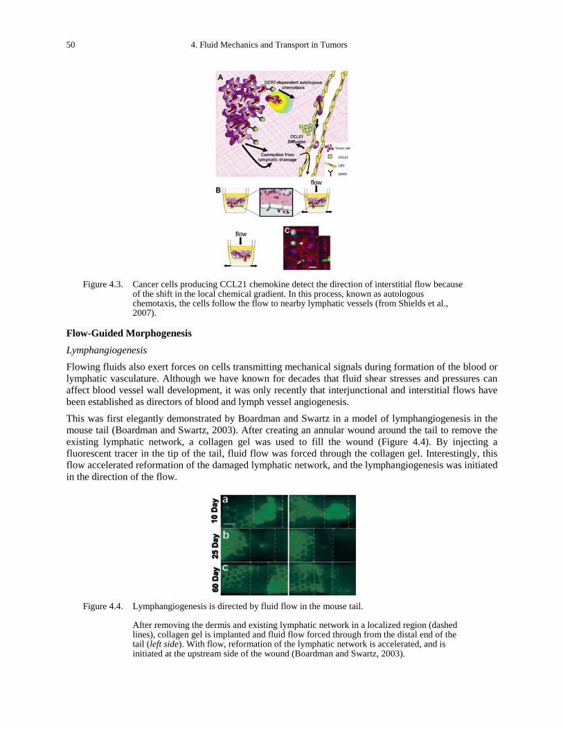
50 4. Fluid Mechanics and Transport in Tumors
Figure 4.3. Cancer cells producing CCL21 chemokine detect the direction of interstitial flow because
of the shift in the local chemical gradient. In this process, known as autologous chemotaxis, the cells follow the flow to nearby lymphatic vessels (from Shields et al., 2007).
Flow-Guided Morphogenesis
Lymphangiogenesis Flowing fluids also exert forces on cells transmitting mechanical signals during formation of the blood or lymphatic vasculature. Although we have known for decades that fluid shear stresses and pressures can affect blood vessel wall development, it was only recently that interjunctional and interstitial flows have been established as directors of blood and lymph vessel angiogenesis. This was first elegantly demonstrated by Boardman and Swartz in a model of lymphangiogenesis in the mouse tail (Boardman and Swartz, 2003). After creating an annular wound around the tail to remove the existing lymphatic network, a collagen gel was used to fill the wound (Figure 4.4). By injecting a fluorescent tracer in the tip of the tail, fluid flow was forced through the collagen gel. Interestingly, this flow accelerated reformation of the damaged lymphatic network, and the lymphangiogenesis was initiated in the direction of the flow.
Figure 4.4. Lymphangiogenesis is directed by fluid flow in the mouse tail.
After removing the dermis and existing lymphatic network in a localized region (dashed lines), collagen gel is implanted and fluid flow forced through from the distal end of the tail (left side). With flow, reformation of the lymphatic network is accelerated, and is initiated at the upstream side of the wound (Boardman and Swartz, 2003).
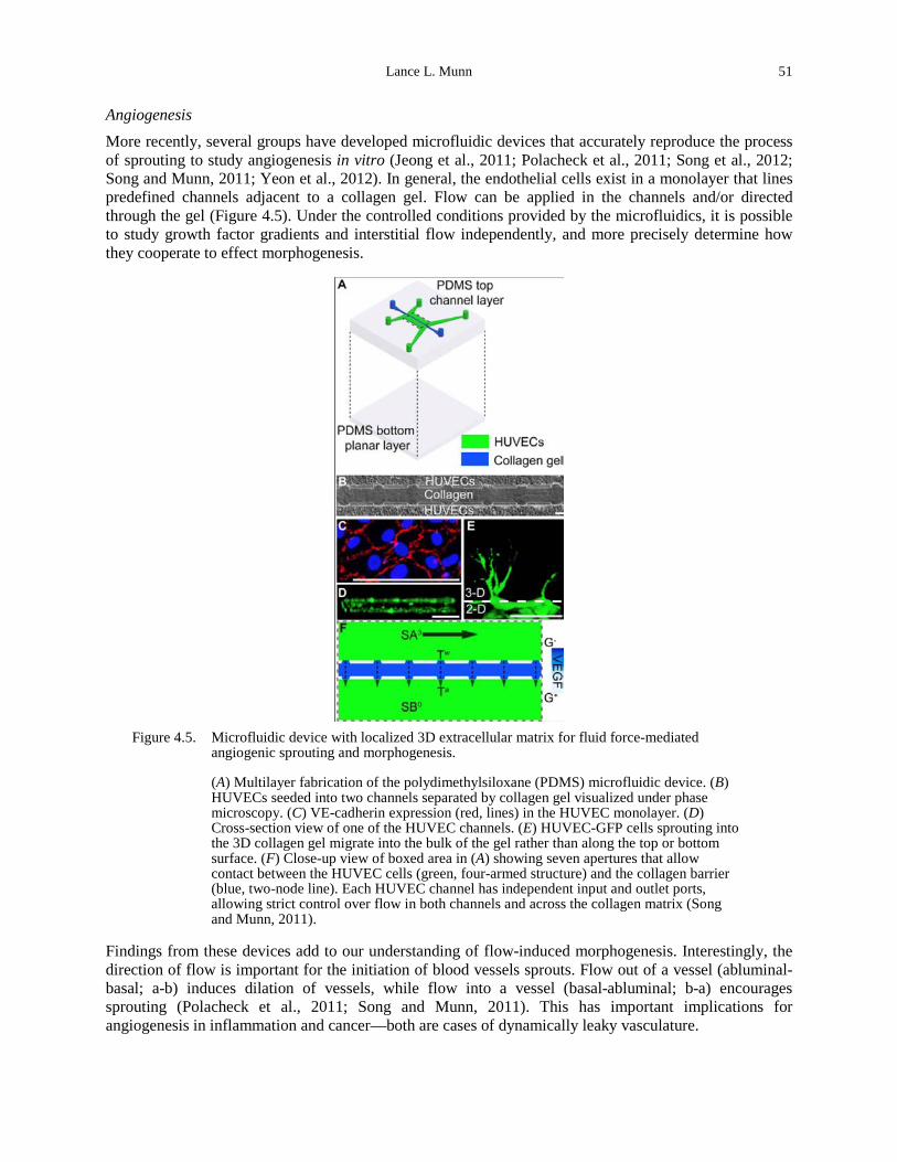
Lance L. Munn 51
Angiogenesis More recently, several groups have developed microfluidic devices that accurately reproduce the process of sprouting to study angiogenesis in vitro (Jeong et al., 2011; Polacheck et al., 2011; Song et al., 2012; Song and Munn, 2011; Yeon et al., 2012). In general, the endothelial cells exist in a monolayer that lines predefined channels adjacent to a collagen gel. Flow can be applied in the channels and/or directed through the gel (Figure 4.5). Under the controlled conditions provided by the microfluidics, it is possible to study growth factor gradients and interstitial flow independently, and more precisely determine how they cooperate to effect morphogenesis.
Figure 4.5. Microfluidic device with localized 3D extracellular matrix for fluid force-mediated
angiogenic sprouting and morphogenesis.
(A) Multilayer fabrication of the polydimethylsiloxane (PDMS) microfluidic device. (B) HUVECs seeded into two channels separated by collagen gel visualized under phase microscopy. (C) VE-cadherin expression (red, lines) in the HUVEC monolayer. (D) Cross-section view of one of the HUVEC channels. (E) HUVEC-GFP cells sprouting into the 3D collagen gel migrate into the bulk of the gel rather than along the top or bottom surface. (F) Close-up view of boxed area in (A) showing seven apertures that allow contact between the HUVEC cells (green, four-armed structure) and the collagen barrier (blue, two-node line). Each HUVEC channel has independent input and outlet ports, allowing strict control over flow in both channels and across the collagen matrix (Song and Munn, 2011).
Findings from these devices add to our understanding of flow-induced morphogenesis. Interestingly, the direction of flow is important for the initiation of blood vessels sprouts. Flow out of a vessel (abluminal-basal; a-b) induces dilation of vessels, while flow into a vessel (basal-abluminal; b-a) encourages sprouting (Polacheck et al., 2011; Song and Munn, 2011). This has important implications for angiogenesis in inflammation and cancer—both are cases of dynamically leaky vasculature.
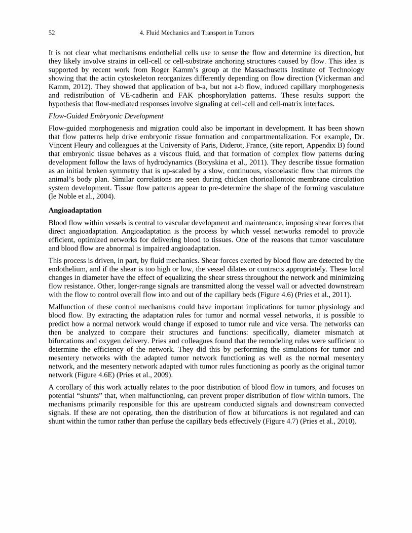
52 4. Fluid Mechanics and Transport in Tumors
It is not clear what mechanisms endothelial cells use to sense the flow and determine its direction, but they likely involve strains in cell-cell or cell-substrate anchoring structures caused by flow. This idea is supported by recent work from Roger Kamm’s group at the Massachusetts Institute of Technology showing that the actin cytoskeleton reorganizes differently depending on flow direction (Vickerman and Kamm, 2012). They showed that application of b-a, but not a-b flow, induced capillary morphogenesis and redistribution of VE-cadherin and FAK phosphorylation patterns. These results support the hypothesis that flow-mediated responses involve signaling at cell-cell and cell-matrix interfaces.
Flow-Guided Embryonic Development Flow-guided morphogenesis and migration could also be important in development. It has been shown that flow patterns help drive embryonic tissue formation and compartmentalization. For example, Dr. Vincent Fleury and colleagues at the University of Paris, Diderot, France, (site report, Appendix B) found that embryonic tissue behaves as a viscous fluid, and that formation of complex flow patterns during development follow the laws of hydrodynamics (Boryskina et al., 2011). They describe tissue formation as an initial broken symmetry that is up-scaled by a slow, continuous, viscoelastic flow that mirrors the animal’s body plan. Similar correlations are seen during chicken chorioallontoic membrane circulation system development. Tissue flow patterns appear to pre-determine the shape of the forming vasculature (le Noble et al., 2004).
Angioadaptation
Blood flow within vessels is central to vascular development and maintenance, imposing shear forces that direct angioadaptation. Angioadaptation is the process by which vessel networks remodel to provide efficient, optimized networks for delivering blood to tissues. One of the reasons that tumor vasculature and blood flow are abnormal is impaired angioadaptation. This process is driven, in part, by fluid mechanics. Shear forces exerted by blood flow are detected by the endothelium, and if the shear is too high or low, the vessel dilates or contracts appropriately. These local changes in diameter have the effect of equalizing the shear stress throughout the network and minimizing flow resistance. Other, longer-range signals are transmitted along the vessel wall or advected downstream with the flow to control overall flow into and out of the capillary beds (Figure 4.6) (Pries et al., 2011). Malfunction of these control mechanisms could have important implications for tumor physiology and blood flow. By extracting the adaptation rules for tumor and normal vessel networks, it is possible to predict how a normal network would change if exposed to tumor rule and vice versa. The networks can then be analyzed to compare their structures and functions: specifically, diameter mismatch at bifurcations and oxygen delivery. Pries and colleagues found that the remodeling rules were sufficient to determine the efficiency of the network. They did this by performing the simulations for tumor and mesentery networks with the adapted tumor network functioning as well as the normal mesentery network, and the mesentery network adapted with tumor rules functioning as poorly as the original tumor network (Figure 4.6E) (Pries et al., 2009).
A corollary of this work actually relates to the poor distribution of blood flow in tumors, and focuses on potential “shunts” that, when malfunctioning, can prevent proper distribution of flow within tumors. The mechanisms primarily responsible for this are upstream conducted signals and downstream convected signals. If these are not operating, then the distribution of flow at bifurcations is not regulated and can shunt within the tumor rather than perfuse the capillary beds effectively (Figure 4.7) (Pries et al., 2010).
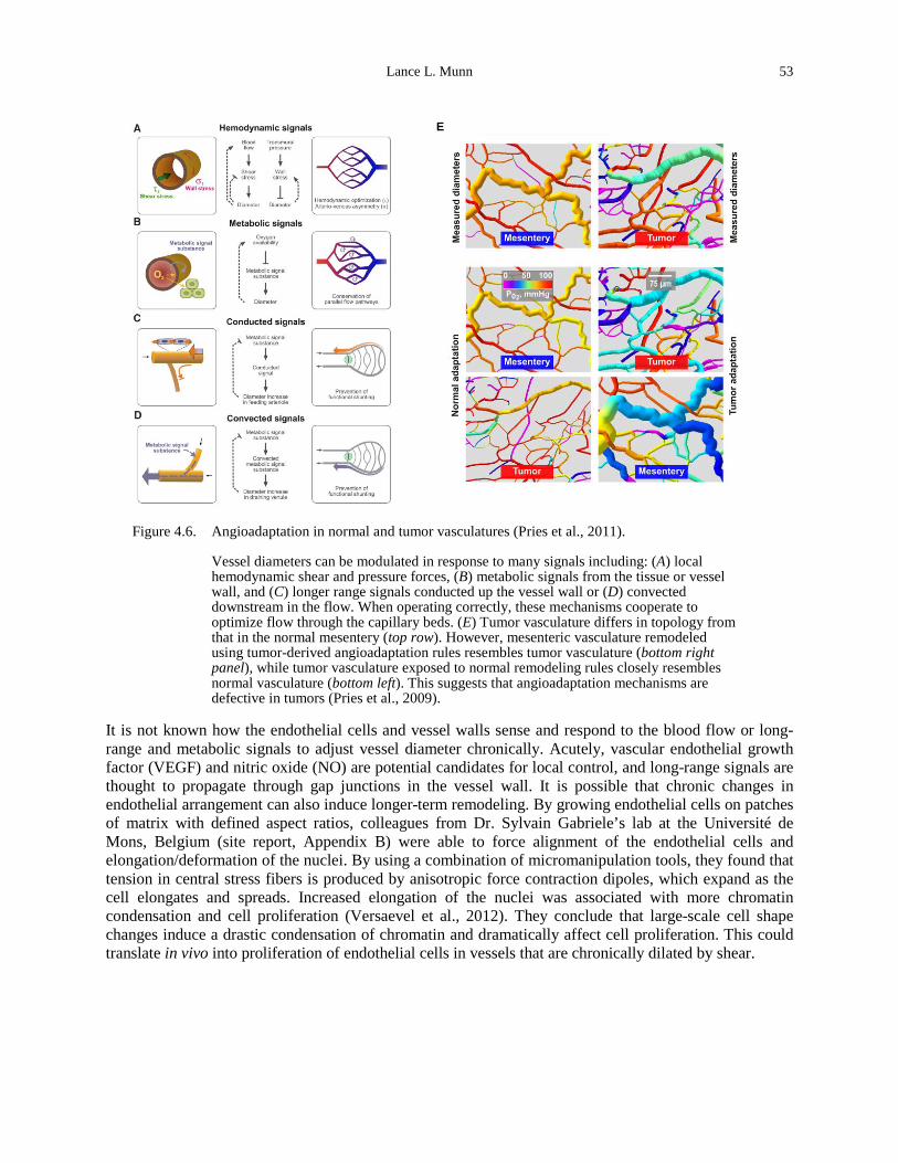
Lance L. Munn 53
E
Figure 4.6. Angioadaptation in normal and tumor vasculatures (Pries et al., 2011).
Vessel diameters can be modulated in response to many signals including: (A) local hemodynamic shear and pressure forces, (B) metabolic signals from the tissue or vessel wall, and (C) longer range signals conducted up the vessel wall or (D) convected downstream in the flow. When operating correctly, these mechanisms cooperate to optimize flow through the capillary beds. (E) Tumor vasculature differs in topology from that in the normal mesentery (top row). However, mesenteric vasculature remodeled using tumor-derived angioadaptation rules resembles tumor vasculature (bottom right panel), while tumor vasculature exposed to normal remodeling rules closely resembles normal vasculature (bottom left). This suggests that angioadaptation mechanisms are defective in tumors (Pries et al., 2009).
It is not known how the endothelial cells and vessel walls sense and respond to the blood flow or long-range and metabolic signals to adjust vessel diameter chronically. Acutely, vascular endothelial growth factor (VEGF) and nitric oxide (NO) are potential candidates for local control, and long-range signals are thought to propagate through gap junctions in the vessel wall. It is possible that chronic changes in endothelial arrangement can also induce longer-term remodeling. By growing endothelial cells on patches of matrix with defined aspect ratios, colleagues from Dr. Sylvain Gabriele’s lab at the Université de Mons, Belgium (site report, Appendix B) were able to force alignment of the endothelial cells and elongation/deformation of the nuclei. By using a combination of micromanipulation tools, they found that tension in central stress fibers is produced by anisotropic force contraction dipoles, which expand as the cell elongates and spreads. Increased elongation of the nuclei was associated with more chromatin condensation and cell proliferation (Versaevel et al., 2012). They conclude that large-scale cell shape changes induce a drastic condensation of chromatin and dramatically affect cell proliferation. This could translate in vivo into proliferation of endothelial cells in vessels that are chronically dilated by shear.
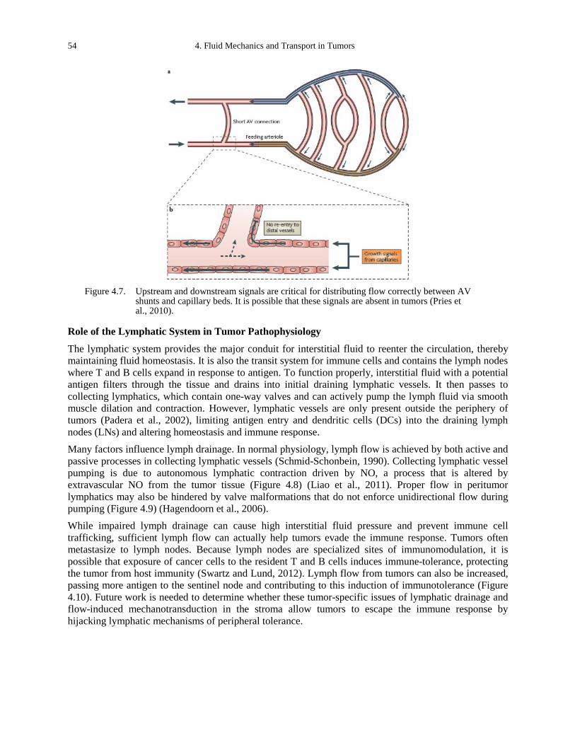
54 4. Fluid Mechanics and Transport in Tumors
Figure 4.7. Upstream and downstream signals are critical for distributing flow correctly between AV
shunts and capillary beds. It is possible that these signals are absent in tumors (Pries et al., 2010).
Role of the Lymphatic System in Tumor Pathophysiology
The lymphatic system provides the major conduit for interstitial fluid to reenter the circulation, thereby maintaining fluid homeostasis. It is also the transit system for immune cells and contains the lymph nodes where T and B cells expand in response to antigen. To function properly, interstitial fluid with a potential antigen filters through the tissue and drains into initial draining lymphatic vessels. It then passes to collecting lymphatics, which contain one-way valves and can actively pump the lymph fluid via smooth muscle dilation and contraction. However, lymphatic vessels are only present outside the periphery of tumors (Padera et al., 2002), limiting antigen entry and dendritic cells (DCs) into the draining lymph nodes (LNs) and altering homeostasis and immune response. Many factors influence lymph drainage. In normal physiology, lymph flow is achieved by both active and passive processes in collecting lymphatic vessels (Schmid-Schonbein, 1990). Collecting lymphatic vessel pumping is due to autonomous lymphatic contraction driven by NO, a process that is altered by extravascular NO from the tumor tissue (Figure 4.8) (Liao et al., 2011). Proper flow in peritumor lymphatics may also be hindered by valve malformations that do not enforce unidirectional flow during pumping (Figure 4.9) (Hagendoorn et al., 2006). While impaired lymph drainage can cause high interstitial fluid pressure and prevent immune cell trafficking, sufficient lymph flow can actually help tumors evade the immune response. Tumors often metastasize to lymph nodes. Because lymph nodes are specialized sites of immunomodulation, it is possible that exposure of cancer cells to the resident T and B cells induces immune-tolerance, protecting the tumor from host immunity (Swartz and Lund, 2012). Lymph flow from tumors can also be increased, passing more antigen to the sentinel node and contributing to this induction of immunotolerance (Figure 4.10). Future work is needed to determine whether these tumor-specific issues of lymphatic drainage and flow-induced mechanotransduction in the stroma allow tumors to escape the immune response by hijacking lymphatic mechanisms of peripheral tolerance.
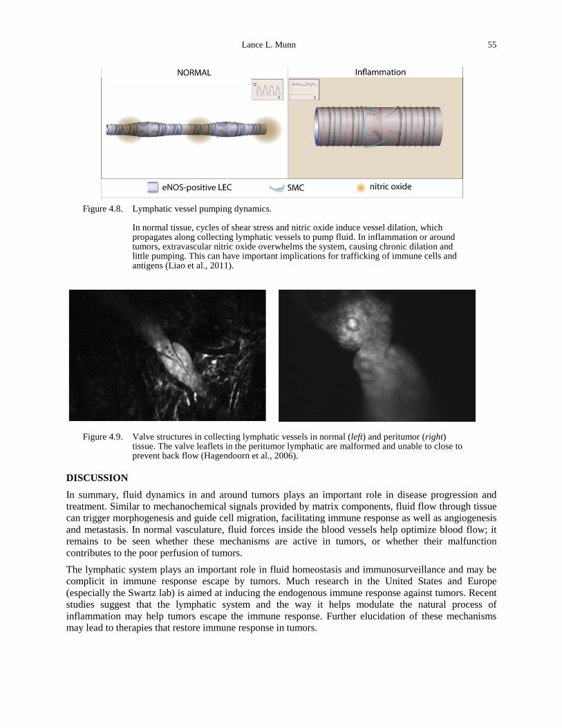
Lance L. Munn 55
Figure 4.8. Lymphatic vessel pumping dynamics.
In normal tissue, cycles of shear stress and nitric oxide induce vessel dilation, which propagates along collecting lymphatic vessels to pump fluid. In inflammation or around tumors, extravascular nitric oxide overwhelms the system, causing chronic dilation and little pumping. This can have important implications for trafficking of immune cells and antigens (Liao et al., 2011).
Figure 4.9. Valve structures in collecting lymphatic vessels in normal (left) and peritumor (right) tissue. The valve leaflets in the peritumor lymphatic are malformed and unable to close to prevent back flow (Hagendoorn et al., 2006).
DISCUSSION
In summary, fluid dynamics in and around tumors plays an important role in disease progression and treatment. Similar to mechanochemical signals provided by matrix components, fluid flow through tissue can trigger morphogenesis and guide cell migration, facilitating immune response as well as angiogenesis and metastasis. In normal vasculature, fluid forces inside the blood vessels help optimize blood flow; it remains to be seen whether these mechanisms are active in tumors, or whether their malfunction contributes to the poor perfusion of tumors. The lymphatic system plays an important role in fluid homeostasis and immunosurveillance and may be complicit in immune response escape by tumors. Much research in the United States and Europe (especially the Swartz lab) is aimed at inducing the endogenous immune response against tumors. Recent studies suggest that the lymphatic system and the way it helps modulate the natural process of inflammation may help tumors escape the immune response. Further elucidation of these mechanisms may lead to therapies that restore immune response in tumors.
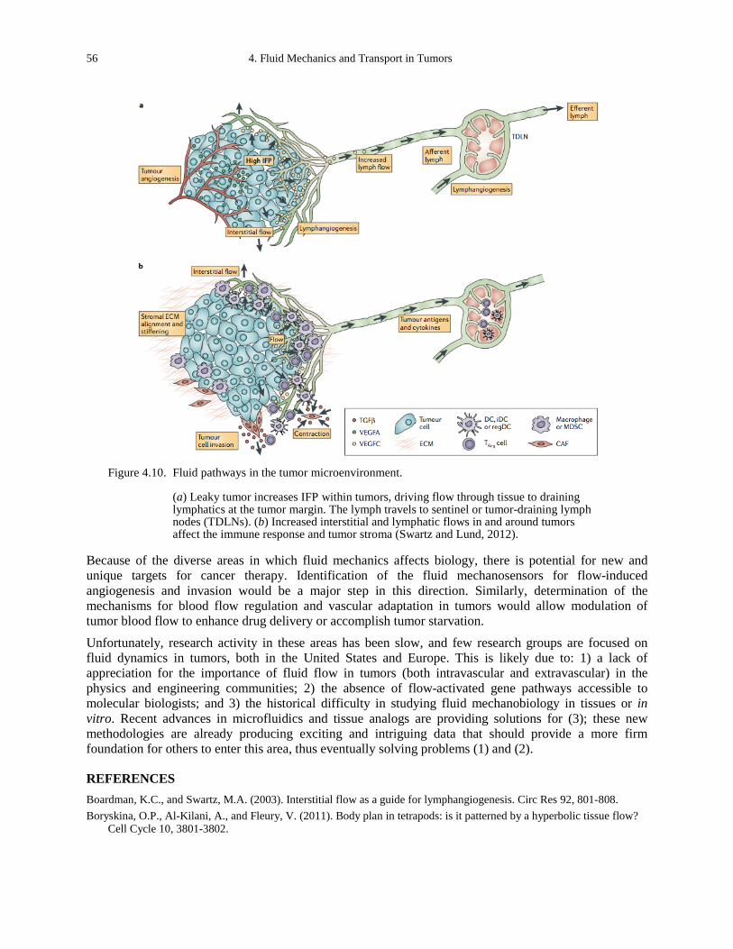
56 4. Fluid Mechanics and Transport in Tumors
Figure 4.10. Fluid pathways in the tumor microenvironment.
(a) Leaky tumor increases IFP within tumors, driving flow through tissue to draining lymphatics at the tumor margin. The lymph travels to sentinel or tumor-draining lymph nodes (TDLNs). (b) Increased interstitial and lymphatic flows in and around tumors affect the immune response and tumor stroma (Swartz and Lund, 2012).
Because of the diverse areas in which fluid mechanics affects biology, there is potential for new and unique targets for cancer therapy. Identification of the fluid mechanosensors for flow-induced angiogenesis and invasion would be a major step in this direction. Similarly, determination of the mechanisms for blood flow regulation and vascular adaptation in tumors would allow modulation of tumor blood flow to enhance drug delivery or accomplish tumor starvation.
Unfortunately, research activity in these areas has been slow, and few research groups are focused on fluid dynamics in tumors, both in the United States and Europe. This is likely due to: 1) a lack of appreciation for the importance of fluid flow in tumors (both intravascular and extravascular) in the physics and engineering communities; 2) the absence of flow-activated gene pathways accessible to molecular biologists; and 3) the historical difficulty in studying fluid mechanobiology in tissues or in vitro. Recent advances in microfluidics and tissue analogs are providing solutions for (3); these new methodologies are already producing exciting and intriguing data that should provide a more firm foundation for others to enter this area, thus eventually solving problems (1) and (2).
REFERENCES Boardman, K.C., and Swartz, M.A. (2003). Interstitial flow as a guide for lymphangiogenesis. Circ Res 92, 801-808. Boryskina, O.P., Al-Kilani, A., and Fleury, V. (2011). Body plan in tetrapods: is it patterned by a hyperbolic tissue flow?
Cell Cycle 10, 3801-3802.
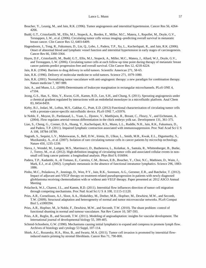
Lance L. Munn 57
Boucher, Y., Leunig, M., and Jain, R.K. (1996). Tumor angiogenesis and interstitial hypertension. Cancer Res 56, 4264-4266.
Budd, G.T., Cristofanilli, M., Ellis, M.J., Stopeck, A., Borden, E., Miller, M.C., Matera, J., Repollet, M., Doyle, G.V., Terstappen, L.W., et al. (2006). Circulating tumor cells versus imaging--predicting overall survival in metastatic breast cancer. Clin Cancer Res 12, 6403-6409.
Hagendoorn, J., Tong, R., Fukumura, D., Lin, Q., Lobo, J., Padera, T.P., Xu, L., Kucherlapati, R., and Jain, R.K. (2006). Onset of abnormal blood and lymphatic vessel function and interstitial hypertension in early stages of carcinogenesis. Cancer Res 66, 3360-3364.
Hayes, D.F., Cristofanilli, M., Budd, G.T., Ellis, M.J., Stopeck, A., Miller, M.C., Matera, J., Allard, W.J., Doyle, G.V., and Terstappen, L.W. (2006). Circulating tumor cells at each follow-up time point during therapy of metastatic breast cancer patients predict progression-free and overall survival. Clin Cancer Res 12, 4218-4224.
Jain, R.K. (1994). Barriers to drug delivery in solid tumors. Scientific American 271, 58-65. Jain, R.K. (1996). Delivery of molecular medicine to solid tumors. Science 271, 1079-1080. Jain, R.K. (2001). Normalizing tumor vasculature with anti-angiogenic therapy: a new paradigm for combination therapy.
Nature medicine 7, 987-989. Jain, A., and Munn, L.L. (2009) Determinants of leukocyte margination in rectangular microchannels. PLoS ONE 4,
e7104. Jeong, G.S., Han, S., Shin, Y., Kwon, G.H., Kamm, R.D., Lee, S.H., and Chung, S. (2011). Sprouting angiogenesis under
a chemical gradient regulated by interactions with an endothelial monolayer in a microfluidic platform. Anal Chem 83, 8454-8459.
Kirby, B.J., Jodari, M., Loftus, M.S., Gakhar, G., Pratt, E.D. (2012) Functional characterization of circulating tumor cells with a prostate-cancer-specific microfluidic device. PLoS ONE 7, e35976.
le Noble, F., Moyon, D., Pardanaud, L., Yuan, L., Djonov, V., Matthijsen, R., Breant, C., Fleury, V., and Eichmann, A. (2004). Flow regulates arterial-venous differentiation in the chick embryo yolk sac. Development 131, 361-375.
Liao, S., Cheng, G., Conner, D.A., Huang, Y., Kucherlapati, R.S., Munn, L.L., Ruddle, N.H., Jain, R.K., Fukumura, D., and Padera, T.P. (2011). Impaired lymphatic contraction associated with immunosuppression. Proc Natl Acad Sci U S A 108, 18784-18789.
Nagrath, S., Sequist, L.V., Maheswaran, S., Bell, D.W., Irimia, D., Ulkus, L., Smith, M.R., Kwak, E.L., Digumarthy, S., Muzikansky, A., et al. (2007). Isolation of rare circulating tumour cells in cancer patients by microchip technology. Nature 450, 1235-1239.
Nieva, J., Wendel, M., Luttgen, M.S., Marrinucci, D., Bazhenova, L., Kolatkar, A., Santala, R., Whittenberger, B., Burke, J., Torrey, M., et al. (2012). High-definition imaging of circulating tumor cells and associated cellular events in non-small cell lung cancer patients: a longitudinal analysis. Phys Biol 9, 016004.
Padera, T.P., Kadambi, A., di Tomaso, E., Carreira, C.M., Brown, E.B., Boucher, Y., Choi, N.C., Mathisen, D., Wain, J., Mark, E.J., et al. (2002). Lymphatic metastasis in the absence of functional intratumor lymphatics. Science 296, 1883-1886.
Pinho, M.C., Polaskova, P., Jennings, D., Wen, P.Y., Jain, R.K., Sorensen, A.G., Gerstner, E.R., and Batchelor, T. (2012). Impact of adjuvant anti-VEGF therapy on treatment-related pseudoprogression in patients with newly diagnosed glioblastoma receiving chemoradiation with or without anti-VEGF therapy. Paper presented at: 2012 ASCO Annual Meeting
Polacheck, W.J., Charest, J.L., and Kamm, R.D. (2011). Interstitial flow influences direction of tumor cell migration through competing mechanisms. Proc Natl Acad Sci U S A 108, 11115-11120.
Pries, A.R., Cornelissen, A.J., Sloot, A.A., Hinkeldey, M., Dreher, M.R., Hopfner, M., Dewhirst, M.W., and Secomb, T.W. (2009). Structural adaptation and heterogeneity of normal and tumor microvascular networks. PLoS Comput Biol 5, e1000394.
Pries, A.R., Hopfner, M., le Noble, F., Dewhirst, M.W., and Secomb, T.W. (2010). The shunt problem: control of functional shunting in normal and tumour vasculature. Nat Rev Cancer 10, 587-593.
Pries, A.R., Reglin, B., and Secomb, T.W. (2011). Modeling of angioadaptation: insights for vascular development. The International journal of developmental biology 55, 399-405.
Schmid-Schonbein, G.W. (1990). Mechanisms causing initial lymphatics to expand and compress to promote lymph flow. Archives of histology and cytology 53 Suppl, 107-114.
Shieh, A.C., Rozansky, H.A., Hinz, B., and Swartz, M.A. (2011). Tumor cell invasion is promoted by interstitial flow-induced matrix priming by stromal fibroblasts. Cancer Res 71, 790-800.
58 4. Fluid Mechanics and Transport in Tumors
Shields, J.D., Fleury, M.E., Yong, C., Tomei, A.A., Randolph, G.J., and Swartz, M.A. (2007). Autologous chemotaxis as a mechanism of tumor cell homing to lymphatics via interstitial flow and autocrine CCR7 signaling. Cancer Cell 11, 526-538.
Song, J.W., Bazou, D., and Munn, L.L. (2012). Anastomosis of endothelial sprouts forms new vessels in a tissue analogue of angiogenesis. Integr Biol (Camb).
Song, J.W., and Munn, L.L. (2011). Fluid forces control endothelial sprouting. Proc Natl Acad Sci U S A 108, 15342-15347.
Swartz, M.A., and Lund, A.W. (2012). Lymphatic and interstitial flow in the tumour microenvironment: linking mechanobiology with immunity. Nat Rev Cancer 12, 210-219.
Versaevel, M., Grevesse, T., and Gabriele, S. (2012). Spatial coordination between cell and nuclear shape within micropatterned endothelial cells. Nat Commun 3, 671.
Vickerman, V., and Kamm, R.D. (2012). Mechanism of a flow-gated angiogenesis switch: early signaling events at cell-matrix and cell-cell junctions. Integr Biol (Camb).
Wendel, M., Bazhenova, L., Boshuizen, R., Kolatkar, A., Honnatti, M., Cho, E.H., Marrinucci, D., Sandhu, A., Perricone, A., Thistlethwaite, P., et al. (2012). Fluid biopsy for circulating tumor cell identification in patients with early-and late-stage non-small cell lung cancer: a glimpse into lung cancer biology. Phys Biol 9, 016005.
Yeon, J.H., Ryu, H.R., Chung, M., Hu, Q.P., and Jeon, N.L. (2012). In vitro formation and characterization of a perfusable three-dimensional tubular capillary network in microfluidic devices. Lab Chip.
Yuan, F., Leunig, M., Huang, S.K., Berk, D.A., Papahadjopoulos, D., and Jain, R.K. (1994a). Microvascular permeability and interstitial penetration of sterically stabilized (stealth) liposomes in a human tumor xenograft. Cancer Res 54, 3352-3356.
Yuan, F., Salehi, H.A., Boucher, Y., Vasthare, U.S., Tuma, R.F., and Jain, R.K. (1994b). Vascular permeability and microcirculation of gliomas and mammary carcinomas transplanted in rat and mouse cranial windows. Cancer Res 54, 4564-4568.
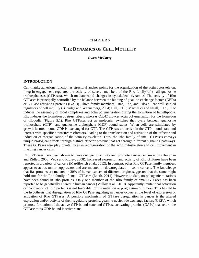
CHAPTER 5
THE DYNAMICS OF CELL MOTILITY
Owen McCarty
INTRODUCTION
Cell-matrix adhesions function as structural anchor points for the organization of the actin cytoskeleton. Integrin engagement regulates the activity of several members of the Rho family of small guanosine triphosphatases (GTPases), which mediate rapid changes in cytoskeletal dynamics. The activity of Rho GTPases is principally controlled by the balance between the binding of guanine-exchange factors (GEFs) or GTPase-activating proteins (GAPs). Three family members—Rac, Rho, and Cdc42—are well-studied regulators of cell motility (Burridge and Wennerberg, 2004; Hall, 1998; Machesky and Insall, 1999). Rac induces the assembly of focal complexes and actin polymerization during the formation of lamellipodia. Rho induces the formation of stress fibers, whereas Cdc42 induces actin polymerization for the formation of filopodia (Figure 5.1). Rho GTPases act as molecular switches that cycle between guanosine triphosphate (GTP)- and guanosine diphosphate (GDP)-bound states. When cells are stimulated by growth factors, bound GDP is exchanged for GTP. The GTPases are active in the GTP-bound state and interact with specific downstream effectors, leading to the translocation and activation of the effector and induction of reorganization of the actin cytoskeleton. Thus, the Rho family of small GTPases conveys unique biological effects through distinct effector proteins that act through different signaling pathways. These GTPases also play pivotal roles in reorganization of the actin cytoskeleton and cell movement in invading cancer cells. Rho GTPases have been shown to have oncogenic activity and promote cancer cell invasion (Heasman and Ridley, 2008; Vega and Ridley, 2008). Increased expression and activity of Rho GTPases have been reported in a variety of cancers (Mardilovich et al., 2012). In contrast, other Rho GTPase family members appear to act as tumor suppressors and are mutated or downregulated in some cancers. The knowledge that Ras proteins are mutated in 30% of human cancers of different origins suggested that the same might hold true for the Rho family of small GTPases (Lauth, 2011). However, to date, no oncogenic mutations have been found in Rho proteins. Only one member of the Rho family of small GTPases has been reported to be genetically altered in human cancer (Mulloy et al., 2010). Apparently, mutational activation or inactivation of Rho proteins is not favorable for the initiation or progression of tumors. This has led to the hypothesis that disregulation of Rho GTPase signaling in cancer occurs at the level of expression or activation of Rho GTPases. A possible mechanism of GTPase deregulation in cancer is the altered expression and/or activity of their regulatory proteins, guanine nucleotide exchange factors (GEFs), which promote formation of the active GTP-bound state and GTPase activating proteins (GAPs) that return the GTPase to its GDP-bound inactive state.
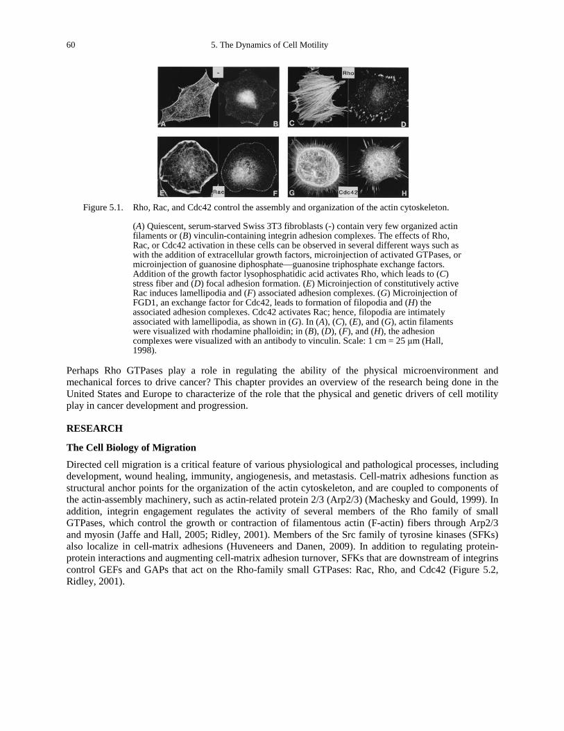
60 5. The Dynamics of Cell Motility
Figure 5.1. Rho, Rac, and Cdc42 control the assembly and organization of the actin cytoskeleton.
(A) Quiescent, serum-starved Swiss 3T3 fibroblasts (-) contain very few organized actin filaments or (B) vinculin-containing integrin adhesion complexes. The effects of Rho, Rac, or Cdc42 activation in these cells can be observed in several different ways such as with the addition of extracellular growth factors, microinjection of activated GTPases, or microinjection of guanosine diphosphate—guanosine triphosphate exchange factors. Addition of the growth factor lysophosphatidic acid activates Rho, which leads to (C) stress fiber and (D) focal adhesion formation. (E) Microinjection of constitutively active Rac induces lamellipodia and (F) associated adhesion complexes. (G) Microinjection of FGD1, an exchange factor for Cdc42, leads to formation of filopodia and (H) the associated adhesion complexes. Cdc42 activates Rac; hence, filopodia are intimately associated with lamellipodia, as shown in (G). In (A), (C), (E), and (G), actin filaments were visualized with rhodamine phalloidin; in (B), (D), (F), and (H), the adhesion complexes were visualized with an antibody to vinculin. Scale: 1 cm = 25 μm (Hall, 1998).
Perhaps Rho GTPases play a role in regulating the ability of the physical microenvironment and mechanical forces to drive cancer? This chapter provides an overview of the research being done in the United States and Europe to characterize of the role that the physical and genetic drivers of cell motility play in cancer development and progression.
RESEARCH
The Cell Biology of Migration
Directed cell migration is a critical feature of various physiological and pathological processes, including development, wound healing, immunity, angiogenesis, and metastasis. Cell-matrix adhesions function as structural anchor points for the organization of the actin cytoskeleton, and are coupled to components of the actin-assembly machinery, such as actin-related protein 2/3 (Arp2/3) (Machesky and Gould, 1999). In addition, integrin engagement regulates the activity of several members of the Rho family of small GTPases, which control the growth or contraction of filamentous actin (F-actin) fibers through Arp2/3 and myosin (Jaffe and Hall, 2005; Ridley, 2001). Members of the Src family of tyrosine kinases (SFKs) also localize in cell-matrix adhesions (Huveneers and Danen, 2009). In addition to regulating protein-protein interactions and augmenting cell-matrix adhesion turnover, SFKs that are downstream of integrins control GEFs and GAPs that act on the Rho-family small GTPases: Rac, Rho, and Cdc42 (Figure 5.2, Ridley, 2001).
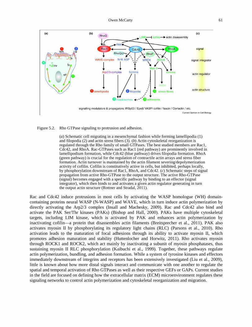
Owen McCarty 61
Figure 5.2. Rho GTPase signaling to protrusion and adhesion.
(a) Schematic cell migrating in a mesenchymal fashion while forming lamellipodia (1) and filopodia (2) and actin stress fibers (3). (b) Actin cytoskeletal reorganization is regulated through the Rho family of small GTPases. The best studied members are Rac1, Cdc42, and RhoA. Rac-GTPases such as Rac1 (red pathway) are prominently involved in lamellipodium formation, while Cdc42 (blue pathway) drives filopodia formation. RhoA (green pathway) is crucial for the regulation of contractile actin arrays and stress fiber formation. Actin turnover is maintained by the actin filament severing/depolymerization activity of cofilin. Cofilin is constitutively active in cells, but inhibited, perhaps locally, by phosphorylation downstream of Rac1, RhoA, and Cdc42. (c) Schematic steps of signal propagation from active Rho-GTPase to the output structure. The active Rho-GTPase (signal) becomes engaged with a specific pathway by binding to an effector (signal integrator), which then binds to and activates a given actin regulator generating in turn the output actin structure (Rottner and Stradal, 2011).
Rac and Cdc42 induce protrusions in most cells by activating the WASP homologue (WH) domain-containing proteins neural WASP (N-WASP) and WAVE, which in turn induce actin polymerization by directly activating the Arp2/3 complex (Insall and Machesky, 2009). Rac and Cdc42 also bind and activate the PAK Ser/Thr kinases (PAKs) (Bishop and Hall, 2000). PAKs have multiple cytoskeletal targets, including LIM kinase, which is activated by PAK and enhances actin polymerization by inactivating cofilin—a protein that disassembles actin filaments (Breitsprecher et al., 2011). PAK also activates myosin II by phosphorylating its regulatory light chains (RLC) (Parsons et al., 2010). Rho activation leads to the maturation of focal adhesions through its ability to activate myosin II, which promotes adhesion maturation and stability (Huttenlocher and Horwitz, 2011). Rho activates myosin through ROCK1 and ROCK2, which act mainly by inactivating a subunit of myosin phosphatases, thus sustaining myosin II RLC phosphorylation (Kaibuchi et al., 1999). Together, these pathways regulate actin polymerization, bundling, and adhesion formation. While a system of tyrosine kinases and effectors immediately downstream of integrins and receptors has been extensively investigated (Liu et al., 2009), little is known about how more distal signals interact and communicate with one another to regulate the spatial and temporal activation of Rho GTPases as well as their respective GEFs or GAPs. Current studies in the field are focused on defining how the extracellular matrix (ECM) microenvironment regulates these signaling networks to control actin polymerization and cytoskeletal reorganization and migration.
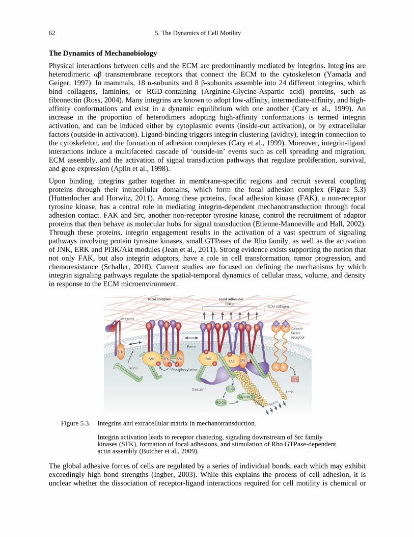
62 5. The Dynamics of Cell Motility
The Dynamics of Mechanobiology
Physical interactions between cells and the ECM are predominantly mediated by integrins. Integrins are heterodimeric αβ transmembrane receptors that connect the ECM to the cytoskeleton (Yamada and Geiger, 1997). In mammals, 18 α-subunits and 8 β-subunits assemble into 24 different integrins, which bind collagens, laminins, or RGD-containing (Arginine-Glycine-Aspartic acid) proteins, such as fibronectin (Ross, 2004). Many integrins are known to adopt low-affinity, intermediate-affinity, and high-affinity conformations and exist in a dynamic equilibrium with one another (Cary et al., 1999). An increase in the proportion of heterodimers adopting high-affinity conformations is termed integrin activation, and can be induced either by cytoplasmic events (inside-out activation), or by extracellular factors (outside-in activation). Ligand-binding triggers integrin clustering (avidity), integrin connection to the cytoskeleton, and the formation of adhesion complexes (Cary et al., 1999). Moreover, integrin-ligand interactions induce a multifaceted cascade of ‘outside-in’ events such as cell spreading and migration, ECM assembly, and the activation of signal transduction pathways that regulate proliferation, survival, and gene expression (Aplin et al., 1998).
Upon binding, integrins gather together in membrane-specific regions and recruit several coupling proteins through their intracellular domains, which form the focal adhesion complex (Figure 5.3) (Huttenlocher and Horwitz, 2011). Among these proteins, focal adhesion kinase (FAK), a non-receptor tyrosine kinase, has a central role in mediating integrin-dependent mechanotransduction through focal adhesion contact. FAK and Src, another non-receptor tyrosine kinase, control the recruitment of adaptor proteins that then behave as molecular hubs for signal transduction (Etienne-Manneville and Hall, 2002). Through these proteins, integrin engagement results in the activation of a vast spectrum of signaling pathways involving protein tyrosine kinases, small GTPases of the Rho family, as well as the activation of JNK, ERK and PI3K/Akt modules (Jean et al., 2011). Strong evidence exists supporting the notion that not only FAK, but also integrin adaptors, have a role in cell transformation, tumor progression, and chemoresistance (Schaller, 2010). Current studies are focused on defining the mechanisms by which integrin signaling pathways regulate the spatial-temporal dynamics of cellular mass, volume, and density in response to the ECM microenvironment.
Figure 5.3. Integrins and extracellular matrix in mechanotransduction.
Integrin activation leads to receptor clustering, signaling downstream of Src family kinases (SFK), formation of focal adhesions, and stimulation of Rho GTPase-dependent actin assembly (Butcher et al., 2009).
The global adhesive forces of cells are regulated by a series of individual bonds, each which may exhibit exceedingly high bond strengths (Ingber, 2003). While this explains the process of cell adhesion, it is unclear whether the dissociation of receptor-ligand interactions required for cell motility is chemical or
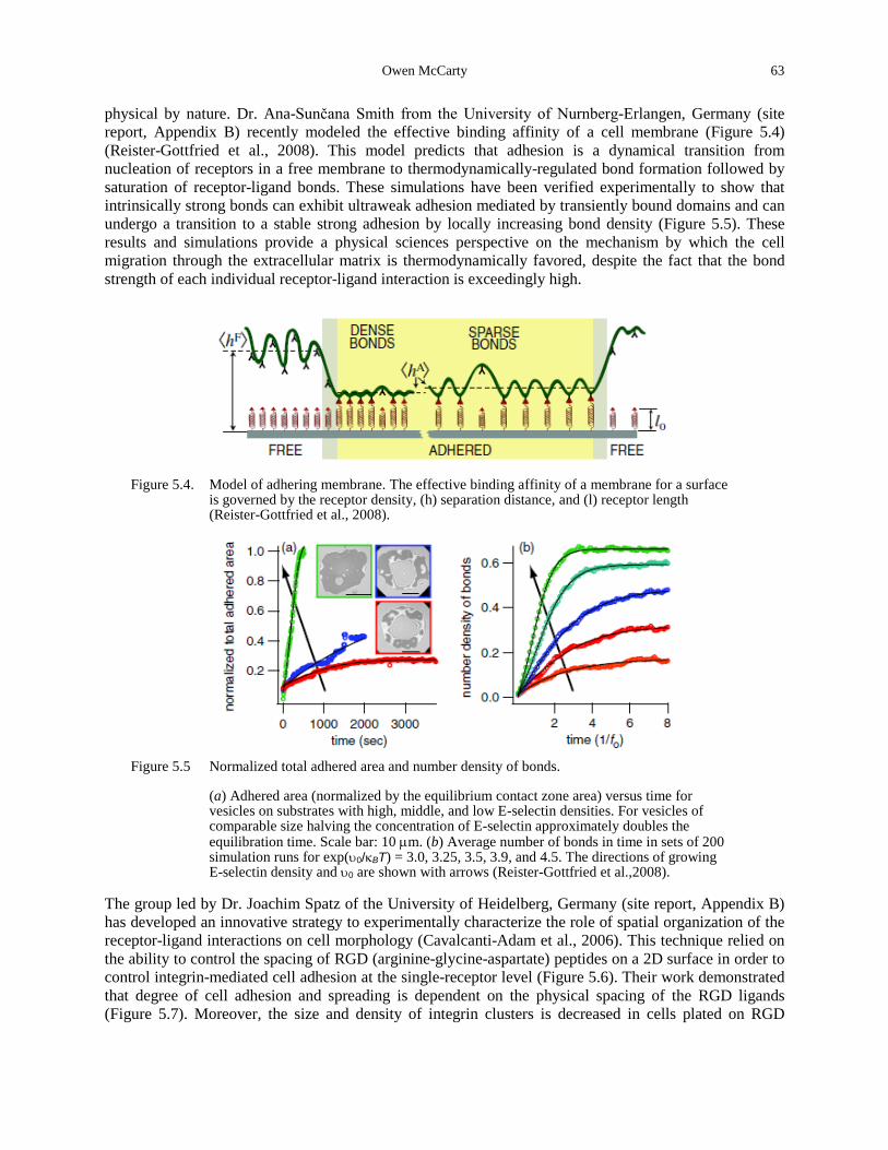
Owen McCarty 63
physical by nature. Dr. Ana-Sunčana Smith from the University of Nurnberg-Erlangen, Germany (site report, Appendix B) recently modeled the effective binding affinity of a cell membrane (Figure 5.4) (Reister-Gottfried et al., 2008). This model predicts that adhesion is a dynamical transition from nucleation of receptors in a free membrane to thermodynamically-regulated bond formation followed by saturation of receptor-ligand bonds. These simulations have been verified experimentally to show that intrinsically strong bonds can exhibit ultraweak adhesion mediated by transiently bound domains and can undergo a transition to a stable strong adhesion by locally increasing bond density (Figure 5.5). These results and simulations provide a physical sciences perspective on the mechanism by which the cell migration through the extracellular matrix is thermodynamically favored, despite the fact that the bond strength of each individual receptor-ligand interaction is exceedingly high.
Figure 5.4. Model of adhering membrane. The effective binding affinity of a membrane for a surface
is governed by the receptor density, (h) separation distance, and (l) receptor length (Reister-Gottfried et al., 2008).
Figure 5.5 Normalized total adhered area and number density of bonds.
(a) Adhered area (normalized by the equilibrium contact zone area) versus time for vesicles on substrates with high, middle, and low E-selectin densities. For vesicles of comparable size halving the concentration of E-selectin approximately doubles the equilibration time. Scale bar: 10 µm. (b) Average number of bonds in time in sets of 200 simulation runs for exp(υ0/κBT) = 3.0, 3.25, 3.5, 3.9, and 4.5. The directions of growing E-selectin density and υ0 are shown with arrows (Reister-Gottfried et al.,2008).
The group led by Dr. Joachim Spatz of the University of Heidelberg, Germany (site report, Appendix B) has developed an innovative strategy to experimentally characterize the role of spatial organization of the receptor-ligand interactions on cell morphology (Cavalcanti-Adam et al., 2006). This technique relied on the ability to control the spacing of RGD (arginine-glycine-aspartate) peptides on a 2D surface in order to control integrin-mediated cell adhesion at the single-receptor level (Figure 5.6). Their work demonstrated that degree of cell adhesion and spreading is dependent on the physical spacing of the RGD ligands (Figure 5.7). Moreover, the size and density of integrin clusters is decreased in cells plated on RGD
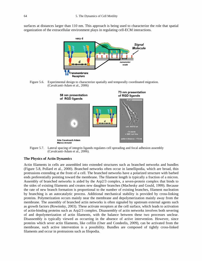
64 5. The Dynamics of Cell Motility
surfaces at distances larger than 110 nm. This approach is being used to characterize the role that spatial organization of the extracellular environment plays in regulating cell-ECM interactions.
Figure 5.6. Experimental design to characterize spatially and temporally coordinated migration.
(Cavalcanti-Adam et al., 2006)
Figure 5.7. Lateral spacing of integrin ligands regulates cell spreading and focal adhesion assembly
(Cavalcanti-Adam et al., 2006).
The Physics of Actin Dynamics
Actin filaments in cells are assembled into extended structures such as branched networks and bundles (Figure 5.8, Pollard et al., 2000). Branched networks often occur in lamellipodia, which are broad, thin protrusions extending at the front of a cell. The branched networks have a polarized structure with barbed ends preferentially pointing toward the membrane. The filament length is typically a fraction of a micron. Assembly of branched networks is aided by the Arp2/3 complex, a seven-protein complex that binds to the sides of existing filaments and creates new daughter branches (Machesky and Gould, 1999). Because the rate of new branch formation is proportional to the number of existing branches, filament nucleation by branching is an autocatalytic process. Additional mechanical stability is provided by cross-linking proteins. Polymerization occurs mainly near the membrane and depolymerization mainly away from the membrane. The assembly of branched actin networks is often signaled by upstream external agents such as growth factors (Rowinsky, 2003). These activate receptors at the cell surface, which leads to activation of actin-binding proteins such as Arp2/3 complex. Disassembly of actin networks involves both severing of and depolymerization of actin filaments, with the balance between these two processes unclear. Disassembly is typically viewed as occurring in the absence of active intervention. However, since proteins which sever actin filaments, like cofilin (Oser and Condeelis, 2009), can be activated from the membrane, such active intervention is a possibility. Bundles are composed of tightly cross-linked filaments and occur in protrusions such as filopodia.
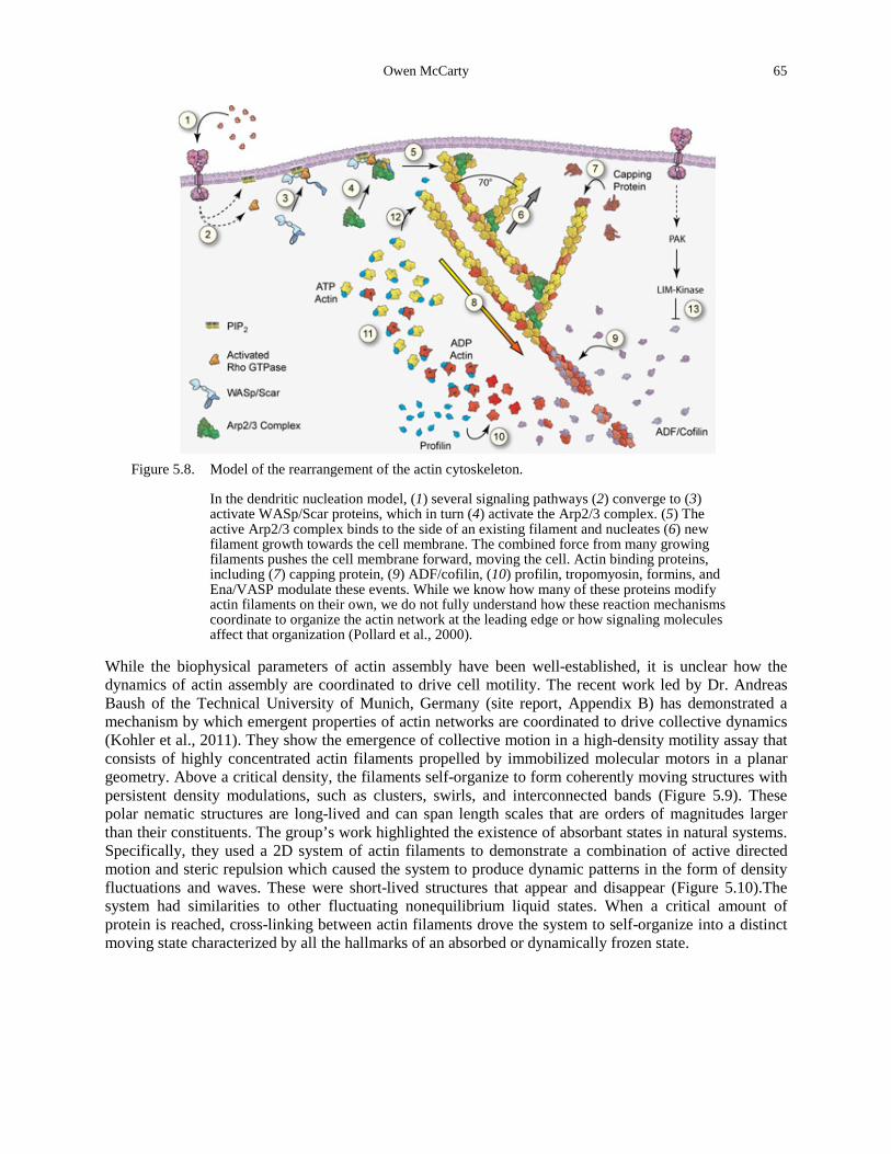
Owen McCarty 65
Figure 5.8. Model of the rearrangement of the actin cytoskeleton.
In the dendritic nucleation model, (1) several signaling pathways (2) converge to (3) activate WASp/Scar proteins, which in turn (4) activate the Arp2/3 complex. (5) The active Arp2/3 complex binds to the side of an existing filament and nucleates (6) new filament growth towards the cell membrane. The combined force from many growing filaments pushes the cell membrane forward, moving the cell. Actin binding proteins, including (7) capping protein, (9) ADF/cofilin, (10) profilin, tropomyosin, formins, and Ena/VASP modulate these events. While we know how many of these proteins modify actin filaments on their own, we do not fully understand how these reaction mechanisms coordinate to organize the actin network at the leading edge or how signaling molecules affect that organization (Pollard et al., 2000).
While the biophysical parameters of actin assembly have been well-established, it is unclear how the dynamics of actin assembly are coordinated to drive cell motility. The recent work led by Dr. Andreas Baush of the Technical University of Munich, Germany (site report, Appendix B) has demonstrated a mechanism by which emergent properties of actin networks are coordinated to drive collective dynamics (Kohler et al., 2011). They show the emergence of collective motion in a high-density motility assay that consists of highly concentrated actin filaments propelled by immobilized molecular motors in a planar geometry. Above a critical density, the filaments self-organize to form coherently moving structures with persistent density modulations, such as clusters, swirls, and interconnected bands (Figure 5.9). These polar nematic structures are long-lived and can span length scales that are orders of magnitudes larger than their constituents. The group’s work highlighted the existence of absorbant states in natural systems. Specifically, they used a 2D system of actin filaments to demonstrate a combination of active directed motion and steric repulsion which caused the system to produce dynamic patterns in the form of density fluctuations and waves. These were short-lived structures that appear and disappear (Figure 5.10).The system had similarities to other fluctuating nonequilibrium liquid states. When a critical amount of protein is reached, cross-linking between actin filaments drove the system to self-organize into a distinct moving state characterized by all the hallmarks of an absorbed or dynamically frozen state.
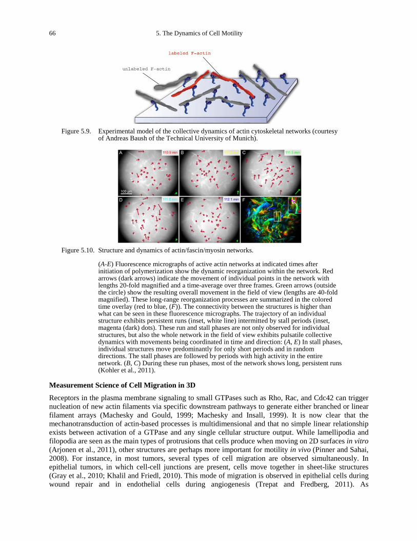
66 5. The Dynamics of Cell Motility
Figure 5.9. Experimental model of the collective dynamics of actin cytoskeletal networks (courtesy
of Andreas Baush of the Technical University of Munich).
Figure 5.10. Structure and dynamics of actin/fascin/myosin networks.
(A-E) Fluorescence micrographs of active actin networks at indicated times after initiation of polymerization show the dynamic reorganization within the network. Red arrows (dark arrows) indicate the movement of individual points in the network with lengths 20-fold magnified and a time-average over three frames. Green arrows (outside the circle) show the resulting overall movement in the field of view (lengths are 40-fold magnified). These long-range reorganization processes are summarized in the colored time overlay (red to blue, (F)). The connectivity between the structures is higher than what can be seen in these fluorescence micrographs. The trajectory of an individual structure exhibits persistent runs (inset, white line) intermitted by stall periods (inset, magenta (dark) dots). These run and stall phases are not only observed for individual structures, but also the whole network in the field of view exhibits pulsatile collective dynamics with movements being coordinated in time and direction: (A, E) In stall phases, individual structures move predominantly for only short periods and in random directions. The stall phases are followed by periods with high activity in the entire network. (B, C) During these run phases, most of the network shows long, persistent runs (Kohler et al., 2011).
Measurement Science of Cell Migration in 3D
Receptors in the plasma membrane signaling to small GTPases such as Rho, Rac, and Cdc42 can trigger nucleation of new actin filaments via specific downstream pathways to generate either branched or linear filament arrays (Machesky and Gould, 1999; Machesky and Insall, 1999). It is now clear that the mechanotransduction of actin-based processes is multidimensional and that no simple linear relationship exists between activation of a GTPase and any single cellular structure output. While lamellipodia and filopodia are seen as the main types of protrusions that cells produce when moving on 2D surfaces in vitro (Arjonen et al., 2011), other structures are perhaps more important for motility in vivo (Pinner and Sahai, 2008). For instance, in most tumors, several types of cell migration are observed simultaneously. In epithelial tumors, in which cell-cell junctions are present, cells move together in sheet-like structures (Gray et al., 2010; Khalil and Friedl, 2010). This mode of migration is observed in epithelial cells during wound repair and in endothelial cells during angiogenesis (Trepat and Fredberg, 2011). As
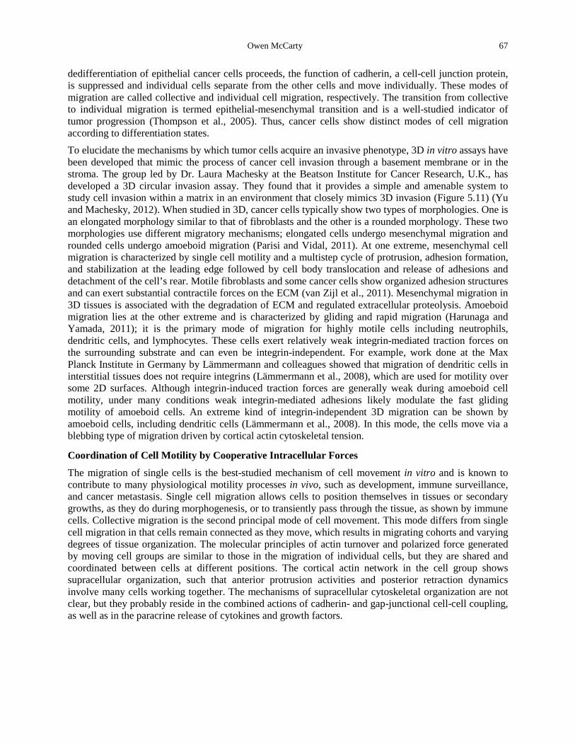
Owen McCarty 67
dedifferentiation of epithelial cancer cells proceeds, the function of cadherin, a cell-cell junction protein, is suppressed and individual cells separate from the other cells and move individually. These modes of migration are called collective and individual cell migration, respectively. The transition from collective to individual migration is termed epithelial-mesenchymal transition and is a well-studied indicator of tumor progression (Thompson et al., 2005). Thus, cancer cells show distinct modes of cell migration according to differentiation states. To elucidate the mechanisms by which tumor cells acquire an invasive phenotype, 3D in vitro assays have been developed that mimic the process of cancer cell invasion through a basement membrane or in the stroma. The group led by Dr. Laura Machesky at the Beatson Institute for Cancer Research, U.K., has developed a 3D circular invasion assay. They found that it provides a simple and amenable system to study cell invasion within a matrix in an environment that closely mimics 3D invasion (Figure 5.11) (Yu and Machesky, 2012). When studied in 3D, cancer cells typically show two types of morphologies. One is an elongated morphology similar to that of fibroblasts and the other is a rounded morphology. These two morphologies use different migratory mechanisms; elongated cells undergo mesenchymal migration and rounded cells undergo amoeboid migration (Parisi and Vidal, 2011). At one extreme, mesenchymal cell migration is characterized by single cell motility and a multistep cycle of protrusion, adhesion formation, and stabilization at the leading edge followed by cell body translocation and release of adhesions and detachment of the cell’s rear. Motile fibroblasts and some cancer cells show organized adhesion structures and can exert substantial contractile forces on the ECM (van Zijl et al., 2011). Mesenchymal migration in 3D tissues is associated with the degradation of ECM and regulated extracellular proteolysis. Amoeboid migration lies at the other extreme and is characterized by gliding and rapid migration (Harunaga and Yamada, 2011); it is the primary mode of migration for highly motile cells including neutrophils, dendritic cells, and lymphocytes. These cells exert relatively weak integrin-mediated traction forces on the surrounding substrate and can even be integrin-independent. For example, work done at the Max Planck Institute in Germany by Lämmermann and colleagues showed that migration of dendritic cells in interstitial tissues does not require integrins (Lämmermann et al., 2008), which are used for motility over some 2D surfaces. Although integrin-induced traction forces are generally weak during amoeboid cell motility, under many conditions weak integrin-mediated adhesions likely modulate the fast gliding motility of amoeboid cells. An extreme kind of integrin-independent 3D migration can be shown by amoeboid cells, including dendritic cells (Lämmermann et al., 2008). In this mode, the cells move via a blebbing type of migration driven by cortical actin cytoskeletal tension.
Coordination of Cell Motility by Cooperative Intracellular Forces
The migration of single cells is the best-studied mechanism of cell movement in vitro and is known to contribute to many physiological motility processes in vivo, such as development, immune surveillance, and cancer metastasis. Single cell migration allows cells to position themselves in tissues or secondary growths, as they do during morphogenesis, or to transiently pass through the tissue, as shown by immune cells. Collective migration is the second principal mode of cell movement. This mode differs from single cell migration in that cells remain connected as they move, which results in migrating cohorts and varying degrees of tissue organization. The molecular principles of actin turnover and polarized force generated by moving cell groups are similar to those in the migration of individual cells, but they are shared and coordinated between cells at different positions. The cortical actin network in the cell group shows supracellular organization, such that anterior protrusion activities and posterior retraction dynamics involve many cells working together. The mechanisms of supracellular cytoskeletal organization are not clear, but they probably reside in the combined actions of cadherin- and gap-junctional cell-cell coupling, as well as in the paracrine release of cytokines and growth factors.
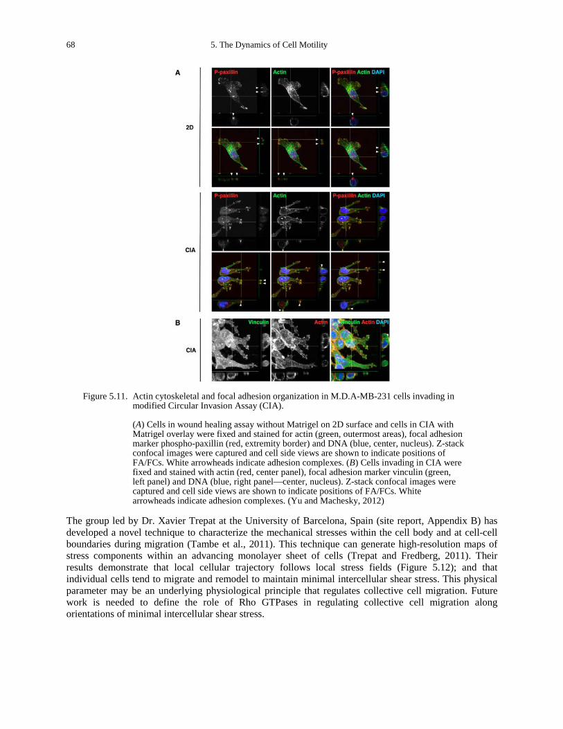
68 5. The Dynamics of Cell Motility
Figure 5.11. Actin cytoskeletal and focal adhesion organization in M.D.A-MB-231 cells invading in
modified Circular Invasion Assay (CIA).
(A) Cells in wound healing assay without Matrigel on 2D surface and cells in CIA with Matrigel overlay were fixed and stained for actin (green, outermost areas), focal adhesion marker phospho-paxillin (red, extremity border) and DNA (blue, center, nucleus). Z-stack confocal images were captured and cell side views are shown to indicate positions of FA/FCs. White arrowheads indicate adhesion complexes. (B) Cells invading in CIA were fixed and stained with actin (red, center panel), focal adhesion marker vinculin (green, left panel) and DNA (blue, right panel—center, nucleus). Z-stack confocal images were captured and cell side views are shown to indicate positions of FA/FCs. White arrowheads indicate adhesion complexes. (Yu and Machesky, 2012)
The group led by Dr. Xavier Trepat at the University of Barcelona, Spain (site report, Appendix B) has developed a novel technique to characterize the mechanical stresses within the cell body and at cell-cell boundaries during migration (Tambe et al., 2011). This technique can generate high-resolution maps of stress components within an advancing monolayer sheet of cells (Trepat and Fredberg, 2011). Their results demonstrate that local cellular trajectory follows local stress fields (Figure 5.12); and that individual cells tend to migrate and remodel to maintain minimal intercellular shear stress. This physical parameter may be an underlying physiological principle that regulates collective cell migration. Future work is needed to define the role of Rho GTPases in regulating collective cell migration along orientations of minimal intercellular shear stress.
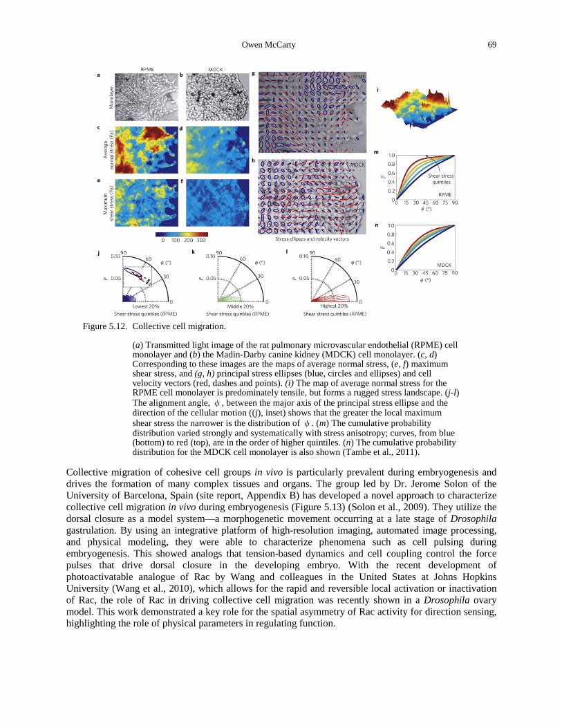
Owen McCarty 69
Figure 5.12. Collective cell migration.
(a) Transmitted light image of the rat pulmonary microvascular endothelial (RPME) cell monolayer and (b) the Madin-Darby canine kidney (MDCK) cell monolayer. (c, d) Corresponding to these images are the maps of average normal stress, (e, f) maximum shear stress, and (g, h) principal stress ellipses (blue, circles and ellipses) and cell velocity vectors (red, dashes and points). (i) The map of average normal stress for the RPME cell monolayer is predominately tensile, but forms a rugged stress landscape. (j-l) The alignment angle, φ, between the major axis of the principal stress ellipse and the direction of the cellular motion ((j), inset) shows that the greater the local maximum shear stress the narrower is the distribution of φ. (m) The cumulative probability distribution varied strongly and systematically with stress anisotropy; curves, from blue (bottom) to red (top), are in the order of higher quintiles. (n) The cumulative probability distribution for the MDCK cell monolayer is also shown (Tambe et al., 2011).
Collective migration of cohesive cell groups in vivo is particularly prevalent during embryogenesis and drives the formation of many complex tissues and organs. The group led by Dr. Jerome Solon of the University of Barcelona, Spain (site report, Appendix B) has developed a novel approach to characterize collective cell migration in vivo during embryogenesis (Figure 5.13) (Solon et al., 2009). They utilize the dorsal closure as a model system—a morphogenetic movement occurring at a late stage of Drosophila gastrulation. By using an integrative platform of high-resolution imaging, automated image processing, and physical modeling, they were able to characterize phenomena such as cell pulsing during embryogenesis. This showed analogs that tension-based dynamics and cell coupling control the force pulses that drive dorsal closure in the developing embryo. With the recent development of photoactivatable analogue of Rac by Wang and colleagues in the United States at Johns Hopkins University (Wang et al., 2010), which allows for the rapid and reversible local activation or inactivation of Rac, the role of Rac in driving collective cell migration was recently shown in a Drosophila ovary model. This work demonstrated a key role for the spatial asymmetry of Rac activity for direction sensing, highlighting the role of physical parameters in regulating function.
70 5. The Dynamics of Cell Motility
Figure 5.13. Collective cell dynamics during dorsal closure (DC).
(A) Cartoon of DC embryos. Colored arrows depict forces produced by AS cells (blue, two center images—central arrows), AC (red, arrows following the curve of the ellipse), and zippering (green, arrows pointing to the median). Black arrows show the direction of LE movement. (B) Typical apical surface area pulsations of an AS cell in a GFP-Arm expressing embryo. The upper panel shows raw data, the lower panel shows the superimposed segmented image. (C) Apical cell surface fluctuations of an AS cell. Time point zero depicts the approximate onset of dorsal closure. (D) The period distribution of 505 pulses measured in 35 AS cells in two embryos is shown. The distribution is narrow and centered at 230 ± 76 s. (E) Image of a GFP-Arm-expressing embryo showing the epidermis (small cells at the bottom), the LE, and part of the AS tissue. AS cells are color-coded depending on their distance from the LE; the ventral-most cell row in red, the second row in green, and the third row in blue. (F) Analysis of the apical surface fluctuations of the AS cells highlighted in (E). Cells sequentially cease pulsing (indicated by arrows): cells contacting the LE (red) are first (top), followed by the cells in the second row (green, middle). Cells in the third row (blue) continue pulsing throughout the analyzed time period (bottom). (G) The mean of the apical surface maxima and minima is shown for the different rows of AS cells in (F). The minima remain virtually constant over time while the maxima decrease sequentially. Consequently, the average surface (dashed lines) decreases mainly as function of the maxima reduction (Solon et al., 2009).
Owen McCarty 71
DISCUSSION
This chapter has given an overview of the research being done in Europe to define the role of physical parameters in the regulation of cell motility and migration. Understanding the mechanisms that govern collective cell migration, in contrast to single cell motility, may lead to the development of strategies to either suppress or enhance collective cell function in health and disease. Research is being done in Europe at length and time scales that span single molecule reactions to macroscopic cell migration in vivo. During our site visits, we witnessed the synergy between basic scientists and theorists and cell biologists that is used to develop novel techniques to study the role of cell motility, cell-cell adhesion, signaling and ECM remodeling to drive collective cell migration. We observed a greater degree of multidisciplinary teamwork, and more importantly, infrastructure to house multidisciplinary teams in the same physical location, than what is typically observed in the United States. Accordingly, the United States must continue to development and support multidisciplinary teamwork in order to continue to make scientific advances in understanding the pathophysiology of diseases such as cancer. A greater degree of emphasis on basic, mechanistic research was observed in Europe, as compared to the translational approach that is being emphasized in the United States. This has given the United States an advantage in the rational design and development of novel therapeutic approaches to treat and prevent disease such as cancer. Continued support of translational medicine in the United States is key to developing the next generation of therapeutics to produce better outcomes for cancer patients.
REFERENCES Aplin, A.E., Howe, A., Alahari, S.K., and Juliano, R.L. (1998). Signal transduction and signal modulation by cell adhesion
receptors: the role of integrins, cadherins, immunoglobulin-cell adhesion molecules, and selectins. Pharmacol Rev 50, 197-263.
Arjonen, A., Kaukonen, R., and Ivaska, J. (2011). Filopodia and adhesion in cancer cell motility. Cell Adh Migr 5, 421-430.
Bishop, A.L., and Hall, A. (2000). Rho GTPases and their effector proteins. Biochem J 348 Pt 2, 241-255. Breitsprecher, D., Koestler, S.A., Chizhov, I., Nemethova, M., Mueller, J., Goode, B.L., Small, J.V., Rottner, K., and Faix,
J. (2011). Cofilin cooperates with fascin to disassemble filopodial actin filaments. J Cell Sci 124, 3305-3318. Burridge, K., and Wennerberg, K. (2004). Rho and Rac take center stage. Cell 116, 167-179. Butcher, D.T., Alliston, T., and Weaver, V.M. (2009). A tense situation: forcing tumour progression. Nat Rev Cancer 9,
108-122. Cary, L.A., Han, D.C., and Guan, J.L. (1999). Integrin-mediated signal transduction pathways. Histol Histopathol 14,
1001-1009. Cavalcanti-Adam, E.A., Micoulet, A., Blummel, J., Auernheimer, J., Kessler, H., and Spatz, J.P. (2006). Lateral spacing of
integrin ligands influences cell spreading and focal adhesion assembly. Eur J Cell Biol 85, 219-224. Etienne-Manneville, S., and Hall, A. (2002). Rho GTPases in cell biology. Nature 420, 629-635. Gray, R.S., Cheung, K.J., and Ewald, A.J. (2010). Cellular mechanisms regulating epithelial morphogenesis and cancer
invasion. Curr Opin Cell Biol 22, 640-650. Hall, A. (1998). Rho GTPases and the actin cytoskeleton. Science 279, 509-514. Harunaga, J.S., and Yamada, K.M. (2011). Cell-matrix adhesions in 3D. Matrix Biol 30, 363-368. Heasman, S.J., and Ridley, A.J. (2008). Mammalian Rho GTPases: new insights into their functions from in vivo studies.
Nat Rev Mol Cell Biol 9, 690-701. Huttenlocher, A., and Horwitz, A.R. (2011). Integrins in cell migration. Cold Spring Harb Perspect Biol 3, a005074. Huveneers, S., and Danen, E.H. (2009). Adhesion signaling - crosstalk between integrins, Src and Rho. J Cell Sci 122,
1059-1069. Ingber, D.E. (2003). Mechanosensation through integrins: cells act locally but think globally. Proc Natl Acad Sci U S A
100, 1472-1474. Insall, R.H., and Machesky, L.M. (2009). Actin dynamics at the leading edge: from simple machinery to complex
networks. Dev Cell 17, 310-322. Jaffe, A.B., and Hall, A. (2005). Rho GTPases: biochemistry and biology. Annu Rev Cell Dev Biol 21, 247-269.
72 5. The Dynamics of Cell Motility
Jean, C., Gravelle, P., Fournie, J.J., and Laurent, G. (2011). Influence of stress on extracellular matrix and integrin biology. Oncogene 30, 2697-2706.
Kaibuchi, K., Kuroda, S., and Amano, M. (1999). Regulation of the cytoskeleton and cell adhesion by the Rho family GTPases in mammalian cells. Annu Rev Biochem 68, 459-486.
Khalil, A.A., and Friedl, P. (2010). Determinants of leader cells in collective cell migration. Integr Biol (Camb) 2, 568-574.
Kohler, S., Schaller, V., and Bausch, A.R. (2011). Collective dynamics of active cytoskeletal networks. PLoS One 6, e23798.
Lammermann, T., Bader, B.L., Monkley, S.J., Worbs, T., Wedlich-Soldner, R., Hirsch, K., Keller, M., Forster, R., Critchley, D.R., Fassler, R., et al. (2008). Rapid leukocyte migration by integrin-independent flowing and squeezing. Nature 453, 51-55.
Lauth, M. (2011). RAS and Hedgehog--partners in crime. Front Biosci 17, 2259-2270. Liu, P., Cheng, H., Roberts, T.M., and Zhao, J.J. (2009). Targeting the phosphoinositide 3-kinase pathway in cancer. Nat
Rev Drug Discov 8, 627-644. Machesky, L.M., and Gould, K.L. (1999). The Arp2/3 complex: a multifunctional actin organizer. Curr Opin Cell Biol 11,
117-121. Machesky, L.M., and Insall, R.H. (1999). Signaling to actin dynamics. J Cell Biol 146, 267-272. Mardilovich, K., Olson, M.F., and Baugh, M. (2012). Targeting Rho GTPase signaling for cancer therapy. Future Oncol 8,
165-177. Mulloy, J.C., Cancelas, J.A., Filippi, M.D., Kalfa, T.A., Guo, F., and Zheng, Y. (2010). Rho GTPases in hematopoiesis
and hemopathies. Blood 115, 936-947. Oser, M., and Condeelis, J. (2009). The cofilin activity cycle in lamellipodia and invadopodia. J Cell Biochem 108, 1252-
1262. Parisi, F., and Vidal, M. (2011). Epithelial delamination and migration: lessons from Drosophila. Cell Adh Migr 5, 366-
372. Parsons, J.T., Horwitz, A.R., and Schwartz, M.A. (2010). Cell adhesion: integrating cytoskeletal dynamics and cellular
tension. Nat Rev Mol Cell Biol 11, 633-643. Pinner, S., and Sahai, E. (2008). Imaging amoeboid cancer cell motility in vivo. J Microsc 231, 441-445. Pollard, T.D., Blanchoin, L., and Mullins, R.D. (2000). Molecular mechanisms controlling actin filament dynamics in
nonmuscle cells. Annu Rev Biophys Biomol Struct 29, 545-576. Reister-Gottfried, E., Sengupta, K., Lorz, B., Sackmann, E., Seifert, U., and Smith, A.S. (2008). Dynamics of specific
vesicle-substrate adhesion: from local events to global dynamics. Phys Rev Lett 101, 208103. Ridley, A.J. (2001). Rho family proteins: coordinating cell responses. Trends Cell Biol 11, 471-477. Ross, R.S. (2004). Molecular and mechanical synergy: cross-talk between integrins and growth factor receptors.
Cardiovasc Res 63, 381-390. Rottner, K., and Stradal, T.E. (2011). Actin dynamics and turnover in cell motility. Curr Opin Cell Biol 23, 569-578. Rowinsky, E.K. (2003). Signal events: Cell signal transduction and its inhibition in cancer. Oncologist 8 Suppl 3, 5-17. Schaller, M.D. (2010). Cellular functions of FAK kinases: insight into molecular mechanisms and novel functions. J Cell
Sci 123, 1007-1013. Solon, J., Kaya-Copur, A., Colombelli, J., and Brunner, D. (2009). Pulsed forces timed by a ratchet-like mechanism drive
directed tissue movement during dorsal closure. Cell 137, 1331-1342. Tambe, D.T., Hardin, C.C., Angelini, T.E., Rajendran, K., Park, C.Y., Serra-Picamal, X., Zhou, E.H., Zaman, M.H.,
Butler, J.P., Weitz, D.A., et al. (2011). Collective cell guidance by cooperative intercellular forces. Nat Mater 10, 469-475.
Thompson, E.W., Newgreen, D.F., and Tarin, D. (2005). Carcinoma invasion and metastasis: a role for epithelial-mesenchymal transition? Cancer Res 65, 5991-5995; discussion 5995.
Trepat, X., and Fredberg, J.J. (2011). Plithotaxis and emergent dynamics in collective cellular migration. Trends Cell Biol 21, 638-646.
van Zijl, F., Krupitza, G., and Mikulits, W. (2011). Initial steps of metastasis: cell invasion and endothelial transmigration. Mutat Res 728, 23-34.
Vega, F.M., and Ridley, A.J. (2008). Rho GTPases in cancer cell biology. FEBS Lett 582, 2093-2101.

Owen McCarty 73
Wang, X., He, L., Wu, Y.I., Hahn, K.M., and Montell, D.J. (2010). Light-mediated activation reveals a key role for Rac in collective guidance of cell movement in vivo. Nat Cell Biol 12, 591-597.
Yamada, K.M., and Geiger, B. (1997). Molecular interactions in cell adhesion complexes. Curr Opin Cell Biol 9, 76-85. Yu, X., and Machesky, L.M. (2012). Cells assemble invadopodia-like structures and invade into matrigel in a matrix
metalloprotease dependent manner in the circular invasion assay. PLoS One 7, e30605.

74 5. The Dynamics of Cell Motility
CHAPTER 6
DEVICES AND NEW DIAGNOSTIC PRINCIPLES
Daniel A. Fletcher
INTRODUCTION
Physical scientists and engineers working in the biological sciences have made significant contributions through the development of devices and new diagnostic principles. Many of the screening, diagnostic, and therapeutic technologies used today have their origins in fundamental discoveries of physical scientists and laboratory instruments developed by engineers. Examples from the past 150 years include the discovery of X-rays and creation of X-ray machines, discovery of radioactivity and its use in radiotherapy, discovery of nuclear magnetic resonance and development of magnetic resonance imaging (MRI), and development of flow cytometry and its use in high-throughput screening.
Despite significant improvements in patient healthcare that have accompanied new clinical technologies, cancer remains a significant medical challenge. No cure exists for cancer, and the precise biological origins of tumors in different tissues remain a mystery. Therapeutic management continues to be a challenge for most forms of the disease. As average life expectancy increases, the likelihood that an individual will be faced with cancer at some point in his or her life also increases. Even the developing world, saddled with the scourge of infectious diseases, must now confront the fact that cancer and other non-communicable diseases are serious public health problems (Hotez and Daar, 2008). Can a revolutionary idea or technology be found that detects cancer early? Will a novel insight from outside of the medical field result in a new and effective therapy? Can the biological origins of cancer be understood and addressed from a physical sciences perspective? Recent research initiatives in the United States and abroad have focused attention on the intersection of physical sciences, biological sciences, and oncology, bringing researchers with fundamental and applied interests together with biologists and oncologists to pursue a new understanding of cancer and new approaches to cancer therapies. These efforts are producing a community poised to make significant contributions to the field. This chapter will describe new ideas and innovations at the intersection of physical sciences and oncology that benefit both the lab and the clinic. The chapter will conclude with a summary of drivers of innovation in Europe and the United States.
RESEARCH
New Ideas and Innovations
The United States and European research institutions have produced a remarkable breadth of new ideas and innovative approaches to the diagnosis and treatment of cancer. The specific examples discussed below are based on the APHELION study trip to European institutions, broken into five clusters of innovation: optical microscopy, force microscopy, new materials, microdevices, and integrated therapies. Examples from research in the United States are also included.

76 6. Devices and New Diagnostic Principles
Optical Microscopy Optical microscopy is a mainstay of cancer research and diagnosis. Traditional histological analysis combines tissue sample staining with microscopic imaging to determine patient diagnoses and treatment plans. In research, optical microscopy using fluorescently labeled molecules has led to a mechanistic understanding of basic cellular processes and the ability to study the molecular details of tissue development and function. Researchers in the United States have pioneered a multitude of fluorescent proteins and nanocrystal tags, including the variations on green fluorescent proteins by Dr. Roger Tsien at University of California, San Diego and quantum dots by Dr. Paul Alivisatos at University of California, Berkeley. While fluorescence microscopy has become routine for localizing molecules, many properties that are harder to measure, such as the local concentration of reactive oxygen within a cell, are important measurements to understanding metabolism. Dr. Jerker Windengren at the Royal Institute of Technology (KTH) in Stockholm, Sweden, (site report, Appendix B) has advanced a new approach to measuring small molecules by using not the fluorescence emission of a fluorescent molecule but rather the population of its triplet state (Hevekerl et al., 2011). By carefully quantifying photon fluxes, Dr. Windengren can carry out triplet-state imaging that may be useful in detecting molecules, such as reactive oxygen, that alter the triplet state. Fluorescence microscopy and its variants are useful for fundamental studies based on cells and tissues that can be genetically or otherwise manipulated to contain fluorescent labels on the molecules of interest. Label-free approaches are desirable if optical microscopy is to be applied to humans for diagnosis and therapy. Research into multiphoton interactions with biological molecules has revealed that high contrast can be achieved without labels by measuring 2nd and even 3rd harmonic interactions. Dr. Peter Friedl at the Nijmegen Centre for Molecular Life Sciences, Netherlands (site report, Appendix B) is a pioneer of multiphoton imaging and has brought the technology to bear on questions of tumor progression and metastasis (Friedl et al., 2007; Ilina et al., 2011). Recent developments in his laboratory have enabled real-time and structure-specific imaging of live tissue with both 2nd harmonic and 3rd harmonic contrast without the need to introduce fluorescent labels or other contrast tabs (Figure 6.1). This technology is already advancing the study of tumor progression in model animals.
Figure 6.1. Images of human dermatofibrosarcoma protuberans from (Friedl et al., 2007).
(A) The tissue was stained for CD34 (red—top and bottom layer of tissue, showing cancer cells) and DAPI (green, dermis). The image also shows second harmonic generation from collagen (green, dermis), which is shown together with DAPI as a grayscale image in (B). (C) Intensity curves show that SHG (top) is reduced at the border with the cancer lesion.
Daniel A. Fletcher 77
Force Microscopy The old observation that solid tumors are stiffer than their surroundings has taken on new importance with the finding that mechanical properties of cells and their matrix are both indicators and drivers of disease. Studies have shown that cancer cell stiffness is increased in leukemia and decreased in breast cancer, suggesting that measuring these properties in hospitals (which do not currently use assays to quantify cellular or extracellular matrix stiffness) may be clinically useful. A variety of methods for measuring the mechanical properties of cells have been developed, including atomic force microscopy (AFM) and parallel plate rheometry (see Chapter 3 by Reinhart-King). Researchers in the United States have been particularly active in the application of AFM and related indentation technologies for imaging and measurement of cells and their properties, including measurements of the correlation between cell stiffness and extracellular matrix stiffness by Dr. Paul Janmey of the University of Pennsylvania and analysis of bone structure by Dr. Paul Hansma at University of California, Santa Barbara. These methods have proven very useful for fundamental studies in which small numbers of samples may be needed to establish a point, but their throughput is woefully inadequate for a clinical setting in which large numbers of blood cells from many different patients would need to be assayed. Dr. Josef Käs at the University of Leipzig, Germany (site report, Appendix B) has developed a high-throughput method for quantifying mechanical properties of cells called the optical stretcher (Figure 6.2) (Guck et al., 2000). This technology takes advantage of refractive index differences between the cell and surrounding media and uses two counter-propagating lasers to actively deform single cells. Video imaging of the cell deformation and subsequent relaxation gives a measure of cell mechanical properties. A major advantage of this technology is that it has been automated to enable high-throughput analysis of cell populations. Since the mechanical properties of patient cells can be heterogeneous, and the cells of interest may represent only a small fraction of the population, assays like the optical stretcher quantify properties of hundreds or thousands of cells to determine statistically reliable data that could be missed by evaluation of only tens of cells with traditional approaches (Fritsch et al., 2010).
New Materials A particularly active field of research is the development of new materials for biological and biomedical applications. Advancements range from the creation of new polymeric gels to direct cell growth to the development of new nanoparticle probes for measuring cell behavior. Researchers in the United States are actively pursuing an understanding of cell-matrix interactions and development of new extracellular matrix materials, such as the alginate-based gels from Dr. David Mooney at Harvard University and the dynamic biomaterials from Dr. Kristi Anseth at the University of Colorado. Dr. Wilhelm Huck at the Nijmegen Centre for Molecular Life Sciences in the Netherlands (site report, Appendix B) has created polyacrylamide gels with crosslinked collagen that are able to alter cell activation (Trappmann et al., 2012). Dr. Pierre Nassoy at the Institute Curie in Paris, France (site report, Appendix B) is developing a novel alginate encapsulation method for cell aggregates that will help to understand how cell growth responds to confinement. Dr. Clair Wilhelm at the University of Paris, Diderot (site report, Appendix B) has produced magnetic nanoparticles and nanorods that are taken up by cells and can be externally manipulated to measure cytoplasmic properties of cells (Figure 6.3). She is also developing technologies to form liposomes from cell membranes that have the potential to be more effective drug delivery materials (Lesieur et al., 2011).
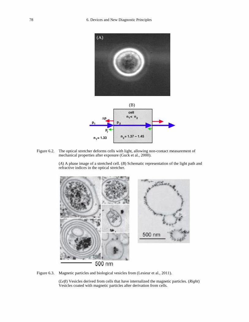
78 6. Devices and New Diagnostic Principles
(B)
Figure 6.2. The optical stretcher deforms cells with light, allowing non-contact measurement of mechanical properties after exposure (Guck et al., 2000).
(A) A phase image of a stretched cell. (B) Schematic representation of the light path and refractive indices in the optical stretcher.
Figure 6.3. Magnetic particles and biological vesicles from (Lesieur et al., 2011).
(Left) Vesicles derived from cells that have internalized the magnetic particles. (Right) Vesicles coated with magnetic particles after derivation from cells.
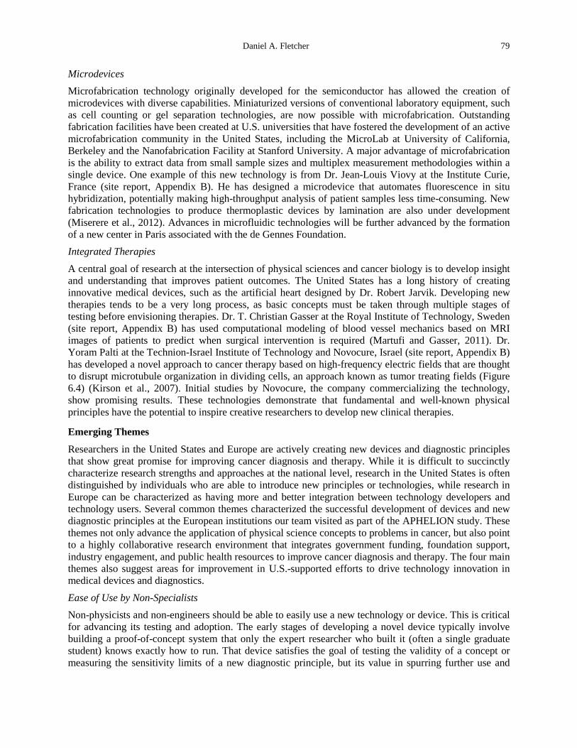
Daniel A. Fletcher 79
Microdevices Microfabrication technology originally developed for the semiconductor has allowed the creation of microdevices with diverse capabilities. Miniaturized versions of conventional laboratory equipment, such as cell counting or gel separation technologies, are now possible with microfabrication. Outstanding fabrication facilities have been created at U.S. universities that have fostered the development of an active microfabrication community in the United States, including the MicroLab at University of California, Berkeley and the Nanofabrication Facility at Stanford University. A major advantage of microfabrication is the ability to extract data from small sample sizes and multiplex measurement methodologies within a single device. One example of this new technology is from Dr. Jean-Louis Viovy at the Institute Curie, France (site report, Appendix B). He has designed a microdevice that automates fluorescence in situ hybridization, potentially making high-throughput analysis of patient samples less time-consuming. New fabrication technologies to produce thermoplastic devices by lamination are also under development (Miserere et al., 2012). Advances in microfluidic technologies will be further advanced by the formation of a new center in Paris associated with the de Gennes Foundation. Integrated Therapies A central goal of research at the intersection of physical sciences and cancer biology is to develop insight and understanding that improves patient outcomes. The United States has a long history of creating innovative medical devices, such as the artificial heart designed by Dr. Robert Jarvik. Developing new therapies tends to be a very long process, as basic concepts must be taken through multiple stages of testing before envisioning therapies. Dr. T. Christian Gasser at the Royal Institute of Technology, Sweden (site report, Appendix B) has used computational modeling of blood vessel mechanics based on MRI images of patients to predict when surgical intervention is required (Martufi and Gasser, 2011). Dr. Yoram Palti at the Technion-Israel Institute of Technology and Novocure, Israel (site report, Appendix B) has developed a novel approach to cancer therapy based on high-frequency electric fields that are thought to disrupt microtubule organization in dividing cells, an approach known as tumor treating fields (Figure 6.4) (Kirson et al., 2007). Initial studies by Novocure, the company commercializing the technology, show promising results. These technologies demonstrate that fundamental and well-known physical principles have the potential to inspire creative researchers to develop new clinical therapies.
Emerging Themes
Researchers in the United States and Europe are actively creating new devices and diagnostic principles that show great promise for improving cancer diagnosis and therapy. While it is difficult to succinctly characterize research strengths and approaches at the national level, research in the United States is often distinguished by individuals who are able to introduce new principles or technologies, while research in Europe can be characterized as having more and better integration between technology developers and technology users. Several common themes characterized the successful development of devices and new diagnostic principles at the European institutions our team visited as part of the APHELION study. These themes not only advance the application of physical science concepts to problems in cancer, but also point to a highly collaborative research environment that integrates government funding, foundation support, industry engagement, and public health resources to improve cancer diagnosis and therapy. The four main themes also suggest areas for improvement in U.S.-supported efforts to drive technology innovation in medical devices and diagnostics.
Ease of Use by Non-Specialists Non-physicists and non-engineers should be able to easily use a new technology or device. This is critical for advancing its testing and adoption. The early stages of developing a novel device typically involve building a proof-of-concept system that only the expert researcher who built it (often a single graduate student) knows exactly how to run. That device satisfies the goal of testing the validity of a concept or measuring the sensitivity limits of a new diagnostic principle, but its value in spurring further use and
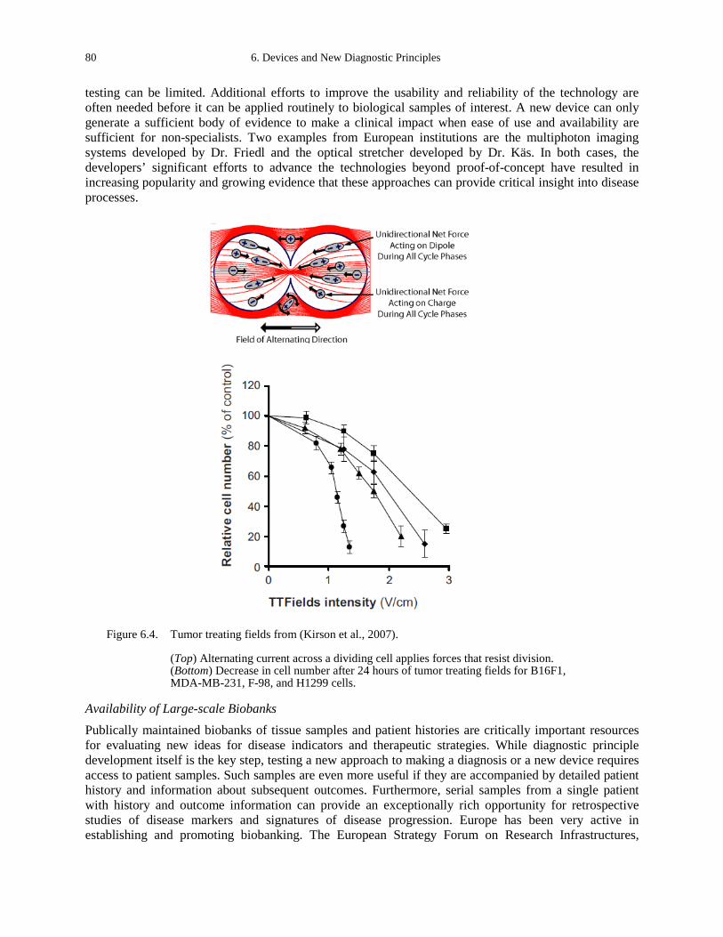
80 6. Devices and New Diagnostic Principles
testing can be limited. Additional efforts to improve the usability and reliability of the technology are often needed before it can be applied routinely to biological samples of interest. A new device can only generate a sufficient body of evidence to make a clinical impact when ease of use and availability are sufficient for non-specialists. Two examples from European institutions are the multiphoton imaging systems developed by Dr. Friedl and the optical stretcher developed by Dr. Käs. In both cases, the developers’ significant efforts to advance the technologies beyond proof-of-concept have resulted in increasing popularity and growing evidence that these approaches can provide critical insight into disease processes.
Figure 6.4. Tumor treating fields from (Kirson et al., 2007).
(Top) Alternating current across a dividing cell applies forces that resist division. (Bottom) Decrease in cell number after 24 hours of tumor treating fields for B16F1, MDA-MB-231, F-98, and H1299 cells.
Availability of Large-scale Biobanks Publically maintained biobanks of tissue samples and patient histories are critically important resources for evaluating new ideas for disease indicators and therapeutic strategies. While diagnostic principle development itself is the key step, testing a new approach to making a diagnosis or a new device requires access to patient samples. Such samples are even more useful if they are accompanied by detailed patient history and information about subsequent outcomes. Furthermore, serial samples from a single patient with history and outcome information can provide an exceptionally rich opportunity for retrospective studies of disease markers and signatures of disease progression. Europe has been very active in establishing and promoting biobanking. The European Strategy Forum on Research Infrastructures,
Daniel A. Fletcher 81
funded by the European Commission, has established the Biobanking and Biomolecular Resources Research Infrastructure, which includes more than 225 associated organizations from over 30 countries (www.bbmri.eu). The goal of this infrastructure is to organize biobanking activities in Europe and strengthen the link between biological specimen collection and use in biological and medical research. Some individual countries have had long-established biobanking systems that now support extensive medical research. The Swedish National Biobank was established more than 20 years ago and has patient information allowing for both retrospective and prospective studies of a broad range of cancers (www.biobanks.se). Biobanks within Sweden now contain more than 10 million pathology samples, 1.7 million plasma samples, and 24,000 fresh-frozen samples.
Co-localization of Multiple Device Modalities Diagnosis and treatment of cancer involves the use of many different devices and technologies. In order to understand how a new device or diagnostic principle compares with current or competing methods, data from several different measurement techniques on the same samples provides the most useful information. Indeed, obtaining the biological specimen, whether it is from a specially engineered mouse model or from a patient with a unique condition, can often be the limiting step in data collection. Therefore, maximizing data collection from an available sample is important. This can be achieved by combining multiple device modalities in a single location with open access to all instruments. Rather than have a separate facility for X-ray imaging, MRI, and a new imaging modality in the testing phase, combine these instruments into one imaging facility. This multipurpose facility will have easy access to commercial instruments as well as instruments under development, and it enables efficient use of samples and provides valuable comparative data. This is exactly what has been developed at the Preclinical Imaging Centre of the Radboud University Nijmegen Medical Centre in the Netherlands, where Dr. Friedl and colleagues in multiple departments have established an imaging center that offers MRI, microSPECT/CT, multiphoton microscopy, and other imaging modalities in a single facility (www.umcn.nl/Research/Departments/cdl/PRIME). Access to multiple instruments in a central location enables researchers to easily interrogate tissue in multiple ways. Close Industry Connections It is necessary to commercialize technologies developed in research laboratories if they are to become broadly available for clinical use. The process of transitioning a technology from a research environment to an industrial environment is a complex one, often presenting significant barriers. Strong connections between academic researchers and industry can provide efficient routes for device commercialization as well as awareness of what problems need solving. Research institutions that are able to foster those strong connections have been successful in spawning start-up companies or partnering with established companies to further develop and commercialize technology. A successful example from our study trip is KTH in Sweden, where multiple technologies have gone from laboratory prototypes to commercial devices, including eXcillum which produces X-ray phase contrast equipment (www.excillum.com) and Adolesco which produces mobile 3D SPECT (www.adolesco.se). Two actions contribute to close collaborations between research and industry: 1) strong government support for collaborations with industry; and 2) a consensus among researchers that transitioning a technology to industry is part of a research project timeline. In Sweden, patent policy is also a contributing factor. Their policy states that the rights to an invention stay with the inventor rather than with the institution, which is counter to the United States policy.
DISCUSSION
The physical sciences have great potential to impact the diagnosis and treatment of cancer. European research institutions have created a supportive environment for the interaction of physical sciences and oncology that has fostered the development of novel devices and new diagnostic principles. This environment is characterized by highly interactive and interdisciplinary researchers spanning academic departments and medical institutions. Collaborative funding initiatives from the European Union and
82 6. Devices and New Diagnostic Principles
country governments have contributed to Europe’s highly creative and productive research, and an emphasis on industry partnerships has promoted commercialization of innovative new technologies. The Institute Curie in France, where internal funding is used to initiate cross-disciplinary projects, is a model of how basic researchers and clinical researchers can be located within a common research infrastructure and work together to advance fundamental principles and identify new therapeutic opportunities. The emerging themes identified from a review of research practices in European institutions point to specific ways in which U.S. funding could further encourage the development of new devices and diagnostic principles for cancer.
REFERENCES Friedl, P., Wolf, K., von Andrian, U.H., and Harms, G. (2007). Biological second and third harmonic generation
microscopy. Current protocols in cell biology / editorial board, Juan S Bonifacino [et al] Chapter 4, Unit 4 15. Fritsch, A., Hockel, M., Kiessling, T., Nnetu, K.D., Wetzel, F., Zink, M., and Kas, J.A. (2010). Are biomechanical
changes necessary for tumour progression? Nat Phys 6, 730-732. Guck, J., Ananthakrishnan, R., Moon, T.J., Cunningham, C.C., and Kas, J. (2000). Optical deformability of soft biological
dielectrics. Physical review letters 84, 5451-5454. Hevekerl, H., Spielmann, T., Chmyrov, A., and Widengren, J. (2011). Forster resonance energy transfer beyond 10 nm:
exploiting the triplet state kinetics of organic fluorophores. The journal of physical chemistry B 115, 13360-13370. Hotez, P.J., and Daar, A.S. (2008). The CNCDs and the NTDs: blurring the lines dividing noncommunicable and
communicable chronic diseases. PLoS neglected tropical diseases 2, e312. Ilina, O., Bakker, G.J., Vasaturo, A., Hofmann, R.M., and Friedl, P. (2011). Two-photon laser-generated microtracks in
3D collagen lattices: principles of MMP-dependent and -independent collective cancer cell invasion. Physical biology 8, 015010.
Kirson, E.D., Dbaly, V., Tovarys, F., Vymazal, J., Soustiel, J.F., Itzhaki, A., Mordechovich, D., Steinberg-Shapira, S., Gurvich, Z., Schneiderman, R., et al. (2007). Alternating electric fields arrest cell proliferation in animal tumor models and human brain tumors. Proceedings of the National Academy of Sciences of the United States of America 104, 10152-10157.
Lesieur, S., Gazeau, F., Luciani, N., Menager, C., and Wilhelm, C. (2011). Multifunctional nanovectors based on magnetic nanoparticles coupled with biological vesicles or synthetic liposomes. J Mater Chem 21, 14387-14393.
Martufi, G., and Gasser, T.C. (2011). A constitutive model for vascular tissue that integrates fibril, fiber and continuum levels with application to the isotropic and passive properties of the infrarenal aorta. Journal of biomechanics 44, 2544-2550.
Miserere, S., Mottet, G., Taniga, V., Descroix, S., Viovy, J.L., and Malaquin, L. (2012). Fabrication of thermoplastics chips through lamination based techniques. Lab Chip 12, 1849-1856.
Trappmann, B., Gautrot, J.E., Connelly, J.T., Strange, D.G., Li, Y., Oyen, M.L., Cohen Stuart, M.A., Boehm, H., Li, B., Vogel, V., et al. (2012). Extracellular-matrix tethering regulates stem-cell fate. Nature materials 11, 642-649.

APPENDIX A. APHELION STUDY PANELISTS AND ADVISORS
PANELISTS
Daniel Fletcher Daniel Fletcher is professor of bioengineering and biophysics at the University of California, Berkeley; and a faculty scientist in the physical sciences at Lawrence Berkeley National Laboratory. He received a Ph.D. from Stanford University and a D.Phil. from Oxford University.
Dr. Fletcher’s research interests include mechanics of leukemic cells, reconstruction of branched actin networks, measurement of platelet contraction, and mechanics of branching morphogenesis. He is currently exploring the mechanics of breast cancer cells, mechanical regulation of actin networks, and development of infectious disease diagnostics.
Sharon Gerecht Sharon Gerecht is assistant professor of chemical and biomolecular engineering at Johns Hopkins University. She received a Ph.D. in biotechnology from Technion Technical Institute of Israel and an M.S. in medical science from Tel Aviv University. Dr. Gerecht’s research interests include stem cell engineering, angiogenesis and vascular biology, and biomaterials. Her new research has branched into investigating microbioreactors, skin regeneration, and the blood/brain barrier.
Paul Janmey Paul Janmey is professor of physiology, physics, and bioengineering at the Institute of Medicine and Engineering at the University of Pennsylvania. He received his Ph.D. in physical chemistry from the University of Wisconsin and completed his postdoc at the Hematology-Oncology Unit at Massachusetts General Hospital.
Dr. Janmey’s research interests include the interaction between cytoskeletal and extracellular matrix stiffness, effects of substrate mechanics on cell structure and function, phosphoinositide signaling for actin assembly, fibrin-based materials for wound healing, and intermediate filament assembly and mechanics. His latest research explores signaling between integrins and HA receptors for cell proliferation and motility, and mechanosensing through cadherins.
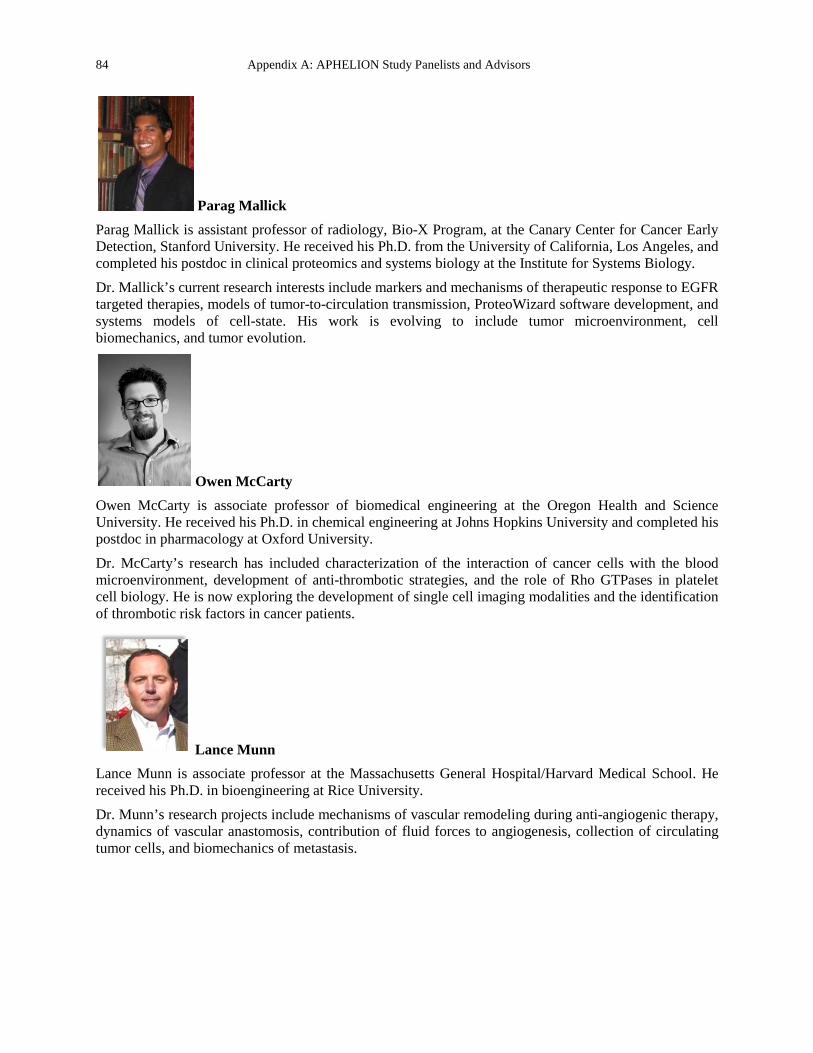
84 Appendix A: APHELION Study Panelists and Advisors
Parag Mallick Parag Mallick is assistant professor of radiology, Bio-X Program, at the Canary Center for Cancer Early Detection, Stanford University. He received his Ph.D. from the University of California, Los Angeles, and completed his postdoc in clinical proteomics and systems biology at the Institute for Systems Biology. Dr. Mallick’s current research interests include markers and mechanisms of therapeutic response to EGFR targeted therapies, models of tumor-to-circulation transmission, ProteoWizard software development, and systems models of cell-state. His work is evolving to include tumor microenvironment, cell biomechanics, and tumor evolution.
Owen McCarty Owen McCarty is associate professor of biomedical engineering at the Oregon Health and Science University. He received his Ph.D. in chemical engineering at Johns Hopkins University and completed his postdoc in pharmacology at Oxford University.
Dr. McCarty’s research has included characterization of the interaction of cancer cells with the blood microenvironment, development of anti-thrombotic strategies, and the role of Rho GTPases in platelet cell biology. He is now exploring the development of single cell imaging modalities and the identification of thrombotic risk factors in cancer patients.
Lance Munn Lance Munn is associate professor at the Massachusetts General Hospital/Harvard Medical School. He received his Ph.D. in bioengineering at Rice University. Dr. Munn’s research projects include mechanisms of vascular remodeling during anti-angiogenic therapy, dynamics of vascular anastomosis, contribution of fluid forces to angiogenesis, collection of circulating tumor cells, and biomechanics of metastasis.
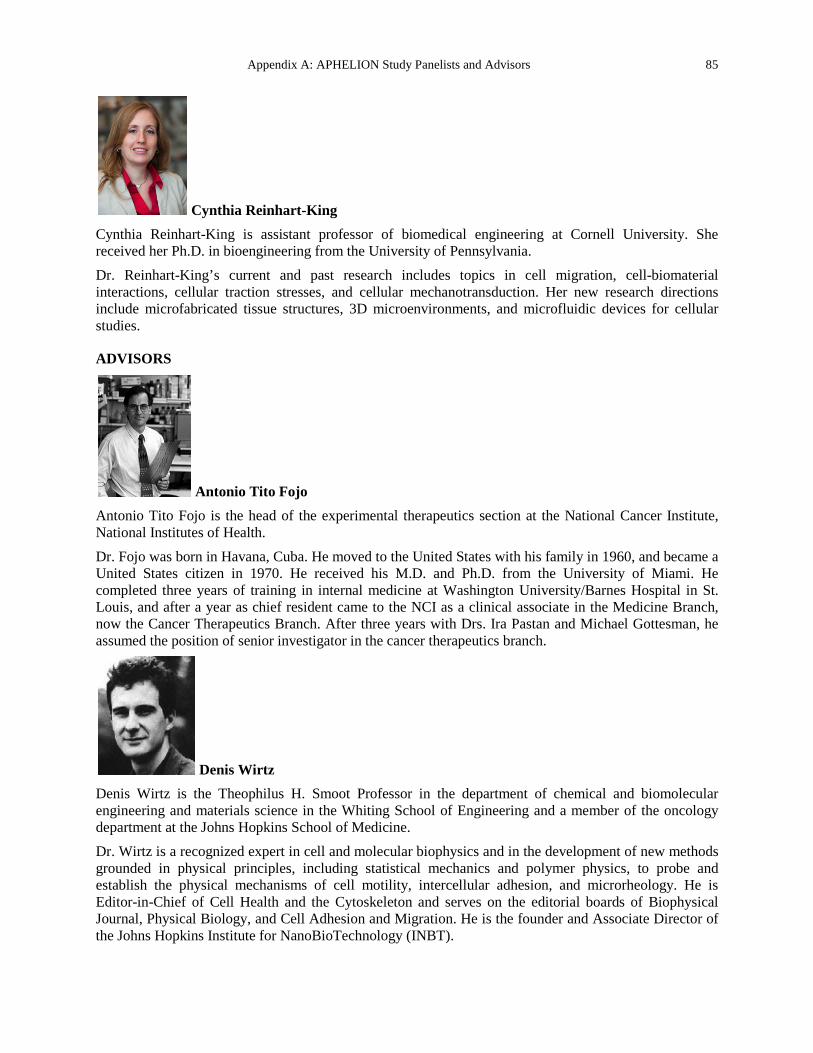
Appendix A: APHELION Study Panelists and Advisors 85
Cynthia Reinhart-King Cynthia Reinhart-King is assistant professor of biomedical engineering at Cornell University. She received her Ph.D. in bioengineering from the University of Pennsylvania. Dr. Reinhart-King’s current and past research includes topics in cell migration, cell-biomaterial interactions, cellular traction stresses, and cellular mechanotransduction. Her new research directions include microfabricated tissue structures, 3D microenvironments, and microfluidic devices for cellular studies.
ADVISORS
Antonio Tito Fojo Antonio Tito Fojo is the head of the experimental therapeutics section at the National Cancer Institute, National Institutes of Health. Dr. Fojo was born in Havana, Cuba. He moved to the United States with his family in 1960, and became a United States citizen in 1970. He received his M.D. and Ph.D. from the University of Miami. He completed three years of training in internal medicine at Washington University/Barnes Hospital in St. Louis, and after a year as chief resident came to the NCI as a clinical associate in the Medicine Branch, now the Cancer Therapeutics Branch. After three years with Drs. Ira Pastan and Michael Gottesman, he assumed the position of senior investigator in the cancer therapeutics branch.
Denis Wirtz Denis Wirtz is the Theophilus H. Smoot Professor in the department of chemical and biomolecular engineering and materials science in the Whiting School of Engineering and a member of the oncology department at the Johns Hopkins School of Medicine. Dr. Wirtz is a recognized expert in cell and molecular biophysics and in the development of new methods grounded in physical principles, including statistical mechanics and polymer physics, to probe and establish the physical mechanisms of cell motility, intercellular adhesion, and microrheology. He is Editor-in-Chief of Cell Health and the Cytoskeleton and serves on the editorial boards of Biophysical Journal, Physical Biology, and Cell Adhesion and Migration. He is the founder and Associate Director of the Johns Hopkins Institute for NanoBioTechnology (INBT).


APPENDIX B. SITE VISIT REPORTS
Site visit reports are arranged in alphabetical order by organization name.
88 Appendix B. Site Visit Reports
École Polytechnique Fédérale de Lausanne (EPFL)
Site Address: School of Life Sciences and School of Engineering Station 15, CH-1015 Lausanne, SWITZERLAND Tel: +41 21 693 96 68 Fax: +41 21 693 96 65 Email: [email protected]
Date Visited: 8 May 2012 WTEC Attendees: Sharon Gerecht, Parag Mallick, Owen McCarty, Lance Munn (site report author), and
Hassan Ali Host(s): Prof. Jeffrey Hubbell
School of Life Sciences/IBI/LMRP, Station 15 Tel: +41 21 693 9681 Fax: +41 21 693 9685 Email: [email protected] http://epfl.ch/
Dr. Jennifer Munson Post-doctoral fellow (Georgia Tech) Prof. Melody Swartz’s lab; EPFL SV IBI-SV LLCB AI 3137 (Bâtiment AI), Station 15 Tel: +41 21 693 31693 Email: [email protected] http://swartz-lab.epfl.ch/
Asst. Prof. Matthias Lutolf Faculty of Life Sciences and Institute of Bioengineering, EPFL Office: AI1109 Tel: +41 21 69 31876, 37281 Email: [email protected] http://lscb.epfl.ch
Prof. Henry Markram Neural Microcircuitry Laboratory EPFL, SV BMI LNMC, AAB 110 (Bâtiment AAB), Station 15 Tel: +41 21 69-39537, 39569 Email: [email protected]
OVERVIEW
Prof. Jeffrey Hubbell opened the meeting with an overview of the EPFL Institute of Bioengineering (IBI). The life sciences program is relatively young, initiated in 2001. Hubbell was recruited to develop the IBI in 2003. The institute has a translational focus and is dedicated to interdisciplinary training: all biologist undergrads get bioengineering training. There are 490 personnel, 33 of whom are faculty. Few of the faculty members are from Switzerland. The WTEC panel heard presentations from four groups with projects related to cancer: Hubbell, Melody Swartz (presented by Jennifer Munson), Matthias Lutolf, and Henry Markram.
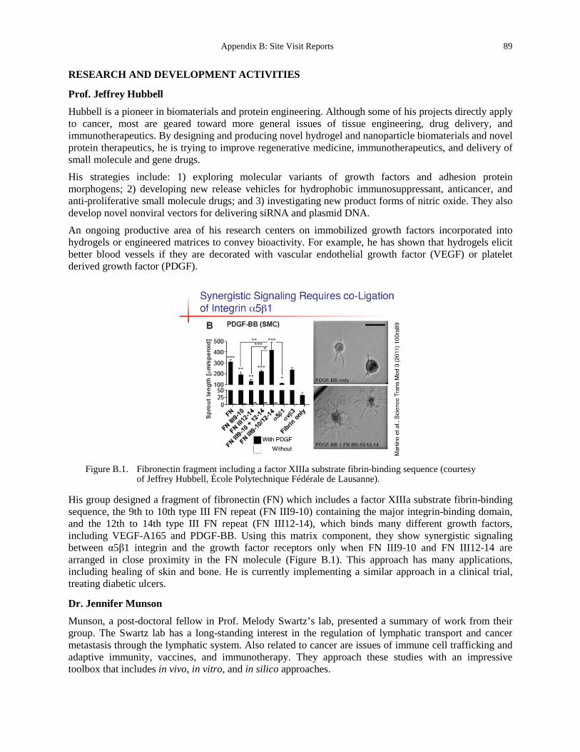
Appendix B: Site Visit Reports 89
RESEARCH AND DEVELOPMENT ACTIVITIES
Prof. Jeffrey Hubbell
Hubbell is a pioneer in biomaterials and protein engineering. Although some of his projects directly apply to cancer, most are geared toward more general issues of tissue engineering, drug delivery, and immunotherapeutics. By designing and producing novel hydrogel and nanoparticle biomaterials and novel protein therapeutics, he is trying to improve regenerative medicine, immunotherapeutics, and delivery of small molecule and gene drugs. His strategies include: 1) exploring molecular variants of growth factors and adhesion protein morphogens; 2) developing new release vehicles for hydrophobic immunosuppressant, anticancer, and anti-proliferative small molecule drugs; and 3) investigating new product forms of nitric oxide. They also develop novel nonviral vectors for delivering siRNA and plasmid DNA. An ongoing productive area of his research centers on immobilized growth factors incorporated into hydrogels or engineered matrices to convey bioactivity. For example, he has shown that hydrogels elicit better blood vessels if they are decorated with vascular endothelial growth factor (VEGF) or platelet derived growth factor (PDGF).
Figure B.1. Fibronectin fragment including a factor XIIIa substrate fibrin-binding sequence (courtesy
of Jeffrey Hubbell, École Polytechnique Fédérale de Lausanne).
His group designed a fragment of fibronectin (FN) which includes a factor XIIIa substrate fibrin-binding sequence, the 9th to 10th type III FN repeat (FN III9-10) containing the major integrin-binding domain, and the 12th to 14th type III FN repeat (FN III12-14), which binds many different growth factors, including VEGF-A165 and PDGF-BB. Using this matrix component, they show synergistic signaling between α5β1 integrin and the growth factor receptors only when FN III9-10 and FN III12-14 are arranged in close proximity in the FN molecule (Figure B.1). This approach has many applications, including healing of skin and bone. He is currently implementing a similar approach in a clinical trial, treating diabetic ulcers.
Dr. Jennifer Munson
Munson, a post-doctoral fellow in Prof. Melody Swartz’s lab, presented a summary of work from their group. The Swartz lab has a long-standing interest in the regulation of lymphatic transport and cancer metastasis through the lymphatic system. Also related to cancer are issues of immune cell trafficking and adaptive immunity, vaccines, and immunotherapy. They approach these studies with an impressive toolbox that includes in vivo, in vitro, and in silico approaches.
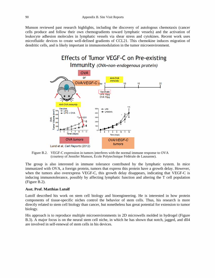
90 Appendix B. Site Visit Reports
Munson reviewed past research highlights, including the discovery of autologous chemotaxis (cancer cells produce and follow their own chemogradients toward lymphatic vessels) and the activation of leukocyte adhesion molecules in lymphatic vessels via shear stress and cytokines. Recent work uses microfluidic devices to create well-defined gradients of CCL21. This chemokine induces migration of dendritic cells, and is likely important in immunomodulation in the tumor microenvironment.
Figure B.2. VEGF-C expression in tumors interferes with the normal immune response to OVA
(courtesy of Jennifer Munson, École Polytechnique Fédérale de Lausanne).
The group is also interested in immune tolerance contributed by the lymphatic system. In mice immunized with OVA, a foreign protein, tumors that express this protein have a growth delay. However, when the tumors also overexpress VEGF-C, this growth delay disappears, indicating that VEGF-C is inducing immunotolerance, possibly by affecting lymphatic function and altering the T cell population (Figure B.2).
Asst. Prof. Matthias Lutolf
Lutolf described his work on stem cell biology and bioengineering. He is interested in how protein components of tissue-specific niches control the behavior of stem cells. Thus, his research is more directly related to stem cell biology than cancer, but nonetheless has great potential for extension to tumor biology.
His approach is to reproduce multiple microenvironments in 2D microwells molded in hydrogel (Figure B.3). A major focus is on the neural stem cell niche, in which he has shown that notch, jagged, and dll4 are involved in self-renewal of stem cells in his devices.
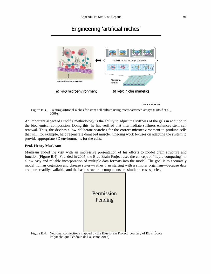
Appendix B: Site Visit Reports 91
Figure B.3. Creating artificial niches for stem cell culture using micropatterned assays (Lutolf et al.,
2009).
An important aspect of Lutolf’s methodology is the ability to adjust the stiffness of the gels in addition to the biochemical composition. Doing this, he has verified that intermediate stiffness enhances stem cell renewal. Thus, the devices allow deliberate searches for the correct microenvironment to produce cells that will, for example, help regenerate damaged muscle. Ongoing work focuses on adapting the system to provide appropriate 3D environments for the cells.
Prof. Henry Markram
Markram ended the visit with an impressive presentation of his efforts to model brain structure and function (Figure B.4). Founded in 2005, the Blue Brain Project uses the concept of “liquid computing” to allow easy and reliable incorporation of multiple data formats into the model. The goal is to accurately model human cognition and disease states—rather than starting with a simpler organism—because data are more readily available, and the basic structural components are similar across species.
Figure B.4. Neuronal connections mapped by the Blue Brain Project (courtesy of BBP/ École
Polytechnique Fédérale de Lausanne 2012).
Permission Pending
92 Appendix B. Site Visit Reports
The overall strategy is to simplify the problem. With current computational power, one dedicated processor is needed to simulate the activity of every single neuron. Thus, the billions of neurons in the brain cannot be modeled neuron-by-neuron. To overcome this limitation, the project uses multi-level simulations in which only highly active groups of neurons are simulated in detail. The resulting “virtual brain,” living in supercomputers, will incorporate “all the data that neuroscience has generated to date.” The Human Brain Project is an extension of the Blue Brain Project, bringing together a consortium of 13 partners from nine European Union member states. In preparation for a full-scale flagship project, the consortium partners are each developing one specific pillar of activity that will be integrated in the final project.
TRANSLATION
Within the IBI, Hubbell’s work is the most translational. He is actively working with clinicians to apply his engineered materials to patients.
SOURCES OF SUPPORT/FUNDING
Each faculty member receives $1.25 million Euros per year, and is expected to match this with grants. Sources of external funding include the Swiss National Science Foundation, Oncosuisse, and the National Centre for Competence in Research in Molecular Oncology.
SUMMARYAND CONCLUSIONS
EPFL has a world-class bioengineering institute, with some emphasis on cancer. Of the sites visited, EPFL is most similar to U.S. institutions in terms of the various research directions and approaches. This is likely due to the fact that all of the PIs represented at our visit had extensive training in the United States. There is an impressive range of projects, spanning from genes to organisms, and inter-group collaborations are extensive. All of this is actively supported by the EPFL, which is obviously dedicated to nurturing these bioengineering efforts.
REFERENCES Bott, K., Upton, Z., Schrobback, K., Ehrbar, M., Hubbell, J.A., Lutolf, M.P., and Rizzi, S.C. (2006). The effect of matrix
characteristics on fibroblast proliferation in 3D gels. Biomaterials 31, 8454-8464. Di Crescenzo, A., Velluto, D., Hubbell, J.A., and Fontana, A. (2011). Biocompatible dispersions of carbon nanotubes: a
potential tool for intracellular transport of anticancer drugs. Nanoscale 3, 925-928. Eming, S.A., and Hubbell, J.A. (2011). Extracellular matrix in angiogenesis: dynamic structures with translational
potential. Exp Dermatol 20, 605-613. Hubbell, J.A. (2010). Drug development: Longer-lived proteins. Nature 467, 1051-1052. Velluto, D., Thomas, S.N., Simeoni, E., Swartz, M.A., and Hubbell, J.A. (2011). PEG-b-PPS-b-PEI micelles and PEG-b-
PPS/PEG-b-PPS-b-PEI mixed micelles as non-viral vectors for plasmid DNA: tumor immunotoxicity in B16F10 melanoma. Biomaterials 32, 9839-9847.
Swartz Haessler, U., Teo, J.C., Foretay, D., Renaud, P., and Swartz, M.A. (2012). Migration dynamics of breast cancer cells in a
tunable 3D interstitial flow chamber. Integr Biol (Camb) 4, 401-409. Miteva, D.O., Rutkowski, J.M., Dixon, J.B., Kilarski, W., Shields, J.D., and Swartz, M.A. (2010). Transmural flow
modulates cell and fluid transport functions of lymphatic endothelium. Circ Res 106, 920-931. Shieh, A.C., and Swartz, M.A. (2011). Regulation of tumor invasion by interstitial fluid flow. Phys Biol 8, 015012. Swartz, M.A., Iida, N., Roberts, E.W., Sangaletti, S., Wong, M.H., Yull, F.E., Coussens, L.M., and DeClerck, Y.A.
(2012). Tumor microenvironment complexity: emerging roles in cancer therapy. Cancer Res 72, 2473-2480. Swartz, M.A., and Lund, A.W. (2012). Lymphatic and interstitial flow in the tumour microenvironment: linking
mechanobiology with immunity. Nat Rev Cancer 12, 210-219.
Appendix B: Site Visit Reports 93
Lutolf Bichsel, C.A., Gobaa, S., Kobel, S., Secondini, C., Thalmann, G.N., Cecchini, M.G., and Lutolf, M.P. (2009). Diagnostic
microchip to assay 3D colony-growth potential of captured circulating tumor cells. Lab Chip 12, 2313-2316. Lutolf, M.P., Gilbert, P.M., and Blau, H.M. (2009). Designing materials to direct stem-cell fate. Nature 462, 433-441. Roccio, M., Gobaa, S., and Lutolf, M.P. (2012). High-throughput clonal analysis of neural stem cells in microarrayed
artificial niches. Integr Biol (Camb) 4, 391-400. Sala, A., Hanseler, P., Ranga, A., Lutolf, M.P., Voros, J., Ehrbar, M., and Weber, F.E. (2011). Engineering 3D cell
instructive microenvironments by rational assembly of artificial extracellular matrices and cell patterning. Integr Biol (Camb) 3, 1102-1111.
Vannini, N., Roch, A., Naveiras, O., Griffa, A., Kobel, S., and Lutolf, M.P. (2012). Identification of in vitro HSC fate regulators by differential lipid raft clustering. Cell Cycle 11, 1535-1543.
Markram Berger, T.K., Silberberg, G., Perin, R., and Markram, H. (2010). Brief bursts self-inhibit and correlate the pyramidal
network. PLoS Biol 8. Khazen, G., Hill, S.L., Schurmann, F., and Markram, H. (2012). Combinatorial expression rules of ion channel genes in
juvenile rat (Rattus norvegicus) neocortical neurons. PLoS One 7, e34786. Lasserre, S., Hernando, J., Hill, S., Schumann, F., Anasagasti Pde, M., Jaoude, G.A., and Markram, H. (2012). A neuron
membrane mesh representation for visualization of electrophysiological simulations. IEEE Trans Vis Comput Graph 18, 214-227.
Markram, H. (2011). Newsmaker interview: Henry Markram. Blue Brain founder responds to critics, clarifies his goals. Interview by Greg Miller. Science 334, 748-749.
94 Appendix B. Site Visit Reports
European Institute of Oncology
Site Address: via Ripamonti 435, 20141 Milan, ITALY (The meeting took place at the Venetian Institute of Molecular Medicine, University of Padua)
Date Visited: 11 May 2012 WTEC Attendees: Sharon Gerecht, Parag Mallick (site report author), Owen McCarty, and Hassan Ali Host(s): Alberto d’Onofrio
Institute of Molecular Oncology Foundation – European Institute of Oncology Italian Foundation for Cancer Research Tel: +39 02 574 891 Fax: +39 02 5748 9208 Email: [email protected] www.ifom-ieo-campus.it/research/donofrio.php
OVERVIEW
The European Institute of Oncology (IEO) project was launched in 1987 as a comprehensive cancer center with research laboratories and clinical services. It is the largest oncology research institute in Italy. Research started in 1991, and clinical work in the current site began in 1994. It has been a private non-profit Scientific Institute for Research, Hospitalization and Health Care (IRCCS) since 1996. IEO has three core activity areas: clinical work, research, and training. IEO is one of Italy’s 44 research hospitals and treatment centers that deal with specific disease sectors. It also has an official agreement with Italy’s National Health Service (NHS), making the IEO equivalent, from the patients’ point of view, to an NHS structure.
RESEARCH AND DEVELOPMENT ACTIVITIES
Dr. Alberto d’Onofrio
The research of the d’Onofrio lab is focused on the application of a wide spectrum of computational and analytical tools of physics and mathematics in the basic science of cancer and clinical oncology. In particular, their research focuses on systems biomedicine. Unlike systems biology, systems biomedicine focuses on: 1) cells interplay and organ physiopathology (as “emergent properties”); 2) single- and multi-scale modeling of therapies; and 3) stressing similarities between bioprocesses that are typically considered significantly distinct. Dr. d’Onofrio’s use of metaphor is an important component of his research and overlaps significantly with the goals of the physical sciences in oncology research initiative. For example, it was described at the outset that the goals of the initiative might be to identify new metaphors that might be broadly applicable, in the same way that “signaling,” which originated in communication theory, has now become pervasive. Dr. d’Onofrio described a number of exciting research directions and metaphors including a metaphor between tumor-immune interactions, and the ecologic analogy of predator-prey relationships. He also highlighted the importance of noise and of bi-stability in biological systems. We briefly discussed the potential impacts of anti-angiogenic therapies and approaches for drug scheduling.

Appendix B: Site Visit Reports 95
SOURCES OF SUPPORT/FUNDING
Sources of support and funding come from the local area and—mainly—from the European Union.
SUMMARY AND CONCLUSIONS
Despite significant funding barriers and a climate that in Italy is not extremely conducive to interdisciplinary studies, the research being conducted at IEO is charting exciting new horizons at the interface between oncology and the physical sciences.
REFERENCES www.ifom-ieo-campus.it/research/donofriopub.php
96 Appendix B. Site Visit Reports
Hubrecht Institute
Site Address: Uppsalalaan 8 3584 CT Utrecht, NETHERLANDS www.hubrecht.eu/
Date Visited: 7 May 2012 WTEC Attendees: Paul Janmey, Cindy Reinhart-King (site report author), Dan Fletcher, Nastaran Kuhn,
Nicole Moore, and Hemant Sarin Host(s): Johan de Rooij
Group Leader of Dynamics of Cell Adhesion Email: [email protected] +31 (0)30 212 19 60)
Jacco Van Rheenen Group Leader of Cancer Biophysics Email: [email protected] +31 (0)30 212 19 05)
OVERVIEW
The Hubrecht Institute is a research institute of the Royal Netherlands Academy of Arts and Sciences (KNAW), located on the Utrecht University campus. KNAW’s research focus is on developmental and stem cell biology. Most of the university faculty as well as the University Medical Center Utrecht are located at the same site. The Hubrecht Institute also houses an imaging center, the Hubrecht Imaging Center (HIC), which was founded in 2009. Since its founding, the center has acquired a number of advanced microscopes that can be used for simple phase-contrast imaging of cells to high-resolution imaging of living tissue. HIC is headed by the microscopy manager Anko de Graaff as part of the groups of Johan de Rooij and Jacco van Rheenen.
RESEARCH AND DEVELOPMENT ACTIVITIES
We heard presentations from Drs. Johan de Rooij and Jacco van Rheenen, whose major works are described below.
Dr. Johan de Rooij
The De Rooij lab is interested in the mechanics of tissue remodeling with specific emphasis on mechanotransduction mechanisms at cell-cell junctions. His lab is addressing several main questions:
• Is mechanical force a signal? How is it transduced into biochemical signals? (cadherin-based mechanotransduction)
• Is mechanical force involved in HGF-induced epithelial cell plasticity in vivo? • Can we identify signaling that is intermediated directly by cell-cell junctions (not through cell
migration or cytoskeletal rearrangements)? • Which cell-cell adhesion complexes are targeted by HGF? The de Rooij lab has focused on both e-cadherin and VE-cadherin (Figure B.5) as force sensors. They have used magnetic tweezers to identify cadherins as mechanosensitive and live imaging of cell-cell
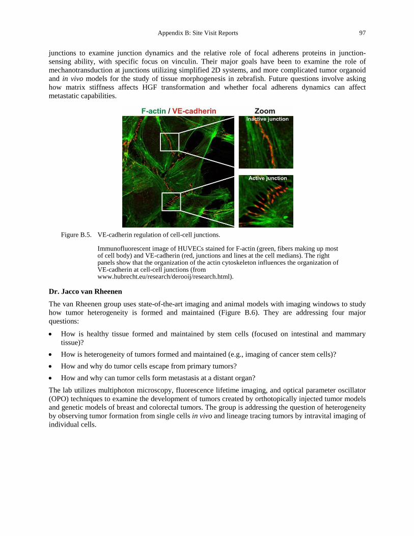
Appendix B: Site Visit Reports 97
junctions to examine junction dynamics and the relative role of focal adherens proteins in junction-sensing ability, with specific focus on vinculin. Their major goals have been to examine the role of mechanotransduction at junctions utilizing simplified 2D systems, and more complicated tumor organoid and in vivo models for the study of tissue morphogenesis in zebrafish. Future questions involve asking how matrix stiffness affects HGF transformation and whether focal adherens dynamics can affect metastatic capabilities.
Figure B.5. VE-cadherin regulation of cell-cell junctions.
Immunofluorescent image of HUVECs stained for F-actin (green, fibers making up most of cell body) and VE-cadherin (red, junctions and lines at the cell medians). The right panels show that the organization of the actin cytoskeleton influences the organization of VE-cadherin at cell-cell junctions (from www.hubrecht.eu/research/derooij/research.html).
Dr. Jacco van Rheenen
The van Rheenen group uses state-of-the-art imaging and animal models with imaging windows to study how tumor heterogeneity is formed and maintained (Figure B.6). They are addressing four major questions:
• How is healthy tissue formed and maintained by stem cells (focused on intestinal and mammary tissue)?
• How is heterogeneity of tumors formed and maintained (e.g., imaging of cancer stem cells)? • How and why do tumor cells escape from primary tumors? • How and why can tumor cells form metastasis at a distant organ? The lab utilizes multiphoton microscopy, fluorescence lifetime imaging, and optical parameter oscillator (OPO) techniques to examine the development of tumors created by orthotopically injected tumor models and genetic models of breast and colorectal tumors. The group is addressing the question of heterogeneity by observing tumor formation from single cells in vivo and lineage tracing tumors by intravital imaging of individual cells.
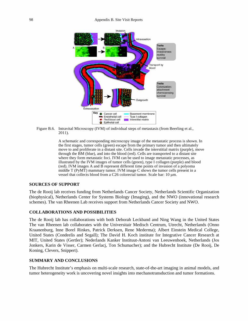
98 Appendix B. Site Visit Reports
Figure B.6. Intravital Microscopy (IVM) of individual steps of metastasis (from Beerling et al.,
2011).
A schematic and corresponding microscopy image of the metastatic process is shown. In the first stages, tumor cells (green) escape from the primary tumor and then ultimately move to and proliferate in a distant site. Cells invade the interstitial matrix (purple), move through the BM (blue), and into the blood (red). Cells are transported to a distant site where they form metastatic foci. IVM can be used to image metastatic processes, as illustrated by the IVM images of tumor cells (green), type I collagen (purple) and blood (red). IVM images A and B represent different time points of invasion of a polyoma middle T (PyMT) mammary tumor. IVM image C shows the tumor cells present in a vessel that collects blood from a C26 colorectal tumor. Scale bar: 10 μm.
SOURCES OF SUPPORT
The de Rooij lab receives funding from Netherlands Cancer Society, Netherlands Scientific Organization (biophysical), Netherlands Center for Systems Biology (Imaging), and the NWO (innovational research schemes). The van Rheenen Lab receives support from Netherlands Cancer Society and NWO.
COLLABORATIONS AND POSSIBILITIES
The de Rooij lab has collaborations with both Deborah Leckband and Ning Wang in the United States The van Rheenen lab collaborates with the Universitair Medisch Centrum, Utrecht, Netherlands (Onno Kraanenburg, Inne Borel Rinkes, Patrick Derksen, Rene Mederma); Albert Einstein Medical College, United States (Condeelis and Segall); The David H. Koch institute for Integrative Cancer Research at MIT, United States (Gertler); Nederlands Kanker Instituut-Antoni van Leeuwenhoek, Netherlands (Jos Jonkers, Karin de Visser, Carmen Gerlacj, Ton Schumacher); and the Hubrecht Institute (De Rooij, De Koning, Clevers, Snippert).
SUMMARY AND CONCLUSIONS
The Hubrecht Institute’s emphasis on multi-scale research, state-of-the-art imaging in animal models, and tumor heterogeneity work is uncovering novel insights into mechanotransduction and tumor formations.
Appendix B: Site Visit Reports 99
REFERENCES Barker, N., Huch, M., Kujala, P., van de Wetering, M., Snippert, H.J., van Es, J.H., Sato, T., Stange, D.E., Begthel, H., van
den Born, M., et al. (2010). Lgr5(+ve) stem cells drive self-renewal in the stomach and build long-lived gastric units in vitro. Cell Stem Cell 6, 25-36.
Beerling, E., Ritsma, L., Vrisekoop, N., Derksen, P.W., and van Rheenen, J. (2011). Intravital microscopy: new insights into metastasis of tumors. J Cell Sci 124, 299-310.
Itzkovitz, S., Blat, I.C., Jacks, T., Clevers, H., and van Oudenaarden, A. (2012). Optimality in the development of intestinal crypts. Cell 148, 608-619.
le Duc, Q., Shi, Q., Blonk, I., Sonnenberg, A., Wang, N., Leckband, D., and de Rooij, J. (2010). Vinculin potentiates E-cadherin mechanosensing and is recruited to actin-anchored sites within adherens junctions in a myosin II-dependent manner. J Cell Biol 189, 1107-1115.
Sato, T., Vries, R.G., Snippert, H.J., van de Wetering, M., Barker, N., Stange, D.E., van Es, J.H., Abo, A., Kujala, P., Peters, P.J., et al. (2009). Single Lgr5 stem cells build crypt-villus structures in vitro without a mesenchymal niche. Nature 459, 262-265.
Snippert, H.J., Haegebarth, A., Kasper, M., Jaks, V., van Es, J.H., Barker, N., van de Wetering, M., van den Born, M., Begthel, H., Vries, R.G., et al. (2010). Lgr6 marks stem cells in the hair follicle that generate all cell lineages of the skin. Science 327, 1385-1389.
Zomer, A., Beerling, E., Vlug, E.J., and van Rheenen, J. (2011). Real-time intravital imaging of cancer models. Clin Transl Oncol 13, 848-854.
www.hubrecht.eu/ www.hubrecht.eu/research/index.html www.hubrecht.eu/research/clevers/research.html www.hubrecht.eu/research/derooij/research.html www.hubrecht.eu/research/vanrheenen/research.html www.hubrecht.eu/research/vanoudenaarden/index.html
100 Appendix B. Site Visit Reports
Institut Curie
Site Address: 26 rue d’Ulm 75248 Paris Cedex 05, FRANCE http://curie.fr/en
Date Visited: 9 May 2012 WTEC Attendees: Paul Janmey, Cindy Reinhart-King, Dan Fletcher (site report author), Jerry Lee, Nastaran
Kuhn, and Hemant Sarin Host(s): Daniel Louvard
Group Leader of Morphogenesis and Signaling Cell Director of Research at Curie Institute Email: [email protected]
Jean-François Joanny Physical Approach of Biological Problems Professor, Exceptional Class University Email: [email protected] +33 1 56 24 67 55
Jacques Prost Physical Approach of Biological Problems Research Director, Exceptional Class CNRS Email: [email protected] Tel: +33 1 56 24 64 72
Bruno Goud Group Leader of the Molecular Mechanisms of Intracellular Transport Email: [email protected] +33 1 56 24 63 98
Jean-Louis Viovy Group Leader of Macromolecules and Microsystems in Biology and Medicine Email: [email protected] Tel: +33 1 56 24 67 52
OVERVIEW
The Curie Institute, founded in 1909, is a recognized public utility foundation. It is solely devoted to multidisciplinary cancer research and treatment, and brings together over 3,000 researchers, physicians, and caregivers for this purpose. Being the birthplace of radiotherapy, it continues to be an innovator for techniques in high-precision radiation therapy, proton therapy, brachytherapy, imaging, and oncogenetics. Its highly-recognized Proton Therapy Centre is located in Orsay and is perfectly adapted for radiotherapy of childhood tumors. Being one of the largest European research centers in cancer, it is composed of 84 teams and 15 units with platforms available for advanced cell imaging, bioinformatics, genomics, and proteomics. Since 1993, Prof. Daniel Louvard has been the director of research of the Curie Institute, where he has also functions as head of the morphogenesis and cell signaling team in the 144 UMR CNRS - Institut Curie.
Appendix B: Site Visit Reports 101
RESEARCH AND DEVELOPMENT ACTIVITIES
Louvard gave an overview of the history and organization of research at the Curie Institute. His priorities for strengthening the Institute include: 1) creating incentives for collaboration; 2) training young researchers; and 3) establishing core facilities. The success of these efforts in establishing the Curie Institute as one of the premier research institution at the intersection of physical science and biology was evident in both the research we heard about and the facilities we toured. Jean-François Joanny is head of physical chemistry unit (UMR 168) at the Curie Institute and a theoretician working on physical approaches to biological problems. Bruno Goud is head of the cell biology unit (UMR 144) and is working on molecular mechanisms of intracellular transport. Joanny and Goud presented an overview of activities in the physical chemistry and cell biology units, both of which are characterized by a highly interdisciplinary group of researchers with expertise in soft matter physics, biochemistry, cell biology, and biophysics (Figure B.7). Major initiatives include CelTisPhyBio and LabEx. The research in cell biology and physical chemistry is supported by an integrated effort to advance interactions between cell and tissue biology and physical sciences known as “CelTisPhyBio.” Researchers at the Curie have pioneered the idea that homeostatic pressure in tissues and tumors regulates their growth. They have developed a theory to describe this homeostatic pressure. Researchers are also pursuing vesicle-based reconstitution of membrane remodeling, curvature sensing, and artificial cell contraction, which is helping to identify fundamental mechanisms that animate cells. A relatively new research direction involves studying the collective behavior of cell aggregates, which show spreading and wetting behaviors that are dependent on cell-cell and cell-matrix adhesion properties. Jean-Louis Viovy is the leader of the group studying macromolecules and microsystems in biology and medicine. He described the development of microfluidic technologies for using basic biological research and clinical applications. Viovy has developed a pressure-based flow control system with switching times under 100ms that is being commercialized by Fluigent. Viovy also described the development of manufacturing technologies, including nanoparticle assembly, magnetic particle arrays, and template self-assembly. To support further development of microfluidic technologies, the de Genne Institute for Microfluidics will be opening in January 2014 and will provide new clean room space for microfluidics research projects. Clinical applications of microfluidics that are underway at the Curie Institute include genetic testing using “FISH in chips” and identification of circulating tumor cells with microfluidic devices. Our tour of the Curie Institute included: 1) Joanny’s theoretical group, where Edouard Hannezo described mechanical models of morphogenesis and tumorigenesis; 2) the laboratory of Pierre Nassoy, where he demonstrated the encapsulation of cells in alginate; 3) the laboratory of Francoise Brochard, where she described the behavior of multi-cellular aggregates; and 4) the core Nikon Imaging facility, with an extensive set of microscopes available for Curie Institute researchers.
TRANSLATIONAL EFFORTS
Close interactions exist between the clinical units at the Curie Institute and the basic research units. These interactions are fostered by internal funding.
SOURCES OF SUPPORT
Researchers at the Curie Institute are very successful at winning European funding, as well as French government funding. Internal funds at the Curie Institute are used to foster collaborations.
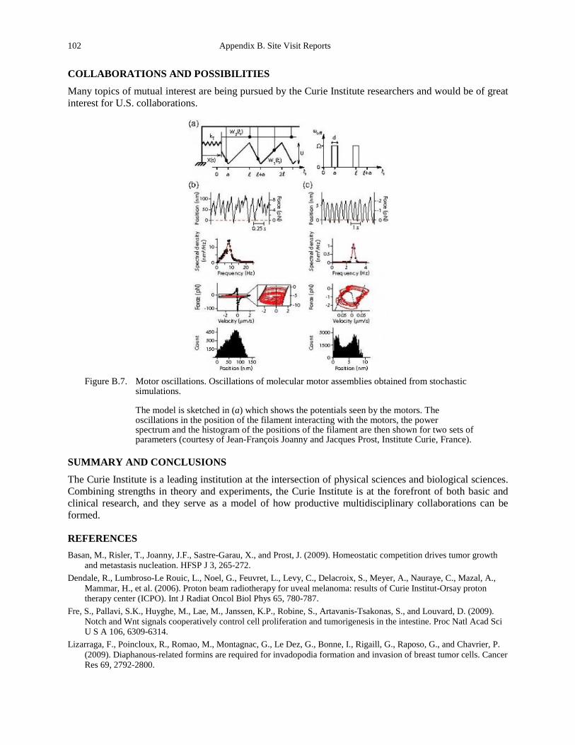
102 Appendix B. Site Visit Reports
COLLABORATIONS AND POSSIBILITIES
Many topics of mutual interest are being pursued by the Curie Institute researchers and would be of great interest for U.S. collaborations.
Figure B.7. Motor oscillations. Oscillations of molecular motor assemblies obtained from stochastic
simulations.
The model is sketched in (a) which shows the potentials seen by the motors. The oscillations in the position of the filament interacting with the motors, the power spectrum and the histogram of the positions of the filament are then shown for two sets of parameters (courtesy of Jean-François Joanny and Jacques Prost, Institute Curie, France).
SUMMARY AND CONCLUSIONS
The Curie Institute is a leading institution at the intersection of physical sciences and biological sciences. Combining strengths in theory and experiments, the Curie Institute is at the forefront of both basic and clinical research, and they serve as a model of how productive multidisciplinary collaborations can be formed.
REFERENCES Basan, M., Risler, T., Joanny, J.F., Sastre-Garau, X., and Prost, J. (2009). Homeostatic competition drives tumor growth
and metastasis nucleation. HFSP J 3, 265-272. Dendale, R., Lumbroso-Le Rouic, L., Noel, G., Feuvret, L., Levy, C., Delacroix, S., Meyer, A., Nauraye, C., Mazal, A.,
Mammar, H., et al. (2006). Proton beam radiotherapy for uveal melanoma: results of Curie Institut-Orsay proton therapy center (ICPO). Int J Radiat Oncol Biol Phys 65, 780-787.
Fre, S., Pallavi, S.K., Huyghe, M., Lae, M., Janssen, K.P., Robine, S., Artavanis-Tsakonas, S., and Louvard, D. (2009). Notch and Wnt signals cooperatively control cell proliferation and tumorigenesis in the intestine. Proc Natl Acad Sci U S A 106, 6309-6314.
Lizarraga, F., Poincloux, R., Romao, M., Montagnac, G., Le Dez, G., Bonne, I., Rigaill, G., Raposo, G., and Chavrier, P. (2009). Diaphanous-related formins are required for invadopodia formation and invasion of breast tumor cells. Cancer Res 69, 2792-2800.

Appendix B: Site Visit Reports 103
Schauer, K., Duong, T., Bleakley, K., Bardin, S., Bornens, M., and Goud, B. (2010). Probabilistic density maps to study global endomembrane organization. Nat Methods 7, 560-566.
http://curie.fr/en http://protontherapie.curie.fr/fr/institut-curie http://umr144.curie.fr/fr/les-equipes/mecanismes-moleculaires-du-transport-intracellulaire-bruno-goud/mecanismes-
moleculaires- http://umr144.curie.fr/fr/equipes-de-recherche/morphogenese-et-signalisation-cellulaires-daniel-louvard/morphogenese-et-
signal
104 Appendix B. Site Visit Reports
Max Planck Society, Dresden
Site Address: Max Planck Institute for the Physics of Complex Systems Dresden (meeting at the University of Leipzig) Linnéstrasse 5 04103 Leipzig, GERMANY www.mpg.de/155526/physik_komplexer_systeme
Date Visited: 11 May 2012 WTEC Attendees: Paul Janmey, Cindy Reinhart-King (site report author), Dan Fletcher, Lance Munn, and
Hemant Sarin Host(s): Dr. Guillaume Salbreux, Group Leader
Physics of the Cytoskeleton Max Planck Institute for the Physics of Complex Systems Nöthnitzer Strasse 38 01187 Dresden, GERMANY Email: [email protected] Tel: +49 351 871 1214
OVERVIEW
The Max Planck Society (MPG) operates a number of research institutions in Germany and internationally. These institutes are independent and autonomous in the selection and conduct of their research pursuits as long as they meet MPG’s excellence criteria. The Max Planck Institutes (MPI) carry out basic research in the life sciences, natural sciences, and the social and human sciences. In 1999, MPG launched the Inter-Institutional Research Initiatives program to promote the interdisciplinary basic research amongst its institutes, including the sharing of laboratory infrastructures.
One of the projects under the Inter-Institutional Research Initiatives program is the “identification of clinical predictive markers and drug development by large-scale translational genomic analysis of lung adenocarcinoma” (2009-2015), in which the Lung Cancer Genome Project (CLCGP), the MPI for Neurological Research (Cologne), and the MPI of Biochemistry (Martinsried) collaborate. It will undertake an in-depth analytical characterization of 600 adenocarcinomas genomes to identify new predicative and prognostic markers and therapeutic targets.
RESEARCH AND DEVELOPMENT ACTIVITIES
We met with Guillaume Salbreux who works in the MPI for Physics of Complex Systems directed by Frank Jülicher. They have a joint research program with MPI Molecular Cell Biology and Genetics, which is also located in Dresden. The Center for Systems Biology is expanding and getting a new building. They want to add informatics and image analysis to the already strong experimental and theoretical activities. The biological physics department of the MPI for Physics of Complex Systems is interested in studying the molecular, cellular, and collective behaviors of molecules, cells, and tissues with four main research thrusts:
• Active molecular processes (force generation) • Collective behaviors of motors and filaments (movements and flows) • Spatiotemporal processes in cells (cell shape control)
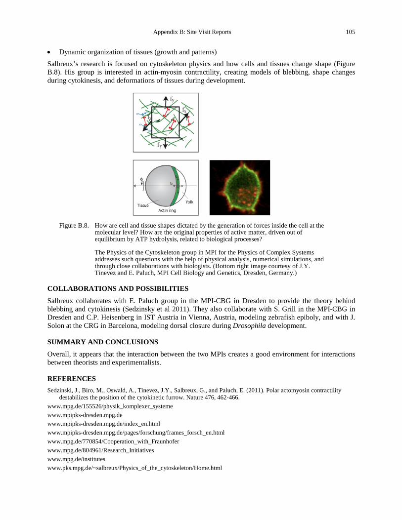
Appendix B: Site Visit Reports 105
• Dynamic organization of tissues (growth and patterns) Salbreux’s research is focused on cytoskeleton physics and how cells and tissues change shape (Figure B.8). His group is interested in actin-myosin contractility, creating models of blebbing, shape changes during cytokinesis, and deformations of tissues during development.
Figure B.8. How are cell and tissue shapes dictated by the generation of forces inside the cell at the
molecular level? How are the original properties of active matter, driven out of equilibrium by ATP hydrolysis, related to biological processes?
The Physics of the Cytoskeleton group in MPI for the Physics of Complex Systems addresses such questions with the help of physical analysis, numerical simulations, and through close collaborations with biologists. (Bottom right image courtesy of J.Y. Tinevez and E. Paluch, MPI Cell Biology and Genetics, Dresden, Germany.)
COLLABORATIONS AND POSSIBILITIES
Salbreux collaborates with E. Paluch group in the MPI-CBG in Dresden to provide the theory behind blebbing and cytokinesis (Sedzinsky et al 2011). They also collaborate with S. Grill in the MPI-CBG in Dresden and C.P. Heisenberg in IST Austria in Vienna, Austria, modeling zebrafish epiboly, and with J. Solon at the CRG in Barcelona, modeling dorsal closure during Drosophila development.
SUMMARY AND CONCLUSIONS
Overall, it appears that the interaction between the two MPIs creates a good environment for interactions between theorists and experimentalists.
REFERENCES Sedzinski, J., Biro, M., Oswald, A., Tinevez, J.Y., Salbreux, G., and Paluch, E. (2011). Polar actomyosin contractility
destabilizes the position of the cytokinetic furrow. Nature 476, 462-466. www.mpg.de/155526/physik_komplexer_systeme www.mpipks-dresden.mpg.de www.mpipks-dresden.mpg.de/index_en.html www.mpipks-dresden.mpg.de/pages/forschung/frames_forsch_en.html www.mpg.de/770854/Cooperation_with_Fraunhofer www.mpg.de/804961/Research_Initiatives www.mpg.de/institutes www.pks.mpg.de/~salbreux/Physics_of_the_cytoskeleton/Home.html
106 Appendix B. Site Visit Reports
Max Planck Institute for Dynamics and Self-Organization, Göttingen
Site Address: (Meeting at University of Leipzig) Linnéstrasse 5 04103 Leipzig, GERMANY www.ds.mpg.de/english/research/index.php
Date Visited: 11 May 2012 WTEC Attendees: Paul Janmey, Cindy Reinhart-King (site report author), Dan Fletcher, Lance Munn, and
Hemant Sarin Host(s): Oskar Hallatschek
Biological Physics and Evolutionary Dynamics Max Planck Research Group Max Planck Institute for Dynamics and Self-Organization Bunsenstrasse 10 D-37073 Göttingen, GERMANY +49 0 551 5176 670 [email protected]
OVERVIEW
The Max Planck Institute for Dynamics and Self-Organization, Göttingen, houses three specialized departments and several researcher groups studying the physical principles underlying biological interactions. The departments include the theory department of Theo Geisel (non-linear dynamics), and experimental departments of Eberhard Bodenschatz (fluid dynamics, pattern formation, and biocomplexity) and Stephan Herminghaus (dynamics of complex fluids). The theory department focuses on theoretical and computational neuroscience, nonlinear dynamics, and transport phenomena in complex systems. Bodenschatz’s experimental department investigates turbulence and other pattern formation phenomena in fluids, the physics of clouds, and self-organization in biological systems. In Herminghaus’ experimental department, dry and wet granular systems are studied as paradigm systems far from thermal equilibrium, as well as self-organization in emulsions, soft autonomous microsystems, and geophysical systems. The research program of these three departments is complemented by independent groups, with research on dynamical networks (Marc Timme); turbulence in shear flows (Björn Hof); evolution on the cellular scale (Oskar Hallatschek); transport phenomena in emulsion systems (Jean-Christophe Baret); self-organized collective behavior of heart muscle cells (Stefan Luther), theoretical neurophysics (Fred Wolf); and polymers, complex fluids, and disordered systems (Annette Zippelius).
RESEARCH AND DEVELOPMENT ACTIVITIES
While in Leipzig, we met with Dr. Oskar Hallatschek, the Group Leader of Biological Physics and Evolutionary Dynamics at the Max Planck Institute for Dynamics and Self-Organization. His presentation topic was “From colony growth to tumor growth.” He is interested in range expansions—the movement of populations to different areas where they evolve separately. These patterns allow investigation of reproductive noise. This was first done in bacteria with Dr. David Nelson, but has been adapted to colon cancer and clonal expansion in neoplastic tissues. In these cases, the work allows for mutations to come in
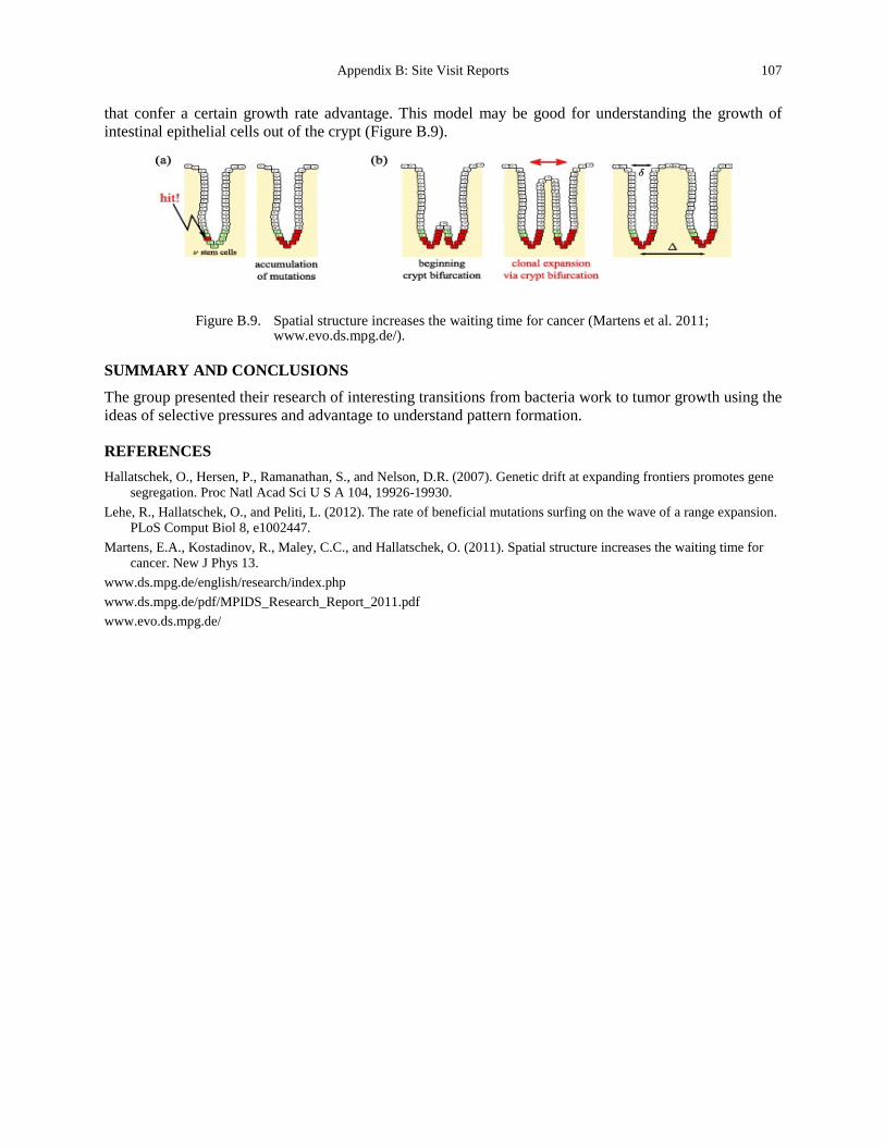
Appendix B: Site Visit Reports 107
that confer a certain growth rate advantage. This model may be good for understanding the growth of intestinal epithelial cells out of the crypt (Figure B.9).
Figure B.9. Spatial structure increases the waiting time for cancer (Martens et al. 2011;
www.evo.ds.mpg.de/).
SUMMARY AND CONCLUSIONS
The group presented their research of interesting transitions from bacteria work to tumor growth using the ideas of selective pressures and advantage to understand pattern formation.
REFERENCES Hallatschek, O., Hersen, P., Ramanathan, S., and Nelson, D.R. (2007). Genetic drift at expanding frontiers promotes gene
segregation. Proc Natl Acad Sci U S A 104, 19926-19930. Lehe, R., Hallatschek, O., and Peliti, L. (2012). The rate of beneficial mutations surfing on the wave of a range expansion.
PLoS Comput Biol 8, e1002447. Martens, E.A., Kostadinov, R., Maley, C.C., and Hallatschek, O. (2011). Spatial structure increases the waiting time for
cancer. New J Phys 13. www.ds.mpg.de/english/research/index.php www.ds.mpg.de/pdf/MPIDS_Research_Report_2011.pdf www.evo.ds.mpg.de/
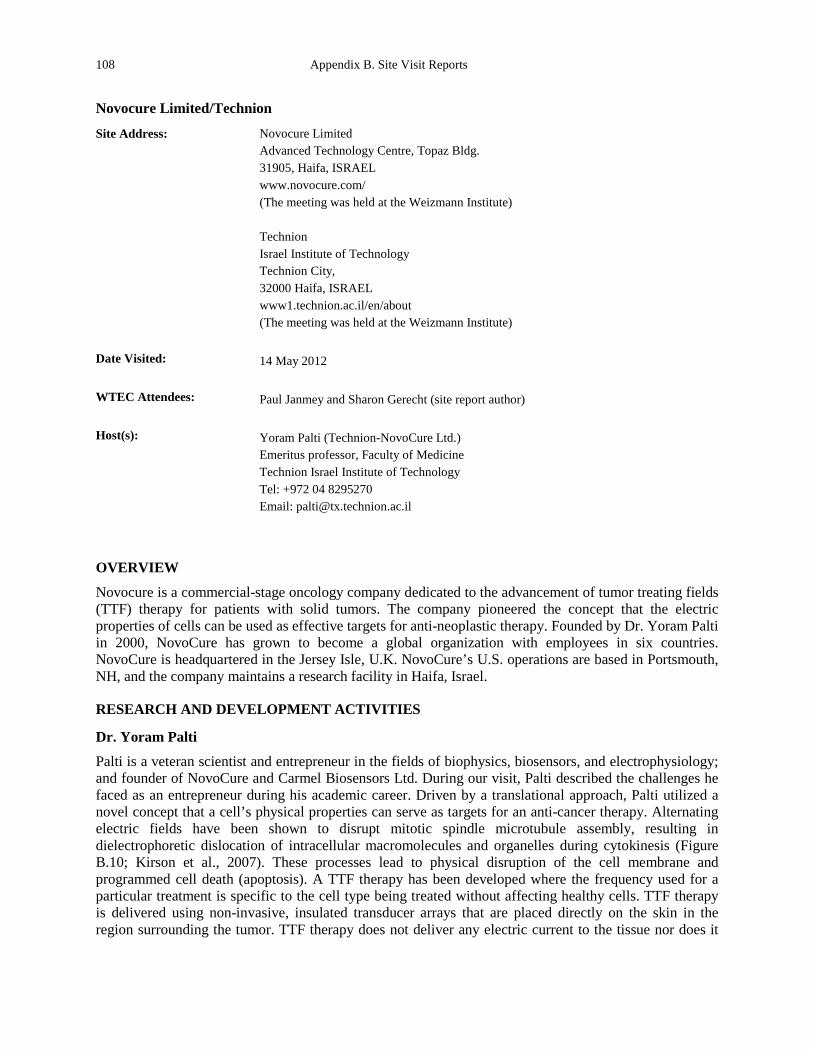
108 Appendix B. Site Visit Reports
Novocure Limited/Technion
Site Address: Novocure Limited Advanced Technology Centre, Topaz Bldg. 31905, Haifa, ISRAEL www.novocure.com/ (The meeting was held at the Weizmann Institute) Technion Israel Institute of Technology Technion City, 32000 Haifa, ISRAEL www1.technion.ac.il/en/about (The meeting was held at the Weizmann Institute)
Date Visited: 14 May 2012 WTEC Attendees: Paul Janmey and Sharon Gerecht (site report author) Host(s): Yoram Palti (Technion-NovoCure Ltd.)
Emeritus professor, Faculty of Medicine Technion Israel Institute of Technology Tel: +972 04 8295270 Email: [email protected]
OVERVIEW
Novocure is a commercial-stage oncology company dedicated to the advancement of tumor treating fields (TTF) therapy for patients with solid tumors. The company pioneered the concept that the electric properties of cells can be used as effective targets for anti-neoplastic therapy. Founded by Dr. Yoram Palti in 2000, NovoCure has grown to become a global organization with employees in six countries. NovoCure is headquartered in the Jersey Isle, U.K. NovoCure’s U.S. operations are based in Portsmouth, NH, and the company maintains a research facility in Haifa, Israel.
RESEARCH AND DEVELOPMENT ACTIVITIES
Dr. Yoram Palti
Palti is a veteran scientist and entrepreneur in the fields of biophysics, biosensors, and electrophysiology; and founder of NovoCure and Carmel Biosensors Ltd. During our visit, Palti described the challenges he faced as an entrepreneur during his academic career. Driven by a translational approach, Palti utilized a novel concept that a cell’s physical properties can serve as targets for an anti-cancer therapy. Alternating electric fields have been shown to disrupt mitotic spindle microtubule assembly, resulting in dielectrophoretic dislocation of intracellular macromolecules and organelles during cytokinesis (Figure B.10; Kirson et al., 2007). These processes lead to physical disruption of the cell membrane and programmed cell death (apoptosis). A TTF therapy has been developed where the frequency used for a particular treatment is specific to the cell type being treated without affecting healthy cells. TTF therapy is delivered using non-invasive, insulated transducer arrays that are placed directly on the skin in the region surrounding the tumor. TTF therapy does not deliver any electric current to the tissue nor does it
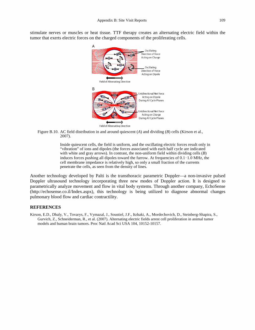
Appendix B: Site Visit Reports 109
stimulate nerves or muscles or heat tissue. TTF therapy creates an alternating electric field within the tumor that exerts electric forces on the charged components of the proliferating cells.
Figure B.10. AC field distribution in and around quiescent (A) and dividing (B) cells (Kirson et al.,
2007).
Inside quiescent cells, the field is uniform, and the oscillating electric forces result only in “vibration” of ions and dipoles (the forces associated with each half cycle are indicated with white and gray arrows). In contrast, the non-uniform field within dividing cells (B) induces forces pushing all dipoles toward the furrow. At frequencies of 0.1–1.0 MHz, the cell membrane impedance is relatively high, so only a small fraction of the currents penetrate the cells, as seen from the density of lines.
Another technology developed by Palti is the transthoracic parametric Doppler—a non-invasive pulsed Doppler ultrasound technology incorporating three new modes of Doppler action. It is designed to parametrically analyze movement and flow in vital body systems. Through another company, EchoSense (http://echosense.co.il/Index.aspx), this technology is being utilized to diagnose abnormal changes pulmonary blood flow and cardiac contractility.
REFERENCES Kirson, E.D., Dbaly, V., Tovarys, F., Vymazal, J., Soustiel, J.F., Itzhaki, A., Mordechovich, D., Steinberg-Shapira, S.,
Gurvich, Z., Schneiderman, R., et al. (2007). Alternating electric fields arrest cell proliferation in animal tumor models and human brain tumors. Proc Natl Acad Sci USA 104, 10152-10157.
110 Appendix B. Site Visit Reports
St Radboud University Nijmegen Medical Center
Site Address: Geert Grooteplein 26-28 6525 GA Nijmegen, NETHERLANDS www.umcn.nl/Pages/default.aspx
Date Visited: 7 May 2012 WTEC Attendees: Paul Janmey (site report author), Cindy Reinhart-King, Dan Fletcher, Nastaran Kuhn,
Nicole Moore, and Hemant Sarin Host(s): Peter Friedl
Professor, Department of Cell Biology Chair, Microscopic Imaging of the Cell Microscopic Imaging Centre Email: [email protected] Tel: +31 24 36 10 907
Wilhelm T.S. Huck Professor, Faculty of Science Group Leader of Physical Organic Chemistry Email: [email protected] Tel: +31 24 36 52 676
Katarina Wolf Scientific Researcher Email: [email protected] Tel: +31 24 36 14 296
OVERVIEW
The mission of the Radboud University Nijmegen Medical Centre (RUNMC) is to advance human knowledge by conducting biomedical, translational, and clinical research. The center’s major strengths are in medical life sciences and clinical practice. RUNMC is made up of departments as well as several transdisciplinary institutes, which include the Research Institute for Oncology; Nijmegen Institute for Infection, Inflammation, and Immunity; Institute for Genetic and Metabolic Disease; Donders Centre for Neuroscience; Nijmegen Centre of Molecular Life Sciences; and Nijmegen Centre for Evidence-Based Practice. The major areas of research in the center’s department of cell biology include cellular mobility, metabolism and immunity, cancer cell metastasis and invasion, and mechanisms of neurodegenerative disease. Its infrastructure is comprised of state-of-the-art technology platforms and translational research facilities. These include the Microscopic Imaging Centre (MIC) for fluorescence and electron microscopy of basic cellular processes. It also works in close collaboration with the Preclinical Imaging Centre (PRIME), which is a partnership between the clinical departments of radiology, nuclear medicine, cell biology and rheumatology of the Medical Center (Bakker et al., 2012; Gritsenko et al., 2012).
RESEARCH AND DEVELOPMENT ACTIVITIES
Drs. Peter Friedl and Katarina Wolf
Friedl is chair for Microscopical Imaging of the Cell at the Nijmegen Centre of Molecular Life Sciences (NCMLS) which includes the core facility for microscopy at RUNMC. He also heads the cell dynamics lab and has a joint-faculty position as head of the imaging section at the David H. Koch Center,
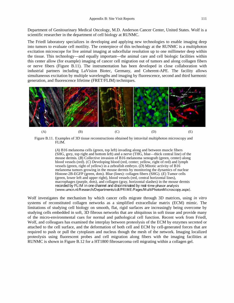
Appendix B: Site Visit Reports 111
Department of Genitourinary Medical Oncology, M.D. Anderson Cancer Center, United States. Wolf is a scientific researcher in the department of cell biology at RUNMC.
The Friedl laboratory specializes in developing and applying new technologies to enable imaging deep into tumors to evaluate cell motility. The centerpiece of this technology at the RUNMC is a multiphoton excitation microscope for live animal imaging at subcellular resolution up to one millimeter deep within the tissue. This technology—and equally important—the animal care and cell biologic facilities within this center allow (for example) imaging of cancer cell migration out of tumors and along collagen fibers or nerve fibers (Figure B.11). The instrumentation has been developed in close collaboration with industrial partners including LaVision Biotec, Germany, and Coherent-APE. The facility allows simultaneous excitation by multiple wavelengths and imaging by fluorescence, second and third harmonic generation, and fluorescence lifetime (FRET/FLIM) techniques.
(A) (B) (C) (D) (E)
Figure B.11. Examples of 3D tissue reconstructions obtained by intravital multiphoton microscopy and FLIM.
(A) B16 melanoma cells (green, top left) invading along and between muscle fibers (SHG, grey, top right and bottom left) and a nerve (THG, blue—thick central line) of the mouse dermis. (B) Collective invasion of B16 melanoma xenograft (green, center) along blood vessels (red). (C) Developing blood (red, center; yellow, right of red) and lymph vessels (green, right of yellow) in a zebrafish embryo. (D) Mitotic activity of B16 melanoma tumors growing in the mouse dermis by monitoring the dynamics of nuclear Histone-2B-EGFP (green, dots). Blue (lines): collagen fibers (SHG). (E) Tumor cells (green, lower left and upper right), blood vessels (red, central horizontal lines), macrophages (purple, dots), and collagen (gray, horizontal slashes) in the mouse dermis recorded by FLIM in one channel and discriminated by real-time phasor analysis (www.umcn.nl/R esearch/Departments/cdl/PR IME /Pages/MultiPhotonMicroscopy.aspx).
Wolf investigates the mechanism by which cancer cells migrate through 3D matrices, using in vitro systems of reconstituted collagen networks as a simplified extracellular matrix (ECM) mimic. The limitations of studying cell biology on smooth, flat, rigid surfaces are increasingly being overcome by studying cells embedded in soft, 3D fibrous networks that are ubiquitous in soft tissue and provide many of the micro-environmental cues for normal and pathological cell function. Recent work from Friedl, Wolf, and colleagues has examined the interplay between proteolysis of the ECM by enzymes secreted or attached to the cell surface, and the deformation of both cell and ECM by cell-generated forces that are required to push or pull the cytoplasm and nucleus though the mesh of the network. Imaging localized proteolysis using fluorescent probes and cell migration along fibers with the imaging facilities at RUNMC is shown in Figure B.12 for a HT1800 fibrosarcoma cell migrating within a collagen gel.
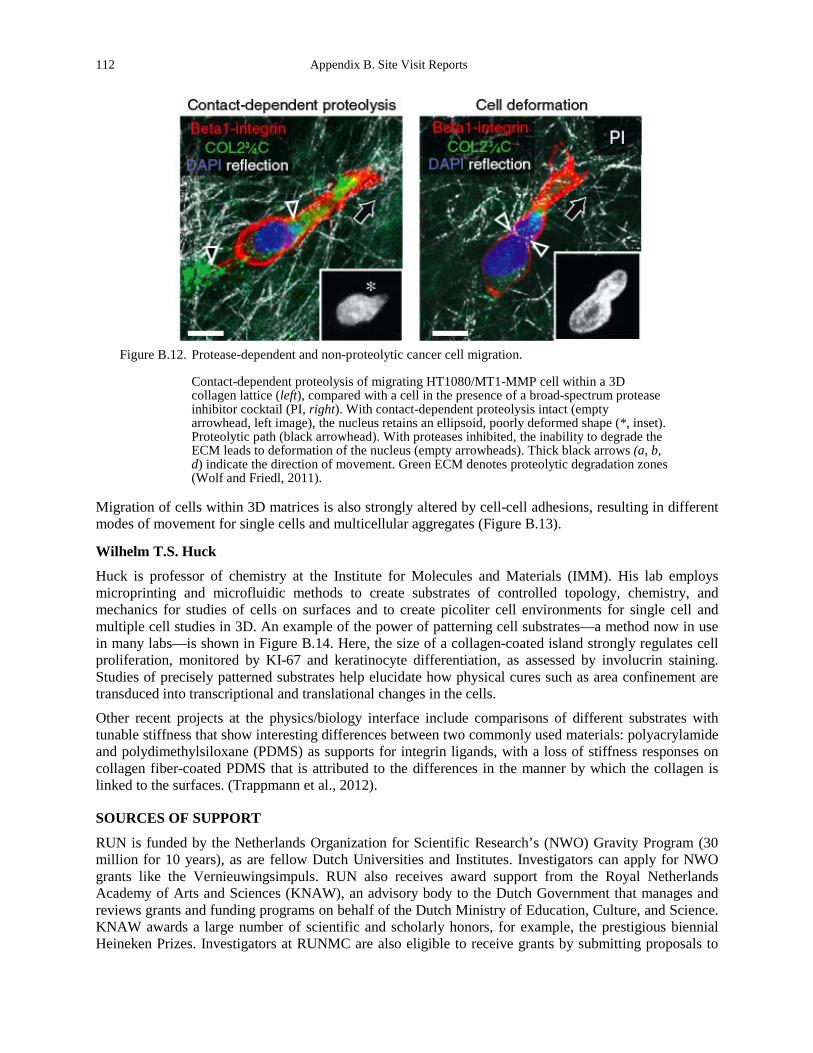
112 Appendix B. Site Visit Reports
Figure B.12. Protease-dependent and non-proteolytic cancer cell migration.
Contact-dependent proteolysis of migrating HT1080/MT1-MMP cell within a 3D collagen lattice (left), compared with a cell in the presence of a broad-spectrum protease inhibitor cocktail (PI, right). With contact-dependent proteolysis intact (empty arrowhead, left image), the nucleus retains an ellipsoid, poorly deformed shape (*, inset). Proteolytic path (black arrowhead). With proteases inhibited, the inability to degrade the ECM leads to deformation of the nucleus (empty arrowheads). Thick black arrows (a, b, d) indicate the direction of movement. Green ECM denotes proteolytic degradation zones (Wolf and Friedl, 2011).
Migration of cells within 3D matrices is also strongly altered by cell-cell adhesions, resulting in different modes of movement for single cells and multicellular aggregates (Figure B.13).
Wilhelm T.S. Huck
Huck is professor of chemistry at the Institute for Molecules and Materials (IMM). His lab employs microprinting and microfluidic methods to create substrates of controlled topology, chemistry, and mechanics for studies of cells on surfaces and to create picoliter cell environments for single cell and multiple cell studies in 3D. An example of the power of patterning cell substrates—a method now in use in many labs—is shown in Figure B.14. Here, the size of a collagen-coated island strongly regulates cell proliferation, monitored by KI-67 and keratinocyte differentiation, as assessed by involucrin staining. Studies of precisely patterned substrates help elucidate how physical cures such as area confinement are transduced into transcriptional and translational changes in the cells.
Other recent projects at the physics/biology interface include comparisons of different substrates with tunable stiffness that show interesting differences between two commonly used materials: polyacrylamide and polydimethylsiloxane (PDMS) as supports for integrin ligands, with a loss of stiffness responses on collagen fiber-coated PDMS that is attributed to the differences in the manner by which the collagen is linked to the surfaces. (Trappmann et al., 2012).
SOURCES OF SUPPORT
RUN is funded by the Netherlands Organization for Scientific Research’s (NWO) Gravity Program (30 million for 10 years), as are fellow Dutch Universities and Institutes. Investigators can apply for NWO grants like the Vernieuwingsimpuls. RUN also receives award support from the Royal Netherlands Academy of Arts and Sciences (KNAW), an advisory body to the Dutch Government that manages and reviews grants and funding programs on behalf of the Dutch Ministry of Education, Culture, and Science. KNAW awards a large number of scientific and scholarly honors, for example, the prestigious biennial Heineken Prizes. Investigators at RUNMC are also eligible to receive grants by submitting proposals to
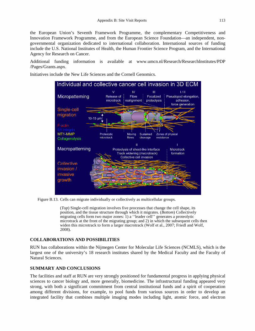
Appendix B: Site Visit Reports 113
the European Union’s Seventh Framework Programme, the complementary Competitiveness and Innovation Framework Programme, and from the European Science Foundation—an independent, non-governmental organization dedicated to international collaboration. International sources of funding include the U.S. National Institutes of Health, the Human Frontier Science Program, and the International Agency for Research on Cancer. Additional funding information is available at www.umcn.nl/Research/ResearchInstitutes/PDP /Pages/Grants.aspx.
Initiatives include the New Life Sciences and the Cornell Genomics.
Figure B.13. Cells can migrate individually or collectively as multicellular groups.
(Top) Single-cell migration involves five processes that change the cell shape, its position, and the tissue structure through which it migrates. (Bottom) Collectively migrating cells form two major zones: 1) a ‘‘leader cell’’ generates a proteolytic microtrack at the front of the migrating group; and 2) in which the subsequent cells then widen this microtrack to form a larger macrotrack (Wolf et al., 2007; Friedl and Wolf, 2008).
COLLABORATIONS AND POSSIBILITIES
RUN has collaborations within the Nijmegen Center for Molecular Life Sciences (NCMLS), which is the largest one of the university’s 18 research institutes shared by the Medical Faculty and the Faculty of Natural Sciences.
SUMMARY AND CONCLUSIONS
The facilities and staff at RUN are very strongly positioned for fundamental progress in applying physical sciences to cancer biology and, more generally, biomedicine. The infrastructural funding appeared very strong, with both a significant commitment from central institutional funds and a spirit of cooperation among different divisions, for example, to pool funds from various sources in order to develop an integrated facility that combines multiple imaging modes including light, atomic force, and electron

114 Appendix B. Site Visit Reports
microscopy, as well as magnetic resonance imaging. There also appeared to be a strong attitude of optimism for the future of this work, with researchers seeing activity at the physics/biology interface being in a rapidly growing stage both in the Netherlands and within Europe.
Figure B.14. Regulation of keratinocyte shape and differentiation on micropatterned substrates.
(a) Overview of the micropatterning strategy. (b) Immunofluorescence microscopy images of type I collagen (top) and phase-contrast microscopy images of primary human keratinocytes (bottom) on 20, 30, and 50 μm diameter islands. (c) Representative immunofluorescence microscopy images of involucrin (green) and Ki67 (red) expression on substrates with 20, 30, or 50 μm diameters at 1 h and 24 h after seeding. Scale bars, 100 μm. (d) Quantification of positive cells at 1 h and 24 h on substrates with adhesive areas ranging from 314 μm2 to 1963 μm2 (20-50 μm diameters). Data represent means ± s.e.m. (n = 4 experiments, asterisk indicates P = 0.0001, compared with the cells on substrates consisting of 50 μm diameter islands). (e) Quantification of Ki67-positive cells at 1 h and 24 h. Data represent means ± s.e.m. (n =experiments; asterisk indicates P = 0.0472, compared with the cells on substrates consisting of 50 μm diameter islands). Involucrin-positive cells at 1 h and 24 h on substrates with adhesive areas ranging from 314 μm2 to 1963 μm2 (20-50 μm diameters). Data represent means ± s.e.m. (n = 4 experiments, asterisk indicates P = 0.0001, compared with the cells on substrates consisting of 50 μm diameter islands). From Connelly et al., 2010, Micropatterning of larger islands allows for creation of multicellular aggregates with defined spatial features such as disks and toroids with specific cell-cell contacts (Gautrot et al., 2012).
Appendix B: Site Visit Reports 115
REFERENCES Alexander, S., and Friedl, P. (2012). Cancer invasion and resistance: interconnected processes of disease progression and
therapy failure. Trends in molecular medicine 18, 13-26. Bakker, G.J., Andresen, V., Hoffman, R.M., and Friedl, P. (2012). Fluorescence lifetime microscopy of tumor cell
invasion, drug delivery, and cytotoxicity. Methods in enzymology 504, 109-125. Connelly, J.T., Gautrot, J.E., Trappmann, B., Tan, D.W., Donati, G., Huck, W.T., and Watt, F.M. (2010). Actin and serum
response factor transduce physical cues from the microenvironment to regulate epidermal stem cell fate decisions. Nature cell biology 12, 711-718.
Friedl, P. (2010). To adhere or not to adhere? Nature reviews Molecular cell biology 11, 3. Friedl, P., and Alexander, S. (2011). Cancer invasion and the microenvironment: plasticity and reciprocity. Cell 147, 992-
1009. Friedl, P., and Wolf, K. (2008). Tube travel: the role of proteases in individual and collective cancer cell invasion. Cancer
Res 68, 7247-7249. Gautrot, J.E., Wang, C., Liu, X., Goldie, S.J., Trappmann, B., Huck, W.T., and Watt, F.M. (2012). Mimicking normal
tissue architecture and perturbation in cancer with engineered micro-epidermis. Biomaterials 33, 5221-5229. Gritsenko, P.G., Ilina, O., and Friedl, P. (2012). Interstitial guidance of cancer invasion. The Journal of pathology 226,
185-199. Trappmann, B., Gautrot, J.E., Connelly, J.T., Strange, D.G., Li, Y., Oyen, M.L., Cohen Stuart, M.A., Boehm, H., Li, B.,
Vogel, V., et al. (2012). Extracellular-matrix tethering regulates stem-cell fate. Nature materials 11, 642-649. Wolf, K., and Friedl, P. (2011). Extracellular matrix determinants of proteolytic and non-proteolytic cell migration. Trends
in cell biology 21, 736-744. Wolf, K., Wu, Y.I., Liu, Y., Geiger, J., Tam, E., Overall, C., Stack, M.S., and Friedl, P. (2007). Multi-step pericellular
proteolysis controls the transition from individual to collective cancer cell invasion. Nature cell biology 9, 893-904. http://www.umcn.nl/Pages/default.aspx http://www.ncmls.eu/principal-investigators/friedl/ http://www.umcn.nl/Research/Departments/cdl/PRIME/Pages/MultiPhotonMicroscopy.aspx http://www.umcn.nl/Research/Departments/cellbiology/Pages/default.aspx
116 Appendix B. Site Visit Reports
The Royal Institute of Technology (KTH)
Site Address: Life Science Technology Platform (meeting at Uppsala University) Dag Hammarskjölds väg 20 752 37 Uppsala, SWEDEN www.kth.se/en
Date Visited: 8 May 2012 WTEC Attendees: Paul Janmey, Cindy Reinhart-King, Dan Fletcher (site report author), Nastaran Kuhn,
Nicole Moore, and Hemant Sarin Host(s): Wouter van der Wijngaart
Professor, KTH School of Electrical Engineering Micro- and Nanosystems Department Osquldas väg 10, 100 44 Stockholm, SWEDEN Email: [email protected] Tel: +46 9 790 6613
Jerker Widengren Professor, Experimental Biomolecular Physics, Department of Applied Physics Email: [email protected] Tel: +46 8 55 37 8030
Ozan Öktem Associate Professor, Department of Mathematics Email: [email protected] Tel: +46 9 790 6606)
Christian Gasser Department of Solid Mechanics School of Engineering Sciences Email: [email protected] Tel: +46 8 790 7793
Jochen Schwenk Associate Professor, Platform Manager at Science for Life Laboratory Human Protein Atlas Project Email: [email protected] Tel: +46 8 524 814 82
OVERVIEW
KTH is Sweden’s premier technical university. One-third of Sweden’s technical research and engineering education takes place here (13,000 undergraduates, 4,500 post-graduates and employees, and 64 research teams). KTH’s core research strengths are categorized into five platforms: 1) energy; 2) materials; 3) life science technologies; 4) information and communication technology; and 5) transport. These platforms serve as the basis for multidisciplinary research initiatives of KTH units as well as with its external partners—which include industry, healthcare, academia, and the general public. Within Life Sciences, KTH has six focus areas: 1) bioimaging; 2) biomolecular tools and biomaterials; 3) fundamental research

Appendix B: Site Visit Reports 117
in life science; 4) medical devices; 5) mathematical and computational sciences; and 6) infrastructure in health.
RESEARCH AND DEVELOPMENT ACTIVITIES
Dr. Wouter van der Wijngaart, the Life Science Technology Platform Director, provided an overview of biomedical research in Sweden and of his research in microsystems with a focus on cancer treatment. The Stockholm-Uppsala region is the third largest medical technology research concentration in Europe, counting over 700 business and academic organizations active within the field. KTH is serving as the central academic technology partner. About 15 to 20 percent of research at KTH is focused on life science related technologies, with six focus areas (described above). These strengths at KTH support a broad range of large efforts, including the Stockholm Brain Project, the Science for Life Lab, the Centre for Technology in Medicine and Health, and the Human Protein Atlas Project. Dr. van der Wijngaart is Professor at the KTH Micro- and Nanosystems Department, one of the internationally leading academic MEMS players (Figure B.15). The Department develops, amongst others, micro- and nanotechnologies to advance and accelerate biomedical research. Examples of cancer treatment related devices under development include a lab-on-a-chip cell encapsulation system, an internal radiation therapy device, a microwave sensor for skin cancer diagnosis, and circulating tumor cell technology.
Figure B.15. Overview of the KTH Life Science Technologies Platform (Wouter van der Wijngaart,
courtesy of The Royal Institute of Technology).
118 Appendix B. Site Visit Reports
Dr. Jerker Widengren is the experimental biomolecular physics group leader and presented his recent research on ultrasensitive spectroscopy and imaging. Widengren’s research includes development and use of fluorescence correlation spectroscopy (FCS) and fluorescence cross-correlation spectroscopy (FCCS) to study molecular interactions and protein densities. A new and potentially powerful imaging method pioneered by Widengren is the use of the triplet state as a source of contrast. This method, known as TRAST, has been used to image metabolism, including O2 concentrations in live cells. Widengren is also applying his broad range of fluorescence imaging modalities to cancer diagnostics, where individual cells from fine needle aspiration samples are being analyzed with advanced spectroscopic and microscopy techniques.
Dr. Ozan Öktem from the department of mathematics gave an overview of bioimaging technologies in the life science technology platform. Regarding instrumentation, KTH has developed several medical imaging technologies that have now been commercialized, including X-ray phase contrast imaging (eXcillum) and mobile 3D SPECT (Adolesco). KTH is also developing advanced photon-counting and energy-resolving detectors. Regarding algorithms for image reconstruction and signal processing, new methods of image processing are needed to extract the most useful clinical information from these and other imaging modalities. Öktem is, together with others at the department of mathematics at KTH, developing image reconstruction and signal processing based on sparse signal processing. The department of mathematics also pursues applied research on methods for optimization of radiation therapy. This is done jointly with an industrial partner, RaySearch Laboratories. Dr. Christian Gasser in the department of solid mechanics presented his research on soft biological tissue modeling, which is aimed at addressing cardiovascular diseases, specifically abdominal aortic aneurysm. A large fraction of the population has this condition, and it is not dangerous until is ruptures, so the clinical challenge is to determine when repair is necessary. Gasser has developed an integrated rupture risk assessment method that uses CT data together with peak wall stress computations that help to guide when surgery is advisable. The mechanical modeling takes into account the collagen organization and active growth of the vessels to predict time and location of rupture. Dr. Jochen Schwenk is the platform manager for Biobank Profiling at the science for life lab (SciLifeLab) and principle investigator within the Human Protein Atlas project. He presented on the major activities of the SciLifeLab, which included genomics, RNA profiling, and bioinformatics efforts. The ambitious Human Protein Atlas provides protein expression profiles based on immunohistochemistry for a large number of human tissues, cancers, and cell lines using antibodies raised in white rabbits. Plasma profiling with antibodies is a major, yet not public effort in the Human Protein Atlas Project.
TRANSLATIONAL EFFORTS
Strong connections with industry are enabling translation of new technology. This is true for most of the medical and biomedical focused research. Furthermore, KTH is one of the driving forces in unifying the Stockholm region with respect to medical and biomedical innovations.
SOURCES OF SUPPORT
Major sources of funding include:
• The Swedish Research Council • The European Commission through the Framework Programs (FP6, FP7, Horizon 2020) • Through industrial collaborations • The Knut and Alice Wallenberg Foundation • The Swedish Human Protein Atlas Project (funded by the Knut and Alice Wallenberg Foundation).
Appendix B: Site Visit Reports 119
Within the coming years, the Science for Life Lab will be supported by industry and the Swedish national government with up to 100 M€ annually.
COLLABORATIONS AND POSSIBILITIES
The extensive data collected as part of the Human Protein Atlas Project are publically available. The large European biobanking efforts would provide an exciting opportunity for collaboration with U.S. researchers.
SUMMARY AND CONCLUSIONS
KTH is a leader in biomedical research and has a particular strength in collaborative biomedical projects. By levering new technologies and biobanking resources in Sweden, KTH is advancing the state-of the art in biomedical research.
REFERENCES Uhlen, M., Oksvold, P., Fagerberg, L., Lundberg, E., Jonasson, K., Forsberg, M., Zwahlen, M., Kampf, C., Wester, K.,
Hober, S., et al. (2010). Towards a knowledge-based Human Protein Atlas. Nature biotechnology 28, 1248-1250. www.kth.se/en www.kth.se/lifescience www.proteinatlas.org/ www.scilifelab.se www.ctmh.se
120 Appendix B. Site Visit Reports
Technical University of Munich
Site Address: James-Franck-Str. 1 85748 Garching, GERMANY http://portal.mytum.de/welcome/
Date Visited: 9 May 2012 WTEC Attendees: Sharon Gerecht, Parag Mallick (site report author), Owen McCarty, and Hassan Ali Host(s): Andreas Bausch
Department of Cellular and Molecular Biophysics; Physik-Department E27 Tel: +49 89 289 124 80 Fax: +49 89 289 144 69 Email: [email protected] http://bio.ph.tum.de/home/e27-prof-dr-bausch/research-areas.html
Franz Pfeiffer Phone: +49 89 289 12552 Fax: +49 89 289 12548 Email: [email protected] www.e17.ph.tum.de/
Martin Zacharias Physik-Department (T38) Tel: +49 89 289 12335 Email: [email protected] www.t38.physik.tu-muenchen.de/
Hendrik Dietz Laboratory for Biomolecular Nanotechnology Am Coulombwall 4a, Walter Schottky Institut/ZNN Tel: +49-89 289 11615, Fax: +49-89 289 11612, Email: [email protected] http://bionano.physik.tu-muenchen.de/coordinates.html
Friedrich C. Simmel Physics Department and ZNN/WSI Am Coulombwall 4a, Room 2.016. Tel: +49 89 289 11610 (secretary), 11611 (office) Fax: +49 89 289 11612. Email: [email protected] www.e14.ph.tum.de/index.php/group-members/friedrich-c-simmel
Thorsten Hugel Department Biophysics and Institute for Medical Engineering Tel: +49 89 289 16781 Fax: +49 89 289 10805 Email: [email protected]

Appendix B: Site Visit Reports 121
Matthias Rief
Chair of Biophysics E22 Tel: +49 89 289 12472 Fax: +49 89 289 12523 Email: [email protected] www.bio.ph.tum.de/index.php?id=44&tx_wtdirectory_pi1
OVERVIEW
The Technical University of Munich (TUM) is one of the most research-focused universities in Germany and Europe. This claim is supported by relevant rankings, such as the DFG-Förderranking (DFG Funding Rankings) or the research rankings of the Centrum für Hochschulentwicklung (CHE-Center for Higher Education Development). TUM was one of three universities which were successful in obtaining funding in all three funding lines from the Excellence Initiative in 2006. Along with the IGSSE Graduate School and TUM’s participation in five Clusters of Excellence, of which TUM is a leading institution, the strategic plan “TUM. The Entrepreneurial University” is also being developed. In addition, the university takes part in 23 collaborative research centers, of which TUM is the leading institution in nine. In the seventh European Union Research Framework Program, TUM coordinates thus far nine projects and also received six Starting Independent Researcher Grants and five Advanced Investigator Grants. The scientists we visited commented on the importance of physics in the German curriculum. Whereas many U.S. institutions typically graduate tens of students, TUM graduates hundreds per year. There is significant demand in Germany for students with physics undergraduate degrees in all sectors of industry.
In addition, TUM has been fortunate to be an active participant in Germany’s Excellence Initiative, which provides a significant funding base for diverse research as diagrammed below. Our visit focused on researchers in the physics department. There are likely additional researchers taking a physical sciences approach to biology whom we were unable to visit with due to time constraints.
RESEARCH AND DEVELOPMENT ACTIVITIES
Dr. Andreas Bausch
Bausch’s group highlighted exciting emergent properties in fibrillar self-assembly. In one example, he explored the emergence of collective motion in a high-density motility assay that consists of highly concentrated actin filaments propelled by immobilized molecular motors in a planar geometry. Above a critical density, the filaments self-organize to form coherently moving structures with persistent density modulations, such as clusters, swirls, and interconnected bands (Figure B.16). These polar nematic structures are long-lived and can span length scales orders of magnitudes larger than their constituents. This property of context-dependent state transitions is likely a general property that applies ubiquitously throughout biology. In another example, he highlighted the existence of absorbant states in natural systems. Specifically, they consider a 2D system of actin filaments. A combination of active directed motion and steric repulsion causes the system to produce dynamic patterns in the form of density fluctuations and waves; however, these are short-lived structures that appear and disappear, so the system has similarities to other fluctuating nonequilibrium liquid states. When a critical amount of protein is reached, cross-linking between actin filaments leads the system to self-organize into a distinct moving state characterized by all the hallmarks of an absorbed or dynamically frozen state. Bausch showed a third example of such a phase transition governed by pH as well.
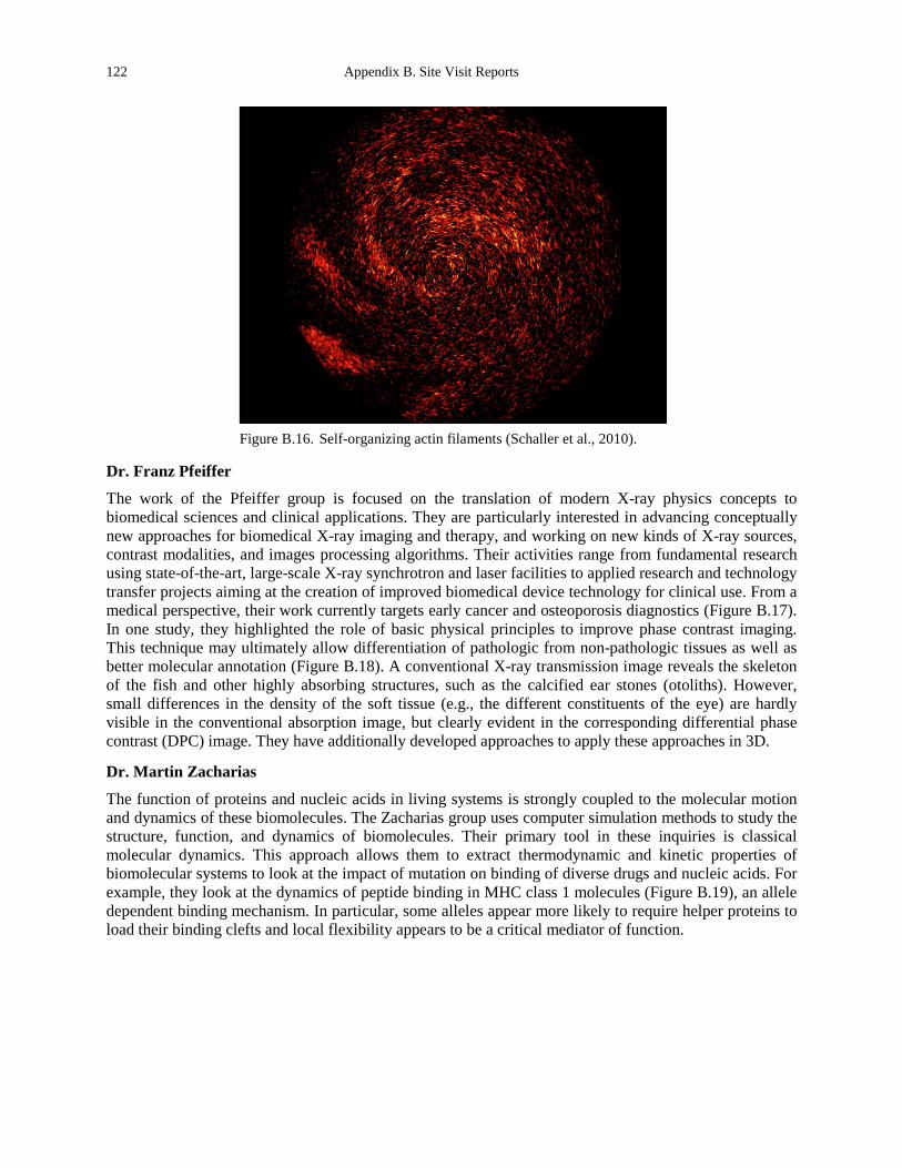
122 Appendix B. Site Visit Reports
Figure B.16. Self-organizing actin filaments (Schaller et al., 2010).
Dr. Franz Pfeiffer
The work of the Pfeiffer group is focused on the translation of modern X-ray physics concepts to biomedical sciences and clinical applications. They are particularly interested in advancing conceptually new approaches for biomedical X-ray imaging and therapy, and working on new kinds of X-ray sources, contrast modalities, and images processing algorithms. Their activities range from fundamental research using state-of-the-art, large-scale X-ray synchrotron and laser facilities to applied research and technology transfer projects aiming at the creation of improved biomedical device technology for clinical use. From a medical perspective, their work currently targets early cancer and osteoporosis diagnostics (Figure B.17). In one study, they highlighted the role of basic physical principles to improve phase contrast imaging. This technique may ultimately allow differentiation of pathologic from non-pathologic tissues as well as better molecular annotation (Figure B.18). A conventional X-ray transmission image reveals the skeleton of the fish and other highly absorbing structures, such as the calcified ear stones (otoliths). However, small differences in the density of the soft tissue (e.g., the different constituents of the eye) are hardly visible in the conventional absorption image, but clearly evident in the corresponding differential phase contrast (DPC) image. They have additionally developed approaches to apply these approaches in 3D.
Dr. Martin Zacharias
The function of proteins and nucleic acids in living systems is strongly coupled to the molecular motion and dynamics of these biomolecules. The Zacharias group uses computer simulation methods to study the structure, function, and dynamics of biomolecules. Their primary tool in these inquiries is classical molecular dynamics. This approach allows them to extract thermodynamic and kinetic properties of biomolecular systems to look at the impact of mutation on binding of diverse drugs and nucleic acids. For example, they look at the dynamics of peptide binding in MHC class 1 molecules (Figure B.19), an allele dependent binding mechanism. In particular, some alleles appear more likely to require helper proteins to load their binding clefts and local flexibility appears to be a critical mediator of function.
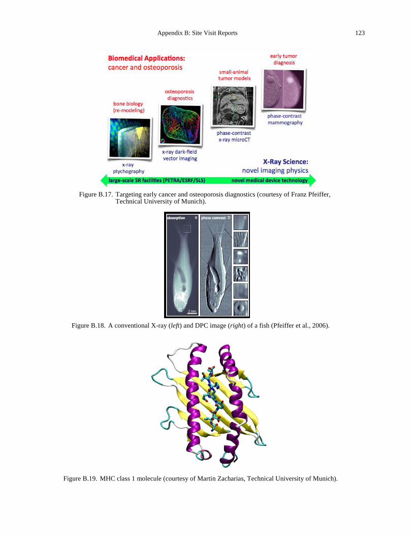
Appendix B: Site Visit Reports 123
Figure B.17. Targeting early cancer and osteoporosis diagnostics (courtesy of Franz Pfeiffer,
Technical University of Munich).
Figure B.18. A conventional X-ray (left) and DPC image (right) of a fish (Pfeiffer et al., 2006).
Figure B.19. MHC class 1 molecule (courtesy of Martin Zacharias, Technical University of Munich).
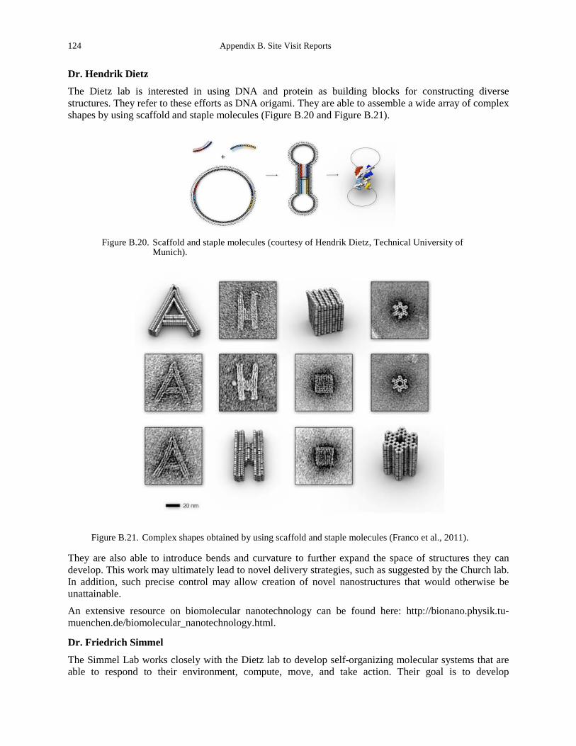
124 Appendix B. Site Visit Reports
Dr. Hendrik Dietz
The Dietz lab is interested in using DNA and protein as building blocks for constructing diverse structures. They refer to these efforts as DNA origami. They are able to assemble a wide array of complex shapes by using scaffold and staple molecules (Figure B.20 and Figure B.21).
Figure B.20. Scaffold and staple molecules (courtesy of Hendrik Dietz, Technical University of
Munich).
Figure B.21. Complex shapes obtained by using scaffold and staple molecules (Franco et al., 2011).
They are also able to introduce bends and curvature to further expand the space of structures they can develop. This work may ultimately lead to novel delivery strategies, such as suggested by the Church lab. In addition, such precise control may allow creation of novel nanostructures that would otherwise be unattainable.
An extensive resource on biomolecular nanotechnology can be found here: http://bionano.physik.tu-muenchen.de/biomolecular_nanotechnology.html.
Dr. Friedrich Simmel
The Simmel Lab works closely with the Dietz lab to develop self-organizing molecular systems that are able to respond to their environment, compute, move, and take action. Their goal is to develop
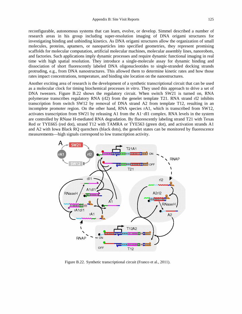
Appendix B: Site Visit Reports 125
reconfigurable, autonomous systems that can learn, evolve, or develop. Simmel described a number of research areas in his group including super-resolution imaging of DNA origami structures for investigating binding and unbinding kinetics. As DNA origami structures allow the organization of small molecules, proteins, aptamers, or nanoparticles into specified geometries, they represent promising scaffolds for molecular computation, artificial molecular machines, molecular assembly lines, nanorobots, and factories. Such applications imply dynamic processes and require dynamic functional imaging in real time with high spatial resolution. They introduce a single-molecule assay for dynamic binding and dissociation of short fluorescently labeled DNA oligonucleotides to single-stranded docking strands protruding, e.g., from DNA nanostructures. This allowed them to determine kinetic rates and how those rates impact concentrations, temperature, and binding site location on the nanostructures. Another exciting area of research is the development of a synthetic transcriptional circuit that can be used as a molecular clock for timing biochemical processes in vitro. They used this approach to drive a set of DNA tweezers. Figure B.22 shows the regulatory circuit. When switch SW21 is turned on, RNA polymerase transcribes regulatory RNA (rI2) from the genelet template T21. RNA strand rI2 inhibits transcription from switch SW12 by removal of DNA strand A2 from template T12, resulting in an incomplete promoter region. On the other hand, RNA species rA1, which is transcribed from SW12, activates transcription from SW21 by releasing A1 from the A1•dI1 complex. RNA levels in the system are controlled by RNase H-mediated RNA degradation. By fluorescently labeling strand T21 with Texas Red or TYE665 (red dot), strand T12 with TAMRA or TYE563 (green dot), and activation strands A1 and A2 with Iowa Black RQ quenchers (black dots), the genelet states can be monitored by fluorescence measurements—high signals correspond to low transcription activity.
Figure B.22. Synthetic transcriptional circuit (Franco et al., 2011).
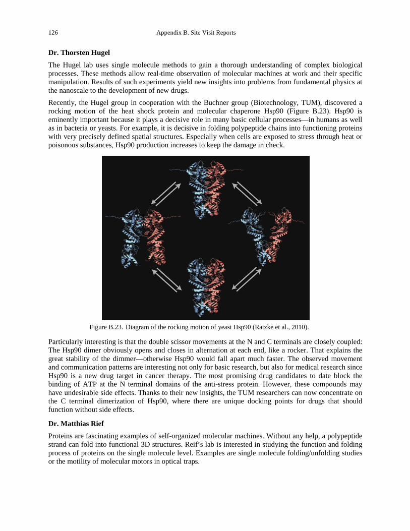
126 Appendix B. Site Visit Reports
Dr. Thorsten Hugel
The Hugel lab uses single molecule methods to gain a thorough understanding of complex biological processes. These methods allow real-time observation of molecular machines at work and their specific manipulation. Results of such experiments yield new insights into problems from fundamental physics at the nanoscale to the development of new drugs. Recently, the Hugel group in cooperation with the Buchner group (Biotechnology, TUM), discovered a rocking motion of the heat shock protein and molecular chaperone Hsp90 (Figure B.23). Hsp90 is eminently important because it plays a decisive role in many basic cellular processes—in humans as well as in bacteria or yeasts. For example, it is decisive in folding polypeptide chains into functioning proteins with very precisely defined spatial structures. Especially when cells are exposed to stress through heat or poisonous substances, Hsp90 production increases to keep the damage in check.
Figure B.23. Diagram of the rocking motion of yeast Hsp90 (Ratzke et al., 2010).
Particularly interesting is that the double scissor movements at the N and C terminals are closely coupled: The Hsp90 dimer obviously opens and closes in alternation at each end, like a rocker. That explains the great stability of the dimmer—otherwise Hsp90 would fall apart much faster. The observed movement and communication patterns are interesting not only for basic research, but also for medical research since Hsp90 is a new drug target in cancer therapy. The most promising drug candidates to date block the binding of ATP at the N terminal domains of the anti-stress protein. However, these compounds may have undesirable side effects. Thanks to their new insights, the TUM researchers can now concentrate on the C terminal dimerization of Hsp90, where there are unique docking points for drugs that should function without side effects.
Dr. Matthias Rief
Proteins are fascinating examples of self-organized molecular machines. Without any help, a polypeptide strand can fold into functional 3D structures. Reif’s lab is interested in studying the function and folding process of proteins on the single molecule level. Examples are single molecule folding/unfolding studies or the motility of molecular motors in optical traps.
Appendix B: Site Visit Reports 127
TRANSLATION
The Pfeiffer group is actively working towards translation of their novel imaging modality. However, the majority of groups we met with are focused more towards basic sciences.
SOURCES OF SUPPORT/FUNDING
Support is from the European Research Council (ERC), the collaborative research center Forces in Biomolecular Systems (SFB 863), Institute for Advanced Study (IAS), and the Center for Integrated Protein Science (CIPS).
SUMMARY AND CONCLUSIONS
The groups at TUM are focused heavily on exploring fundamental molecular biophysics. Their unique approach focuses on identifying specific questions to ask, developing novel experimental systems to ask and answer those questions and then answering them. Through their studies, they have uncovered numerous exciting properties of biomolecules and have a particular strength in studying group behaviors.
REFERENCES Franco, E., Friedrichs, E., Kim, J., Jungmann, R., Murray, R., Winfree, E., and Simmel, F.C. (2011). Timing molecular
motion and production with a synthetic transcriptional clock. Proc Natl Acad Sci U S A 108, E784-793. Ratzke, C., Mickler, M., Hellenkamp, B., Buchner, J., and Hugel, T. (2010). Dynamics of heat shock protein 90 C-
terminal dimerization is an important part of its conformational cycle. PNAS doi: 10.1073/pnas.10000916107. Schaller, V., Weber, C., Semmrich, C., Frey, E., and Bausch, A.R. (2010) Polar patterns of driven filaments. Nature 467,
73-77. Pfeiffer, F., Weitkamp, T., Bunk, O., and David, C. (2006) Phase retrieval and differential phase-contrast imaging with
low-brilliance X-ray sources. Nature Physics 2, 258-261. Smith, D.M., Schuller, V., Forthmann, C., Schreiber, R., Tinnefeld, P., and Liedl, T. (2011). A structurally variable hinged
tetrahedron framework from DNA origami. Journal of nucleic acids 2011, 360954.
128 Appendix B. Site Visit Reports
University of Leipzig
Site Address: Soft Matter Physics Division Institute of Experimental Physics I Linnéstrasse 5 04103 Leipzig, GERMANY www.softmatterphysics.com
Date Visited: 11 May 2012 WTEC Attendees: Paul Janmey, Cindy Reinhart-King (site report author), Dan Fletcher, Lance Munn, and
Hemant Sarin Host(s): Josef A. Käs, Director of Experimental Physics I
Institute of Experimental Physics I Faculty of Physics and Earth Sciences [email protected] Tel: +49 341 97 32471
OVERVIEW
The University of Leipzig’s research paradigm is based on an interdisciplinary bottom-up approach to modern materials, which was the basis for establishment of the Graduate School, Leipzig School of Natural Sciences – Building with Molecules and Nano-objects (BuildMoNa). The school’s research focus is on the development of smart molecules and studying multifunctional scaffolds and complex nanostructures. This includes strong research in cell biophysics on the cytoskeleton. BuildMoNa is grouped into three focus areas that are quantum coherent structures, smart and active assemblies and physiochemical oncology. These focus areas have also helped to intensify relations with industrial partners and external research institutions. Moreover, a unique materials characterization facility is provided including micro- and nanostructures, nano-analysis, catalyst testing, biophotonics and magnetic resonance imaging.
RESEARCH AND DEVELOPMENT ACTIVITIES
Dr. Josef A. Käs spent about half a day with the site team, giving us a tour of the facilities and demonstrating several experimental set-ups, including the optical stretcher setup developed and built in his lab, and atomic force microscopy. The optical stretching device can—in an automated fashion—measure the deformability of cells. The advantages of this system over systems like atomic force microscopy are its high throughput compared to many other systems (30 cell/min), which allows for the testing of many more cells. It can also operate with minimal user direction. Recent advances include the ability to image calcium dynamics during stretching (Gyger et al., 2011). Use of the optical stretching device has shown clear differences in the deformability of normal, malignant and metastatic cells, and future plans for the device include potential commercialization for use in the clinic for diagnosis. The group has a clear strength in understanding the challenges with translating technologies into the clinic and appears to be addressing them head-on.
Käs’ presentation was titled “Are fundamental changes in a cell’s material properties necessary for tumor progression?” The group has become very good at utilizing primary cells from patients and has demonstrated almost seamless collaborations with clinicians. His research has shown that with tumor progression the amount of cells that behave soft under small deformations increases (Figure B.24). This is
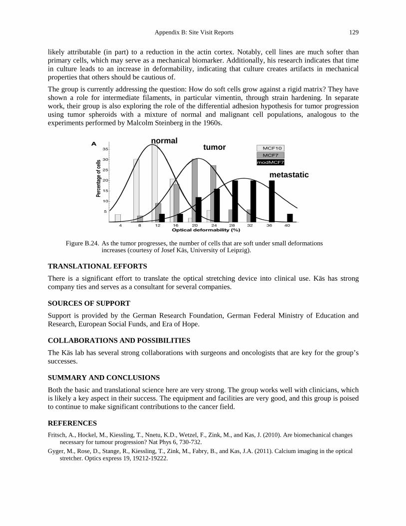
Appendix B: Site Visit Reports 129
likely attributable (in part) to a reduction in the actin cortex. Notably, cell lines are much softer than primary cells, which may serve as a mechanical biomarker. Additionally, his research indicates that time in culture leads to an increase in deformability, indicating that culture creates artifacts in mechanical properties that others should be cautious of. The group is currently addressing the question: How do soft cells grow against a rigid matrix? They have shown a role for intermediate filaments, in particular vimentin, through strain hardening. In separate work, their group is also exploring the role of the differential adhesion hypothesis for tumor progression using tumor spheroids with a mixture of normal and malignant cell populations, analogous to the experiments performed by Malcolm Steinberg in the 1960s.
Figure B.24. As the tumor progresses, the number of cells that are soft under small deformations
increases (courtesy of Josef Käs, University of Leipzig).
TRANSLATIONAL EFFORTS
There is a significant effort to translate the optical stretching device into clinical use. Käs has strong company ties and serves as a consultant for several companies.
SOURCES OF SUPPORT
Support is provided by the German Research Foundation, German Federal Ministry of Education and Research, European Social Funds, and Era of Hope.
COLLABORATIONS AND POSSIBILITIES
The Käs lab has several strong collaborations with surgeons and oncologists that are key for the group’s successes.
SUMMARY AND CONCLUSIONS
Both the basic and translational science here are very strong. The group works well with clinicians, which is likely a key aspect in their success. The equipment and facilities are very good, and this group is poised to continue to make significant contributions to the cancer field.
REFERENCES Fritsch, A., Hockel, M., Kiessling, T., Nnetu, K.D., Wetzel, F., Zink, M., and Kas, J. (2010). Are biomechanical changes
necessary for tumour progression? Nat Phys 6, 730-732. Gyger, M., Rose, D., Stange, R., Kiessling, T., Zink, M., Fabry, B., and Kas, J.A. (2011). Calcium imaging in the optical
stretcher. Optics express 19, 19212-19222.
normal tumor
metastatic

130 Appendix B. Site Visit Reports
Leal-Egana, A., Fritsch, A., Heidebrecht, F., Diaz-Cuenca, A., Nowicki, M., Bader, A., and Kas, J. (2012). Tuning liver stiffness against tumours: an in vitro study using entrapped cells in tumour-like microcapsules. Journal of the mechanical behavior of biomedical materials 9, 113-121.
www.uni-leipzig.de/~pwm/web/ www.zv.uni-leipzig.de/en/ www.zv.uni-leipzig.de/en/university/profile-and-management/mission-profile-and-history/research-profile.html www.dfg.de/en/dfg_profile/index.jsp

Appendix B: Site Visit Reports 131
University Medical Center Utrecht
Site Address: (Meeting at the Hubrecht Institute) Uppsalalaan 8 3584 CT Utrecht, NETHERLANDS www.uu.nl/
Date Visited: 7 May 2012 WTEC Attendees: Paul Janmey, Cindy Reinhart-King (site report author), Dan Fletcher, Nastaran Kuhn,
Nicole Moore, and Hemant Sarin Host(s): Philip de Groot
Prof. of Thrombosis and Hemostasis, Department of Hematology University Medical Center Utrecht Heidelberglaan 100 3584 CX Utrecht, NETHERLANDS [email protected] Tel: +31 88 7557769)
OVERVIEW
The University Medical Center, Utrecht (UMC Utrecht), is comprised of the Faculty of Medicine of Utrecht University, the former Academic Hospital, and the Wilhelmina Kinderziekenhuis Children’s Hospital. Faculty members conduct interdisciplinary research in biomedical genetics, biomedical image sciences, and clinical epidemiology. The medical center’s research areas include brain, infection and immunity, circulatory health, personalized cancer care, regenerative medicine, and child health. UMC Utrecht and the University closely liaise in large-scale research programs such as TI Pharma, the Centre for Translational and Molecular Medicine, and Biomedical Materials. The development of Science Park Utrecht at De Uithof has also increased collaboration between both institutions.
RESEARCH AND DEVELOPMENT ACTIVITIES
While we were at the Hubrecht Institute, our group met with Dr. Philip de Groot from UMC Utrecht. Even though he is a biochemist by training, he spoke about work at the intersection of hemostasis and engineering. The department includes both a diagnostic lab that employs 300 people. The research labs cover four major topics and extensively collaborate with engineers in several major areas:
• Flow models for hemostasis • Point-of-care diagnostic assays • Improved assays to detect microparticles • The study of the intersection of primary and secondary hemostasis Questions that continue to be a focus for the research labs include:
• Why do some patients bleed? • Can we create a reliable model for thrombosis that incorporates elements of flow and adhesion, which
are essential for drug testing? • Can we create better point-of-care tests for bleeding?
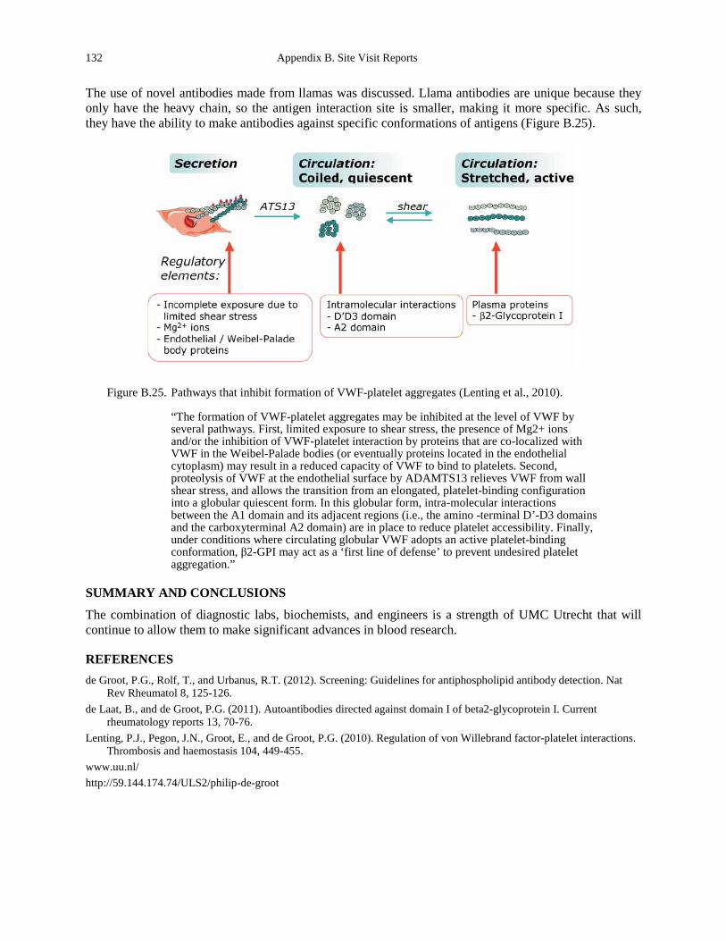
132 Appendix B. Site Visit Reports
The use of novel antibodies made from llamas was discussed. Llama antibodies are unique because they only have the heavy chain, so the antigen interaction site is smaller, making it more specific. As such, they have the ability to make antibodies against specific conformations of antigens (Figure B.25).
Figure B.25. Pathways that inhibit formation of VWF-platelet aggregates (Lenting et al., 2010).
“The formation of VWF-platelet aggregates may be inhibited at the level of VWF by several pathways. First, limited exposure to shear stress, the presence of Mg2+ ions and/or the inhibition of VWF-platelet interaction by proteins that are co-localized with VWF in the Weibel-Palade bodies (or eventually proteins located in the endothelial cytoplasm) may result in a reduced capacity of VWF to bind to platelets. Second, proteolysis of VWF at the endothelial surface by ADAMTS13 relieves VWF from wall shear stress, and allows the transition from an elongated, platelet-binding configuration into a globular quiescent form. In this globular form, intra-molecular interactions between the A1 domain and its adjacent regions (i.e., the amino -terminal D’-D3 domains and the carboxyterminal A2 domain) are in place to reduce platelet accessibility. Finally, under conditions where circulating globular VWF adopts an active platelet-binding conformation, β2-GPI may act as a ‘first line of defense’ to prevent undesired platelet aggregation.”
SUMMARY AND CONCLUSIONS
The combination of diagnostic labs, biochemists, and engineers is a strength of UMC Utrecht that will continue to allow them to make significant advances in blood research.
REFERENCES de Groot, P.G., Rolf, T., and Urbanus, R.T. (2012). Screening: Guidelines for antiphospholipid antibody detection. Nat
Rev Rheumatol 8, 125-126. de Laat, B., and de Groot, P.G. (2011). Autoantibodies directed against domain I of beta2-glycoprotein I. Current
rheumatology reports 13, 70-76. Lenting, P.J., Pegon, J.N., Groot, E., and de Groot, P.G. (2010). Regulation of von Willebrand factor-platelet interactions.
Thrombosis and haemostasis 104, 449-455. www.uu.nl/ http://59.144.174.74/ULS2/philip-de-groot
Appendix B: Site Visit Reports 133
University of Barcelona
Site Address: Institute for Bioengineering of Catalonia www.ibecbarcelona.eu/ Baldiri Reixac, 10-12, 08028 Barcelona, SPAIN
Date Visited: 7 May 2012 WTEC Attendees: Sharon Gerecht, Parag Mallick, Owen McCarty (site report author), Lance Munn, and
Hassan Ali Host(s): Prof. Josep A. Planell, Director
Tel: +34 93 403 97 06 Fax: +34 93 403 97 02 Email: [email protected]
Dr. Jordi Alcaraz, Principal Investigator Translational Lung Biomechanics Laboratory Unitat de Biofísica i Bioenginyeria Facultat de Medicina Casanova 143, 08036 Barcelona Tel: (office) +34 93 402 4515, (lab): +34 93 403 9764 Fax: +34 93 403 5278 Email: [email protected] www.ub.edu/biofisica/jalcaraz.htm
Prof. Xavier Trepat ICREA Research Professor and Group Leader Edifici Hèlix Baldiri Reixac 15-21, 08028 Barcelona Tel: +34 93 402 0265 Email: xtrepat ibecbarcelona.eu www.ibecbarcelona.eu/xtrepat
Dr. Jerome Solon Centre for Genomic Regulation C/ Dr. Aiguader 88, 08003 Barcelona Tel. +34 93 316 01 00 Fax +34 93 316 00 99 Email: [email protected] http://pasteur.crg.es/portal/page/portal/Internet/02_Research
Prof. Pere Roca-Cusachs, Principal Investigator C/ Baldiri Reixac 10-12, 08028 Barcelona Tel: +34 934 020 863 Email: [email protected] www.ub.edu/biofisica/procacusachs.htm
134 Appendix B. Site Visit Reports
Dr. Marc Marti-Renom
Genome Biology Group Leader, Structural Genomics Team National Center for Genomic Analysis Parc Cientific de Barcelona - Torre I Baldiri Reixac, 4 - 2a.p – 08028, Barcelona Tel: +34 934 033 743 Fax: +34 934 037 279 Email: [email protected] http://marciuslab.org or http://cnag.cat
OVERVIEW
The Institute for Bioengineering of Catalonia (IBEC) is an interdisciplinary research center in Barcelona, Spain, focused on bioengineering and nanomedicine. IBEC was established in 2005 by the Ministries of Innovation, Universities and Enterprises and Health of the Generalitat de Catalunya (Autonomous Government of Catalonia), the University of Barcelona (UB), and the Technical University of Catalonia (UPC). Today, IBEC’s relationship with the UB and UPC researchers continues to operate under a framework agreement signed in 2008. IBEC’s mission is to conduct high-quality research that creates knowledge while contributing to a better quality of life, improving health, and creating wealth. The institute establishes close links with international research centers, universities, hospitals, and industry to exchange talent and develop and execute projects. The institute currently has 15 research groups and 250 researchers and staff from 20 different countries. IBEC’s groups and their activities are organized into six research programs:
• Cellular biotechnology • Biomechanics and cellular biophysics • Nanobiotechnology • Biomaterials, implants, and tissue engineering • Medical signals and instrumentation • Robotics and biomedical engineering The location of IBEC in the Parc Científic de Barcelona offers a highly stimulating biomedical environment in which the institute can work closely with organizations from the public and private sector interested in the biomedical application of nanotechnology. In addition, IBEC has access to powerful technological facilities, including the nanotechnology platform that offers services in nanofabrication, nanomanipulation and nanocharacterization.
RESEARCH AND DEVELOPMENT ACTIVITIES
Dr. Jordi Alcaraz
The Alcaraz group is focused on determining the role that tissue and cell mechanics play in regulating cellular functions in health and disease. Dr. Alcaraz has shown how epithelial cell phenotype is regulated by the biochemical and mechanical extracellular matrix (ECM) microenvironment, and how changes in these parameters drive breast cancer development (Alcaraz et al., 2008). They have recently shown the role that increased collagen deposition and decreased ECM degrading activity by matrix metalloproteinases (MMPs) plays in driving abnormal extracellular matrix mechanical properties (Alcaraz et al., 2011). They have developed a novel cylindrical flat-ended atomic force microscopy (AFM) tip in
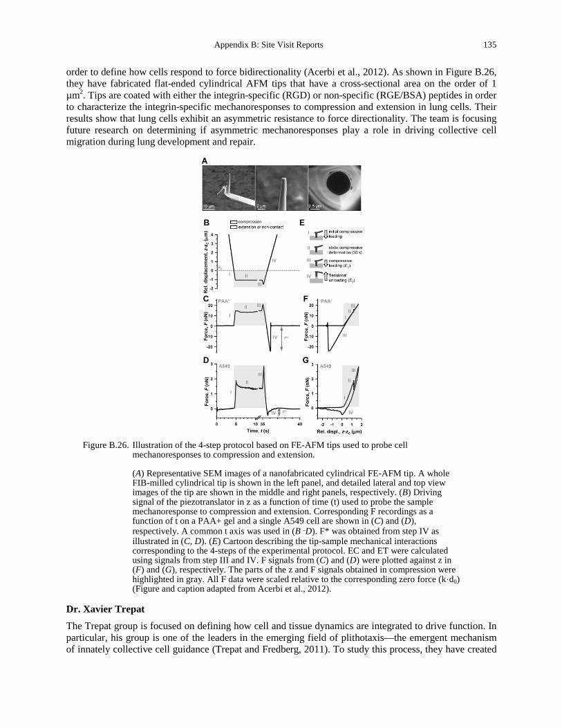
Appendix B: Site Visit Reports 135
order to define how cells respond to force bidirectionality (Acerbi et al., 2012). As shown in Figure B.26, they have fabricated flat-ended cylindrical AFM tips that have a cross-sectional area on the order of 1 µm2. Tips are coated with either the integrin-specific (RGD) or non-specific (RGE/BSA) peptides in order to characterize the integrin-specific mechanoresponses to compression and extension in lung cells. Their results show that lung cells exhibit an asymmetric resistance to force directionality. The team is focusing future research on determining if asymmetric mechanoresponses play a role in driving collective cell migration during lung development and repair.
Figure B.26. Illustration of the 4-step protocol based on FE-AFM tips used to probe cell
mechanoresponses to compression and extension.
(A) Representative SEM images of a nanofabricated cylindrical FE-AFM tip. A whole FIB-milled cylindrical tip is shown in the left panel, and detailed lateral and top view images of the tip are shown in the middle and right panels, respectively. (B) Driving signal of the piezotranslator in z as a function of time (t) used to probe the sample mechanoresponse to compression and extension. Corresponding F recordings as a function of t on a PAA+ gel and a single A549 cell are shown in (C) and (D), respectively. A common t axis was used in (B–D). F* was obtained from step IV as illustrated in (C, D). (E) Cartoon describing the tip-sample mechanical interactions corresponding to the 4-steps of the experimental protocol. EC and ET were calculated using signals from step III and IV. F signals from (C) and (D) were plotted against z in (F) and (G), respectively. The parts of the z and F signals obtained in compression were highlighted in gray. All F data were scaled relative to the corresponding zero force (k·d0) (Figure and caption adapted from Acerbi et al., 2012).
Dr. Xavier Trepat
The Trepat group is focused on defining how cell and tissue dynamics are integrated to drive function. In particular, his group is one of the leaders in the emerging field of plithotaxis—the emergent mechanism of innately collective cell guidance (Trepat and Fredberg, 2011). To study this process, they have created
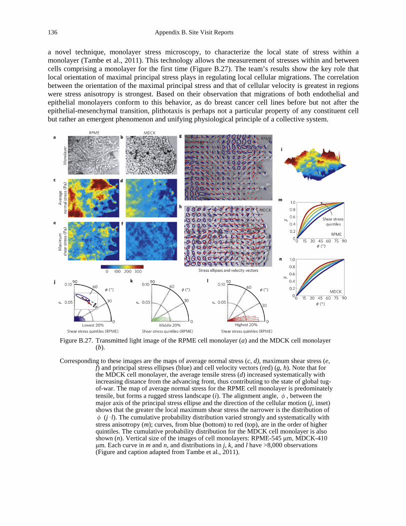
136 Appendix B. Site Visit Reports
a novel technique, monolayer stress microscopy, to characterize the local state of stress within a monolayer (Tambe et al., 2011). This technology allows the measurement of stresses within and between cells comprising a monolayer for the first time (Figure B.27). The team’s results show the key role that local orientation of maximal principal stress plays in regulating local cellular migrations. The correlation between the orientation of the maximal principal stress and that of cellular velocity is greatest in regions were stress anisotropy is strongest. Based on their observation that migrations of both endothelial and epithelial monolayers conform to this behavior, as do breast cancer cell lines before but not after the epithelial-mesenchymal transition, plithotaxis is perhaps not a particular property of any constituent cell but rather an emergent phenomenon and unifying physiological principle of a collective system.
Figure B.27. Transmitted light image of the RPME cell monolayer (a) and the MDCK cell monolayer
(b).
Corresponding to these images are the maps of average normal stress (c, d), maximum shear stress (e, f) and principal stress ellipses (blue) and cell velocity vectors (red) (g, h). Note that for the MDCK cell monolayer, the average tensile stress (d) increased systematically with increasing distance from the advancing front, thus contributing to the state of global tug-of-war. The map of average normal stress for the RPME cell monolayer is predominately tensile, but forms a rugged stress landscape (i). The alignment angle, φ, between the major axis of the principal stress ellipse and the direction of the cellular motion (j, inset) shows that the greater the local maximum shear stress the narrower is the distribution of φ (j–l). The cumulative probability distribution varied strongly and systematically with stress anisotropy (m); curves, from blue (bottom) to red (top), are in the order of higher quintiles. The cumulative probability distribution for the MDCK cell monolayer is also shown (n). Vertical size of the images of cell monolayers: RPME-545 μm, MDCK-410 μm. Each curve in m and n, and distributions in j, k, and l have >8,000 observations (Figure and caption adapted from Tambe et al., 2011).
Appendix B: Site Visit Reports 137
Dr. Jerome Solon
The Solon group is focused on characterizing the biomechanics of morphogenesis. Their group uses an integrative approach of high-resolution imaging, automated image processing, and physical modeling to characterize phenomena such as cell pulsing during embryogenesis (Figure B.28). They utilize the dorsal closure (DC) as a model system—a morphogenetic movement occurring at a late stage of Drosophila gastrulation. DC comprises the closure of a gap in the epidermis at the dorsal side of the embryo. The process begins with the dorsal convergence of two lateral, epidermal cell layers and terminates with the dorsal zippering of the leading cells from both layers. DC combines many cellular behaviors including cooperative cell movement, tissue force generation, and cell shape changes that are fundamental to the development and functioning of multicellular organisms. By coupling quantitative analysis, combined with laser cutting experiments and simulations, they were able to show that tension-based dynamics and cell coupling control the force pulses that drive dorsal closure in the developing Drosophila embryo (Solon et al., 2009). Their current aim is to unravel how the forces driving such collective cell movements are generated and coordinated.
Dr. Pere Roca-Cusachs
The Roca-Cusachs group is focused on defining the mechanisms by which molecules detect and respond to forces, triggering downstream cellular response. In particular, they utilize biophysical techniques to characterize the mechanical link between integrins and the actin cytoskeleton. The group has focused defining the role that integrin clustering and integrin-talin linkages play in regulating adhesion strength and mechanotransduction (Figure B.29). This study was enabled by the development of a magnetic tweezers apparatus able to exert forces of 1 nN on 2.8-μm diameter magnetic beads coated with a four-domain segment of fibronectin responsible for cell binding and containing the RGD and PHSRN motifs (Roca-Cusachs et al., 2009). Their results suggest a model for the mechanics of fibronectin-cell contacts in which fibronectin clustering leads to integrin binding, clustering, and recruitment.
Dr. Marc Marti-Renom
Dr. Marti-Renom is the Genome Biology Group Leader for the National Center for Genomic Analysis (CNAG). He and his structural genomics team are interested in the molecular mechanisms that regulate cell fate. To study such mechanisms, they employ the laws of physics and the rules of evolution to develop and apply computational methods for predicting the 3D structures of macromolecules and their complexes. Their three areas of research include:
• Protein-ligand interactions. They have developed methods for comparative docking of small chemical compounds and their target proteins. Such methods have already been applied to identify drug targets in 10 genomes that cause tropical diseases.
• Comparative RNA structure prediction. The recent interest in RNA, specifically non-coding RNA molecules, has prompted the team to develop a series of tools for the alignment of RNA structures and the prediction of their functions.
• Structure determination of genomes. More recently, they have engaged collaboration with experimentalists to study the 3D organization of the chromatin. Such work is resulting in the first ever structures of genomic domains and entire genomes.
SOURCES OF SUPPORT/FUNDING
This work is supported in part by the European Union European Research Council (ERC) and the Spanish Ministerio de Ciencia e Innovación. In addition, the work at the CNAG has received funding from the Tropical Disease Initiative, the Marie Curie Actions grant program, and a Generalitat Valenciana research grant.
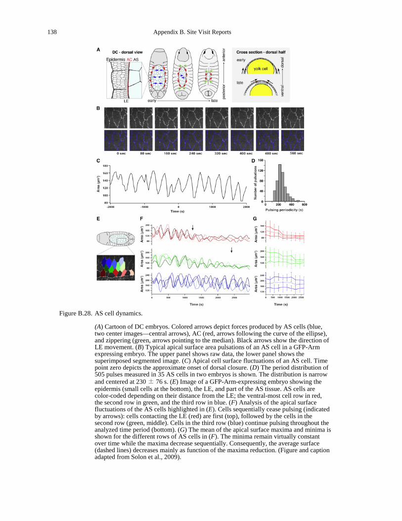
138 Appendix B. Site Visit Reports
Figure B.28. AS cell dynamics.
(A) Cartoon of DC embryos. Colored arrows depict forces produced by AS cells (blue, two center images—central arrows), AC (red, arrows following the curve of the ellipse), and zippering (green, arrows pointing to the median). Black arrows show the direction of LE movement. (B) Typical apical surface area pulsations of an AS cell in a GFP-Arm expressing embryo. The upper panel shows raw data, the lower panel shows the superimposed segmented image. (C) Apical cell surface fluctuations of an AS cell. Time point zero depicts the approximate onset of dorsal closure. (D) The period distribution of 505 pulses measured in 35 AS cells in two embryos is shown. The distribution is narrow and centered at 230 ± 76 s. (E) Image of a GFP-Arm-expressing embryo showing the epidermis (small cells at the bottom), the LE, and part of the AS tissue. AS cells are color-coded depending on their distance from the LE; the ventral-most cell row in red, the second row in green, and the third row in blue. (F) Analysis of the apical surface fluctuations of the AS cells highlighted in (E). Cells sequentially cease pulsing (indicated by arrows): cells contacting the LE (red) are first (top), followed by the cells in the second row (green, middle). Cells in the third row (blue) continue pulsing throughout the analyzed time period (bottom). (G) The mean of the apical surface maxima and minima is shown for the different rows of AS cells in (F). The minima remain virtually constant over time while the maxima decrease sequentially. Consequently, the average surface (dashed lines) decreases mainly as function of the maxima reduction. (Figure and caption adapted from Solon et al., 2009).
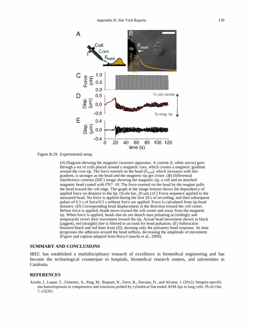
Appendix B: Site Visit Reports 139
Figure B.29. Experimental setup.
(A) Diagram showing the magnetic tweezers apparatus. A current (I, white arrow) goes through a set of coils placed around a magnetic core, which creates a magnetic gradient around the core tip. The force exerted on the bead (Fbead), which increases with this gradient, is stronger as the bead and the magnetic tip get closer. (B) Differential interference contrast (DIC) image showing the magnetic tip, a cell and an attached magnetic bead coated with FN7–10. The force exerted on the bead by the magnet pulls the bead toward the cell edge. The graph at the image bottom shows the dependency of applied force on distance to the tip. (Scale bar, 20 μm.) (C) Force sequence applied to the measured bead. No force is applied during the first 20 s of recording, and then subsequent pulses of 0.5 s of force/0.5 s without force are applied. Force is calculated from tip-bead distance. (D) Corresponding bead displacement in the direction toward the cell center. Before force is applied, beads move toward the cell center and away from the magnetic tip. When force is applied, beads that do not detach start pulsating accordingly and temporarily revert their movement toward the tip. Actual bead movement shown in black (jagged), red (straight) line is filtered to account for bead pulsation. (E) Subtraction between black and red lines from (D), showing only the pulsatory bead response. As time progresses the adhesion around the bead stiffens, decreasing the amplitude of movement. (Figure and caption adapted from Roca-Cusachs et al., 2009).
SUMMARY AND CONCLUSIONS
IBEC has established a multidisciplinary research of excellence in biomedical engineering and has become the technological counterpart to hospitals, biomedical research centers, and universities in Catalonia.
REFERENCES Acerbi, I., Luque, T., Gimenez, A., Puig, M., Reguart, N., Farre, R., Navajas, D., and Alcaraz, J. (2012). Integrin-specific
mechanoresponses to compression and extension probed by cylindrical flat-ended AFM tips in lung cells. PLoS One 7, e32261.
140 Appendix B. Site Visit Reports
Alcaraz, J., Mori, H., Ghajar, C.M., Brownfield, D., Galgoczy, R., and Bissell, M.J. (2011). Collective epithelial cell invasion overcomes mechanical barriers of collagenous extracellular matrix by a narrow tube-like geometry and MMP14-dependent local softening. Integr Biol (Camb) 3, 1153-1166.
Alcaraz, J., Xu, R., Mori, H., Nelson, C.M., Mroue, R., Spencer, V.A., Brownfield, D., Radisky, D.C., Bustamante, C., and Bissell, M.J. (2008). Laminin and biomimetic extracellular elasticity enhance functional differentiation in mammary epithelia. The EMBO journal 27, 2829-2838.
Roca-Cusachs, P., Gauthier, N.C., Del Rio, A., and Sheetz, M.P. (2009). Clustering of alpha(5)beta(1) integrins determines adhesion strength whereas alpha(v)beta(3) and talin enable mechanotransduction. Proc Natl Acad Sci U S A 106, 16245-16250.
Solon, J., Kaya-Copur, A., Colombelli, J., and Brunner, D. (2009). Pulsed forces timed by a ratchet-like mechanism drive directed tissue movement during dorsal closure. Cell 137, 1331-1342.
Tambe, D.T., Hardin, C.C., Angelini, T.E., Rajendran, K., Park, C.Y., Serra-Picamal, X., Zhou, E.H., Zaman, M.H., Butler, J.P., Weitz, D.A., et al. (2011). Collective cell guidance by cooperative intercellular forces. Nature materials 10, 469-475.
Trepat, X., and Fredberg, J.J. (2011). Plithotaxis and emergent dynamics in collective cellular migration. Trends in cell biology 21, 638-646.
Appendix B: Site Visit Reports 141
University of Basel
Site Address: Biozentrum, University of Basel Klingelbergstrasse 50/70 CH - 4056 Basel, SWITZERLAND (meeting took place at EPFL, Lausanne)
Date Visited: 8 May 2012 WTEC Attendees: Sharon Gerecht, Parag Mallick, Owen McCarty, Lance Munn (site report author), and
Hassan Ali Host(s): Cora-Ann Schoenenberger
Tel: +41 61 267 22 60 Email: [email protected] www.biozentrum.unibas.ch/research/groups-platforms/overview/unit/schoenenberger/
OVERVIEW
The Biozentrum at the Universitat Basel, Center for Molecular Life Sciences, consists of 33 research groups with scientific focus on growth and development, infection biology, neurobiology, structural biology and biophysics, and computational and systems biology. A few of these groups are interested in the molecular underpinnings of cancer, and the relationship between cancer and stem cell biology. Two groups at this site (Prof. R. Lim, a nanobiologist and Dr. C-A. Schoenenberger) are overtly adapting concepts from nanomechanics to study oncology. The WTEC panel heard a presentation by Schoenenberger.
RESEARCH AND DEVELOPMENT ACTIVITIES
Schoenenberger, originally trained as a cell biologist, now studies the structural and mechanical plasticity of healthy cells and tumors, with the premise that the cytoskeleton is an essential mediator of structural and nanomechanical plasticity of cells in health and disease. She believes that pathologically altered nanomechanical properties may serve as diagnostic markers for cancer. Schoenenberger presented a summary of her early work which used atomic force microscopy (AFM) to image cytoskeletal dynamics in cultured cells. She then described studies pioneered by Plodinec et al., where breakthrough AFM-based nanotechnology known as ARTIDIS (“Automated and Reliable Tissue Diagnostics”) is used to measure the nanomechanical properties of breast cancer biopsies. The feasibility of these studies was first demonstrated on tissue specimens of breast cancer in the mouse model, showing that different types of tumors and stages of tumor development can be distinguished on the basis of their stiffness signature. Schoenenberger is a close collaborator of a team headed by Prof. Lim investigating the potential of AFM measurements for the diagnosis and prognosis of breast cancer in humans. An interesting aspect of the data is the spatially-resolved measurements, which result in histograms of stiffness over the surface of the cylindrical biopsies. Distinct peaks in the histogram can be attributed to cancer cells, normal cells, and matrix components and can be used in diagnosis. In the final few slides, Schoenenberger outlined a more recent project aimed at understanding the relationship between tissue hypoxia, mechanical elasticity, and progression of cancer. Besides her collaborations with Prof. Roderick Lim within the Biozentrum, Schoenenberger has outside collaborations with the Friedrich Miescher Institute, Basel (Dr. Mohamed Bentires-Alj) regarding the MMTV-PyMT mouse model. Collaboration with the Department of Gynecology and Gynecological
142 Appendix B. Site Visit Reports
Oncology, University Hospital, Basel, is the source of the clinical biopsies for elasticity analyses. Histopathological aspects are covered by the Department of Pathology, University Hospital, Basel.
TRANSLATION
On the topic of translation, Schoenenberger pointed out that the inventors of ARTIDIS (Dr. med. Marko Loparic, Dr. Marija Plodinec and Prof. Lim), in partnership with the Swiss company Nanosurf AG, are actively working with the University Hospital, Basel to further develop it for the diagnosis of breast cancer. Patents have been filed on the technology and intellectual property related to breast cancer diagnostics by the University of Basel.
SOURCES OF SUPPORT/FUNDING
Funding comes from the Swiss National Science Foundation; Swiss Nanoscience Institute, Basel; and Swiss Commission for Technology and Innovation.
SUMMARY AND CONCLUSIONS
Schoenenberger’s research has at its foundation the concept that cell mechanical properties are a result of—and contribute to—cancer progression. Her interdisciplinary collaborations provide a good example of how physics and oncology can be integrated so as to promote the translation of these concepts to the bedside.
REFERENCES Plodinec, M., Loparic, M., Monnier, C.A., Obermann, E.C., Zanetti-Dallenbach, R., Oertle, P., Hyotyla, J.T., Aebi, U.,
Bentires-Alj, M., Lim, R.Y.H., Schoenenberger, C-A. The nanomechanical signature of breast cancer. Nature nanotechnology 2012, in press.
Plodinec, M., Loparic, M., Suetterlin, R., Herrmann, H., Aebi, U., and Schoenenberger, C.A. (2011). The nanomechanical properties of rat fibroblasts are modulated by interfering with the vimentin intermediate filament system. Journal of structural biology 174, 476-484.
Plodinec, M., and Schoenenberger, C.A. (2010). Spatial organization acts on cell signaling: how physical force contributes to the development of cancer. Breast cancer research : BCR 12, 308.
Schoenenberger, C.A., Mannherz, H.G., and Jockusch, B.M. (2011). Actin: from structural plasticity to functional diversity. European journal of cell biology 90, 797-804.
Silvan, U., Boiteux, C., Sutterlin, R., Schroeder, U., Mannherz, H.G., Jockusch, B.M., Berneche, S., Aebi, U., and Schoenenberger, C.A. (2012). An antiparallel actin dimer is associated with the endocytic pathway in mammalian cells. Journal of structural biology 177, 70-80.
Appendix B: Site Visit Reports 143
University of the Basque Country
Site Address: P.O. Box 644, 48080 Bilbao, SPAIN www.ehu.es/p200-hmencont/en/contenidos/informacion/basic_facts/en_inf/basicfacts.html (The meeting took place at the University of Barcelona Institute for Bioengineering of Catalonia)
Date Visited: 7 May 2012 WTEC Attendees: Sharon Gerecht, Parag Mallick (site report author), Owen McCarty, Lance Munn, and
Hassan Ali Host(s): Prof. José M.G. Vilar
Biophysics Unit (CSIC-UPV/EHU) Department of Biochemistry and Molecular Biology Tel: +34-94 601 3450 Fax: +34-94 601 3360 Email: [email protected] www.vilarlab.org
OVERVIEW
The University of the Basque Country (UPV/EHU) has been certified as a Campus of International Excellence by the Spanish Ministry of Education. UPV/EHU and its partners stimulate research, creativity, and innovation in order to establish a new, sustainable economic and social model. Researchers at UPV/EHU seek to extend their international influence by working on a “brain-gain” basis and establishing cross-border campuses. It is clear that they are significantly connected to the broader international community. Towards this, UPV/EHU hosts an unrestricted and multicultural community. Their international relations office manages over 900 exchange agreements with other Spanish, European, and international universities. Every year, the three campuses turn into lively melting pots where over 900 foreign students blend in with local trainees. The university takes part in SICUE, the Spanish universities students’ exchange program; ERASMUS, the most extended European mobility program; and they have created their own exchange programs with Latin America (UPV/EHU-AMÉRICA LATINA) and other destinations (United States, Canada, The Philippine Islands, New Zealand, and Russia). A number of teaching staff travels to Central and South American universities. The UPV/EHU and the National Research Council of Spain (CSIC) host together the Biophysics Unit (CSIC-UPV/EHU), a cross-disciplinary center at the interface between physics, biochemistry and molecular biology.
RESEARCH AND DEVELOPMENT ACTIVITIES
Dr. Jose Vilar
The Vilar group is broadly interested in understanding and accurately predicting molecular, cellular, and cell-population behaviors in terms of the interactions of the components and vice versa. Although traditional approaches have been successful at identifying cellular components and their interactions, the Vilar lab believes it is critical to piece back together all the genetic, biochemical,
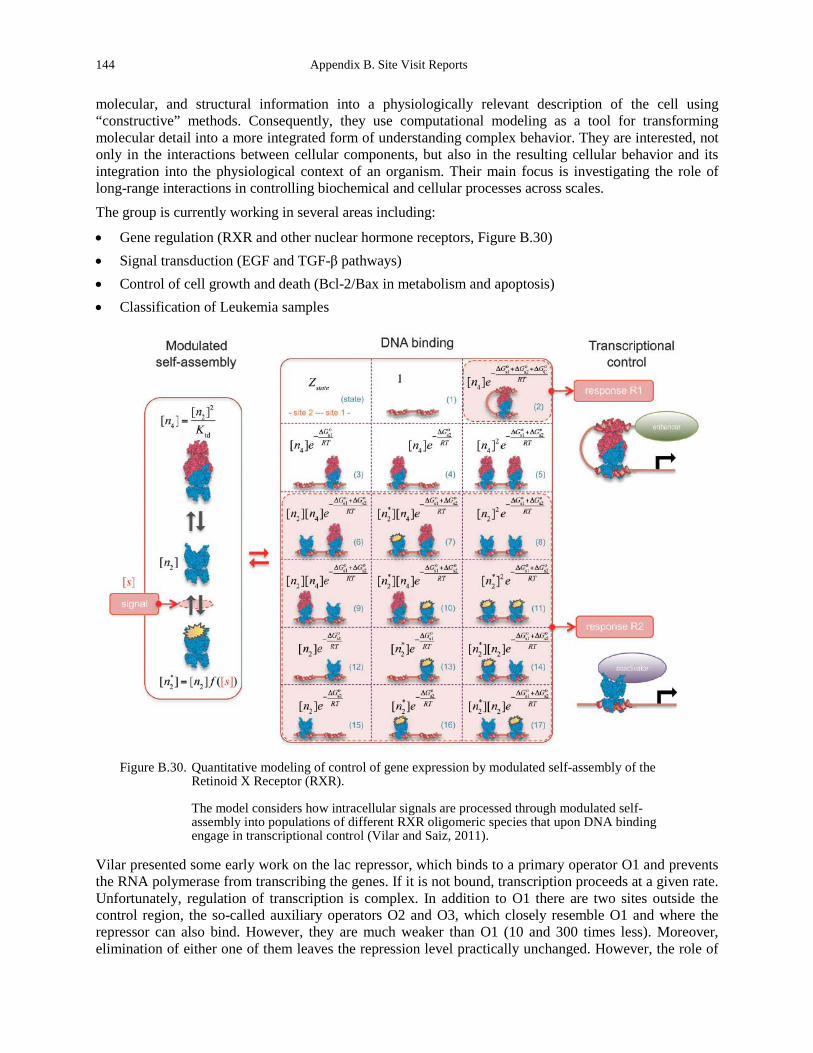
144 Appendix B. Site Visit Reports
molecular, and structural information into a physiologically relevant description of the cell using “constructive” methods. Consequently, they use computational modeling as a tool for transforming molecular detail into a more integrated form of understanding complex behavior. They are interested, not only in the interactions between cellular components, but also in the resulting cellular behavior and its integration into the physiological context of an organism. Their main focus is investigating the role of long-range interactions in controlling biochemical and cellular processes across scales. The group is currently working in several areas including:
• Gene regulation (RXR and other nuclear hormone receptors, Figure B.30) • Signal transduction (EGF and TGF-β pathways) • Control of cell growth and death (Bcl-2/Bax in metabolism and apoptosis) • Classification of Leukemia samples
Figure B.30. Quantitative modeling of control of gene expression by modulated self-assembly of the
Retinoid X Receptor (RXR).
The model considers how intracellular signals are processed through modulated self-assembly into populations of different RXR oligomeric species that upon DNA binding engage in transcriptional control (Vilar and Saiz, 2011).
Vilar presented some early work on the lac repressor, which binds to a primary operator O1 and prevents the RNA polymerase from transcribing the genes. If it is not bound, transcription proceeds at a given rate. Unfortunately, regulation of transcription is complex. In addition to O1 there are two sites outside the control region, the so-called auxiliary operators O2 and O3, which closely resemble O1 and where the repressor can also bind. However, they are much weaker than O1 (10 and 300 times less). Moreover, elimination of either one of them leaves the repression level practically unchanged. However, the role of
Appendix B: Site Visit Reports 145
O2 and O3 are actually quite significant: simultaneous elimination of both of these operators reduced the repression level about 100 times. Deeper investigations and computational modeling were able to detail how DNA looping could explain this result. Vilar then proceeded to demonstrate how general processes of self-assembly could lead to these sorts of emergent behaviors in regulation by mechanisms previously thought to be distinct. Vilar also used an entropy-based approach to develop the highest performing method in the DREAM 6 competition where the goal was to diagnose acute myeloid leukemia using flow cytometry.
SOURCES OF SUPPORT/FUNDING
Vilar receives funding from Ikerbasque, the Basque Foundation for Science, for both research and endowing his professorship, and from the Spanish Ministry of Science and Innovation.
SUMMARY AND CONCLUSIONS
Vilar’s research program at Ikerbasque and the Biophysics Unit (CSIC-UPV/EHU) is extremely exciting. His research focus is on an important intersection between biologically important problems and novel, highly formal approaches to attack them. In particular, he has been leading efforts to identify mechanisms of emergent behavior in cellular regulation and information transfer in cancer.
REFERENCES Vilar, J.M., and Saiz, L. (2011). Control of gene expression by modulated self-assembly. Nucleic acids research 39, 6854-
6863. www.istem.eu/ewb_pages/s/statutes.php (http://194.146.175.227) www.ehu.es/biologiacomputacional/#Publications www.ehu.es/biologiacomputacional/#Research www.ikerbasque.net/ www.unidaddebiofisica.org/
146 Appendix B. Site Visit Reports
University of Freiburg
Site Address: Hermann-Herder Straβe 3, 79104 Freiburg im Breisgau, GERMAMY www.uni-freiburg.de/forschung-en (meeting held at the University of Heidelberg)
Date Visited: 10 May 2012 WTEC Attendees: Sharon Gerecht, Parag Mallick (site report author), Owen McCarty, and Hassan Ali Host(s): Jens Timmer
Institute of Physics Tel: +49 761 203 5829 Fax: +49 761 203 8541 Email: [email protected] http://webber.physik.uni-freiburg.de/~jeti/
Hauke Busch Freiburg Institute for Advanced Studies School of Life Sciences Tel: +49 761 203 97154 Fax: +49 761 203 97334 Email: [email protected] www.frias.uni-freiburg.de/lifenet/fellows/hauke-busch
OVERVIEW
The Freiburg Institute for Advanced Studies (FRIAS) is a key component of the University of Freiburg. FRIAS is organized into four schools: 1) School of History; 2) School of Language & Literature; 3) School of Life Sciences – LifeNet; and 4) School of Soft Matter Research. There are also several interdisciplinary research groups in annual rotation. These schools and research groups work closely with the university’s eleven faculties. FRIAS’s work pivots around four key concepts that also lie at the heart of the university’s institutional strategy:
• “Windows for Research” - FRIAS provides leading researchers with privileged conditions to conduct their work.
• “New Universitas” - FRIAS opens up new opportunities for interdisciplinary contact and collaboration.
• “Internationalisation” - FRIAS reinforces the university’s international networking and visibility. • “Promotion of Early-Stage Researchers” - FRIAS offers outstanding conditions for young academics. In 2009, an important component was added to the four-school architecture: the interdisciplinary research group competition. This program provides University of Freiburg professors with the opportunity to apply for a 10-month FRIAS fellowship in order to carry out an innovative, interdisciplinary research project at the institute. This can also include international partners, if applicable. Two or three research groups are supported each year.
Appendix B: Site Visit Reports 147
The major FRIAS interdisciplinary symposia also offer new options for projects. They aim to promote dialogue between the humanities, social sciences, and natural sciences and to shed light on a topic of key academic and social relevance from different perspectives (2009: Evolution, 2011: Catastrophes). The same is true of many smaller, inter-school event formats such as monthly dinner speeches and after hours conversations. FRIAS School of Life Sciences-LifeNet focuses its research on the biology of complex systems. Its multidisciplinary, system-oriented approach includes a wide range of researchers whose expertise spans mathematics and physics to biology and medicine.
RESEARCH AND DEVELOPMENT ACTIVITIES
Both the Timmer and Busch groups highlighted how mathematical modeling is used to create hypotheses, which, in turn, are validated using experiments in high-throughput biological systems (genomic, proteomic, and metabolomic methods) and imaging technologies. This system-oriented approach is designed to improve prediction of normal functions in plants, animals, and humans. Similarly, the approach can be used to disclose causes and progress of illnesses and to assess how successful different treatment options will be.
Dr. Jens Timmer
Dynamic processes are ubiquitous in the life sciences. They can be found from the regulation in cells up to oscillations in tremor. Malfunction of these dynamical processes can be a cause or a sign of diseases. In interdisciplinary projects, the Timmer group develops and applies mathematical methods to analyze and model these processes based on measured data. The final aim of these efforts is to help to turn the life sciences from a qualitative descriptive science into a quantitative predictive science. The group is exploring a number of exciting models of regulation ranging from testing the accuracy of sensor network interaction maps, to understanding the design principles underlying biological systems (particularly with regards to robustness), as well as understanding randomness in gene expression and pattern formation in development. One study that was highlighted investigates why biology has selected a particular level of complexity to chemotaxis networks. This study used a combined experimental and computational approach to look at the very simple and measurable system of Che proteins (Figure B.31). Unlike traditional systems oriented approaches that are focused on tens of thousands of genes and on discovering the interactions between them, the Timmer group focused on a simple, compact system. Regulation of this system is driven by a handful of proteins (CheR, CheB, CheA, CheZ and CheY). After measuring the extent of noise using YFP and CFP reporter, the authors were able to look at both the protein covariation and noise. In order for the system to function appropriately, it must be sensitive to signaling molecules (e.g., chemoattractants and chemorepellants), but insensitive to random noise fluctuations. After evaluating the common ‘BL’ model (Figure B.31, left), the authors found that it was unable to accurately reproduce the measured data. However, more complex topologies (Figure B.31, right) allow cells in the population to respond accurately to changes in ligand concentration. Furthermore, the topologies are sufficiently robust to compensate for co-variations in expression levels and some suppression of noise-based fluctuations. Though more complex structures may be envisioned, they appear not necessary for maintaining system responsiveness and stability. It is highly likely that this is a general principle of biological signaling networks.
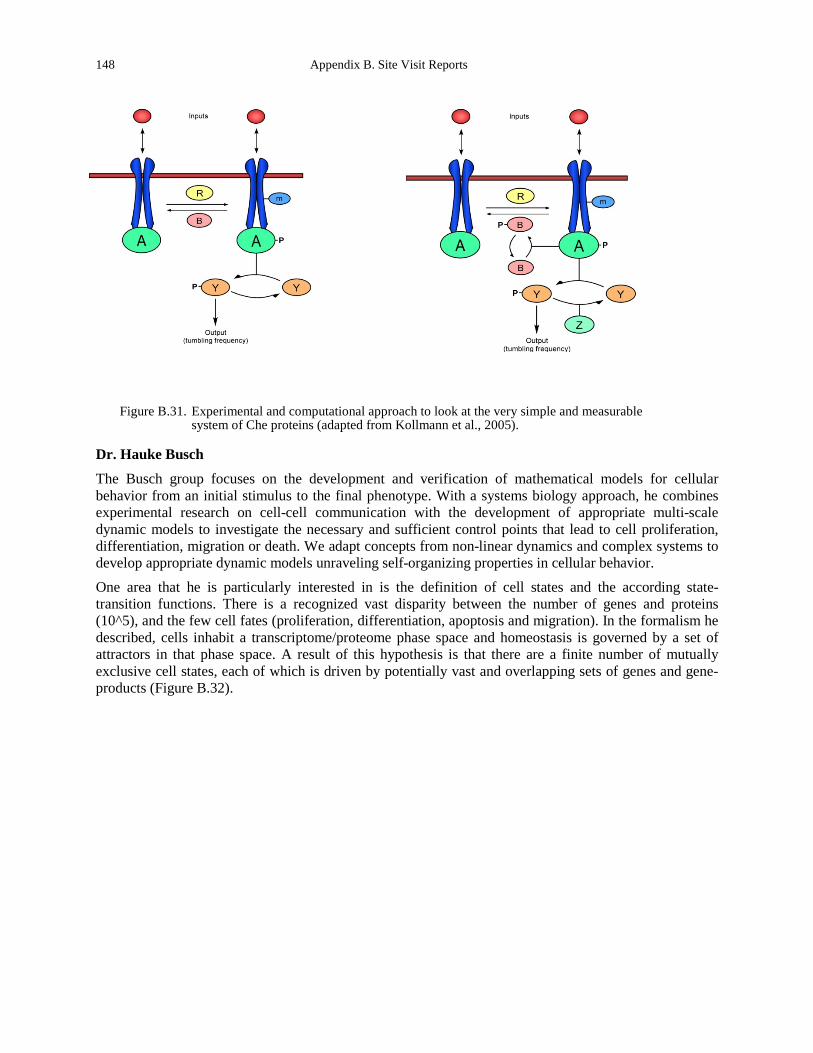
148 Appendix B. Site Visit Reports
Figure B.31. Experimental and computational approach to look at the very simple and measurable system of Che proteins (adapted from Kollmann et al., 2005).
Dr. Hauke Busch
The Busch group focuses on the development and verification of mathematical models for cellular behavior from an initial stimulus to the final phenotype. With a systems biology approach, he combines experimental research on cell-cell communication with the development of appropriate multi-scale dynamic models to investigate the necessary and sufficient control points that lead to cell proliferation, differentiation, migration or death. We adapt concepts from non-linear dynamics and complex systems to develop appropriate dynamic models unraveling self-organizing properties in cellular behavior.
One area that he is particularly interested in is the definition of cell states and the according state-transition functions. There is a recognized vast disparity between the number of genes and proteins (10^5), and the few cell fates (proliferation, differentiation, apoptosis and migration). In the formalism he described, cells inhabit a transcriptome/proteome phase space and homeostasis is governed by a set of attractors in that phase space. A result of this hypothesis is that there are a finite number of mutually exclusive cell states, each of which is driven by potentially vast and overlapping sets of genes and gene-products (Figure B.32).
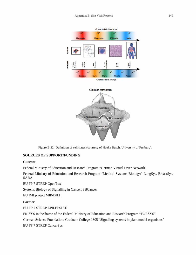
Appendix B: Site Visit Reports 149
Figure B.32. Definition of cell states (courtesy of Hauke Busch, University of Freiburg).
SOURCES OF SUPPORT/FUNDING
Current
Federal Ministry of Education and Research Program “German Virtual Liver Network” Federal Ministry of Education and Research Program “Medical Systems Biology:” LungSys, BreastSys, SARA
EU FP 7 STREP OpenTox Systems Biology of Signalling in Cancer: SBCancer EU IMI project MIP-DILI
Former
EU FP 7 STREP EPILEPSIAE FRISYS in the frame of the Federal Ministry of Education and Research Program “FORSYS” German Science Foundation: Graduate College 1305 “Signaling systems in plant model organisms” EU FP 7 STREP CancerSys
150 Appendix B. Site Visit Reports
EU FP 6 IP Sens-it-iv: Novel Testing Strategies for In Vitro Assessment of Allergens Bernstein Center for Computational Neuroscience Freiburg Federal Ministry of Education and Research Program “Systems Biology of Hepatocytes” (HepatoSys)
German Science Foundation: Statistical Modelling in Neurology Federal Ministry of Education and Research Program “QuantPro” EU FP 6 STREP COSBICS: Computational Systems Biology in Cellular Signalling Nationales Genomforschungsnetz NGFN Explorative Project: Mircoarray Validation of cardio-vascular Risk German Research Foundation Priority Program 1114 “Mathematical methods for time series analysis and digital image processing” State of Baden-Württemberg, Funding Initiative RNA/RNAi Federal Ministry of Education and Research Program on Non-linear Dynamics Graduate College “Nonlinear Differential Equations: Modelling, Theory, Numerics, Visualisation”
SUMMARY AND CONCLUSIONS
The research at the University of Freiburg is extremely exciting. There are a number of funding mechanisms to support interdisciplinary research. In addition, interdisciplinary research is strongly encouraged. One notable component of the proposed research is the significant focus on integrating experiment and computation. Among the most exciting aspects at the University of Freiburg was the focus on formalizing hypotheses mathematically and then identifying experiments to test those mathematical models.
REFERENCES Kollmann, M., Løvdok, L., Bartholomé, K., Timmer, J., and Sourjik, V. (2005). Design principles of a bacterial signaling network. Nature 438, 504-507.
http://webber.physik.uni-freiburg.de/~jeti/papers/Julie_JIM.pdf www.frias.uni-freiburg.de/institute/frias-im-ueberblick-en/frias-overview
Appendix B: Site Visit Reports 151
University of Heidelberg
Site Address: BioQuant, In Neuheimer Feld 27 Ute Greenuier Saal D-69120, GERMANY www.bioquant.uni-heidelberg.de/
The German Cancer Research Center (DKFZ)
Im Neuenheimer Feld 280 69120 Heidelberg, GERMANY www.dkfz.de/en/index.html
Center for Molecular Biology of the University of Heidelberg (ZMBH)
Im Neuenheimer Feld 282 D-69120 Heidelberg, GERMANY
Date Visited: 10 May 2012 WTEC Attendees: Sharon Gerecht (site report author), Parag Mallick, Owen McCarty, and Hassan Ali Host(s): Joachim Spatz
Max Planck Institute (MPI) for Metals Research Heisenbergstr. 3 D - 70 569Stuttgart, GERMANY Email: [email protected] www.mf.mpg.de/en/abteilungen/spatz/index.html University of Heidelberg INF 253 D - 69 120Heidelberg, GERMANY Email: [email protected] www.pci.uni-heidelberg.de/bpc/biophysik.html
Ulrich Schwartz Institute for Theoretical Physics Tel: +49 6221 54 9399 Fax: +49 6221 54 9331 Email: [email protected] www.tphys.uni-heidelberg.de
Evgeny Gladilin Group Leader, Cellular Mechanics, Bioquant DKFZ Email: [email protected]
Ralf Kemkemer MPI for Intelligent Systems Stuttgart, GERMANY Tel: +49 711 689 3516 Fax: +49 711 689 3612 Email: [email protected] www.is.mpg.dle
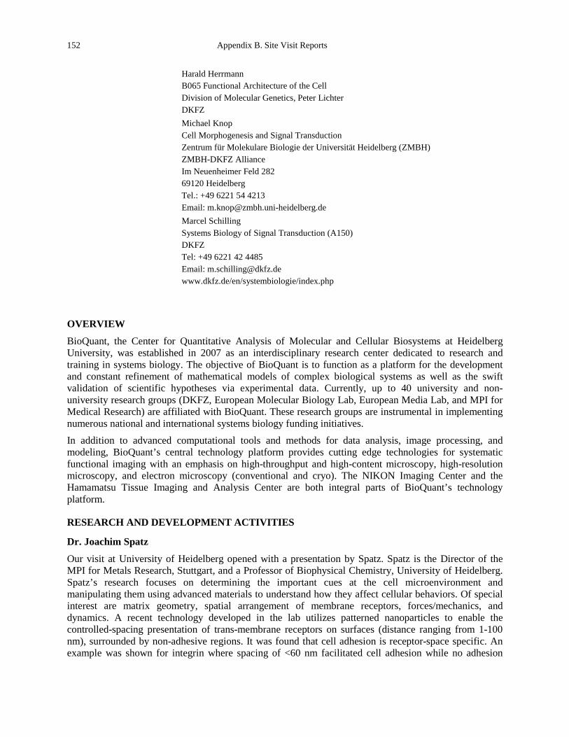
152 Appendix B. Site Visit Reports
Harald Herrmann B065 Functional Architecture of the Cell Division of Molecular Genetics, Peter Lichter DKFZ
Michael Knop Cell Morphogenesis and Signal Transduction Zentrum für Molekulare Biologie der Universität Heidelberg (ZMBH) ZMBH-DKFZ Alliance Im Neuenheimer Feld 282 69120 Heidelberg Tel.: +49 6221 54 4213 Email: [email protected]
Marcel Schilling Systems Biology of Signal Transduction (A150) DKFZ Tel: +49 6221 42 4485 Email: [email protected] www.dkfz.de/en/systembiologie/index.php
OVERVIEW
BioQuant, the Center for Quantitative Analysis of Molecular and Cellular Biosystems at Heidelberg University, was established in 2007 as an interdisciplinary research center dedicated to research and training in systems biology. The objective of BioQuant is to function as a platform for the development and constant refinement of mathematical models of complex biological systems as well as the swift validation of scientific hypotheses via experimental data. Currently, up to 40 university and non-university research groups (DKFZ, European Molecular Biology Lab, European Media Lab, and MPI for Medical Research) are affiliated with BioQuant. These research groups are instrumental in implementing numerous national and international systems biology funding initiatives. In addition to advanced computational tools and methods for data analysis, image processing, and modeling, BioQuant’s central technology platform provides cutting edge technologies for systematic functional imaging with an emphasis on high-throughput and high-content microscopy, high-resolution microscopy, and electron microscopy (conventional and cryo). The NIKON Imaging Center and the Hamamatsu Tissue Imaging and Analysis Center are both integral parts of BioQuant’s technology platform.
RESEARCH AND DEVELOPMENT ACTIVITIES
Dr. Joachim Spatz
Our visit at University of Heidelberg opened with a presentation by Spatz. Spatz is the Director of the MPI for Metals Research, Stuttgart, and a Professor of Biophysical Chemistry, University of Heidelberg. Spatz’s research focuses on determining the important cues at the cell microenvironment and manipulating them using advanced materials to understand how they affect cellular behaviors. Of special interest are matrix geometry, spatial arrangement of membrane receptors, forces/mechanics, and dynamics. A recent technology developed in the lab utilizes patterned nanoparticles to enable the controlled-spacing presentation of trans-membrane receptors on surfaces (distance ranging from 1-100 nm), surrounded by non-adhesive regions. It was found that cell adhesion is receptor-space specific. An example was shown for integrin where spacing of <60 nm facilitated cell adhesion while no adhesion
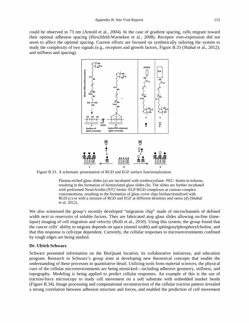
Appendix B: Site Visit Reports 153
could be observed in 73 nm (Arnold et al., 2004). In the case of gradient spacing, cells migrate toward their optimal adhesion spacing (Hirschfeld-Warneken et al., 2008). Receptor over-expression did not seem to affect the optimal spacing. Current efforts are focused on synthetically tailoring the system to study the complexity of two signals (e.g., receptors and growth factors, Figure B.33 (Shahal et al., 2012); and stiffness and spacing).
Figure B.33. A schematic presentation of RGD and EGF surface functionalization.
Plasma-etched glass slides (a) are incubated with triethoxysilane–PEG–biotin in toluene, resulting in the formation of biotinylated glass slides (b). The slides are further incubated with preformed NeutrAvidin (NT)–biotin–EGF/RGD complexes at various complex concentrations, resulting in the formation of glass cover slips biofunctionalized with RGD (c) or with a mixture of RGD and EGF at different densities and ratios (d) (Shahal et al. 2012).
We also witnessed the group’s recently developed “migration chip” made of microchannels of defined width next to reservoirs of soluble factors. They are fabricated atop glass slides allowing on-line (time-lapse) imaging of cell migration and velocity (Rolli et al., 2010). Using this system, the group found that the cancer cells’ ability to migrate depends on space (tunnel width) and sphingosylphosphorylcholine, and that this response is cell-type dependent. Currently, the cellular responses to microenvironments confined by rough edges are being studied.
Dr. Ulrich Schwarz
Schwarz presented information on the BioQuant location, its collaborative initiatives, and education program. Research in Schwarz’s group aims at developing new theoretical concepts that enable the understanding of these processes in quantitative detail. Utilizing tools from material sciences, the physical cues of the cellular microenvironments are being mimicked—including adhesive geometry, stiffness, and topography. Modeling is being applied to predict cellular responses. An example of this is the use of traction-force microscopy to study cell movement on a soft substrate with embedded marker beads (Figure B.34). Image processing and computational reconstruction of the cellular traction pattern revealed a strong correlation between adhesion structure and forces, and enabled the prediction of cell movement
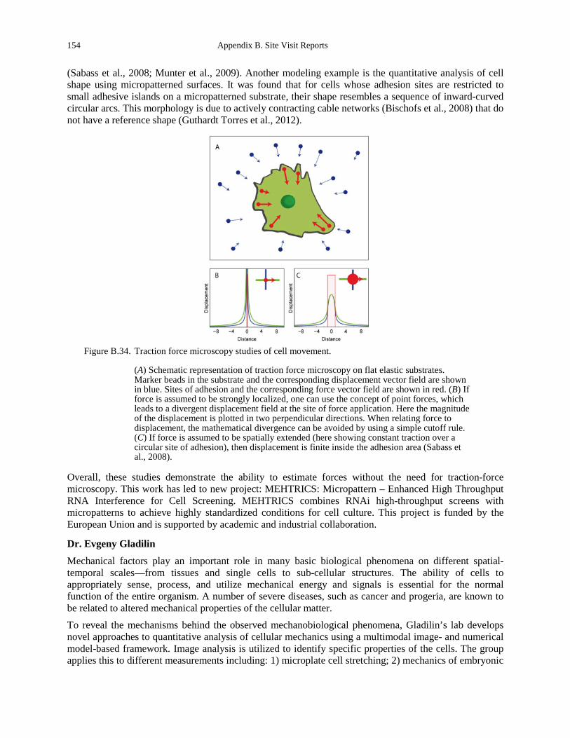
154 Appendix B. Site Visit Reports
(Sabass et al., 2008; Munter et al., 2009). Another modeling example is the quantitative analysis of cell shape using micropatterned surfaces. It was found that for cells whose adhesion sites are restricted to small adhesive islands on a micropatterned substrate, their shape resembles a sequence of inward-curved circular arcs. This morphology is due to actively contracting cable networks (Bischofs et al., 2008) that do not have a reference shape (Guthardt Torres et al., 2012).
Figure B.34. Traction force microscopy studies of cell movement.
(A) Schematic representation of traction force microscopy on flat elastic substrates. Marker beads in the substrate and the corresponding displacement vector field are shown in blue. Sites of adhesion and the corresponding force vector field are shown in red. (B) If force is assumed to be strongly localized, one can use the concept of point forces, which leads to a divergent displacement field at the site of force application. Here the magnitude of the displacement is plotted in two perpendicular directions. When relating force to displacement, the mathematical divergence can be avoided by using a simple cutoff rule. (C) If force is assumed to be spatially extended (here showing constant traction over a circular site of adhesion), then displacement is finite inside the adhesion area (Sabass et al., 2008).
Overall, these studies demonstrate the ability to estimate forces without the need for traction-force microscopy. This work has led to new project: MEHTRICS: Micropattern – Enhanced High Throughput RNA Interference for Cell Screening. MEHTRICS combines RNAi high-throughput screens with micropatterns to achieve highly standardized conditions for cell culture. This project is funded by the European Union and is supported by academic and industrial collaboration.
Dr. Evgeny Gladilin
Mechanical factors play an important role in many basic biological phenomena on different spatial-temporal scales—from tissues and single cells to sub-cellular structures. The ability of cells to appropriately sense, process, and utilize mechanical energy and signals is essential for the normal function of the entire organism. A number of severe diseases, such as cancer and progeria, are known to be related to altered mechanical properties of the cellular matter. To reveal the mechanisms behind the observed mechanobiological phenomena, Gladilin’s lab develops novel approaches to quantitative analysis of cellular mechanics using a multimodal image- and numerical model-based framework. Image analysis is utilized to identify specific properties of the cells. The group applies this to different measurements including: 1) microplate cell stretching; 2) mechanics of embryonic
Appendix B: Site Visit Reports 155
stem cell division; 3) drug-induced nuclear deformation; 4) substrate cell stretching; and 5) optical cell stretching. Future directions include combining chemical perturbation (drugs, knockdowns) and mechanical cell phenotyping, 3D culture experiments with fluorescent microscopy, high-throughput methods based on a combination of microfluidics and different deformation induction techniques, development of algorithms for unbiased data assessment (automatization) processing and analysis of 3D microscopic images, and numerical solvers based on mesh-free methods.
Dr. Ralf Kemkemer
The research of Kemkemer focuses on the understanding of the mechanics of cells as it relates to their cytoskeleton and the study of cell migration. To address these topics, materials are being utilized to develop advanced tissue culture tools. For example, a photo-switchable wound healing assay where non-adhesive regions are cleaved upon exposure to UV light (365 nm) result in creating adhesive sites to cells. Using this technology, the researchers were able to measure expansion kinetics of cell clusters upon exposure to UV. Using epithelial cells, they have found that cluster size and boundary curvature modulate the expansion of the cell sheet and the formation of leader cells (Rolli et al., 2011). Additional studies utilize inert hydrogels to explore the effect of stiffness/mechanics on early tumor growth in 3D.
Dr. Harald Herrmann
Located in the Division of Molecular Genetics at DKFZ, Herrmann’s group researches the structure and assembly of intermediate filament proteins. Intermediate filaments build two distinct systems in animal cells, one in the nucleus and one in the cytoplasm. The major function of intermediate filaments is speculated to be that of a mechanical stress absorber and an integrating device for the entire cytoskeleton. Herrmann’s research is focused on the nuclear lamins as they relates to extracellular matrix forces acting on the nucleus during cell migration. The group has recently demonstrated that lamin A and lamin C do not form heterodimers, but almost completely segregate (Kolb et al., 2011). Current studies focus on studying how fragmented genomic DNA binds to the lamin scaffold through comparison of in vivo and in vitro data.
Dr. Michael Knop
Research in Knop’s lab focuses on the processes that regulate cellular morphogenesis and cell signaling. They also study the cellular response to external stimuli that are processed via mitogen-activated protein kinase (MAPK) signaling pathways and lead to specific adaptations. The group utilizes high-throughput genetics, microscopy, and systems biology with yeast for functional imaging of protein behavior. An example of a systems approach to studying MAPK singling was shown. To quantify the abundance of complexes in the cytoplasm among different MAPKs in yeast pheromone signaling, the group used fluorescence cross-correlation spectroscopy (FCCS). They have found that specific MPAKs—Fus 3—forms a gradient of activity across the cell via a reaction-diffusion mechanism (Maeder et al., 2007). The group built on the FCCS technology to develop a microscope based on light-sheet illumination that allows massively parallel fluorescence correlation spectroscopy measurements (Figure B.35). Based on knowledge about the optical properties of the setup, it is possible to calculate spatially resolved maps of protein concentrations and mobilities—especially maps of diffusion coefficients and interaction properties. Using this technology, the group reported the diffusion and interactions of proteins in mammalian cells and in isolated fly tissue (Capoulade et al., 2011).

156 Appendix B. Site Visit Reports
Figure B.35. Microscope based on light-sheet illumination that allows massively parallel fluorescence
correlation spectroscopy measurements.
(a) Front view of the main components of the light-pad microscope. The specimen is contained in a Petri dish. The first objective lens illuminates a thin slice in the specimen. Optical sectioning is performed under 45° with respect to the bottom of the Petri dish. The high aperture angle of the illumination light-sheet (74°) leads to weak illumination of cells lying outside the light-pad area [inset (i)]. Fluorescence is detected at a right angle to the illumination plane by the detection lens. Spatial filters in the illumination path and the detection path confine the observed area to a rectangular array of volume elements, the light-pad [inset (ii)]. An inverted microscope allows convenient positioning of the specimen. (b) Each individual pixel of the EM-CCD records a fluctuating fluorescence signal over time. These fluctuations are analyzed by temporal correlation analysis resulting in one ACF for each pixel. The ACF provides information about the diffusion coefficient D (dashed line) and the concentration C of diffusing fluorescently labeled molecules (amplitude of the curve) (Capoulade et al., 2011).
Dr. Marcel Schilling
Schilling’s work in the division of Dr. Ursula Klingmüller focuses on systems biology of signal transduction. The research aims are: 1) unraveling principal mechanisms of erythropoietin (Epo)-mediated cellular decisions in the hematopoietic system as well as the role of the EpoR system in lung
Appendix B: Site Visit Reports 157
cancer; 2) bridging from the cellular to the whole organ level during liver regeneration; and 3) prediction of strategies for efficient intervention in diseases. These projects are enabled by collaborative efforts of biology (Klingmüller and Schilling), theory (Drs. Jens Timmer and Hauke Busch), technology (Drs. Dirk-Peter Herten and Wolf-Dieter Lehmann), and medicine (Drs. Michael Thomas and Anthony D. Ho). This approach allows the conversion of time-resolved quantitative experimental data to mathematical models. As an example the study of Epo-mediated cellular decisions in the hematopoietic system was described. By mathematical modeling of quantitative data combined with experimental validation, the group has shown that rapid ligand depletion and replenishment of the cell surface receptor are characteristic features of the Epo receptor. They have also found a linear relation of Epo levels and Epo receptor activation over a broad range of ligand concentrations (Becker et al., 2010). Ongoing efforts apply data-based mathematical models for the rapid testing of hypotheses to uncover deregulation in cancer and to predict strategies of intervention in diseases towards personalized medicine.
Several large initiatives are centered around BioQuant:
• BIOMS: Center for Modeling and Simulation in Bioscience; www.bioms.de/ • CellNetworks: From molecular mechanisms to quantitative understanding of complex functions;
www.cellnetworks.uni-hd.de/ • VIROQUANT: Systems biology of virus-cell interactions; www.viroquant.uni-hd.de/ • SBCANCER: Systems biology of signaling in cancer; www.dkfz.de/en/sbcancer/ Interdisciplinary science education programs are offered for Heidelberg life sciences:
• EMBL international Ph.D. program • DKFZ Helmholtz International Graduate School for Cancer Research • HBIGS molecular and cell biology • HGS MathComp (computational sciences) • HGS fundamental physics (plans to include biophysics in second funding period)
SUMMARY AND CONCLUSIONS
BioQuant is Europe’s first quantitative biology center. It supports interdisciplinary collaboration between research groups from the biological and biomedical sciences and research from chemistry, physics,
158 Appendix B. Site Visit Reports
mathematics, and computer sciences. Emphasis on collaborative effort, through large initiatives and interdisciplinary education programs, enables the exploration and development of pioneering research approaches.
REFERENCES Arnold, M., Cavalcanti-Adam, E.A., Glass, R., Blummel, J., Eck, W., Kantlehner, M., Kessler, H., and Spatz, J.P. (2004).
Activation of integrin function by nanopatterned adhesive interfaces. Chemphyschem : a European journal of chemical physics and physical chemistry 5, 383-388.
Becker, V., Schilling, M., Bachmann, J., Baumann, U., Raue, A., Maiwald, T., Timmer, J., and Klingmuller, U. (2010). Covering a broad dynamic range: information processing at the erythropoietin receptor. Science 328, 1404-1408.
Bischofs, I.B., Klein, F., Lehnert, D., Bastmeyer, M., and Schwarz, U.S. (2008). Filamentous network mechanics and active contractility determine cell and tissue shape. Biophysical journal 95, 3488-3496.
Capoulade, J., Wachsmuth, M., Hufnagel, L., and Knop, M. (2011). Quantitative fluorescence imaging of protein diffusion and interaction in living cells. Nature biotechnology 29, 835-839.
Guthardt Torres, P., Bischofs, I.B., and Schwarz, U.S. (2012). Contractile network models for adherent cells. Physical review E, Statistical, nonlinear, and soft matter physics 85, 011913.
Hirschfeld-Warneken, V.C., Arnold, M., Cavalcanti-Adam, A., Lopez-Garcia, M., Kessler, H., and Spatz, J.P. (2008). Cell adhesion and polarisation on molecularly defined spacing gradient surfaces of cyclic RGDfK peptide patches. European journal of cell biology 87, 743-750.
Kolb, T., Maass, K., Hergt, M., Aebi, U., and Herrmann, H. (2011). Lamin A and lamin C form homodimers and coexist in higher complex forms both in the nucleoplasmic fraction and in the lamina of cultured human cells. Nucleus 2, 425-433.
Maeder, C.I., Hink, M.A., Kinkhabwala, A., Mayr, R., Bastiaens, P.I., and Knop, M. (2007). Spatial regulation of Fus3 MAP kinase activity through a reaction-diffusion mechanism in yeast pheromone signalling. Nature cell biology 9, 1319-1326.
Munter, S., Sabass, B., Selhuber-Unkel, C., Kudryashev, M., Hegge, S., Engel, U., Spatz, J.P., Matuschewski, K., Schwarz, U.S., and Frischknecht, F. (2009). Plasmodium sporozoite motility is modulated by the turnover of discrete adhesion sites. Cell host & microbe 6, 551-562.
Rolli, C.G., Nakayama, H., Yamaguchi, K., Spatz, J.P., Kemkemer, R., and Nakanishi, J. (2011). Switchable adhesive substrates: revealing geometry dependence in collective cell behavior. Biomaterials 33, 2409-2418.
Rolli, C.G., Seufferlein, T., Kemkemer, R., and Spatz, J.P. (2010). Impact of tumor cell cytoskeleton organization on invasiveness and migration: a microchannel-based approach. PLoS One 5, e8726.
Sabass, B., Gardel, M.L., Waterman, C.M., and Schwarz, U.S. (2008). High resolution traction force microscopy based on experimental and computational advances. Biophysical journal 94, 207-220.
Shahal, T., Geiger, B., Dunlop, I.E., and Spatz, J.P. (2012). Regulation of integrin adhesions by varying the density of substrate-bound epidermal growth factor. Biointerphases 7, 23.
Appendix B: Site Visit Reports 159
Instituut-Lorentz
Site Address: (meeting at the Hubrecht Institute) Uppsalalaan 8 3584 CT Utrecht, NETHERLANDS www.leidenuniv.nl/
Date Visited: 7 May 2012 WTEC Attendees: Paul Janmey, Cindy Reinhart-King (site report author), Dan Fletcher, Nastaran Kuhn,
Nicole Moore, and Hemant Sarin Host(s): Helmut Schiessel, Group Leader
Statistical Physics of Biological Matter Instituut-Lorentz for Theoretical Physics Niels Bohrweg 2 2333 CA Leiden, NETHERLANDS [email protected] Tel: +31 71 5275505
OVERVIEW
There is a long, rich tradition of physics in Leiden. The Leiden Institute of Physics (LION) was established in the fall of 1993 in order to promote and stimulate research of the highest level in a number of priority fields in experimental and theoretical physics; and to create and sustain the necessary infrastructure for an outstanding graduate education in experimental and theoretical physics. In addition, LION is responsible for teaching physics at the undergraduate level. The institute consists of the Kamerlingh Onnes Laboratorium, the Huygens Laboratorium, and the Instituut-Lorentz founded in 1921. The research groups of LION participate in three research schools that include the Casimir Research School with Delft University of Technology, Dutch Research School for Theoretical Physics, and the Graduate School for the Structure and Function of Bio-macromolecules between research groups at the Leiden Institute for Chemistry and LION.
RESEARCH AND DEVELOPMENT ACTIVITIES
While at the Hubrecht Institute, we met with Helmut Schiessel from the Instituut-Lorentz. He works in the area of chromatin mechanics, including the mechanics of single base-pair steps and the structure of chromosomes and chromatin fibers. He has developed a theory that can explain data from the Seidel Lab of the extension of DNA when twisted using magnetic tweezers. In addition, he has found that there is an energy barrier to nucleosome unwrapping from DNA and force-induced strengthening that prevents unwinding (Figure B.36). A change in conformation is necessary for nucleosome unwrapping, and just simply pulling is not sufficient to induce unwrapping. Schiessel has authored a book entitled Biophysics for Beginners: A Journey Through the Cell Nucleus through Pan Stanford Publishing.
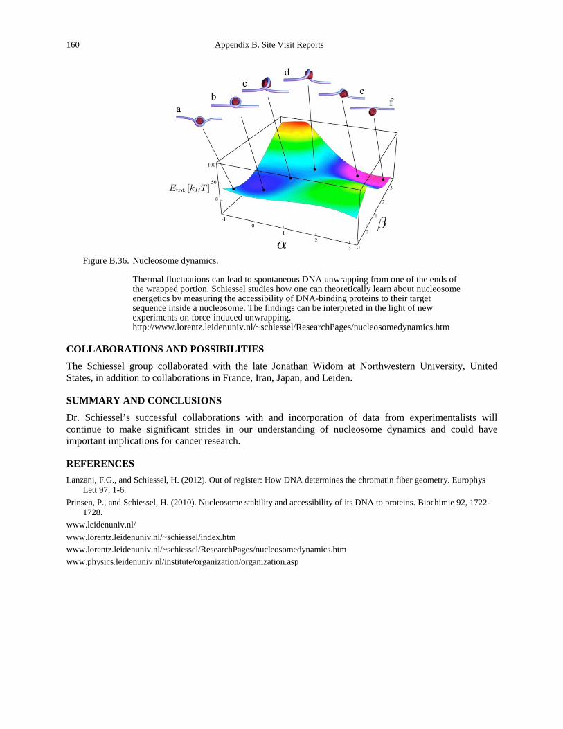
160 Appendix B. Site Visit Reports
Figure B.36. Nucleosome dynamics.
Thermal fluctuations can lead to spontaneous DNA unwrapping from one of the ends of the wrapped portion. Schiessel studies how one can theoretically learn about nucleosome energetics by measuring the accessibility of DNA-binding proteins to their target sequence inside a nucleosome. The findings can be interpreted in the light of new experiments on force-induced unwrapping. http://www.lorentz.leidenuniv.nl/~schiessel/ResearchPages/nucleosomedynamics.htm
COLLABORATIONS AND POSSIBILITIES
The Schiessel group collaborated with the late Jonathan Widom at Northwestern University, United States, in addition to collaborations in France, Iran, Japan, and Leiden.
SUMMARY AND CONCLUSIONS
Dr. Schiessel’s successful collaborations with and incorporation of data from experimentalists will continue to make significant strides in our understanding of nucleosome dynamics and could have important implications for cancer research.
REFERENCES Lanzani, F.G., and Schiessel, H. (2012). Out of register: How DNA determines the chromatin fiber geometry. Europhys
Lett 97, 1-6. Prinsen, P., and Schiessel, H. (2010). Nucleosome stability and accessibility of its DNA to proteins. Biochimie 92, 1722-
1728. www.leidenuniv.nl/ www.lorentz.leidenuniv.nl/~schiessel/index.htm www.lorentz.leidenuniv.nl/~schiessel/ResearchPages/nucleosomedynamics.htm www.physics.leidenuniv.nl/institute/organization/organization.asp
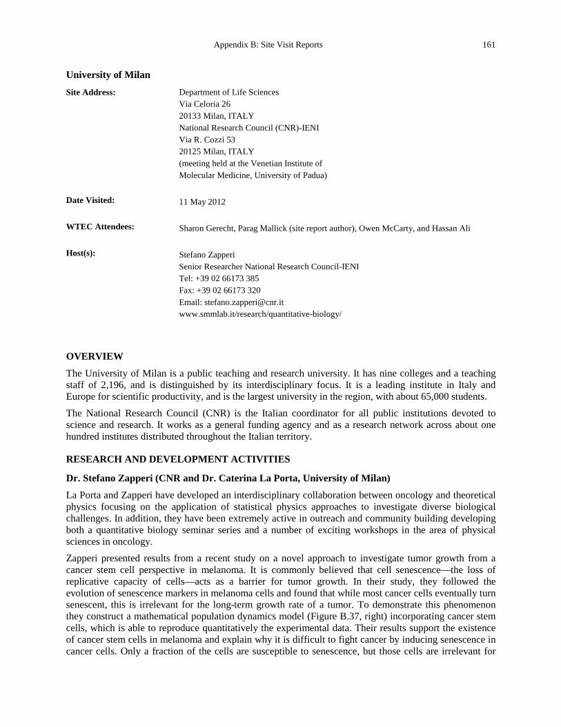
Appendix B: Site Visit Reports 161
University of Milan
Site Address: Department of Life Sciences Via Celoria 26 20133 Milan, ITALY National Research Council (CNR)-IENI Via R. Cozzi 53 20125 Milan, ITALY (meeting held at the Venetian Institute of Molecular Medicine, University of Padua)
Date Visited: 11 May 2012 WTEC Attendees: Sharon Gerecht, Parag Mallick (site report author), Owen McCarty, and Hassan Ali Host(s): Stefano Zapperi
Senior Researcher National Research Council-IENI Tel: +39 02 66173 385 Fax: +39 02 66173 320 Email: [email protected] www.smmlab.it/research/quantitative-biology/
OVERVIEW
The University of Milan is a public teaching and research university. It has nine colleges and a teaching staff of 2,196, and is distinguished by its interdisciplinary focus. It is a leading institute in Italy and Europe for scientific productivity, and is the largest university in the region, with about 65,000 students.
The National Research Council (CNR) is the Italian coordinator for all public institutions devoted to science and research. It works as a general funding agency and as a research network across about one hundred institutes distributed throughout the Italian territory.
RESEARCH AND DEVELOPMENT ACTIVITIES
Dr. Stefano Zapperi (CNR and Dr. Caterina La Porta, University of Milan)
La Porta and Zapperi have developed an interdisciplinary collaboration between oncology and theoretical physics focusing on the application of statistical physics approaches to investigate diverse biological challenges. In addition, they have been extremely active in outreach and community building developing both a quantitative biology seminar series and a number of exciting workshops in the area of physical sciences in oncology. Zapperi presented results from a recent study on a novel approach to investigate tumor growth from a cancer stem cell perspective in melanoma. It is commonly believed that cell senescence—the loss of replicative capacity of cells—acts as a barrier for tumor growth. In their study, they followed the evolution of senescence markers in melanoma cells and found that while most cancer cells eventually turn senescent, this is irrelevant for the long-term growth rate of a tumor. To demonstrate this phenomenon they construct a mathematical population dynamics model (Figure B.37, right) incorporating cancer stem cells, which is able to reproduce quantitatively the experimental data. Their results support the existence of cancer stem cells in melanoma and explain why it is difficult to fight cancer by inducing senescence in cancer cells. Only a fraction of the cells are susceptible to senescence, but those cells are irrelevant for
162 Appendix B. Site Visit Reports
tumor growth. A successful therapeutic strategy should instead target cancer stem cells, which are, however, likely to be strongly resistant to drug-induced senescence. This result is quite important and highlights the importance of evolutionary modeling of tumor growth as well as the possible insights that come from formal modeling approaches.
Figure B.37. Stochastic and cancer stem cell model for tumors (La Porta et al., 2012).
SOURCES OF SUPPORT/FUNDING
European Science Foundation, Centre Européen de Calcul Atomique et Moléculaire
SUMMARY AND CONCLUSIONS
The interdisciplinary research underway in Milan is extremely exciting. They are looking at cancer through an evolutionary lens and developing rigorous approaches to describe evolutionary dynamics.
REFERENCES www.smmlab.it/ www.cecam.org/workshop-0-751.html www.cancerphysics.unimi.it www.ploscompbiol.org/article/info%3Adoi%2F10.1371%2Fjournal.pcbi.1002316 www.nature.com/srep/2012/120607/srep00441/full/srep00441.html La Porta, C.A., Zapperi, S., and Sethna, J.P. (2012). Senescent cells in growing tumors: population dynamics and cancer
stem cells. PLoS Computational Biology 8, e1002316.
Appendix B: Site Visit Reports 163
University of Mons
Site Address: (meeting at Institute Curie, Paris) 26 rue d’Ulm 75248 Paris Cedex 05, FRANCE http://portail.umons.ac.be/en2/pages/default.aspx
Date Visited: 9 May 2012 WTEC Attendees: Paul Janmey, Cindy Reinhart-King, Dan Fletcher (site report author), Jerry Lee, Nastaran
Kuhn, and Hemant Sarin Host(s): Sylvain Gabriele
Group Leader of Mechanobiology and Soft Matter University of Mons Campus of Sciences and Medicine, Mendeleïev Building, 1-059 Maistriau B-7000 Avenue Mons, BELGIUM Email: [email protected] Tel: +32 65 37 3824
OVERVIEW
The University of Mons is the founding partner of the Hainaut higher education consortium, which consists of the university and other higher education institutions. The University of Mons was represented by Dr. Sylvain Gabriele, Mechanobiology and Soft Matter group leader.
RESEARCH AND DEVELOPMENT ACTIVITIES
Gabriele presented an overview of his basic and applied research projects. He has developed a microfluidic system to mimic lung capillaries. This work was motivated by acute respiratory distress syndrome (ARDS), in which blood cells become lodged in the microvasculature. The microfluidic system passes cells through small capillaries and allows quantification of the likelihood of aggregation and blockage. Use of devices such as this could provide an early warning for ARDS. In a second project on blast-induced traumatic brain injury, Gabriele has developed a high-speed uniaxial stretcher that can simulate the effects of blast-induced injuries. This work has the potential to identify key events in blast-induced trauma at the tissue and cell level using model systems. Gabriele’s basic research is focused on understanding mechanical interactions of cells, such as the role of integrins in the propagation of strain-induced injury. Using magnetic tweezers, he has recently been able to apply and measure localized strains. His ongoing research uses micropatterning to control nuclear shape and evaluate its impact on chromatin organization and proliferation, as well as determining integrin-cadherin crosstalk (Figure B.38).
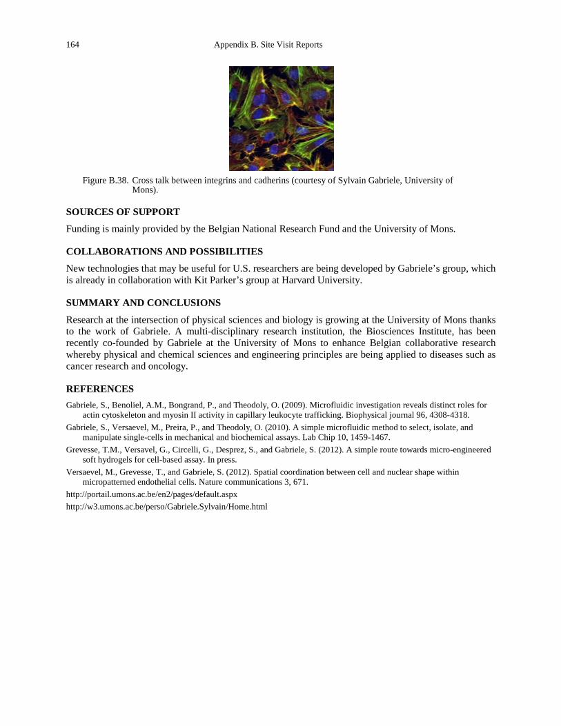
164 Appendix B. Site Visit Reports
Figure B.38. Cross talk between integrins and cadherins (courtesy of Sylvain Gabriele, University of
Mons).
SOURCES OF SUPPORT
Funding is mainly provided by the Belgian National Research Fund and the University of Mons.
COLLABORATIONS AND POSSIBILITIES
New technologies that may be useful for U.S. researchers are being developed by Gabriele’s group, which is already in collaboration with Kit Parker’s group at Harvard University.
SUMMARY AND CONCLUSIONS
Research at the intersection of physical sciences and biology is growing at the University of Mons thanks to the work of Gabriele. A multi-disciplinary research institution, the Biosciences Institute, has been recently co-founded by Gabriele at the University of Mons to enhance Belgian collaborative research whereby physical and chemical sciences and engineering principles are being applied to diseases such as cancer research and oncology.
REFERENCES Gabriele, S., Benoliel, A.M., Bongrand, P., and Theodoly, O. (2009). Microfluidic investigation reveals distinct roles for
actin cytoskeleton and myosin II activity in capillary leukocyte trafficking. Biophysical journal 96, 4308-4318. Gabriele, S., Versaevel, M., Preira, P., and Theodoly, O. (2010). A simple microfluidic method to select, isolate, and
manipulate single-cells in mechanical and biochemical assays. Lab Chip 10, 1459-1467. Grevesse, T.M., Versavel, G., Circelli, G., Desprez, S., and Gabriele, S. (2012). A simple route towards micro-engineered
soft hydrogels for cell-based assay. In press. Versaevel, M., Grevesse, T., and Gabriele, S. (2012). Spatial coordination between cell and nuclear shape within
micropatterned endothelial cells. Nature communications 3, 671. http://portail.umons.ac.be/en2/pages/default.aspx http://w3.umons.ac.be/perso/Gabriele.Sylvain/Home.html
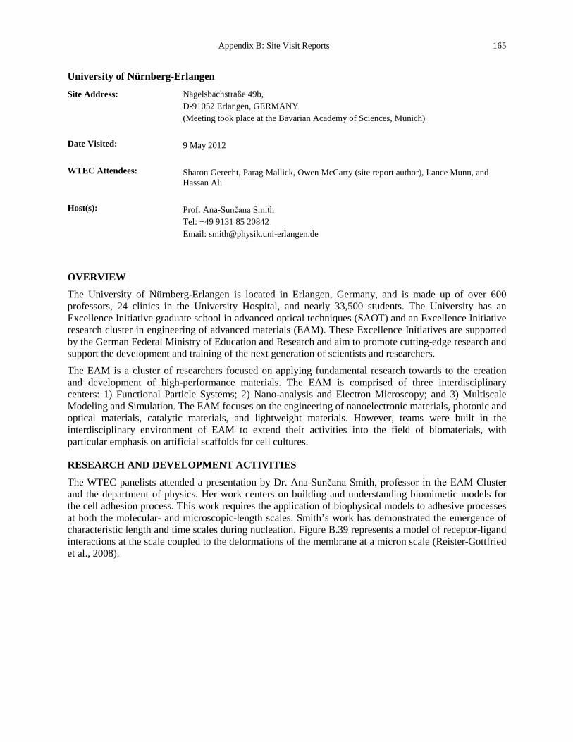
Appendix B: Site Visit Reports 165
University of Nürnberg-Erlangen
Site Address: Nägelsbachstraße 49b, D-91052 Erlangen, GERMANY (Meeting took place at the Bavarian Academy of Sciences, Munich)
Date Visited: 9 May 2012 WTEC Attendees: Sharon Gerecht, Parag Mallick, Owen McCarty (site report author), Lance Munn, and
Hassan Ali Host(s): Prof. Ana-Sunčana Smith
Tel: +49 9131 85 20842 Email: [email protected]
OVERVIEW
The University of Nürnberg-Erlangen is located in Erlangen, Germany, and is made up of over 600 professors, 24 clinics in the University Hospital, and nearly 33,500 students. The University has an Excellence Initiative graduate school in advanced optical techniques (SAOT) and an Excellence Initiative research cluster in engineering of advanced materials (EAM). These Excellence Initiatives are supported by the German Federal Ministry of Education and Research and aim to promote cutting-edge research and support the development and training of the next generation of scientists and researchers. The EAM is a cluster of researchers focused on applying fundamental research towards to the creation and development of high-performance materials. The EAM is comprised of three interdisciplinary centers: 1) Functional Particle Systems; 2) Nano-analysis and Electron Microscopy; and 3) Multiscale Modeling and Simulation. The EAM focuses on the engineering of nanoelectronic materials, photonic and optical materials, catalytic materials, and lightweight materials. However, teams were built in the interdisciplinary environment of EAM to extend their activities into the field of biomaterials, with particular emphasis on artificial scaffolds for cell cultures.
RESEARCH AND DEVELOPMENT ACTIVITIES
The WTEC panelists attended a presentation by Dr. Ana-Sunčana Smith, professor in the EAM Cluster and the department of physics. Her work centers on building and understanding biomimetic models for the cell adhesion process. This work requires the application of biophysical models to adhesive processes at both the molecular- and microscopic-length scales. Smith’s work has demonstrated the emergence of characteristic length and time scales during nucleation. Figure B.39 represents a model of receptor-ligand interactions at the scale coupled to the deformations of the membrane at a micron scale (Reister-Gottfried et al., 2008).
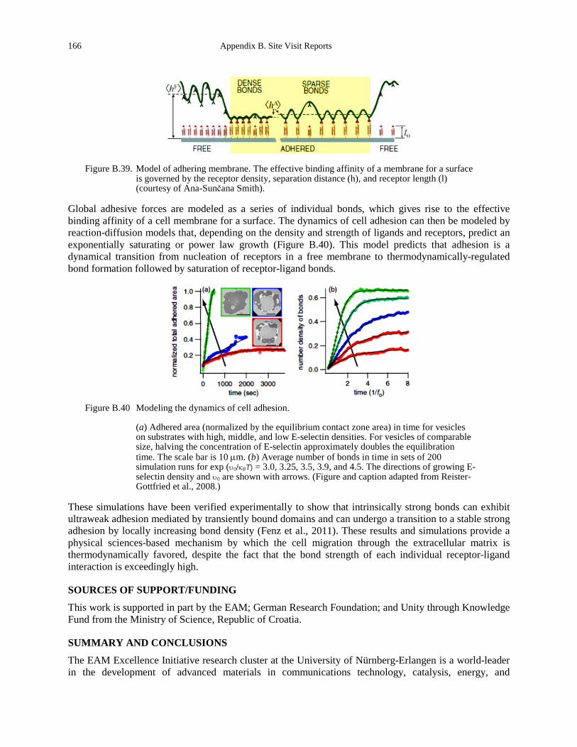
166 Appendix B. Site Visit Reports
Figure B.39. Model of adhering membrane. The effective binding affinity of a membrane for a surface
is governed by the receptor density, separation distance (h), and receptor length (l) (courtesy of Ana-Sunčana Smith).
Global adhesive forces are modeled as a series of individual bonds, which gives rise to the effective binding affinity of a cell membrane for a surface. The dynamics of cell adhesion can then be modeled by reaction-diffusion models that, depending on the density and strength of ligands and receptors, predict an exponentially saturating or power law growth (Figure B.40). This model predicts that adhesion is a dynamical transition from nucleation of receptors in a free membrane to thermodynamically-regulated bond formation followed by saturation of receptor-ligand bonds.
Figure B.40 Modeling the dynamics of cell adhesion.
(a) Adhered area (normalized by the equilibrium contact zone area) in time for vesicles on substrates with high, middle, and low E-selectin densities. For vesicles of comparable size, halving the concentration of E-selectin approximately doubles the equilibration time. The scale bar is 10 µm. (b) Average number of bonds in time in sets of 200 simulation runs for exp (υ0/κBT) = 3.0, 3.25, 3.5, 3.9, and 4.5. The directions of growing E-selectin density and υ0 are shown with arrows. (Figure and caption adapted from Reister-Gottfried et al., 2008.)
These simulations have been verified experimentally to show that intrinsically strong bonds can exhibit ultraweak adhesion mediated by transiently bound domains and can undergo a transition to a stable strong adhesion by locally increasing bond density (Fenz et al., 2011). These results and simulations provide a physical sciences-based mechanism by which the cell migration through the extracellular matrix is thermodynamically favored, despite the fact that the bond strength of each individual receptor-ligand interaction is exceedingly high.
SOURCES OF SUPPORT/FUNDING
This work is supported in part by the EAM; German Research Foundation; and Unity through Knowledge Fund from the Ministry of Science, Republic of Croatia.
SUMMARY AND CONCLUSIONS
The EAM Excellence Initiative research cluster at the University of Nürnberg-Erlangen is a world-leader in the development of advanced materials in communications technology, catalysis, energy, and

Appendix B: Site Visit Reports 167
transportation fields. The strength of the EAM is bridging the length scales from the molecular level (10-9 m) to macroscopic level (10-3 m) for the creation and application of new materials for emerging technologies.
REFERENCES Fenz, S.F., Bihr, T., Merkel, R., Seifert, U., Sengupta, K., and Smith, A.S. (2011). Switching from ultraweak to strong
adhesion. Advanced materials 23, 2622-2626. Reister-Gottfried, E., Sengupta, K., Lorz, B., Sackmann, E., Seifert, U., and Smith, A.S. (2008). Dynamics of specific
vesicle-substrate adhesion: from local events to global dynamics. Physical review letters 101, 208103.
168 Appendix B. Site Visit Reports
Venetian Institute of Molecular Medicine
Site Address: University of Padua Via Orus 2, 35129 Padua, ITALY www.vimm.it
Date Visited: 11 May 2012 WTEC Attendees: Sharon Gerecht (site report author), Parag Mallick, Owen McCarty, and Hassan Ali Host(s): Nicola Elvassore
Venetian Institute of Molecular Medicine Via Orus 2, 35129 Padua Tel: +39 049 7923 211 Fax: +39 049 7923 250 www.vimm.it/research/group.php?PI=172 Department of Chemical Engineering University of Padova via Marzolo 9, 35131 Padova Tel: +39 049 827 5469 Fax: +39 049 827 5461 Email: [email protected] www.bioera.dipic.unipd.it/Bioera/
Giacinto Scoles European Research Council Grantee at the Azienda Ospedaliera Universitaria dell’Universita’ di Udine, Padiglione #13 Udine 3300, ITALY Tel: +39 0432 559 413 Fax: +39 040 375 8565 Email: [email protected] www.princeton.edu/~chemdept/Scoles/SCOLES%20NEW/Scolessite/index.html
Barbara Molon Foundation for Advanced Biomedical Research Venetian Institute of Molecular Medicine - VIMM via Orus 2 - 35129 Padua, ITALY www.vimm.it , www.fondbiomed.it Email: [email protected]
OVERVIEW
The Venetian Institute of Molecular Medicine (VIMM) is a new center for advanced biomedical research. It was established 10 years ago by the Foundation for Advanced Biomedical Research (a private consortium) as a joint venture with the University of Padua and the Padua Health Authority. VIMM is strategically located between the University Hospital and the Preclinical Science Campus. It occupies an area of about 3,000 m2. Once the remaining 8,000 m2 is finished, the complex will become the major center for research in molecular medicine in the Veneto region. Creation of VIMM, a modern
Appendix B: Site Visit Reports 169
building with facilities for advanced research, was made possible by private fundraising, which actively involved industries and financial institutions.
The scientific goal of the institute is to integrate basic and clinical research in order to achieve a faster transfer of the developments of molecular and cellular biology into the clinic. The basic research themes of the institute revolve around four basic areas: 1) structural biology; 2) cellular biology; 3) host-pathogen interactions and its implications for gene therapy; and 4) cellular and molecular oncology. Currently, there are 20 principal investigators, of which three are young investigators. Half of the investigators hold dual appointments with the University of Padua. VIMM’s heart is represented by newly recruited postdoctoral fellows and faculty members. A special effort is being made to attract promising scientists in Europe and the United States.
Dr. Nicola Elvassore
Sharing facilities in VIMM and University of Padua, research in Elvassore’s laboratory applies engineering principles with biological approaches to rationally understand the mechanisms governing cell behavior. The main engineering tools to recapitulate the cell microenvironment include substrate engineering, micropatterning technology, and microfluidics technology. Utilizing substrates with stiffness ranging from 12-21 kPa, the group has shown that sarcomere formation during myoblast differentiation occurs on softer substrate (15 kPa) which in turn enables the maturation and functionality of myotubes (Serena et al., 2010). This model is now being used to study Muscular Dystrophy, where the soft surrounding tissue of the diseased muscle results in dysfunctional myotubes with impaired calcium release and upregulation in dystrophin expression. In a recent collaboration with Piccolo, the groups identified the YAP/TAZ [Yorkie-homologues YAP (Yes-associated protein) and TAZ (transcriptional coactivator with PDZ-binding motif)] as nuclear relays of mechanical signals exerted by extracellular rigidity and cell shape, independently from Hippo pathways (Dupont et al., 2011). Utilizing micropillar substrates with varying rigidity, the groups were able to decouple matrix stiffness and adhesion. The ease of use of the technology facilitated robust examination and in-depth understanding of the underlying mechanism. Piccolo counted 7,000 experiments in which the technology enabled discoveries to move forward. Control over substrate topography (using micropatterning) was shown to affect myoblast proliferation and differentiation (Zatti et al., 2012) and recently is being applied to study cardiac differentiation of human embryonic stem cells. Final examples were presented for the use of microfluidic technology to fabricate a miniature culture system for the robust study of cellular behaviors in response to a soluble factor. One device is comprised of culture chambers that allow control over the soluble environment with on-line monitoring of the cultured cells. Another microfluidic technology includes a gas exchanger for control over dissolved oxygen levels for the study of the hypoxic effect on calcium transients in response to electrical stimulation of muscle cell derivatives (Figure B.41).
Dr. Giacinto Scoles
Scoles is well-known in North America as a chemical physicist. He works at the University Hospital of the University of Udine, the largest city in the northeast region of Italy. He has recently obtained support through the ERC for building a network of laboratories in Italy’s northwest region that comprises, in addition to Scoles’ lab and Dr. C.A. Beltrami’s pathology lab in Udine, the IOM microfabrication lab and the synchrotron light lab elettra in Basovizza, Trieste, and the oncopharmacology lab of Dr. G. Toffoli at the Center of Reference for Oncology in Aviano, Pordenone. The aim of this collaboration is to use MEMS- and AFM-based nanotechnologies to impact the diagnostics and eventually the cure of metastatic cancer.
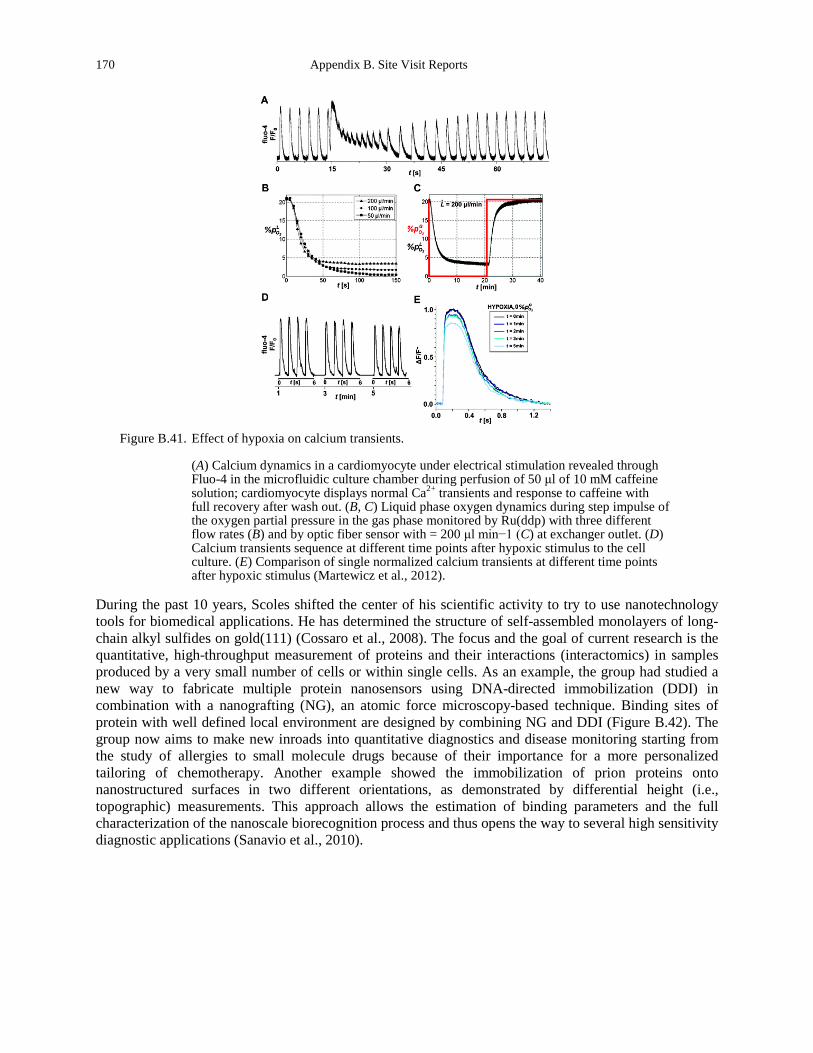
170 Appendix B. Site Visit Reports
Figure B.41. Effect of hypoxia on calcium transients.
(A) Calcium dynamics in a cardiomyocyte under electrical stimulation revealed through Fluo-4 in the microfluidic culture chamber during perfusion of 50 μl of 10 mM caffeine solution; cardiomyocyte displays normal Ca2+ transients and response to caffeine with full recovery after wash out. (B, C) Liquid phase oxygen dynamics during step impulse of the oxygen partial pressure in the gas phase monitored by Ru(ddp) with three different flow rates (B) and by optic fiber sensor with = 200 μl min−1 (C) at exchanger outlet. (D) Calcium transients sequence at different time points after hypoxic stimulus to the cell culture. (E) Comparison of single normalized calcium transients at different time points after hypoxic stimulus (Martewicz et al., 2012).
During the past 10 years, Scoles shifted the center of his scientific activity to try to use nanotechnology tools for biomedical applications. He has determined the structure of self-assembled monolayers of long-chain alkyl sulfides on gold(111) (Cossaro et al., 2008). The focus and the goal of current research is the quantitative, high-throughput measurement of proteins and their interactions (interactomics) in samples produced by a very small number of cells or within single cells. As an example, the group had studied a new way to fabricate multiple protein nanosensors using DNA-directed immobilization (DDI) in combination with a nanografting (NG), an atomic force microscopy-based technique. Binding sites of protein with well defined local environment are designed by combining NG and DDI (Figure B.42). The group now aims to make new inroads into quantitative diagnostics and disease monitoring starting from the study of allergies to small molecule drugs because of their importance for a more personalized tailoring of chemotherapy. Another example showed the immobilization of prion proteins onto nanostructured surfaces in two different orientations, as demonstrated by differential height (i.e., topographic) measurements. This approach allows the estimation of binding parameters and the full characterization of the nanoscale biorecognition process and thus opens the way to several high sensitivity diagnostic applications (Sanavio et al., 2010).
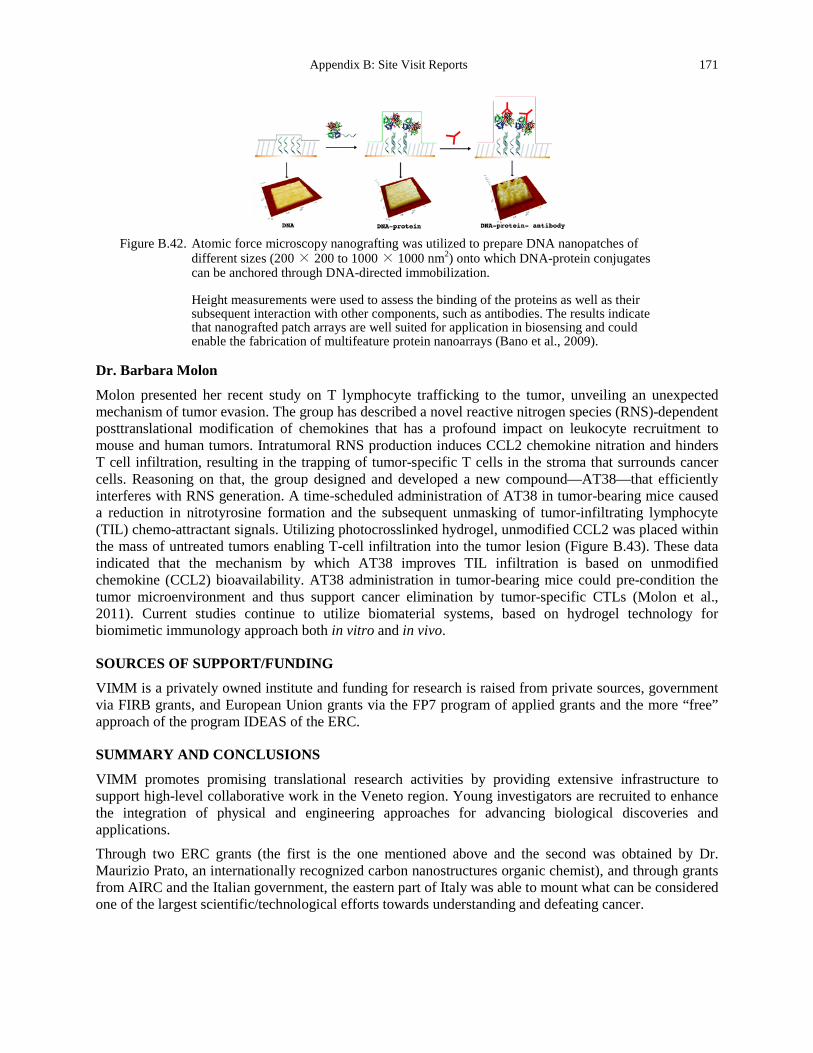
Appendix B: Site Visit Reports 171
Figure B.42. Atomic force microscopy nanografting was utilized to prepare DNA nanopatches of
different sizes (200 × 200 to 1000 × 1000 nm2) onto which DNA-protein conjugates can be anchored through DNA-directed immobilization.
Height measurements were used to assess the binding of the proteins as well as their subsequent interaction with other components, such as antibodies. The results indicate that nanografted patch arrays are well suited for application in biosensing and could enable the fabrication of multifeature protein nanoarrays (Bano et al., 2009).
Dr. Barbara Molon
Molon presented her recent study on T lymphocyte trafficking to the tumor, unveiling an unexpected mechanism of tumor evasion. The group has described a novel reactive nitrogen species (RNS)-dependent posttranslational modification of chemokines that has a profound impact on leukocyte recruitment to mouse and human tumors. Intratumoral RNS production induces CCL2 chemokine nitration and hinders T cell infiltration, resulting in the trapping of tumor-specific T cells in the stroma that surrounds cancer cells. Reasoning on that, the group designed and developed a new compound—AT38—that efficiently interferes with RNS generation. A time-scheduled administration of AT38 in tumor-bearing mice caused a reduction in nitrotyrosine formation and the subsequent unmasking of tumor-infiltrating lymphocyte (TIL) chemo-attractant signals. Utilizing photocrosslinked hydrogel, unmodified CCL2 was placed within the mass of untreated tumors enabling T-cell infiltration into the tumor lesion (Figure B.43). These data indicated that the mechanism by which AT38 improves TIL infiltration is based on unmodified chemokine (CCL2) bioavailability. AT38 administration in tumor-bearing mice could pre-condition the tumor microenvironment and thus support cancer elimination by tumor-specific CTLs (Molon et al., 2011). Current studies continue to utilize biomaterial systems, based on hydrogel technology for biomimetic immunology approach both in vitro and in vivo.
SOURCES OF SUPPORT/FUNDING
VIMM is a privately owned institute and funding for research is raised from private sources, government via FIRB grants, and European Union grants via the FP7 program of applied grants and the more “free” approach of the program IDEAS of the ERC.
SUMMARY AND CONCLUSIONS
VIMM promotes promising translational research activities by providing extensive infrastructure to support high-level collaborative work in the Veneto region. Young investigators are recruited to enhance the integration of physical and engineering approaches for advancing biological discoveries and applications. Through two ERC grants (the first is the one mentioned above and the second was obtained by Dr. Maurizio Prato, an internationally recognized carbon nanostructures organic chemist), and through grants from AIRC and the Italian government, the eastern part of Italy was able to mount what can be considered one of the largest scientific/technological efforts towards understanding and defeating cancer.
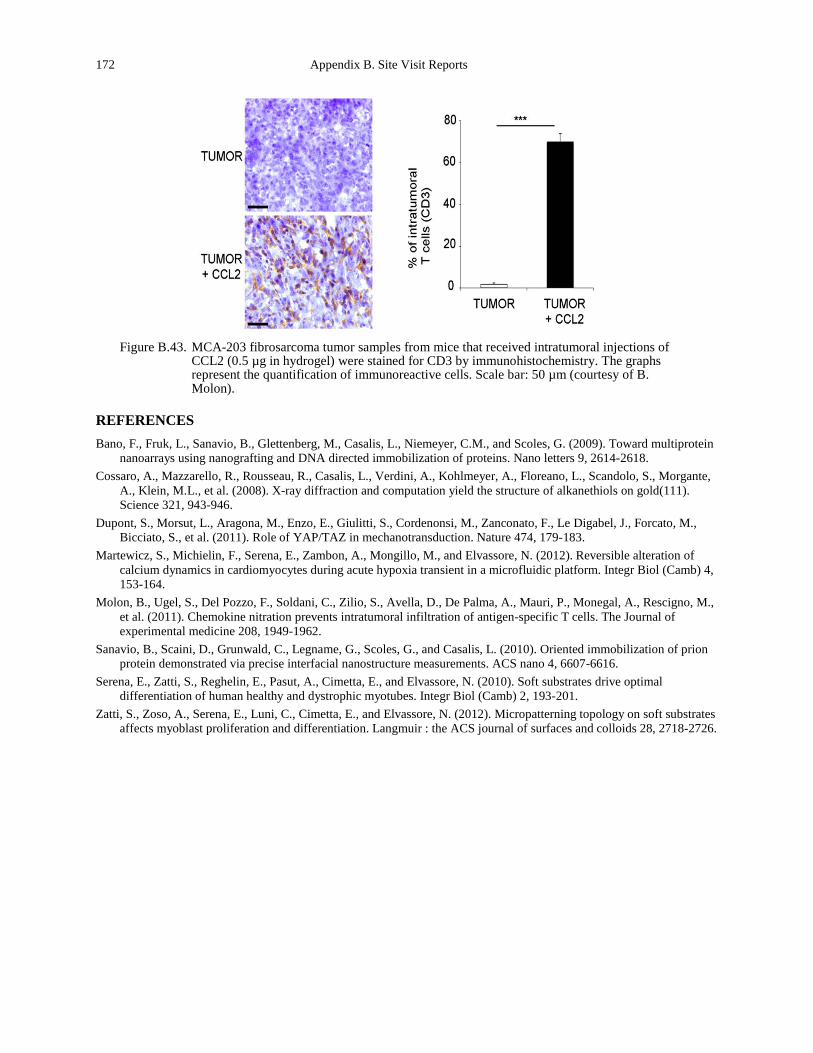
172 Appendix B. Site Visit Reports
Figure B.43. MCA-203 fibrosarcoma tumor samples from mice that received intratumoral injections of
CCL2 (0.5 µg in hydrogel) were stained for CD3 by immunohistochemistry. The graphs represent the quantification of immunoreactive cells. Scale bar: 50 µm (courtesy of B. Molon).
REFERENCES Bano, F., Fruk, L., Sanavio, B., Glettenberg, M., Casalis, L., Niemeyer, C.M., and Scoles, G. (2009). Toward multiprotein
nanoarrays using nanografting and DNA directed immobilization of proteins. Nano letters 9, 2614-2618. Cossaro, A., Mazzarello, R., Rousseau, R., Casalis, L., Verdini, A., Kohlmeyer, A., Floreano, L., Scandolo, S., Morgante,
A., Klein, M.L., et al. (2008). X-ray diffraction and computation yield the structure of alkanethiols on gold(111). Science 321, 943-946.
Dupont, S., Morsut, L., Aragona, M., Enzo, E., Giulitti, S., Cordenonsi, M., Zanconato, F., Le Digabel, J., Forcato, M., Bicciato, S., et al. (2011). Role of YAP/TAZ in mechanotransduction. Nature 474, 179-183.
Martewicz, S., Michielin, F., Serena, E., Zambon, A., Mongillo, M., and Elvassore, N. (2012). Reversible alteration of calcium dynamics in cardiomyocytes during acute hypoxia transient in a microfluidic platform. Integr Biol (Camb) 4, 153-164.
Molon, B., Ugel, S., Del Pozzo, F., Soldani, C., Zilio, S., Avella, D., De Palma, A., Mauri, P., Monegal, A., Rescigno, M., et al. (2011). Chemokine nitration prevents intratumoral infiltration of antigen-specific T cells. The Journal of experimental medicine 208, 1949-1962.
Sanavio, B., Scaini, D., Grunwald, C., Legname, G., Scoles, G., and Casalis, L. (2010). Oriented immobilization of prion protein demonstrated via precise interfacial nanostructure measurements. ACS nano 4, 6607-6616.
Serena, E., Zatti, S., Reghelin, E., Pasut, A., Cimetta, E., and Elvassore, N. (2010). Soft substrates drive optimal differentiation of human healthy and dystrophic myotubes. Integr Biol (Camb) 2, 193-201.
Zatti, S., Zoso, A., Serena, E., Luni, C., Cimetta, E., and Elvassore, N. (2012). Micropatterning topology on soft substrates affects myoblast proliferation and differentiation. Langmuir : the ACS journal of surfaces and colloids 28, 2718-2726.
Appendix B: Site Visit Reports 173
University of Paris Diderot
Site Address: Material and Complex Systems Laboratory, UMR 7057 Centre National de la Recherche Scientifique and Université Paris-Diderot, CC 7056 10 Rue A. Domon et L. Duquet 75205 Paris Cedex 13, FRANCE www.msc.univ-paris-diderot.fr/?lang=en
Date Visited: 10 May 2012 WTEC Attendees: Paul Janmey, Cindy Reinhart-King, Dan Fletcher, Lance Munn (site report author), Jerry
Lee, Nastaran Kuhn, Larry Nagahara, and Hemant Sarin Host(s): François Gallet
Head of Physics Department Email: [email protected] Tel: +33 1 57 27 61 03
Loic Auvray Principal Investigator at CNRS, Biophysics Department Email: [email protected]
Atef Asnacios Group Leader Physics of Single Cell Mechanosensing Biophysics Department Email: [email protected]
Sylvie Hénon Professor in Material and Complex Systems Laboratory Email: [email protected] Tel: +33 1 57 27 62 15
Florence Gazeau Professor in Material and Complex Systems Laboratory Biophysics Department Email: [email protected]
Jean-Francois Berret Professor in Material and Complex Systems Laboratory Email: [email protected] Tel: +33 1 44 27 44 97
Vincent Fleury Professor in Material and Complex Systems Laboratory Email: [email protected]
OVERVIEW
The University Paris Diderot science sector teams mainly work with the French National Center for Scientific Research (CNRS) and National Institute of Health and Medical Research (INSERM). A large number of science sector teams also work with other universities or institutions, such as Paris Descartes, Pierre et Marie Curie, Paris Sud, Paris Est, the École Normale Supérieure Paris, the École Supérieure de Physique et de Chimie Industrielles de la Ville de Paris, and the Observatoire de Paris. Some of these
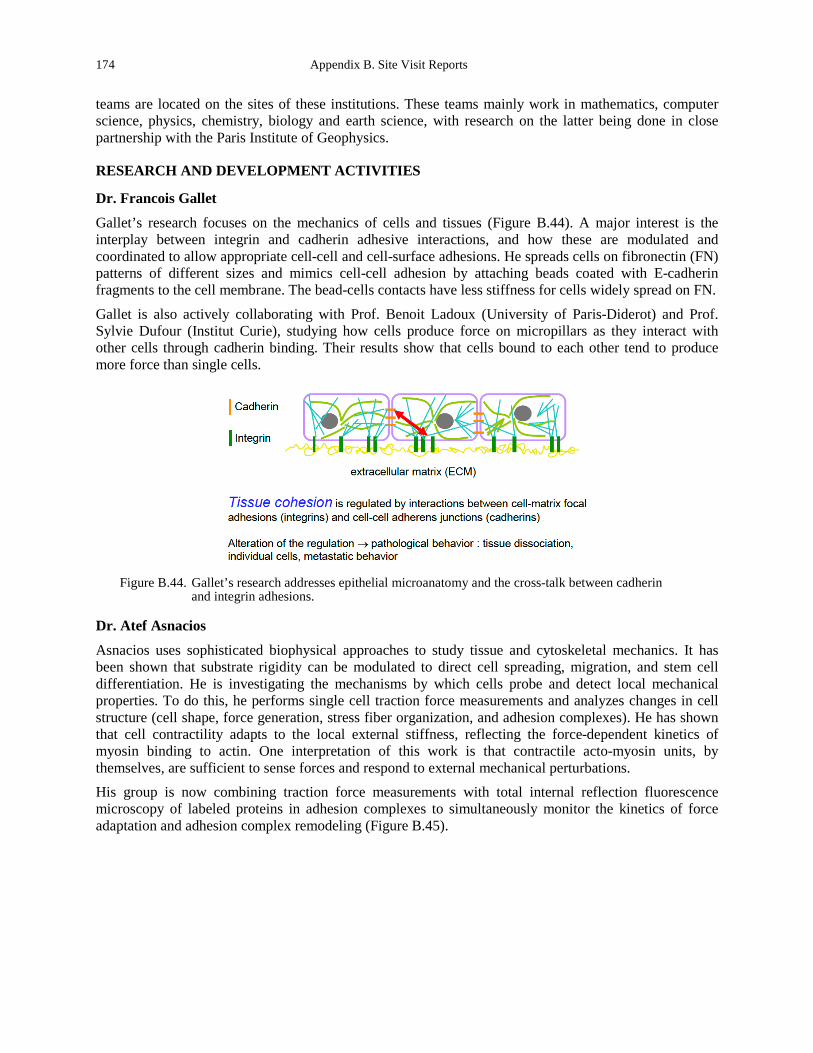
174 Appendix B. Site Visit Reports
teams are located on the sites of these institutions. These teams mainly work in mathematics, computer science, physics, chemistry, biology and earth science, with research on the latter being done in close partnership with the Paris Institute of Geophysics.
RESEARCH AND DEVELOPMENT ACTIVITIES
Dr. Francois Gallet
Gallet’s research focuses on the mechanics of cells and tissues (Figure B.44). A major interest is the interplay between integrin and cadherin adhesive interactions, and how these are modulated and coordinated to allow appropriate cell-cell and cell-surface adhesions. He spreads cells on fibronectin (FN) patterns of different sizes and mimics cell-cell adhesion by attaching beads coated with E-cadherin fragments to the cell membrane. The bead-cells contacts have less stiffness for cells widely spread on FN.
Gallet is also actively collaborating with Prof. Benoit Ladoux (University of Paris-Diderot) and Prof. Sylvie Dufour (Institut Curie), studying how cells produce force on micropillars as they interact with other cells through cadherin binding. Their results show that cells bound to each other tend to produce more force than single cells.
Figure B.44. Gallet’s research addresses epithelial microanatomy and the cross-talk between cadherin
and integrin adhesions.
Dr. Atef Asnacios
Asnacios uses sophisticated biophysical approaches to study tissue and cytoskeletal mechanics. It has been shown that substrate rigidity can be modulated to direct cell spreading, migration, and stem cell differentiation. He is investigating the mechanisms by which cells probe and detect local mechanical properties. To do this, he performs single cell traction force measurements and analyzes changes in cell structure (cell shape, force generation, stress fiber organization, and adhesion complexes). He has shown that cell contractility adapts to the local external stiffness, reflecting the force-dependent kinetics of myosin binding to actin. One interpretation of this work is that contractile acto-myosin units, by themselves, are sufficient to sense forces and respond to external mechanical perturbations. His group is now combining traction force measurements with total internal reflection fluorescence microscopy of labeled proteins in adhesion complexes to simultaneously monitor the kinetics of force adaptation and adhesion complex remodeling (Figure B.45).
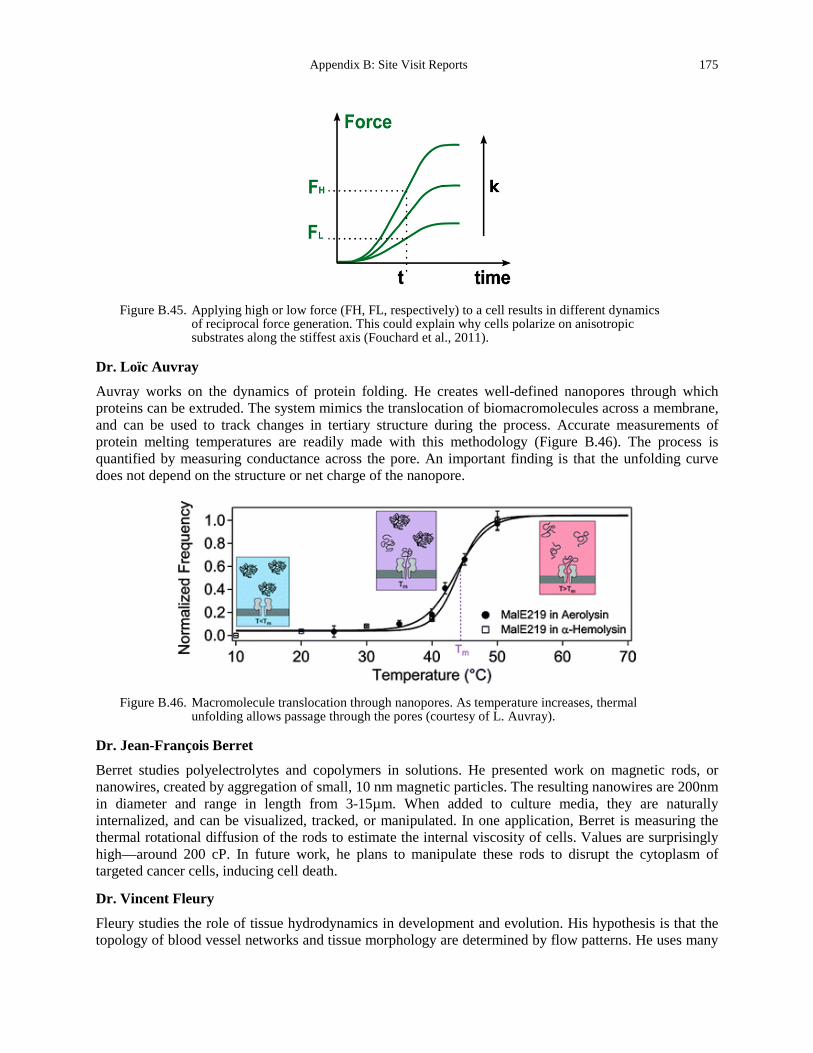
Appendix B: Site Visit Reports 175
Figure B.45. Applying high or low force (FH, FL, respectively) to a cell results in different dynamics
of reciprocal force generation. This could explain why cells polarize on anisotropic substrates along the stiffest axis (Fouchard et al., 2011).
Dr. Loïc Auvray
Auvray works on the dynamics of protein folding. He creates well-defined nanopores through which proteins can be extruded. The system mimics the translocation of biomacromolecules across a membrane, and can be used to track changes in tertiary structure during the process. Accurate measurements of protein melting temperatures are readily made with this methodology (Figure B.46). The process is quantified by measuring conductance across the pore. An important finding is that the unfolding curve does not depend on the structure or net charge of the nanopore.
Figure B.46. Macromolecule translocation through nanopores. As temperature increases, thermal
unfolding allows passage through the pores (courtesy of L. Auvray).
Dr. Jean-François Berret
Berret studies polyelectrolytes and copolymers in solutions. He presented work on magnetic rods, or nanowires, created by aggregation of small, 10 nm magnetic particles. The resulting nanowires are 200nm in diameter and range in length from 3-15µm. When added to culture media, they are naturally internalized, and can be visualized, tracked, or manipulated. In one application, Berret is measuring the thermal rotational diffusion of the rods to estimate the internal viscosity of cells. Values are surprisingly high—around 200 cP. In future work, he plans to manipulate these rods to disrupt the cytoplasm of targeted cancer cells, inducing cell death.
Dr. Vincent Fleury
Fleury studies the role of tissue hydrodynamics in development and evolution. His hypothesis is that the topology of blood vessel networks and tissue morphology are determined by flow patterns. He uses many

176 Appendix B. Site Visit Reports
models, including chicken embryos and jellyfish, to study angiogenesis and branching morphogenesis (Figure B.47). Using time-lapse video-microscopy, he tracks arterial-venous differentiation, and has demonstrated that flow shapes the global patterning of the arterial tree and regulates the activation of the arterial markers ephrinB2 and neuropilin 1.
Figure B.47. Development of the chick vasculature.
(A) Formation of the vitelline artery (VA). Red and blue arrows indicate arterial and venous flow, respectively. The heart is marked by the asterisk. (B) Outline of the vascular system in the boxed area in A. (C) After 26 hours, significant remodeling has occurred. (D) Illustration of the vascular system in the boxed area in C. Veins are white, arteries are gray; at this stage, veins and arteries run in parallel. SV: sinus vein. Scale bar: 1100 µm (Freund et al., 2012).
Drs. Florence Gazeau and Claire Wilhelm
Gazeau and Wilhelm are developing magnetic nanoparticles for medical imaging and therapy. The particles are easily taken up by cells and can be imaged using standard technologies. Immediate applications include cell tracking via MRI, and MRI imaging of inflammation. It is also possible to manipulate the particles or particle-laden cells with a magnetic field, so cell therapy and tissue engineering might be directed or facilitated by externally-applied magnetic force. Using this approach, Gazeau and Wilhelm have shown directed localization of particles in tumors. This could be used to deliver photoactivatable drugs or therapeutic cells, or induce nanoparticle-mediated hyperthermia. Another interesting application is the manipulation of cells in culture to force aggregation in pre-designated shapes, with defined kinetics.
Dr. Sylvie Hénon
Hénon studies cell mechanics, focusing on the role of intermediate filaments. She attaches functionalized beads to cells and manipulates them with optical tweezers to measure cell mechanical properties. She applies relatively sophisticated methods to measure viscoelastic moduli and cell mechanical “creep” in addition to Young’s moduli. Using these methods, she discovered that cells submitted to successive creep
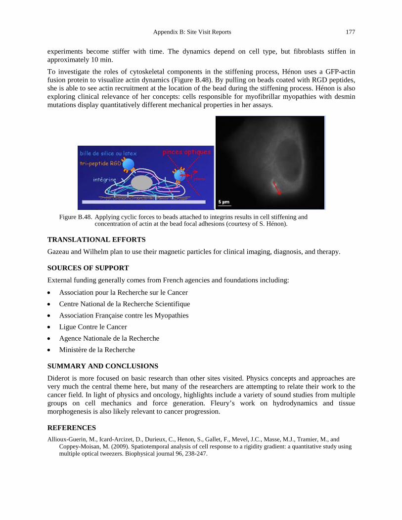
Appendix B: Site Visit Reports 177
experiments become stiffer with time. The dynamics depend on cell type, but fibroblasts stiffen in approximately 10 min.
To investigate the roles of cytoskeletal components in the stiffening process, Hénon uses a GFP-actin fusion protein to visualize actin dynamics (Figure B.48). By pulling on beads coated with RGD peptides, she is able to see actin recruitment at the location of the bead during the stiffening process. Hénon is also exploring clinical relevance of her concepts: cells responsible for myofibrillar myopathies with desmin mutations display quantitatively different mechanical properties in her assays.
Figure B.48. Applying cyclic forces to beads attached to integrins results in cell stiffening and
concentration of actin at the bead focal adhesions (courtesy of S. Hénon).
TRANSLATIONAL EFFORTS
Gazeau and Wilhelm plan to use their magnetic particles for clinical imaging, diagnosis, and therapy.
SOURCES OF SUPPORT
External funding generally comes from French agencies and foundations including:
• Association pour la Recherche sur le Cancer • Centre National de la Recherche Scientifique • Association Française contre les Myopathies • Ligue Contre le Cancer • Agence Nationale de la Recherche • Ministère de la Recherche
SUMMARY AND CONCLUSIONS
Diderot is more focused on basic research than other sites visited. Physics concepts and approaches are very much the central theme here, but many of the researchers are attempting to relate their work to the cancer field. In light of physics and oncology, highlights include a variety of sound studies from multiple groups on cell mechanics and force generation. Fleury’s work on hydrodynamics and tissue morphogenesis is also likely relevant to cancer progression.
REFERENCES Allioux-Guerin, M., Icard-Arcizet, D., Durieux, C., Henon, S., Gallet, F., Mevel, J.C., Masse, M.J., Tramier, M., and
Coppey-Moisan, M. (2009). Spatiotemporal analysis of cell response to a rigidity gradient: a quantitative study using multiple optical tweezers. Biophysical journal 96, 238-247.
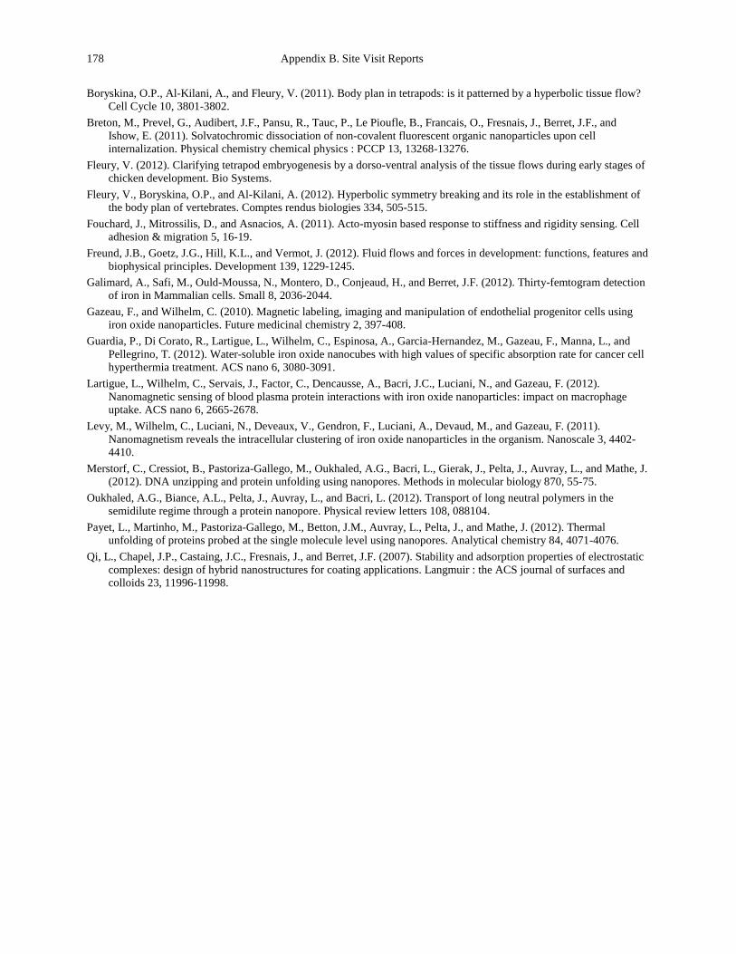
178 Appendix B. Site Visit Reports
Boryskina, O.P., Al-Kilani, A., and Fleury, V. (2011). Body plan in tetrapods: is it patterned by a hyperbolic tissue flow? Cell Cycle 10, 3801-3802.
Breton, M., Prevel, G., Audibert, J.F., Pansu, R., Tauc, P., Le Pioufle, B., Francais, O., Fresnais, J., Berret, J.F., and Ishow, E. (2011). Solvatochromic dissociation of non-covalent fluorescent organic nanoparticles upon cell internalization. Physical chemistry chemical physics : PCCP 13, 13268-13276.
Fleury, V. (2012). Clarifying tetrapod embryogenesis by a dorso-ventral analysis of the tissue flows during early stages of chicken development. Bio Systems.
Fleury, V., Boryskina, O.P., and Al-Kilani, A. (2012). Hyperbolic symmetry breaking and its role in the establishment of the body plan of vertebrates. Comptes rendus biologies 334, 505-515.
Fouchard, J., Mitrossilis, D., and Asnacios, A. (2011). Acto-myosin based response to stiffness and rigidity sensing. Cell adhesion & migration 5, 16-19.
Freund, J.B., Goetz, J.G., Hill, K.L., and Vermot, J. (2012). Fluid flows and forces in development: functions, features and biophysical principles. Development 139, 1229-1245.
Galimard, A., Safi, M., Ould-Moussa, N., Montero, D., Conjeaud, H., and Berret, J.F. (2012). Thirty-femtogram detection of iron in Mammalian cells. Small 8, 2036-2044.
Gazeau, F., and Wilhelm, C. (2010). Magnetic labeling, imaging and manipulation of endothelial progenitor cells using iron oxide nanoparticles. Future medicinal chemistry 2, 397-408.
Guardia, P., Di Corato, R., Lartigue, L., Wilhelm, C., Espinosa, A., Garcia-Hernandez, M., Gazeau, F., Manna, L., and Pellegrino, T. (2012). Water-soluble iron oxide nanocubes with high values of specific absorption rate for cancer cell hyperthermia treatment. ACS nano 6, 3080-3091.
Lartigue, L., Wilhelm, C., Servais, J., Factor, C., Dencausse, A., Bacri, J.C., Luciani, N., and Gazeau, F. (2012). Nanomagnetic sensing of blood plasma protein interactions with iron oxide nanoparticles: impact on macrophage uptake. ACS nano 6, 2665-2678.
Levy, M., Wilhelm, C., Luciani, N., Deveaux, V., Gendron, F., Luciani, A., Devaud, M., and Gazeau, F. (2011). Nanomagnetism reveals the intracellular clustering of iron oxide nanoparticles in the organism. Nanoscale 3, 4402-4410.
Merstorf, C., Cressiot, B., Pastoriza-Gallego, M., Oukhaled, A.G., Bacri, L., Gierak, J., Pelta, J., Auvray, L., and Mathe, J. (2012). DNA unzipping and protein unfolding using nanopores. Methods in molecular biology 870, 55-75.
Oukhaled, A.G., Biance, A.L., Pelta, J., Auvray, L., and Bacri, L. (2012). Transport of long neutral polymers in the semidilute regime through a protein nanopore. Physical review letters 108, 088104.
Payet, L., Martinho, M., Pastoriza-Gallego, M., Betton, J.M., Auvray, L., Pelta, J., and Mathe, J. (2012). Thermal unfolding of proteins probed at the single molecule level using nanopores. Analytical chemistry 84, 4071-4076.
Qi, L., Chapel, J.P., Castaing, J.C., Fresnais, J., and Berret, J.F. (2007). Stability and adsorption properties of electrostatic complexes: design of hybrid nanostructures for coating applications. Langmuir : the ACS journal of surfaces and colloids 23, 11996-11998.
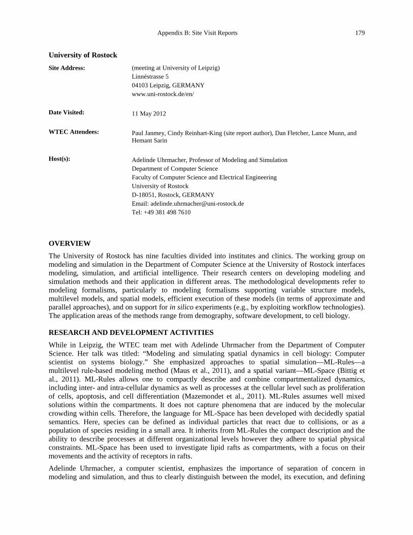
Appendix B: Site Visit Reports 179
University of Rostock
Site Address: (meeting at University of Leipzig) Linnéstrasse 5 04103 Leipzig, GERMANY www.uni-rostock.de/en/
Date Visited: 11 May 2012 WTEC Attendees: Paul Janmey, Cindy Reinhart-King (site report author), Dan Fletcher, Lance Munn, and
Hemant Sarin Host(s): Adelinde Uhrmacher, Professor of Modeling and Simulation
Department of Computer Science Faculty of Computer Science and Electrical Engineering University of Rostock D-18051, Rostock, GERMANY Email: [email protected] Tel: +49 381 498 7610
OVERVIEW
The University of Rostock has nine faculties divided into institutes and clinics. The working group on modeling and simulation in the Department of Computer Science at the University of Rostock interfaces modeling, simulation, and artificial intelligence. Their research centers on developing modeling and simulation methods and their application in different areas. The methodological developments refer to modeling formalisms, particularly to modeling formalisms supporting variable structure models, multilevel models, and spatial models, efficient execution of these models (in terms of approximate and parallel approaches), and on support for in silico experiments (e.g., by exploiting workflow technologies). The application areas of the methods range from demography, software development, to cell biology.
RESEARCH AND DEVELOPMENT ACTIVITIES
While in Leipzig, the WTEC team met with Adelinde Uhrmacher from the Department of Computer Science. Her talk was titled: “Modeling and simulating spatial dynamics in cell biology: Computer scientist on systems biology.” She emphasized approaches to spatial simulation—ML-Rules—a multilevel rule-based modeling method (Maus et al., 2011), and a spatial variant—ML-Space (Bittig et al., 2011). ML-Rules allows one to compactly describe and combine compartmentalized dynamics, including inter- and intra-cellular dynamics as well as processes at the cellular level such as proliferation of cells, apoptosis, and cell differentiation (Mazemondet et al., 2011). ML-Rules assumes well mixed solutions within the compartments. It does not capture phenomena that are induced by the molecular crowding within cells. Therefore, the language for ML-Space has been developed with decidedly spatial semantics. Here, species can be defined as individual particles that react due to collisions, or as a population of species residing in a small area. It inherits from ML-Rules the compact description and the ability to describe processes at different organizational levels however they adhere to spatial physical constraints. ML-Space has been used to investigate lipid rafts as compartments, with a focus on their movements and the activity of receptors in rafts. Adelinde Uhrmacher, a computer scientist, emphasizes the importance of separation of concern in modeling and simulation, and thus to clearly distinguish between the model, its execution, and defining
180 Appendix B. Site Visit Reports
an in silico experiment with this model. The latter can mean parameter scanning and optimization. Similar to a wet-lab experiment, the in silico experiment needs to be carefully documented to be of any value—including how has the model been validated and how have the simulation results been achieved. Uhrmacher’s group has designed a general purpose plugin-based modeling and simulation framework which has already been applied to develop different modeling and simulation tools for cell biology. Currently, the framework includes more than 700 plugins and more than 100 plugin types (e.g., different modeling formalisms, execution algorithms, steady state analyzers, etc.). It also provides intelligent support to configure suitable experiments on demand. Figure B.49 shows an illustration of the hierarchical modeling concept. Different-shaped nodes correspond to different species names while attributes are color-coded. Stacking of identical nodes represents the amount of a certain species. In the figure, A is a graphical representation of a hierarchical model structure via nested nodes. B shows the same model structure alternatively depicted as a directed tree graph. Note that besides atomic species (triangles and diamonds), species containing a sub-solution (squares) might be attributed so that each species at each level might have its own state. In C are examples of matching different reactant patterns within the hierarchical model structure. The rainbow shadings in the second and third pattern illustrate variable instead of defined colors, i.e., attributes (Maus et al., 2011).
Figure B.49. Nested model structure (Maus et al., 2011).
Figure B.50 shows an example model consisting of three hierarchical levels.
SUMMARY AND CONCLUSIONS
A multi-scale simulation of cell processes should be useful in understanding biological processes.

Appendix B: Site Visit Reports 181
Figure B.50. Schematic description of the example model.
At the bottom level, interacting proteins describe the intracellular dynamics of a fission yeast cell (reactions 1-5). The intermediate level describes dynamics of entire cell states, i.e., cell growth (6), cell cycle phase transitions (7-9), and division including mating type switching (9). In addition, cells may secrete pheromone molecules (P-factor and M-factor) to the extracellular medium (10). Various inter-level causalities between the intermediate and the bottom level influence processes both in an upward (7-9) and downward causation manner (4,11-12). The top level discretizes the environment of cells into multiple fictive compartments in order to study spatial dynamics of pheromone diffusion and displacement of cells (13-14). Although spatial dynamics referring to compartments and particle diffusion between cells can be modeled, excluded volume effects cannot be described in ML-Rules; therefore one has to move to ML-Space (Bittig et al., 2011).
REFERENCES Bittig, A.T., Haack, F., Maus, C., and Uhrmacher, A.M. Adapting rule-based model descriptions for simulating in
continuous and hybrid space. Proceedings of the 9th International Conference on Computational Methods in Systems Biology 2011;161-170, New York, NY, USA, ACM. CMSB ‘11.
Maus, C., Rybacki, S., and Uhrmacher, A.M. (2011). Rule-based multi-level modeling of cell biological systems. BMC systems biology 5, 166.
Mazemondet, O., Hubner, R., Frahm, J., Koczan, D., Bader, B.M., Weiss, D.G., Uhrmacher, A.M., Frech, M.J., Rolfs, A., and Luo, J. (2011). Quantitative and kinetic profile of Wnt/beta-catenin signaling components during human neural progenitor cell differentiation. Cellular & molecular biology letters 16, 515-538.
www.mosi.informatik.uni-rostock.de/mosi/
182 Appendix B. Site Visit Reports
Uppsala University and Science for Life Laboratory Uppsala
Site Address: Meeting at Uppsala University Dag Hammarskjölds väg 20 752 37 Uppsala, SWEDEN www.uu.se/
Date Visited: 8 May 2012 WTEC Attendees: Paul Janmey, Cindy Reinhart-King, Dan Fletcher (site report author), Nastaran Kuhn,
Nicole Moore, and Hemant Sarin Host(s): Karin Forsberg Nilsson
Professor of Stem Cell Research Department of Immunology, Genetics, and Pathology Vice Director, Science for Life Laboratory Uppsala Email: [email protected] Tel: +46 18 471 41 58
Fredrik Nikolajeff Associate Professor, Center Manager, Explorative Research Uppsala Berzelii Technology Centre for Neurodiagnostics Email: [email protected] Tel: +46 18 471 30 36
Magnus Malmqvist Associate Professor, Biomedical Radiation Sciences Department of Radiology, Oncology, and Radiation Science Email: [email protected] Tel: +46 18 55 53 00
OVERVIEW
Uppsala University is one of the world’s oldest universities, with a rich academic and research history. Uppsala University, along with the Royal Institute of Technology (KTH), has some of Europe’s most advanced laboratories for nanomaterial science with applications in pharmaceutics, biotechnology, and energy. Science for Life Laboratory (SciLifeLab) is a joint venture between four universities; Karolinska Institutet, KTH, Stockholm University, and Uppsala University. At Uppsala University, the SciLifeLab project is based at the Biomedical Center. SciLifeLab researchers study the molecular basis for human complex disease. Their methods include identifying genetic risk factors, biomarkers, and mechanisms by applying novel technologies to patient samples unique to the Nordic countries, as well as comparative biology proteomic and genomic approaches. The efforts of SciLifeLab in Uppsala are coordinated with SciLifeLab in Stockholm to provide a national infrastructure for molecular biosciences.
RESEARCH AND DEVELOPMENT ACTIVITIES
Fredrik Nikolajeff described the activities of the Uppsala Berzelii Technology Centre for Neurodiagnostics, a unique collaboration of researchers in academic, medical, and commercial institutions that focuses on Alzheimer’s disease, chronic pain, and the development of new technologies. The goal of the Centre is to identify new biomarkers and methods that can be used for early diagnosis of diseases. It draws on the strong medical and academic resources in the Uppsala-Stockholm area. For
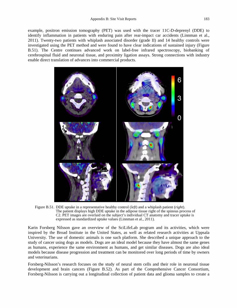
Appendix B: Site Visit Reports 183
example, positron emission tomography (PET) was used with the tracer 11C-D-deprenyl (DDE) to identify inflammation in patients with enduring pain after rear-impact car accidents (Linnman et al., 2011). Twenty-two patients with whiplash associated disorder (grade II) and 14 healthy controls were investigated using the PET method and were found to have clear indications of sustained injury (Figure B.51). The Centre continues advanced work on label-free infrared spectroscopy, biobanking of cerebrospinal fluid and neuronal tissue, and proximity ligation assays. Strong connections with industry enable direct translation of advances into commercial products.
Figure B.51. DDE uptake in a representative healthy control (left) and a whiplash patient (right).
The patient displays high DDE uptake in the adipose tissue right of the spinous process of C2. PET images are overlaid on the subject’s individual CT anatomy and tracer uptake is expressed as standardized uptake values (Linnman et al., 2011).
Karin Forsberg Nilsson gave an overview of the SciLifeLab program and its activities, which were inspired by the Broad Institute in the United States, as well as related research activities at Uppsala University. The use of domestic animals is one such platform. She described a unique approach to the study of cancer using dogs as models. Dogs are an ideal model because they have almost the same genes as humans, experience the same environment as humans, and get similar diseases. Dogs are also ideal models because disease progression and treatment can be monitored over long periods of time by owners and veterinarians. Forsberg-Nilsson’s research focuses on the study of neural stem cells and their role in neuronal tissue development and brain cancers (Figure B.52). As part of the Comprehensive Cancer Consortium, Forsberg-Nilsson is carrying out a longitudinal collection of patient data and glioma samples to create a
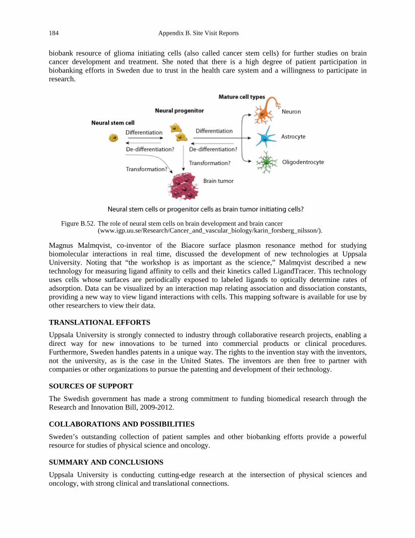
184 Appendix B. Site Visit Reports
biobank resource of glioma initiating cells (also called cancer stem cells) for further studies on brain cancer development and treatment. She noted that there is a high degree of patient participation in biobanking efforts in Sweden due to trust in the health care system and a willingness to participate in research.
Figure B.52. The role of neural stem cells on brain development and brain cancer
(www.igp.uu.se/Research/Cancer_and_vascular_biology/karin_forsberg_nilsson/).
Magnus Malmqvist, co-inventor of the Biacore surface plasmon resonance method for studying biomolecular interactions in real time, discussed the development of new technologies at Uppsala University. Noting that “the workshop is as important as the science,” Malmqvist described a new technology for measuring ligand affinity to cells and their kinetics called LigandTracer. This technology uses cells whose surfaces are periodically exposed to labeled ligands to optically determine rates of adsorption. Data can be visualized by an interaction map relating association and dissociation constants, providing a new way to view ligand interactions with cells. This mapping software is available for use by other researchers to view their data.
TRANSLATIONAL EFFORTS
Uppsala University is strongly connected to industry through collaborative research projects, enabling a direct way for new innovations to be turned into commercial products or clinical procedures. Furthermore, Sweden handles patents in a unique way. The rights to the invention stay with the inventors, not the university, as is the case in the United States. The inventors are then free to partner with companies or other organizations to pursue the patenting and development of their technology.
SOURCES OF SUPPORT
The Swedish government has made a strong commitment to funding biomedical research through the Research and Innovation Bill, 2009-2012.
COLLABORATIONS AND POSSIBILITIES
Sweden’s outstanding collection of patient samples and other biobanking efforts provide a powerful resource for studies of physical science and oncology.
SUMMARY AND CONCLUSIONS
Uppsala University is conducting cutting-edge research at the intersection of physical sciences and oncology, with strong clinical and translational connections.

Appendix B: Site Visit Reports 185
REFERENCES Forsberg, M., Holmborn, K., Kundu, S., Dagalv, A., Kjellen, L., and Forsberg-Nilsson, K. (2012). Undersulfation of
heparan sulfate restricts differentiation potential of mouse embryonic stem cells. The Journal of biological chemistry 287, 10853-10862.
Linnman, C., Appel, L., Fredrikson, M., Gordh, T., Soderlund, A., Langstrom, B., and Engler, H. (2011). Elevated [11C]-D-deprenyl uptake in chronic Whiplash Associated Disorder suggests persistent musculoskeletal inflammation. PLoS One 6, e19182.
Niklasson, M., Bergstrom, T., Zhang, X.Q., Gustafsdottir, S.M., Sjogren, M., Edqvist, P.H., Vennstrom, B., Forsberg, M., and Forsberg-Nilsson, K. (2010). Enlarged lateral ventricles and aberrant behavior in mice overexpressing PDGF-B in embryonic neural stem cells. Experimental cell research 316, 2779-2789.
www.uu.se/ www.uu.se/en/research/ http://suls.se/ http://suls.se/research/how/research-infrastructure/ www.igp.uu.se/Research/Cancer_and_vascular_biology/karin_forsberg_nilsson/ www.igp.uu.se/Research/Cancer_and_vascular_biology/karin_forsberg_nilsson/more/ www.teknik.uu.se/material/staff/haen/index.html www.teknik.uu.se/material/Tribomaterials/mim/index.html www.nanobridge.eu www.uuinnovation.uu.se/ www.berzelii.uu.se/
186 Appendix B. Site Visit Reports
Weizmann Institute of Science
Site Address: 234 Herzl Street Rehovot 76100, ISRAEL Tel: +972 8 934 3111 Fax: +972 8 934 4107 www.weizmann.ac.il/
Date Visited: 14 May 2012 WTEC Attendees: Paul Janmey (site report author) and Sharon Gerecht Host(s): Ronen Alon
The Linda Jacobs Chair in Immune and Stem Cell Research Department of Immunology Tel: +972 8 934 2482 Fax: +972 8 934 2724 Email: [email protected] www.weizmann.ac.il/immunology/AlonPage.html
Tom Shemesh Post-doctoral fellow, Department of Molecular Cell Biology Laboratory of Professor Alexander Bershadsky Tel: +972 8 934 2936, 4071 Fax: +972 8 934 4125 Email: [email protected]
Roy Bar-Ziv Faculty of Chemistry, Department of Materials and Interfaces Helen and Martin Kimmel Center for Nanoscale Science Perlman Building Rehovot 76100 Tel: +972 8 934 2069 Email: [email protected] www.weizmann.ac.il/materials/barziv/
Sam Safran Fern and Manfred Steinfeld Professorial Chair Faculty of Chemistry Department of Materials and Interfaces Tel: +972 8 934 3362 Fax +972 8 934 4138 Email: [email protected] www.weizmann.ac.il/fluids/SafranGroup
OVERVIEW
The Weizmann Institute of Science (WIS) is one of the foremost research institutions in Israel and has a long tradition of interdisciplinary work and especially strong interactions between theorists and experimentalists in biophysics. The mission of WIS is to “address the most urgent challenges facing humanity.” One of the institute’s major focus areas is cancer research.
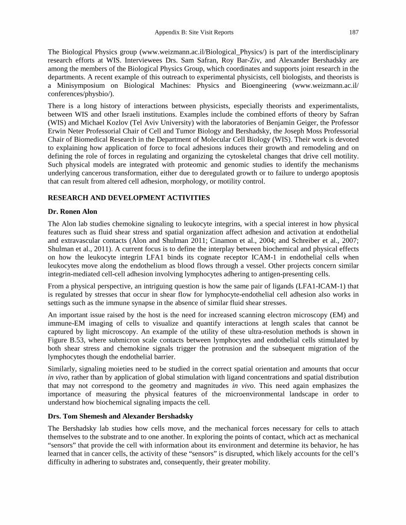
Appendix B: Site Visit Reports 187
The Biological Physics group (www.weizmann.ac.il/Biological_Physics/) is part of the interdisciplinary research efforts at WIS. Interviewees Drs. Sam Safran, Roy Bar-Ziv, and Alexander Bershadsky are among the members of the Biological Physics Group, which coordinates and supports joint research in the departments. A recent example of this outreach to experimental physicists, cell biologists, and theorists is a Minisymposium on Biological Machines: Physics and Bioengineering (www.weizmann.ac.il/ conferences/physbio/). There is a long history of interactions between physicists, especially theorists and experimentalists, between WIS and other Israeli institutions. Examples include the combined efforts of theory by Safran (WIS) and Michael Kozlov (Tel Aviv University) with the laboratories of Benjamin Geiger, the Professor Erwin Neter Professorial Chair of Cell and Tumor Biology and Bershadsky, the Joseph Moss Professorial Chair of Biomedical Research in the Department of Molecular Cell Biology (WIS). Their work is devoted to explaining how application of force to focal adhesions induces their growth and remodeling and on defining the role of forces in regulating and organizing the cytoskeletal changes that drive cell motility. Such physical models are integrated with proteomic and genomic studies to identify the mechanisms underlying cancerous transformation, either due to deregulated growth or to failure to undergo apoptosis that can result from altered cell adhesion, morphology, or motility control.
RESEARCH AND DEVELOPMENT ACTIVITIES
Dr. Ronen Alon
The Alon lab studies chemokine signaling to leukocyte integrins, with a special interest in how physical features such as fluid shear stress and spatial organization affect adhesion and activation at endothelial and extravascular contacts (Alon and Shulman 2011; Cinamon et al., 2004; and Schreiber et al., 2007; Shulman et al., 2011). A current focus is to define the interplay between biochemical and physical effects on how the leukocyte integrin LFA1 binds its cognate receptor ICAM-1 in endothelial cells when leukocytes move along the endothelium as blood flows through a vessel. Other projects concern similar integrin-mediated cell-cell adhesion involving lymphocytes adhering to antigen-presenting cells.
From a physical perspective, an intriguing question is how the same pair of ligands (LFA1-ICAM-1) that is regulated by stresses that occur in shear flow for lymphocyte-endothelial cell adhesion also works in settings such as the immune synapse in the absence of similar fluid shear stresses.
An important issue raised by the host is the need for increased scanning electron microscopy (EM) and immune-EM imaging of cells to visualize and quantify interactions at length scales that cannot be captured by light microscopy. An example of the utility of these ultra-resolution methods is shown in Figure B.53, where submicron scale contacts between lymphocytes and endothelial cells stimulated by both shear stress and chemokine signals trigger the protrusion and the subsequent migration of the lymphocytes though the endothelial barrier. Similarly, signaling moieties need to be studied in the correct spatial orientation and amounts that occur in vivo, rather than by application of global stimulation with ligand concentrations and spatial distribution that may not correspond to the geometry and magnitudes in vivo. This need again emphasizes the importance of measuring the physical features of the microenvironmental landscape in order to understand how biochemical signaling impacts the cell.
Drs. Tom Shemesh and Alexander Bershadsky
The Bershadsky lab studies how cells move, and the mechanical forces necessary for cells to attach themselves to the substrate and to one another. In exploring the points of contact, which act as mechanical “sensors” that provide the cell with information about its environment and determine its behavior, he has learned that in cancer cells, the activity of these “sensors” is disrupted, which likely accounts for the cell’s difficulty in adhering to substrates and, consequently, their greater mobility.
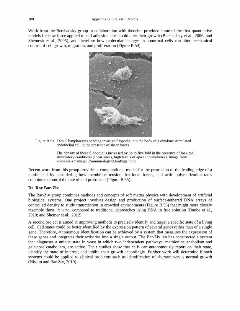
188 Appendix B. Site Visit Reports
Work from the Bershadsky group in collaboration with theorists provided some of the first quantitative models for how force applied to cell adhesion sites could alter their growth (Bershadsky et al., 2006; and Shemesh et al., 2005), and therefore how molecular changes in abnormal cells can alter mechanical control of cell growth, migration, and proliferation (Figure B.54).
Figure B.53. Two T lymphocytes sending invasive filopodia into the body of a cytokine stimulated
endothelial cell in the presence of shear forces.
The density of these filopodia is increased by up to five fold in the presence of maximal stimulatory conditions (shear stress, high levels of apical chemokines). Image from www.weizmann.ac.il/immunology/AlonPage.html.
Recent work from this group provides a computational model for the protrusion of the leading edge of a motile cell by considering how membrane tension, frictional forces, and actin polymerization rates combine to control the rate of cell protrusion (Figure B.55).
Dr. Roy Bar-Ziv
The Bar-Ziv group combines methods and concepts of soft matter physics with development of artificial biological systems. One project involves design and production of surface-tethered DNA arrays of controlled density to study transcription in crowded environments (Figure B.56) that might more closely resemble those in vitro, compared to traditional approaches using DNA in free solution (Daube et al., 2010; and Shemer et al., 2012). A second project is aimed at improving methods to precisely identify and target a specific state of a living cell. Cell states could be better identified by the expression pattern of several genes rather than of a single gene. Therefore, autonomous identification can be achieved by a system that measures the expression of these genes and integrates their activities into a single output. The Bar-Ziv lab has constructed a system that diagnoses a unique state in yeast in which two independent pathways, methionine anabolism and galactose catabolism, are active. Their studies show that cells can autonomously report on their state, identify the state of interest, and inhibit their growth accordingly. Further work will determine if such systems could be applied to clinical problems such as identification of aberrant versus normal growth (Nissim and Bar-Ziv, 2010).
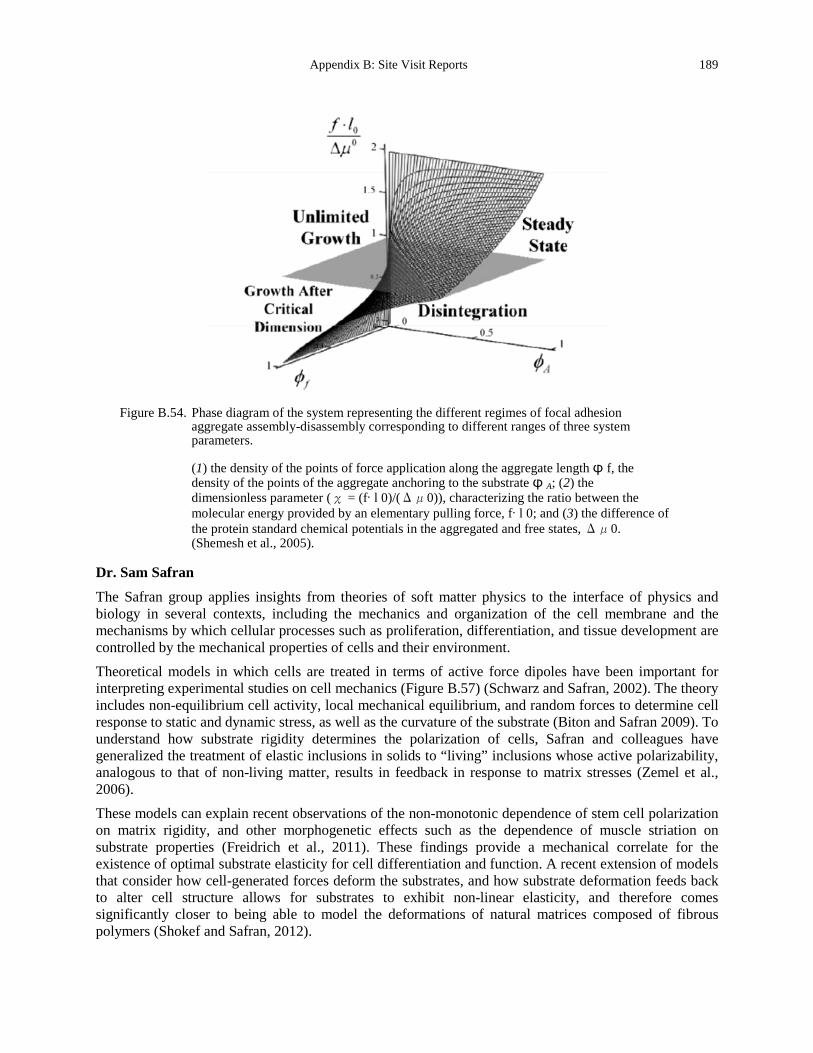
Appendix B: Site Visit Reports 189
Figure B.54. Phase diagram of the system representing the different regimes of focal adhesion
aggregate assembly-disassembly corresponding to different ranges of three system parameters.
(1) the density of the points of force application along the aggregate length ϕ f, the density of the points of the aggregate anchoring to the substrate ϕ A; (2) the dimensionless parameter (χ = (f·l 0)/(Δμ0)), characterizing the ratio between the molecular energy provided by an elementary pulling force, f·l 0; and (3) the difference of the protein standard chemical potentials in the aggregated and free states, Δμ0. (Shemesh et al., 2005).
Dr. Sam Safran
The Safran group applies insights from theories of soft matter physics to the interface of physics and biology in several contexts, including the mechanics and organization of the cell membrane and the mechanisms by which cellular processes such as proliferation, differentiation, and tissue development are controlled by the mechanical properties of cells and their environment. Theoretical models in which cells are treated in terms of active force dipoles have been important for interpreting experimental studies on cell mechanics (Figure B.57) (Schwarz and Safran, 2002). The theory includes non-equilibrium cell activity, local mechanical equilibrium, and random forces to determine cell response to static and dynamic stress, as well as the curvature of the substrate (Biton and Safran 2009). To understand how substrate rigidity determines the polarization of cells, Safran and colleagues have generalized the treatment of elastic inclusions in solids to “living” inclusions whose active polarizability, analogous to that of non-living matter, results in feedback in response to matrix stresses (Zemel et al., 2006). These models can explain recent observations of the non-monotonic dependence of stem cell polarization on matrix rigidity, and other morphogenetic effects such as the dependence of muscle striation on substrate properties (Freidrich et al., 2011). These findings provide a mechanical correlate for the existence of optimal substrate elasticity for cell differentiation and function. A recent extension of models that consider how cell-generated forces deform the substrates, and how substrate deformation feeds back to alter cell structure allows for substrates to exhibit non-linear elasticity, and therefore comes significantly closer to being able to model the deformations of natural matrices composed of fibrous polymers (Shokef and Safran, 2012).
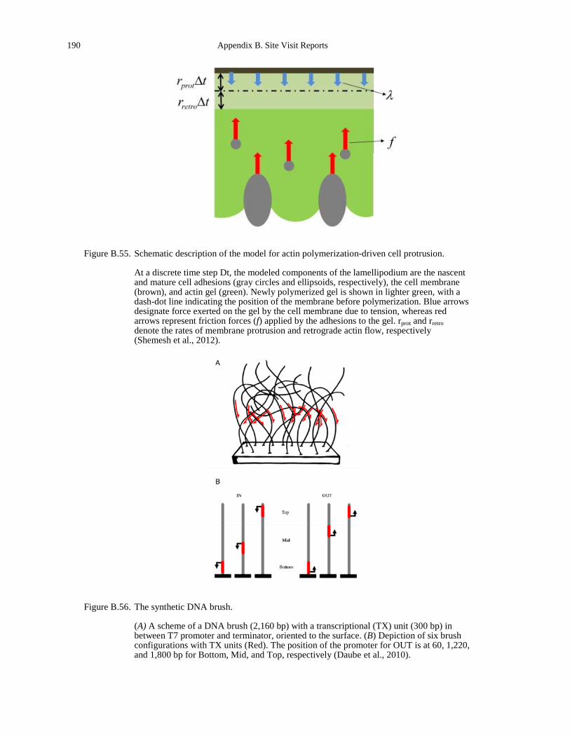
190 Appendix B. Site Visit Reports
Figure B.55. Schematic description of the model for actin polymerization-driven cell protrusion.
At a discrete time step Dt, the modeled components of the lamellipodium are the nascent and mature cell adhesions (gray circles and ellipsoids, respectively), the cell membrane (brown), and actin gel (green). Newly polymerized gel is shown in lighter green, with a dash-dot line indicating the position of the membrane before polymerization. Blue arrows designate force exerted on the gel by the cell membrane due to tension, whereas red arrows represent friction forces (f) applied by the adhesions to the gel. rprot and rretro denote the rates of membrane protrusion and retrograde actin flow, respectively (Shemesh et al., 2012).
Figure B.56. The synthetic DNA brush.
(A) A scheme of a DNA brush (2,160 bp) with a transcriptional (TX) unit (300 bp) in between T7 promoter and terminator, oriented to the surface. (B) Depiction of six brush configurations with TX units (Red). The position of the promoter for OUT is at 60, 1,220, and 1,800 bp for Bottom, Mid, and Top, respectively (Daube et al., 2010).
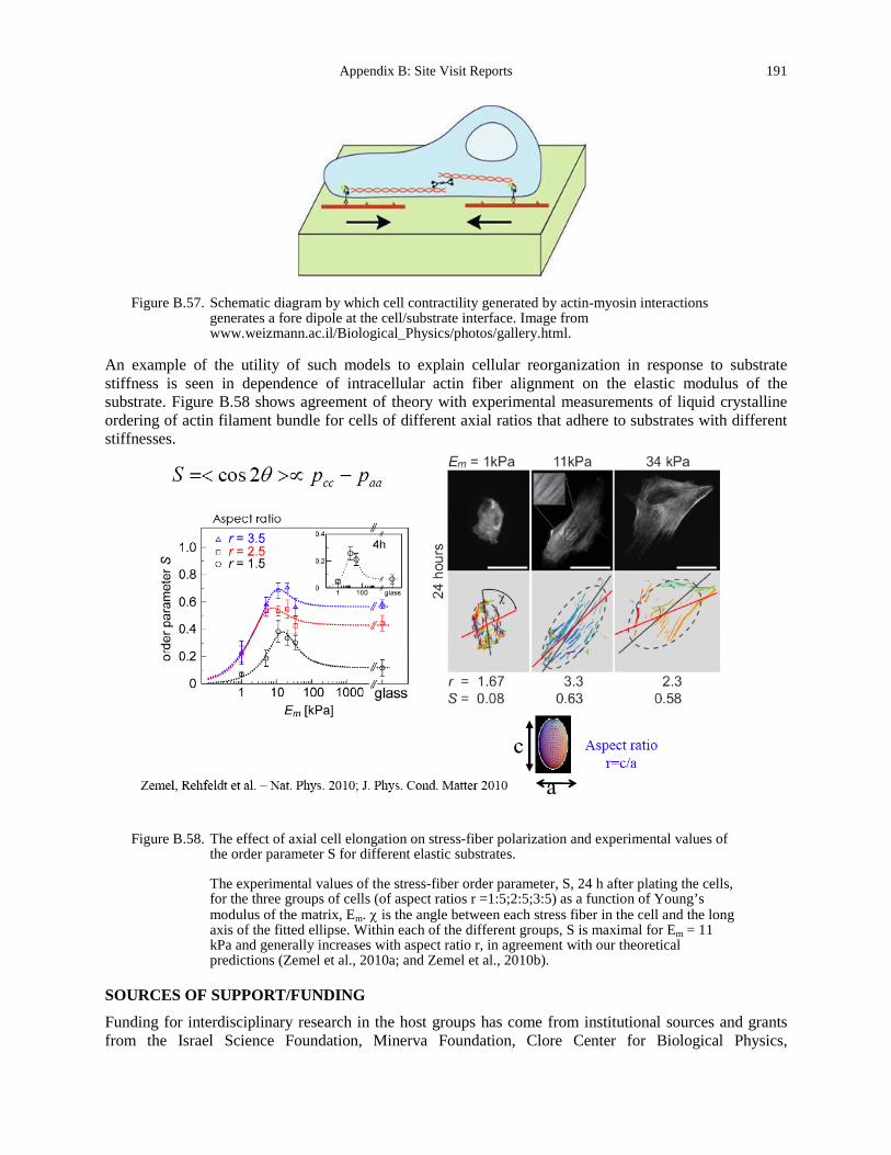
Appendix B: Site Visit Reports 191
Figure B.57. Schematic diagram by which cell contractility generated by actin-myosin interactions
generates a fore dipole at the cell/substrate interface. Image from www.weizmann.ac.il/Biological_Physics/photos/gallery.html.
An example of the utility of such models to explain cellular reorganization in response to substrate stiffness is seen in dependence of intracellular actin fiber alignment on the elastic modulus of the substrate. Figure B.58 shows agreement of theory with experimental measurements of liquid crystalline ordering of actin filament bundle for cells of different axial ratios that adhere to substrates with different stiffnesses.
Figure B.58. The effect of axial cell elongation on stress-fiber polarization and experimental values of the order parameter S for different elastic substrates.
The experimental values of the stress-fiber order parameter, S, 24 h after plating the cells, for the three groups of cells (of aspect ratios r =1:5;2:5;3:5) as a function of Young’s modulus of the matrix, Em. χ is the angle between each stress fiber in the cell and the long axis of the fitted ellipse. Within each of the different groups, S is maximal for Em = 11 kPa and generally increases with aspect ratio r, in agreement with our theoretical predictions (Zemel et al., 2010a; and Zemel et al., 2010b).
SOURCES OF SUPPORT/FUNDING
Funding for interdisciplinary research in the host groups has come from institutional sources and grants from the Israel Science Foundation, Minerva Foundation, Clore Center for Biological Physics,
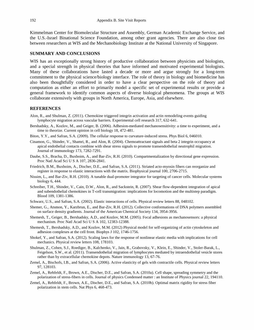
192 Appendix B. Site Visit Reports
Kimmelman Center for Biomolecular Structure and Assembly, German Academic Exchange Service, and the U.S.-Israel Binational Science Foundation, among other grant agencies. There are also close ties between researchers at WIS and the Mechanobiology Institute at the National University of Singapore.
SUMMARY AND CONCLUSIONS
WIS has an exceptionally strong history of productive collaboration between physicists and biologists, and a special strength in physical theories that have informed and motivated experimental biologists. Many of these collaborations have lasted a decade or more and argue strongly for a long-term commitment to the physical science/biology interface. The role of theory in biology and biomedicine has also been thoughtfully considered in order to have a clear perspective on the role of theory and computation as either an effort to primarily model a specific set of experimental results or provide a general framework to identify common aspects of diverse biological phenomena. The groups at WIS collaborate extensively with groups in North America, Europe, Asia, and elsewhere.
REFERENCES Alon, R., and Shulman, Z. (2011). Chemokine triggered integrin activation and actin remodeling events guiding
lymphocyte migration across vascular barriers. Experimental cell research 317, 632-641. Bershadsky, A., Kozlov, M., and Geiger, B. (2006). Adhesion-mediated mechanosensitivity: a time to experiment, and a
time to theorize. Current opinion in cell biology 18, 472-481. Biton, Y.Y., and Safran, S.A. (2009). The cellular response to curvature-induced stress. Phys Biol 6, 046010. Cinamon, G., Shinder, V., Shamri, R., and Alon, R. (2004). Chemoattractant signals and beta 2 integrin occupancy at
apical endothelial contacts combine with shear stress signals to promote transendothelial neutrophil migration. Journal of immunology 173, 7282-7291.
Daube, S.S., Bracha, D., Buxboim, A., and Bar-Ziv, R.H. (2010). Compartmentalization by directional gene expression. Proc Natl Acad Sci U S A 107, 2836-2841.
Friedrich, B.M., Buxboim, A., Discher, D.E., and Safran, S.A. (2011). Striated acto-myosin fibers can reorganize and register in response to elastic interactions with the matrix. Biophysical journal 100, 2706-2715.
Nissim, L., and Bar-Ziv, R.H. (2010). A tunable dual-promoter integrator for targeting of cancer cells. Molecular systems biology 6, 444.
Schreiber, T.H., Shinder, V., Cain, D.W., Alon, R., and Sackstein, R. (2007). Shear flow-dependent integration of apical and subendothelial chemokines in T-cell transmigration: implications for locomotion and the multistep paradigm. Blood 109, 1381-1386.
Schwarz, U.S., and Safran, S.A. (2002). Elastic interactions of cells. Physical review letters 88, 048102. Shemer, G., Atsmon, Y., Karzbrun, E., and Bar-Ziv, R.H. (2012). Collective conformations of DNA polymers assembled
on surface density gradients. Journal of the American Chemical Society 134, 3954-3956. Shemesh, T., Geiger, B., Bershadsky, A.D., and Kozlov, M.M. (2005). Focal adhesions as mechanosensors: a physical
mechanism. Proc Natl Acad Sci U S A 102, 12383-12388. Shemesh, T., Bershadsky, A.D., and Kozlov, M.M. (2012) Physical model for self-organizing of actin cytoskeleton and
adhesion complexes at the cell front. Biophys J 102, 1746-1756. Shokef, Y., and Safran, S.A. (2012). Scaling laws for the response of nonlinear elastic media with implications for cell
mechanics. Physical review letters 108, 178103. Shulman, Z., Cohen, S.J., Roediger, B., Kalchenko, V., Jain, R., Grabovsky, V., Klein, E., Shinder, V., Stoler-Barak, L.,
Feigelson, S.W., et al. (2011). Transendothelial migration of lymphocytes mediated by intraendothelial vesicle stores rather than by extracellular chemokine depots. Nature immunology 13, 67-76.
Zemel, A., Bischofs, I.B., and Safran, S.A. (2006). Active elasticity of gels with contractile cells. Physical review letters 97, 128103.
Zemel, A., Rehfeldt, F., Brown, A.E., Discher, D.E., and Safran, S.A. (2010a). Cell shape, spreading symmetry and the polarization of stress-fibers in cells. Journal of physics Condensed matter : an Institute of Physics journal 22, 194110.
Zemel, A., Rehfeldt, F., Brown, A.E., Discher, D.E., and Safran, S.A. (2010b). Optimal matrix rigidity for stress fiber polarization in stem cells. Nat Phys 6, 468-473.