Embed Size (px)
Citation preview

http://academic.brooklyn.cuny.edu/biology/bio4fv/page/prot_gc.htm
What are proteins
Proteins are polymers of amino acids in which the adjacent amino acids
are connected by peptide bonds.
This polypeptide is short but has several features that are common
to all proteins.
All proteins have an N-terminal end.
All proteins have a C-terminal end.

http://academic.brooklyn.cuny.edu/biology/bio4fv/page/peptide_.htm
What is a peptide bond?
Peptide bonds
A C-N covalent bond between the Nitrogen of one amino acid
and the carboxyl carbon of an adjacent amino acid.

Amino acids
What are amino acids?
The term amino acids refers to a groups of molecules which comprise
the primary structure of a protein
In general, amino acids have several features in common.
The figure shows that amino acids have
a central carbon, referred to as the alpha carbon.
The alpha carbon is covalently bounded to:
- A hydrogen (H)
- A carboxyl functional group (COO -).
- An amine functional group (NH3 +).
- A side chain that distinguishes one kind of amino acid
from another kind.
http://academic.brooklyn.cuny.edu/biology/bio4fv/page/amino_a.htm

http://academic.brooklyn.cuny.edu/biology/bio4fv/page/carboxyl.htm
It is an acidic functional group frequently found in biological molecules.
It is found in amino acids, proteins. fatty acids, acetic acids and other
organic acid.
Because the carboxyl functional group is a weak acid it will dissociate.
At a pH of 7 the carboxyl group is in the dissociated form (COO-).
The carboxyl group of an amino acid will contribute a negative charge
at neutral pH.
Carboxyl Functional Group

Amino Functional Group
The amino functional group is basic and found in proteins, amino acids
and the nitrogenous bases of DNA and RNA .
Under certain pH conditions the amino group can accept a proton and
gain a positive charge of +1.
At a pH of 7 the amino group of an amino acid has a positive charge.
The amino group then contributes a positive charge to the amino acid
at pH 7 .
http://academic.brooklyn.cuny.edu/biology/bio4fv/page/amine_f.htm

N-terminal end of polypeptide
The N-terminal end of the polypeptide has a free amine group.
C-terminal end of polypeptide The C-terminal end of the polypeptide has a free carboxyl group.


Primary protein structure: It is a sequence of chain of amino acids.
Secondary protein structure: It occurs when the sequence of
amino acids are linked by hydrogen bonds.
Tertiary protein structure: It occurs when certain attractions
are present between α-helices and β-sheets.
Quaternary protein structure: It is a protein consisting of more than
one amino acid chain
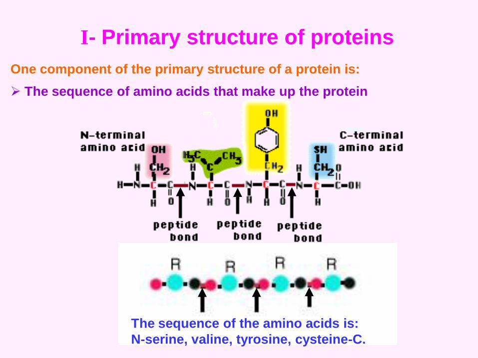
The sequence of the amino acids is:
N-serine, valine, tyrosine, cysteine-C.
I- Primary structure of proteins
One component of the primary structure of a protein is:
The sequence of amino acids that make up the protein

Another component of the primary structure is:
The covalent bonds in the protein.
A single covalent bond between the sulfur
atoms to two amino acids called cysteine .
What is the significance of disulfide bonds?
Because it is a covalent bond, the disulfide bond can be considered as
part of the primary structure of a protein.
They are very important in determining the tertiary structure of proteins.
they are very important in determining the quaternary structure of
some proteins.
The structure of antibody molecules is a very prominent example showing
the role of disulfide bonds.

http://academic.brooklyn.cuny.edu/biology/bio4fv/page/second.htm
II- The Secondary Structure of Protein
Secondary protein structure: It occurs when the sequence of amino
acids are linked by hydrogen bonds.
The secondary structure of protein is formed of α-helical regions
and β-pleated sheets.
What is an alpha helix?
An alpha helix can be formed by making a rope coil in a left handed
direction.
The rope is represented by the N-C-C-N-C-C-N .... backbone of the
polypeptide chain.

The alpha helix structure of protein depends on; bond angles, lengths
and rotations.
The alpha helix could be a very stable structure because intra-chain
hydrogen bonds could be formed that stabilized the helix.
http://academic.brooklyn.cuny.edu/biology/bio4fv/page/alpha_h.htm

Significance of alpha helix
The alpha helix is one of the structures that together with the β- pleated
sheets are called the secondary structures of proteins.
Some proteins like keratin and collagen are almost entirely α-helical
in structure.
Most globular proteins have α-helical and β-pleated sheet regions
in addition to regions that are neither alpha helical or beta pleated
sheets.
Charge amino acid side chains have a tendency to destabilize the
α-helical or β-pleated sheet structures.
Amino acids with hydrophobic side chains are compatible with the
formation of α-helices and β-pleated sheets.

What is a beta pleated sheet?
A beta pleated sheet is a pleat ed structure that is composed of the
C-C-N-C-C backbone of a polypeptide .
Each chain of CCNCC… has a N to C polarity in
the direction opposite to that of its neighbor.
The line on the left and far right have the
N-to-C polarity from top to bottom .
The line in the middle has the N-to-C polarity
from bottom to top.
These chains are said to be antiparallel
because they run in the opposite directions.
If the chains run in the same N-C direction
they are said to be parallel.
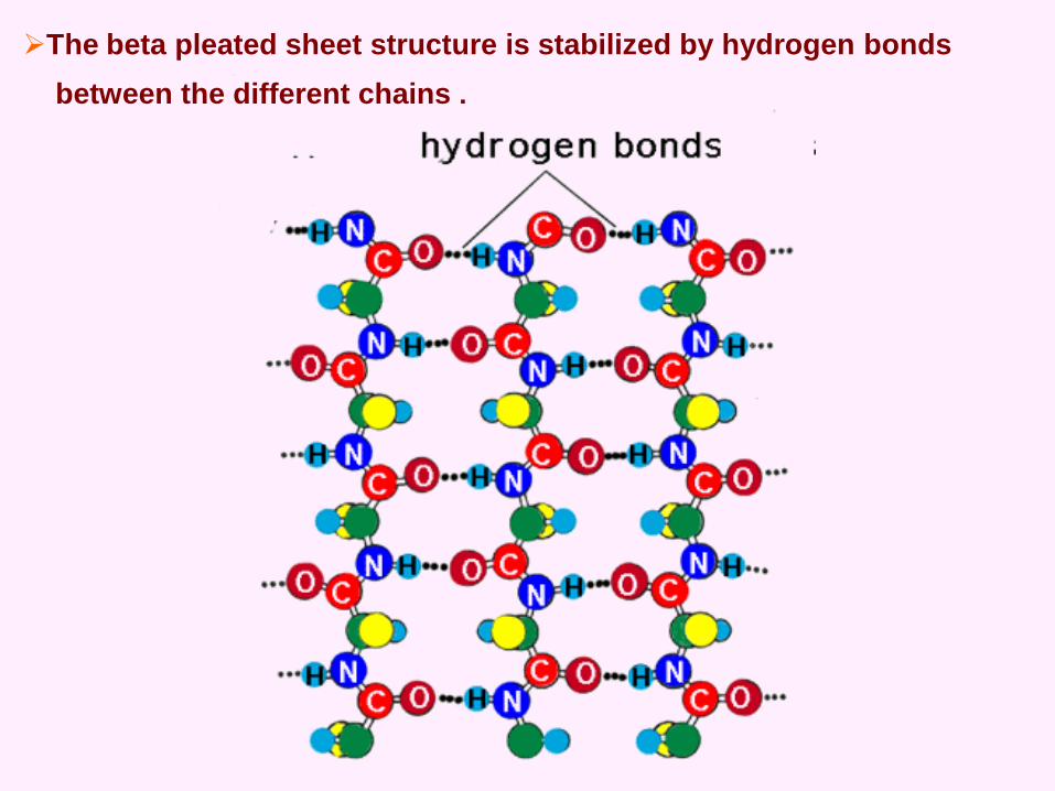
The beta pleated sheet structure is stabilized by hydrogen bonds
between the different chains .

III- Tertiary Structure of Proteins
recoginze the β-pleated sheets
(ribbons with arrows) and the
α-helical regions (barrel shaped
structures.
In addition to these secondary
structures, the protein has
additional twists and turns which
give the protein its unique shape. .
http://academic.brooklyn.cuny.edu/biology/bio4fv/page/terti.htm

Forces that give rise to tertiary structure
Ionic bonding.
Hydrogen bonding.
Hydrophobic interaction.
Disulfide bonds.
Hydrophobicinteraction

1- Ionic bonding.
They are forces of attraction between ions of opposite charge ( + and - )
What kinds of biological molecules form ionic bonds?
Any kind of biological molecule that can form ions
An example of a functional group that can enter into ionic bonds is
shown. The carboxyl group is shown.
Under the right conditions of pH the carboxyl group will ionize and form
the negatively charged COO ion and a positively charged H ion
(or proton)

Here is another representation of the carboxyl group. In this case the
covalent bonds are shown by the lines and the shared electrons are
shown by the black dots .
When ionization of the carboxyl group occurs a proton dissociates from
the OH group, leaving the shared electrons behind with the oxygen.
Thus the COO ion has an excess of electrons over protons and is an
anion. The proton that is released has no associated electron and is
therefore a cation .

Functions of ionic bonds in biology?
They are important in all biological processes. A few examples are:
They play an important role in determining the shapes (tertiary and
quaternary structures) of proteins
They are involved in the process of enzymatic catalysis
They are important in determining the shapes of chromosomes.
They play a role in muscle contraction and cell shape
They are important in establishing polarized membranes for neuron
function and muscle contraction

2- Hydrogen BondsProperties of hydrogen bonds.
Is formed when a charged part of a molecule
having polar covalent bonds forms an electrostatic
interaction with a substance of opposite charge.
Strenght.
Hydrogen bonds are classified as weak bonds
because they are easily and rapidly formed and
broken under normal biological conditions.
What classes of compounds can form hydrogen bonds?
Under the right environmental conditions, any compound that
has polar covalent bonds can form hydrogen bonds.
Importance in biological systems.
* Stabilizing and determining the structure of large
macormolecules like proteins and nucleic acids.
* They are involved in the mechanism of enzyme catalysis
http://academic.brooklyn.cuny.edu/biology/bio4fv/page/hydroge.htm

Hydrocarbons
The Covalent Bond
Non-polar
Covalent Bond
Polar
Covalent Bond
Water
In Biological systems:
It allow the formation of the weak
Hydrogen bond.
In Biological systems:
The molecule that predominance
in non-polar covalent bonds is
called hydrophobic

3- Hydrophobic Interactions
Hydrophobic interactions are more correctly called
hydrophobic exclusions .
http://academic.brooklyn.cuny.edu/biology/bio4fv/page/hydropho.htm
There are two regions containing
hydrophobic substances.
Each of the substances is excluded
from the water matrix.
The two areas of hydrophobic substances will
encounter one another, combine and form one
larger hydrophobic region that is excluded
from the water matix.
This combined state is more energetically
favorable than the one in which the hydrophobic
substances were separate.
Thus this combined state will persist.
Over period of time

VI- Quaternary Structure of Proteins
The quaternary structure of proteins is the shape that results from the
orderly interaction of the polypeptides of a multisubunit protein .
Multisubunit protein
Some proteins are composed of more than one polypeptide.
Each polypeptide is called a subunit. For example, if a protein is
composed of two polypeptides, then it has two subunits.
The polypeptides may or may not be different in primary structure.
This is dependent upon the nature of the protein.

Forces that give rise to the quaternary structure
Ionic bonding
hydrogen bonding
hydrophobic interaction
disulfide bonds
covalent bonds
http://academic.brooklyn.cuny.edu/biology/bio4fv/page/quarte.htm

http://barleyworld.org/css430_09/lecture%209-09/figure-09-03.JPG

http://barleyworld.org/css430_09/lecture%209-09/figure-09-03.JPG

http://www.stat.rice.edu/~marina/figure/protein_structure.jpg
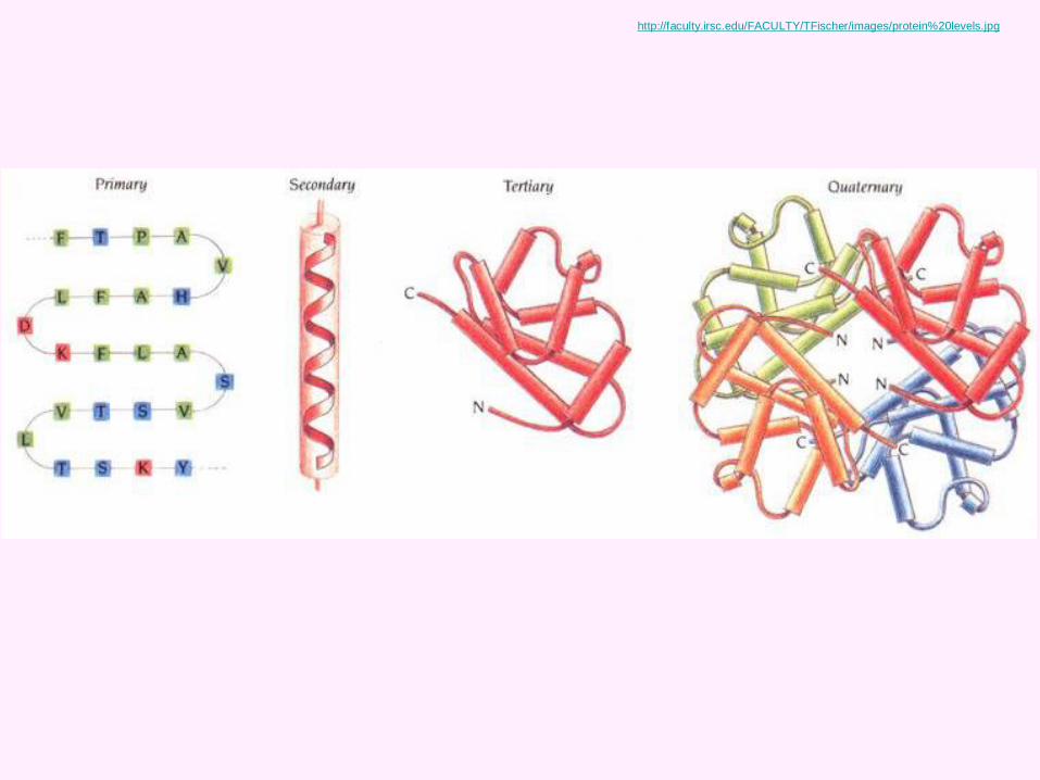
http://faculty.irsc.edu/FACULTY/TFischer/images/protein%20levels.jpg

http://bio73.files.wordpress.com/2008/10/protein-gambar.jpg

http://bio1151b.nicerweb.net/Locked/media/ch05/05_21-ProteinStructure-L.jpg

http://bio1151b.nicerweb.net/Locked/media/ch05/05_21cProteinStructure-L.jpg

http://www-3.unipv.it/webbio/anatcomp/freitas/2008-2009/protein_structure.jpg