Embed Size (px)
Citation preview
What are tissues? • Groups of similar cells that are similar in structure and
perform a common function. • Histology—The study of tissues. • There are 4 categories:
– Epithelial- linings and coverings of organs and body cavities, secretory part of organs and glands, transport membranes of capillaries and alveolar sacs, and membranes which lubricate organs.--covering
– Connective—supports as bone, cartilage, tendons, and ligaments, protects as the bony cavities and as protective immune cells in the blood, and stores nutrients.--support
– Nervous—carries information in the form of impulses throughout the body.--control
– Muscle—contracts to perform movements such as skeletal muscle movements, propulsion in the GI tract, and pumping blood in the heart.--movement
Video Clip: https://www.youtube.com/watch?v=tKWTJ3_-1E8
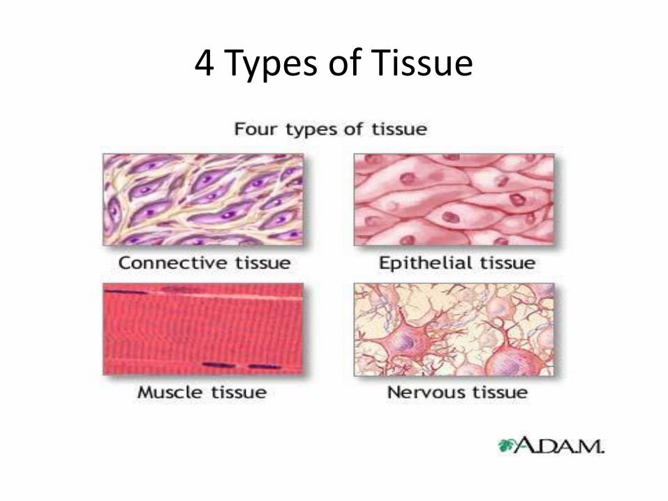
4 Types of Tissue
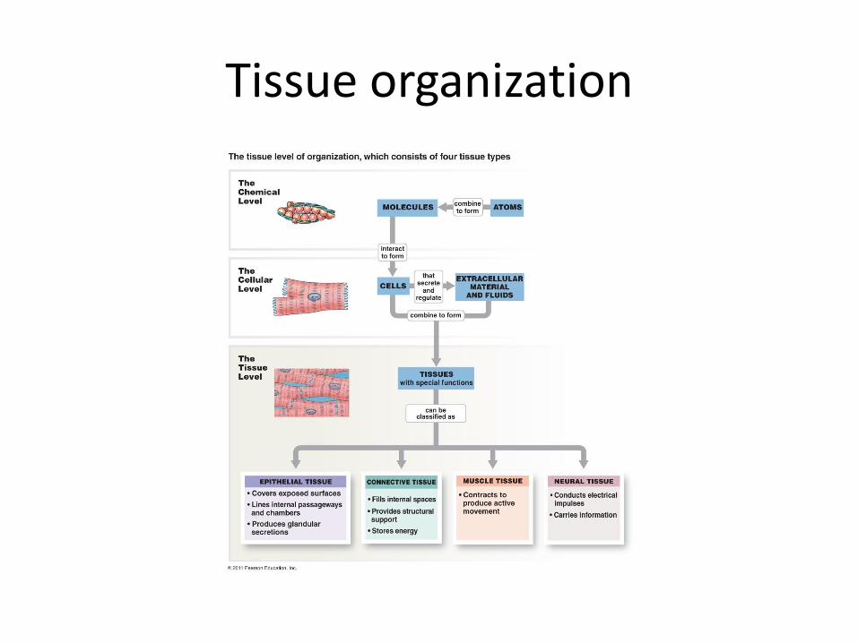
Tissue organization
Epithelial • Epithelial tissue protects your body from moisture loss,
bacteria, and internal injury; form covering layers.
• There are 2 types of epithelial cells: – A. Covering and lining epithelium covers or lines almost all
of your internal and external body surfaces; ex. Outer layer of skin and organs, internal surface lining of your lymph vessels and digestive tract.
– B. Glandular epithelium secretes hormones or other products such as stomach acid, sweat, saliva, and milk.
• Functions: Protection, Absorption, Filtration, Secretion, Excretion, Sensory Reception
• Epithelial Song: https://www.youtube.com/watch?v=KyHxa0t8uac
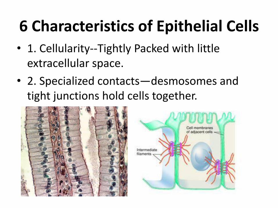
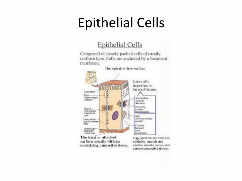
6 Characteristics of Epithelial Cells • 1. Cellularity--Tightly Packed with little
extracellular space.
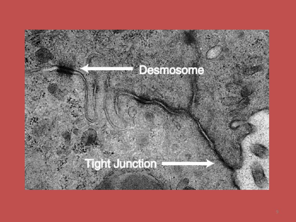
• 2. Specialized contacts—desmosomes and tight junctions hold cells together.
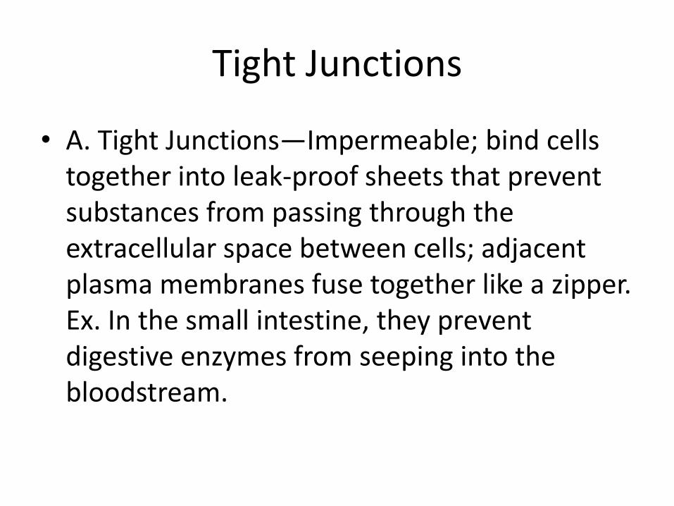
Tight Junctions
• A. Tight Junctions—Impermeable; bind cells together into leak-proof sheets that prevent substances from passing through the extracellular space between cells; adjacent plasma membranes fuse together like a zipper. Ex. In the small intestine, they prevent digestive enzymes from seeping into the bloodstream.
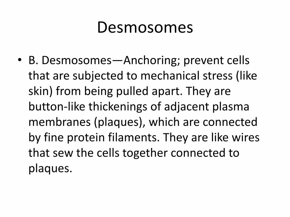
Desmosomes
• B. Desmosomes—Anchoring; prevent cells that are subjected to mechanical stress (like skin) from being pulled apart. They are button-like thickenings of adjacent plasma membranes (plaques), which are connected by fine protein filaments. They are like wires that sew the cells together connected to plaques.
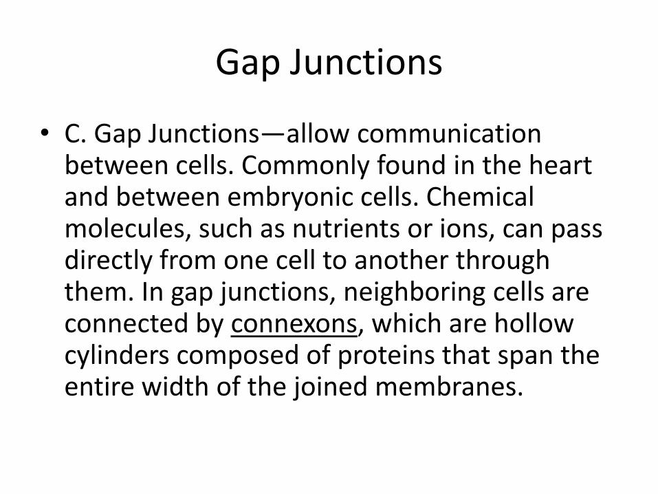
Gap Junctions
• C. Gap Junctions—allow communication between cells. Commonly found in the heart and between embryonic cells. Chemical molecules, such as nutrients or ions, can pass directly from one cell to another through them. In gap junctions, neighboring cells are connected by connexons, which are hollow cylinders composed of proteins that span the entire width of the joined membranes.
9
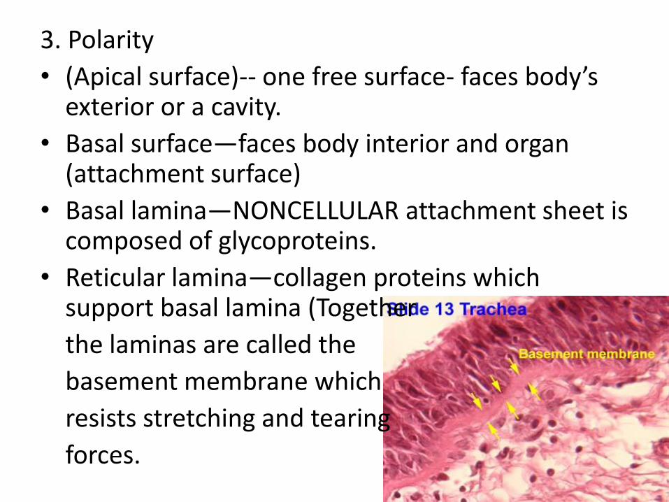
3. Polarity
• (Apical surface)-- one free surface- faces body’s exterior or a cavity.
• Basal surface—faces body interior and organ (attachment surface)
• Basal lamina—NONCELLULAR attachment sheet is composed of glycoproteins.
• Reticular lamina—collagen proteins which support basal lamina (Together
the laminas are called the
basement membrane which
resists stretching and tearing
forces.
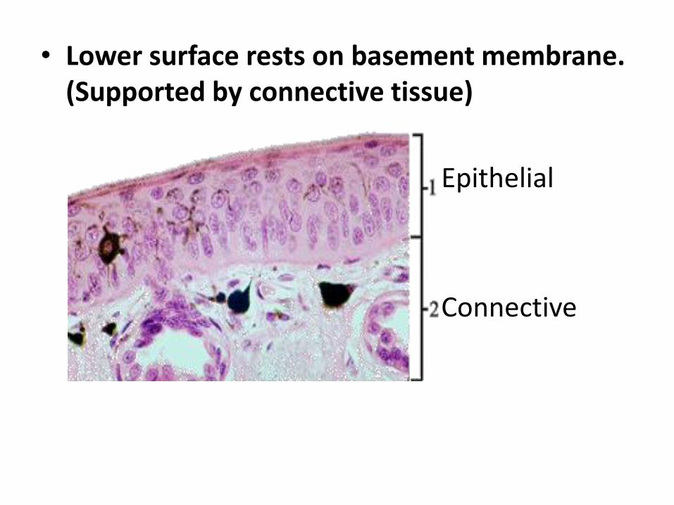
• Lower surface rests on basement membrane. (Supported by connective tissue)
Epithelial
Connective
• 4. They are avascular and innervated-- Cells have no blood supply of their own and depend on underlying connective tissue for food and oxygen through diffusion. They do contain nerves.
5. Regeneration/Reproduce readily. (Rapid healing)
--cells are highly mitotic to replace lost cells 6. Prevents entrance for bacteria or viruses. Video Clip: Epithelia https://www.youtube.com/watch?v=Ucgm0qX4T0k
Epithelial Cells
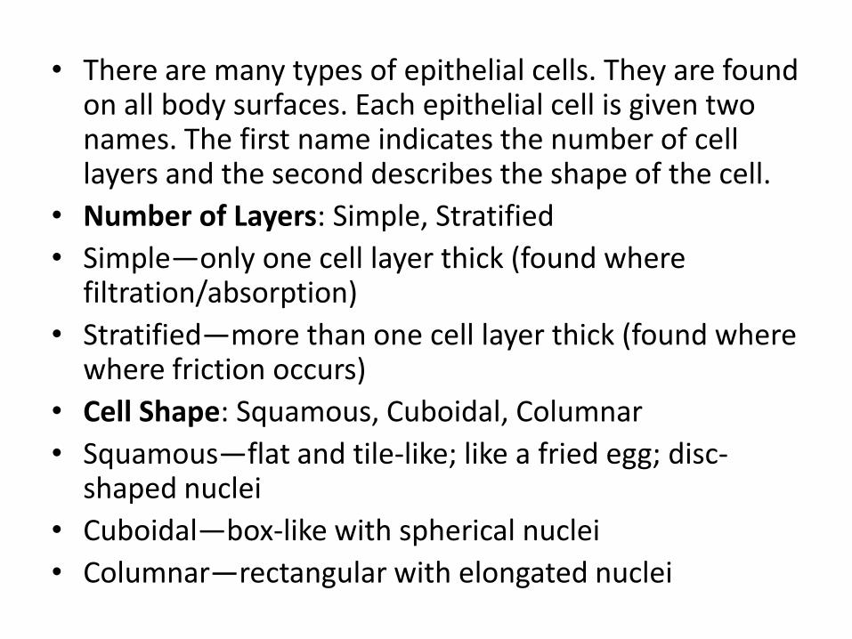
• There are many types of epithelial cells. They are found on all body surfaces. Each epithelial cell is given two names. The first name indicates the number of cell layers and the second describes the shape of the cell.
• Number of Layers: Simple, Stratified
• Simple—only one cell layer thick (found where filtration/absorption)
• Stratified—more than one cell layer thick (found where where friction occurs)
• Cell Shape: Squamous, Cuboidal, Columnar
• Squamous—flat and tile-like; like a fried egg; disc-shaped nuclei
• Cuboidal—box-like with spherical nuclei
• Columnar—rectangular with elongated nuclei
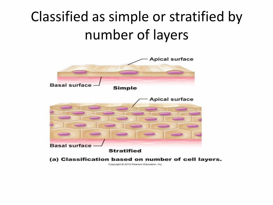
Classified as simple or stratified by number of layers
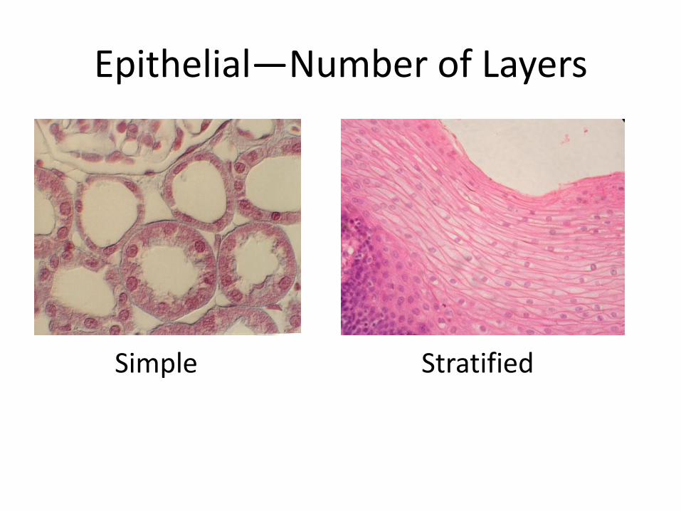
Epithelial—Number of Layers
Simple Stratified
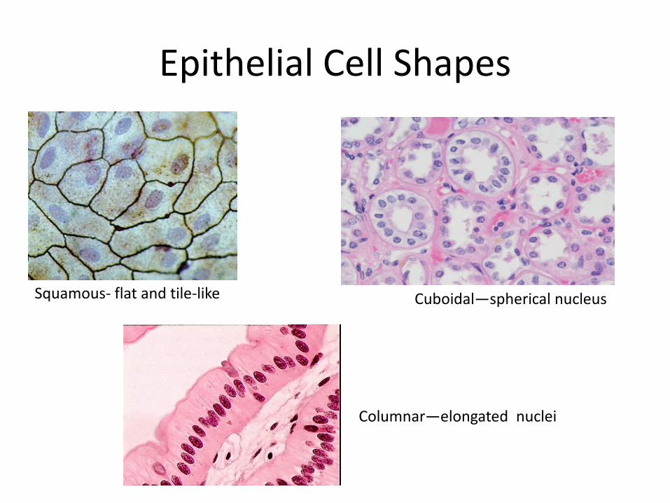
Epithelial Cell Shapes
Squamous- flat and tile-like Cuboidal—spherical nucleus
Columnar—elongated nuclei
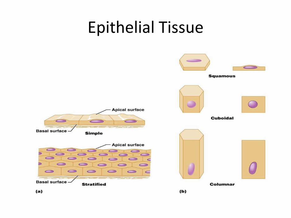
Epithelial Tissue

Epithelial Tissue
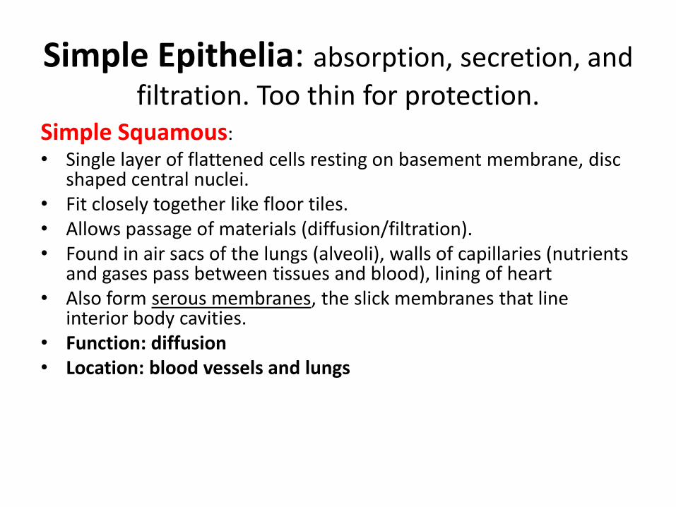
Simple Epithelia: absorption, secretion, and
filtration. Too thin for protection. Simple Squamous:
• Single layer of flattened cells resting on basement membrane, disc shaped central nuclei.
• Fit closely together like floor tiles. • Allows passage of materials (diffusion/filtration). • Found in air sacs of the lungs (alveoli), walls of capillaries (nutrients
and gases pass between tissues and blood), lining of heart • Also form serous membranes, the slick membranes that line
interior body cavities. • Function: diffusion • Location: blood vessels and lungs
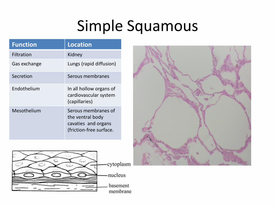
Simple Squamous Function Location
Filtration Kidney
Gas exchange Lungs (rapid diffusion)
Secretion Serous membranes
Endothelium In all hollow organs of cardiovascular system (capillaries)
Mesothelium Serous membranes of the ventral body cavaties and organs (friction-free surface.
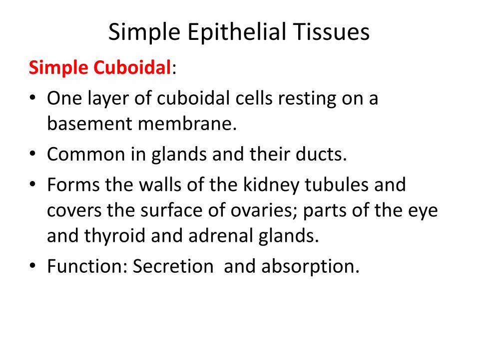
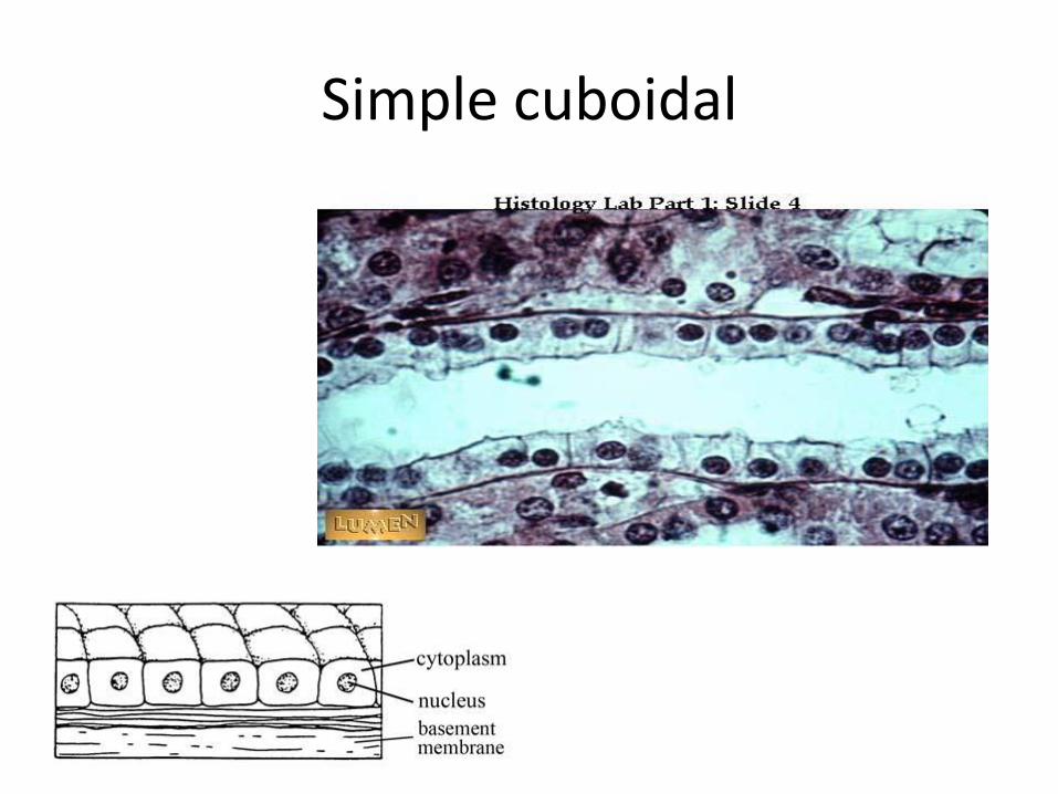
Simple Epithelial Tissues
Simple Cuboidal:
• One layer of cuboidal cells resting on a basement membrane.
• Common in glands and their ducts.
• Forms the walls of the kidney tubules and covers the surface of ovaries; parts of the eye and thyroid and adrenal glands.
• Function: Secretion and absorption.
Simple cuboidal
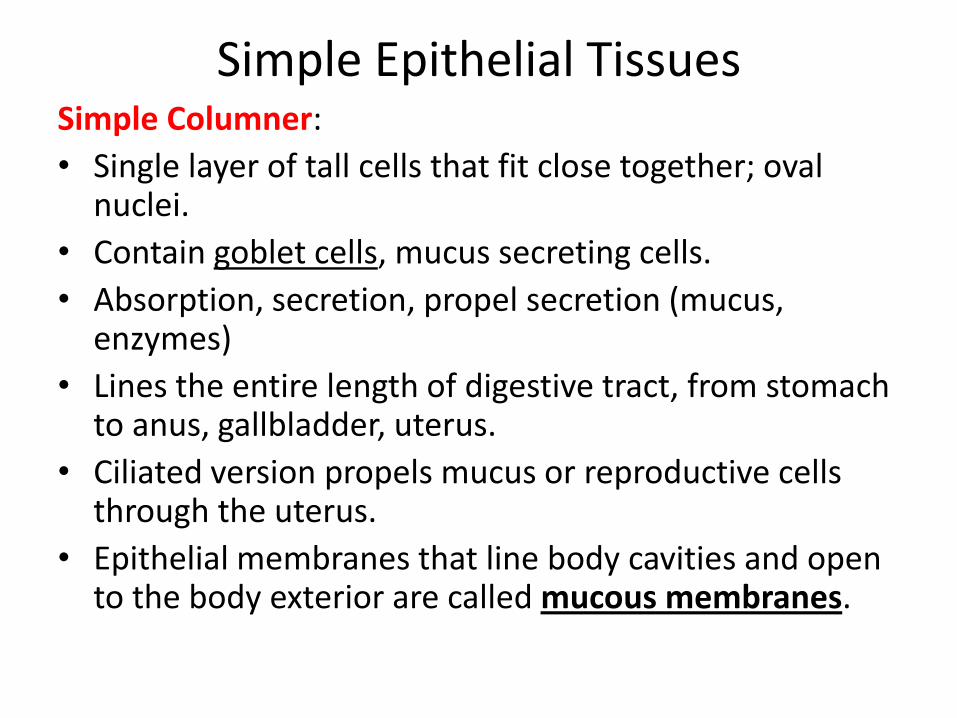
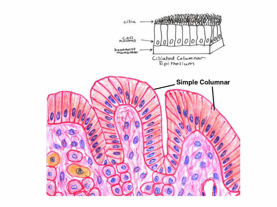
Simple Epithelial Tissues Simple Columner:
• Single layer of tall cells that fit close together; oval nuclei.
• Contain goblet cells, mucus secreting cells.
• Absorption, secretion, propel secretion (mucus, enzymes)
• Lines the entire length of digestive tract, from stomach to anus, gallbladder, uterus.
• Ciliated version propels mucus or reproductive cells through the uterus.
• Epithelial membranes that line body cavities and open to the body exterior are called mucous membranes.
Simple Epithelial Tissues
Pseudostratified Columnar Epithlium:
• All of the cells rest on the basement membrane, but some of its cells are shorter than others, and their nuclei appear at different heights above the basement membrane. This gives the false impression that it is stratified.
• Function: Absorption and secretion.
• A ciliated variety lines most of the respiratory tract (bronchi and trachea).
• The mucus produced by the goblet cells in this epithelium traps dust and other debris, and the cilia propel the mucus upward and away from the lungs.
Stratified Epithelial Tissues—2 or more
layers; more durable. Primary function is to protect.
Stratified Squamous: • Most common stratified epithelium. • Several layers of cells; surface cells full of keratin
(dead).—epidermis of skin • The cells at the free edge are squamous, but those
close to basement membrane are cuboidal or columnar.
• Regenerate from basal layer as apical layer is removed. • Protect underlying tissue in areas subject to abrasion. • Esophagus, mouth, and outer portion of the skin.

Stratified Epithelial Tissues
Stratified Cuboidal: • Usually 2 layers with (at least) the surface being
cuboidal. • They protect areas such as the ducts of sweat
glands, mammary glands, and salivary glands. Stratified Columnar: • Surface cells are columnar, but basal cells vary in
size and shape. • Found in the ocular conjunctiva of the eye, in
parts of the pharynx and anus, the male urethra, and large glands such as the pancreas.
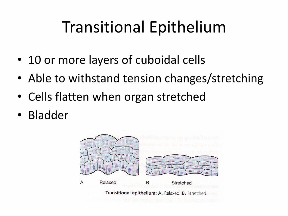
• Transitional Epithelium • A highly modified, stratified squamous epithelium that
forms the lining of only a few organs: the urinary bladder, and the uterus. They are subject to stretching.
• Basal layer are cuboidal or columnar; those of free surface vary in appearance.
• Unstretched: membrane is many-layered, superficial cells are rounded and dome-like.
• Stretched and full: epithelium thins, surface flatten and become squamous-like.
• The ability of transitional cells to slide past one another and change their shape (transitions) allows the ureter wall to stretch as greater volume of urine flows through the tube-like organ. Thus, more urine can be stored.
Transitional Epithelium
• 10 or more layers of cuboidal cells
• Able to withstand tension changes/stretching
• Cells flatten when organ stretched
• Bladder
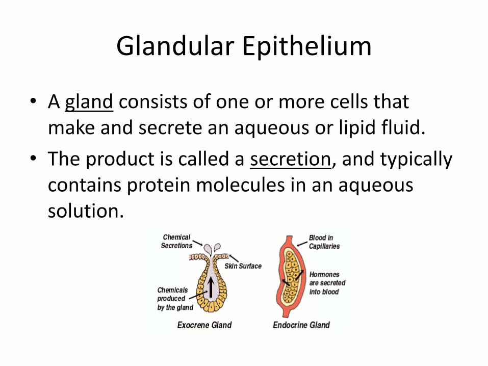
Glandular Epithelium
• A gland consists of one or more cells that make and secrete an aqueous or lipid fluid.
• The product is called a secretion, and typically contains protein molecules in an aqueous solution.
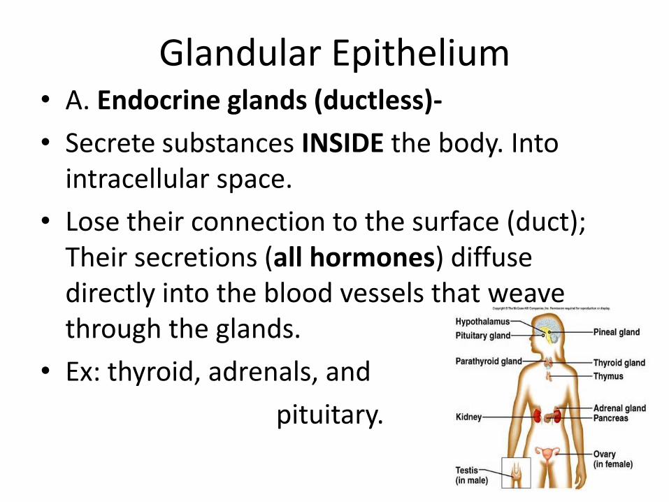
Glandular Epithelium • A. Endocrine glands (ductless)-
• Secrete substances INSIDE the body. Into intracellular space.
• Lose their connection to the surface (duct); Their secretions (all hormones) diffuse directly into the blood vessels that weave through the glands.
• Ex: thyroid, adrenals, and
pituitary.
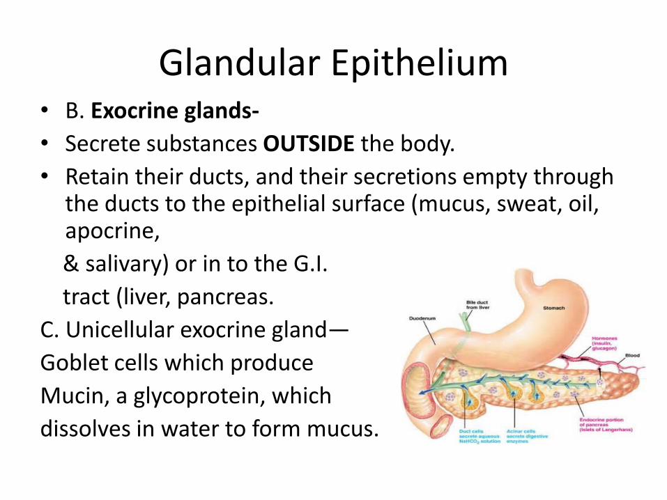
Glandular Epithelium • B. Exocrine glands-
• Secrete substances OUTSIDE the body.
• Retain their ducts, and their secretions empty through the ducts to the epithelial surface (mucus, sweat, oil, apocrine,
& salivary) or in to the G.I.
tract (liver, pancreas.
C. Unicellular exocrine gland—
Goblet cells which produce
Mucin, a glycoprotein, which
dissolves in water to form mucus.
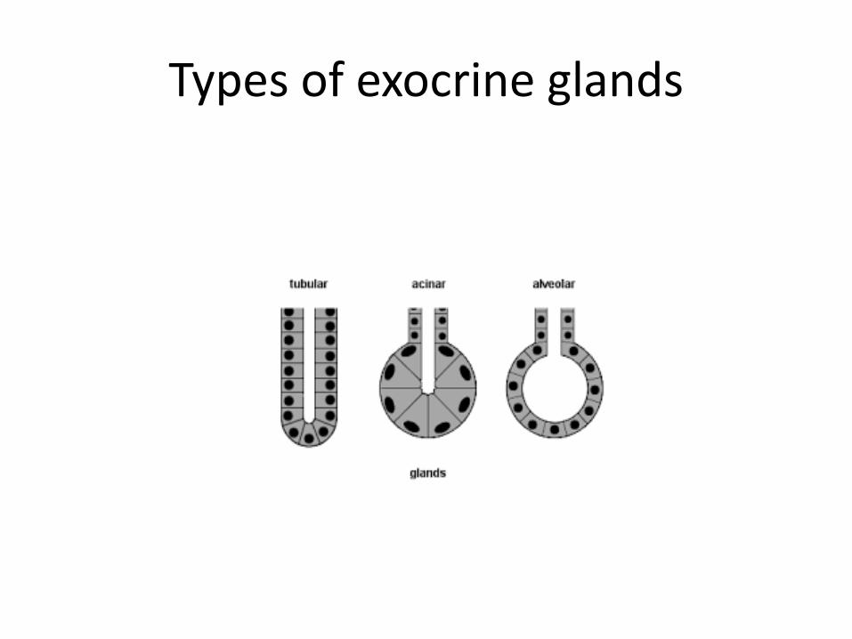
Types of exocrine glands
Connective Tissue
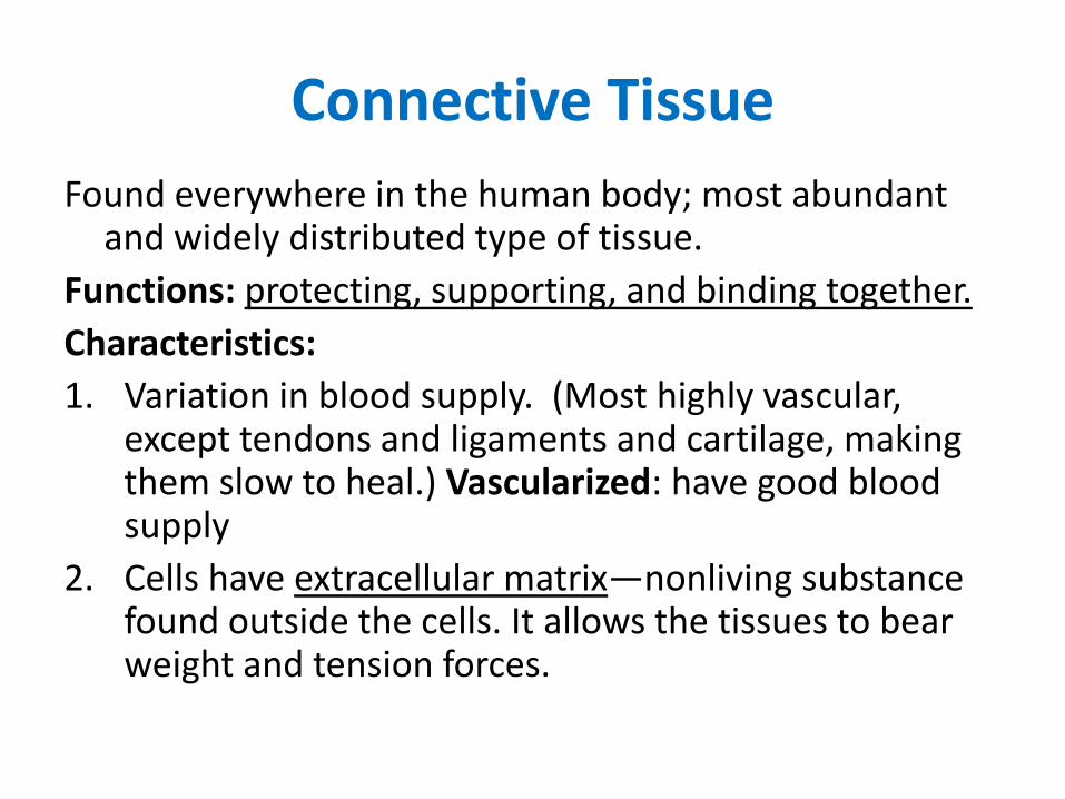
Found everywhere in the human body; most abundant and widely distributed type of tissue.
Functions: protecting, supporting, and binding together.
Characteristics:
1. Variation in blood supply. (Most highly vascular, except tendons and ligaments and cartilage, making them slow to heal.) Vascularized: have good blood supply
2. Cells have extracellular matrix—nonliving substance found outside the cells. It allows the tissues to bear weight and tension forces.
Connective Tissues – general functions
• 3. Connect epithelium to the rest of the body (basal lamina)
• 4. Have no contact with environment (usually covered by epithelium)
• Also:
– Protect delicate organs
– Provide structure and support (bone)
– Insulate and store energy (fat)
– Transport materials (blood)
38
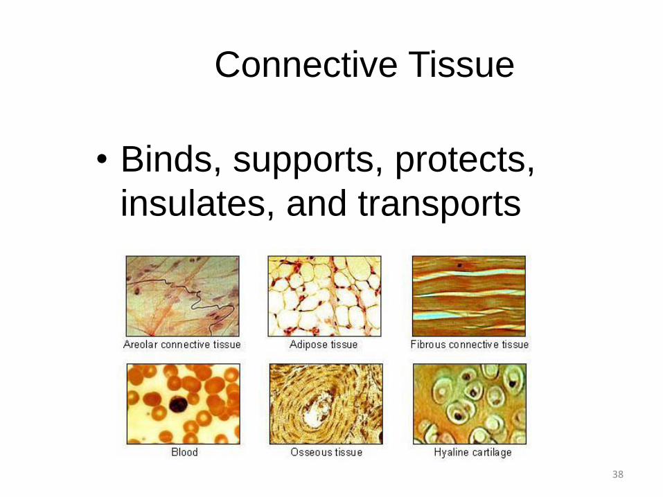
Connective Tissue
• Binds, supports, protects,
insulates, and transports
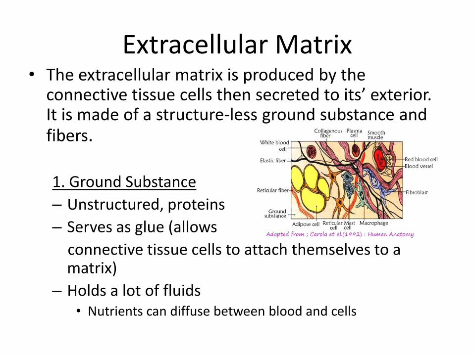
Extracellular Matrix • The extracellular matrix is produced by the
connective tissue cells then secreted to its’ exterior. It is made of a structure-less ground substance and fibers.
1. Ground Substance
– Unstructured, proteins
– Serves as glue (allows
connective tissue cells to attach themselves to a matrix)
– Holds a lot of fluids • Nutrients can diffuse between blood and cells
40
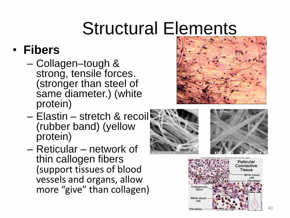
Structural Elements • Fibers
– Collagen–tough & strong, tensile forces. (stronger than steel of same diameter.) (white protein)
– Elastin – stretch & recoil (rubber band) (yellow protein)
– Reticular – network of thin callogen fibers (support tissues of blood vessels and organs, allow more “give” than collagen)
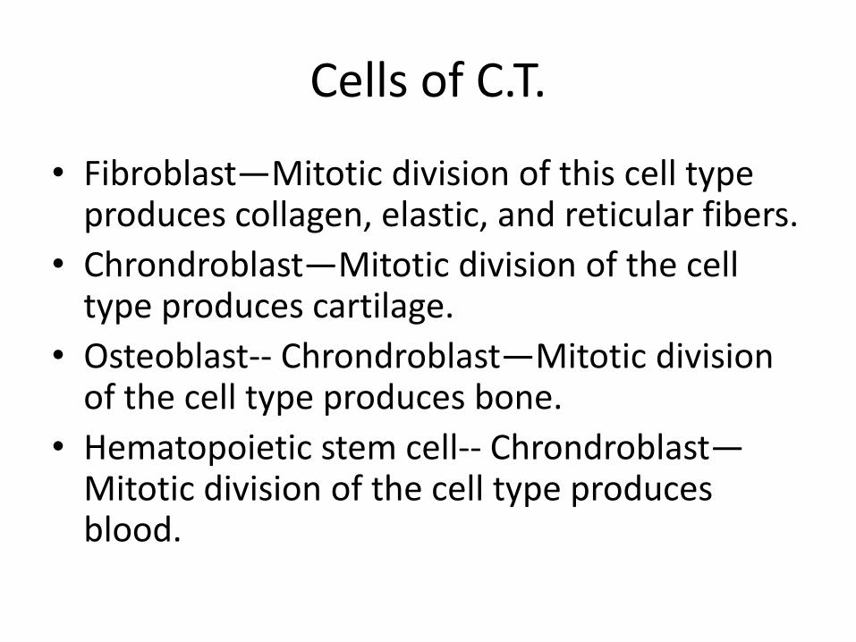
Cells of C.T.
• Fibroblast—Mitotic division of this cell type produces collagen, elastic, and reticular fibers.
• Chrondroblast—Mitotic division of the cell type produces cartilage.
• Osteoblast-- Chrondroblast—Mitotic division of the cell type produces bone.
• Hematopoietic stem cell-- Chrondroblast—Mitotic division of the cell type produces blood.
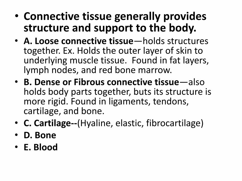
• Connective tissue generally provides structure and support to the body.
• A. Loose connective tissue—holds structures together. Ex. Holds the outer layer of skin to underlying muscle tissue. Found in fat layers, lymph nodes, and red bone marrow.
• B. Dense or Fibrous connective tissue—also holds body parts together, buts its structure is more rigid. Found in ligaments, tendons, cartilage, and bone.
• C. Cartilage--(Hyaline, elastic, fibrocartilage) • D. Bone • E. Blood
43
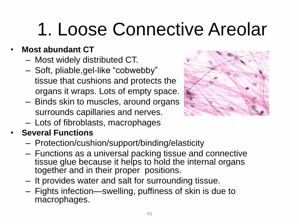
1. Loose Connective Areolar • Most abundant CT
– Most widely distributed CT.
– Soft, pliable,gel-like “cobwebby”
tissue that cushions and protects the
organs it wraps. Lots of empty space.
– Binds skin to muscles, around organs,
surrounds capillaries and nerves.
– Lots of fibroblasts, macrophages • Several Functions
– Protection/cushion/support/binding/elasticity
– Functions as a universal packing tissue and connective tissue glue because it helps to hold the internal organs together and in their proper positions.
– It provides water and salt for surrounding tissue.
– Fights infection—swelling, puffiness of skin is due to macrophages.
44
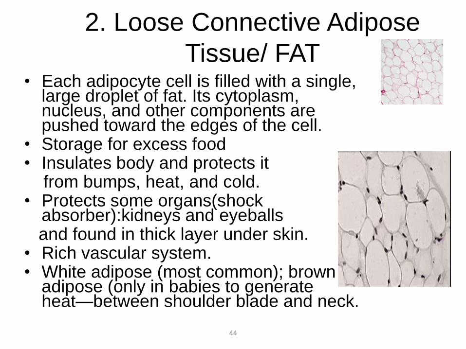
2. Loose Connective Adipose
Tissue/ FAT • Each adipocyte cell is filled with a single,
large droplet of fat. Its cytoplasm, nucleus, and other components are pushed toward the edges of the cell.
• Storage for excess food • Insulates body and protects it from bumps, heat, and cold. • Protects some organs(shock
absorber):kidneys and eyeballs and found in thick layer under skin. • Rich vascular system. • White adipose (most common); brown
adipose (only in babies to generate heat—between shoulder blade and neck.
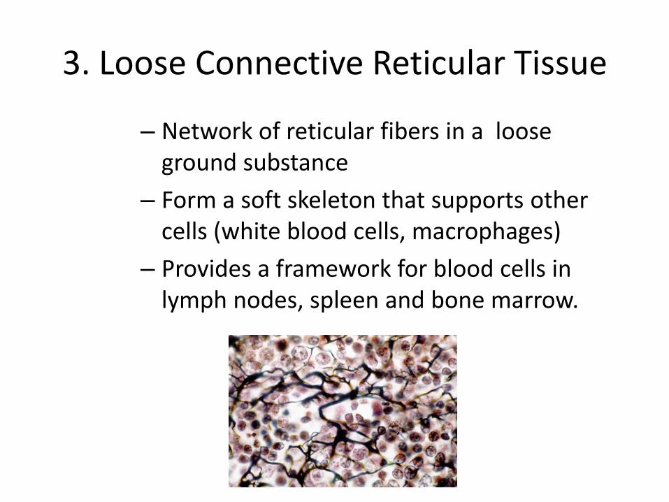
3. Loose Connective Reticular Tissue
– Network of reticular fibers in a loose ground substance
– Form a soft skeleton that supports other cells (white blood cells, macrophages)
– Provides a framework for blood cells in lymph nodes, spleen and bone marrow.
45
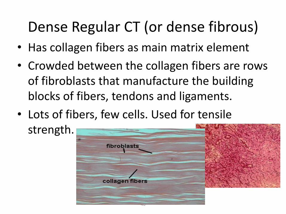
Dense Regular CT (or dense fibrous) • Has collagen fibers as main matrix element
• Crowded between the collagen fibers are rows of fibroblasts that manufacture the building blocks of fibers, tendons and ligaments.
• Lots of fibers, few cells. Used for tensile strength.
46
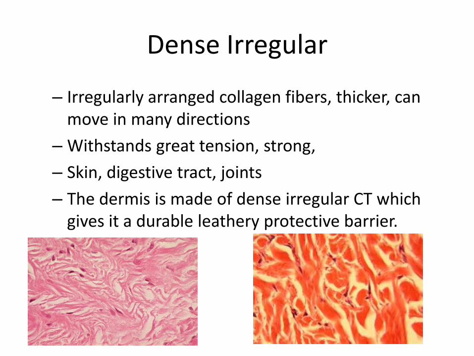
Dense Irregular
– Irregularly arranged collagen fibers, thicker, can move in many directions
– Withstands great tension, strong,
– Skin, digestive tract, joints
– The dermis is made of dense irregular CT which gives it a durable leathery protective barrier.
48
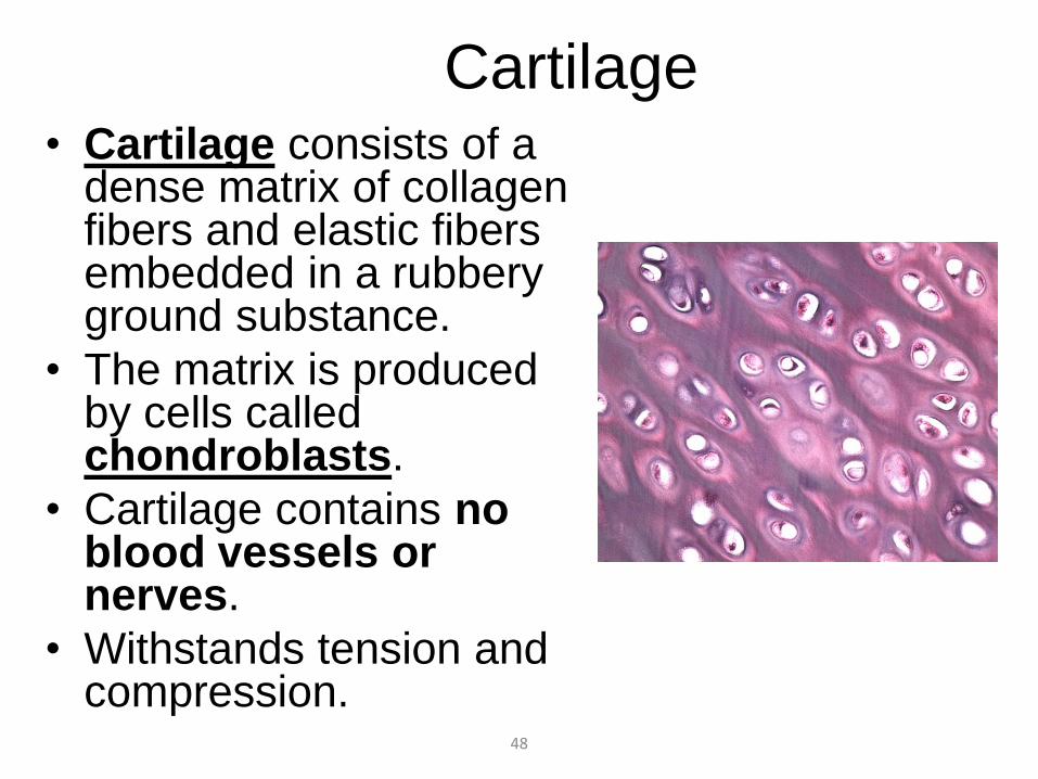
Cartilage • Cartilage consists of a
dense matrix of collagen fibers and elastic fibers embedded in a rubbery ground substance.
• The matrix is produced by cells called chondroblasts.
• Cartilage contains no blood vessels or nerves.
• Withstands tension and compression.
49
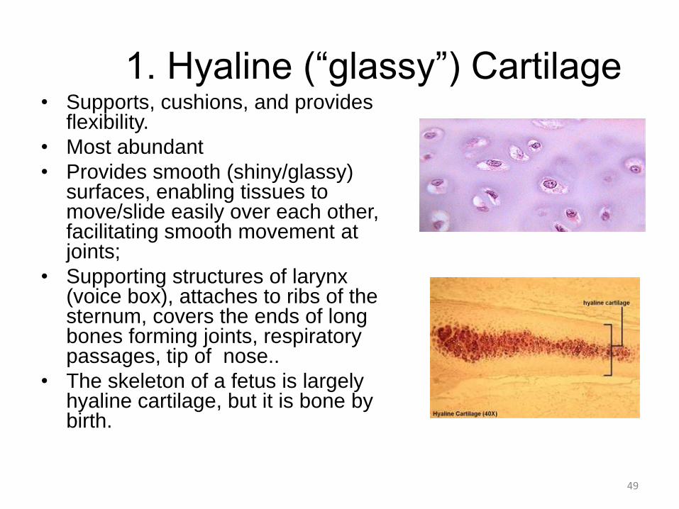
1. Hyaline (“glassy”) Cartilage • Supports, cushions, and provides
flexibility.
• Most abundant
• Provides smooth (shiny/glassy) surfaces, enabling tissues to move/slide easily over each other, facilitating smooth movement at joints;
• Supporting structures of larynx (voice box), attaches to ribs of the sternum, covers the ends of long bones forming joints, respiratory passages, tip of nose..
• The skeleton of a fetus is largely hyaline cartilage, but it is bone by birth.
50
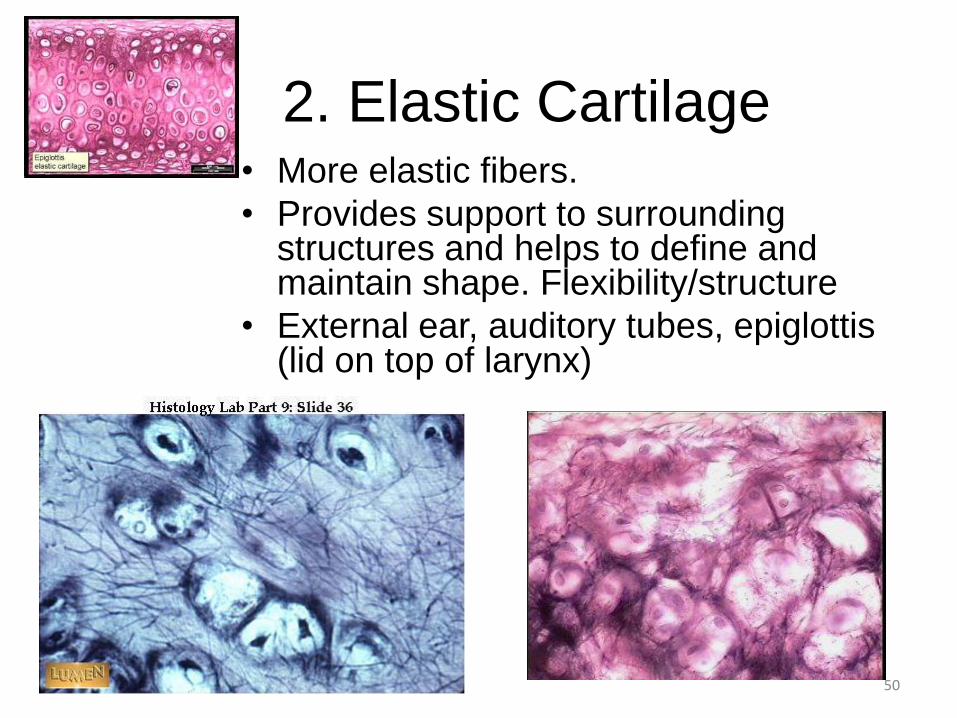
2. Elastic Cartilage • More elastic fibers.
• Provides support to surrounding structures and helps to define and maintain shape. Flexibility/structure
• External ear, auditory tubes, epiglottis (lid on top of larynx)
51
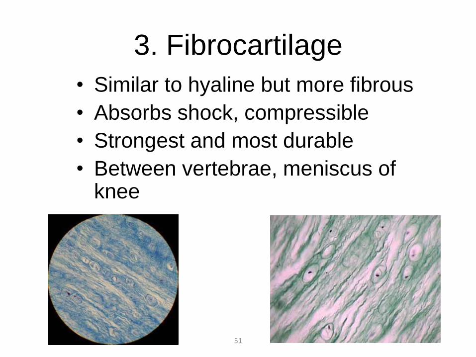
3. Fibrocartilage
• Similar to hyaline but more fibrous
• Absorbs shock, compressible
• Strongest and most durable
• Between vertebrae, meniscus of knee
52
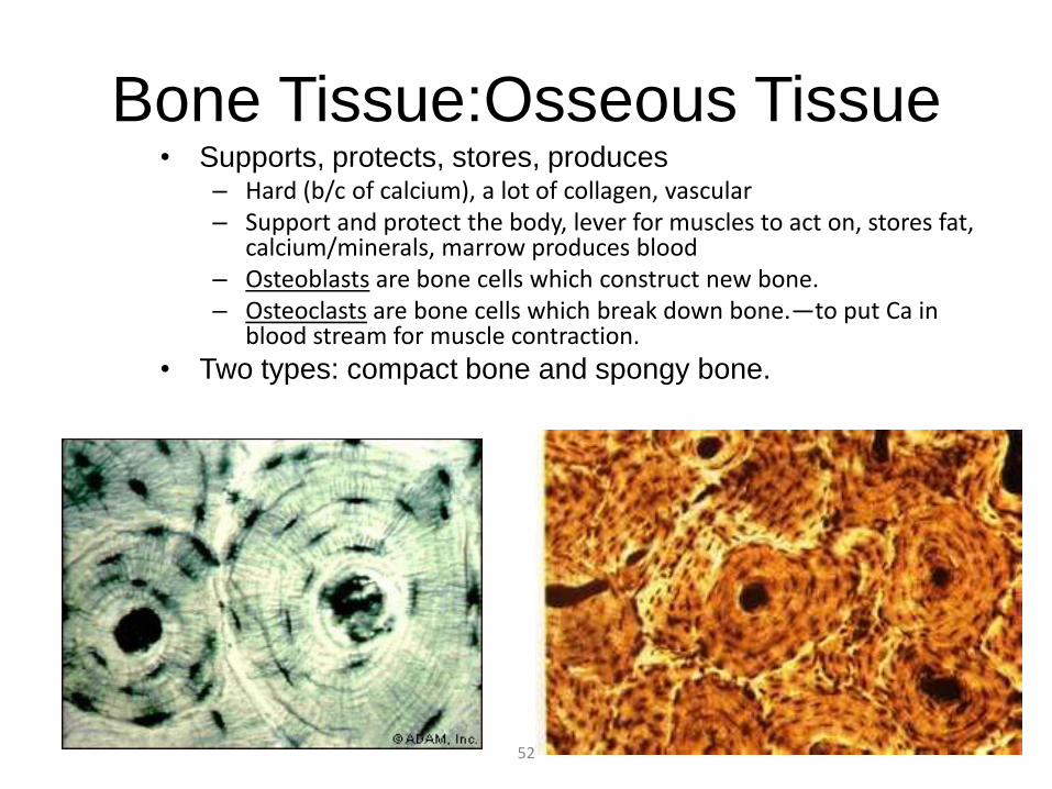
Bone Tissue:Osseous Tissue • Supports, protects, stores, produces
– Hard (b/c of calcium), a lot of collagen, vascular – Support and protect the body, lever for muscles to act on, stores fat,
calcium/minerals, marrow produces blood – Osteoblasts are bone cells which construct new bone. – Osteoclasts are bone cells which break down bone.—to put Ca in
blood stream for muscle contraction.
• Two types: compact bone and spongy bone.
53
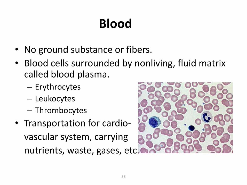
Blood
• No ground substance or fibers.
• Blood cells surrounded by nonliving, fluid matrix called blood plasma. – Erythrocytes
– Leukocytes
– Thrombocytes
• Transportation for cardio-
vascular system, carrying
nutrients, waste, gases, etc.
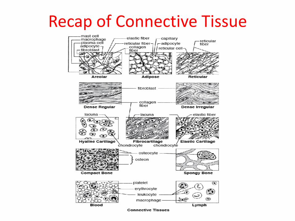
Recap of Connective Tissue
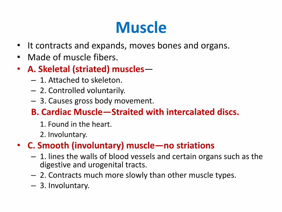
Muscle • It contracts and expands, moves bones and organs. • Made of muscle fibers. • A. Skeletal (striated) muscles—
– 1. Attached to skeleton. – 2. Controlled voluntarily. – 3. Causes gross body movement.
B. Cardiac Muscle—Straited with intercalated discs. 1. Found in the heart. 2. Involuntary.
• C. Smooth (involuntary) muscle—no striations – 1. lines the walls of blood vessels and certain organs such as the
digestive and urogenital tracts. – 2. Contracts much more slowly than other muscle types. – 3. Involuntary.
56
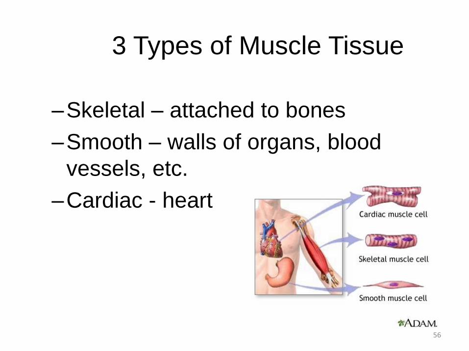
3 Types of Muscle Tissue
–Skeletal – attached to bones
–Smooth – walls of organs, blood
vessels, etc.
–Cardiac - heart
57
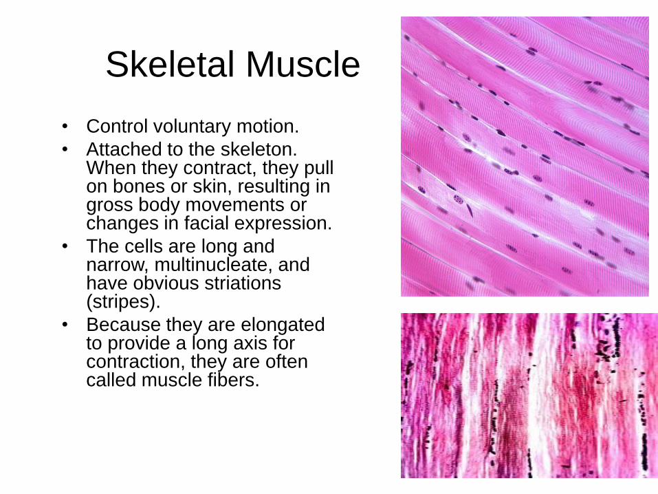
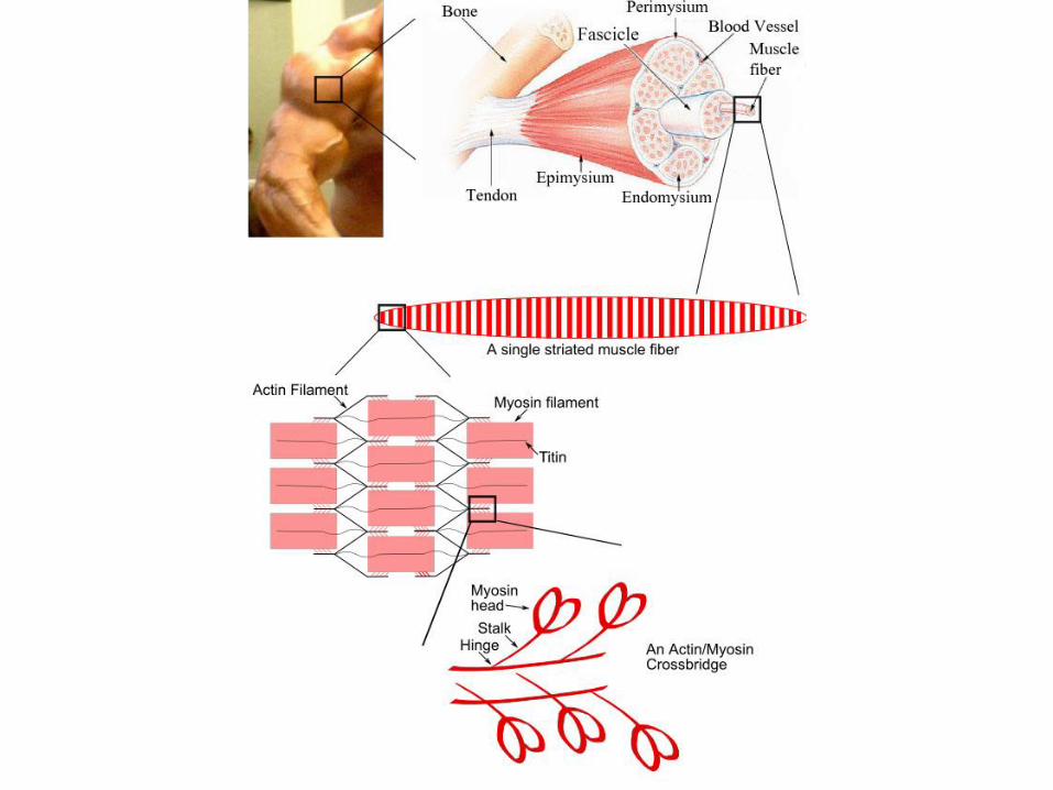
Skeletal Muscle
• Control voluntary motion.
• Attached to the skeleton. When they contract, they pull on bones or skin, resulting in gross body movements or changes in facial expression.
• The cells are long and narrow, multinucleate, and have obvious striations (stripes).
• Because they are elongated to provide a long axis for contraction, they are often called muscle fibers.
59
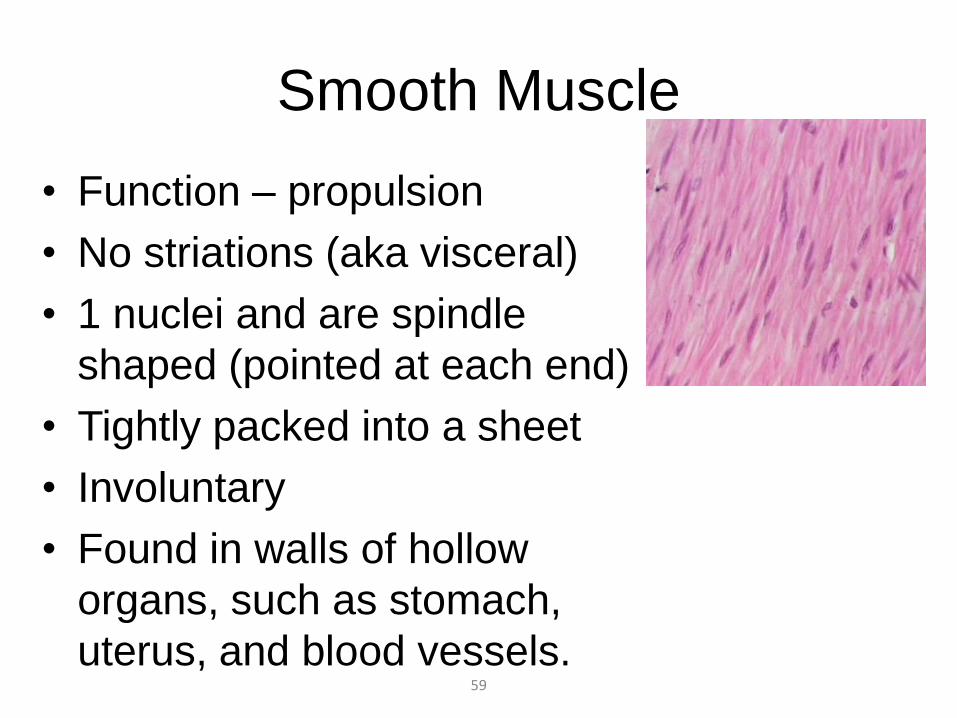
Smooth Muscle
• Function – propulsion
• No striations (aka visceral)
• 1 nuclei and are spindle
shaped (pointed at each end)
• Tightly packed into a sheet
• Involuntary
• Found in walls of hollow
organs, such as stomach,
uterus, and blood vessels.
60
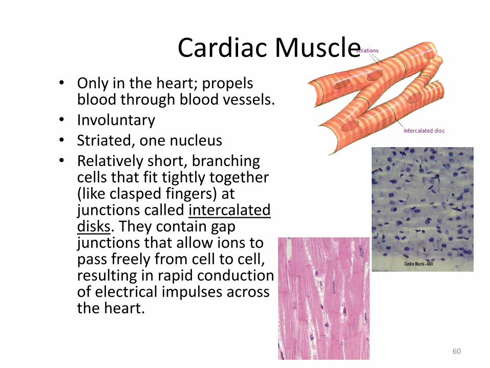
Cardiac Muscle • Only in the heart; propels
blood through blood vessels. • Involuntary • Striated, one nucleus • Relatively short, branching
cells that fit tightly together (like clasped fingers) at junctions called intercalated disks. They contain gap junctions that allow ions to pass freely from cell to cell, resulting in rapid conduction of electrical impulses across the heart.

Nervous • Forms the nervous system, which is responsible for
coordinating the activities and movements of your body through its network of nerves. Includes the brain, spinal cord, and nerves that branch off of those two key parts.
• A. Neurons—basic structural unit of CNS. Each cell consists of the cell body, dendrites, and axon.
• B. Neuroglia (glial cells)—insulate and protect the delicate neurons or anchoring neurons to blood vessels.
• Functions: irritability and conductivity • Neurons receive and conduct electrochemical
messages from one part of the body to another.
62
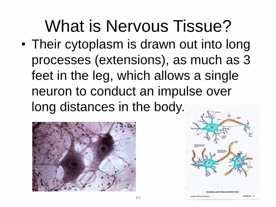
What is Nervous Tissue? • Their cytoplasm is drawn out into long
processes (extensions), as much as 3
feet in the leg, which allows a single
neuron to conduct an impulse over
long distances in the body.

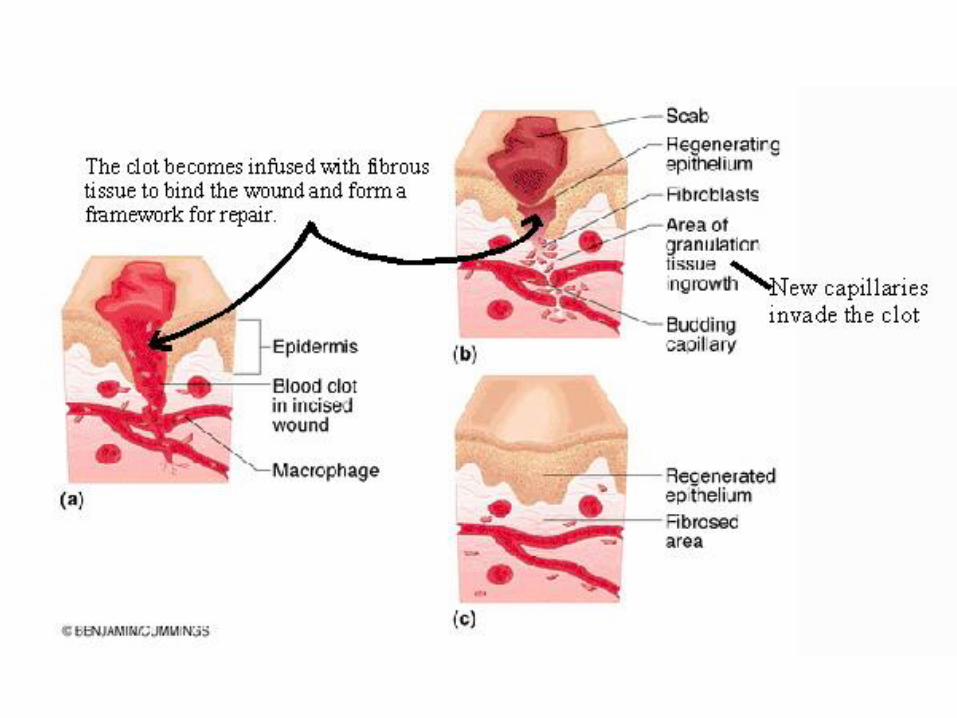
Tissue Repair
• 1. Inflammation
– Tissue DRAMA! Injured cell releases chemicals
– Chemicals cause tissue to dilate more permeable
– White blood cells/plasma/antibodies seep into area
– Construct a clot
Tissue Repair
• 2. Organization restores blood supply
– Clot is replaced by granulation tissue (delicate pink tissue that contains capillaries)
– Granulation tissue bleeds easily (pick at a scab)
– Produce growth factors
– Becomes scar tissue (highly resistant to infection bacteria inhibiting substances)
Tissue Repair
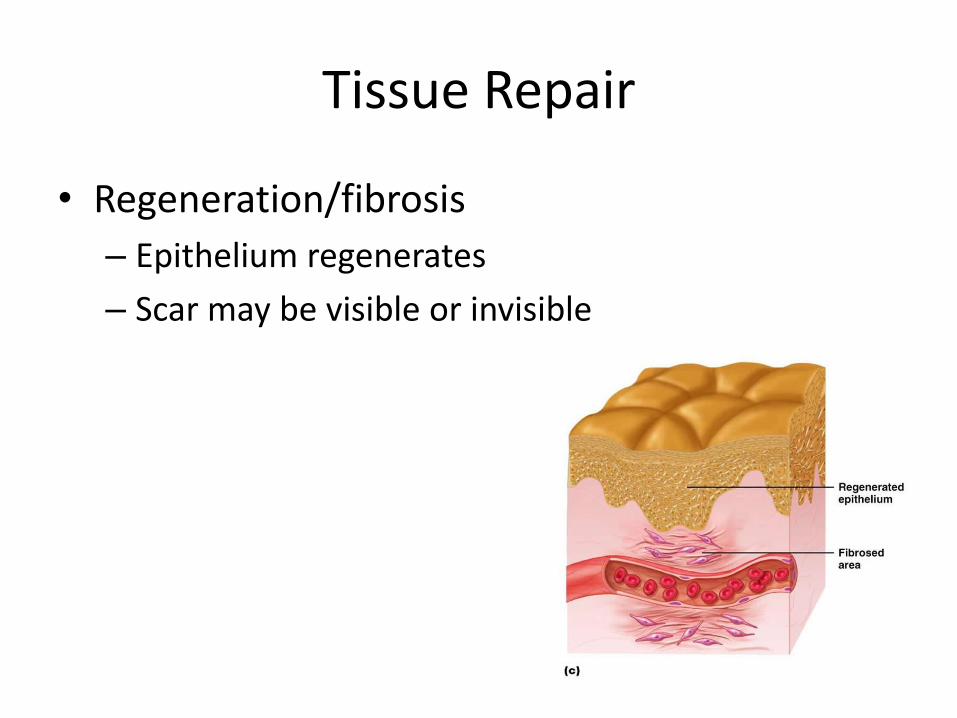
• Regeneration/fibrosis
– Epithelium regenerates
– Scar may be visible or invisible
Tissue Repair
• 1. Inflammation
– Tissue DRAMA! Injured cell releases chemicals
– Chemicals cause tissue to dilate more permeable
– White blood cells/plasma/antibodies seep into area
– Construct a clot

Tissue Repair
• 2. Organization restores blood supply
– Clot is replaced by granulation tissue (delicate pink tissue that contains capillaries)
– Granulation tissue bleeds easily (pick at a scab)
– Produce growth factors
– Becomes scar tissue (highly resistant to infection bacteria inhibiting substances)

Tissue Repair
• Regeneration/fibrosis
– Epithelium regenerates
– Scar may be visible or invisible