Embed Size (px)
Citation preview

WHAT PNEUMATICITY TELLS US ABOUT
‘PROSAUROPODS’, AND VICE VERSA
by MATHEW WEDELUniversity of California Museum of Paleontology and Department of Integrative Biology, 1101 Valley Life Sciences Building, Berkeley, CA 94720-4780, USA;
e-mail: [email protected]
Typescript received 15 February 2006; accepted in revised form 24 October 2006
Abstract: Pneumatic (air-filled) bones are an important fea-
ture of the postcranial skeleton in pterosaurs, theropods and
sauropods. However, there is no unambiguous evidence for
postcranial pneumaticity in basal sauropodomorphs and even
the ambiguous evidence is scant. Patterns of skeletal pneumati-
zation in early sauropods and theropods suggest that basal sau-
rischians had cervical air sacs like those of birds. Furthermore,
patterns of pneumaticity in most pterosaurs, theropods and
sauropods are diagnostic for abdominal air sacs. The air sacs
necessary for flow-through lung ventilation like that of birds
may have evolved once (at the base of Ornithodira), twice
(independently in pterosaurs and saurischians) or three times
(independently in pterosaurs, theropods and sauropods). Skel-
etal pneumaticity appears to be more evolutionarily malleable
than the air sacs and diverticula that produce it. The evolution
of air sacs probably pre-dated the appearance of skeletal pneu-
maticity in ornithodirans.
Key words: Prosauropoda, Sauropodomorpha, Saurischia,
Ornithodira, pneumaticity, air sacs, diverticula.
Pneumaticity is a prominent feature of the postcra-
nial skeleton in theropod and sauropod dinosaurs. In con-
trast, there is little evidence for postcranial pneumaticity in
basal sauropodomorphs (informally referred to as ‘prosaur-
opods’ in this paper), although from time to time some
aspects of prosauropod osteology have been posited as evi-
dence of pneumaticity (Britt 1997) or compared with
unequivocal pneumatic structures in sauropods (Yates
2003; Galton and Upchurch 2004). My goals in this paper
are to review the evidence for postcranial skeletal pneuma-
ticity (PSP) in prosauropods and to discuss the origin of air
sacs and pneumaticity in early dinosaurs and their relatives.
Prosauropod taxonomy is currently in a state of flux,
as other papers in this volume attest (Sereno 2007;
Upchurch et al. 2007; Yates 2007). Prosauropods were
traditionally considered a paraphyletic assemblage that
gave rise to sauropods. Sereno (1998) recovered a mono-
phyletic Prosauropoda, defined this clade (anchored upon
Plateosaurus) as a monophyletic sister taxon to Sauropoda
and united the two in a node-based Sauropodomorpha. A
similar phylogenetic hypothesis was described by Galton
and Upchurch (2004). However, other recent phylogenet-
ic analyses (Yates 2003, 2004; Yates and Kitching 2003)
have found that some prosauropods are closer to Salta-
saurus than to Plateosaurus; thus, under current phylo-
genetic definitions they should be regarded as basal
sauropods. Some other taxa (e.g. Saturnalia) lie outside
Sauropodomorpha as defined by Sereno (1998) altogether
(Yates 2003, 2004; Yates and Kitching 2003; Langer 2004).
However, the monophyly or paraphyly of the group of
taxa traditionally called prosauropods is not critical to the
purposes of this paper. What is important is that all tra-
ditional ‘prosauropods’ have two things in common: they
lack unequivocal evidence of pneumatic cavities in their
vertebrae and ribs, and they are phylogenetically brack-
eted by sauropods and theropods (Text-fig. 1).
Institutional abbreviations. BMNH, The Natural History
Museum, London; CM, Carnegie Museum of Natural His-
tory, Pittsburgh, USA; FMNH, Field Museum of Natural History,
Chicago, USA; MSM, Mesa Southwest Museum, Mesa, USA;
OMNH, Oklahoma Museum of Natural History, Norman, USA;
SMNS, Staatliches Museum fur Naturkunde, Stuttgart, Germany.
Anatomical abbreviations. ACDL, anterior centrodiapophyseal
lamina; AL, accessory lamina; AVF, anteroventral fossa; NAF,
neural arch fossa; PCDL, posterior centrodiapophyseal lamina;
PDF, posterodorsal fossa; PODL, postzygodiapophyseal lamina;
PPDL, paradiapophyseal lamina; PRDL, prezygodiapophyseal
lamina; SPOL, spinopostzygapophyseal lamina; SPRL, spinoprezy-
gapophyseal lamina (lamina abbreviations after Wilson 1999).
POSTCRANIAL PNEUMATICITY INTHEROPOD AND SAUROPODDINOSAURS
Before examining the evidence for PSP in ‘prosauropods’,
I will review the conditions present in other saurischian
[Special Papers in Palaeontology 77, 2007, pp. 207–222]
ª The Palaeontological Association 207
dinosaurs. The sister taxon of Sauropodomorpha is Ther-
opoda; consequently, ‘prosauropods’ are phylogenetically
bracketed in part by birds, the only clade of extant verte-
brates with extensive PSP. The relationship between the
respiratory system and pneumatic postcranial bones in
birds has been described many times (e.g. Muller 1908;
King 1966; Duncker 1971; O’Connor 2004), and is briefly
summarized here. The relatively small, constant-volume,
unidirectional flow-through lungs of birds are ventilated
by the attached air sacs, which are large, flexible and
devoid of parenchymal tissue. The lungs and air sacs also
produce air-filled tubes called diverticula that pass
between the viscera, between the muscles, and under the
skin. Where a diverticulum comes into contact with a
bone, it may (but does not always) induce bone resorp-
tion, which can produce pneumatic tracks, fossae or for-
amina. If resorption of the cortex produces a foramen,
the diverticulum may enter the medullary space and
replace the existing internal structure with a series of air-
filled chambers of varying complexity. The best descrip-
tion of this process is provided by Bremer (1940).
The extent of PSP varies in different avian clades.
Almost any postcranial bones can become pneumatized;
in large soaring birds such as pelicans, almost the entire
skeleton is pneumatic, including the distal limb elements
(O’Connor 2004). Although many large volant and flight-
less birds have highly pneumatic skeletons, the correlation
between body size and the extent of PSP in birds is weak
(O’Connor 2004). PSP tends to be reduced or absent in
diving birds (Gier 1952; O’Connor 2004). Different parts
of the skeleton become pneumatized by diverticula of dif-
ferent air sacs in extant birds (Table 1); this is important
because it allows us to make inferences regarding the evo-
lution of air sacs in fossil taxa. PSP in non-avian thero-
pods generally follows the avian model (Britt 1993, 1997;
O’Connor and Claessens 2005; O’Connor 2006). Patterns
of pneumatization along the vertebral column indicate
that both anterior and posterior air sacs (presumably cer-
vical and abdominal) had evolved by the time of the cer-
atosaur-tetanuran divergence (O’Connor and Claessens
2005).
Fossae are present in the presacral vertebrae of basal
sauropods such as Shunosaurus and Barapasaurus (Britt
1993; Wilson and Sereno 1998). These fossae are similar
to the unequivocally pneumatic foramina and camerae of
more derived sauropods, both in their position on indi-
vidual vertebrae and in their distribution along the ver-
tebral column, and because of these similarities they have
usually been regarded as pneumatic in origin (Britt 1993,
1997; Wedel 2003a). However, similar fossae are present
in other tetrapods that lack PSP, so the presence of fossae
alone is at best equivocal evidence for PSP (O’Connor
2006; see below). The vertebrae of more derived sauro-
pods have foramina that communicate with large internal
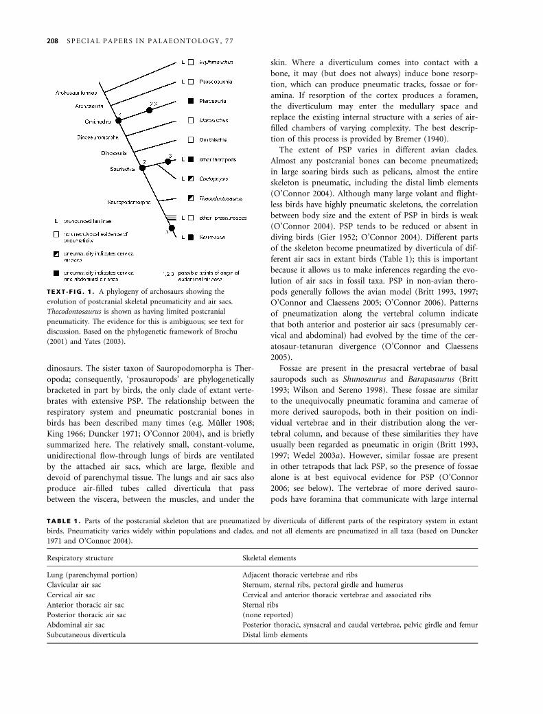
TEXT -F IG . 1 . A phylogeny of archosaurs showing the
evolution of postcranial skeletal pneumaticity and air sacs.
Thecodontosaurus is shown as having limited postcranial
pneumaticity. The evidence for this is ambiguous; see text for
discussion. Based on the phylogenetic framework of Brochu
(2001) and Yates (2003).
TABLE 1 . Parts of the postcranial skeleton that are pneumatized by diverticula of different parts of the respiratory system in extant
birds. Pneumaticity varies widely within populations and clades, and not all elements are pneumatized in all taxa (based on Duncker
1971 and O’Connor 2004).
Respiratory structure Skeletal elements
Lung (parenchymal portion) Adjacent thoracic vertebrae and ribs
Clavicular air sac Sternum, sternal ribs, pectoral girdle and humerus
Cervical air sac Cervical and anterior thoracic vertebrae and associated ribs
Anterior thoracic air sac Sternal ribs
Posterior thoracic air sac (none reported)
Abdominal air sac Posterior thoracic, synsacral and caudal vertebrae, pelvic girdle and femur
Subcutaneous diverticula Distal limb elements
208 S P E C I A L P A P E R S I N P A L A E O N T O L O G Y , 7 7

chambers; the combination of foramina and large internal
chambers is an unambiguous indicator of PSP (O’Connor
2006). There is a general trend in sauropod evolution for
PSP to spread posteriorly along the vertebral column,
albeit to different extents in different clades and with a
few reversals (Wedel 2003b; Text-fig. 2). In both sauro-
pods and theropods, fossae in basal forms were replaced
by large-chambered (camerate) vertebrae and eventually
small-chambered (camellate) vertebrae in more derived
taxa (Britt 1993, 1997; Wedel 2003a).
The evolution of PSP in sauropods mirrors in detail
that of non-avian theropods. At the level of individual
elements (e.g. vertebrae and ribs), pneumatic features in
sauropods compare very closely with those of both avian
and non-avian theropods (Text-fig. 3). In terms of the
ratio of bony tissue to air space within a pneumatic ele-
ment, sauropod vertebrae are, on average, comparable
with the limb bones of many extant birds: about 60 per
cent air by volume (Wedel 2004, 2005; Woodward 2005;
Schwarz and Fritsch 2006). At the level of the skeleton,
osteological indicators of pneumaticity spread as far back
as the mid-caudal vertebrae in at least two groups of
sauropods, the diplodocines and saltasaurines (Osborn
1899; Powell 1992). Among non-avian theropods, exten-
sive pneumatization of the caudal series evolved only in
oviraptorosaurs (Osmolska et al. 2004). Finally, limited
appendicular pneumaticity was probably present in both
sauropods and non-avian theropods. The dromaeosaur
Buitreraptor has a pneumatic furcula (Makovicky et al.
2005), and a large foramen in the proximal femur of the
oviraptorid Shixinggia is probably also pneumatic in ori-
gin (Lu and Zhang 2005). Large chambers have been
reported in the ilia of the basal diplodocoid Amazonsau-
rus (Carvalho et al. 2003) and in several titanosaurs
(Powell 1992; Sanz et al. 1999; Xu et al. 2006). Although
these chambers are similar to unequivocally pneumatic
spaces in the other saurischians, it has not yet been
shown that the ilial chambers are connected to foramina,
which are necessary for pneumatization to occur (see
O’Connor 2006).
EVIDENCE OF PNEUMATICITY IN‘PROSAUROPODS’
Historically, postcranial pneumaticity in ‘prosauropods’
has received little attention, which is to be expected given
the paucity of available evidence. Janensch (1947) posited
that a foramen in a dorsal vertebra of Plateosaurus might
have been pneumatic, but he attached no great weight to
this hypothesis. Britt (1997) considered vertebral laminae
evidence of pneumaticity in ‘prosauropods’. Most
recently, Yates (2003, p. 14, fig. 12) identified ‘pleurocoel-
like pits’ in the mid-cervical vertebrae of Thecodontosau-
rus caducus, and Galton and Upchurch (2004, p. 245)
referred to fossae in the posterior dorsals of some pro-
sauropods as ‘pleurocoelar indentations’. The ‘pleurocoel-
like’ structures were not explicitly described as pneumatic
in either work. Although fossae are not unambiguous
indicators of pneumaticity (O’Connor 2006), vertebral
fossae seem to be an early step toward full pneumatiza-
tion, both ontogenetically and phylogenetically (Wedel
2003a). Putative pneumatic characters in ‘prosauropods’
can be divided into three categories: vertebral laminae,
foramina and fossae, which will be discussed in this
order, below.
Vertebral laminae. Vertebral laminae are struts or plates of
bone that connect the various apophyses of a vertebra to
each other and to the centrum. The landmarks that are
usually connected in this way are the pre- and postzygapo-
physes, the diapophyses and parapophyses, and the neu-
rapophysis. The form and occurrence of the major
laminae in saurischian dinosaurs were reviewed by Wilson
TEXT -F IG . 2 . A diagram showing the
distribution of fossae and pneumatic
chambers (black boxes) along the
vertebral column in sauropods. Only the
lineage leading to diplodocines is shown
here. The same caudal extension of
pneumatic features also occurred
independently in macronarian
sauropods, culminating in saltasaurines,
and several times in theropods. The
format of the diagram is based on
Wilson and Sereno (1998, fig. 47).
Phylogeny based on Wilson (2002),
Yates (2003) and Upchurch et al. (2004).
W E D E L : W H A T P N E U M A T I C I T Y T E L L S U S A B O U T ‘ P R O S A U R O P O D S ’ 209
(1999). In addition to a basic set of laminae common to
all saurischians, many sauropods and theropods have other
irregularly developed laminae that are usually not named
but are collectively called accessory laminae. Laminae tend
to be more numerous and more sharply defined in camer-
ate than camellate vertebrae (Wilson and Sereno 1998;
Wedel 2003a). Camellate vertebrae evolved relatively early
in the radiation of non-avian theropods (Britt 1993, 1997),
and most derived theropods have less elaborate systems of
laminae than neosauropods. This may explain why the lit-
erature on laminae has tended to focus on sauropods (e.g.
Osborn 1899; Osborn and Mook 1921; Janensch 1929,
1950; Wilson 1999).
Two problems with the identification of laminae that
are relevant to the question of pneumaticity are how well
developed a ridge of bone must be before we call it a
lamina, and whether laminae are primarily additive struc-
tures formed by the deposition of new bone, or are sim-
ply bone that is left over following the formation of
fossae. The first problem is important because, as shown
below, incipient laminae are broadly distributed among
archosaurs. To what extent are the distinct laminae of
saurischian dinosaurs new (¼ apomorphic) structures,
rather than modifications of pre-existing ones? This ques-
tion has ramifications for the evolution of laminae
and for coding of laminae as characters in phylogenetic
analyses.
The second question can be stated: do laminae grow
out from the corpus of the vertebra to define the fossae
that they bound, or do we only recognize laminae as dis-
tinct structures because the bone between them has been
removed? For example, the cervical vertebra of Nigersau-
rus illustrated by Sereno and Wilson (2005, fig. 5.8) has
on the lateral face of the neural spine two fossae divided
by an accessory lamina (Text-fig. 4). At its edges, the
anteroventral fossa approaches both the prezygapophysis
and the diapophysis. This region is flat or convex in most
other neosauropods, which have a lateral fossa in roughly
the same position as the posterodorsal fossa in Nigersau-
rus. It seems likely therefore that the anteroventral fossa
in Nigersaurus is a new morphological feature, and that
the accessory lamina can only be recognized as a lamina
because a fossa has been excavated below it. Conversely,
the vertebrae of most tetrapods do not have straight bars
of bone that connect the zygapophyses to the neurapoph-
ysis, but this is exactly what the spinopre- and
spinopostzygapophyseal laminae of some sauropods do
(Text-fig. 4). In comparison with the condition in other
tetrapods, including prosauropods, these laminae appear
to be additive structures. These potentially opposing pro-
cesses of lamina formation should be kept in mind while
reading the following descriptions.
The laminae of sauropods often form the boundaries of
fossae that have been interpreted as pneumatic, either
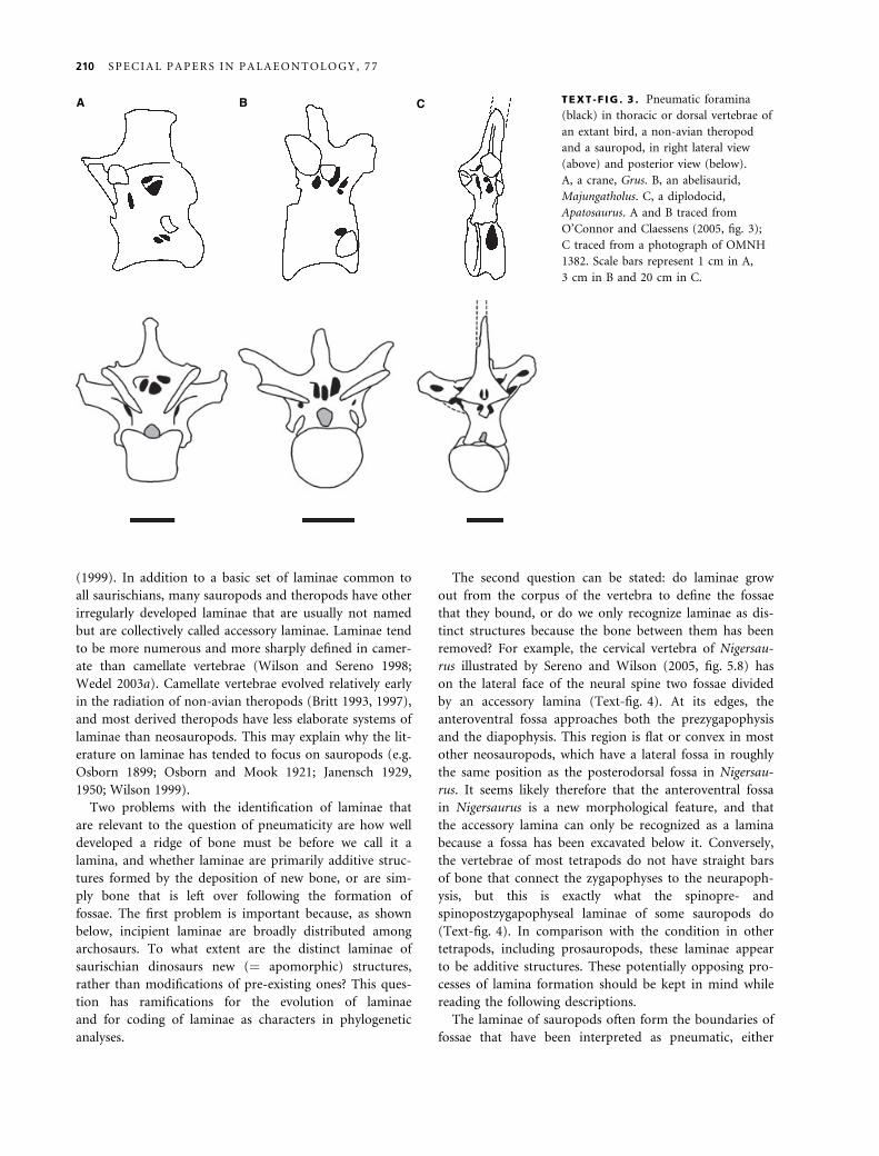
A B C TEXT -F IG . 3 . Pneumatic foramina
(black) in thoracic or dorsal vertebrae of
an extant bird, a non-avian theropod
and a sauropod, in right lateral view
(above) and posterior view (below).
A, a crane, Grus. B, an abelisaurid,
Majungatholus. C, a diplodocid,
Apatosaurus. A and B traced from
O’Connor and Claessens (2005, fig. 3);
C traced from a photograph of OMNH
1382. Scale bars represent 1 cm in A,
3 cm in B and 20 cm in C.
210 S P E C I A L P A P E R S I N P A L A E O N T O L O G Y , 7 7

because they contain foramina that lead to internal cham-
bers or because they are heavily sculpted, with numerous
subfossae (sensu Wilson 1999) and a distinct bony texture
(although texture alone is not necessarily a good indicator
of pneumaticity; see O’Connor 2006). Wilson (1999) con-
sidered whether sauropod laminae existed to provide
mechanical support or to subdivide pneumatic diverti-
cula, and concluded that they probably served both func-
tions simultaneously. Following from the aforementioned
discussion, we might also ask if sauropod laminae exist
because the pneumatic diverticula are subdivided, as they
often are in birds (e.g. Wedel 2003b, fig. 2), and these
subdivisions are impressed into the bone, leaving laminae
between them. Rather than try to determine which struc-
ture has morphogenetic precedence, it may be more use-
ful to view sauropod vertebrae in light of Witmer’s
(1997) hypothesis that the form of a pneumatic bone can
be viewed as the outcome of a struggle between bone tis-
sue, which grows partly in response to biomechanical
stress, and pneumatic diverticula, which are opportunistic
and invasive and spread wherever possible (see Sadler
et al. 1996 and Anorbe et al. 2000 for examples of prolif-
erating diverticula).
The laminae of ‘prosauropods’ differ from those of
sauropods in three important ways. The first is that pro-
sauropods have fewer laminae. The laminae that connect
the diapophysis to the centrum, parapophysis and zygap-
ophyses are usually present (Wilson 1999), but those that
connect the neurapophysis to other landmarks are absent
(Text-fig. 5; but see Bonaparte 1999, figs 13–16 on Les-
semsaurus). The second is that laminae are confined to
the presacral vertebrae in ‘prosauropods’, whereas the sac-
ral vertebrae of neosauropods and the caudal vertebrae of
diplodocids also bear laminae.
The third and most important difference between the
laminae of sauropods and ‘prosauropods’ is that the fos-
sae bounded by the latter are blind. These fossae do not
contain foramina or subfossae and they do not have a
distinctive bone texture. Consequently, there is no strong
reason to suspect that they contained pneumatic diverti-
cula. O’Connor (2006) found that similar fossae in extant
crocodilians and birds may contain cartilage or adipose
tissue. Considering whether the laminae are additive
structures or remnants of fossa formation sheds little light
on the problem. Some laminae, such as the PRDLs of
Plateosaurus cervicals, are straight-line structures that
TEXT -F IG . 4 . Laminae, fossae and
foramina in cervical vertebrae of
Nigersaurus and Apatosaurus. A, fifth
cervical vertebra of Nigersaurus, traced
from Sereno and Wilson (2005, fig. 5.8).
B, tenth cervical vertebra of Apatosaurus,
traced from Gilmore (1936, pl. 24).
Scale bars represent 5 cm in A and
20 cm in B.
A B
PRDL PRDL
PPDL
PODLPODL
PCDL
PCDL groove
TEXT -F IG . 5 . Vertebrae of Plateosaurus trossingensis (SMNS 13200) in left lateral view. A, the eighth cervical vertebra. B, dorsal
vertebrae 1–4. Scale bars represent 5 cm.
W E D E L : W H A T P N E U M A T I C I T Y T E L L S U S A B O U T ‘ P R O S A U R O P O D S ’ 211
appear to have been added, compared with the condition
in vertebrae that lack laminae (Text-fig. 5). Others, such
as the PODLs in the same vertebrae, are only detectable
because they have been undercut by a fossa. The form of
the fossae themselves provides no obvious clues to their
contents in vivo.
Laminae like those of ‘prosauropods’ occur in many
other archosaurs. Desojo et al. (2002) and Parker (2003)
recognized that many of the laminae described by Wilson
(1999) for saurischian dinosaurs are also present in basal
archosaurs and pseudosuchians. The full complement of
diapophyseal laminae is present in dorsal vertebrae of the
basal archosauriform Erythrosuchus and in those of popo-
saurs such as Sillosuchus and Arizonasaurus, including the
PCDL, PODL, PPDL and PRDL (Text-fig. 6; see Alcober
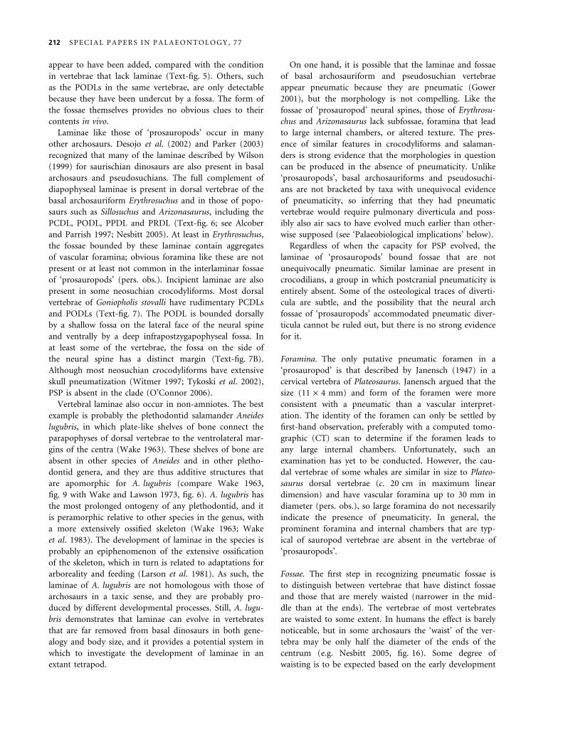
and Parrish 1997; Nesbitt 2005). At least in Erythrosuchus,
the fossae bounded by these laminae contain aggregates
of vascular foramina; obvious foramina like these are not
present or at least not common in the interlaminar fossae
of ‘prosauropods’ (pers. obs.). Incipient laminae are also
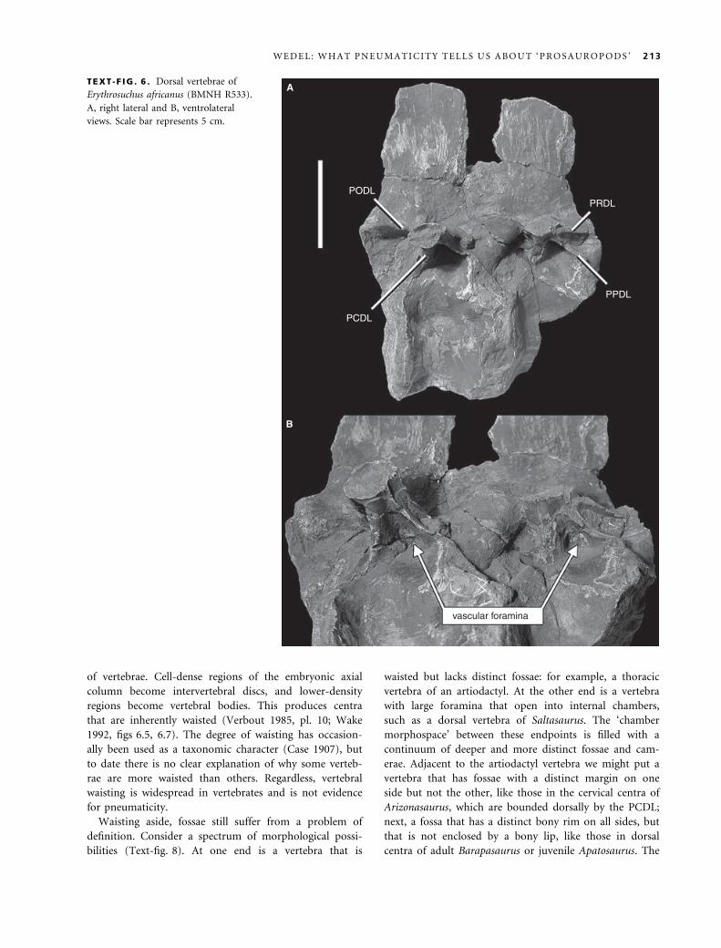
present in some neosuchian crocodyliforms. Most dorsal
vertebrae of Goniopholis stovalli have rudimentary PCDLs
and PODLs (Text-fig. 7). The PODL is bounded dorsally
by a shallow fossa on the lateral face of the neural spine
and ventrally by a deep infrapostzygapophyseal fossa. In
at least some of the vertebrae, the fossa on the side of
the neural spine has a distinct margin (Text-fig. 7B).
Although most neosuchian crocodyliforms have extensive
skull pneumatization (Witmer 1997; Tykoski et al. 2002),
PSP is absent in the clade (O’Connor 2006).
Vertebral laminae also occur in non-amniotes. The best
example is probably the plethodontid salamander Aneides
lugubris, in which plate-like shelves of bone connect the
parapophyses of dorsal vertebrae to the ventrolateral mar-
gins of the centra (Wake 1963). These shelves of bone are
absent in other species of Aneides and in other pletho-
dontid genera, and they are thus additive structures that
are apomorphic for A. lugubris (compare Wake 1963,
fig. 9 with Wake and Lawson 1973, fig. 6). A. lugubris has
the most prolonged ontogeny of any plethodontid, and it
is peramorphic relative to other species in the genus, with
a more extensively ossified skeleton (Wake 1963; Wake
et al. 1983). The development of laminae in the species is
probably an epiphenomenon of the extensive ossification
of the skeleton, which in turn is related to adaptations for
arboreality and feeding (Larson et al. 1981). As such, the
laminae of A. lugubris are not homologous with those of
archosaurs in a taxic sense, and they are probably pro-
duced by different developmental processes. Still, A. lugu-
bris demonstrates that laminae can evolve in vertebrates
that are far removed from basal dinosaurs in both gene-
alogy and body size, and it provides a potential system in
which to investigate the development of laminae in an
extant tetrapod.
On one hand, it is possible that the laminae and fossae
of basal archosauriform and pseudosuchian vertebrae
appear pneumatic because they are pneumatic (Gower
2001), but the morphology is not compelling. Like the
fossae of ‘prosauropod’ neural spines, those of Erythrosu-
chus and Arizonasaurus lack subfossae, foramina that lead
to large internal chambers, or altered texture. The pres-
ence of similar features in crocodyliforms and salaman-
ders is strong evidence that the morphologies in question
can be produced in the absence of pneumaticity. Unlike
‘prosauropods’, basal archosauriforms and pseudosuchi-
ans are not bracketed by taxa with unequivocal evidence
of pneumaticity, so inferring that they had pneumatic
vertebrae would require pulmonary diverticula and poss-
ibly also air sacs to have evolved much earlier than other-
wise supposed (see ‘Palaeobiological implications’ below).
Regardless of when the capacity for PSP evolved, the
laminae of ‘prosauropods’ bound fossae that are not
unequivocally pneumatic. Similar laminae are present in
crocodilians, a group in which postcranial pneumaticity is
entirely absent. Some of the osteological traces of diverti-
cula are subtle, and the possibility that the neural arch
fossae of ‘prosauropods’ accommodated pneumatic diver-
ticula cannot be ruled out, but there is no strong evidence
for it.
Foramina. The only putative pneumatic foramen in a
‘prosauropod’ is that described by Janensch (1947) in a
cervical vertebra of Plateosaurus. Janensch argued that the
size (11 · 4 mm) and form of the foramen were more
consistent with a pneumatic than a vascular interpret-
ation. The identity of the foramen can only be settled by
first-hand observation, preferably with a computed tomo-
graphic (CT) scan to determine if the foramen leads to
any large internal chambers. Unfortunately, such an
examination has yet to be conducted. However, the cau-
dal vertebrae of some whales are similar in size to Plateo-
saurus dorsal vertebrae (c. 20 cm in maximum linear
dimension) and have vascular foramina up to 30 mm in
diameter (pers. obs.), so large foramina do not necessarily
indicate the presence of pneumaticity. In general, the
prominent foramina and internal chambers that are typ-
ical of sauropod vertebrae are absent in the vertebrae of
‘prosauropods’.
Fossae. The first step in recognizing pneumatic fossae is
to distinguish between vertebrae that have distinct fossae
and those that are merely waisted (narrower in the mid-
dle than at the ends). The vertebrae of most vertebrates
are waisted to some extent. In humans the effect is barely
noticeable, but in some archosaurs the ‘waist’ of the ver-
tebra may be only half the diameter of the ends of the
centrum (e.g. Nesbitt 2005, fig. 16). Some degree of
waisting is to be expected based on the early development
212 S P E C I A L P A P E R S I N P A L A E O N T O L O G Y , 7 7
of vertebrae. Cell-dense regions of the embryonic axial
column become intervertebral discs, and lower-density
regions become vertebral bodies. This produces centra
that are inherently waisted (Verbout 1985, pl. 10; Wake
1992, figs 6.5, 6.7). The degree of waisting has occasion-
ally been used as a taxonomic character (Case 1907), but
to date there is no clear explanation of why some verteb-
rae are more waisted than others. Regardless, vertebral
waisting is widespread in vertebrates and is not evidence
for pneumaticity.
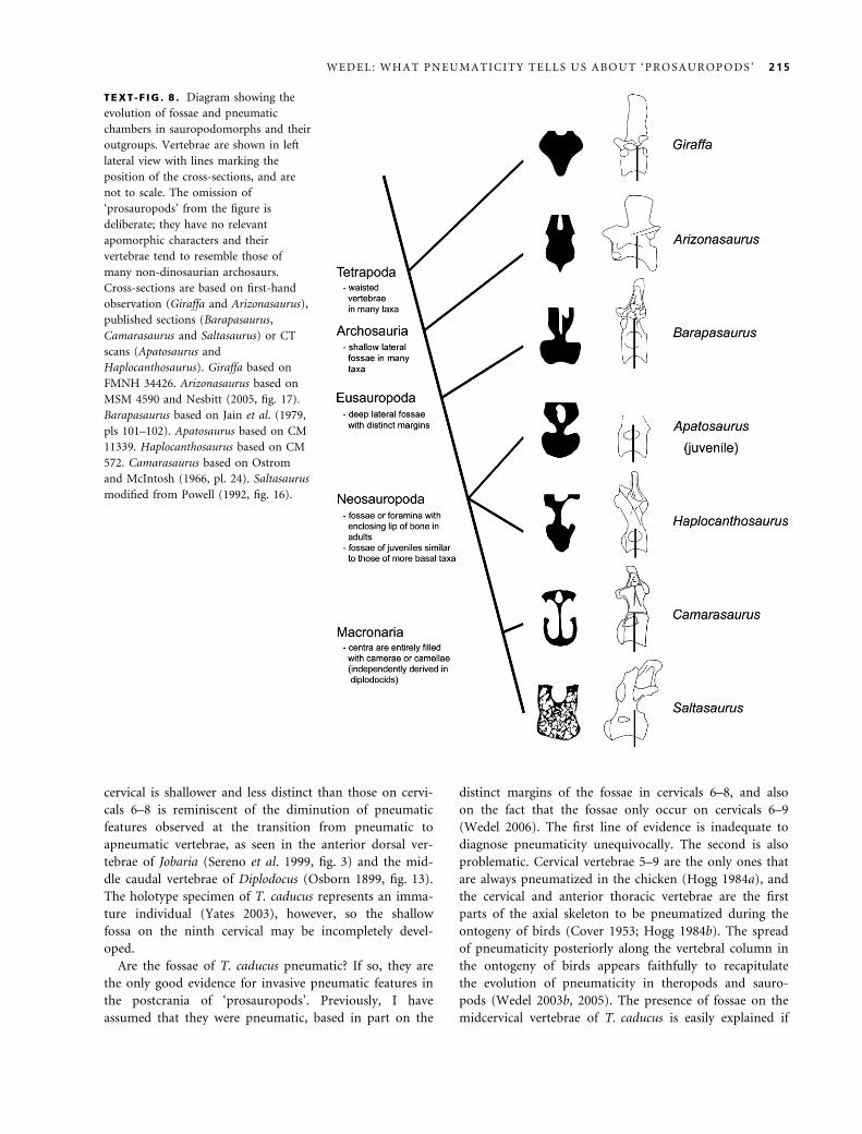
Waisting aside, fossae still suffer from a problem of
definition. Consider a spectrum of morphological possi-
bilities (Text-fig. 8). At one end is a vertebra that is
waisted but lacks distinct fossae: for example, a thoracic
vertebra of an artiodactyl. At the other end is a vertebra
with large foramina that open into internal chambers,
such as a dorsal vertebra of Saltasaurus. The ‘chamber
morphospace’ between these endpoints is filled with a
continuum of deeper and more distinct fossae and cam-
erae. Adjacent to the artiodactyl vertebra we might put a
vertebra that has fossae with a distinct margin on one
side but not the other, like those in the cervical centra of
Arizonasaurus, which are bounded dorsally by the PCDL;
next, a fossa that has a distinct bony rim on all sides, but
that is not enclosed by a bony lip, like those in dorsal
centra of adult Barapasaurus or juvenile Apatosaurus. The
A
B
PODL
PCDL
PRDL
PPDL
vascular foramina
TEXT -F IG . 6 . Dorsal vertebrae of
Erythrosuchus africanus (BMNH R533).
A, right lateral and B, ventrolateral
views. Scale bar represents 5 cm.
W E D E L : W H A T P N E U M A T I C I T Y T E L L S U S A B O U T ‘ P R O S A U R O P O D S ’ 213
penultimate example is a fossa that is enclosed by a bony
lip, but that is little expanded beyond the boundaries of
the opening, such as the fossae in presacral centra of
Haplocanthosaurus [Britt (1993) referred to these cham-
bers as camerae, whereas Wedel termed them fossae
(Wedel et al. 2000; Wedel 2003a). The morphology of
these features is intermediate between that of fossae and
camerae, and either term could reasonably be applied].
Finally, in neosauropods such as Camarasaurus and Salta-
saurus the space beyond the bony lip is greatly expanded,
so that the result is a foramen that leads to camerae or
camellae.
The fossae along this spectrum vary in geometry and
they are not all pneumatic. Although Goniopholis is
extinct and not part of the crown-group Crocodylia, it is
highly unlikely that the caudal vertebrae of this semi-
aquatic neosuchian were pneumatic. Nevertheless, they
bear lateral fossae with distinct margins that are very sim-
ilar to structures that are sometimes interpreted as pneu-
matic in dinosaurs, such as the sacral ‘pleurocoels’ of
ornithomimosaurs. However, distinct margins alone are
not compelling evidence of pneumaticity. Conversely,
truly pneumatic fossae need not have distinct margins.
For example, the fossae behind the prezygapophyses of
ratites lack clear margins, but CT scans show that they
house pneumatic diverticula, and they sometimes contain
pneumatic foramina (Text-fig. 9). In extant birds, the
pneumatic canalis intertransversarius lies alongside the
centrum (Muller 1908), but many birds have cervical cen-
tra that are laterally convex and lack any fossae (the
pneumatic foramina are usually located inside the cervical
rib loop or ansa costotransversaria).
The foregoing discussion implies that where chambers
lack a distinct lip of bone, geometry alone is a poor clue
to whether or not a given fossa has a pneumatic origin.
Other lines of evidence must be used, such as position in
the body, the presence or absence of adjacent pneumatic
foramina, subfossae, or textural differences (and even the
last two may be misleading; see O’Connor 2006).
Vertebral centra of ‘prosauropods’ can be quite nar-
row-waisted, and some have lateral grooves or fossae that
are bounded on one side by a lamina. As with neural arch
laminae, these features are sometimes associated with
pneumaticity but they are not diagnostic for it. The
‘pleurocoelar indentations’ mentioned by Galton and
Upchurch (2004) do not have a distinct boundary or lip
in any of the specimens that I have examined (e.g. Moser
2003, pl. 16). The only known ‘prosauropod’ with dis-
tinctly emarginated lateral fossae is Thecodontosaurus
caducus (Yates 2003). Cervical vertebrae 6–8 of BMNH
P24, the holotype of T. caducus, have small, distinct fos-
sae just behind the diapophyses (Text-fig. 10). The fossae
are high on the centra and may have crossed the neuro-
central sutures, which are open. The fossa on the eighth
cervical looks darker than it should because it is coated
with glue. The ninth cervical has a very shallow, tear-
drop-shaped hollow in the same region of the centrum.
The bone texture in this hollow is noticeably smoother
than on the rest of the centrum (this is especially appar-
ent under low-angle lighting). That the fossa on the ninth
A B
C D
PODL
PCDL
fossa fossa
PODL
NAF
incipient
TEXT -F IG . 7 . Dorsal and caudal
vertebrae of Goniopholis stovalli. These
vertebrae are part of an associated
collection of several individuals from the
type locality of the species. A, a dorsal
vertebra (OMNH 2504) in left
posterolateral view. B, a dorsal vertebra
(OMNH 2470) in right lateral view. C, a
middle caudal centrum (OMNH 2448)
in right lateral view. D, a distal caudal
centrum (OMNH 2454) in left lateral
view. White arrows in B and D highlight
the margins of fossae. Scale bar
represents 1 cm.
214 S P E C I A L P A P E R S I N P A L A E O N T O L O G Y , 7 7
cervical is shallower and less distinct than those on cervi-
cals 6–8 is reminiscent of the diminution of pneumatic
features observed at the transition from pneumatic to
apneumatic vertebrae, as seen in the anterior dorsal ver-
tebrae of Jobaria (Sereno et al. 1999, fig. 3) and the mid-
dle caudal vertebrae of Diplodocus (Osborn 1899, fig. 13).
The holotype specimen of T. caducus represents an imma-
ture individual (Yates 2003), however, so the shallow
fossa on the ninth cervical may be incompletely devel-
oped.
Are the fossae of T. caducus pneumatic? If so, they are
the only good evidence for invasive pneumatic features in
the postcrania of ‘prosauropods’. Previously, I have
assumed that they were pneumatic, based in part on the
distinct margins of the fossae in cervicals 6–8, and also
on the fact that the fossae only occur on cervicals 6–9
(Wedel 2006). The first line of evidence is inadequate to
diagnose pneumaticity unequivocally. The second is also
problematic. Cervical vertebrae 5–9 are the only ones that
are always pneumatized in the chicken (Hogg 1984a), and
the cervical and anterior thoracic vertebrae are the first
parts of the axial skeleton to be pneumatized during the
ontogeny of birds (Cover 1953; Hogg 1984b). The spread
of pneumaticity posteriorly along the vertebral column in
the ontogeny of birds appears faithfully to recapitulate
the evolution of pneumaticity in theropods and sauro-
pods (Wedel 2003b, 2005). The presence of fossae on the
midcervical vertebrae of T. caducus is easily explained if
TEXT -F IG . 8 . Diagram showing the
evolution of fossae and pneumatic
chambers in sauropodomorphs and their
outgroups. Vertebrae are shown in left
lateral view with lines marking the
position of the cross-sections, and are
not to scale. The omission of
‘prosauropods’ from the figure is
deliberate; they have no relevant
apomorphic characters and their
vertebrae tend to resemble those of
many non-dinosaurian archosaurs.
Cross-sections are based on first-hand
observation (Giraffa and Arizonasaurus),
published sections (Barapasaurus,
Camarasaurus and Saltasaurus) or CT
scans (Apatosaurus and
Haplocanthosaurus). Giraffa based on
FMNH 34426. Arizonasaurus based on
MSM 4590 and Nesbitt (2005, fig. 17).
Barapasaurus based on Jain et al. (1979,
pls 101–102). Apatosaurus based on CM
11339. Haplocanthosaurus based on CM
572. Camarasaurus based on Ostrom
and McIntosh (1966, pl. 24). Saltasaurus
modified from Powell (1992, fig. 16).
W E D E L : W H A T P N E U M A T I C I T Y T E L L S U S A B O U T ‘ P R O S A U R O P O D S ’ 215

the fossae are pneumatic; their appearance in that part of
the skeleton mirrors early ontogeny in birds and is also
consistent with later trends in the evolution of PSP in
sauropodomorphs (Text-fig. 2). On all four vertebrae, the
fossae are not closely associated with laminae and cannot
be dismissed as epiphenomena of lamina formation (see
O’Connor 2006); a specific soft-tissue influence was caus-
ally related to the formation of the fossae. The geometry
of the fossae is not sufficient to specify that soft-tissue
influence because adipose, muscular and pulmonary tis-
sues have all been found to occupy similar fossae in other
tetrapods (O’Connor 2006). On the other hand, the pres-
ence of the fossae only on the midcervical vertebrae is dif-
ficult to explain if they were not produced by pneumatic
diverticula like those of more derived sauropods.
Summary. Vertebral laminae and shallow depressions on
the centra are widespread in archosauriforms and not
diagnostic of pneumaticity, although it is difficult to rule
out the possibility that they may have been associated
with pneumatic diverticula. ‘Prosauropods’ have fewer
laminae than most sauropods, fewer vertebrae with lam-
inae, and the fossae adjacent to the laminae are almost
always blind (with no large foramina or chambers). A
foramen in a vertebra of Plateosaurus and distinct fossae
in the cervical vertebrae of Thecodontosaurus caducus are
the best evidence for potential pneumaticity in ‘prosauro-
pods’, but neither is an unambiguous indicator of PSP
and both would benefit from further study. In any case,
the diagnostic osteological correlatives of pneumaticity
that are common in sauropods and theropods are absent
or extremely rare in ‘prosauropods’, and the putative
pneumatic features that are widespread in ‘prosauropods’
(laminae and shallow fossae) are not compelling evidence
of pneumaticity. To leave aside for a moment the ques-
tion of ‘prosauropod’ monophyly, ‘prosauropods’ are
unusual as the only sizeable group (or grade) of saurischi-
an dinosaurs that lack extensive PSP.
PALAEOBIOLOGICAL IMPLICATIONS
Pneumatic bones are of palaeobiological interest in two
ways. We may be interested in the bones themselves: in
their external and internal morphology, in the ratio of
bone to air space, and in the ways that they develop.
They are also important, arguably more important, as
osteological markers of the pulmonary system. In this sec-
tion I discuss the origins of pneumaticity and of air sacs,
and the implications for the respiratory physiology of
sauropodomorphs.
Origin of the diverticular lung and PSP. The first part of
the postcranial skeleton to be pneumatized in any sauris-
chian dinosaur is the cervical column. The fossae in the
mid-cervical vertebrae of Thecodontosaurus caducus are
not definitely pneumatic on the basis of geometry alone.
However, their placement in the skeleton is suspiciously
similar to the early stages of pneumatization in birds. The
same is true of fossae in the cervical column of the basal
sauropod Shunosaurus (Wilson and Sereno 1998). Among
basal theropods, Coelophysis bauri is the earliest well-rep-
resented taxon with evidence of pneumaticity. The post-
axial cervical vertebrae of C. bauri have pneumatic
cavities that occupy most of the neural spine and that
communicate with the outside through several large for-
amina (Colbert 1989).
The pattern of pneumatization in these early diverging
saurischians indicates the presence of cervical air sacs like
those of birds. It is true that in sauropsids diverticula
may develop from practically any portion of the respirat-
ory system. However, it does not follow that the diverti-
cula that pneumatize the skeleton can come from
anywhere (contra Hillenius and Ruben 2004), for two rea-
sons. First, in extant birds the cervical vertebrae are only
pneumatized by diverticula of cervical air sacs. Diverticula
of the cranial air spaces, larynx and trachea are never
known to pneumatize the postcranial skeleton (King
1966), and diverticula of the parenchymal portion of the
lung only pneumatize the vertebrae and ribs adjacent to
pneumatic foramen
pneumatic fossa
TEXT -F IG . 9 . An uncatalogued cervical vertebra of an emu
(Dromaius novaehollandiae) from the OMNH comparative
collection. Scale bar represents 2 cm.
216 S P E C I A L P A P E R S I N P A L A E O N T O L O G Y , 7 7

the lungs (O’Connor 2004). Second, as discussed below,
pneumatization of the posterior half of the body is
accomplished only by diverticula of abdominal air sacs
(O’Connor and Claessens 2005). These observations of
extant taxa provide valuable guidelines for interpreting
patterns of skeletal pneumatization in fossil taxa.
Pneumatization by diverticula of cervical air sacs is the
only mechanism for pneumatizing the neck that is (1)
known to occur in extant taxa and (2) consistent with the
pattern of pneumatization found in basal saurischians
(Wedel 2006).
Pneumaticity in basal saurischians is extremely limited.
The bone removed by pneumatization of the postcranial
skeleton (or fossa formation, if the fossae of Thecodonto-
saurus are not pneumatic) accounted for much less than
1 per cent of the total body volume in both Coelophysis
and Thecodontosaurus (see Appendix), compared with
several per cent for more derived sauropods and thero-
pods (Wedel 2004, 2005). PSP probably did not evolve as
an adaptation for lightening the skeleton, although it
seems to have been exapted for that purpose later in sau-
rischian evolution (Wedel 2003b).
Furthermore, diverticula did not evolve to pneumatize
the skeleton. In the first place, many of the diverticula of
birds are visceral, subcutaneous or intermuscular, and do
not pneumatize any bones (Duncker 1971). Skeletal
pneumatization cannot be invoked to explain the pres-
ence of these diverticula. In the second place, the presence
of diverticula is a prerequisite for pneumatization of the
skeleton. The immediate ancestors of Coelophysis and
Thecodontosaurus must have already had cervical diverti-
cula (assuming that the fossae of the latter are pneumatic
in origin). Pneumatization of the cervical series could not
happen until these diverticula were already in place, so
the diverticula must have evolved for some other reason.
Alternatively, the origins of paravertebral diverticula
and of PSP may have been coincident. The first step may
have been a developmental accident that allowed the
diverticula to push beyond the coelom and these
‘unleashed’ diverticula may have pneumatized the verteb-
ral column immediately. This sort of morphogenetic
behaviour on the part of diverticula is plausible on the
basis of cases in the human clinical literature (e.g. Sadler
et al. 1996; Anorbe et al. 2000). The main argument
A
B
TEXT -F IG . 10 . Vertebrae of Thecodontosaurus caducus, BMNH P24. A, cervical vertebrae 6–8 in left lateral view. B, cervical vertebra
9 in right lateral view. Scale bars represent 1 cm.
W E D E L : W H A T P N E U M A T I C I T Y T E L L S U S A B O U T ‘ P R O S A U R O P O D S ’ 217
against this near-saltational scenario is that the first ver-
tebrae to be pneumatized in both sauropodomorphs
(Thecodontosaurus, Shunosaurus) and theropods (Coeloph-
ysis) are cervicals that are not adjacent to the lungs
(Wedel 2006).
Origin of flow-through ventilation. Flow-through ventila-
tion requires that air sacs be present both anterior and
posterior to the parenchymal portion of the lung. Given
the pattern of pneumatization found in pterosaurs, sauro-
pods and theropods, we may infer that cervical air sacs
were present in the ancestral ornithodiran (or evolved
independently in pterosaurs and saurischians). The next
problem is to determine when abdominal air sacs origin-
ated and how many times.
In extant birds, the posterior thoracic, synsacral and
caudal vertebrae, pelvic girdle and hindlimb are only
pneumatized by diverticula of abdominal air sacs (O’Con-
nor and Claessens 2005; contra Ruben et al. 2003; Chinsa-
my and Hillenius 2004; Hillenius and Ruben 2004). So if
a fossil archosaur is discovered with pneumatic vertebrae
posterior to the mid-thorax, we have a compelling case
for inferring that the animal had abdominal air sacs.
Pneumatic vertebrae in the ‘posterior compartment’ are
present in pterosaurs, diplodocid and macronarian sauro-
pods, and in most clades of neotheropods, but are absent
in non-dinosaurian dinosauromorphs, ornithischians,
herrerasaurids, ‘prosauropods’, basal sauropods, dicraeo-
saurids, and in basal members of most neotheropod
clades (e.g. Baryonyx, Ceratosaurus and Allosaurus; pers.
obs.).
How many times did abdominal air sacs evolve? Poss-
ibly just once, before the ornithodiran divergence; poss-
ibly twice, in pterosaurs and saurischians; or possibly
three times, in pterosaurs, sauropods and theropods
(Text-fig. 1). We could take this to its logical conclusion
and assume that abdominal air sacs evolved afresh in
every group with posterior compartment pneumaticity;
this would require the independent origin of abdominal
air sacs in ceratosaurs, allosauroids and coelurosaurs, for
example (not to mention several independent derivations
within coelurosaurs).
The alternative is that some or all of the groups listed
above had abdominal air sacs but failed to pneumatize
any elements in the posterior compartment. The same
condition pertains in many extant birds (O’Connor 2004,
table 2). O’Connor and Claessens (2005) posited an ori-
gin of abdominal air sacs by the time of the ceratosaur-
tetanuran divergence, based on the presence of posterior
compartment pneumatization in Majungatholus, and
despite its absence in basal ceratosaurs and basal tetanu-
rans.
In terms of evolutionary change, ventilation mecha-
nisms are highly conserved, PSP is highly labile and diver-
ticula seem to lie between these extremes. All birds have
essentially the same lung architecture; the biggest differ-
ence among living forms is the presence or absence of a
neopulmo (Duncker 1971). On the other hand, PSP var-
ies widely within small clades and even within popula-
tions (King 1966; Hogg 1984a; O’Connor 2004).
Diverticula appear to be more conserved than PSP,
although a dedicated study comparing the evolution of
the two is needed. For example, most birds have femoral
and perirenal diverticula, but the femur and pelvis are
only pneumatized in a subset of these taxa (Muller 1908;
King 1966; Duncker 1971). These observations are neces-
sarily tentative, given the paucity of phylogenetically
based comparative studies of pneumatic diverticula and
PSP (but see O’Connor 2004). Furthermore, our know-
ledge of variation in the pulmonary system and its diver-
ticula is based entirely on extant birds, and may not be
applicable to other saurischians.
Nevertheless, the evolutionary malleability of lungs,
diverticula and PSP in birds should not be ignored in
reconstructing the pulmonary systems of fossil archosaurs.
The absence of unequivocal PSP in most ‘prosauropods’
does not mean that they lacked air sacs. Depending on
the preferred phylogenetic hypothesis, Sauropodomorpha
is only one or two nodes away from Neotheropoda. Most
neosauropods have pneumatic vertebrae in the posterior
compartment. If these sauropods found some way to
pneumatize the posterior compartment without abdom-
inal air sacs, then surely the same could be true of some
or all non-avian theropods. Likewise, if posterior com-
partment pneumaticity is prima facie evidence of abdom-
inal air sacs in theropods, then abdominal air sacs must
also have been present in sauropods (and, by extension,
pterosaurs). What is good for the goose is good for Gong-
xianosaurus. It is more parsimonious to infer that cervical
and abdominal air sacs were present in the ancestral sau-
rischian, but did not pneumatize the skeleton in ‘prosaur-
opods’, than to infer independent origins of air sacs in
sauropods and theropods.
Most pterosaurs have extensively pneumatized skele-
tons, although it is not clear whether pneumaticity is
present in any of the Triassic forms (Bonde and Chris-
tiansen 2003). The presence of PSP in pterosaurs, sau-
ropodomorphs and theropods suggests that air sacs
may have been present in the ancestral ornithodiran.
An apparent problem with pushing the origin of air-
sac-driven breathing back before the origin of Sauris-
chia is the utter absence of PSP in ornithischians. PSP
appeared in pterosaurs, sauropodomorphs and thero-
pods relatively quickly after the divergence of each
clade: by the Norian in theropods (Colbert 1989) and
no later than the Early Jurassic in pterosaurs and sau-
ropodomorphs (Bonde and Christiansen 2003; Wedel
2005). If ornithischians had air sacs and diverticula
218 S P E C I A L P A P E R S I N P A L A E O N T O L O G Y , 7 7
then it is odd that they never evolved PSP during the
160 million years of their existence. However, this
problem may be more illusory than real. The invasion
of bone by pneumatic epithelium is essentially oppor-
tunistic (Witmer 1997). Although pneumatic diverticula
may radically remodel both the exterior and the inter-
ior of an affected bone, this remodelling cannot occur
if the diverticula never come into contact with the
bone, and may not occur even if they do. Furthermore,
for all of the potential advantages it conveys, PSP is
still an exaptation of a pre-existing system: in an adap-
tive sense, lineages that lack PSP do not know what
they are missing. Recall that PSP in basal saurischians
did little to lighten the skeleton (see above). Ornithis-
chians may have had air sacs without diverticula, or
diverticula without PSP. It is pointless to consider the
advantages that ornithischians ‘lost’ by never evolving
PSP, because that evolution would have hinged on the
incidental contact of bone and air sac and could not
have been anticipated or sought by natural selection.
The problem of determining when abdominal air sacs
evolved is challenging because it forces us to decide
between events of unknown probability: the possibility
that ornithischians had an air sac system and never ‘dis-
covered’ PSP (if abdominal air sacs are primitive for
Ornithodira), vs. the possibility that a system of cervical
and abdominal air sacs evolved independently in ptero-
saurs and saurischians. Currently, available evidence is
insufficient to falsify either hypothesis.
Sauropodomorph palaeobiology. It is likely that ‘prosauro-
pods’ had cervical and abdominal air sacs, given the
strong evidence for both in sauropods and theropods. We
may not be able to determine for certain whether ‘pro-
sauropods’ had a bird-like flow-through lung, but the
requisite air sacs were almost certainly present. Our null
hypothesis for the respiratory physiology of ‘prosauro-
pods’ should take into account some form of air-sac-dri-
ven ventilation.
The air sacs of birds mitigate the problem of tracheal
dead space (Schmidt-Nielsen 1972), and some birds
have improbably long tracheae (i.e. longer than the
entire body of the bird; see McClelland 1989). In addi-
tion, birds can ventilate their air sacs without blowing
air through the lungs, which allows them to avoid alka-
losis during thermoregulatory panting (Schmidt-Nielsen
et al. 1969). Finally, flow-through breathing allows birds
to extract much more oxygen from the air than mam-
mals can (Bernstein 1976). In general, sauropods were
larger and longer-necked than ‘prosauropods’, and the
aforementioned capabilities of a bird-like ventilation
system may have helped sauropods overcome the phy-
siological challenges imposed by long necks and large
bodies, including tracheal dead space, heat retention
and oxygen uptake.
The one obvious advantage that ‘prosauropods’ did not
share with sauropods is the very lightweight skeletal con-
struction afforded by pneumaticity. In life, the average
pneumatic sauropod vertebra was approximately 60 per
cent air by volume (Wedel 2005; Woodward 2005;
Schwarz and Fritsch 2006). All else being equal, a sauro-
pod could have a neck two-thirds longer than that of a
prosauropod for the same skeletal mass. Pneumaticity
helped sauropods overcome constraints on neck length,
and thereby opened feeding opportunities that were not
available to ‘prosauropods’. How important that differ-
ence was is unknown, but it is worth considering in
reconstructions of sauropodomorph evolution and pal-
aeobiology.
Acknowledgements. This work was completed as part of a doc-
toral dissertation in the Department of Integrative Biology,
University of California, Berkeley. I am grateful to my advi-
sors, K. Padian and W. A. Clemens, and to the members of
my dissertation committee, F. C. Howell, D. Wake and
M. Wake, for advice and encouragement. Many thanks to
P. M. Barrett and T. Fedak for organizing the symposium
that gave rise to this manuscript. I am grateful for the hospi-
tality and patience of curators and collections managers every-
where, especially P. Barrett, S. Chapman, R. Schoch,
M. Moser, A. Henrici, M. Lamanna, R. Cifelli, N. Czaplewski
and J. Person. L. Claessens, R. Irmis, S. Nesbitt and P.
O’Connor provided many inspiring discussions and gracious
access to their unpublished work. R. Irmis and M. Taylor
read early drafts of this paper and made many helpful sugges-
tions, as did my dissertation committee members. L. Claessens
and J. Harris provided thoughtful review comments that
greatly improved this paper, and I thank them for their time
and effort. A translation of Janensch (1947) was made by
G. Maier, whose effort is gratefully acknowledged. Funding for
this project was provided by the Jurassic Foundation, Sigma
Xi, the Department of Integrative Biology at the University of
California, Berkeley, the University of California, Museum of
Paleontology, and the UCMP Doris and Samuel P. Welles
Fund. This is UCMP Contribution no. 1919.
REFERENCES
A L C OB E R , O. and PA R R I S H, J. M. 1997. A new poposaurid
from the Upper Triassic of Argentina. Journal of Vertebrate
Paleontology, 17, 548–556.
A N O R B E , E., A I S A , P. and S A E N Z D E O R M I J A N A , J.
2000. Spontaneous pneumatocele and pneumocephalus associ-
ated with mastoid hyperpneumatization. European Journal of
Radiology, 36, 158–160.
B E N T O N , M. J., J U UL , L., S T OR R S , G. W. and G A L TO N ,
P. M. 2000. Anatomy and systematics of the prosauropod
dinosaur Thecondontosaurus antiquus from the Upper Triassic
W E D E L : W H A T P N E U M A T I C I T Y T E L L S U S A B O U T ‘ P R O S A U R O P O D S ’ 219
of southwest England. Journal of Vertebrate Paleontology, 20,
77–108.
B E R N S T E I N , M. H. 1976. Ventilation and respiratory evapor-
ation in the flying crow, Corvus ossifragus. Respiration Physiol-
ogy, 26, 371–382.
B ON A PA R T E , J. F. 1999. Evolucion de las vertebras presacras
en Sauropodomorpha. Ameghiniana, 36, 115–187.
B ON DE , N. and C HR I S T I A N S E N , P. 2003. The detailed
anatomy of Rhamphorhynchus: axial pneumaticity and its
implications. Geological Society, London, Special Publication,
217, 217–232.
B R E M E R , J. L. 1940. The pneumatization of the humerus in
the common fowl and the associated activity of theelin. Ana-
tomical Record, 77, 197–211.
B R I T T , B. B. 1993. Pneumatic postcranial bones in dinosaurs
and other archosaurs. Unpublished PhD thesis, University of
Calgary, 383 pp.
—— 1997. Postcranial pneumaticity. 590–593. In C U RR I E , P. J.
and PA D I A N , K. (eds). The encyclopedia of dinosaurs. Aca-
demic Press, San Diego, CA, 869 pp.
B R OC H U, C. A. 2001. Progress and future directions in archo-
saur phylogenetics. Journal of Paleontology, 75, 1185–1201.
C A R V A L H O, I. S., A V I L L A , L. S. and S A L G A D O, L. 2003.
Amazonsaurus maranhensis gen. et sp. nov. (Sauropoda, Dip-
lodocoidea) from the Lower Cretaceous (Aptian–Albian) of
Brazil. Cretaceous Research, 24, 697–713.
C A S E , E. C. 1907. Revision of the Pelycosauria of North
America. Carnegie Institution of Washington Publication, 55,
1–175.
C H I N S A M Y , A. and H I L L E N I U S , W. J. 2004. Physiology of
nonavian dinosaurs. 643–659. In W E I S H A M P E L , D. B.,
D O D S O N , P. and O S M O L S K A , H. (eds). The Dinosauria.
Second edition. University of California Press, Berkeley, CA,
861 pp.
C OL B E R T , E. H. 1989. The Triassic dinosaur Coelophysis. Bul-
letin of the Museum of Northern Arizona, 57, 1–160.
C OV E R , M. S. 1953. Gross and microscopic anatomy of the
respiratory system of the turkey. III. The air sacs. American
Journal of Veterinary Research, 14, 239–245.
D E S OJ O, J. B., A R C U CC I , A. B. and M A R S I CA N O, C. A.
2002. Reassessment of Cuyosuchus huenei, a Middle–Late Tri-
assic archosauriform from the Cuyo Basin, west-central Argen-
tina. Bulletin of the New Mexico Museum of Natural History
and Science, 21, 143–148.
D UN C KE R, H.-R. 1971. The lung air sac system of birds.
Advances in Anatomy, Embryology, and Cell Biology, 45, 1–171.
G A L T ON , P. M. and U PC H UR C H, P. 2004. Prosauropoda.
232–258. In W E I S H A M P E L , D. B., D OD S O N , P. and
O S M O LS KA , H. (eds). The Dinosauria. Second edition.
University of California Press, Berkeley, CA, 861 pp.
G I E R , H. T. 1952. The air sacs of the loon. Auk, 69, 40–49.
G I L M OR E , C. W. 1936. Osteology of Apatosaurus with special
reference to specimens in the Carnegie Museum. Memoirs of
the Carnegie Museum, 11, 175–300.
G O W E R , D. J. 2001. Possible postcranial pneumaticity in the
last common ancestor of birds and crocodilians: evidence
from Erythrosuchus and other Mesozoic archosaurs. Naturwis-
senschaften, 88, 119–122.
H I L L E N I US , W. J. and R UB E N , J. A. 2004. The evolution of
endothermy in terrestrial vertebrates: Who? When? Why? Phy-
siological and Biochemical Zoology, 77, 1019–1042.
H OG G , D. A. 1984a. The distribution of pneumatisation in the
skeleton of the adult domestic fowl. Journal of Anatomy, 138,
617–629.
—— 1984b. The development of pneumatisation in the postcra-
nial skeleton of the domestic fowl. Journal of Anatomy, 139,
105–113.
H UR L B UR T , G. 1999. Comparison of body mass estimation
techniques, using Recent reptiles and the pelycosaur Edapho-
saurus boanerges. Journal of Vertebrate Paleontology, 19, 338–
350.
J A I N , S. L., KU TT Y , T. S., R O Y - C HO W D HU R Y , T. K.
and CH A T T E R J E E , S. 1979. Some characteristics of Bara-
pasaurus tagorei, a sauropod dinosaur from the Lower Jurassic
of Deccan, India. Proceedings of the IV International Gondwana
Symposium, 1, 204–216.
J A N E N S C H, W. 1929. Material und Formengehalt der Sau-
ropoden in der Ausbeute der Tendaguru-expedition. Palaeon-
tographica (Supplement 7), 2 (1), 1–34.
—— 1947. Pneumatizitat bei Wirbeln von Sauropoden und and-
eren Saurischien. Palaeontographica (Supplement 7), 3 (1), 1–25.
—— 1950. Die Wirbelsaule von Brachiosaurus brancai. Palaeon-
tographica (Supplement 7), 3 (2), 27–93.
J E RI S O N , H. J. 1973. Evolution of the brain and intelligence.
Academic Press, New York, NY, 482 pp.
K I N G , A. S. 1966. Structural and functional aspects of the
avian lungs and air sacs. International Review of General and
Experimental Zoology, 2, 171–267.
L A N G E R , M. C. 2004. Basal Saurischia. 25–46. In W E I S -
HA M P E L , D. B., D OD S O N , P. and O S M O L S K A , H.
(eds). The Dinosauria. Second edition. University of California
Press, Berkeley, CA, 861 pp.
L A R S O N , A., W A K E , D. B., M A X S O N , L. R. and H I GH -
T ON , R. 1981. A molecular phylogenetic perspective on the
origins of morphological novelties in the salamanders of the
tribe Plethodontini (Amphibia, Plethodontidae). Evolution, 35,
405–422.
L U JUN-CHANG and Z H A N G BAO-KUN 2005. A new ovi-
raptorid (Theropod: Oviraptorosauria) from the Upper Creta-
ceous of the Nanxiong Basin, Guangdong Province of
southern China. Acta Palaeontologica Sinica, 44, 412–422.
M A KO V I C K Y , P. J., A PE S TE GU I A , S. and A G N O LI N ,
F. L. 2005. The earliest dromaeosaurid theropod from South
America. Nature, 437, 1007–1011.
M C CL E L L A N D , J. 1989. Larynx and trachea. 69–103. In
KI N G , A. S. and M C C L E L L A N D, J. (eds). Form and
function in birds. Volume 4. Academic Press, London, 608
pp.
M OS E R , M. 2003. Plateosaurus engelhardti Meyer, 1837 (Dino-
sauria: Sauropodomorpha) from the Feuerletten (Mittelkeu-
per; Obertrias) of Bavaria. Zitteliana, B24, 1–188.
M U L L E R , B. 1908. The air-sacs of the pigeon. Smithsonian
Miscellaneous Collections, 50, 365–420.
M UR R A Y , P. F. and V I C K E R S - R I C H , P. 2004. Magnificent
mihirungs. Indiana University Press, Bloomington, IN, 410 pp.
220 S P E C I A L P A P E R S I N P A L A E O N T O L O G Y , 7 7

N E S BI TT , S. J. 2005. Osteology of the Middle Triassic pseu-
dosuchian archosaur Arizonasaurus babbitti. Historical Biology,
17, 19–47.
O ’ CO N N O R , P. M. 2004. Pulmonary pneumaticity in the
postcranial skeleton of extant Aves: a case study examining
Anseriformes. Journal of Morphology, 261, 141–161.
—— 2006. Postcranial pneumaticity: an evaluation of soft-tissue
influences on the postcranial axial skeleton and the recon-
struction of pulmonary anatomy in archosaurs. Journal of
Morphology, 267, 1199–1226.
—— and C L A E S S E N S , L. P. A. M. 2005. Basic avian pulmon-
ary design and flow-through ventilation in non-avian thero-
pod dinosaurs. Nature, 436, 253–256.
O S B OR N , H. F. 1899. A skeleton of Diplodocus. Memoirs of the
American Museum of Natural History, 1, 191–214.
—— and M O OK , C. C. 1921. Camarasaurus, Amphicoelias, and
other sauropods of Cope. Memoirs of the American Museum of
Natural History, 3, 247–287.
O S M O L S K A , H., C UR R I E , P. J. and B A R S B O L D, R. 2004.
Oviraptorosauria. 165–183. In W E I S H A M P E L , D. B.,
D O D S O N , P. and O S M O L S K A , H. (eds). The Dinosauria.
Second edition. University of California Press, Berkeley, CA,
861 pp.
O S T R OM , J. H. and M C I N T O S H , J. S. 1966. Marsh’s dino-
saurs: the collections from Como Bluff. Yale University Press,
New Haven, CT, xxiv + 388 pp.
P A R KE R, W. G. 2003. Description of a new specimen of Des-
matosuchus haplocerus from the Late Triassic of northern Ari-
zona. Unpublished MS thesis, Northern Arizona University,
Flagstaff, AZ, 312 pp.
P A UL , G. S. 1997. Dinosaur models: the good, the bad, and
using them to estimate the mass of dinosaurs. 129–154. In
W O L BE R G , D. L., S T UM P, E. and R O S E N B E R G , G.
(eds). Dinofest International: proceedings of a symposium spon-
sored by Arizona State University. Academy of Natural
Sciences, Philadelphia, PA, 587 pp.
P E C ZK I S , J. 1994. Implications of body mass estimates for
dinosaurs. Journal of Vertebrate Paleontology, 14, 520–533.
P OW E L L , J. E. 1992. Osteologıa de Saltasaurus loricatus (Saur-
opoda – Titanosauridae) del Cretacico Superior del noroeste
Argentino. 165–230. In S A N Z , J. L. and B U S C A L I O N I , A.
D. (eds). Los dinosaurios y su entorno biotico: Actas del Segundo
Curso de Paleontologıa en Cuenca. Instituto Juan de Valdes,
Cuenca, Argentina, 397 pp.
R U BE N , J. A., J ON E S , T. D. and GE I S T , N. R. 2003. Respir-
atory and reproductive paleophysiology of dinosaurs and early
birds. Physiological and Biochemical Zoology, 76, 141–164.
S A D L E R , D. J., D OY L E , G. J., H A L L , K. and C R A W -
F OR D , P. J. 1996. Craniocervical bone pneumatisation. Neu-
roradiology, 38, 330–332.
S A N Z, J. L., P O W E L L , J. E., L E LO E U FF , J., M A R T I -
N E Z , R. and P E R E DA S U PE R B I O L A , X. 1999. Sauropod
remains from the Upper Cretaceous of Lano (north central
Spain). Titanosaur phylogenetic relationships. Estudios del
Museo de Ciencias Naturales de Alava, 14 (Numero Especial
1), 235–255.
S C H M I DT - N I E L S E N , K. 1972. How animals work. Cam-
bridge University Press, Cambridge, vi + 114 pp.
—— K A N W I S HE R, J., L A S I E W S K I , R. C., CO H N , J. E.
and B R E T Z , W. L. 1969. Temperature regulation and respir-
ation in the ostrich. Condor, 71, 341–352.
S C H W A R Z , D. and F R I T S C H, G. 2006. Pneumatic struc-
tures in the cervical vertebrae of the Late Jurassic Tendaguru
sauropods Brachiosaurus brancai and Dicraeosaurus. Eclogae
Geologicae Helvetiae, 99, 65–78.
S E R E N O , P. C. 1998. A rationale for phylogenetic definitions,
with application to the higher-level taxonomy of Dinosauria.
Neues Jahrbuch fur Geologie und Palaontologie, Abhandlungen,
20, 41–83.
—— 2007. Basal Sauropodomorpha: historical and recent phylo-
genetic hypotheses, with comments on Ammosaurus major
(Marsh, 1891). 261–289. In BA R R E T T , P. M. and B A T T E N ,
D. J. (eds). Evolution and palaeobiology of early sauropodomorph
dinosaurs. Special Papers in Palaeontology, 77, 289 pp.
—— and W I L S O N , J. A. 2005. Structure and evolution of a
sauropod tooth battery. 157–177. In W I L S O N , J. A. and
CU R R Y - R O G E R S , K. (eds). The sauropods: evolution and
paleobiology. University of California Press, Berkeley, CA, 349
pp.
—— B E C K, A. L., D UT H E I L , D. B., L A R S S O N , H. C. E.,
L Y O N , G. H., M O US S A , B., S A DL E I R , R. W., S I DO R ,
C. A., V A R R I C CH I O , D. J., W I L S O N , G. P. and W I L -
S O N , J. A. 1999. Cretaceous sauropods and the uneven rate
of skeletal evolution among dinosaurs. Science, 286, 1342–
1347.
T Y K O S KI , R. S., R O W E , T. B., K E T C HA M , R. A. and
CO L B E R T , M. W. 2002. Calsoyasuchus valliceps, a new croc-
odyliform from the Early Jurassic Kayenta Formation of Ari-
zona. Journal of Vertebrate Paleontology, 22, 593–611.
U PC H UR C H, P., BA R R E T T , P. M. and D OD S O N , P.
2004. Sauropoda. 259–324. In W E I S H A M P E L , D. B.,
D O D S O N , P. and O S M O L S K A , H. (eds). The Dinosauria.
Second edition. University of California Press, Berkeley, CA,
861 pp.
—— —— and GA LT O N , P. M. 2007. The phylogenetic rela-
tionships of basal sauropodomorphs: implications for the ori-
gin of sauropods. 57–90. In BA R R E T T , P. M. and
B A TT E N , D. J. (eds). Evolution and palaeobiology of early
sauropodomorph dinosaurs. Special Papers in Palaeontology,
77, 289 pp.
V E R B O UT , A. J. 1985. The development of the vertebral col-
umn. Advances in Anatomy, Embryology and Cell Biology, 90,
1–122.
W A K E , D. B. 1963. Comparative osteology of the plethodontid
salamander genus Aneides. Journal of Morphology, 113, 77–118.
—— 1992. The endoskeleton: the comparative anatomy of the
vertebral column and ribs. 192–237. In W A K E , M. H. (ed.).
Hyman’s comparative vertebrate anatomy. Third edition. Uni-
versity of Chicago Press, Chicago, IL, 788 pp.
—— and L A W S O N , R. 1973. Developmental and adult mor-
phology of the vertebral column in the plethodontid salaman-
der Eurycea bislineata, with comments on vertebral evolution
in the Amphibia. Journal of Morphology, 139, 251–300.
—— W A K E , D. B. and W A K E , M. H. 1983. The ossification
sequence of Aneides lugubris, with comments on heterochrony.
Journal of Herpetology, 17, 10–22.
W E D E L : W H A T P N E U M A T I C I T Y T E L L S U S A B O U T ‘ P R O S A U R O P O D S ’ 221
W E D E L , M. J. 2003a. The evolution of vertebral pneumaticity
in sauropod dinosaurs. Journal of Vertebrate Paleontology, 23,
344–357.
—— 2003b. Vertebral pneumaticity, air sacs, and the physiology
of sauropod dinosaurs. Paleobiology, 29, 243–255.
—— 2004. Skeletal pneumaticity in saurischian dinosaurs and
its implications for mass estimates. Journal of Vertebrate Pale-
ontology, 24 (Supplement to No. 3), 127A.
—— 2005. Postcranial skeletal pneumaticity in sauropods and its
implications for mass estimates. 201–228. In W I L S O N , J. A.
and CU R R Y - R OG E R S , K., (eds). The sauropods: evolution
and paleobiology. University of California Press, Berkeley, CA,
349 pp.
—— 2006. The origins of postcranial skeletal pneumaticity in
dinosaurs. Integrative Zoology, 2, 80–85.
—— CI F E L LI , R. L. and S A N DE R S , R. K. 2000. Osteology,
paleobiology, and relationships of the sauropod dinosaur Sau-
roposeidon. Acta Palaeontologica Polonica, 45, 343–388.
W I L S ON , J. A. 1999. A nomenclature for vertebral laminae in
sauropods and other saurischian dinosaurs. Journal of Verte-
brate Paleonotology, 19, 639–653.
—— 2002. Sauropod dinosaur phylogeny: critique and cladistic
analysis. Zoological Journal of the Linnean Society, 136, 217–276.
—— and S E R E N O , P. C. 1998. Early evolution and higher-
level phylogeny of sauropod dinosaurs. Memoir of the Society
of Vertebrate Paleontology, 5, 1–68.
W I T M E R , L. M. 1997. The evolution of the antorbital cavity
of archosaurs: a study in soft-tissue reconstruction in the
fossil record with an analysis of the function of pneuma-
ticity. Memoir of the Society of Vertebrate Paleontology, 3,
1–73.
W O O DW A R D , H. 2005. Bone histology of the titanosaurid
sauropod Alamosaurus sanjuanensis from the Javelina Forma-
tion, Texas. Journal of Vertebrate Paleontology, 25 (Supplement
to No. 3), 132A.
X U XING, Z H A N G XIAO-HONG, TA N QING-WEI, Z H A O
XI-JIN and T A N LIN 2006. A new titanosaurian sauropod
from Late Cretaceous of Nei Mongol, China. Acta Geologica
Sinica, 80, 20–26.
Y A T E S , A. M. 2003. A new species of the primitive dinosaur
Thecodontosaurus (Saurischia: Sauropodomorpha) and its
implications for the systematics of basal dinosaurs. Journal of
Systematic Paleontology, 1, 1–42.
—— 2004. Anchisaurus polyzelus (Hitchcock): the smallest
known sauropod dinosaur and the evolution of gigantism
among sauropodomorph dinosaurs. Postilla, 230, 1–58.
—— 2007. The first complete skull of the Triassic dinosaur
Melanorosaurus Haughton (Sauropodomorpha: Anchisauria).
9–55. In B A R R E T T, P. M. and B A T TE N , D. J. (eds). Evo-
lution and palaeobiology of early sauropodomorph dinosaurs.
Special Papers in Palaeontology, 77, 289 pp.
—— and K I T CH I N G, J. W. 2003. The earliest known sauro-
pod dinosaur and the first steps toward sauropod locomotion.
Proceedings of the Royal Society of London, Series B, 270, 1753–
1758.
APPENDIX
The method of calculating the volumes of bone removed by
pneumatization in Coelophysis and Thecondontosaurus (see ‘Pal-
aeobiological implications’ above) is provided here. To estimate
the whole body volumes of the dinosaurs I used graphic double
integration (GDI: Jerison 1973; Hurlburt 1999; Murray and
Vickers-Rich 2004). I traced over the skeletal reconstructions of
Colbert (1989, fig. 103) and Benton et al. (2000, fig. 19) to make
lateral view body outlines. Dorsal view body outlines were drawn
by hand based on those of Paul (1997) and digitally manipulated
to match the dimensions of the skeletal reconstructions. Using
GDI, I obtained whole body volumes of 23Æ5 L for Coelophysis
and 3Æ3 L for the holotypic individual of Thecodontosaurus cadu-
cus; the latter animal is a small juvenile. Adjusted for scale, these
results are consistent with previous mass estimates for both taxa
(Peczkis 1994).
Pneumaticity is present throughout the cervical series of Coel-
ophysis. The total length of the cervical series is c. 50 cm, and
the vertebral centra have a mean diameter of 1 cm, based on
measurements of uncatalogued CM specimens. The neural spines
are roughly the same size as the centra. The combined cervical
centra are treated as a simple cylinder 50 cm long with a diam-
eter of 1 cm, which yields a volume of 40 cm3. If the neural
spines are assumed to be equal in volume to the centra, the
combined volume of the cervical vertebrae is 80 cm3. The cervi-
cal vertebrae of Coelophysis are probably not more than 50 per
cent air by volume, based on observations of broken specimens,
so the volume of bone removed during pneumatization of the
cervical vertebrae was c. 40 cm3, or 0Æ17 per cent of the volume
of the body.
For Thecondontosaurus caducus it is simpler to calculate the
volumes of the individual fossae. The fossae on cervicals 6–8 are
each c. 5 mm long, 2Æ5 mm tall and 1Æ25 mm deep. The paired
fossae on each vertebra can be thought of as forming the two
halves of an oblate spheroid with x, y and z diameters of 5, 2Æ5and 2Æ5 mm, respectively. The volume of this spheroid, and thus
the volume of the paired fossae, is 0Æ016 cm3. The fossae on
cervicals 6–8 are all roughly the same size, and the visible fossa
on the ninth cervical is only about half as deep. The volume of
bone removed during fossa formation is therefore 0Æ057 cm3, or
0Æ0017 per cent of the volume of the body.
These calculations are all approximate, but they are sufficient
to demonstrate that PSP did not have a noticeable effect on the
skeletal mass of basal saurischians. In the case that I have under-
estimated the volume of the pneumatic chambers in Coelophysis
relative to the body volume by a factor of six: the volume of
these chambers would still only be 1 per cent of the volume of
the body. In contrast, the volume of air in the pneumatic verte-
brae of Tyrannosaurus and Diplodocus accounted for 4–6 per
cent of the volume of the animals. These air spaces replaced
bone, a relatively dense tissue, and lightened the animals by
7–10 per cent (Wedel 2004, 2005).
222 S P E C I A L P A P E R S I N P A L A E O N T O L O G Y , 7 7


![2016 - littlegroveps.wa.edu.auThe longest dinosaur name is the micropachycephalosaurus. The giant plant eating Sauropods [Dreadnoughtus] were the biggest ... EVERY SATURDAY a car park](https://img.pdfslide.net/doc/110x75/5e6ddcb9f456f316f351a654/2016-the-longest-dinosaur-name-is-the-micropachycephalosaurus-the-giant-plant.jpg)