Embed Size (px)
Citation preview

What’s wrong with my mouse?
A beginner’s guide to necropsy and pathology evaluation of mice
Kelli Boyd DVM, PhD, DACVPDirector, Comparative Pathology and Research Histology, Translational Pathology Shared Resource
Associate Director, Comparative Pathology, Division of Animal CareAssociate Professor, Department of Pathology, Microbiology and Immunology
Vanderbilt University Medical Center
Overview
• Mouse use and history• Things all mouse users should know• Pathology Evaluation• Tissue collection• Fixation
– Considerations for ancillary testing• Resources

Why use the mouse• Economic housing of large cohorts• Easy to share• Defined genetics• Defined environment• Short life span equals disease in fast forward• Ability to genetically manipulate mouse
genome • Human and mice are genetically similar

History
• Humans have studied mouse inheritance traits for 3,000 years
• Circa 1664 mouse as first lab animal‐– Robert Hooke studied properties of air
• Circa 1900‐Modern era of mouse genetics– William Castle Harvard researcher– Abby Lathrop‐mouse breeder and
entrepreneur– These mice are the ancestors of most strains
routinely used today

Inbred mice• Hundreds of inbred strains
– Different characteristics • Prone to different diseases
• Provide define genetic background– Experimental therapies quantified and
assessed in standard framework
• Cross breeding inbred strains – Provide new models for mapping genes
and genetic traits
What do you need to know when planning your mouse study?
• Intrinsic influences• Strain characteristics• Environmental influences• Infectious disease impact
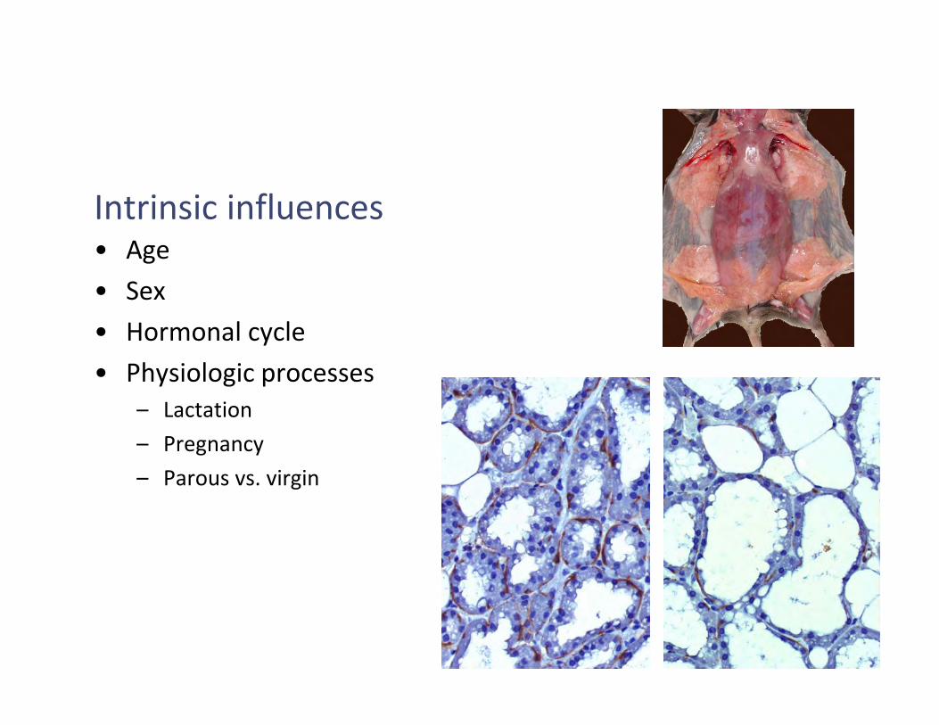
Intrinsic influences• Age • Sex• Hormonal cycle• Physiologic processes
– Lactation– Pregnancy– Parous vs. virgin

Strain effects

Beware of The Known! (background lesions)
Lesions considered normal for the particular background strain• C57BL/6‐microphthalmia, hydrocephalus, dermaitis, osteoporosis, lymphoma or histiocytic sarcoma, amyloidosis, macrophage pneumonia, hydronephrosis
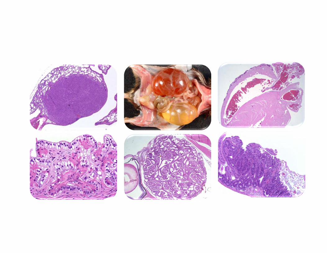
• 129‐ teratomas, Harderian gland tumors, lung tumors, nephropathy, acidophilic crystalline/ macrophage pneumonia, hyalinosis
• FVB‐ retinal degeneration, seizures, lung tumors, acidophilic macrophage pneumonia
• Retinal degeneration‐FVB,C3H, CBA, SJL• Absent corpus callosum‐ Balb‐c• Distrophic cardiac calcification‐ Balb‐c, C3H, DBA
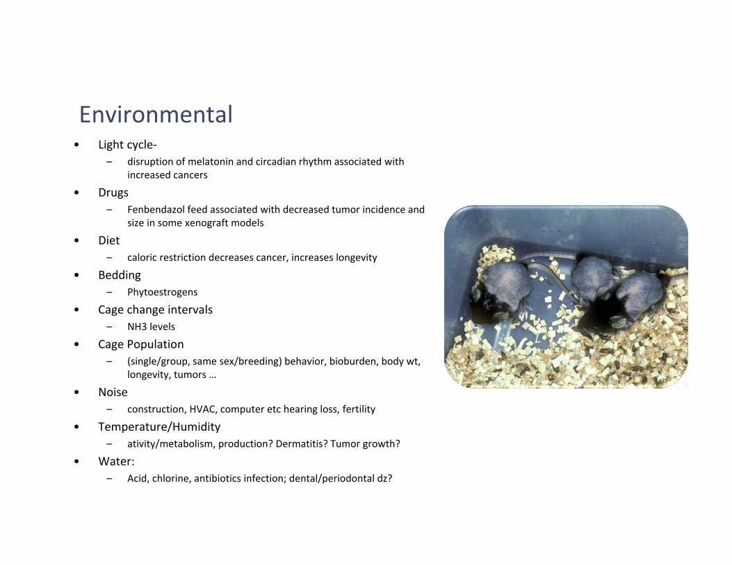
Environmental• Light cycle‐
– disruption of melatonin and circadian rhythm associated with increased cancers
• Drugs– Fenbendazol feed associated with decreased tumor incidence and
size in some xenograft models
• Diet– caloric restriction decreases cancer, increases longevity
• Bedding– Phytoestrogens
• Cage change intervals– NH3 levels
• Cage Population– (single/group, same sex/breeding) behavior, bioburden, body wt,
longevity, tumors …
• Noise – construction, HVAC, computer etc hearing loss, fertility
• Temperature/Humidity– ativity/metabolism, production? Dermatitis? Tumor growth?
• Water: – Acid, chlorine, antibiotics infection; dental/periodontal dz?

TOP (Mouse) Viral agents of interest or concern
In approximate order of seroprevalence & testing recommendations in US & EU
1. MNV Norovirus*2. MHV3. Parvoviruses4. MRV5. TMEV
EDIM‐ Rotavirus? Emerging?

MNV Murine Norovirus
• Most prevalent viral pathogen– 30‐60% in research colonies
• Calicivirus(es) ‐ ss RNA non enveloped– MNV 1‐4 + possibly many genetic variants (like human noroviruses)
• Detection: – RtPCR – Feces or mesenteric Lymph node– Serology
• Innate immunity is essential for Norovirus resistance.• Transmission: Fecal oral with massive shedding early in infection,
persistent in environment (non‐enveloped –hard to get out of cruise ships too…)
• Research impact: immunomodulation (innate immunity)• Control: REDERIVE (foster)

MHV (Mouse Hepatitis Virus)• Coronavirus ss RNA, enveloped
– Highly infectious but don’t survive well in environment – Mutate & recombine new ‘strains’– Enterotropic strains (gut only)– Polytropic strains e.g. laboratory strains used to model demyelinating diseases– Epizootic Dz – dt enterotropic strains – High mortality– Enzootic‐ lower mortality but possible diarrhea and runting
• Transmission: direct contact, aerosol, fomites, biologicals• Susceptibility varies with virus strain; also
– BALB/c susceptible;– SJL/J resistant dt lack functional receptor– Mouse age (all ages susceptible to infection, but Age related susceptibility to Dz)
• Morbidity/mortality with decrease with age
– Immunocompetent mice will clear virus
• Dx/Detection: ELISA, IFA, RtPCR• Rx/Control: Rederive • Research impact: Immunomodulation; liver, gut, CNS phenotypes

MPV (Mouse Parvovirus)
• Parvoviruses ss DNA, NON‐enveloped (tough, persistent), small
• Transmission: fecal oral, fomites/environment , contaminated biologicals
• Dx/Detection: CHALLENGING: ELISA, MFIA, IFA, PCR (VI, IHC etc)– seroconversion may be slow– shedding may be low = slow sporadic seroconversion in
sentinels– C57BL/6 and DBA/2 strains may seroconvert later or may NOT
seroconvert
• PCR: mesenteric nodes, gold standard for testing– Feces + when/if shedding
• Control: Rederive• Research impact: Immunomodulation (many are
lymphotropic); oncotropic/oncolytic; Developmental?

Other Infectious agents• MMTV and MuLV
– Retrovirus and retroviral elements– Exogenous virus MMTV‐ eliminated from commercial strains
• Salivary, milk (Bittner agent) semen transmission • If maintained strain name should include mtv
– Endogenous murine retroviruses• About 10% of mouse genome (8% of human genome)• Phenotypes‐AKR thymic lymphoma, Moloney sarcoma, Friend Leukemia Lymphoma sarcoma• Dilute color in DBA• Hairlessness in HR
• Helicobacter– Colonic cancer– Hepatic cancer (A/J mice)– Immunomodulation
• Internal Parasites (Pin worms)– Aspicularis tetrapta– Syphacia Oblevata
• External Parasites– Fur Mites(Radfordia, Mycoptes, etc.)– Ornythonissus sp

Pathology phenotyping‐setting up controls
• Equal numbers males and females• Littermate controls when possible• Usually perform at 8‐12 weeks of age• Initial screen is 8 mice
– 2 female mutants– 2 male mutants– 2 female wildtype– 2 male wildtype
• If there is no phenotype early in life, subset of mice may be set aside for later analysis.

Baseline phenotyping evaluation
• Observation of Live animal• Blood and urine collection
– Complete blood count– Chemistry Panel– Urinalysis
• Gross necropsy– External and internal exam
• Body weight and organ weights• Digital images• Histologic examination of organs
– Digital images of results

Live animal observation

Terminal blood collection
Retro-orbital Intracardiac

Complete Blood Count
– Five‐part differential Seg (segmented neutrophils), Lymph (lymphocytes), Mono (monocytes), Eos (eosinophils), Baso (basophils)
– RBC (red blood cell count) – HB (hemoglobin) – HCT (hematocrit) – MCV (mean corpuscular volume) – MCH (mean corpuscular hemoglobin) – MCHC (mean corpuscular hemoglobin
concentration) – RDW (red cell distribution width) – PLT (platelet count) – MPV (mean platelet volume)

Clinical Chemistries• Electrolytes: Sodium (NA), Potassium (K), Chloride (Cl)• Cholesterol (CHOL)• Triglyceride (TRIG)• Amylase (AMY)• Aspartate Aminotransferase (AST)• Alanine Aminotransferase (ALT)• Creatinine Kinase (CK)• Gamma‐Glutamyl Transferase (GGT)• Total Bilirubin (TBILI)• Creatinine (CREAT)• Blood Urea Nitrogen (BUN)• Glucose (GLU)• Total Protein (TP)• Calcium (CA)• Albumin (ALB)

• Skin• Oral cavity• Eyes• External ear • Reproductive tract• General body condition
External examination

Internal organ evaluation
• Performed the same way every time• Record findings on each individual animal• Knowledge of normal mouse anatomy is
essential• Knowledge of normal diseases in
background strains is essential• Accurate tissue identification and lesion
description• Organ weights

Instruments

Opening the mouse
Pin mouseWet fur with alcohol
Use forceps to raise skinUsing scissors cut opening in skin

Create pocket in the subcutaneous tissue along each rear legCut skin and peel back
Create pocket in the subcutaneous tissue to the anterior of the mouseCut skin along the pocket and peel back
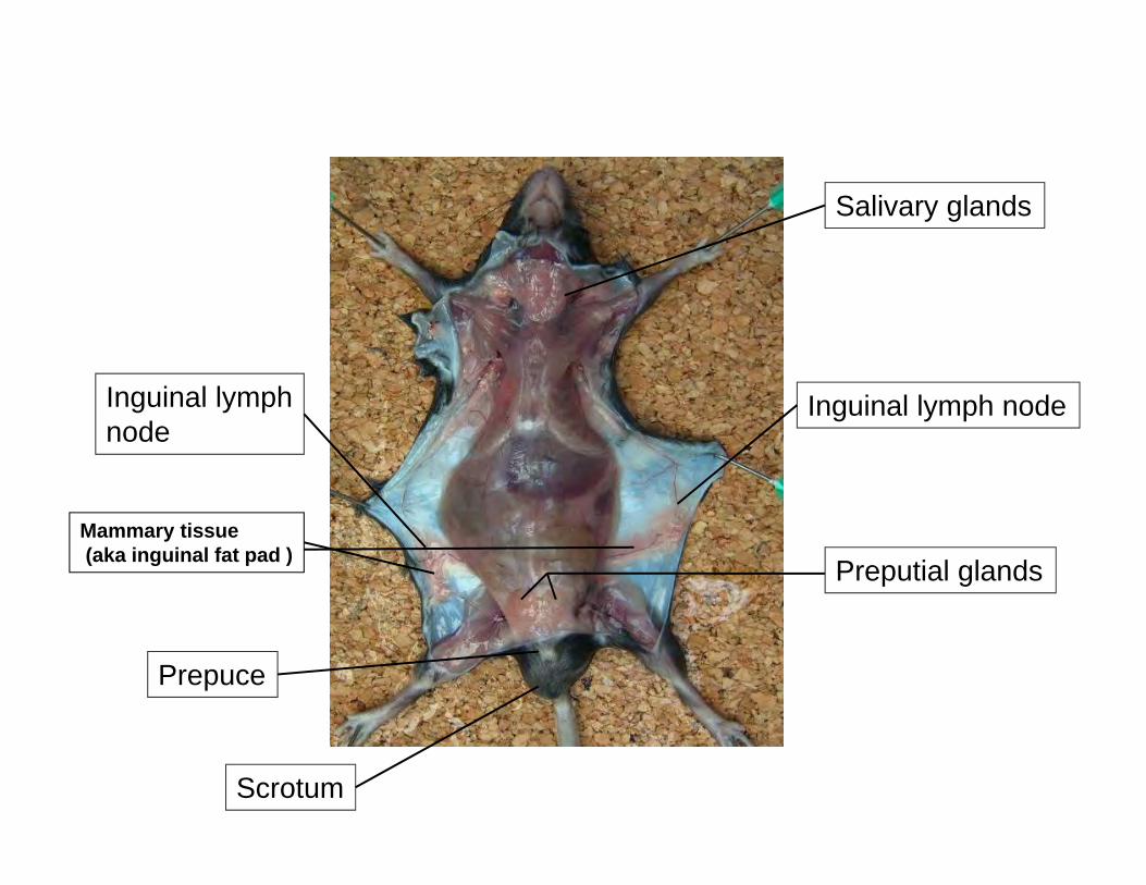
Salivary glands
Preputial glandsMammary tissue(aka inguinal fat pad )
Prepuce
Scrotum
Inguinal lymph nodeInguinal lymph node

Gonadalfat pad

Gonadalfat pad
Gonadalfat pad
Uterus
Ovaries

Pancreas

Lymph nodes

Mesenteric lymph node

Tissue Handling
• Collection– Tissue samples should not be greater than
1cm thick for adequate fixation– Try to hold the connective tissue around the
tissue to prevent crush artifact• Lung inflation
– Infuse 1ml‐1.5ml of fixative via the trachea• Swiss roll
– Flush intestine with fixative– Roll in cassette
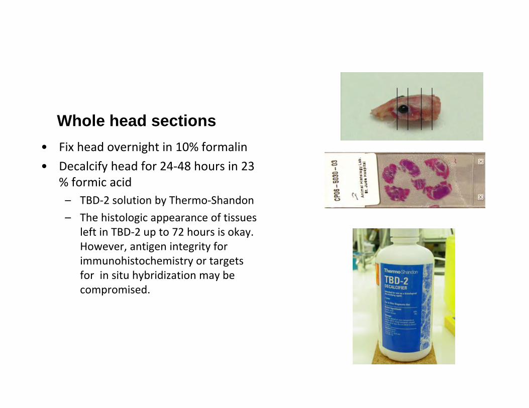
• Fix head overnight in 10% formalin• Decalcify head for 24‐48 hours in 23
% formic acid– TBD‐2 solution by Thermo‐Shandon– The histologic appearance of tissues
left in TBD‐2 up to 72 hours is okay. However, antigen integrity for immunohistochemistry or targets for in situ hybridization may be compromised.
Whole head sections

Autolysis happens!• If the animal is found dead in the cage in the morning it will
most likely be too autolyzed for necropsy
• Refrigeration is good • It slows down autolysis
• How long is too long between death and necropsy• If not refrigerated >4 hours is too long• If refrigerated about 6 hours max for most tissues
• The Gastrointestinal tract autolyzes quickly b/c of bacterial load;
• If doing GIT work, fixation should immediately follow death
• If you sac the animal and you want to perform histology on the tissue, put the tissue in fixative immediately
• Never put the carcass in the freezer or put tissues in the freezer prior to histology.
• Note: This does not include flash freezing for frozen sectioning.
• Use 10x the amount of fixative as tissue

What are the microscopic consequences of autolysis or poor fixation?

Animals are sick or dying…Not part of the plan
I have five surgeries, PCR running, it’s almost 5:00 and my daycare is going to charge me $20/minute after 5:30!!!!!
Getting the tissues in formalin quickly with excellent tissue preservation.
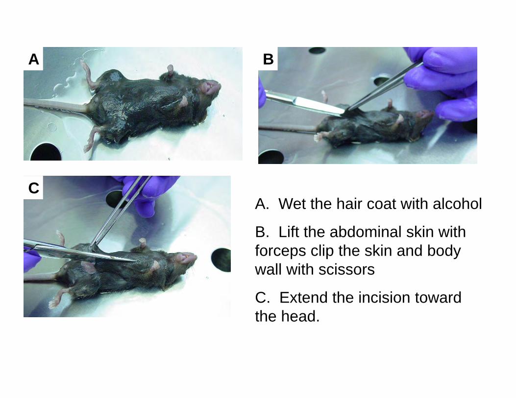
A. Wet the hair coat with alcohol
B. Lift the abdominal skin with forceps clip the skin and body wall with scissors
C. Extend the incision toward the head.
A
C
B
A B
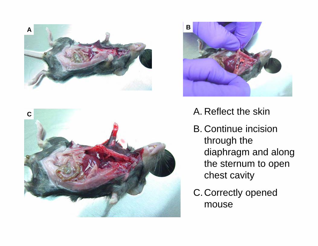
C A. Reflect the skin
B. Continue incision through the diaphragm and along the sternum to open chest cavity
C.Correctly opened mouse

Remove skin on the top of head to allow for brain fixation
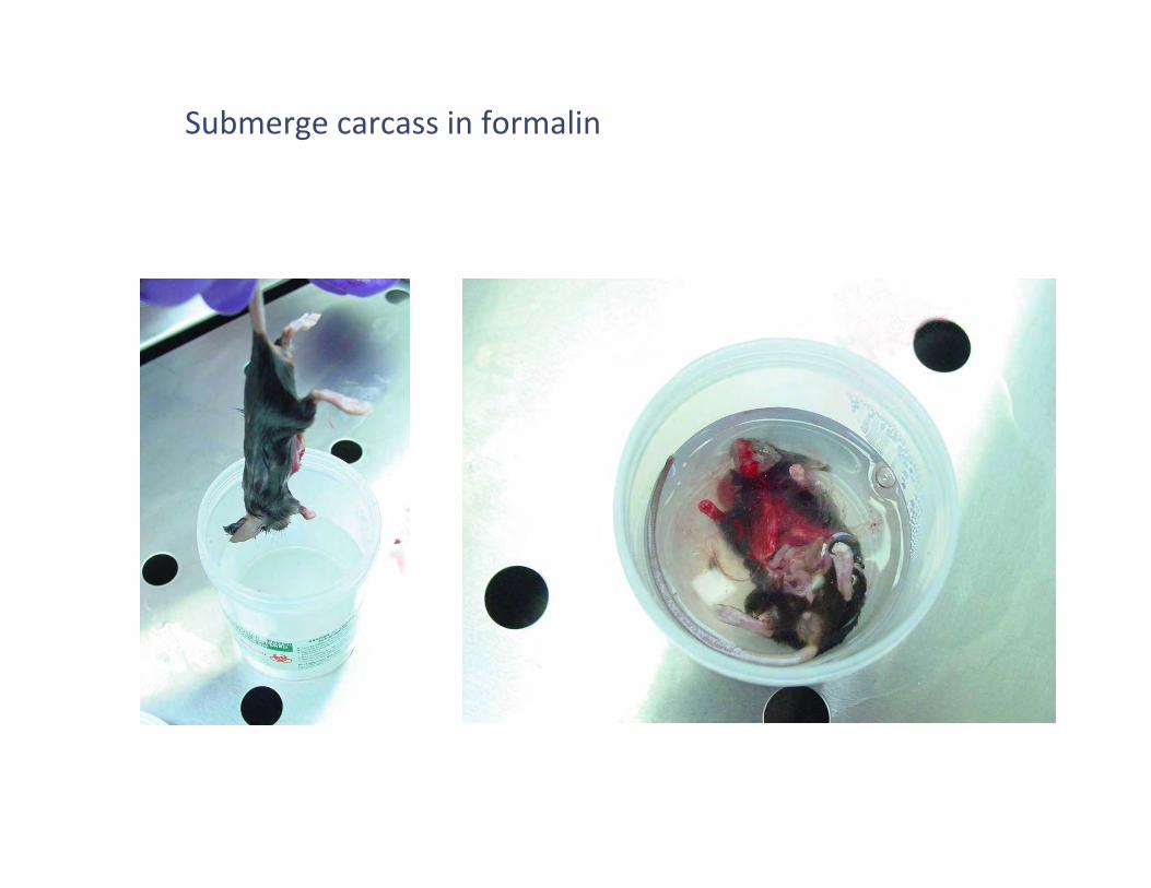
Submerge carcass in formalin

Fixation
• Most Common Types used – 10% Neutral Buffered Formalin– 4% paraformaldehyde– Bouins fixative– Davidson’s solution– Fekete's acid‐alcohol‐formalin– Glutaraldehyde (most commonly 4%)
• Immersion fixation• Perfusion

10% NBF
• Standard fixative• Easy to acquire and store• Works for most applications• Recent manuscript reports with HIER(heat induced
epitope retrieval) – long term fixation up to 6 weeks can be
overcome for 61 common antibodies– Tissues were stained same day of sectioning– Dog mainly small number of cat samples– CK7, HMWCK and Laminin diminished
– Webster et al. – Journal of Histochemistry April 2009

• Commonly used in research• Must be prepared fresh
– >48hours old = DON’T USE• Works for most applications• May be prepared with a variety of buffers to fit
application• For routine fixation basically the same as formalin
4% paraformaldehyde

Bouins• Slightly acidic• A little better penetration than formalin• Often used for
– Embryo– Reproductive tract
• >48 hrs in Bouin’s Tissues will become brittle– Switch to 70% alcohol
• Order pre‐made– Picric acid is explosive – EHS won’t be happy
• Can inhibit IHC
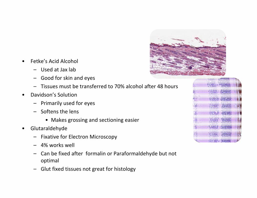
• Fetke’s Acid Alcohol– Used at Jax lab – Good for skin and eyes– Tissues must be transferred to 70% alcohol after 48 hours
• Davidson’s Solution– Primarily used for eyes– Softens the lens
• Makes grossing and sectioning easier• Glutaraldehyde
– Fixative for Electron Microscopy– 4% works well– Can be fixed after formalin or Paraformaldehyde but not
optimal– Glut fixed tissues not great for histology
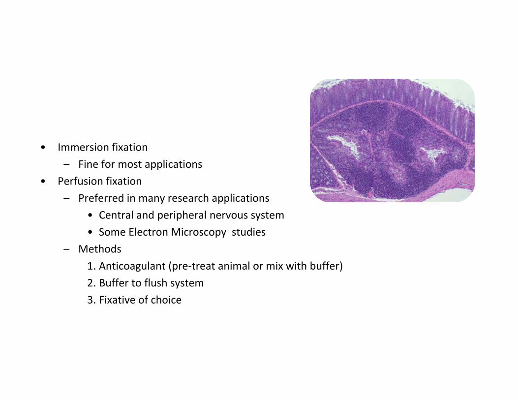
• Immersion fixation– Fine for most applications
• Perfusion fixation– Preferred in many research applications
• Central and peripheral nervous system• Some Electron Microscopy studies
– Methods1. Anticoagulant (pre‐treat animal or mix with buffer)2. Buffer to flush system3. Fixative of choice

Perfusion fixation• Animal is deeply anesthetized or immediately post‐mortem• Open thorax• Clip right atrium or right ventricle• Insert needle into left ventricle• Infusion of buffer typically 30‐60 ml until solution is clear and organs are pale• Infusion of fixative – 30‐60 ml• Carcass will be stiff and organs will be pale and vessels will be cleared of red blood cells

General principles for perfusion‐ Buffer
• Buffer used for initial flush – Routine histo
• Normal saline or Phospate buffered saline +/‐ heparin
– Electron microscopy• 0.1M cacodylate buffer with 4%sucrose, 0.2calcium
chlorideOR
• 0.1M Phosphate buffered saline
• Depending on application may need to be ice cold
• Flush pre‐wash buffer until the exiting fluid is clear– For mice the minimum is between 30‐60 mls– More flush is okay. Less is not

General principles for perfusions
• TO PUMP or NOT TO PUMP– Gravity* Best method
• Mean Arterial Pressure in awake mouse is 100mmHg• Hanging fluids no higher than 24 inches above carcass will
prevent artifact– Mechanical perfusion using calibrated pump– Manual perfusion via syringe
• Flush with 30‐60mls of fixative– More fixative is okay. Less is not!
• Artifacts– Pressure to high (usually with Manual and Mechanical )
• Vessel dilation and rupture • Cell and organelle swelling and rupture can occur
– These artifacts are especially problematic for EM studies
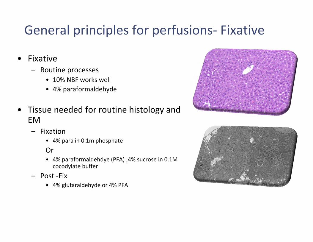
General principles for perfusions‐ Fixative
• Fixative– Routine processes
• 10% NBF works well• 4% paraformaldehyde
• Tissue needed for routine histology and EM– Fixation
• 4% para in 0.1m phosphate
Or • 4% paraformaldehdye (PFA) ;4% sucrose in 0.1M
cocodylate buffer
– Post ‐Fix• 4% glutaraldehyde or 4% PFA
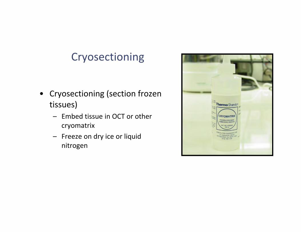
Cryosectioning
• Cryosectioning (section frozen tissues)– Embed tissue in OCT or other
cryomatrix– Freeze on dry ice or liquid
nitrogen

• Molecular testing– Flash freeze tissue – Store at ‐80– Preserve in RNA‐later

Placing tissues in cassettes
• Tissue should not be thicker than cassette– >0.5 cm
• Tissue should not fill the entire cassette• Use a pencil to write on cassette
– Sharpie or other markers will wash off during processing
Questions???Find your histologist!

Tissue Cassettes
The entire mouse in 12 cassettes
1. Heart, thymus, skeletal muscle2. Tongue, trachea, thyroid,
esophagus, lung, mediastinal LN3. Kidneys with adrenals attached4. Liver, spleen5. Peripheral lymph nodes
6. Salivary glands, stomach, pancreas7. Small Intestines (swiss roll)8. Cecum, Colon, Rectum9. Urogenital tract10. Skin 11. Whole Head sections12. Sternum (Bone marrow)


Teratomas in the Skin
Fu et al, CELL 111: 41, 2002

Conclusion
• Use a systemic approach to evaluate your mouse
• Know what is normal for your mouse strain(C57BL/6, FVB, etc..)
• Know what is normal for your experiment• Use appropriate fixation techniques• Find a Comparative Pathologist• Utilize local expertise• Utilize online resources

Online links-

Questions?