Embed Size (px)
Citation preview

Necropsy of GEM:The good, the bad, and the ugly
Kelli Boyd DVM, PhD, DACVPDepartment of Pathology
Vanderbilt University Medical Center
July 21, 2009

Overview
• Necropsy Evaluation• Tissue collection• Fixation
– Considerations for ancillary testing• “Normal Pathology”
– Stain Background Pathology– Unique mouse lesions
• Resources

There is always something new or unexpected
There is always another way

Necropsy Evaluation• General principles• Observation of Live animal • Terminal blood collection• External examination• Evaluation of internal organs• Lesion description

Autolysis happens!• If the animal is found dead in the cage in the morning it will
most likely be too autolyzed for necropsy
• Refrigeration is good • It slows down autolysis
• How long is too long between death and necropsy• If not refrigerated >4 hours is too long• If refrigerated about 6 hours max for most tissues
• The Gastrointestinal tract autolyzes quickly b/c of bacterial load;
• If doing GIT work, fixation should immediately follow death
• If you sac the animal and you want to perform histology on the tissue, put the tissue in fixative immediately
• Never put the carcass in the freezer or put tissues in the freezer prior to histology.
• Note: This does not include flash freezing for frozen sectioning.
• Use 10x the amount of fixative as tissue

Live animal observation

Terminal blood collection
Retro-orbital Intracardiac

• Skin• Oral cavity• Eyes• External ear • Reproductive tract• General body condition
External examination
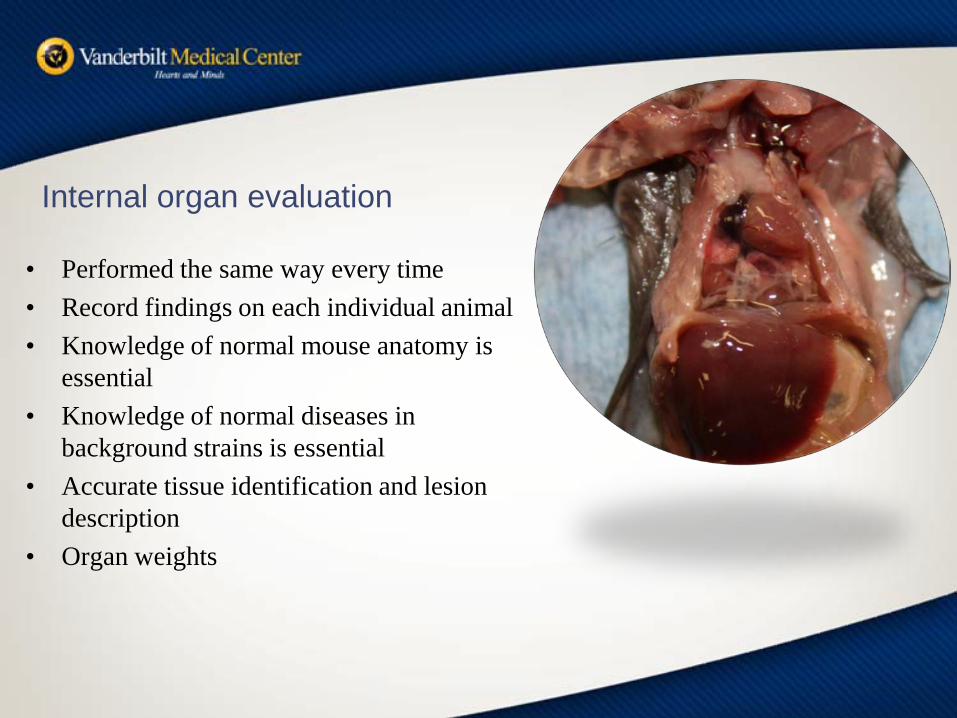
Internal organ evaluation
• Performed the same way every time• Record findings on each individual animal• Knowledge of normal mouse anatomy is
essential• Knowledge of normal diseases in
background strains is essential• Accurate tissue identification and lesion
description• Organ weights

Opening the mouse
Pin mouseWet fur with alcohol
Use forceps to raise skinUsing scissors cut opening in skin

Create pocket in the subcutaneous tissue along each rear legCut skin and peel back
Create pocket in the subcutaneous tissue to the anterior of the mouseCut skin along the pocket and peel back
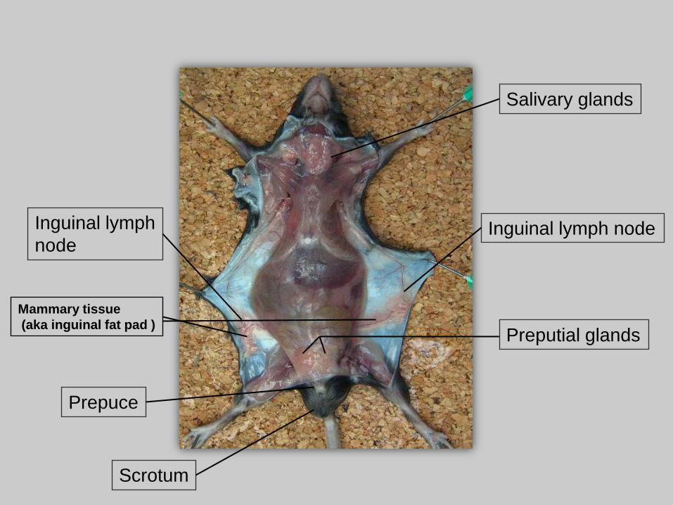
Salivary glands
Preputial glandsMammary tissue(aka inguinal fat pad )
Prepuce
Scrotum
Inguinal lymph nodeInguinal lymph node
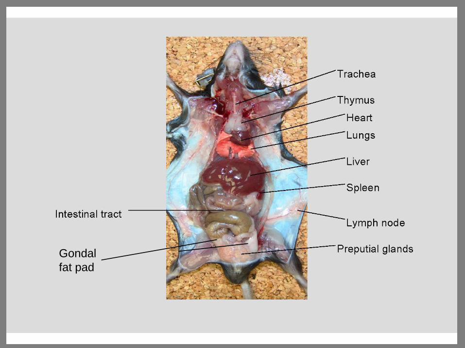
Gondalfat pad


Pancreas

Mesenteric lymph node

Tissue Handling
• Collection– Tissue samples should not be greater than
1cm thick for adequate fixation– Try to hold the connective tissue around the
tissue to prevent crush artifact• Lung inflation
– Infuse 1ml-1.5ml of fixative via the trachea• Swiss roll
– Flush intestine with fixative– Roll in cassette

• Fix head overnight in 10% formalin
• Decalcify head for 24-48 hours in 23 % formic acid– TBD-2 solution by Thermo-
Shandon– The histologic appearance of
tissues left in TBD-2 up to 72 hours is okay. However, antigen integrity for immunohistochemistry or targets for in situ hybridization may be compromised.
Whole head sections
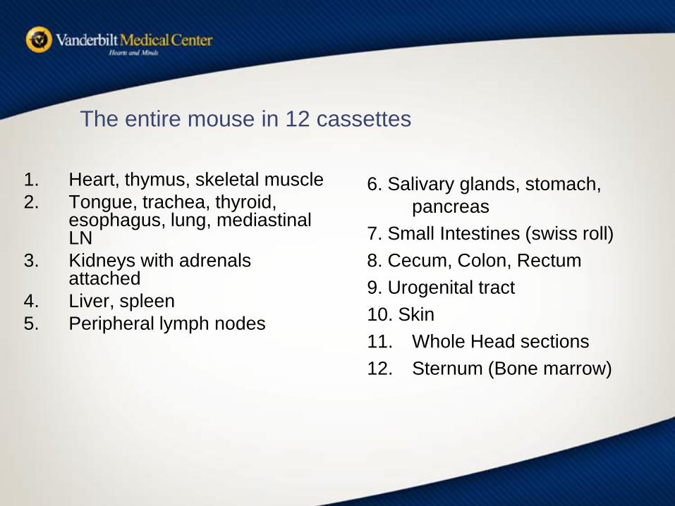
The entire mouse in 12 cassettes
1. Heart, thymus, skeletal muscle2. Tongue, trachea, thyroid,
esophagus, lung, mediastinal LN
3. Kidneys with adrenals attached
4. Liver, spleen5. Peripheral lymph nodes
6. Salivary glands, stomach, pancreas
7. Small Intestines (swiss roll)8. Cecum, Colon, Rectum9. Urogenital tract10. Skin 11. Whole Head sections12. Sternum (Bone marrow)

There’s no time for a necropsy. What can I do?!


Fixation
• Most Common Types used – 10% Neutral Buffered Formalin– 4% paraformaldehyde– Bouins fixative– Davidson’s solution– Fekete's acid-alcohol-formalin– Glutaraldehyde (most commonly 4%)
• Immersion fixation• Perfusion

10% NBF
• Standard fixative• Easy to acquire and store• Works for most applications• Recent manuscript reports with HIER
– long term fixation up to 6 weeks can be overcome for 61 common antibodies
– Tissues were stained same day of sectioning– Dog mainly small number of cat samples– CK7, HMWCK and Laminin diminished
– Webster et al. – Journal of Histochemistry April 2009
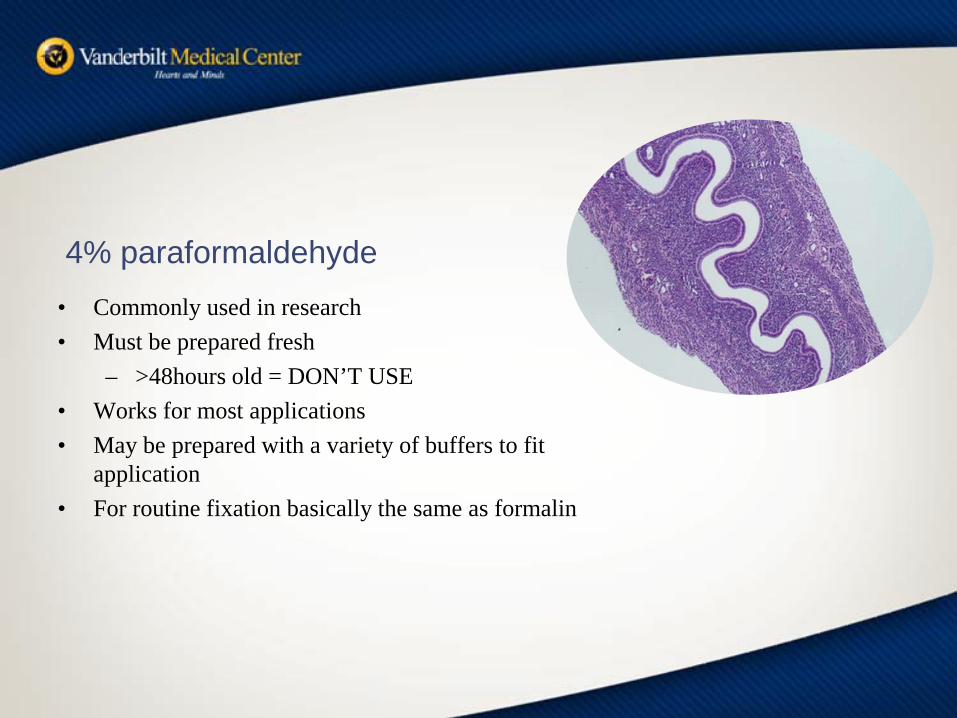
• Commonly used in research• Must be prepared fresh
– >48hours old = DON’T USE• Works for most applications• May be prepared with a variety of buffers to fit
application• For routine fixation basically the same as formalin
4% paraformaldehyde

Bouins• Slightly acidic• A little better penetration than formalin• Often used for
– Embryo– Reproductive tract
• >48 hrs in Bouin’s Tissues will become brittle– Switch to 70% alcohol
• Order pre-made– Picric acid is explosive – EHS won’t be happy
• Can inhibit IHC

• Fetke’s Acid Alcohol– Used at Jax lab – Good for skin and eyes– Tissues must be transferred to 70% alcohol after 48 hours
• Davidson’s Solution– Primarily used for eyes– Softens the lens
• Makes grossing and sectioning easier• Glutaraldehyde
– Fixative for Electron Microscopy– 4% works well– Can be fixed after formalin or Paraformaldehyde but not
optimal– Glut fixed tissues not great for histology

• Immersion fixation– Fine for most applications
• Perfusion fixation– Preferred in many research applications
• Central and peripheral nervous system• Some Electron Microscopy studies
– Methods1. Anticoagulant (pre-treat animal or mix with buffer)2. Buffer to flush system3. Fixative of choice
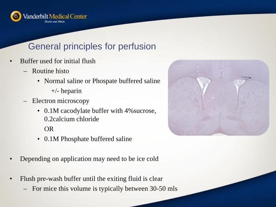
General principles for perfusion• Buffer used for initial flush
– Routine histo• Normal saline or Phospate buffered saline
+/- heparin– Electron microscopy
• 0.1M cacodylate buffer with 4%sucrose, 0.2calcium chlorideOR
• 0.1M Phosphate buffered saline
• Depending on application may need to be ice cold
• Flush pre-wash buffer until the exiting fluid is clear– For mice this volume is typically between 30-50 mls

General principles for perfusions • Fixative
– Routine processes• 10% NBF works well• 4% paraformaldehyde
• Tissue needed for routine histology and EM– 4% para in 0.1m phosphate
Or – 4% paraformaldehdye ;4% sucrose in 0.1M
cocodylate buffer– Then fix over night in 4% glutaraldehyde

General principles for perfusions
• TO PUMP or NOT TO PUMP– Manual perfusion via syringe– Mechanical perfusion using pump– Gravity* Best method
• Artifacts– Pressure to high (usually with Manual and Mecanical )
• Vessel dilation and rupture • Cell and organelle swelling and rupture can occur
– These artifacts are especially problematic for EM studies

Cryosectioning
• Cryosectioning (section frozen tissues)– Embed tissue in OCT or other
cryomatrix– Freeze on dry ice or liquid
nitrogen
Placing tissues in cassettes
• Tissue should not be thicker than cassette– >0.5 cm
• Tissue should not fill the entire cassette• Use a pencil to write on cassette
– Sharpie or other markers will wash off during processing
Questions???Find your histologist!
Beware of The Known! (background lesions)
– Lesions considered normal for the particular background strain• C57BL/6- microphthalmia, hydrocephalus, dermaitis,
osteoporosis, lymphoma or histiocytic sarcoma, amyloidosis, acidophilic crystalline/ macrophage pneumonia, hyalinosis
• 129- teratomas, Harderian gland tumors, lung tumors, nephropathy, acidophilic crstalline/ macrophage pneumonia, hyalinosis
• FVB- retinal degeneration, seizures, lung tumors, acidophilic macrophage pneumonia
• Retinal degeneration-FVB,C3H, CBA, SJL• Absent corpus callosum- Balb-c• Distrophic cardiac calcification- Balb-c, C3H, DBA


Gastroenterology. 2007 Apr;132(4):1299-308. Links
Precancerous lesions upon sporadic activation of beta-catenin in mice.
Coste I, Freund JN, Spaderna S, Brabletz T, Renno T.

Teratomas in the Skin
Fu et al, CELL 111: 41, 2002
Avoid interpretation pitfalls
• Set up appropriate controls• Know what is normal for your mouse strain
(C57BL/6, FVB, etc..) • Find a Comparative Pathologist• Utilize online resources

Online links-
Conclusion
• There are many techniques to accomplish similar goals
• Use a systemic approach to evaluate your mouse
• Remember mouse strains are unique and understanding the influence of background strain is important
• Find an expert if you need help

Acknowledgements
• SJCRH Vet Path Core– Brenda McGowan– Debra Williams– Hermitta McLaurine– Pam Johnson– Lourie West– Joe Emmons
• SJCRH EM Core Facility– Jackie Williams– Fara Sudlow– Sharon Frase
• Vanderbilt University IHC Core– Frances Shook– Melissa Downing
– Division of Animal Care– Dr. Ken Salleng– Dr. Troy Apple
– Photography– Michelle Endres
The end!
Questions?

Useful Links Mouse Pathology
• Mouse anatomy http://www.informatics.jax.org/cookbook/chapters/contents2.shtml
• Normal mouse histology http://www.deltagen.com/target/histologyatlas/HistologyAtlas.html
• Virtual mouse necropsy http://www.geocities.com/virtualbiology/
• Diseases of Laboratory animals http://www.radil.missouri.edu/info/dora/Dora.htm
• The Center for Genomic Pathology http://ctrgenpath.net/
• Revised guides for organ sampling and trimming in rats and mice published in 2003 and 2004 in three parts in Experimental and Toxicologic Pathology. http://www.item.fraunhofer.de/reni/trimming/index.php
• Proliferative lesions in the lung http://emice.nci.nih.gov/emice/nikitin/appendix/index.html
Atlases
• Atlas of Laboratory Mouse Histology http://www.ctrgenpath.org/static/atlas/mousehistology/
• MBL – mouse brain library http://www.mbl.org/procedures/procedure.php
• Edinburg mouse atlas project http://genex.hgu.mrc.ac.uk/
Resources
• Mouse nomenclature http://www.informatics.jax.org/mgihome/nomen/index.shtml
• Coat colors of mice http://www.informatics.jax.org/wksilvers/
• Mouse Genetics http://www.informatics.jax.org/silver/
• European tumor database http://www.pathbase.net/
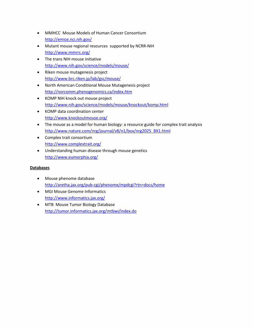
• MMHCC Mouse Models of Human Cancer Consortium http://emice.nci.nih.gov/
• Mutant mouse regional resources supported by NCRR-NIH http://www.mmrrc.org/
• The trans NIH mouse initiative http://www.nih.gov/science/models/mouse/
• Riken mouse mutagenesis project http://www.brc.riken.jp/lab/gsc/mouse/
• North American Conditional Mouse Mutagenesis project http://norcomm.phenogenomics.ca/index.htm
• KOMP NIH knock out mouse project http://www.nih.gov/science/models/mouse/knockout/komp.html
• KOMP data coordination center http://www.knockoutmouse.org/
• The mouse as a model for human biology: a resource guide for complex trait analysis http://www.nature.com/nrg/journal/v8/n1/box/nrg2025_BX1.html
• Complex trait consortium http://www.complextrait.org/
• Understanding human disease through mouse genetics http://www.eumorphia.org/
Databases
• Mouse phenome database http://aretha.jax.org/pub-cgi/phenome/mpdcgi?rtn=docs/home
• MGI Mouse Genome Informatics http://www.informatics.jax.org/
• MTB Mouse Tumor Biology Database http://tumor.informatics.jax.org/mtbwi/index.do