Embed Size (px)
Citation preview

Why sign language is good for your brain
Bencie Woll Deafness Cognition and Language Research Centre, UCL
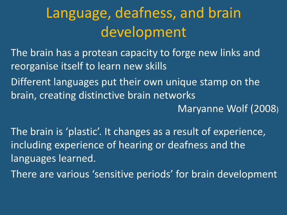
Language, deafness, and brain development
The brain has a protean capacity to forge new links and reorganise itself to learn new skills
Different languages put their own unique stamp on the brain, creating distinctive brain networks
Maryanne Wolf (2008)
The brain is ‘plastic’. It changes as a result of experience, including experience of hearing or deafness and the languages learned.
There are various ‘sensitive periods’ for brain development
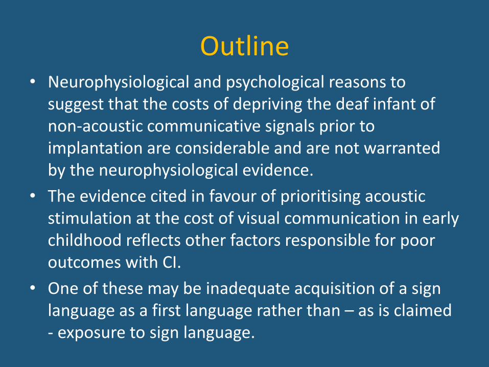
Outline • Neurophysiological and psychological reasons to
suggest that the costs of depriving the deaf infant of non-acoustic communicative signals prior to implantation are considerable and are not warranted by the neurophysiological evidence.
• The evidence cited in favour of prioritising acoustic stimulation at the cost of visual communication in early childhood reflects other factors responsible for poor outcomes with CI.
• One of these may be inadequate acquisition of a sign language as a first language rather than – as is claimed - exposure to sign language.
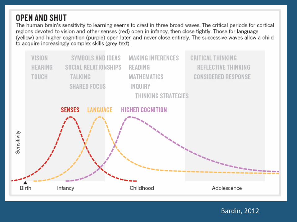
Sensitive Periods
Bardin, 2012
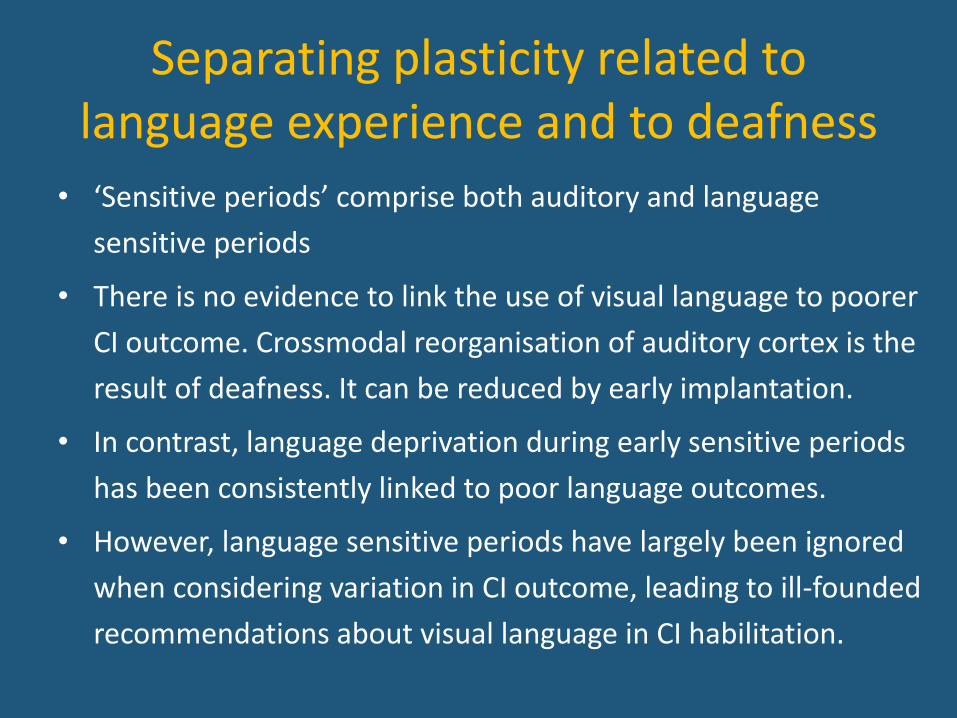
Separating plasticity related to language experience and to deafness
• ‘Sensitive periods’ comprise both auditory and language
sensitive periods
• There is no evidence to link the use of visual language to poorer
CI outcome. Crossmodal reorganisation of auditory cortex is the
result of deafness. It can be reduced by early implantation.
• In contrast, language deprivation during early sensitive periods
has been consistently linked to poor language outcomes.
• However, language sensitive periods have largely been ignored
when considering variation in CI outcome, leading to ill-founded
recommendations about visual language in CI habilitation.
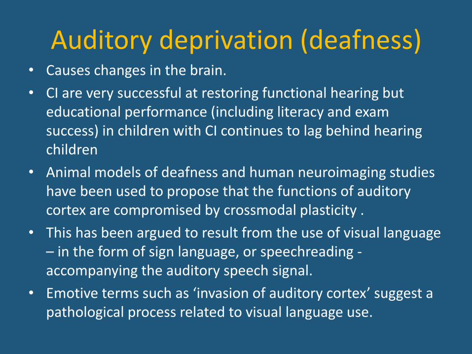
Auditory deprivation (deafness) • Causes changes in the brain.
• CI are very successful at restoring functional hearing but educational performance (including literacy and exam success) in children with CI continues to lag behind hearing children
• Animal models of deafness and human neuroimaging studies have been used to propose that the functions of auditory cortex are compromised by crossmodal plasticity .
• This has been argued to result from the use of visual language – in the form of sign language, or speechreading - accompanying the auditory speech signal.
• Emotive terms such as ‘invasion of auditory cortex’ suggest a pathological process related to visual language use.
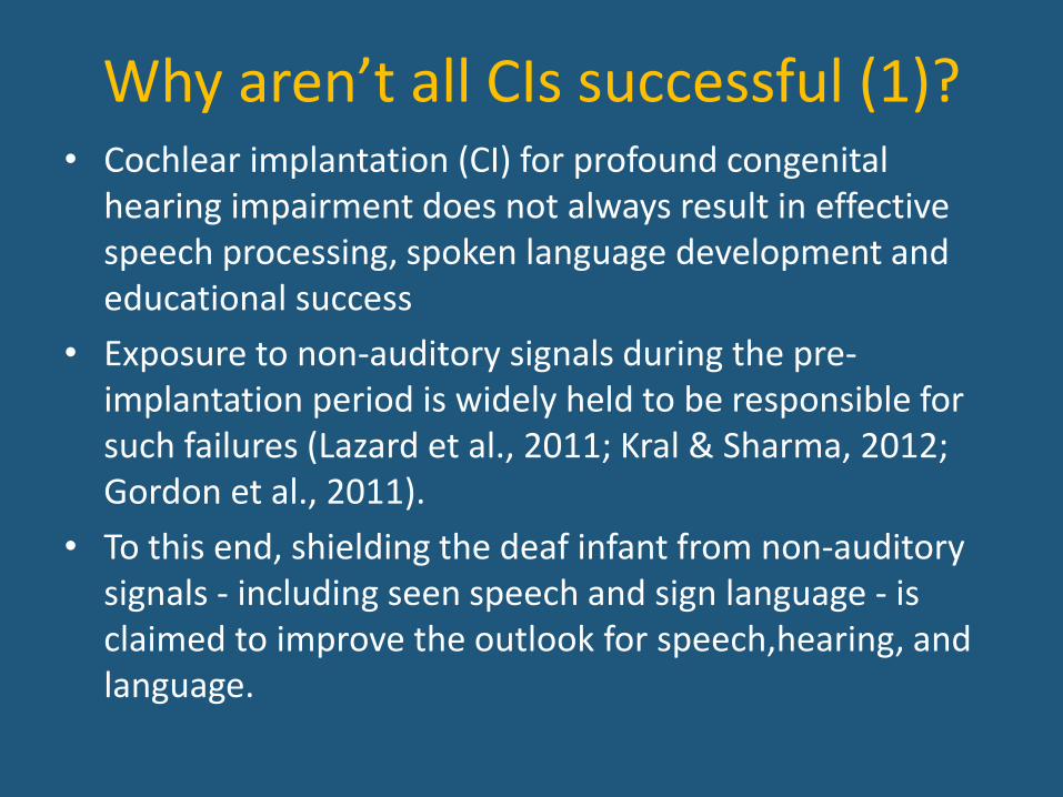
Why aren’t all CIs successful (1)? • Cochlear implantation (CI) for profound congenital
hearing impairment does not always result in effective speech processing, spoken language development and educational success
• Exposure to non-auditory signals during the pre-implantation period is widely held to be responsible for such failures (Lazard et al., 2011; Kral & Sharma, 2012; Gordon et al., 2011).
• To this end, shielding the deaf infant from non-auditory signals - including seen speech and sign language - is claimed to improve the outlook for speech,hearing, and language.
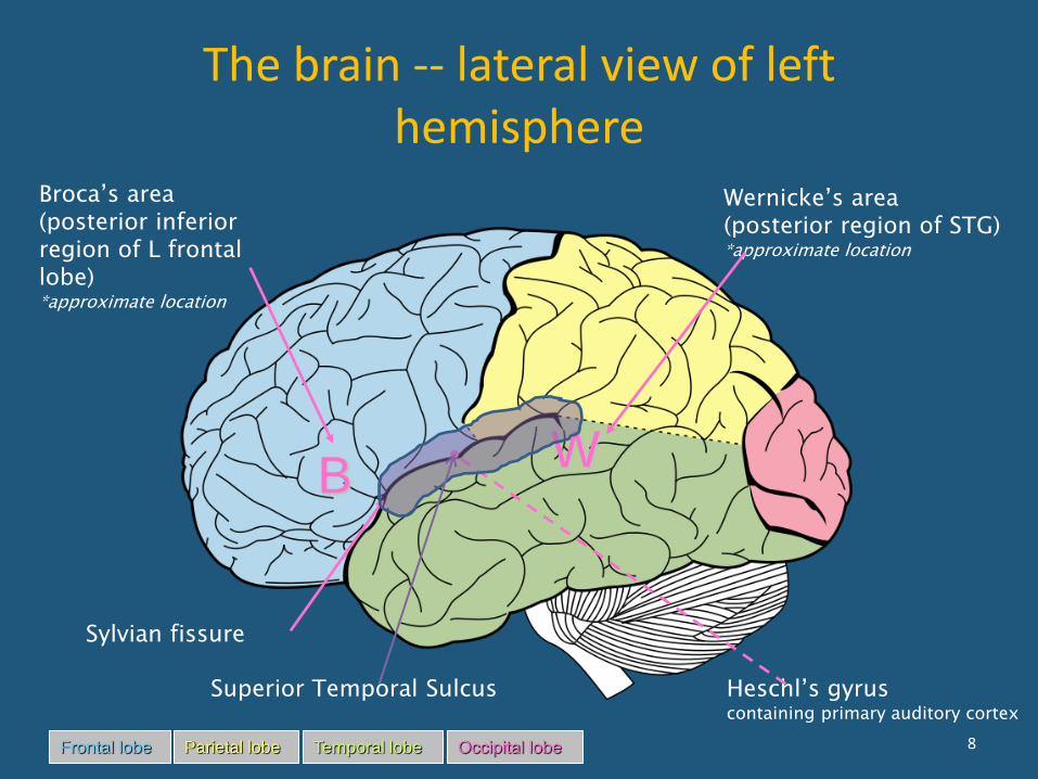
The brain -- lateral view of left hemisphere
B
Frontal lobe Temporal lobe Parietal lobe Occipital lobe
Broca’s area (posterior inferior region of L frontal lobe) *approximate location
W
Wernicke’s area (posterior region of STG) *approximate location
Sylvian fissure
Heschl’s gyrus containing primary auditory cortex
Superior Temporal Sulcus
8
We question the inference that visual language irreparably distorts the function
of auditory cortex, negatively impacting on CI success
• The ‘functional decoupling hypothesis’ (Kral et al., 2005) suggests that a disconnection between activation in primary auditory cortex (A1) and activation in secondary auditory cortex (A2) resulting from early deafness, reflects abnormal organization within A1 and disconnections between A1 and A2.
• One factor contributing to such decoupling is assumed to be the activation of A1 by visual projections in the deaf brain.
• Such ‘abnormal’ activation by the sight of sign language does not routinely occur
Current practice • Current practice in relation to speech training pre- and post-CI
stresses that exposure to non-auditory signals must be minimised because of its assumed deleterious effects on the dynamic development of auditory cortical circuits.
• In ‘auditory-verbal’ training regimes the adult is required to train the child’s acoustic skills by reducing (hiding) the visibility of oral actions, and parents are advised not to use sign language prior to implantation (Chan et al., 2000; Rhoades & Chisholm, 2001; Yoshida et al., 2008).
• Clinical practice follows a neurological hypothesis which suggests that seeing speech or SL may disrupt auditory cortical development during the sensitive period.
Is such advice warranted?
The argument: exposure to visual stimulation results in functional
decoupling of auditory cortex in the deaf infant brain
• Exposure of primary auditory cortex to non-auditory signals is claimed to lead to functional decoupling of the cortical network for speech processing.
• The benefit of CI should therefore be greatest when brains have been shielded from non-auditory stimulation, and least when there is earlier exposure to non-auditory stimuli.
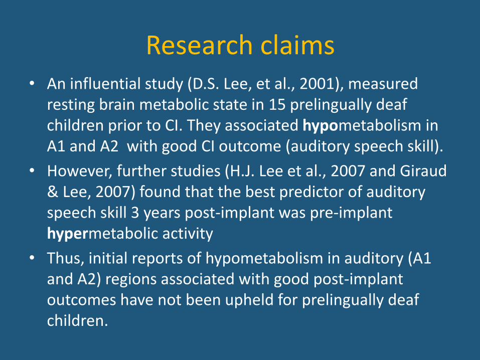
Research claims • An influential study (D.S. Lee, et al., 2001), measured
resting brain metabolic state in 15 prelingually deaf children prior to CI. They associated hypometabolism in A1 and A2 with good CI outcome (auditory speech skill).
• However, further studies (H.J. Lee et al., 2007 and Giraud & Lee, 2007) found that the best predictor of auditory speech skill 3 years post-implant was pre-implant hypermetabolic activity
• Thus, initial reports of hypometabolism in auditory (A1 and A2) regions associated with good post-implant outcomes have not been upheld for prelingually deaf children.
Re-examining the ‘visual takeover’ hypothesis: a direct test
• The assumption persists that the colonization of
A1 by visual projections may contribute to functional decoupling of elements within the cortical auditory processing network – and to poor CI outcomes for prelingually deaf children exposed to such material. So, for example, with regard to preparation for and rehabilitation with CI, the aim is “..to limit activity which restricts auditory function” (Gordon et al., 2011, p 217) .
The consequences for clinical practice and rehabilitation are profound and
far-reaching. Is this assumption valid?
• There are inconsistencies in the literature and in the inferences made from various results. This claim is also at odds with Giraud and Lee (2007), who found no correlation between pre-implant metabolic status in A1 and post-implant auditory processing proficiency, and who concluded that “primary auditory regions do not appear to re-organise in a cross-modal manner…” (p 386).
Primary auditory cortex (A1) • Leonard et al (2012) compared sign language processing in
deaf native signers to speech processing in hearing non-signers. While lexico-semantic processing activated an equivalent left superior temporal network in deaf signers and hearing speakers, signs did not activate A1 in deaf signers. A1 was activated by speech in normally hearing people.
• Since A1 is activated following CI , the argument about the relationship between non-auditory cortical stimulation and poor CI outcome must shift to one focusing on functional decoupling between primary and secondary auditory cortex, and/or ‘malfunction’ within A2, rather than a failure within A1 itself.
Secondary auditory cortex: activation by visual signals in deafness
• Is secondary auditory cortex (A2), activated by visual stimuli?
• If yes, does this negatively affect prognosis for CI with respect to
learning spoken language?
– While strongly activated in sign language processing (Emmorey et
al., 2011) the specialization of superior temporal regions is not for
language only
– This region responds to the processing of a wide range of
communicative actions performed by plausible (social) agents in
whatever modality they occur, and however ‘incomplete’ they are
(Hein & Knight, 2008; Nummenmaa & Calder, 2009)
– Left hemisphere lateralization depends on the extent to which such
visual events are possible language processes – i.e. sign language
processing in Deaf native users (Husain et al., 2012).
Language is supramodal
• Posterior superior temporal regions, comprising secondary auditory cortex in hearing people, are not only critical for the integration of heard and seen speech, but are highly and dynamically multimodal.
• Lee et al. (2007) consider this region to show ‘latent multimodal connectivity’ for speech. That is, A2 can be readily and immediately activated by one modality when the other is absent.
Visible and audiovisual speech in CI
• Does A2 continue to support the effective processing of speech when it is delivered to A1 by CI?
• Two possibilities: – either visual speech interferes with the new
auditory signal, reflecting reduced cross-modal plasticity in A2, and supporting the functional decoupling hypothesis
– or, A2 supports the integration of visual speech with projections from the newly activated A1 to deliver good (auditory) CI outcomes.
What about visual speech (speechreading)?
• Far from interfering with auditory speech processing, silent
speechreading experience enhances auditory speech processing post-CI.
• Pre-CI speechreading in prelingually deaf children is a good predictor of
post-CI auditory speech processing abilities (Bergeson, et al, 2005).
• Just one case study (Hirano et al., 2000) suggests a negative relationship
between (pre-implant) speechreading skill and post-implant auditory
success, but most studies find a positive correlation between
speechreading and good CI outcomes
• Rather than vision and language interfering with audition after CI,
correlations between patterns of neural activations in visual cortex and
increasingly successful performance with CI suggest the opposite.
(Giraud et al., 2001,Giraud & Truy, 2002; Lazard et al, 2011).
Why aren’t all CIs successful (2)?
• Since early infancy is a critical period for the acquisition of language, deaf children born to hearing parents are at risk of developing inefficient neural structures to support skilled language processing (Mayberry et al., 2011).
• Sign language, acquired by a deaf child as a first language in a signing environment, is cortically organized like a heard spoken language (MacSweeney et al., 2008).
• The cortical signatures for individuals showing poor outcome for CI may thus reflect the effects of impaired language experience and acquisition in the earliest years, rather than the effects of exposure to non-auditory signals.
Consequences and correlates of impaired first language learning on
cortical organization and CI • If the evidence for functional decoupling is weak,
how do we to explain poor outcomes of CI in some prelingually deaf children? – They may reflect late and incomplete acquisition of a
first language which, in turn, is reflected in anomalous cortical organization.
• The neural correlates of late first language acquisition may provide clues about causes and clinical management of language acquisition with CI.

16
Comparing deaf native signers processing BSL sentences and
hearing non-signers processing audiovisual English
BSL English
Separating experience of deafness and experience of language
• Plastic effects induced by auditory deprivation are expected, independently of linguistic access, in Deaf Signers and Deaf Oral groups, but not in Hearing Non-Signers
• Sign language-induced plasticity should be observed only in Deaf Signers, who have access to the linguistic content of BSL and not in Deaf Oral and Hearing Non-Signers.
Differential activations depend on experience of hearing and of language
• Differential activations observed in the left superior temporal sulcus, are driven by experience with sign language, and not by auditory deprivation. Non-signers (deaf and hearing) cannot process the linguistic information.
• In the right STC, differential activations are driven both by auditory deprivation and experience of sign language.
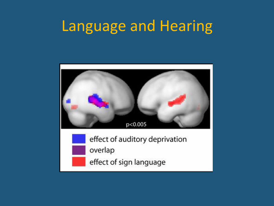
Language and Hearing
Conclusions on language experience
• Plastic effects in the left STC are of linguistic origin, and are shaped by sign language experience. The right STC shows plasticity due to sensory deprivation.
• Life-long sign language experience and life-long sensori-motor adaptation to auditory deprivation drive plasticity in separate portions of the cortex.
• Plastic effects in the left STC have a linguistic origin, and are shaped by sign language experience, whereas the right STC also shows plasticity due to sensory deprivation.
Sensitive periods for language acquisition: late acquisition of a first
language • Most deaf children (90 – 95%) are born to hearing parents
and cannot benefit from a natural, language-rich environment
• The existence of sensitive periods suggests that if a child fails to learn language in early childhood s/he will never reach the normal level of mastery, with full command of syntax, phonology and verbal working memory.
• Do late first language learners – who constitute the vast majority of prelingually deaf people - show atypical structural and functional circuitry for language processing as adults?
28
Comparing phonology in BSL and English
• In an English phonology task, hearing and deaf participants had to decide whether the English labels for two pictures rhymed
• In the BSL phonology task, deaf participants had to decide if the BSL labels for two pictures shared the same location
If similar processing is required to make phonological similarity judgments about BSL and English, similar brain areas should be recruited during both tasks
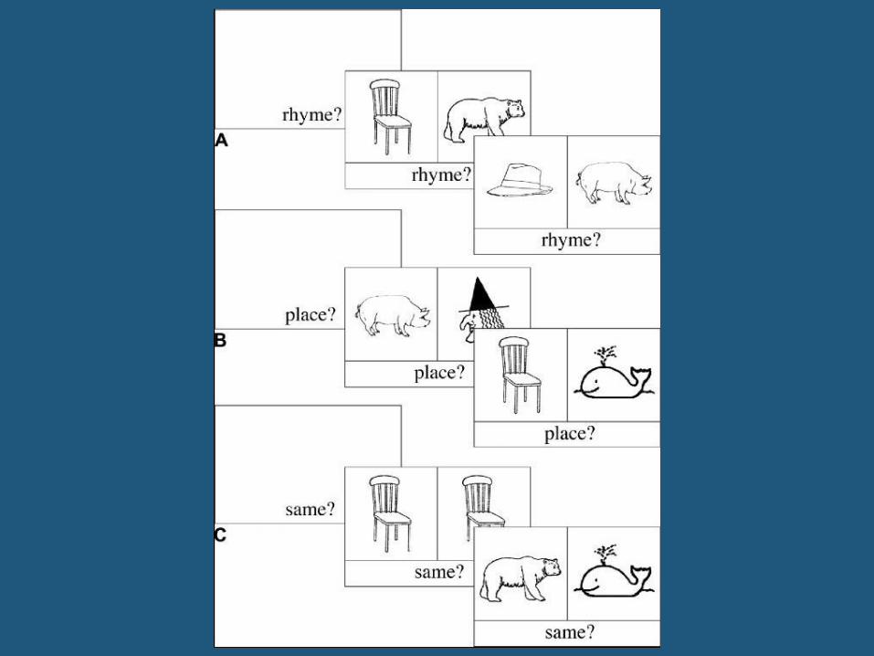
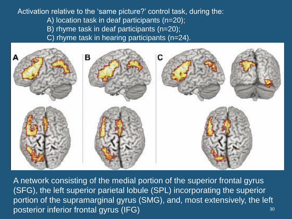
Activation relative to the ‘same picture?’ control task, during the:
A) location task in deaf participants (n=20);
B) rhyme task in deaf participants (n=20);
C) rhyme task in hearing participants (n=24).
30
A network consisting of the medial portion of the superior frontal gyrus
(SFG), the left superior parietal lobule (SPL) incorporating the superior
portion of the supramarginal gyrus (SMG), and, most extensively, the left
posterior inferior frontal gyrus (IFG)

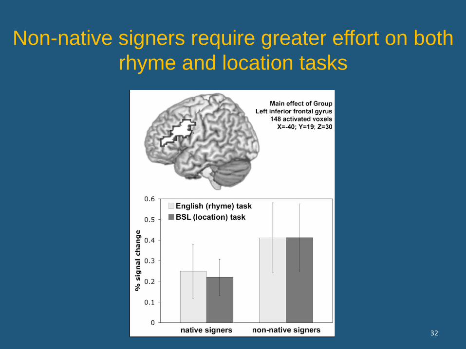
The task is harder (in English as
well as BSL)for those who
acquired English as a first
language
• Deaf non-native signers (with delayed L1 English)
activated the left inferior frontal gyrus more than
native signers during the BSL task, and also
during the task performed in English
– phonological processing required greater effort when a
first language was delayed
31
32
Non-native signers require greater effort on both
rhyme and location tasks
What about CI outcomes in children with sign language as a first language?
• A direct test of the impact of first (sign) language acquisition on CI outcomes has been provided (Hassanzadeh, 2012).
• This study compared CI outcome in native signing deaf children with deaf parents to CI outcome in deaf children with hearing parents and no sign language background .
• In contrast to the predictions from an auditory-neural critical period hypothesis, the deaf children who were exposed to sign language early in life had better speech and language outcomes following implant.
• This suggests that linguistic development of the relevant cortical circuits is critical to successful outcome with CI – whatever the role of auditory-neural developmental processes.
Assessing language proficiency • The few studies of deaf late sign language learners point to abnormal
cortical circuitry for language
• To date, sign language proficiency assessments have not been
administered in any studies of non-native signers
• ‘Uses a sign language’ is no indicator of proficiency in phonological,
morphological, syntactic and discourse skills.
• No assessment of SL proficiency means that a potential factor in the
efficiency of CI for speech outcome has been ignored.
• An early and well-established visual language may be critical in assisting
a CI to deliver a ‘new’ language source.
• Yet inferences are made concerning the functional decoupling
hypothesis, and recommendations put forward for shielding from non-
auditory inputs
Summary
• The disordered cortical circuitry attributed to exposure to
visual stimuli affecting auditory regions is more likely
associated with disordered language and pre-linguistic
experience. Giraud & Lee (2007, p 381) say
– “First language acquisition thus appears to be a major
landmark in brain organisation signalling a major
constraint on language-related brain plasticity”.
• Similar arguments apply to acquiring a SL as a first language.
Not one study has reported measures of sign language
proficiency in relation to CI outcome in prelingually deaf
children
What do these arguments mean for clinical management of CI?
• Far from shielding the developing infant from visual communication, the
deaf child awaiting CI needs language and communicative input of any and
all sorts to enable effective cognitive development to proceed.
• The early months and years are crucial for the development of language –
not just heard speech
• hile auditory rehabilitation is necessary to enable effective functioning of the
CI, there is no compelling evidence that the rehabilitation of hearing – on its
own– predicts satisfactory speech and language progress.
• Early CI is an astonishing breakthrough in delivering hearing to the child born
deaf, but its success should be measured in terms of language skills and
cognitive development – not in terms of auditory impact.
• The best guarantee of such success is good first language acquisition within
the early years – however that may be achieved

THANK YOU
www.dcal.ucl.ac.uk

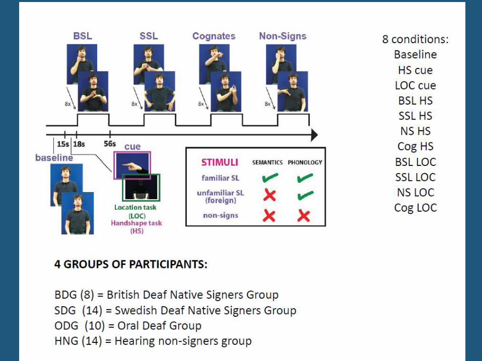
Study 1.Methods and Participants • 2 groups of 7 congenitally or early (before 3 years of age) severely-to-
profoundly deaf individuals. • These were either Deaf Signers (DS), who have deaf parents, and are
native signers of British Sign Language (BSL); or Deaf Oral (DO), who have hearing parents, and are native speakers of English who access language through speechreading and who have never learned a sign language.
• A third group of participants with normal hearing, native speakers of English (Hearing Non-signers – HN), as a separate control group.
• Deaf groups matched for: – sensory loss [better-ear Pure Tone Average (PTA; 1KHz, 2KHz, 4KHz;
maximum output of equipment= 100 dB): DS= 98.1 dB ± 3.7 SEM; DO= 94.5 dB ± 3.3; t[6]= 0.64, p= 0.54];
– age (DS= 46.3 years ± 4.4 SEM; DO= 47.3 ± 1; HN= 47.6 ± 3.3; t[6]DO,DS =
0.23, p= 0.82; t[6]DO,HG= 0.09, p= 0.93; t[6]
HG,DS= 0.2, p= 0.81); – gender (3 male and 4 female in each group).
Language skills • All deaf participants learned their preferred language from
infancy.
• The Deaf Signers group were native signers of BSL (at least
one deaf parent), and they indicated their level of proficiency
of BSL to be 6.17 on a scale of 1-7 (1=not very good at all;
7=excellent). All Deaf Signers communicated with the
researchers in BSL.
• The Deaf Oral group had on average adult reading skills (35.6
points ± 1.19 SEM), as measured with the revised Vernon-
Warden Reading Comprehension Test16, ranging from 32-38.
• All Deaf Oral participants communicated with the researchers
in English.
Experimental Design • Stimuli consisted of videos of sign-based material, each one of 2-3s of
duration. • Four types of signs:
– a) British Sign Language; – b) Swedish Sign Language ; – c) Cognates (signs shared by both languages due to their iconic nature); – d) Non-signs.
• Four scanning runs, each consisting of 3 blocks of 12 videos per condition (12 blocks/run), with an inter-trial interval (ITI) of 4.5s on average. A baseline period of 15s, consisting of the image of the model without making any movement with his hands, appeared between blocks.
• Participants’ task was to indicate with a button-press if the sign presented in each video had the same hand-shape or same location as a cue presented just before the onset of the block. The cues consisted of static pictures of handshapes or highlighted parts of the model’s body.
• The task could be performed by anyone independently of sign language knowledge but may tap phonological knowledge of sign language (there were no significant differences in performance across groups).
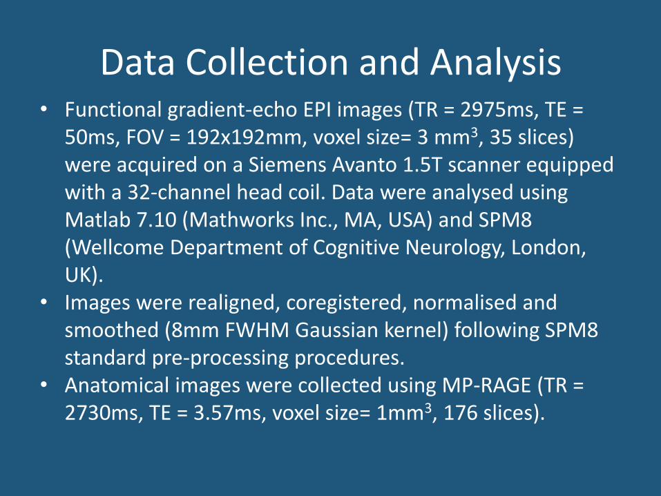
Data Collection and Analysis • Functional gradient-echo EPI images (TR = 2975ms, TE =
50ms, FOV = 192x192mm, voxel size= 3 mm3, 35 slices) were acquired on a Siemens Avanto 1.5T scanner equipped with a 32-channel head coil. Data were analysed using Matlab 7.10 (Mathworks Inc., MA, USA) and SPM8 (Wellcome Department of Cognitive Neurology, London, UK).
• Images were realigned, coregistered, normalised and smoothed (8mm FWHM Gaussian kernel) following SPM8 standard pre-processing procedures.
• Anatomical images were collected using MP-RAGE (TR = 2730ms, TE = 3.57ms, voxel size= 1mm3, 176 slices).