Embed Size (px)
Citation preview

World Journal of Gastrointestinal EndoscopyWorld J Gastrointest Endosc 2017 August 16; 9(8): 346-427
ISSN 1948-5190 (online)
Published by Baishideng Publishing Group Inc

Contents Monthly Volume 9 Number 8 August 16, 2017
August 16, 2017|Volume 9|Issue 8|WJGE|www.wjgnet.com I
REVIEW
346 Terahertzendoscopicimagingforcolorectalcancerdetection:Currentstatusandfutureperspectives
Doradla P, Joseph C, Giles RH
MINIREVIEWS359 Useandbarrierstochromoendoscopyfordysplasiasurveillanceininflammatoryboweldisease
Shukla R, Salem M, Hou JK
368 Evolutionofstereoscopicimaginginsurgeryandrecentadvances
Schwab K, Smith R, Brown V, Whyte M, Jourdan I
378 Endoscopicultrasonography-emergingapplicationsinhepatology
Magalhães J, Monteiro S, Xavier S, Leite S, de Castro FD, Cotter J
ORIGINAL ARTICLE
Retrospective Cohort Study
389 Clinicalimpactofconfocallaserendomicroscopyinthemanagementofgastrointestinallesionswithan
uncertaindiagnosis
Robles-Medranda C, Vargas M, Ospina J, Puga-Tejada M, Valero M, Soria M, Bravo G, Robles-Jara C, Lukashok HP
396 Impactoflaparoscopicliverresectiononbleedingcomplicationsinpatientsreceivingantithrombotics
Fujikawa T, Kawamoto H, Kawamura Y, Emoto N, Sakamoto Y, Tanaka A
Retrospective Study
405 Correlationofabnormalhistologywithendoscopicfindingsamongmycophenolatemofetiltreatedpatients
Izower MA, Rahman M, Molmenti EP, Bhaskaran MC, Amin VG, Khan S, Sultan K
411 UsefulnessoftheHookknifeinflexibleendoscopicmyotomyforZenker’sdiverticulum
Rouquette O, Abergel A, Mulliez A, Poincloux L
CASE REPORT417 RussellbodygastritiswithDutcherbodiesevaluatedusingmagnificationendoscopy
Yorita K, Iwasaki T, Uchita K, Kuroda N, Kojima K, Iwamura S, Tsutsumi Y, Ohno A, Kataoka H
425 SimultaneousCourvoisier’sanddoubleductsigns
Agrawal S, Vohra S
ContentsWorld Journal of Gastrointestinal Endoscopy
Volume 9 Number 8 August 16, 2017
EDITORS FOR THIS ISSUE
Responsible Assistant Editor: Xiang Li Responsible Science Editor: Fang-Fang JiResponsible Electronic Editor: Ya-Jing Lu Proofing Editorial Office Director: Jin-Lei WangProofing Editor-in-Chief: Lian-Sheng Ma
NAMEOFJOURNALWorld Journal of Gastrointestinal Endoscopy
ISSNISSN 1948-5190 (online)
LAUNCHDATEOctober 15, 2009
FREQUENCYMonthly
EDITORS-IN-CHIEFAtsushi Imagawa, PhD, Director, Doctor, Depart-ment of Gastroenterology, Mitoyo General Hospital, Kan-onji, Kagawa 769-1695, Japan
Juan Manuel Herrerias Gutierrez, PhD, Academic Fellow, Chief Doctor, Professor, Unidad de Gestión Clínica de Aparato Digestivo, Hospital Universitario Virgen Macarena, Sevilla 41009, Spain
EDITORIALBOARDMEMBERSAll editorial board members resources online at http://
www.wjgnet.com/1948-5190/editorialboard.htm
EDITORIALOFFICEXiu-Xia Song, DirectorWorld Journal of Gastrointestinal EndoscopyBaishideng Publishing Group Inc7901 Stoneridge Drive, Suite 501, Pleasanton, CA 94588, USATelephone: +1-925-2238242Fax: +1-925-2238243E-mail: [email protected] Desk: http://www.f6publishing.com/helpdeskhttp://www.wjgnet.com
PUBLISHERBaishideng Publishing Group Inc7901 Stoneridge Drive, Suite 501, Pleasanton, CA 94588, USATelephone: +1-925-2238242Fax: +1-925-2238243E-mail: [email protected] Desk: http://www.f6publishing.com/helpdeskhttp://www.wjgnet.com
PUBLICATIONDATEAugust 16, 2017
COPYRIGHT© 2017 Baishideng Publishing Group Inc. Articles published by this Open-Access journal are distributed under the terms of the Creative Commons Attribution Non-commercial License, which permits use, distribution, and reproduction in any medium, provided the original work is properly cited, the use is non commercial and is otherwise in compliance with the license.
SPECIALSTATEMENTAll articles published in journals owned by the Baishideng Publishing Group (BPG) represent the views and opinions of their authors, and not the views, opinions or policies of the BPG, except where otherwise explicitly indicated.
INSTRUCTIONSTOAUTHORShttp://www.wjgnet.com/bpg/gerinfo/204
ONLINESUBMISSIONhttp://www.f6publishing.com
ABOUT COVER
August 16, 2017|Volume 9|Issue 8|WJGE|www.wjgnet.com II
EditorialBoardMemberofWorldJournalofGastrointestinalEndoscopy, JorgGAlbert,MD,PhD,AssociateProfessor,DepartmentofInternalMedicineI,JohannWolfgangGoethe-UniversityHospital,D-60590Frankfurt,Germany
World Journal of Gastrointestinal Endoscopy (World J Gastrointest Endosc, WJGE, online ISSN 1948-5190, DOI: 10.4253) is a peer-reviewed open access (OA) academic journal that aims to guide clinical practice and improve diagnostic and therapeutic skills of clinicians. WJGE covers topics concerning gastroscopy, intestinal endoscopy, colonoscopy, capsule endoscopy, laparoscopy, interventional diagnosis and therapy, as well as advances in technology. Emphasis is placed on the clinical practice of treating gastrointestinal diseases with or under endoscopy. We encourage authors to submit their manuscripts to WJGE. We will give priority to manuscripts that are supported by major national and international foundations and those that are of great clinical significance.
World Journal of Gastrointestinal Endoscopy is now indexed in Emerging Sources Citation Index (Web of Science), PubMed, and PubMed Central.
I-III EditorialBoard
AIM AND SCOPE
INDEXING/ABSTRACTING
FLYLEAF

Joana Magalhães, Sara Monteiro, Sofia Xavier, Sílvia Leite, Francisca Dias de Castro, José Cotter
MINIREVIEWS
378 August 16, 2017|Volume 9|Issue 8|WJGE|www.wjgnet.com
Endoscopic ultrasonography - emerging applications in hepatology
Joana Magalhães, Sara Monteiro, Sofia Xavier, Sílvia Leite, Francisca Dias de Castro, José Cotter, Gastroenterology Department, Hospital Senhora da Oliveira, 4835-044 Guimarães, Portugal
Joana Magalhães, Sara Monteiro, Sofia Xavier, Sílvia Leite, Francisca Dias de Castro, José Cotter, School of Medicine, University of Minho, 4704-553 Braga, Portugal
Joana Magalhães, Sara Monteiro, Sofia Xavier, Sílvia Leite, Francisca Dias de Castro, José Cotter, Life and Health Sciences Research Institute/3B’s, PT Government Associate Laboratory, 4710-057 Braga/Guimarães, Portugal
Author contributions: Magalhães J wrote and conceived the study; Monteiro S and Xavier S contributed to the literature search and acquisition of data; Leite S, de Castro FD and Cotter J contributed to the critical appraisal of the work, revising the ar-ticle critically for important intellectual content; all authors read and approved the final version of the manuscript.
Conflict-of-interest statement: All authors disclose any potential or actual personal, political or financial conflict of interest in the material, information or techniques described in the paper.
Open-Access: This article is an open-access article which was selected by an in-house editor and fully peer-reviewed by external reviewers. It is distributed in accordance with the Creative Commons Attribution Non Commercial (CC BY-NC 4.0) license, which permits others to distribute, remix, adapt, build upon this work non-commercially, and license their derivative works on different terms, provided the original work is properly cited and the use is non-commercial. See: http://creativecommons.org/licenses/by-nc/4.0/
Manuscript source: Invited manuscript
Correspondence to: Joana Magalhães, MD, Gastroenterology Department, Hospital Senhora da Oliveira, Rua dos Cuteleiros, Creixomil, 4835-044 Guimarães, Portugal. [email protected]: +351-253-540330Fax: +351-253-513592
Submit a Manuscript: http://www.f6publishing.com
DOI: 10.4253/wjge.v9.i8.378
World J Gastrointest Endosc 2017 August 16; 9(8): 378-388
ISSN 1948-5190 (online)
Received: January 23, 2017 Peer-review started: February 2, 2017First decision: February 20, 2017Revised: March 30, 2017 Accepted: July 7, 2017Article in press: July 10, 2017Published online: August 16, 2017
AbstractThe inspection of the liver is a valuable part of the upper endoscopic ultrasonography (EUS) studies, regardless of the primary indication for the exami-nation. The detailed images of the liver segments provided by EUS allows the use of this technique in the study of parenchymal liver disease and even in the diagnosis and classification of focal liver lesions. EUS has also emerged as an important tool in understanding the complex collateral circulation in patients with portal hypertension and their clinical and prognostic value. Recently, EUS-guided portal vein catheterization has been performed for direct portal pressure measurement as an alternative method to evaluate portal hemody-namics. In this review, the authors summarize the available evidence regarding the application of EUS to patients with liver diseases and how we can apply it in our current clinical practice.
Key words: Endoscopic ultrasonography; Portal hyper-tension; Gastroesophageal varices; Focal liver lesions; Liver biopsy
© The Author(s) 2017. Published by Baishideng Publishing Group Inc. All rights reserved.
Core tip: This review summarizes the current status of the available evidence regarding the application of endoscopic ultrasonography (EUS) to patients with liver diseases, focusing on recent breakthroughs and its potential application on clinical practice. We highlight the
379 August 16, 2017|Volume 9|Issue 8|WJGE|www.wjgnet.com
Magalhães J et al . EUS - emerging applications in hepatology
emerging role of EUS in the study of parenchymal liver disease as well as in the diagnosis and classification of focal liver lesions. Finally, we emphasise the crucial role of EUS in the understanding of the complex collateral circulation in patients with portal hypertension.
Magalhães J, Monteiro S, Xavier S, Leite S, de Castro FD, Cotter J. Endoscopic ultrasonography - emerging applications in hepatology. World J Gastrointest Endosc 2017; 9(8): 378-388 Available from: URL: http://www.wjgnet.com/1948-5190/full/v9/i8/378.htm DOI: http://dx.doi.org/10.4253/wjge.v9.i8.378
INTRODUCTIONIn the recent years, there has been remarkable improvement in hepatology, with new treatments for viral hepatitis, recommendations for the followup of cirrhotic patients and treatment of portal hypertension complications. These advances have brought an increased need for the assessment of liver function and liver histologic characterization.
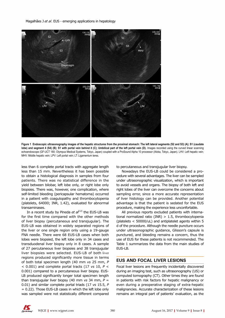
Endoscopic ultrasonography (EUS) has become an important tool, not only in the diagnosis of several gastrointestinal lesions, but also in performing various therapeutic manoeuvres[1]. Due to the close proximity of the transducer to the liver, from the transgastric and transduodenal routes, EUS allows a clear visualization of the liver anatomy and its vasculature providing accurate and detailed images[2,3] (Figure 1). As experience grows with this technique new indications for EUS continue to emerge, and endosonographers have made an effort to define a clinical role for EUS in liver diseases.
This review summarizes the available evidence regarding the application of EUS to patients with liver diseases and how it can be applied in a current clinical practice.
EUS AND LIVER PARENCHYMAL DISEASEAlthough noninvasive tests, such as elastography or serologic markers for liver fibrosis, have been developed, the liver biopsy remains an important part of the liver disease evaluation and management[4].
Liver biopsy has been commonly performed by percutaneously imageguided. A transjugular fluoroscopyguided approach is used when the percutaneous route is not safe, because of coagulopathy or ascites[5,6].
EUSguidance represents an emerging method of liver biopsy. EUS provides images of both lobes of the liver, moreover biopsy needle can be safely directed into the liver under image guidance, and intervening vessels and organs can be avoided.
EUSguided liver biopsy (EUSLB) for studying parenchymal liver disease has largely been studied with the use of different needles. Since a tissue core biopsy with a preserved architecture is crucial to diagnosis and fully characterization of the hepatic diseases,
needles specifically designed for core biopsy have been used. The ability to obtain specimens of liver tissue for histologic examination with a TruCut biopsy needle dedicated for EUSguided biopsy, the QuickCore® needle (Cook® Medical), were demonstrated in some published studies[7,8]. In a study by DeWitt et al[8], 21 consecutive patients underwent liver biopsy by using a QuickCore® needle. Liver biopsy specimens were able to provide diagnostic clinical information in only 15 of 21 patients (71%), the total specimen length was a median of 9 mm, with a median of 2 complete portal tracts. The technique was safe and feasible. However the samples were smaller than those traditionally considered adequate for histologic assessment.
The TruCut biopsy needle failed to reach widespread use due to technical difficulties with its utilization. To overcome the main limitations of a TruCut biopsy needle, the same manufacture developed a new needle, the ProCore® needle (Cook® Medical). Sey et al[9] compared the diagnostic yield of a 19gauge ProCore®
needle with a QuickCore® needle. A total of 45 patients underwent liver biopsy by using the QuickCore® and 30 patients the ProCore® needle. The ProCore® needle group required fewer passes (median 2 vs 3, P < 0.0001), produced a longer median specimen length (median 20 mm vs 9 mm, P < 0.0001) with more complete portal tracts (median 5 vs 2, P = 0.0003) and also allowed a histologic diagnosis more frequently (97% vs 73%).
Other studies have also been published demonstrating the adequacy of liver tissue sampling by a 19gauge FNA needle. Stavropoulos et al[10] presented a study in which patients underwent a EUSLB with a 19gauge FNA needle. All patients underwent EUS with a 7.5MHz linear echoendoscope (Olympus GFUC140PAL5; Olympus, Tokyo, Japan) as the initial procedure. Twentytwo patients underwent a EUSLB of the left lobe of the liver, a median of 2 passes (range 13) yielded a median specimen length of 36.9 mm, with a median of 9 complete portal tracts and a diagnostic yield of 91%, without postprocedure complications. The authors concluded that EUSLB by using a 19gauge FNA needle was feasible, safe, with an excellent diagnostic yield and sample adequacy for histologic examination.
To evaluate the diagnostic yield of EUSLB in a large patient cohort, Diehl et al[11] recently presented a prospective, multicentre study with 110 patients who underwent EUSLB at eight centres. EUS examination was performed with a linear echoendoscope (GFUC140P, Olympus America, Center Valley, PA, United States). The biopsy was performed using a 19gauge FNA needle, with or without a stylet, 710 toandfro motions of the needle were made per pass (12 pass were made), using the fanning technique and almost all endoscopists preferred to use full suction for the needle aspiration. Adequate liver biopsy specimens for pathological diagnosis were obtained in 98% of patients, with a median specimen length of 38 mm, with median of 14 complete portal tracts. There were five patients whose tissue yield was
380 August 16, 2017|Volume 9|Issue 8|WJGE|www.wjgnet.com
less than 6 complete portal tracts with aggregate length less than 15 mm. Nevertheless it has been possible to obtain a histological diagnosis in samples from four patients. There was no statistical difference in the yield between bilobar, left lobe only, or right lobe only biopsies. There was, however, one complication, where selflimited bleeding (pericapsular hematoma) occurred in a patient with coagulopathy and thrombocytopenia (platelets, 64000; INR, 1.42), evaluated for abnormal transaminases.
In a recent study by Pineda et al[12] the EUSLB was for the first time compared with the other methods of liver biopsy (percutaneous and transjugular). The EUSLB was obtained in widely separated regions of the liver or one single region only using a 19gauge FNA needle. There were 68 EUSLB cases when both lobes were biopsied, the left lobe only in 34 cases and transduodenal liver biopsy only in 8 cases. A sample of 27 percutaneous liver biopsies and 38 transjugular liver biopsies were selected. EUSLB of both liver regions produced significantly more tissue in terms of both total specimen length (40 mm vs 25 mm, P < 0.001) and complete portal tracts (17 vs 10, P < 0.001) compared to a percutaneous liver biopsy. EUSLB produced significantly longer total specimen length than transjugular liver biopsy (40 mm vs 34 mm, P = 0.01) and similar complete portal triads (17 vs 15.5, P = 0.22). Those EUSLB cases in which the left lobe only was sampled were not statistically different compared
to percutaneous and transjugular liver biopsy. Nowadays the EUSLB could be considered a pro
cedure with several advantages. The liver can be sampled under ultrasonographic visualization, which is important to avoid vessels and organs. The biopsy of both left and right lobes of the liver can overcome the concerns about sampling error, since a more accurate representation of liver histology can be provided. Another potential advantage is that the patient is sedated for the EUS procedure, making the experience less unconfortable.
All previous reports excluded patients with international normalized ratio (INR) > 1.5, thrombocytopenia (platelets < 50000/uL) and antiplatelet agents within 5 d of the procedure. Although the needle puncture occurs under ultrasonographic guidance, Glisson’s capsule is punctured, and bleeding remains a concern, thus the use of EUS for these patients is not recommended. The Table 1 summarizes the data from the main studies of EUSLB.
EUS AND FOCAL LIVER LESIONSFocal liver lesions are frequently incidentally discovered during an imaging test, such as ultrasonography (US) or computed tomography (CT). Other times they are found in patients with risk factors for hepatic malignancy or even during a preoperative staging of extrahepatic malignancies. Accurate characterization of these lesions remains an integral part of patients’ evaluation, as the
A B
C D
S2
S3
S1
S1
S4
LHV
Diaphragm
LPV LPV
LT
MHV
Figure 1 Endoscopic ultrasonography images of the hepatic structures from the proximal stomach: The left lateral segments (S2 and S3) (A); S1 (caudate lobe) and segment 4 (S4) (B); S1 with portal vein behind it (C); Umbilical part of the left portal vein (D). Images recorded using the curved linear scanning echoendoscope (GF-UCT 180; Olympus Medical Systems, Tokyo, Japan) coupled with a ProSound Alpha 10 processor (Aloka, Tokyo, Japan). LHV: Left hepatic vein; MHV: Middle hepatic vein; LPV: Left portal vein; LT: Ligamentum teres.
Magalhães J et al . EUS - emerging applications in hepatology

381 August 16, 2017|Volume 9|Issue 8|WJGE|www.wjgnet.com
extent of liver involvement may change clinical stage and management.
The inspection of the liver is a valuable part of the upper EUS studies, regardless of the primary indication for the examination. Recently, EUS and EUSguided fine needle aspiration (EUS-FNA) has emerged as an important tool in the diagnosis and classification of liver lesions. Most of the liver segments can be visualized with the echoendoscope[3] and the proximity of the ultrasound probe to the liver parenchyma provides exceptional images of the liver parenchyma, which may have a key role in the detection, characterization and even in the definitive diagnosis of liver lesions.
Awad et al[13] evaluated the feasibility of EUS for the detection and diagnosis of liver lesions in 14 patients with known or suspected hepatocellular carcinoma (HCC) and metastatic liver lesions. Consecutive patients referred for EUS with suspected liver lesions were evaluated. EUS not only successfully identified all previously hepatic lesions described by CT scan, but also identified new or additional lesions in 4 patients (28%), all less than 0.5 cm in size. Nine patients underwent EUSFNA of hepatic lesions, with a 22gauge needle and two passes for each lesion, and all FNA yielded adequate specimens. The authors suggested that EUS is an adequate preoperative staging tool for liver lesions suspected to be HCC or metastatic lesions, as EUS can detect small hepatic lesions previously undetected by dynamic CT scans.
Singh et al[14] have conducted a prospective trial to compare the accuracy of EUS and EUSFNA with other imaging modalities for the detection of primary liver tumors in subjects at high risk of HCC. Seventeen subjects were enrolled in the study. The EUS has detected more HCC lesions than US (8 vs 2, P = 0.06), CT (19 vs 8, P = 0.06) or magnetic resonance imaging (MRI) (14 vs 7, P = 0.25), although not statistically significant. Moreover, EUS has detected small HCC lesions that has been missed by CT and MRI, with the smallest lesion visualized by EUS and confirmed by FNA having 4 mm in size. Thus, EUSFNA helped in the determination of the cytological nature of liver nodular lesions that were
indeterminate on CT and MRI. A diagnostic algorithm has been proposed in which EUS could be used for highrisk patients with inconclusive CT, or poorly accessible lesions requiring tissue confirmation.
In a study by DeWitt et al[15], the sensitivity of EUS features and EUSFNA for benign and malignant solid liver lesions was described. The EUSFNA was performed on 77 different liver lesions, a total of 45 aspirates (58%) were diagnostic for malignancy (true positives), of these, 44 were metastatic and one was a HCC. In 25 patients (55%), the FNA provided both the primary diagnosis and upstaged the malignancy and in nine subjects (20%) the EUSFNA made the initial diagnosis, upstaged the tumor, and prevented surgery. Three lesions previously classified as benign were lately, by intraoperative findings or percutaneous-FNA, reclassified as malignant (false negatives). The EUS features predictive of malignant hepatic lesions were the presence of regular outer margins (60% vs 27%, P = 0.02) and the detection of two or more lesions (38% vs 9%, P = 0.03). EUSFNA was performed using a 22gauge needle and no complications were reported. This study concluded that EUS is a safe and sensitive procedure that can have a significant impact on patient management. The Table 2 summarizes the reported data from the studies of EUS of Focal liver lesions.
The diagnosis of portal vein thrombosis (PVT) secondary to HCC invasion is of paramount importance since it preclude a therapeutic approach[16]. Nontumor PVT has usually a similar appearance to portal vein tumor thrombosis, the last could enhance with contrast or have Doppler sign, however sometimes this differentiation is difficult and the diagnosis remains doubtful until proven otherwise. Although percutaneous USguided FNA of a PVT has been well documented[17], this technique presents some difficulties, especially in accessing thrombus in the centrally located main portal vein. The EUSFNA could overcome some limitations of a percutaneous USguided FNA, as it provides an excellent view of the liver hilum which facilitates the puncture of a PVT. Some case reports have been published which the EUSFNA was used to diagnose
Table 1 Data from the main studies of endoscopic ultrasonography-guided liver biopsy
Ref. Study design Needle Passes Specimen length (median) Complete portal tracts (median) Histological diagnosis
DeWitt et al[8] Prospectiveunicentre study
n = 21
Quick-Core1 1-4 9 mm 2 71%
Diehl et al[11] Prospectivemulticentre study
n = 110
19G (FNA)Expect1
1-2 38 mm 14 98%
Stavropoulos et al[10] Prospectiveunicentre study
n = 22
19G (FNA)Echotip2
1-3 36.9 mm 9 91%
Sey et al[9] Prospectiveunicentre study
n = 75
Quick-Core1 1-7 9 mm 2 73%ProCore 19G1 1-3 20 mm 5 97%
1Cook® Medical; 2Boston Scientific. FNA: Fine needle aspiration.
Magalhães J et al . EUS - emerging applications in hepatology
382 August 16, 2017|Volume 9|Issue 8|WJGE|www.wjgnet.com
HCC in patients with portal vein thrombosis[1821]. In two cases the procedure was performed with a 25gauge needle[18,19], while the other cases were performed with a 22gauge needle[20,21] and all patient have tolerated the procedure well, without any immediate or delayed complications.
After a careful study and analysis of these articles we can easily conclude that the EUS and EUSFNA may be helpful in the management of a subset of patients with a high suspicion for small liver lesions and to approach lesions that remain difficult to sample by percutaneous USguided techniques. However some important issues remain unanswered[22,23], the risk of needle track spread of HCC from EUSFNA remains undefined and the quality of the visualization of peripheral lesions, such as the areas under the dome of the diaphragm and the inferiorposterior portion of the right lobe of the liver.
Other potential concerns are related to the risks associated with EUSFNA. In a large international survey[24], in which centres with large experience participated, the EUSFNA of the liver lesions, in expert hands, proved to be a safe procedure. The complication rate was 4%, although this included one major complication (death) and several minor complications (bleeding, infection, abdominal pain). The dead occurred in a patient with a pancreatic mass. The patient was suspected to have an occluded biliary stent at the time of the EUS and a cholangitis resulted from the introduction of bacteria into an obstructed bile duct by the needle. For this reason it is recommended that antibiotics are administered prophylactically and biliary drainage is established rapidly if fine needle aspiration of the liver is to be performed in the setting of obstructive jaundice. Despite these results more information about the risks and complications in specific groups is necessary, especially in patients with a particular propensity for liver lesions, such as patients with cirrhosis or portal hypertension. Prospective studies comparing the accuracy and complication rate of the EUSFNA and percutaneous FNA techniques for the diagnosis of liver tumors are also still needed.
The therapy of HCC guided by EUS has also been
reported in some case reports. In 2011, Di Matteo et al[25] reported a case of a hepatocellular carcinoma located in the caudate lobe unsuitable for surgical resection, liver transplant and percutaneous treatment. The embolization failed and an EUSguided neodymium:Yttriumaluminiumgarnet (Nd: YAG) laser ablation was performed. The ablation of hepatocellular carcinoma was effective without adverse events. Nakaji et al[26] reported another case of EUSguided hepatocellular carcinoma treatment this time with ethanol injection. These two cases have shown the significant innovative options to treat lesions that are difficult to reach by conventional methods.
EUS AND ENDOSCOPIC THERAPY OF GASTROESOPHAGEAL VARICESGastroesophageal varices are the most important portosystemic collaterals that can be developed as a consequence of portal hypertension[27]. The venous anatomy of the lower esophagus and stomach in patients with portosystemic collaterals is complex. The dilated submucosal veins can be readily seen during an upper endoscopy. This superficial venous plexus is connected, through the perforating vessels, with the deep venous plexus, periesophageal and paraesophageal veins[28].
The endoscopic and ultrasound images provided by the EUS allows the visualization of the collateral vessels within and outside the esophageal wall[2931] (Figure 2), and its role in the diagnosis and management of gastroesophageal varices is now well established.
In a study by Faigel et al[32] the presence and diameter of varices surrounding the esophagus and proximal stomach (paraesophageal and paragastric varices) were correlated with the presence and degree of liver disease and portal hypertension and represented a risk factor for variceal bleeding.
Since the previous reports about the role of collaterals in patients with portal hypertension and its clinical significance, some studies have analyzed the role of the EUS in the evaluation of the outcome of endoscopic
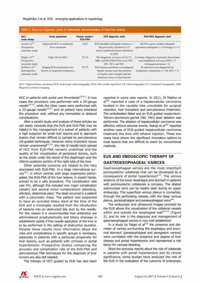
Table 2 Reported diagnostic yields of endoscopic ultrasonography of focal liver lesions
Ref./study design
Study population Patient number/EUS-FNA
EUS diagnostic yield EUS-FNA diagnostic yield
Awad et al[13] Prospectiveunicenter study
Suspected HCC or metastatic liver carcinoma
14/9 EUS identified all hepatic lesions (n = 14) previously reported by CT
4 new/additional lesions identified by EUS
All FNA passes yielded adequate specimens (malignant: n = 8; benign: n = 1)
Singh et al[14]
Prospectiveunicenter study
High risk for HCC 17/16 The diagnostic accuracy of US, CT, MRI, and EUS/EUS-FNA were 38%,
69%, 92%, and 94%
Cytologic diagnosis of primary liver tumor was established in 8 cases (HCC = 7;
cholangiocarcinoma = 1)DeWitt et al[15]
Retrospectiveunicenter study
Staging EUS examinations for known or suspected malignancy
77/77 EUS features predictive of malignant hepatic lesions were the presence of regular outer margins and the detection of two or more lesions
45 aspirates were diagnostic for malignancy (metastasis: n = 44; HCC = 1)
HCC: Hepatocellular carcinoma; EUS: Endoscopic ultrasonography; FNA; Fine needle aspiration; US: Ultrasonography; CT: Computed tomography; MRI: Magnetic resonance imaging.
Magalhães J et al . EUS - emerging applications in hepatology

383 August 16, 2017|Volume 9|Issue 8|WJGE|www.wjgnet.com
therapeutics for esophageal varices, thereby allowing the selection of these patients for more intensive therapy or followup[33].
Endoscopic band ligation (EBL) has become the preferred method of treatment for esophageal varices, as it has proved to be as effective as injection sclerotherapy with fewer serious adverse events[34], however the risks of recurrence and rebleeding still remain a concern.
Recently, Masalaite et al[35] have assessed the role of the EUS in predicting the recurrence of esophageal varices following EBL. The study has shown that the presence of severe or multiple periesophageal collateral veins were independent prognostic factors for variceal recurrence. Similar results have been previously reported[3638].
The effects of sclerotherapy and EBL on esophageal varices are considered to be different, which might be explained by different results of each technique in the ablation of collateral veins.
Lo et al[39] conducted a study to access the correlation between paraesophageal varices and esophageal variceal recurrence/rebleeding in patients who underwent sclerotherapy and EBL. Patients with more severe paraesophageal varices have presented a significantly higher rate of variceal recurrence and rebleeding. The prevalence of paraesophageal varices was 86% in the EBL group compared to 51% in the sclerotherapy group (P = 0.002).
In a study by de Paulo et al[40], the EUS was used to guide sclerotherapy for esophageal varices and although no significant benefit was found in the EUS-guided sclerotherapy in relation to the mean number of sessions necessary for eradication, the presence of collateral vessels, associated with bleeding recurrence, was less frequent in the EUSguided group.
A possible explanation for these results could be appointed as the sclerotherapy causes fibrosis and obliteration of the perforating veins, while during EBL collateral vessels, in deeper layers, could remain untouched.
In order to identify factors that contributed to recurrence of varices and bleeding after endoscopic treatment some authors have also used color Doppler EUS. The
association of Doppler to ultrasound images obtained by EUS allows both the visualization of varices and its collaterals and the understanding of the hemodynamics of the portal venous system and even the effects of endoscopic and pharmacological therapeutics for esophageal varices[41]. In a study by Hino et al[42] the color Doppler EUS was used to study the hemodynamics changes and morphology pattern of the left gastric vein (the main feeder vessel of esophageal varices). The hepatofugal flow velocity in the left gastric vein was studied in 31 patients with high risk esophageal varices. This study has demonstrated that patients showing anterior branch dominant pattern of left gastric vein and high hepatofugal flow velocity may present a high risk of an early recurrence of esophageal varices. Posteriorly, these results were validated by the same authors in a larger study of 68 patients[43].
The Table 3 summarizes the reports about the role of EUS in the evaluation of the outcome of endoscopic therapeutics for esophageal varices.
Currently, there are no specific recommendations for the EUS in the diagnosis or treatment management of patients with esophageal varices. However, the previously reported studies report information that may be important for the selection of optimal treatment for esophageal varices. The identification of collateral veins after endoscopic treatment would allow us to identify patients who are at higher risk of variceal recurrence and rebleeding and to select those who require a closer followup and even a more aggressive endoscopic approach.
Gastric varices occur in approximately 17% of patients with portal hypertension[44]. The endoscopic diagnosis of high risk for bleeding of gastric varices is not always easy to assess, and sometimes they are mistaken for large gastric folds or submucosal tumors. The magnetic resonance and CT allow the visualization of the entire portal venous system, however the accuracy of these techniques in distinguishing between submucosal gastric varices and perigastric collateral veins remains limited[41].
The EUS equipped with Doppler can significantly
A B C
Figure 2 Esophageal collateral vessels (arrow) (A), esophageal varices seen as hypoechoic structures inside the esophageal wall (arrows) (B); and paraesophageal varices and perforating veins (C). Images recorded using the radial scanning echoendoscope (GF-UE160-AL5; Olympus Medical Systems, Tokyo, Japan) coupled with a ProSound Alpha 10 processor (Aloka, Tokyo, Japan).
Magalhães J et al . EUS - emerging applications in hepatology

384 August 16, 2017|Volume 9|Issue 8|WJGE|www.wjgnet.com
improve the detection of gastric varices and the understanding of the feeding vein, according to each type and the evaluation of vascular blood flow, which could be important in defining the therapeutic strategy[45,46]. With EUSColor Doppler, Iwase et al[47] visualized small gastric varices that were difficult to detect by endoscopic observation, and were able to identify the feeding vein for each type of gastric varices. In a recent study by Imamura et al[48] the gastric varices diameter, which was independent from endoscopic view, ChildPugh classification and the presence of hepatocellular carcinoma, have been correlated with flow volume measured by the EUS.
Sato et al[49] have also studied the role of the EUSColor Doppler in the diagnosis and prediction of bleeding risk of gastric varices. The EUSColor Doppler has allowed a clear sonographic visualization of the gastric varices and the evaluation of its morphology. In addition, the authors have showed that a smaller thickness of the gastric wall was a significant predictor of a high bleeding risk.
The presence of isolated gastric varices without esophageal varices can also be observed in patients with noncirrhotic portal hypertension, which can occur in patients with splenic vein obstruction (leftsided portal hypertension). The role of the EUS color Doppler in patients with isolated gastric varices related to splenic vein occlusion has also been studied by Sato et al[50]. In this study the authors have provide specific findings that may be regarded as hallmarks of gastric varices due to splenic vein occlusion, namely a flow clearly depicted a round fundal region at the centre, with varices expanding to the curvatura major of the gastric body.
Endoscopic procedures, mainly the injection of tissue adhesives, such as cyanoacrylate (CYA), have become the therapy of choice for the treatment of gastric varices[51], although it is known to be associated with risk of clinical adverse events[52]. An innovative endoscopic option for the management of gastric varices includes the EUSguided therapy.
The EUS can not only provide a clear image of the
varix lumen, but also of the main feeding vein, and thus guiding the treatment directly to the perforating feeder vessel, which may theoretically minimize the amount of CYA needed to achieve the obliteration of gastric varices.
In a small study conducted by RomeroCastro et al[53] the EUS was used to guide the CYA injection in gastric varices. The EUSguided CYA injection at the entrance of the perforating veins was successful in eradicating gastric varices in all the 5 patients treated, without recurrent bleeding or other subsequent complications. The authors have reported that the most difficult and time-consuming issue was the identification of the perforating vein of gastric varices and rule out what would be the outflowing vein. To be sure that the targeted vessel was the perforator, they carefully displayed the vascular anatomy by EUS and checked by fluoroscopy that the CYAlipiodol mixture would not go downstream if an outflowing vein was mistakenly punctured.
Despite the reported success of the EUSguided CYA injection, the concerns about the risks of embolization still remain. In a study by Binmoeller et al[54], coils, that are currently used for intravascular embolization treatments, were delivered into the varix under the EUSguidance and previous to CYA injection, in order to reduce or eliminate the risk of glue embolization. The procedure was successful in all patients (thirty patients) with immediate hemostasis achieved in patients with active gastric varices bleeding (two patients). There was no damage to the echoendoscope, related to glue injections and nonprocedurerelated complications
In a multicentre study by RomeroCastro et al[55], EUSguided coil application vs cyanoacrylate for the embolization of feeding gastric varices was studied. Thirty patients, 11 patients in the coil group and 19 patients in cyanoacrylate group, were included. Both techniques were effective in the gastric variceal obliteration. However coil application required fewer endoscopies and tended to have fewer adverse events.
An advantage of the EUSguided treatment is the lack of dependency on direct varix visualization. In a case study reported by Tang et al[56] the point
Table 3 Role of endoscopic ultrasonography in the evaluation of the outcome of endoscopic therapeutics for esophageal varices
Ref. Study design Endoscopic findings EUS findings
Masalaite et al[35] ProspectiveThe role of EUS in predicting the recurrence/
rebleeding of esophageal varices:EBL (n = 40)
Recurrence of esophageal varices: 19 (47.5%) within 12 mo of EBL
EUS independent prognostic factors for variceal recurrence: Severe esophageal
collaterals (OR= 24.39) multiple esophageal collaterals (OR =
24.39)Lo et al[39] Prospective
The role of EUS in predicting the recurrence of esophageal varices: ES (n = 35) vs EBL (n = 44)
Recurrence of esophageal varices: 43% ES vs 70% EBL
Paraesophageal varices: 51% ES vs 86% EBL
de Paulo et al[40] ProspectiveThe role of EUS-guided ES: ES (n = 25) vs EUS-
guided ES (n = 25) of esophageal collateral vessels
Mean number of sessions until eradication: 4.3 ES group vs 4.1 for the EUS-ESRecurrence of esophageal varices:
16.7% ES vs 8.3% EUS-ES
Esophageal collaterals at the end of the sclerotherapy program: 8 patients in
ES vs 0 patients in EUS-ES
EUS: Endoscopic ultrasonography; EBL: Endoscopic band ligation; ES: Endoscopic sclerotherapy.
Magalhães J et al . EUS - emerging applications in hepatology

385 August 16, 2017|Volume 9|Issue 8|WJGE|www.wjgnet.com
of rebleeding of a fundal gastric varices, which was persistently obscured due to ongoing bleeding and blood clots, was identified by the EUS, followed by CYA injection and realtime Doppler confirmation of vascular signal loss in gastric varices.
Transesophageal EUSguided coil or CYA injection of gastric varices is feasible and deserves further studies to determine whether these approaches can improve safety and efficiency over standard endoscopic injection of CYA alone. Although the EUSguided gastric variceal therapy offers many potential advantages, a review by FujiiLau et al[46] lists several pitfalls that should be considered before applying the technique, such as the risk of damage the echoendoscope if glue lodged within the channel, the smaller aspiration channel, compared to a therapeutic endoscope, which could be important in cases of active bleeding, the limited retroflexion of the echoendoscope making the approximation to the fundal mucosa difficult, the importance of a fluoroscopy guidance to monitor for the immediate embolization and the complexity of the entire procedure making it timeconsuming.
EUS FOR THE EVALUATION OF HEMODYNAMIC CHANGES IN PORTAL HYPERTENSIONPortal hypertension is a common adverse event of liver cirrhosis as this syndrome develops in the majority of patients with cirrhosis being responsible for severe complications such as gastrointestinal variceal bleeding, ascites, hepatorenal syndrome and hepatic encephalopathy[57]. The hepatic venous pressure gradient, an acceptable indirect measurement of portal pressure, predicts the development of complications of portal hypertension[58], whilst its use has also been proposed in the evaluation of the efficacy of pharmacological therapeutics in patients with portal hypertension[59]. Hepatic venous pressure gradient is traditionally measured by a transjugular approach, an invasive procedure, with radiation and intravenous contrast exposure and not readily available in all centres. The EUSGuided portal vein catheterization for direct portal pressure measurement has been reported in some studies.
The possibility of direct EUSguided portal vein catheterization using a 25gauge needle and accurate pressure measurement has been demonstrated in animal models. In a study by Huang et al[60] a novel EUSguided system using a 25gauge FNA needle (Cook® Medical, WinstonSalem, NC, United States), and a compact manometer with noncompressible tubing (Cook® Medical, Bloomington, Ind, United States) has been used to directly measure portal pressure gradient and to evaluate its performance and clinical feasibility. Under the EUS guidance a 25gauge FNA needle with attached manometer has been used to puncture (transgastrictranshepatic approach) and to measure pressures in the portal vein, right hepatic vein, inferior vena cava, and
aorta in 3 animal models and the results were correlated with the standard transjugular approach. There has been an excellent correlation between the two methods and no adverse events have been reported. Recently, the same group[61] has presented the first human pilot study of the EUSguided portal pressure gradient measurement (EUSPPGM) in patients with liver disease. The procedure has been performed with a linear echoendoscope and the same equipment previously described. Twentyeight patients underwent EUSPPGM, 15 of 28 (57.1%) had evidence of portal hypertension based on portal pressure gradient of which 10 of 15 (66.7%) had clinical significant portal hypertension. There has been an excellent association between portal pressure gradient and clinical evidence of cirrhosis, presence of varices, portal hypertensive gastropathy and thrombocytopenia. There have not been technical failures or reported intraprocedural or postprocedural adverse events. This was the first study demonstrating that the EUSPPGM can be safe and accurate in humans, even in the context of suspected cirrhosis.
The EUSguided measurements of portal pressure gradient provide an alternative method to evaluate portal hemodynamics. More studies are still needed, mainly in cirrhotic patients with impaired hemostasis, and therefore there is a possibility to use this new method to evaluate the effect of pharmacological therapy on portal hypertension.
CONCLUSIONThere is evidence to suggest that the EUS alone or with FNA represent a significant advance in the evaluation and treatment of liver diseases and its complications. The EUS is able to provide an early detection and the biopsy of small focal liver lesions that are either not visualized by other imaging modalities or visualized during routine staging procedures of gastrointestinal malignancies. Thus, the EUS is another potential method for a guided liver biopsy for study parenchymal liver disease.
The EUS proves to be really helpful in managing portal hypertension being used to stratify patients who are at risk of recurrence and rebleeding of gastroesophageal varices and providing support for more aggressive therapy with frequent endoscopic treatments including direct treatment to the perforating veins. Concerning gastric varices, it can be used to guide cyanoacrylate injection in an effort to achieve total occlusion of the varices and decrease the recurrence rate and complications.
More recently, the EUS has been described as a method for guiding interventions such as portal vein catheterization for direct portal pressure measurement. However most of the studies in this field are performed in animal models, and safety date in humans, mainly cirrhotic patients, are still lacking.
The diagnostic and therapeutic role of EUS in hepatology is emerging and the available evidence suggests that the EUS has the potential to be a valuable
Magalhães J et al . EUS - emerging applications in hepatology

386 August 16, 2017|Volume 9|Issue 8|WJGE|www.wjgnet.com
alternative imaging modality in the study of liver diseases and its complications. Several methods are still under development and need to be validated, but the authors expect that in the near future applications of the EUS in hepatology will become an integral part of the evaluation of patients with liver diseases.
REFERENCES1 Luthra AK, Evans JA. Review of current and evolving clinical
indications for endoscopic ultrasound. World J Gastrointest Endosc 2016; 8: 157-164 [PMID: 26862365 DOI: 10.4253/wjge.v8.i3.157]
2 Rameshbabu CS, Wani ZA, Rai P, Abdulqader A, Garg S, Sharma M. Standard imaging techniques for assessment of portal venous system and its tributaries by linear endoscopic ultrasound: a pictorial essay. Endosc Ultrasound 2013; 2: 16-34 [PMID: 24949362 DOI: 10.7178/eus.04.005]
3 Bhatia V, Hijioka S, Hara K, Mizuno N, Imaoka H, Yamao K. Endoscopic ultrasound description of liver segmentation and anatomy. Dig Endosc 2014; 26: 482-490 [PMID: 24355092 DOI: 10.1111/den.12216]
4 European Association for Study of Liver; Asociacion Latinoameri-cana para el Estudio del Higado. EASL-ALEH Clinical Practice Guidelines: Non-invasive tests for evaluation of liver disease severity and prognosis. J Hepatol 2015; 63: 237-264 [PMID: 25911335 DOI: 10.1016/j.jhep.2015.04.006]
5 Rockey DC, Caldwell SH, Goodman ZD, Nelson RC, Smith AD; American Association for the Study of Liver Diseases. Liver biopsy. Hepatology 2009; 49: 1017-1044 [PMID: 19243014 DOI: 10.1002/hep.22742]
6 Shiha G, Ibrahim A, Helmy A, Sarin SK, Omata M, Kumar A, Bernstien D, Maruyama H, Saraswat V, Chawla Y, Hamid S, Abbas Z, Bedossa P, Sakhuja P, Elmahatab M, Lim SG, Lesmana L, Sollano J, Jia JD, Abbas B, Omar A, Sharma B, Payawal D, Abdallah A, Serwah A, Hamed A, Elsayed A, AbdelMaqsod A, Hassanein T, Ihab A, GHaziuan H, Zein N, Kumar M. Asian-Pacific Association for the Study of the Liver (APASL) consensus guidelines on invasive and non-invasive assessment of hepatic fibrosis: a 2016 update. Hepatol Int 2017; 11: 1-30 [PMID: 27714681 DOI: 10.1007/s12072-016-9760-3]
7 Gleeson FC, Clayton AC, Zhang L, Clain JE, Gores GJ, Rajan E, Smyrk TC, Topazian MD, Wang KK, Wiersema MJ, Levy MJ. Adequacy of endoscopic ultrasound core needle biopsy specimen of nonmalignant hepatic parenchymal disease. Clin Gastroenterol Hepatol 2008; 6: 1437-1440 [PMID: 19081532 DOI: 10.1016/j.cgh.2008.07.015]
8 Dewitt J, McGreevy K, Cummings O, Sherman S, Leblanc JK, McHenry L, Al-Haddad M, Chalasani N. Initial experience with EUS-guided Tru-cut biopsy of benign liver disease. Gastrointest Endosc 2009; 69: 535-542 [PMID: 19231495 DOI: 10.1016/j.gie.2008.09.056]
9 Sey MS, Al-Haddad M, Imperiale TF, McGreevy K, Lin J, DeWitt JM. EUS-guided liver biopsy for parenchymal disease: a comparison of diagnostic yield between two core biopsy needles. Gastrointest Endosc 2016; 83: 347-352 [PMID: 26278654 DOI: 10.1016/j.gie.2015.08.012]
10 Stavropoulos SN, Im GY, Jlayer Z, Harris MD, Pitea TC, Turi GK, Malet PF, Friedel DM, Grendell JH. High yield of same-session EUS-guided liver biopsy by 19-gauge FNA needle in patients undergoing EUS to exclude biliary obstruction. Gastrointest Endosc 2012; 75: 310-318 [PMID: 22248599 DOI: 10.1016/j.gie.2011.09.043]
11 Diehl DL, Johal AS, Khara HS, Stavropoulos SN, Al-Haddad M, Ramesh J, Varadarajulu S, Aslanian H, Gordon SR, Shieh FK, Pineda-Bonilla JJ, Dunkelberger T, Gondim DD, Chen EZ. Endoscopic ultrasound-guided liver biopsy: a multicenter experience. Endosc Int Open 2015; 3: E210-E215 [PMID: 26171433 DOI: 10.1055/s-0034-1391412]
12 Pineda JJ, Diehl DL, Miao CL, Johal AS, Khara HS, Bhanushali A, Chen EZ. EUS-guided liver biopsy provides diagnostic samples comparable with those via the percutaneous or transjugular route. Gastrointest Endosc 2016; 83: 360-365 [PMID: 26301407 DOI: 10.1016/j.gie.2015.08.025]
13 Awad SS, Fagan S, Abudayyeh S, Karim N, Berger DH, Ayub K. Preoperative evaluation of hepatic lesions for the staging of hepatocellular and metastatic liver carcinoma using endoscopic ultrasonography. Am J Surg 2002; 184: 601-604; discussion 604-605 [PMID: 12488184]
14 Singh P, Erickson RA, Mukhopadhyay P, Gopal S, Kiss A, Khan A, Ulf Westblom T. EUS for detection of the hepatocellular carcinoma: results of a prospective study. Gastrointest Endosc 2007; 66: 265-273 [PMID: 17543307 DOI: 10.1016/j.gie.2006.10.053]
15 DeWitt J, LeBlanc J, McHenry L, Ciaccia D, Imperiale T, Chappo J, Cramer H, McGreevy K, Chriswell M, Sherman S. Endoscopic ultrasound-guided fine needle aspiration cytology of solid liver lesions: a large single-center experience. Am J Gastroenterol 2003; 98: 1976-1981 [PMID: 14499774 DOI: 10.1111/j.1572-0241.2003.07638.x]
16 Bruix J, Reig M, Sherman M. Evidence-Based Diagnosis, Staging, and Treatment of Patients With Hepatocellular Carcinoma. Gastroenterology 2016; 150: 835-853 [PMID: 26795574 DOI: 10.1053/j.gastro.2015.12.041]
17 Tarantino L, Francica G, Sordelli I, Esposito F, Giorgio A, Sorrentino P, de Stefano G, Di Sarno A, Ferraioli G, Sperlongano P. Diagnosis of benign and malignant portal vein thrombosis in cirrhotic patients with hepatocellular carcinoma: color Doppler US, contrast-enhanced US, and fine-needle biopsy. Abdom Imaging 2006; 31: 537-544 [PMID: 16865315 DOI: 10.1007/s00261-005-0150-x]
18 Michael H, Lenza C, Gupta M, Katz DS. Endoscopic Ultrasound -guided Fine-Needle Aspiration of a Portal Vein Thrombus to Aid in the Diagnosis and Staging of Hepatocellular Carcinoma. Gastroenterol Hepatol (N Y) 2011; 7: 124-129 [PMID: 21475421]
19 Lai R, Stephens V, Bardales R. Diagnosis and staging of hepatocellular carcinoma by EUS-FNA of a portal vein thrombus. Gastrointest Endosc 2004; 59: 574-577 [PMID: 15044903]
20 Moreno M, Gimeno-García AZ, Corriente MM, Nicolás-Pérez D, Brito-García A, García-Castro C, Quintero E. EUS-FNA of a portal vein thrombosis in a patient with a hidden hepatocellular carcinoma: confirmation technique after contrast-enhanced ultrasound. Endoscopy 2014; 46 Suppl 1 UCTN: E590-E591 [PMID: 25502254 DOI: 10.1055/s-0034-1390734]
21 Storch I, Gomez C, Contreras F, Schiff E, Ribeiro A. Hepatocellular carcinoma (HCC) with portal vein invasion, masquerading as pancreatic mass, diagnosed by endoscopic ultrasound-guided fine needle aspiration (EUS-FNA). Dig Dis Sci 2007; 52: 789-791 [PMID: 17268833 DOI: 10.1007/s10620-006-9325-8]
22 Thuluvath PJ. EUS-guided FNA could be another important tool for the early diagnosis of hepatocellular carcinoma. Gastrointest Endosc 2007; 66: 274-276 [PMID: 17643699 DOI: 10.1016/j.gie.2006.12.045]
23 Maheshwari A, Kantsevoy S, Jagannath S, Thuluvath PJ. Endo-scopic ultrasound and fine-needle aspiration for the diagnosis of hepatocellular carcinoma. Clin Liver Dis 2010; 14: 325-332 [PMID: 20682238 DOI: 10.1016/j.cld.2010.03.014]
24 tenBerge J, Hoffman BJ, Hawes RH, Van Enckevort C, Giovannini M, Erickson RA, Catalano MF, Fogel R, Mallery S, Faigel DO, Ferrari AP, Waxman I, Palazzo L, Ben-Menachem T, Jowell PS, McGrath KM, Kowalski TE, Nguyen CC, Wassef WY, Yamao K, Chak A, Greenwald BD, Woodward TA, Vilmann P, Sabbagh L, Wallace MB. EUS-guided fine needle aspiration of the liver: indications, yield, and safety based on an international survey of 167 cases. Gastrointest Endosc 2002; 55: 859-862 [PMID: 12024141]
25 Di Matteo F, Grasso R, Pacella CM, Martino M, Pandolfi M, Rea R, Luppi G, Silvestri S, Zardi E, Costamagna G. EUS-guided Nd:YAG laser ablation of a hepatocellular carcinoma in the caudate lobe. Gastrointest Endosc 2011; 73: 632-636 [PMID: 21030019 DOI: 10.1016/j.gie.2010.08.019]
26 Nakaji S, Hirata N, Iwaki K, Shiratori T, Kobayashi M, Inase M. Endoscopic ultrasound (EUS)-guided ethanol injection for hepatocellular carcinoma difficult to treat with percutaneous local treatment. Endoscopy 2012; 44 Suppl 2 UCTN: E380 [PMID: 23139031 DOI: 10.1055/s-0032-1309918]
27 Garcia-Tsao G, Abraldes JG, Berzigotti A, Bosch J. Portal hypertensive bleeding in cirrhosis: Risk stratification, diagnosis, and
Magalhães J et al . EUS - emerging applications in hepatology

387 August 16, 2017|Volume 9|Issue 8|WJGE|www.wjgnet.com
management: 2016 practice guidance by the American Association for the study of liver diseases. Hepatology 2017; 65: 310-335 [PMID: 27786365 DOI: 10.1002/hep.28906]
28 El-Saadany M, Jalil S, Irisawa A, Shibukawa G, Ohira H, Bhutani MS. EUS for portal hypertension: a comprehensive and critical appraisal of clinical and experimental indications. Endoscopy 2008; 40: 690-696 [PMID: 18609464 DOI: 10.1055/s-2008-1077400]
29 Choudhuri G, Dhiman RK, Agarwal DK. Endosonographic evaluation of the venous anatomy around the gastro-esophageal junction in patients with portal hypertension. Hepatogastroenterology 1996; 43: 1250-1255 [PMID: 8908559]
30 Irisawa A, Shibukawa G, Obara K, Saito A, Takagi T, Shishido H, Odajima H, Abe M, Sugino T, Suzuki T, Kasukawa R, Sato Y. Collateral vessels around the esophageal wall in patients with portal hypertension: comparison of EUS imaging and microscopic findings at autopsy. Gastrointest Endosc 2002; 56: 249-253 [PMID: 12145605]
31 Caletti G, Brocchi E, Baraldini M, Ferrari A, Gibilaro M, Barbara L. Assessment of portal hypertension by endoscopic ultrasonography. Gastrointest Endosc 1990; 36: S21-S27 [PMID: 2184082]
32 Faigel DO, Rosen HR, Sasaki A, Flora K, Benner K. EUS in cirrhotic patients with and without prior variceal hemorrhage in comparison with noncirrhotic control subjects. Gastrointest Endosc 2000; 52: 455-462 [PMID: 11023560]
33 Masalaite L, Valantinas J, Stanaitis J. The role of collateral veins detected by endosonography in predicting the recurrence of esophageal varices after endoscopic treatment: a systematic review. Hepatol Int 2014; 8: 339-351 [PMID: 26202637 DOI: 10.1007/s12072-014-9547-3]
34 Garcia-Tsao G, Sanyal AJ, Grace ND, Carey W; Practice Guide-lines Committee of the American Association for the Study of Liver Diseases; Practice Parameters Committee of the American College of Gastroenterology. Prevention and management of gastroesophageal varices and variceal hemorrhage in cirrhosis. Hepatology 2007; 46: 922-938 [PMID: 17879356 DOI: 10.1002/hep.21907]
35 Masalaite L, Valantinas J, Stanaitis J. Endoscopic ultrasound findings predict the recurrence of esophageal varices after endoscopic band ligation: a prospective cohort study. Scand J Gastroenterol 2015; 50: 1322-1330 [PMID: 25956657 DOI: 10.3109/00365521.2015.1043640]
36 Leung VK, Sung JJ, Ahuja AT, Tumala IE, Lee YT, Lau JY, Chung SC. Large paraesophageal varices on endosonography predict recurrence of esophageal varices and rebleeding. Gastroenterology 1997; 112: 1811-1816 [PMID: 9178670]
37 Kume K, Yamasaki M, Watanabe T, Yoshikawa I, Otsuki M, Harada M. Mild collateral varices and a fundic plexus without perforating veins on EUS predict endoscopic non-recurrence of esophageal varices after EVL. Hepatogastroenterology 2011; 58: 798-801 [PMID: 21830393]
38 Konishi Y, Nakamura T, Kida H, Seno H, Okazaki K, Chiba T. Catheter US probe EUS evaluation of gastric cardia and perigastric vascular structures to predict esophageal variceal recurrence. Gastrointest Endosc 2002; 55: 197-203 [PMID: 11818922 DOI: 10.1067/mge.2002.121338]
39 Lo GH, Lai KH, Cheng JS, Huang RL, Wang SJ, Chiang HT. Prevalence of paraesophageal varices and gastric varices in patients achieving variceal obliteration by banding ligation and by injection sclerotherapy. Gastrointest Endosc 1999; 49: 428-436 [PMID: 10202054]
40 de Paulo GA, Ardengh JC, Nakao FS, Ferrari AP. Treatment of esophageal varices: a randomized controlled trial comparing endoscopic sclerotherapy and EUS-guided sclerotherapy of esophageal collateral veins. Gastrointest Endosc 2006; 63: 396-402; quiz 463 [PMID: 16500386 DOI: 10.1016/j.gie.2005.10.039]
41 Sato T, Yamazaki K. Endoscopic color Doppler ultrasonography for esophagogastric varices. Diagn Ther Endosc 2012; 2012: 859213 [PMID: 23213271 DOI: 10.1155/2012/859213]
42 Hino S, Kakutani H, Ikeda K, Yasue H, Kitamura Y, Sumiyama K, Uchiyama Y, Kuramochi A, Matsuda K, Arakawa H, Hachiya K, Kawamura M, Masuda K, Suzuki H. Hemodynamic analysis of esophageal varices using color Doppler endoscopic ultrasonography to predict recurrence after endoscopic treatment. Endoscopy 2001; 33:
869-872 [PMID: 11571684 DOI: 10.1055/s-2001-17339]43 Kuramochi A, Imazu H, Kakutani H, Uchiyama Y, Hino S, Urashima
M. Color Doppler endoscopic ultrasonography in identifying groups at a high-risk of recurrence of esophageal varices after endoscopic treatment. J Gastroenterol 2007; 42: 219-224 [PMID: 17380280 DOI: 10.1007/s00535-006-1992-x]
44 Garcia-Pagán JC, Barrufet M, Cardenas A, Escorsell A. Management of gastric varices. Clin Gastroenterol Hepatol 2014; 12: 919-928.e1; quiz e51-52 [PMID: 23899955 DOI: 10.1016/j.cgh.2013.07.015]
45 Boustière C, Dumas O, Jouffre C, Letard JC, Patouillard B, Etaix JP, Barthélémy C, Audigier JC. Endoscopic ultrasonography classifi-cation of gastric varices in patients with cirrhosis. Comparison with endoscopic findings. J Hepatol 1993; 19: 268-272 [PMID: 8301060]
46 Fujii-Lau LL, Law R, Wong Kee Song LM, Levy MJ. Novel techniques for gastric variceal obliteration. Dig Endosc 2015; 27: 189-196 [PMID: 25079248 DOI: 10.1111/den.12337]
47 Iwase H, Suga S, Morise K, Kuroiwa A, Yamaguchi T, Horiuchi Y. Color Doppler endoscopic ultrasonography for the evaluation of gastric varices and endoscopic obliteration with cyanoacrylate glue. Gastrointest Endosc 1995; 41: 150-154 [PMID: 7721004]
48 Imamura H, Irisawa A, Shibukawa G, Takagi T, Hikichi T, Obara K, Ohira H. Echo-endoscopic analysis of variceal hemodynamics in patient with isolated gastric varices. Endosc Ultrasound 2014; 3: 238-244 [PMID: 25485272 DOI: 10.4103/2303-9027.144542]
49 Sato T, Yamazaki K, Toyota J, Karino Y, Ohmura T, Akaike J. Observation of gastric variceal flow characteristics by endoscopic ultrasonography using color Doppler. Am J Gastroenterol 2008; 103: 575-580 [PMID: 18028507 DOI: 10.1111/j.1572-0241.2007.01644.x]
50 Sato T, Yamazaki K, Kimura M, Toyota J, Karino Y. Endoscopic Color Doppler Ultrasonographic Evaluation of Gastric Varices Secondary to Left-Sided Portal Hypertension. Diagnostics (Basel) 2014; 4: 94-103 [PMID: 26852679 DOI: 10.3390/diagnostics4030094]
51 Koch D. Update in the management of gastric varices. Curr Opin Gastroenterol 2016; 32: 166-171 [PMID: 27054775 DOI: 10.1097/mog.0000000000000267]
52 Cheng LF, Wang ZQ, Li CZ, Lin W, Yeo AE, Jin B. Low incidence of complications from endoscopic gastric variceal obturation with butyl cyanoacrylate. Clin Gastroenterol Hepatol 2010; 8: 760-766 [PMID: 20621678 DOI: 10.1016/j.cgh.2010.05.019]
53 Romero-Castro R, Pellicer-Bautista FJ, Jimenez-Saenz M, Marcos-Sanchez F, Caunedo-Alvarez A, Ortiz-Moyano C, Gomez-Parra M, Herrerias-Gutierrez JM. EUS-guided injection of cyanoacrylate in perforating feeding veins in gastric varices: results in 5 cases. Gastrointest Endosc 2007; 66: 402-407 [PMID: 17643723 DOI: 10.1016/j.gie.2007.03.008]
54 Binmoeller KF, Weilert F, Shah JN, Kim J. EUS-guided transesophageal treatment of gastric fundal varices with combined coiling and cyanoacrylate glue injection (with videos). Gastrointest Endosc 2011; 74: 1019-1025 [PMID: 21889139 DOI: 10.1016/j.gie.2011.06.030]
55 Romero-Castro R, Ellrichmann M, Ortiz-Moyano C, Subtil-Inigo JC, Junquera-Florez F, Gornals JB, Repiso-Ortega A, Vila-Costas J, Marcos-Sanchez F, Muñoz-Navas M, Romero-Gomez M, Brullet-Benedi E, Romero-Vazquez J, Caunedo-Alvarez A, Pellicer-Bautista F, Herrerias-Gutierrez JM, Fritscher-Ravens A. EUS-guided coil versus cyanoacrylate therapy for the treatment of gastric varices: a multicenter study (with videos). Gastrointest Endosc 2013; 78: 711-721 [PMID: 23891417 DOI: 10.1016/j.gie.2013.05.009]
56 Tang RS, Teoh AY, Lau JY. EUS-guided cyanoacrylate injection for treatment of endoscopically obscured bleeding gastric varices. Gastrointest Endosc 2016; 83: 1032-1033 [PMID: 26551730 DOI: 10.1016/j.gie.2015.10.043]
57 Bosch J, García-Pagán JC. Complications of cirrhosis. I. Portal hypertension. J Hepatol 2000; 32: 141-156 [PMID: 10728801]
58 La Mura V, Nicolini A, Tosetti G, Primignani M. Cirrhosis and portal hypertension: The importance of risk stratification, the role of hepatic venous pressure gradient measurement. World J Hepatol 2015; 7: 688-695 [PMID: 25866605 DOI: 10.4254/wjh.v7.i4.688]
59 Addley J, Tham TC, Cash WJ. Use of portal pressure studies in the management of variceal haemorrhage. World J Gastrointest Endosc 2012; 4: 281-289 [PMID: 22816007 DOI: 10.4253/wjge.v4.i7.281]
Magalhães J et al . EUS - emerging applications in hepatology

388 August 16, 2017|Volume 9|Issue 8|WJGE|www.wjgnet.com
60 Huang JY, Samarasena JB, Tsujino T, Chang KJ. EUS-guided portal pressure gradient measurement with a novel 25-gauge needle device versus standard transjugular approach: a comparison animal study. Gastrointest Endosc 2016; 84: 358-362 [PMID: 26945557 DOI: 10.1016/j.gie.2016.02.032]
61 Huang JY, Samarasena JB, Tsujino T, Lee J, Hu KQ, McLaren CE, Chen WP, Chang KJ. EUS-guided portal pressure gradient measurement with a simple novel device: a human pilot study. Gastrointest Endosc 2017; 85: 996-1001 [PMID: 27693644 DOI: 10.1016/j.gie.2016.09.026]
P- Reviewer: Cui J, Kim GH, Sato T S- Editor: Ji FF L- Editor: A E- Editor: Lu YJ
Magalhães J et al . EUS - emerging applications in hepatology

© 2017 Baishideng Publishing Group Inc. All rights reserved.
Published by Baishideng Publishing Group Inc7901 Stoneridge Drive, Suite 501, Pleasanton, CA 94588, USA
Telephone: +1-925-223-8242Fax: +1-925-223-8243
E-mail: [email protected] Desk: http://www.f6publishing.com/helpdesk
http://www.wjgnet.com