Embed Size (px)
Citation preview
Materials Characterization Labwww.mri.psu.edu/mcl
X-Ray Diffraction Nichole Wonderling
159 Materials Research LaboratoryUniversity Park, PA. 16802
Wednesday, June 29, 2005
Materials Characterization Labwww.mri.psu.edu/mcl
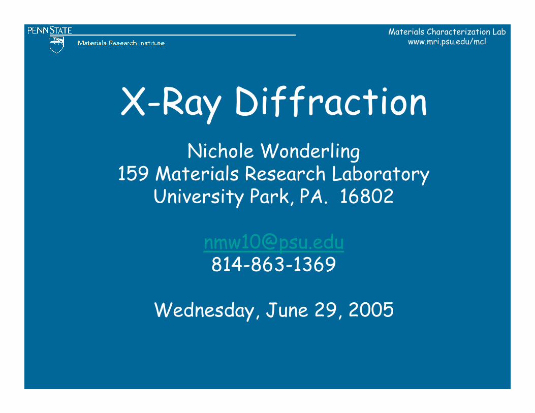
250 MRLAugust 179:45 AMParticle Characterization
114 MRI BldgAugust 249:45 AMX-ray photoelectron spectroscopy (XPS/ESCA)
114 MRI BldgAugust 2411:00 AMAuger Electron Spectroscopy (AES)
541 Deike Bldg.July 279:45 AMChemical analysis (ICP, ICP-MS)
541 Deike Bldg.August 109:45 AMSmall angle x-ray scattering (SAXS)
114 MRI Bldg August 39:45 AMAtomic Force Microscopy (AFM)
250 MRL Bldg.July 209:45 AMOrientation imaging microscopy (OIM/EBSD)
114 MRI BldgJuly 1311:00 AMTEM sample preparation
114 MRI BldgJuly 139:45 AMFocused Ion Beam (FIB)
250 MRL Bldg.July 610:15 AMHigh temperature sintering lab (20 min lecture only)
250 MRL bldg.July 69:45 AMDielectric Characterization (25 min lecture only)
250 MRL Bldg.June 299:45 AMX-ray Diffraction (XRD)
541 Deike Bldg.June 2211:00 AMAnalytical SEM
541 Deike Bldg.June 229:45 AMScanning electron microscopy (SEM)
114 MRI BldgJune 159:45 AMTransmission Electron Microscopy (TEM/STEM)
250 MRL Bldg.June 89:45 AMThermal analysis (TGA, DTA, DSC)
LocationDateTimeTechnique
NOTE LOCATIONS: The MRI Bldg is in the Innovation Park near the Penn Stater Hotel; MRL Bldg. is on Hastings Road.More information: www.mri.psu.edu/mcl
Summer Characterization Open HousesSummer Characterization Open Houses
Materials Characterization Labwww.mri.psu.edu/mcl
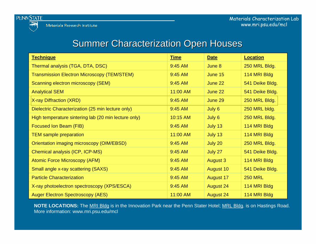
BeaverStadium
Park Ave.
Park Ave.
Porter RoadPollock Road
University Drive
College Ave.
ShortlidgeR
oad North
Bur ro w
esR
oa d
00
00
00
00
00
00
00
00
00
Centre Community
Hospital
E&ES Bldg:SEM
Hosler Bldg:SEM, ESEM, FE-SEM, EPMA, ICP, ICP-MS,BET, SAXS,XRD
MRI Bldg:XPS/ESCA, SIMS, TEM, HR-TEM, FE-Auger, AFM, XRD
Atherton Street
(322 Business)
MRL Bldg:SEM, XRD, OIM, DTA, DSC, TGA, FTIR, AFM, Powder, dielectric, prep, shop, IC, UV-Vis
Hastings Road
Penn StaterHotel
00
Materials Characterization Lab LocationsBldg TelephoneMRL 863-7844MRI 865-0337Hosler 865-1981E&ES 863-4225
Route 322
I-99 00
Steidle Bldg:Nanoindenter
Deike Bldg:
Materials Characterization Labwww.mri.psu.edu/mcl
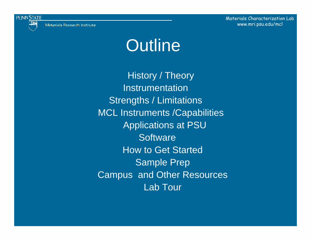
OutlineHistory / Theory
InstrumentationStrengths / Limitations
MCL Instruments /CapabilitiesApplications at PSU
SoftwareHow to Get Started
Sample PrepCampus and Other Resources
Lab Tour

Materials Characterization Labwww.mri.psu.edu/mcl
HISTORY
Materials Characterization Labwww.mri.psu.edu/mcl
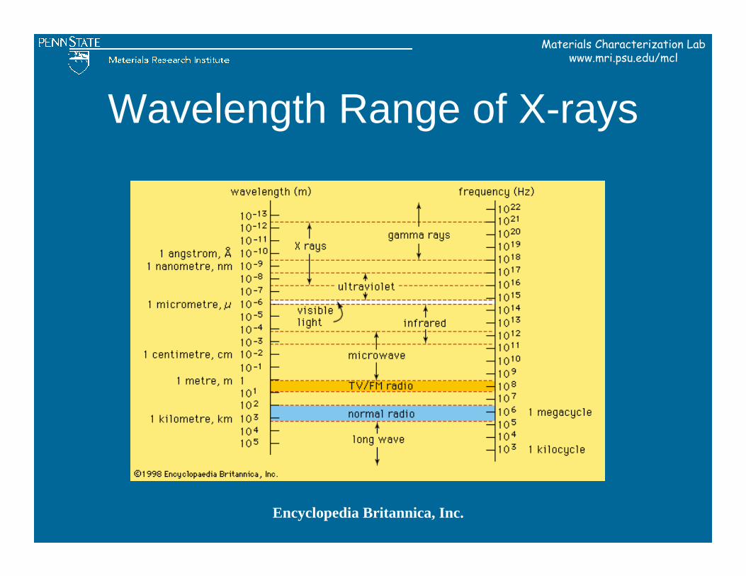
Wavelength Range of X-rays
Encyclopedia Britannica, Inc.
Materials Characterization Labwww.mri.psu.edu/mcl
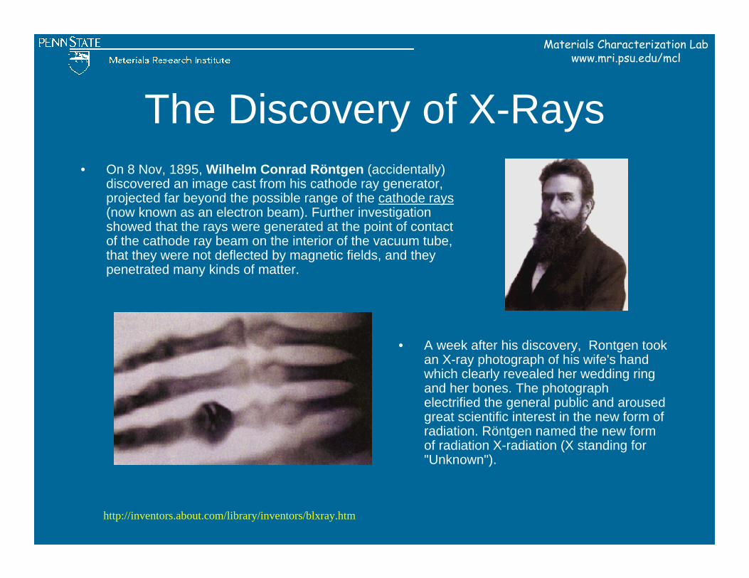
The Discovery of X-Rays• On 8 Nov, 1895, Wilhelm Conrad Röntgen (accidentally)
discovered an image cast from his cathode ray generator, projected far beyond the possible range of the cathode rays(now known as an electron beam). Further investigation showed that the rays were generated at the point of contact of the cathode ray beam on the interior of the vacuum tube, that they were not deflected by magnetic fields, and they penetrated many kinds of matter.
• A week after his discovery, Rontgen took an X-ray photograph of his wife's hand which clearly revealed her wedding ring and her bones. The photograph electrified the general public and aroused great scientific interest in the new form of radiation. Röntgen named the new form of radiation X-radiation (X standing for "Unknown").
http://inventors.about.com/library/inventors/blxray.htm
Materials Characterization Labwww.mri.psu.edu/mcl
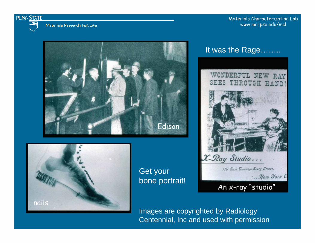
It was the Rage……..
Get your bone portrait!
Images are copyrighted by Radiology Centennial, Inc and used with permission
Edison
nails
An x-ray “studio”
Materials Characterization Labwww.mri.psu.edu/mcl
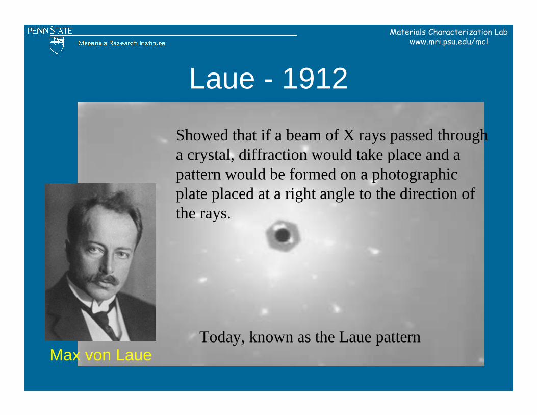
Laue - 1912
Max von Laue
Showed that if a beam of X rays passed through a crystal, diffraction would take place and a pattern would be formed on a photographic plate placed at a right angle to the direction of the rays.
Today, known as the Laue pattern
Materials Characterization Labwww.mri.psu.edu/mcl
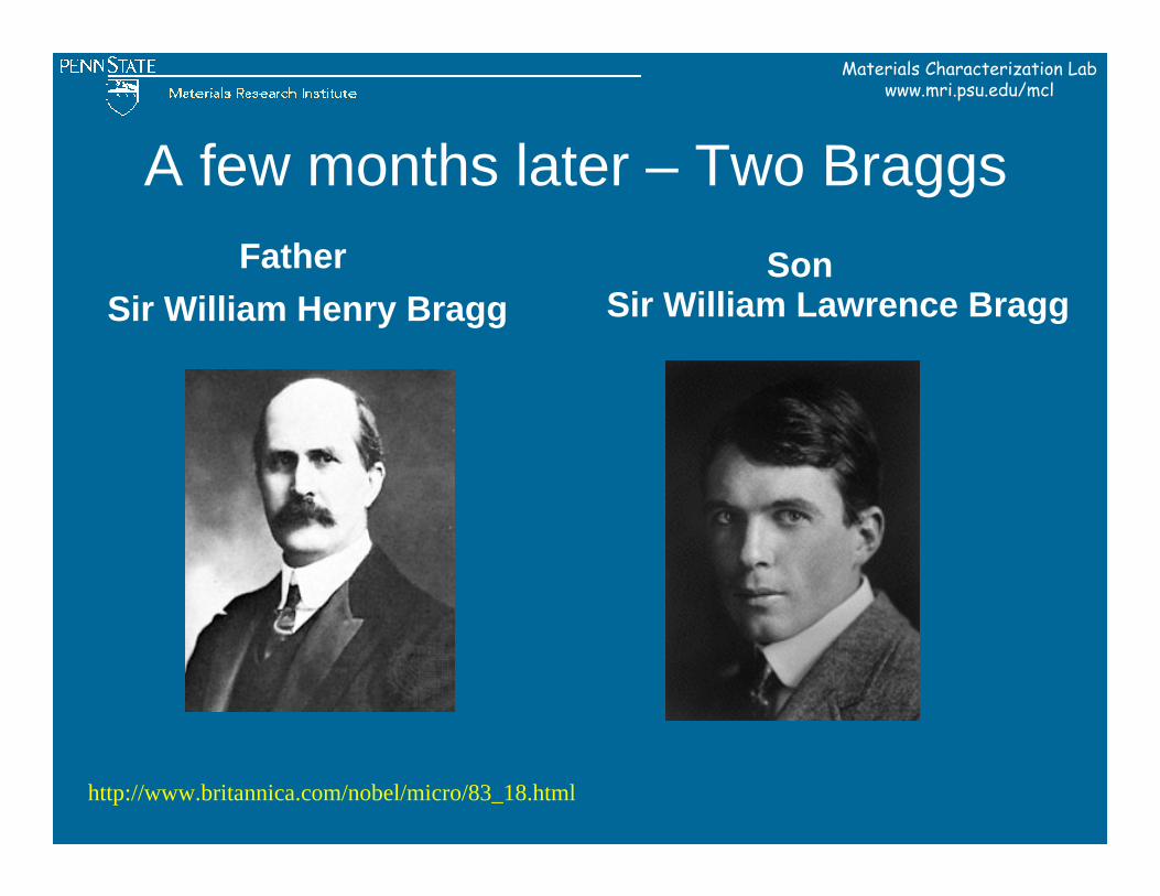
A few months later – Two Braggs
Sir William Henry BraggSonFather
Sir William Lawrence Bragg
http://www.britannica.com/nobel/micro/83_18.html
Materials Characterization Labwww.mri.psu.edu/mcl
THEORY
Materials Characterization Labwww.mri.psu.edu/mcl
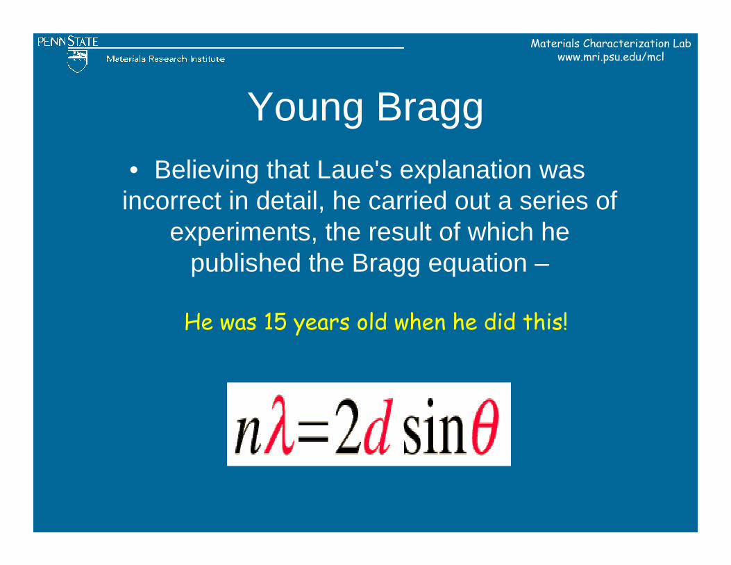
Young Bragg• Believing that Laue's explanation was incorrect in detail, he carried out a series of
experiments, the result of which he published the Bragg equation –
He was 15 years old when he did this!
Materials Characterization Labwww.mri.psu.edu/mcl
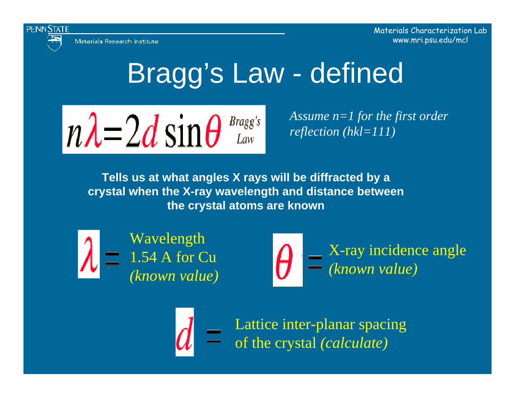
Bragg’s Law - defined
Wavelength1.54 A for Cu(known value)
X-ray incidence angle(known value)
Assume n=1 for the first order reflection (hkl=111)
Lattice inter-planar spacing of the crystal (calculate)
Tells us at what angles X rays will be diffracted by a crystal when the X-ray wavelength and distance between
the crystal atoms are known
Materials Characterization Labwww.mri.psu.edu/mcl
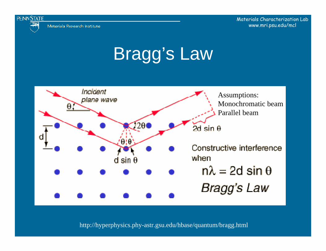
Bragg’s Law
Assumptions:Monochromatic beamParallel beam
http://hyperphysics.phy-astr.gsu.edu/hbase/quantum/bragg.html
Materials Characterization Labwww.mri.psu.edu/mcl
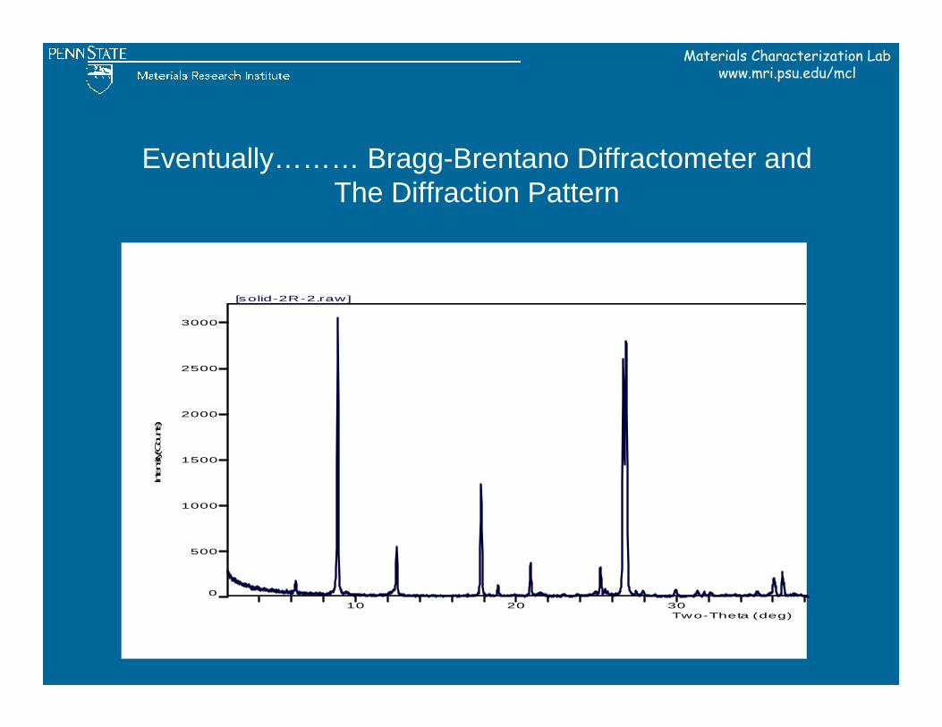
Eventually……… Bragg-Brentano Diffractometer andThe Diffraction Pattern
10 20 30Two-Theta (deg)
0
500
1000
1500
2000
2500
3000
Intensity(C
ounts)
[solid-2R-2.raw]
Materials Characterization Labwww.mri.psu.edu/mcl
Development of Modern Spectrometers
Materials Characterization Labwww.mri.psu.edu/mcl
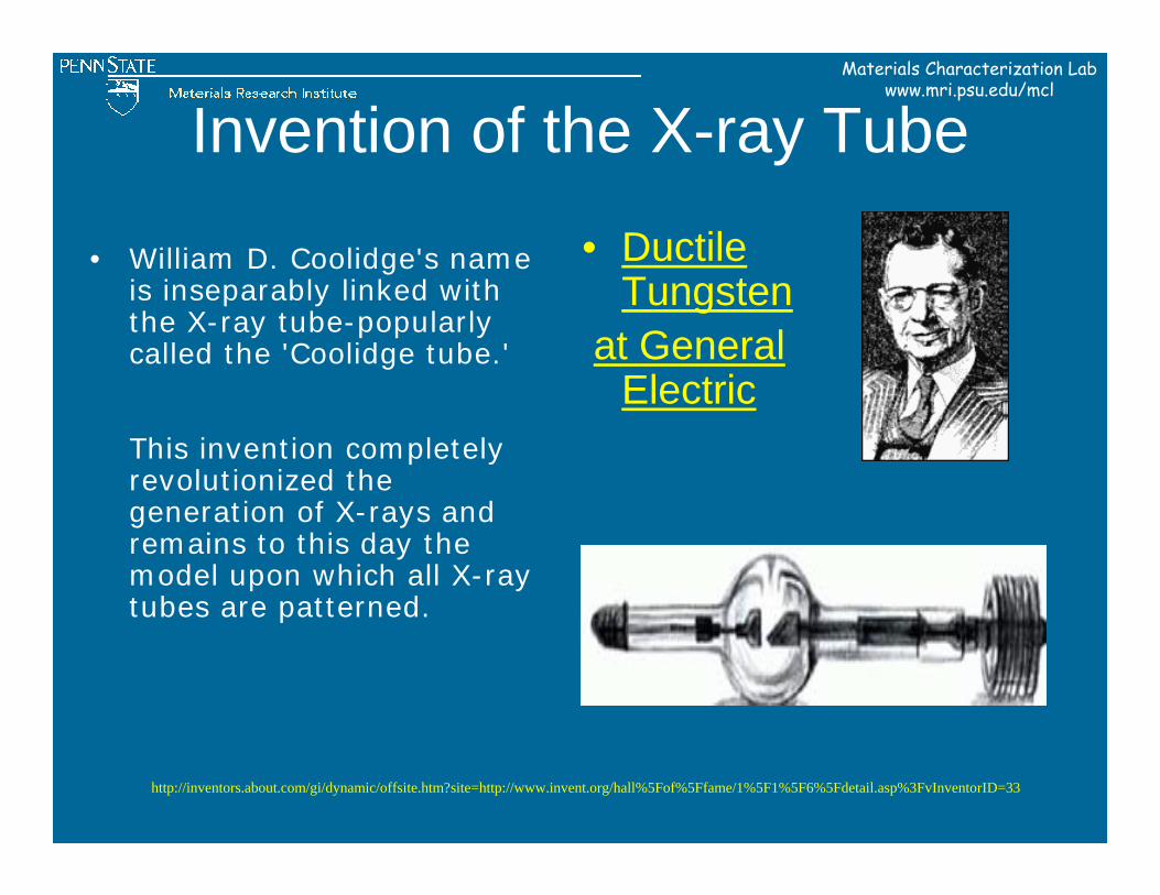
Invention of the X-ray Tube
• William D. Coolidge's name is inseparably linked with the X-ray tube-popularly called the 'Coolidge tube.'
This invention completely revolutionized the generation of X-rays and remains to this day the model upon which all X-ray tubes are patterned.
http://inventors.about.com/gi/dynamic/offsite.htm?site=http://www.invent.org/hall%5Fof%5Ffame/1%5F1%5F6%5Fdetail.asp%3FvInventorID=33
• Ductile Tungsten
at General Electric
Materials Characterization Labwww.mri.psu.edu/mcl
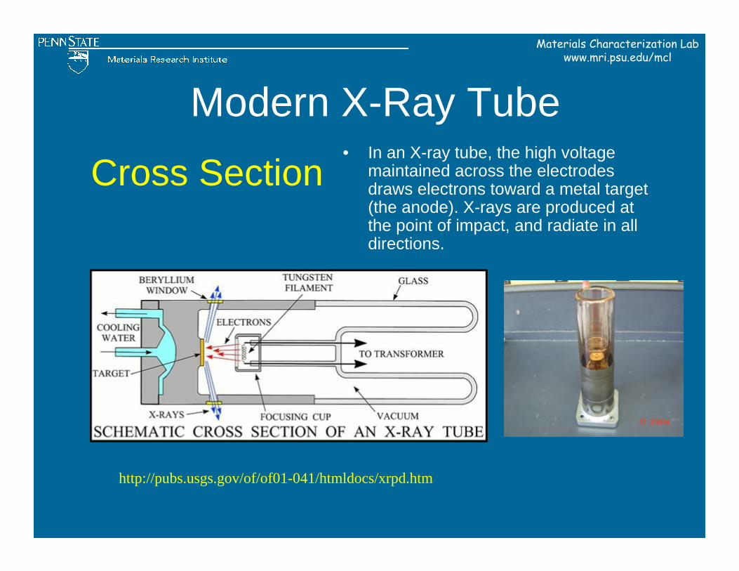
Modern X-Ray Tube
Cross Section• In an X-ray tube, the high voltage
maintained across the electrodes draws electrons toward a metal target (the anode). X-rays are produced at the point of impact, and radiate in all directions.
http://pubs.usgs.gov/of/of01-041/htmldocs/xrpd.htm
Materials Characterization Labwww.mri.psu.edu/mcl
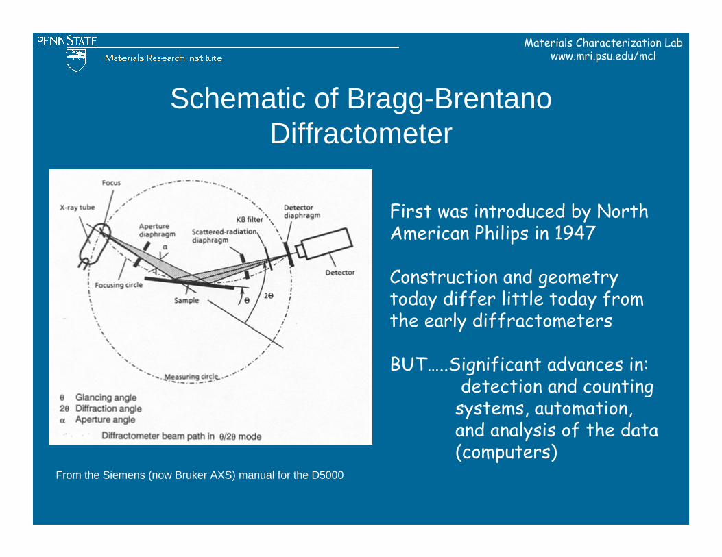
Schematic of Bragg-Brentano Diffractometer
From the Siemens (now Bruker AXS) manual for the D5000
First was introduced by North American Philips in 1947
Construction and geometry today differ little today from the early diffractometers
BUT…..Significant advances in:detection and counting
systems, automation, and analysis of the data (computers)
Materials Characterization Labwww.mri.psu.edu/mcl
Types of X-ray DiffractionInstruments
Materials Characterization Labwww.mri.psu.edu/mcl
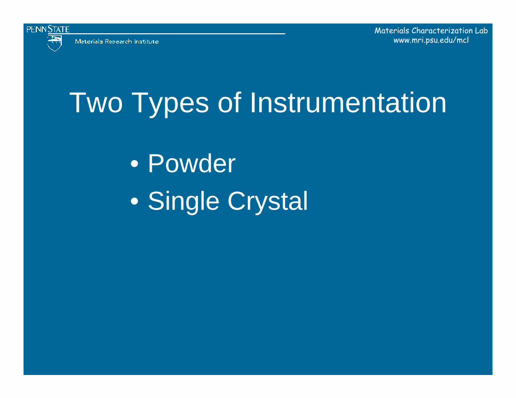
Two Types of Instrumentation
• Powder• Single Crystal
Materials Characterization Labwww.mri.psu.edu/mcl
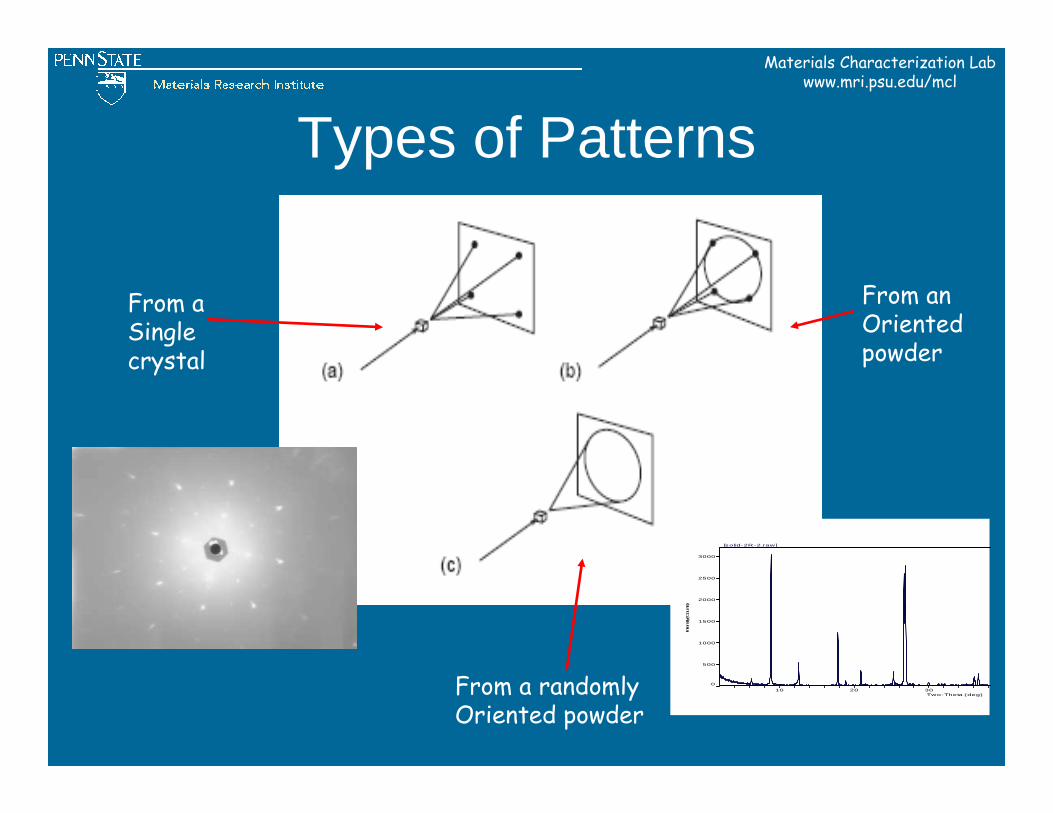
Types of Patterns
From a Singlecrystal
From anOrientedpowder
From a randomly Oriented powder
10 20 30Two-Theta (deg)
0
500
1000
1500
2000
2500
3000
Intens
ity(C
ounts)
[solid-2R-2.raw]
Materials Characterization Labwww.mri.psu.edu/mcl
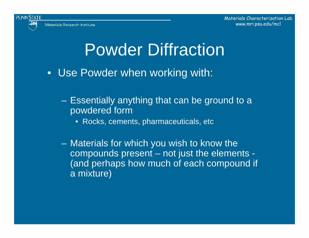
Powder Diffraction• Use Powder when working with:
– Essentially anything that can be ground to a powdered form
• Rocks, cements, pharmaceuticals, etc
– Materials for which you wish to know the compounds present – not just the elements -(and perhaps how much of each compound if a mixture)
Materials Characterization Labwww.mri.psu.edu/mcl
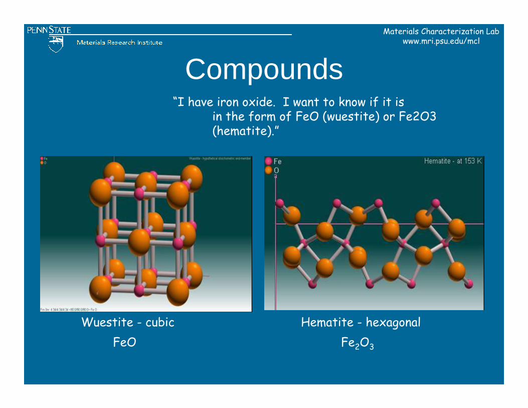
Compounds
Wuestite - cubic Hematite - hexagonalFeO Fe2O3
“I have iron oxide. I want to know if it is in the form of FeO (wuestite) or Fe2O3 (hematite).”
Materials Characterization Labwww.mri.psu.edu/mcl
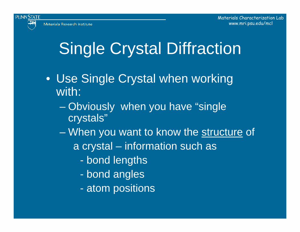
Single Crystal Diffraction
• Use Single Crystal when working with:– Obviously when you have “single
crystals”– When you want to know the structure of
a crystal – information such as- bond lengths- bond angles- atom positions
Materials Characterization Labwww.mri.psu.edu/mcl
Strengths / Limitations of Powder Diffraction
Materials Characterization Labwww.mri.psu.edu/mcl
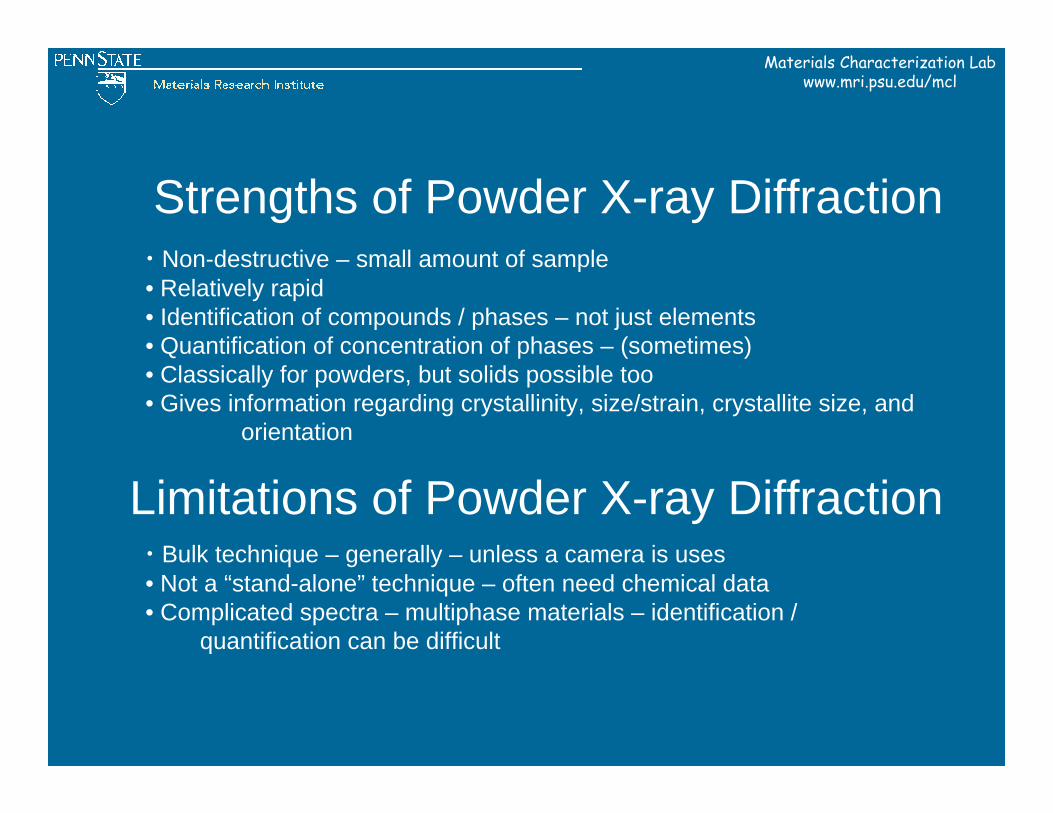
Strengths of Powder X-ray Diffraction• Non-destructive – small amount of sample• Relatively rapid• Identification of compounds / phases – not just elements• Quantification of concentration of phases – (sometimes)• Classically for powders, but solids possible too• Gives information regarding crystallinity, size/strain, crystallite size, and
orientation
Limitations of Powder X-ray Diffraction• Bulk technique – generally – unless a camera is uses• Not a “stand-alone” technique – often need chemical data• Complicated spectra – multiphase materials – identification /
quantification can be difficult
Materials Characterization Labwww.mri.psu.edu/mcl
MCL Instruments / Capabilities
Materials Characterization Labwww.mri.psu.edu/mcl
Powder Diffraction
Materials Characterization Labwww.mri.psu.edu/mcl
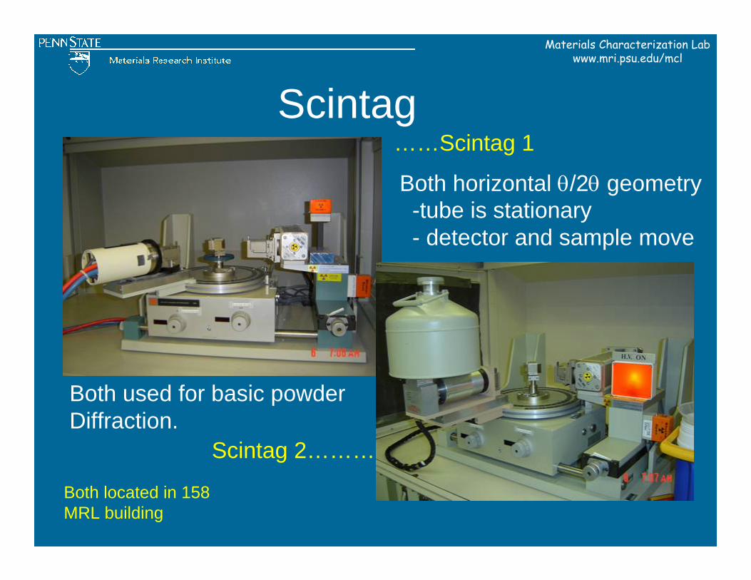
Scintag……Scintag 1
Scintag 2………
Both used for basic powderDiffraction.
Both horizontal θ/2θ geometry-tube is stationary- detector and sample move
Both located in 158 MRL building
Materials Characterization Labwww.mri.psu.edu/mcl
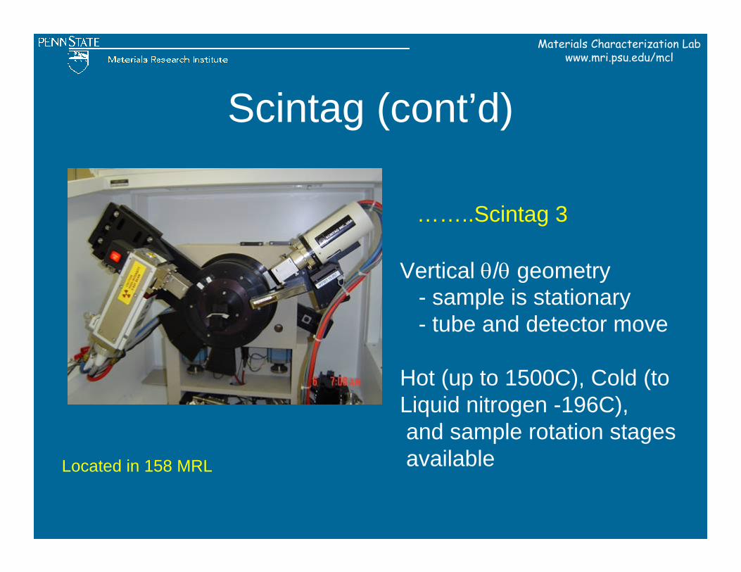
Scintag (cont’d)
……..Scintag 3
Vertical θ/θ geometry- sample is stationary- tube and detector move
Hot (up to 1500C), Cold (toLiquid nitrogen -196C), and sample rotation stagesavailableLocated in 158 MRL
Materials Characterization Labwww.mri.psu.edu/mcl
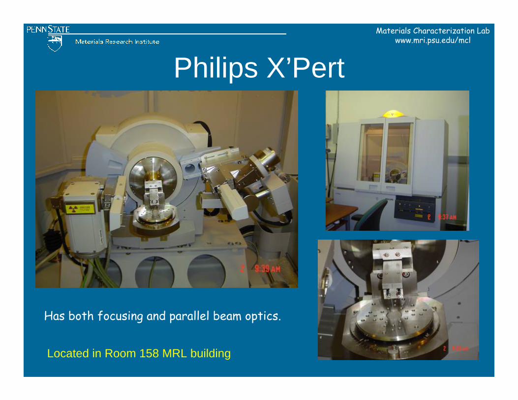
Philips X’Pert
Located in Room 158 MRL building
Has both focusing and parallel beam optics.
Materials Characterization Labwww.mri.psu.edu/mcl
Single Crystal Diffractometers
Materials Characterization Labwww.mri.psu.edu/mcl
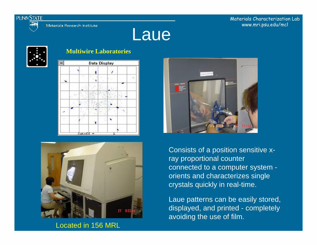
LaueMultiwire Laboratories
Consists of a position sensitive x-ray proportional counter connected to a computer system -orients and characterizes single crystals quickly in real-time.
Laue patterns can be easily stored, displayed, and printed - completely avoiding the use of film.
Located in 156 MRL
Materials Characterization Labwww.mri.psu.edu/mcl
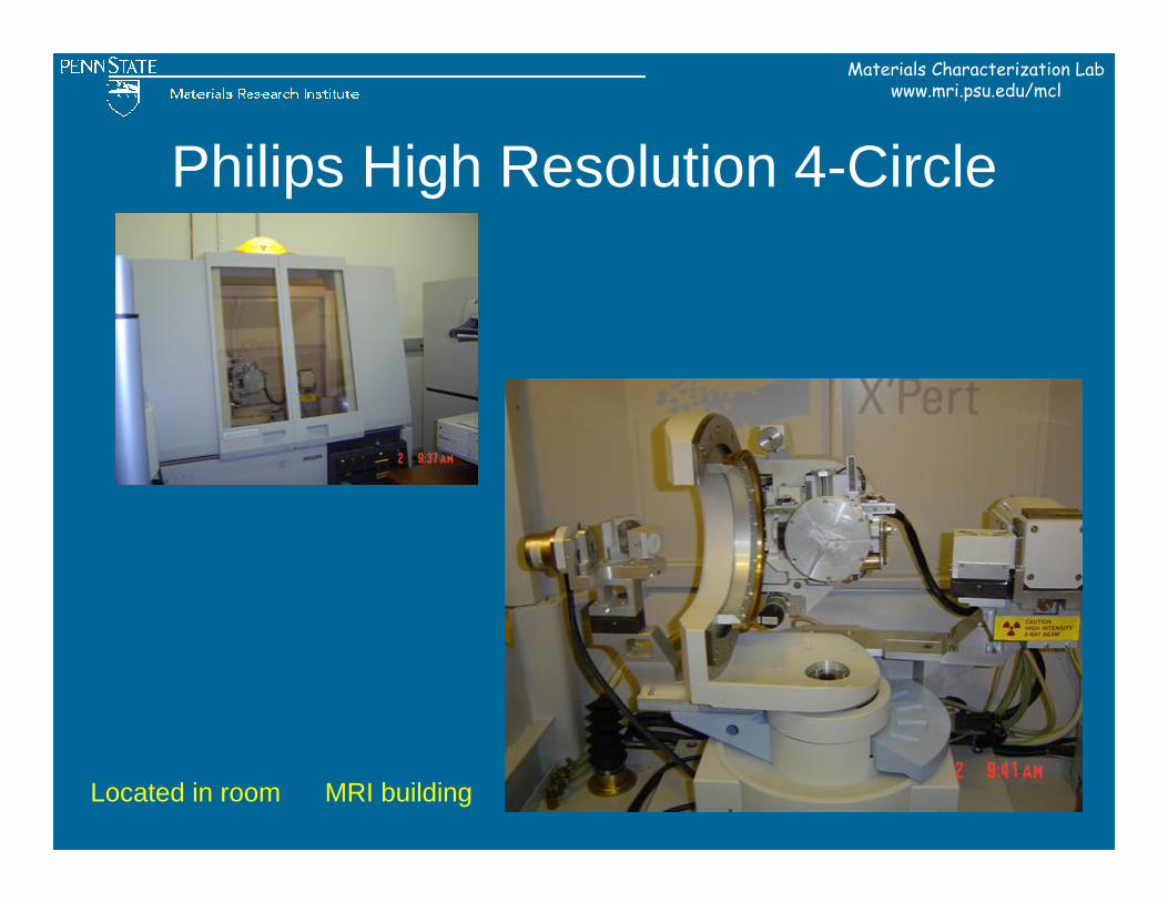
Philips High Resolution 4-Circle
Located in room MRI building
Materials Characterization Labwww.mri.psu.edu/mcl
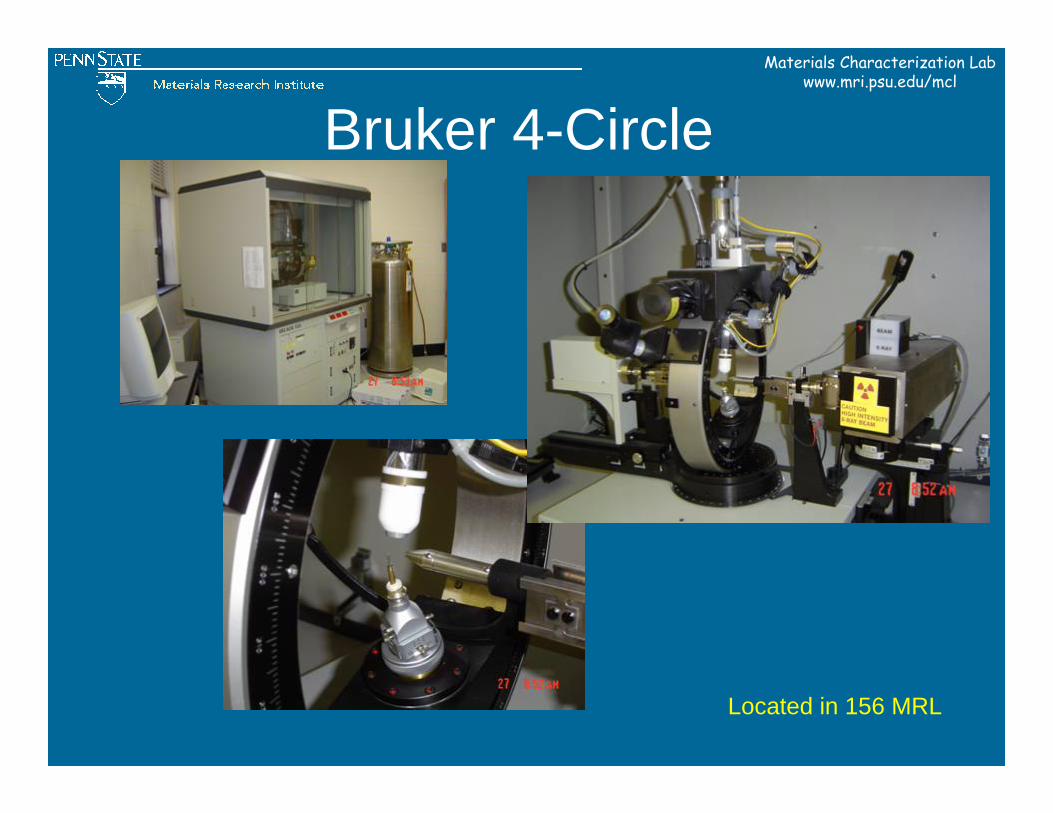
Bruker 4-Circle
Located in 156 MRL
Materials Characterization Labwww.mri.psu.edu/mcl
Applications at PSU
Materials Characterization Labwww.mri.psu.edu/mcl
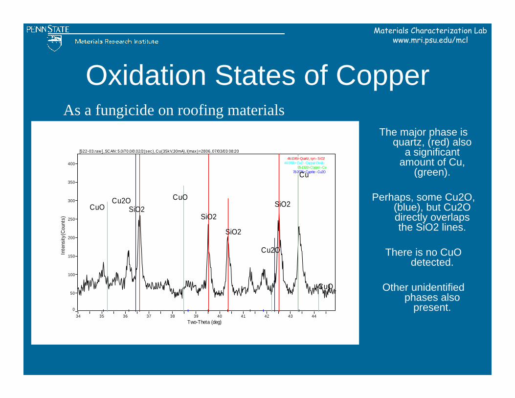
Oxidation States of Copper
The major phase is quartz, (red) also
a significant amount of Cu,
(green).
Perhaps, some Cu2O, (blue), but Cu2O directly overlaps the SiO2 lines.
There is no CuO detected.
Other unidentified phases also
present. 34 35 36 37 38 39 40 41 42 43 44
Two-Theta (deg)
0
50
100
150
200
250
300
350
400
Inte
nsity
(Cou
nts)
[522-03.raw] , SCAN: 5.0/70.0/0.02/2(sec), Cu(35kV,30mA), I(max)=2806, 07/03/03 08:20
46-1045> Quartz, syn - SiO244-0706> CuO - Copper Oxide
85-1326> Copper - Cu78-2076> Cuprite - Cu2O
CuOCuO
CuO
Cu2OSiO2
SiO2
SiO2
Cu2O
SiO2
Cu
As a fungicide on roofing materials
Materials Characterization Labwww.mri.psu.edu/mcl
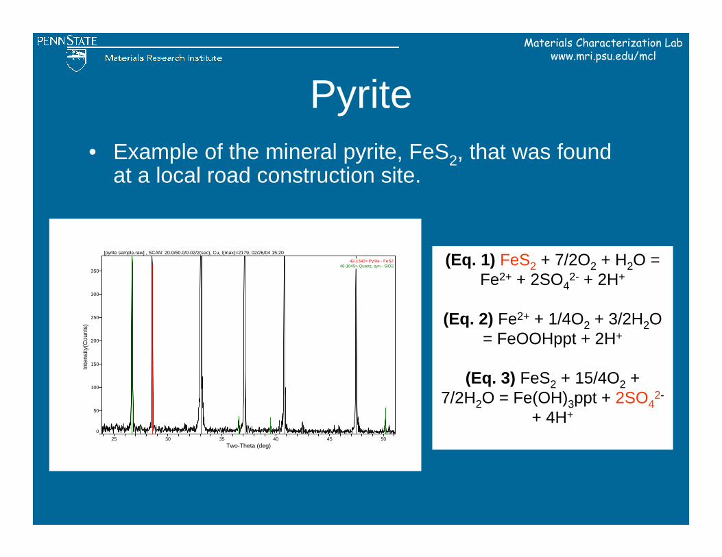
Pyrite• Example of the mineral pyrite, FeS2, that was found
at a local road construction site.
25 30 35 40 45 50Two-Theta (deg)
0
50
100
150
200
250
300
350
Inte
nsity
(Cou
nts)
[pyrite sample.raw] , SCAN: 20.0/60.0/0.02/2(sec), Cu, I(max)=2179, 02/26/04 15:20
42-1340> Pyrite - FeS246-1045> Quartz, syn - SiO2 (Eq. 1) FeS2 + 7/2O2 + H2O =
Fe2+ + 2SO42- + 2H+
(Eq. 2) Fe2+ + 1/4O2 + 3/2H2O = FeOOHppt + 2H+
(Eq. 3) FeS2 + 15/4O2 + 7/2H2O = Fe(OH)3ppt + 2SO4
2-
+ 4H+
Materials Characterization Labwww.mri.psu.edu/mcl
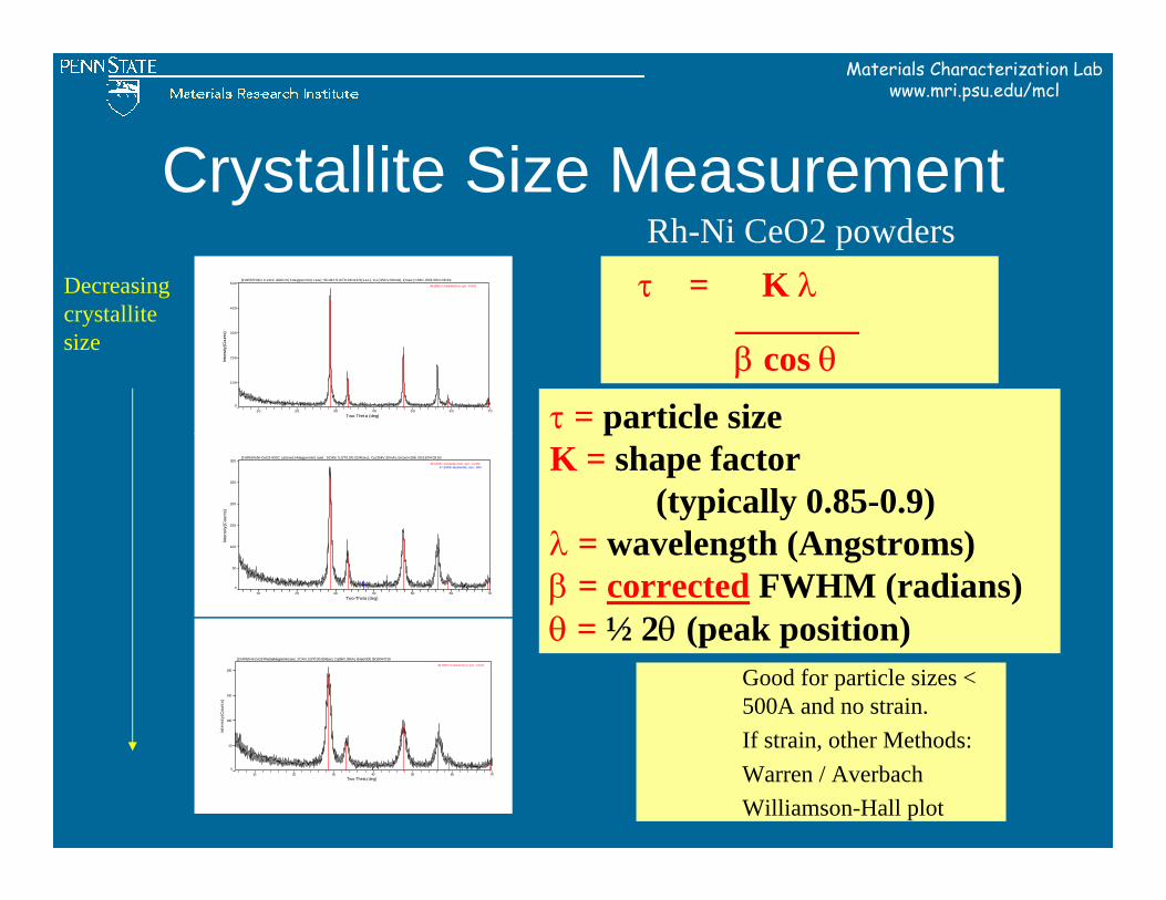
Crystallite Size Measurement
10 20 30 40 50 60 70Two-Theta (deg)
0
50
100
150
200
250
300
Inte
nsity
(Cou
nts)
[1%Rh5%Ni-CeO2-600C calcined (4degpermin).raw] , SCAN: 5.0/70.0/0.02/4(sec), Cu(35kV,30mA), I(max)=288, 05/13/04 09:50
43-1002> Cerianite-(Ce), syn - CeO247-1049> Bunsenite, syn - NiO
10 20 30 40 50 60 70Two-Theta (deg)
0
100
200
300
400
500
Inte
nsity
(Cou
nts)
[1%Rh5%Ni-CeO2-Aldrich(4degpermin).raw] , SCAN: 5.0/70.0/0.02/4(sec), Cu(35kV,30mA), I(max )=480, 05/13/04 08:39
43-1002> Cerianite-(Ce), syn - CeO2
10 20 30 40 50 60 70Two-Theta (deg)
0
50
100
150
200
Inte
nsity
(Cou
nts)
[1%Rh5%Ni-CeO2-Rhodia(4degpermin).raw] , SCAN: 5.0/70.0/0.02/4(sec), Cu(35kV,30mA), I(max)=208, 05/13/04 07:55
43-1002> Cerianite-(Ce), syn - CeO2
Rh-Ni CeO2 powdersτ = K λ
_______β cos θ
τ = particle sizeK = shape factor
(typically 0.85-0.9)λ = wavelength (Angstroms)β = corrected FWHM (radians)θ = ½ 2θ (peak position)
Decreasing crystallite size
Good for particle sizes < 500A and no strain.If strain, other Methods:Warren / AverbachWilliamson-Hall plot
Materials Characterization Labwww.mri.psu.edu/mcl
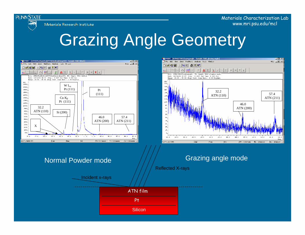
Grazing Angle Geometry
32.2 ATN (110)
46.0 ATN (200)
57.4 ATN (211)
Pt (111)
X
W Lα Pt (111)
Cu Kβ Pt (111)
Si (200)
32.2 ATN (110)
46.0 ATN (200)
57.4 ATN (211)
Normal Powder mode Grazing angle modeReflected X-rays
Incident x-rays
Silicon
Pt
ATN film
Materials Characterization Labwww.mri.psu.edu/mcl
Materials Characterization Labwww.mri.psu.edu/mcl
Software
Materials Characterization Labwww.mri.psu.edu/mcl
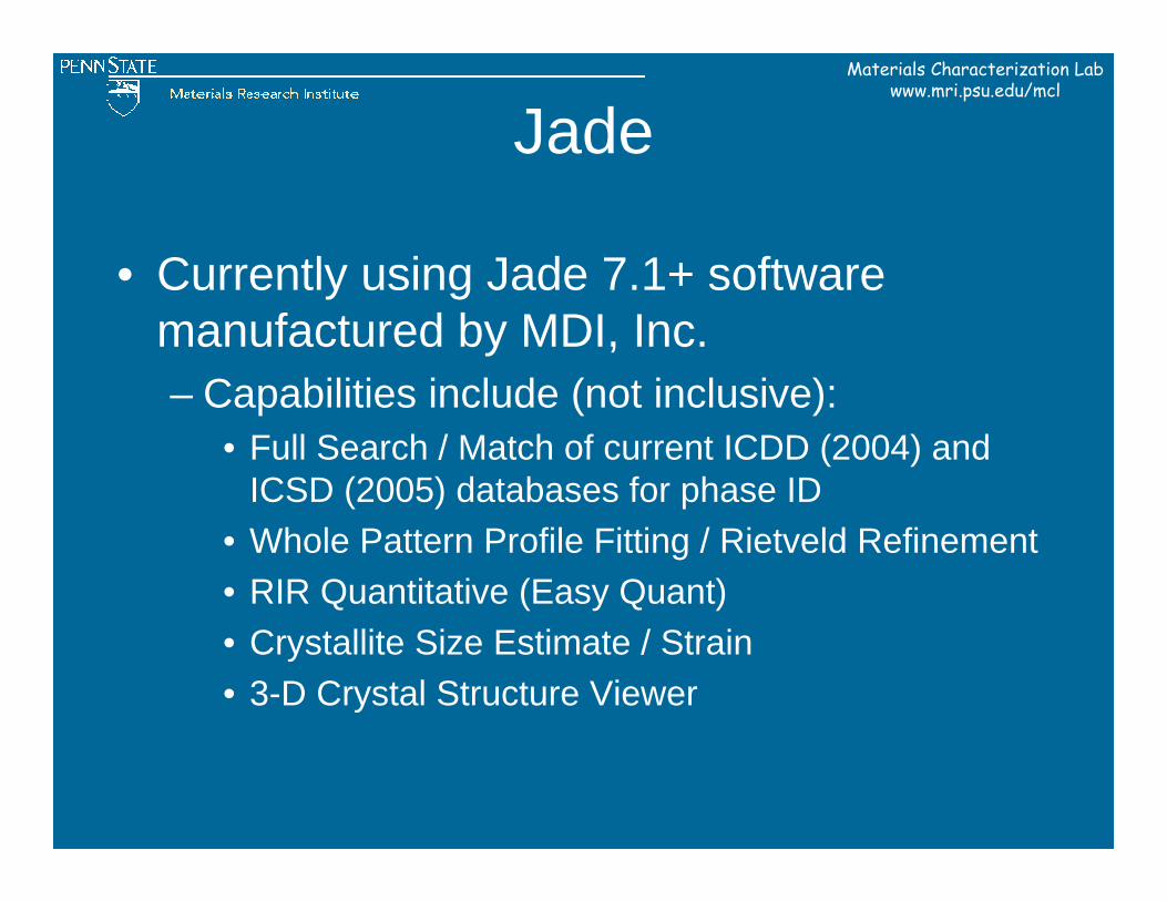
Jade
• Currently using Jade 7.1+ software manufactured by MDI, Inc.– Capabilities include (not inclusive):
• Full Search / Match of current ICDD (2004) and ICSD (2005) databases for phase ID
• Whole Pattern Profile Fitting / Rietveld Refinement• RIR Quantitative (Easy Quant)• Crystallite Size Estimate / Strain• 3-D Crystal Structure Viewer
Materials Characterization Labwww.mri.psu.edu/mcl
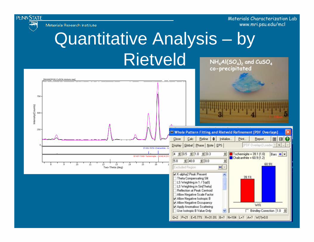
Quantitative Analysis – by Rietveld
0
250
500
750
Inte
nsity
(Cou
nts)
97-001-7679> Chalcanthite - Cu S O4 (H2 O)5
97-007-7246> Tschermigite - (N H4) Al (S O4)2 (H2 O)12
8 9 10 11 12 13 14 15 16 17 18Two-Theta (deg)
[NH4Al(SO4)-CuSO4-mixture.raw]
NH4Al(SO4)2 and CuSO4co-precipitated
Materials Characterization Labwww.mri.psu.edu/mcl
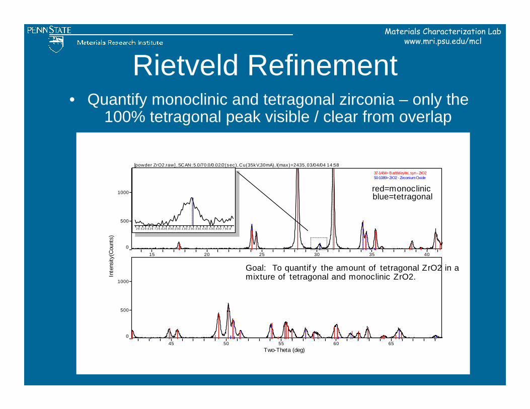
Rietveld Refinement• Quantify monoclinic and tetragonal zirconia – only the
100% tetragonal peak visible / clear from overlap
15 20 25 30 35 400
500
1000
45 50 55 60 65Two-Theta (deg)
0
500
1000
[powder ZrO2.raw] , SCAN: 5.0/70.0/0.02/2(sec), Cu(35kV,30mA), I(max)=2435, 03/04/04 14:58
37-1484> Baddeleyite, syn - ZrO250-1089> ZrO2 - Zirconium Oxide
Inte
nsity
(Cou
nts)
29 .5 29.6 29.7 29.8 29.9 30.0 30.1 30.2 30.3 30.4 30 .5 30.6 30.7 30 .8
mixture of tetragonal and monoclinic ZrO2.Goal: To quantif y the amount of tetragonal ZrO2 in a
red=monoclinicblue=tetragonal
Materials Characterization Labwww.mri.psu.edu/mcl
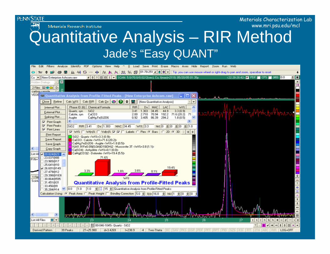
Quantitative Analysis – RIR MethodJade’s “Easy QUANT”
Materials Characterization Labwww.mri.psu.edu/mcl
How to Get Started

Materials Characterization Labwww.mri.psu.edu/mcl
Environmental Health and Safety X-ray safety training
course
http://www.ehs.psu.edu/radprot/x-ray_safety_training.cfm
Materials Characterization Labwww.mri.psu.edu/mcl
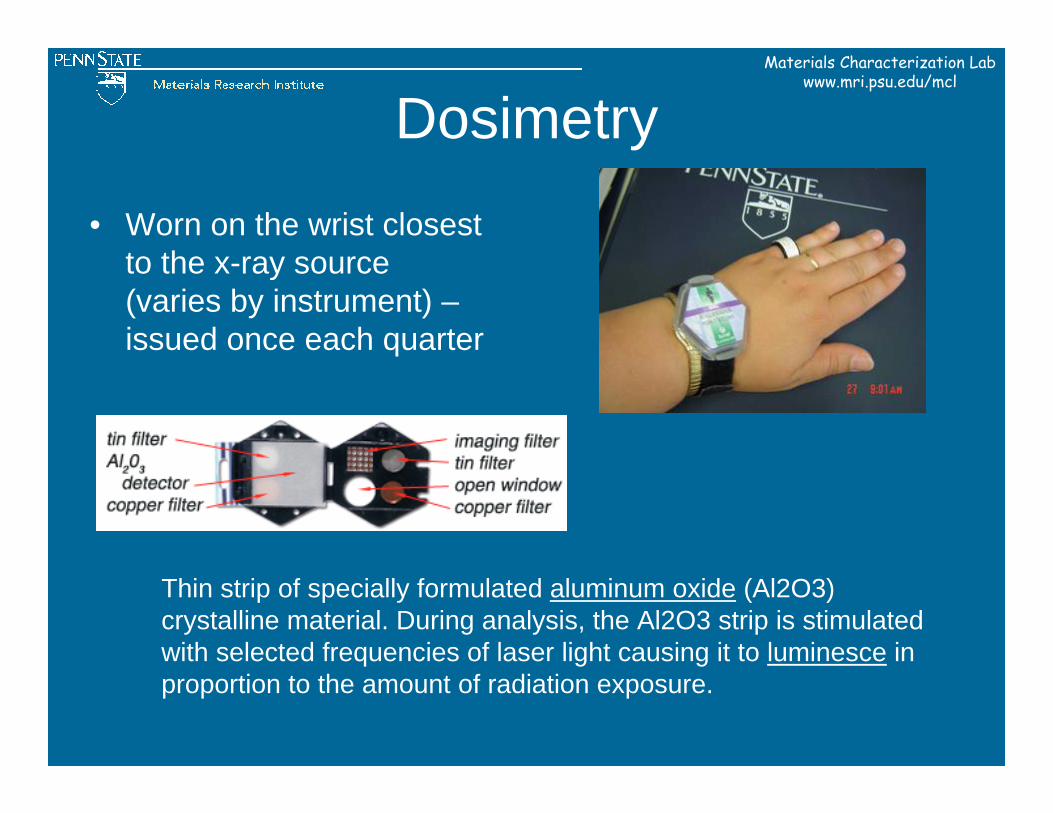
Dosimetry• Worn on the wrist closest
to the x-ray source (varies by instrument) –issued once each quarter
Thin strip of specially formulated aluminum oxide (Al2O3) crystalline material. During analysis, the Al2O3 strip is stimulated with selected frequencies of laser light causing it to luminesce in proportion to the amount of radiation exposure.
Materials Characterization Labwww.mri.psu.edu/mcl
Who Do I Contact to use X-ray Diffraction Equipment at PSU?
Materials Characterization Labwww.mri.psu.edu/mcl
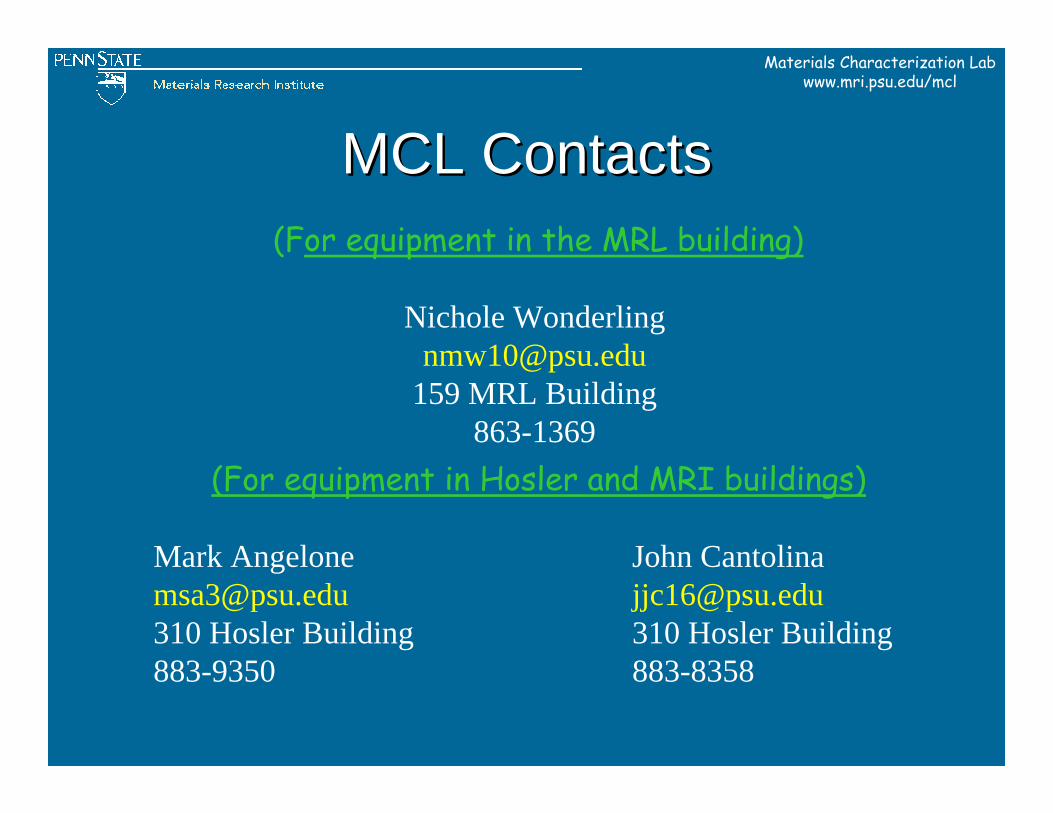
MCL ContactsMCL Contacts
(For equipment in Hosler and MRI buildings)
Mark Angelone John Cantolina [email protected] [email protected] Hosler Building 310 Hosler Building883-9350 883-8358
(For equipment in the MRL building)
Nichole [email protected]
159 MRL Building863-1369
Materials Characterization Labwww.mri.psu.edu/mcl
Sample Preparation
Materials Characterization Labwww.mri.psu.edu/mcl
In Search of the Elusive “Perfect” Powder Sample
• A “Representative” sample
Materials Characterization Labwww.mri.psu.edu/mcl
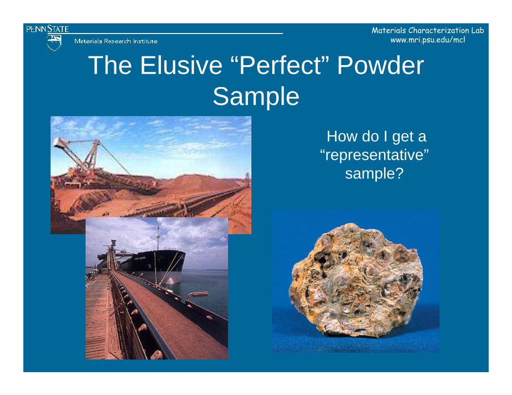
The Elusive “Perfect” Powder Sample
How do I get a “representative”
sample?
Materials Characterization Labwww.mri.psu.edu/mcl
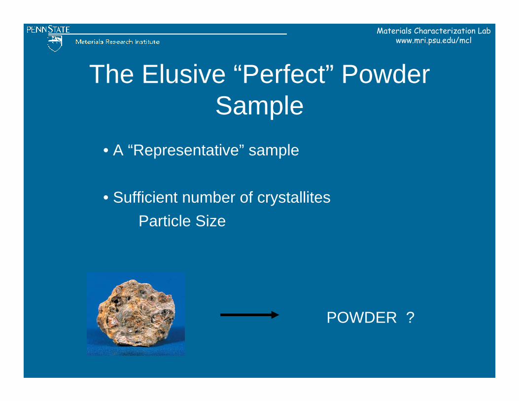
The Elusive “Perfect” Powder Sample
• A “Representative” sample
• Sufficient number of crystallites Particle Size
POWDER ?
Materials Characterization Labwww.mri.psu.edu/mcl
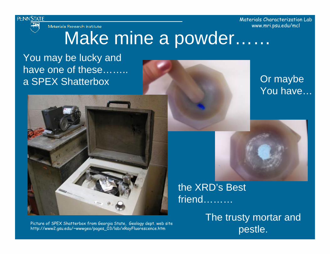
Make mine a powder……
Picture of SPEX Shatterbox from Georgia State, Geology dept. web sitehttp://www2.gsu.edu/~wwwgeo/pages_03/lab/xRayFluorescence.htm
the XRD’s Bestfriend………
You may be lucky andhave one of these……..a SPEX Shatterbox Or maybe
You have…
The trusty mortar and pestle.
Materials Characterization Labwww.mri.psu.edu/mcl
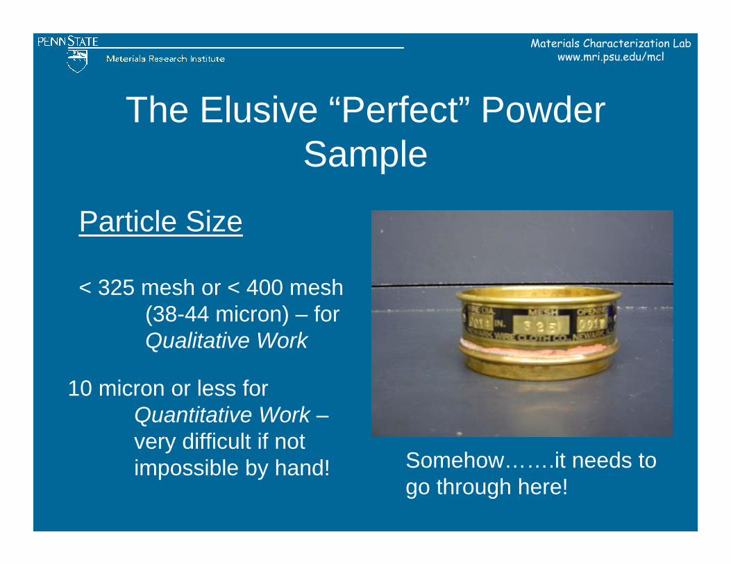
The Elusive “Perfect” Powder Sample
Particle Size
< 325 mesh or < 400 mesh (38-44 micron) – for Qualitative Work
Somehow…….it needs to go through here!
10 micron or less for Quantitative Work –very difficult if notimpossible by hand!
Materials Characterization Labwww.mri.psu.edu/mcl
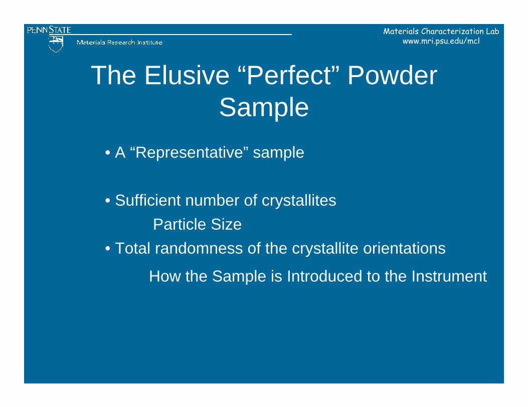
The Elusive “Perfect” Powder Sample
• A “Representative” sample
• Total randomness of the crystallite orientations
• Sufficient number of crystallites Particle Size
How the Sample is Introduced to the Instrument
Materials Characterization Labwww.mri.psu.edu/mcl
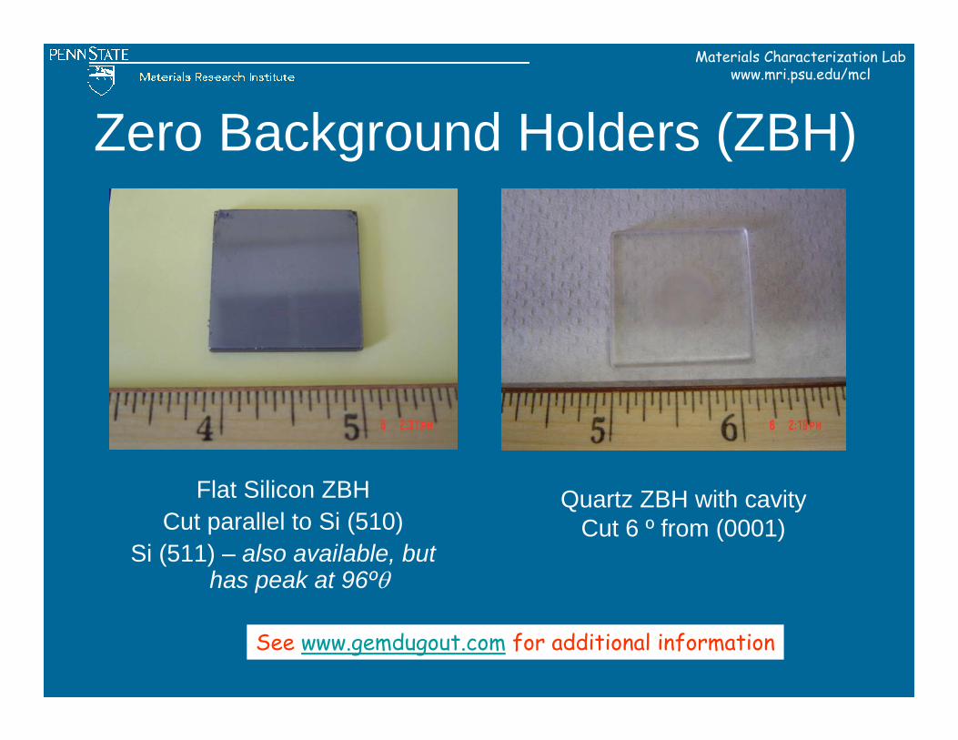
Zero Background Holders (ZBH)
Flat Silicon ZBHCut parallel to Si (510)
Si (511) – also available, but has peak at 96ºθ
Quartz ZBH with cavityCut 6 º from (0001)
See www.gemdugout.com for additional information
Materials Characterization Labwww.mri.psu.edu/mcl
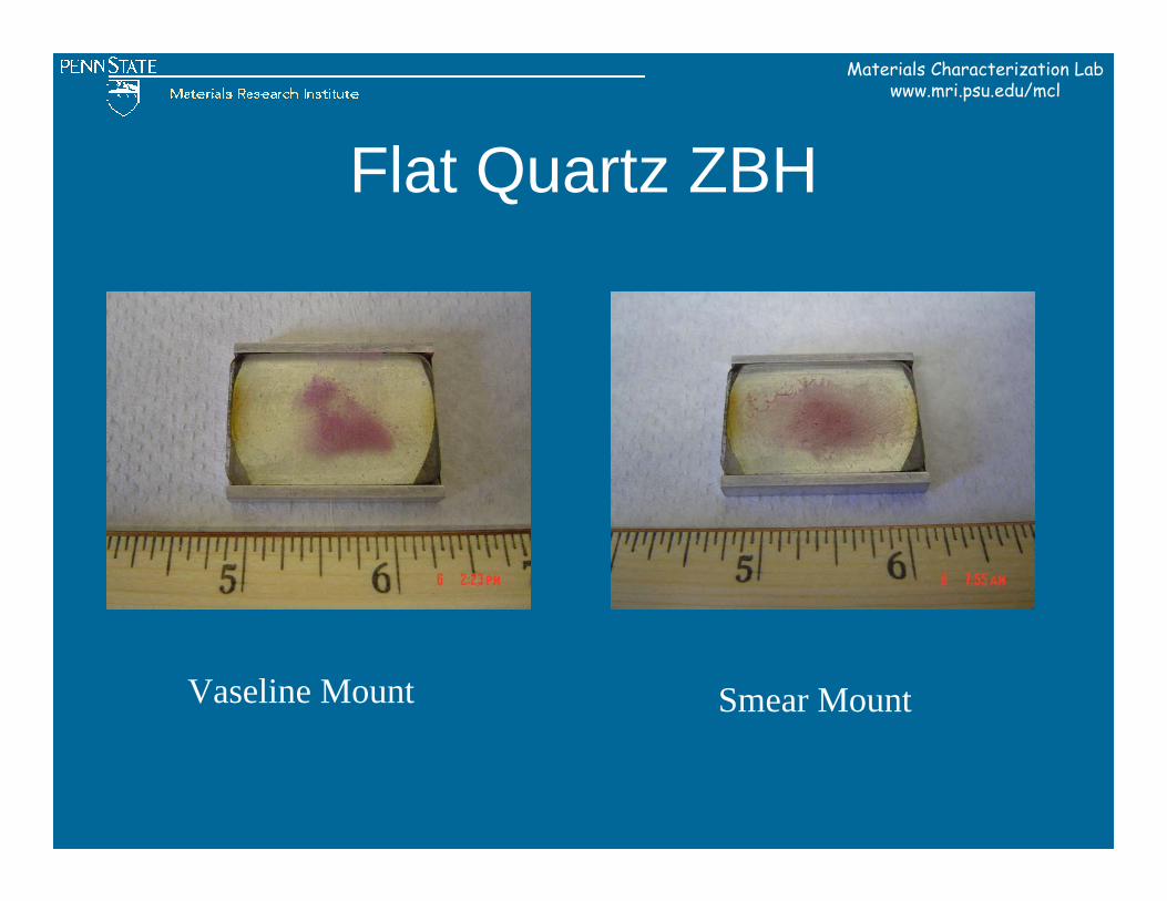
Flat Quartz ZBH
Vaseline Mount Smear Mount
Materials Characterization Labwww.mri.psu.edu/mcl
Back Filled Sample Holder
Materials Characterization Labwww.mri.psu.edu/mcl
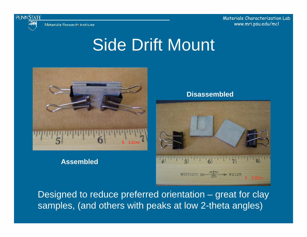
Side Drift Mount
Assembled
Disassembled
Designed to reduce preferred orientation – great for clay samples, (and others with peaks at low 2-theta angles)
Materials Characterization Labwww.mri.psu.edu/mcl
• Sufficient intensity – limit of detection ~5%
The Elusive “Perfect” Powder Sample
• A “Representative” sample
• Total randomness of the crystallite orientations
• Sufficient number of crystallites Particle Size
How the Sample is Introduced to the Instrument
Enough sample area presented to the beam and enough of the phase of interest present
Materials Characterization Labwww.mri.psu.edu/mcl
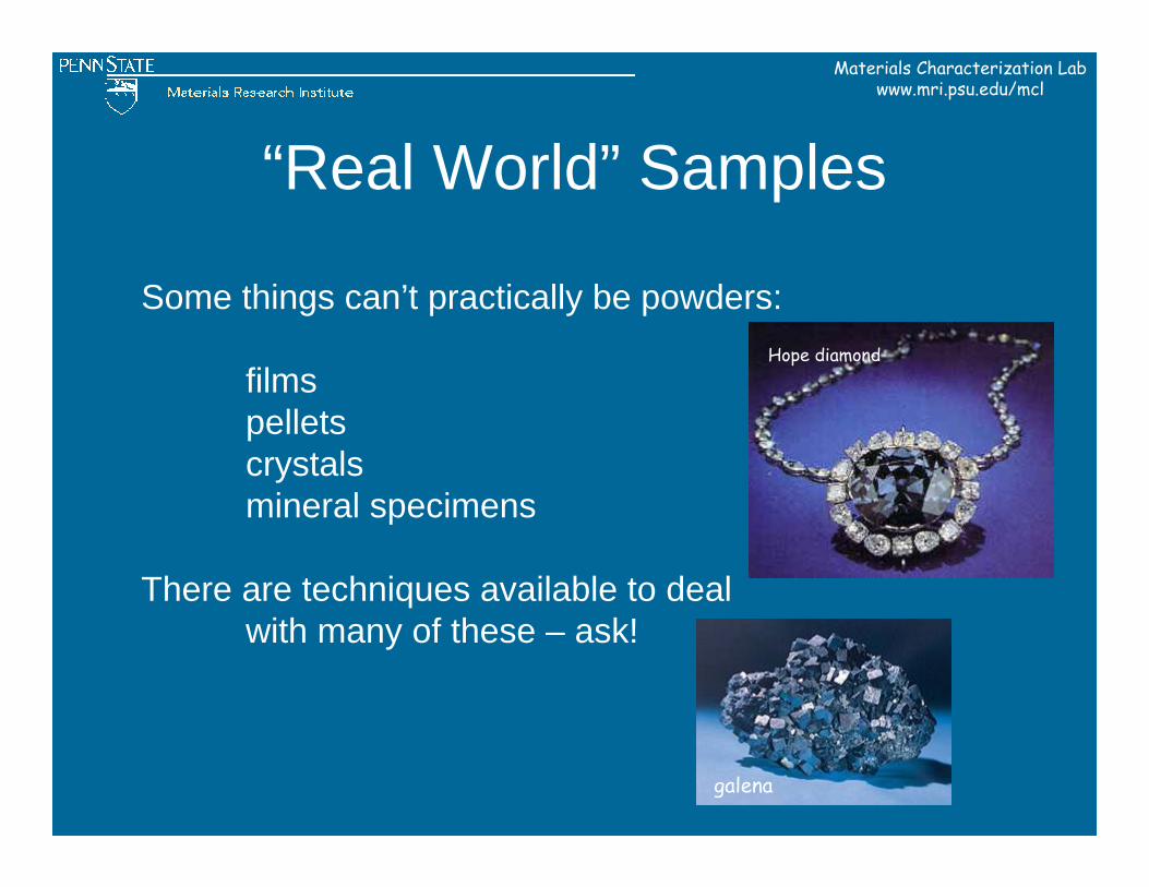
Some things can’t practically be powders:
filmspelletscrystalsmineral specimens
There are techniques available to deal with many of these – ask!
“Real World” Samples
galena
Hope diamond
Materials Characterization Labwww.mri.psu.edu/mcl
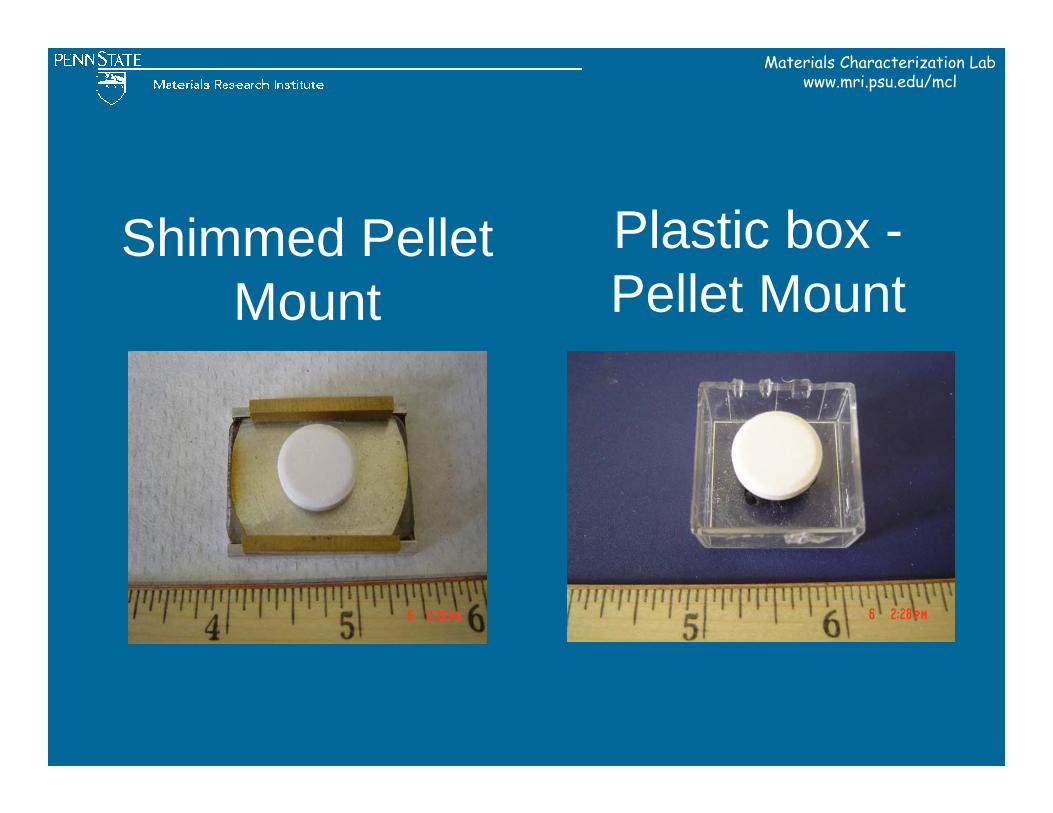
Shimmed Pellet Mount
Plastic box -Pellet Mount
Materials Characterization Labwww.mri.psu.edu/mcl
Campus and Other Resources
Materials Characterization Labwww.mri.psu.edu/mcl
Courses• MATSE 430 Materials Characterization
– Fall semester only, 3 credit course, instructor Elizabeth Dickey
Books“Elements of X-ray Diffraction,” Cullity and Stock
“Introduction to X-ray Powder Diffractometry,” Jenkins and Snyder
“Fundamentals of Powder Diffraction and Structural Characterization of Materials,” Pecharsky and Vitalij
“A Practical Guide for the Preparation of Specimens for X-ray Fluorescence and X-ray Diffraction Analysis,” Buhrke, Jenkins, Smith
Materials Characterization Labwww.mri.psu.edu/mcl
Journals“Powder Diffraction”
“Acta Crystallographica”“Zeitschrift für Kristalographie”
On-linehttp://www.ccp14.ac.uk/
http://www.icdd.com/
Materials Characterization Labwww.mri.psu.edu/mcl
Faculty Experts at PSU
• Elizabeth Dickey, 195 MRI • Peter Heaney, 309 Deike Building• Gerald Johnson, Jr.; Prof. Emeritus, 153 MRL• Earl Ryba, 304B Steidle Building• Barry Scheetz, 107 MRL

Materials Characterization Labwww.mri.psu.edu/mcl
Lab Tour