Embed Size (px)
Citation preview
IAP UG Teaching slides 2015-16
TUBERCULER MENINGITIS
1

IAP UG Teaching slides 2015-16
INTRODUCTION
Tuberculosis • Tuberculosis ‐ major global health problem
• 2nd leading cause of death from an infectious disease worldwide ‐ after HIV
• In 2013– 9.0 million new TB cases– 1.5 million TB deaths
totaldeath
2
IAP UG Teaching slides 2015-16
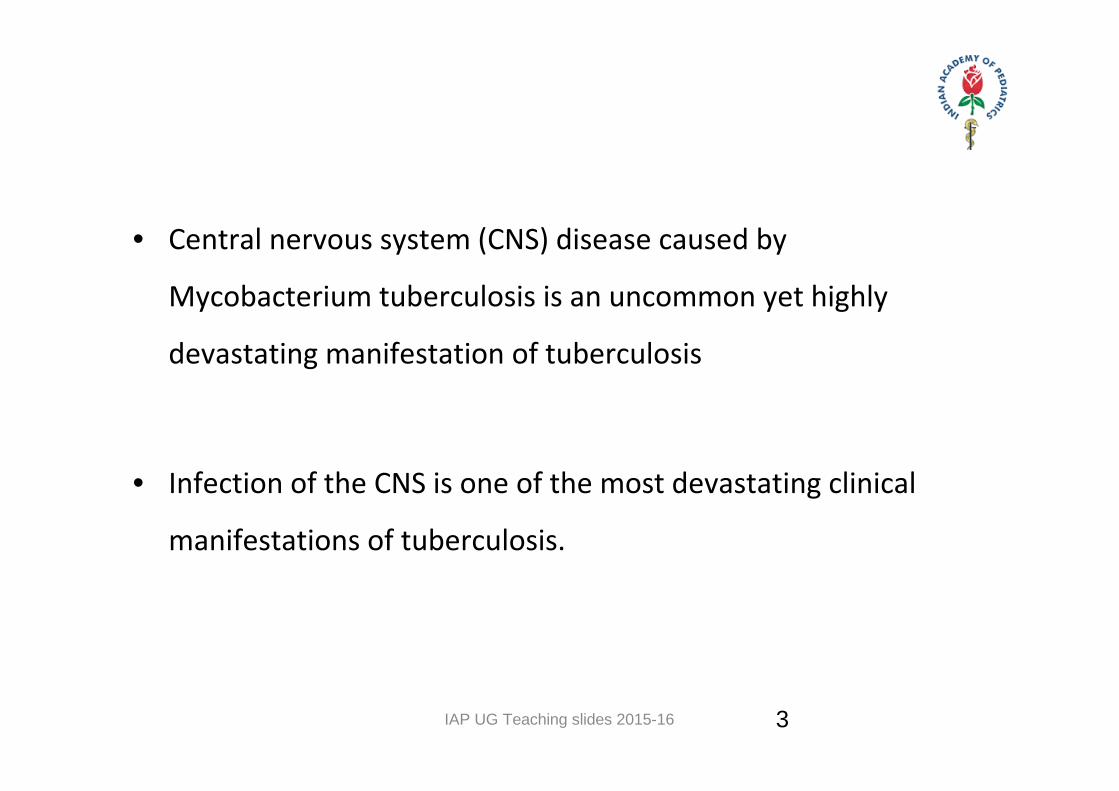
• Central nervous system (CNS) disease caused by
Mycobacterium tuberculosis is an uncommon yet highly
devastating manifestation of tuberculosis
• Infection of the CNS is one of the most devastating clinical
manifestations of tuberculosis.
3
IAP UG Teaching slides 2015-16
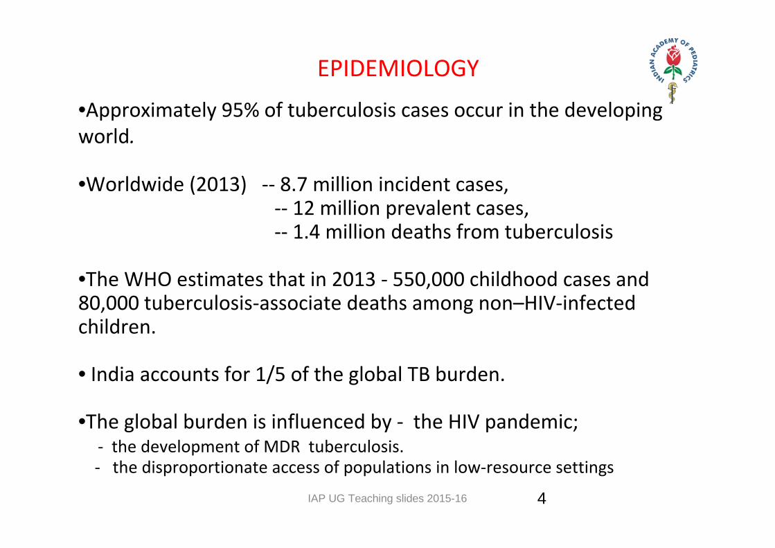
EPIDEMIOLOGY
•Approximately 95% of tuberculosis cases occur in the developing world.
•Worldwide (2013) ‐‐ 8.7 million incident cases, ‐‐ 12 million prevalent cases, ‐‐ 1.4 million deaths from tuberculosis
•The WHO estimates that in 2013 ‐ 550,000 childhood cases and 80,000 tuberculosis‐associate deaths among non–HIV‐infected children.
• India accounts for 1/5 of the global TB burden.
•The global burden is influenced by ‐ the HIV pandemic; ‐ the development of MDR tuberculosis. ‐ the disproportionate access of populations in low‐resource settings
4
IAP UG Teaching slides 2015-16
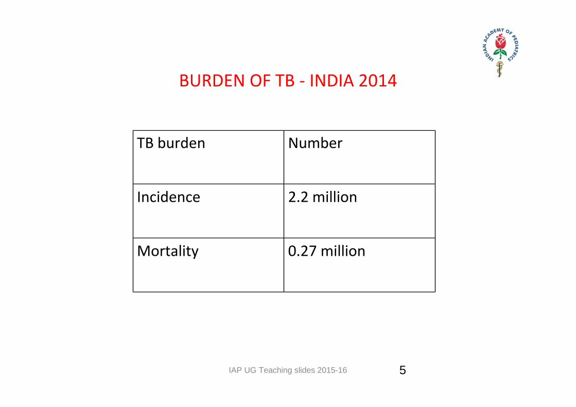
BURDEN OF TB ‐ INDIA 2014
TB burden Number
Incidence 2.2 million
Mortality 0.27 million
5
IAP UG Teaching slides 2015-16
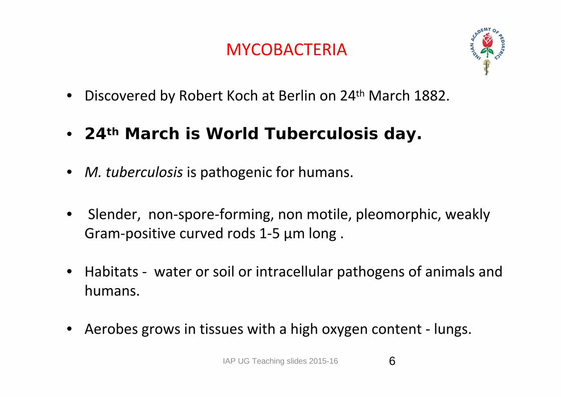
MYCOBACTERIA
• Discovered by Robert Koch at Berlin on 24th March 1882.
• 24th March is World Tuberculosis day.
• M. tuberculosis is pathogenic for humans.
• Slender, non‐spore‐forming, non motile, pleomorphic, weakly Gram‐positive curved rods 1‐5 μm long .
• Habitats ‐ water or soil or intracellular pathogens of animals and humans.
• Aerobes grows in tissues with a high oxygen content ‐ lungs.
6
IAP UG Teaching slides 2015-16
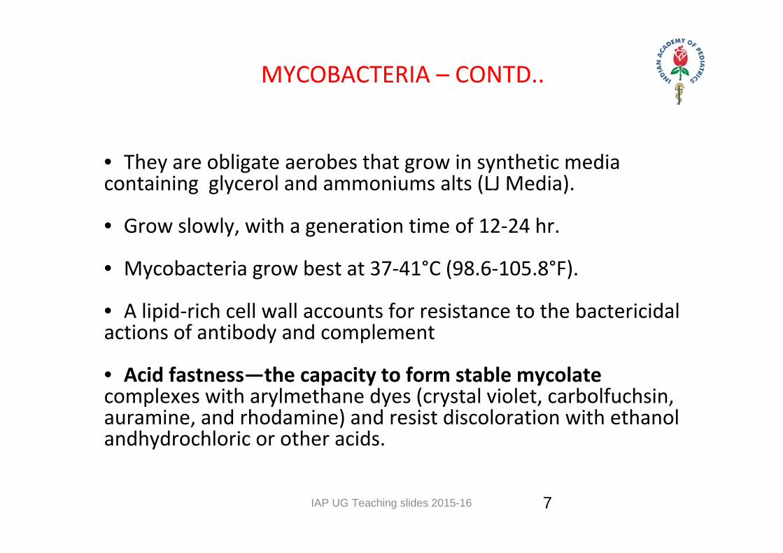
MYCOBACTERIA – CONTD..
• They are obligate aerobes that grow in synthetic media containing glycerol and ammoniums alts (LJ Media). • Grow slowly, with a generation time of 12‐24 hr.
• Mycobacteria grow best at 37‐41°C (98.6‐105.8°F).
• A lipid‐rich cell wall accounts for resistance to the bactericidal actions of antibody and complement
• Acid fastness—the capacity to form stable mycolate complexes with arylmethane dyes (crystal violet, carbolfuchsin, auramine, and rhodamine) and resist discoloration with ethanol andhydrochloric or other acids.
7
IAP UG Teaching slides 2015-16
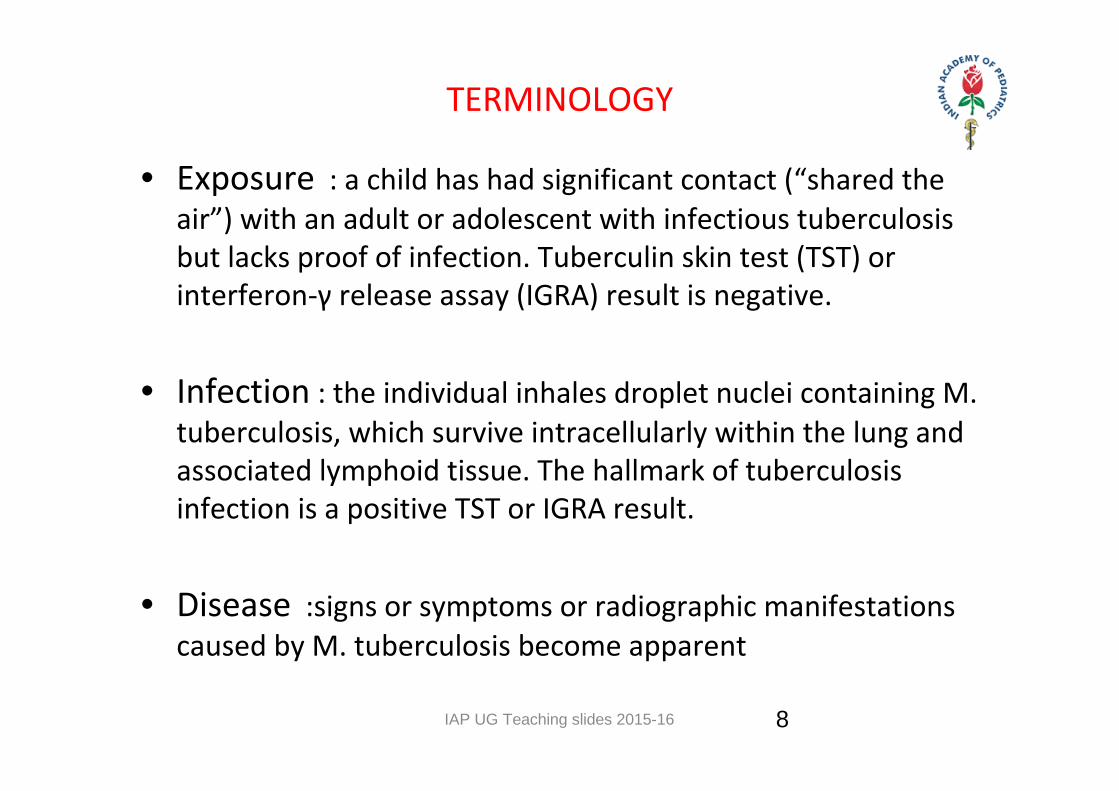
TERMINOLOGY
• Exposure : a child has had significant contact (“shared the air”) with an adult or adolescent with infectious tuberculosis but lacks proof of infection. Tuberculin skin test (TST) or interferon‐γ release assay (IGRA) result is negative.
• Infection : the individual inhales droplet nuclei containing M. tuberculosis, which survive intracellularly within the lung and associated lymphoid tissue. The hallmark of tuberculosis infection is a positive TST or IGRA result.
• Disease :signs or symptoms or radiographic manifestations caused by M. tuberculosis become apparent
8
IAP UG Teaching slides 2015-16
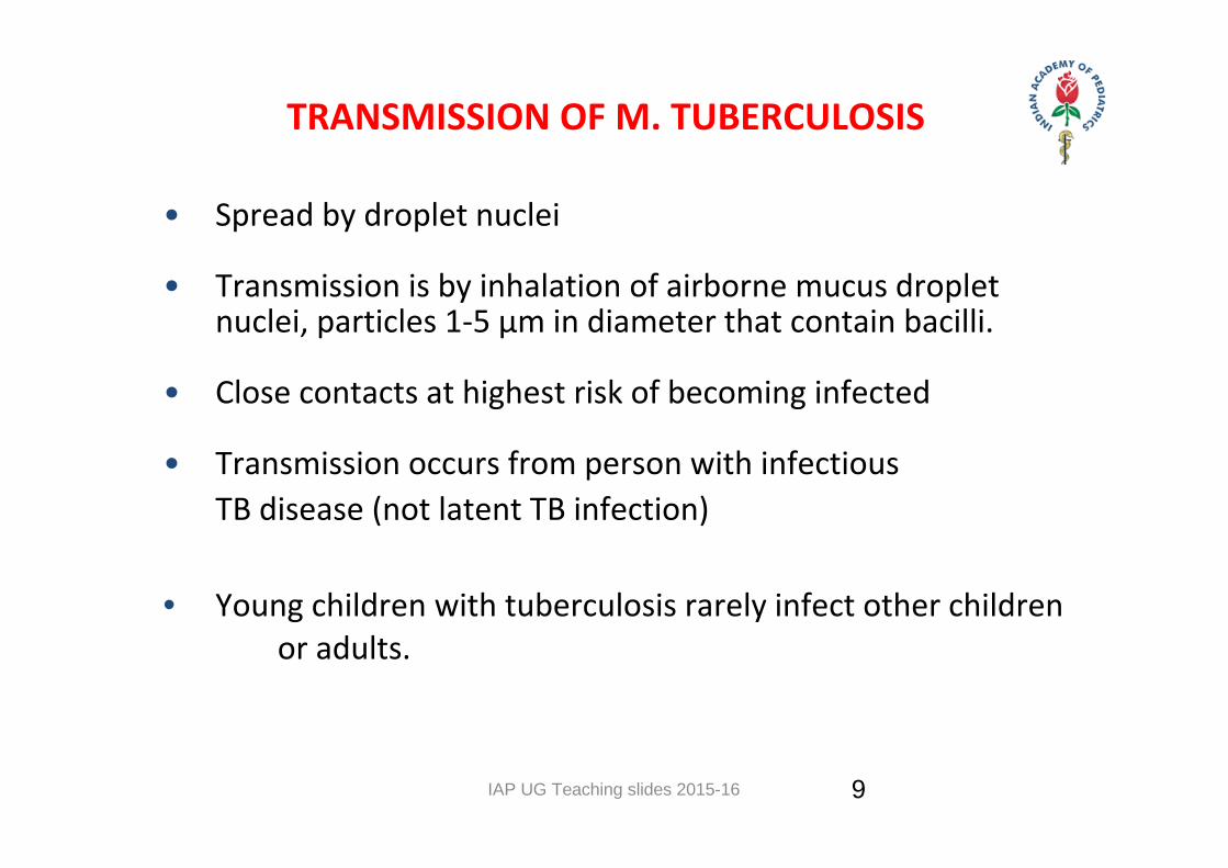
TRANSMISSION OF M. TUBERCULOSIS
• Spread by droplet nuclei
• Transmission is by inhalation of airborne mucus droplet nuclei, particles 1‐5 μm in diameter that contain bacilli.
• Close contacts at highest risk of becoming infected
• Transmission occurs from person with infectious TB disease (not latent TB infection)
• Young children with tuberculosis rarely infect other children or adults.
9
IAP UG Teaching slides 2015-16
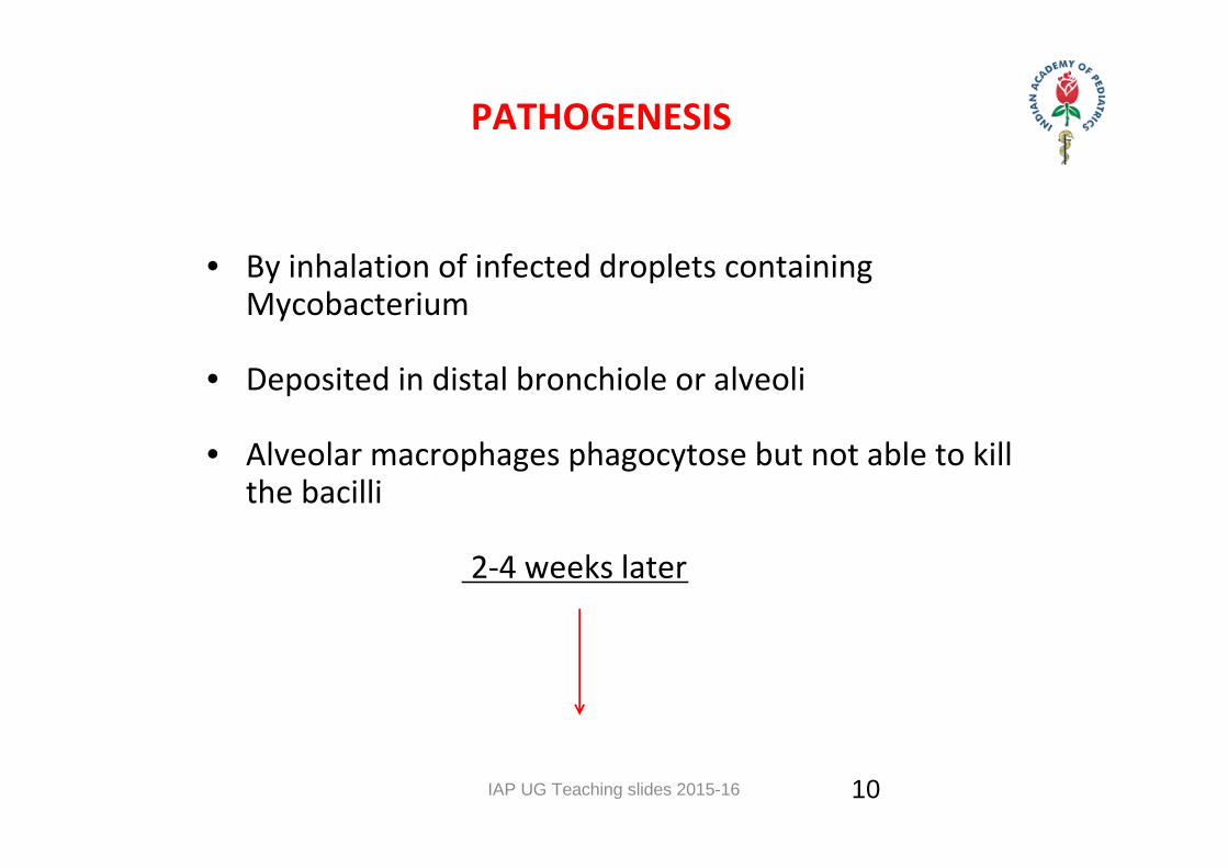
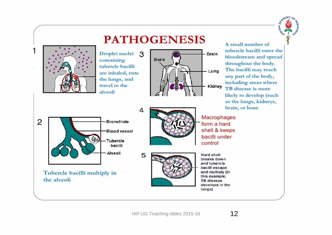
PATHOGENESIS
• By inhalation of infected droplets containing Mycobacterium
• Deposited in distal bronchiole or alveoli
• Alveolar macrophages phagocytose but not able to kill the bacilli
2‐4 weeks later
10
IAP UG Teaching slides 2015-16
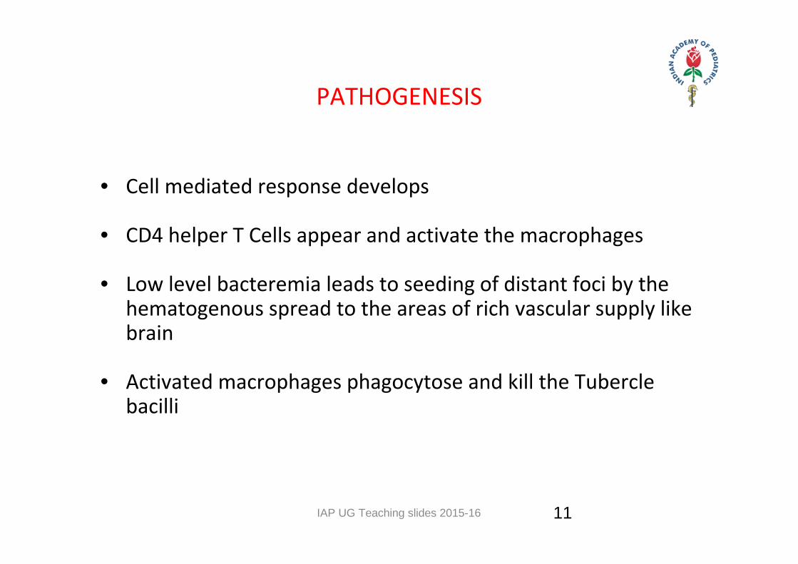
PATHOGENESIS
• Cell mediated response develops
• CD4 helper T Cells appear and activate the macrophages
• Low level bacteremia leads to seeding of distant foci by the hematogenous spread to the areas of rich vascular supply like brain
• Activated macrophages phagocytose and kill the Tubercle bacilli
11
IAP UG Teaching slides 2015-16 12
IAP UG Teaching slides 2015-16
LYMPHO HEMATOGENOUS SPREAD
• CNS involvement in 2‐ 6 months
• Endobronchial TB in 3‐9 months
• Bone and joint lesion after several years
• Renal
13
IAP UG Teaching slides 2015-16
EVOLUTION OF TBM
• Usually as hematogenous spread.
• May also result from direct rupture or extension of a subependymal or subpial focus (Rich focus) and may be located in the meninges, brain or direct extension from cerebrospinal fluid (CSF) infection
• Location of these foci and the capacity to control them ultimately decide the form of CNS Tuberculosis.
14
IAP UG Teaching slides 2015-16
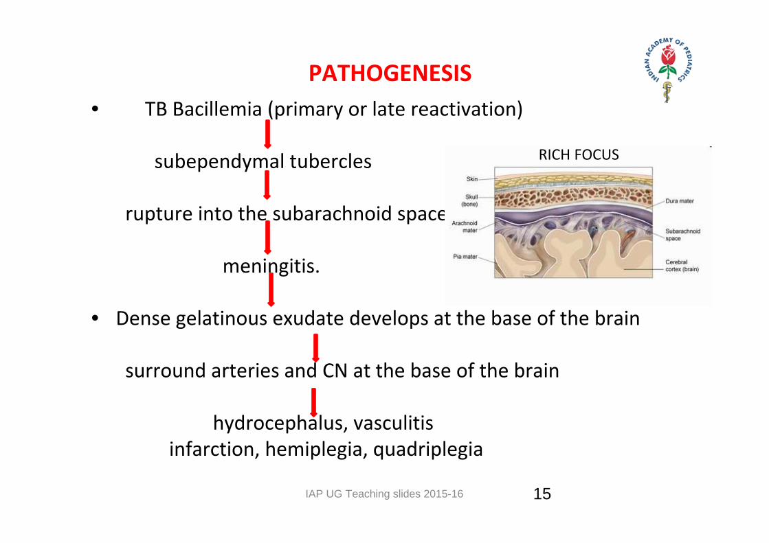
PATHOGENESIS • TB Bacillemia (primary or late reactivation) subependymal tubercles rupture into the subarachnoid space meningitis.
• Dense gelatinous exudate develops at the base of the brain
surround arteries and CN at the base of the brain
hydrocephalus, vasculitis infarction, hemiplegia, quadriplegia
RICH FOCUS
15
IAP UG Teaching slides 2015-16 16neuropathology.neoucom.edu
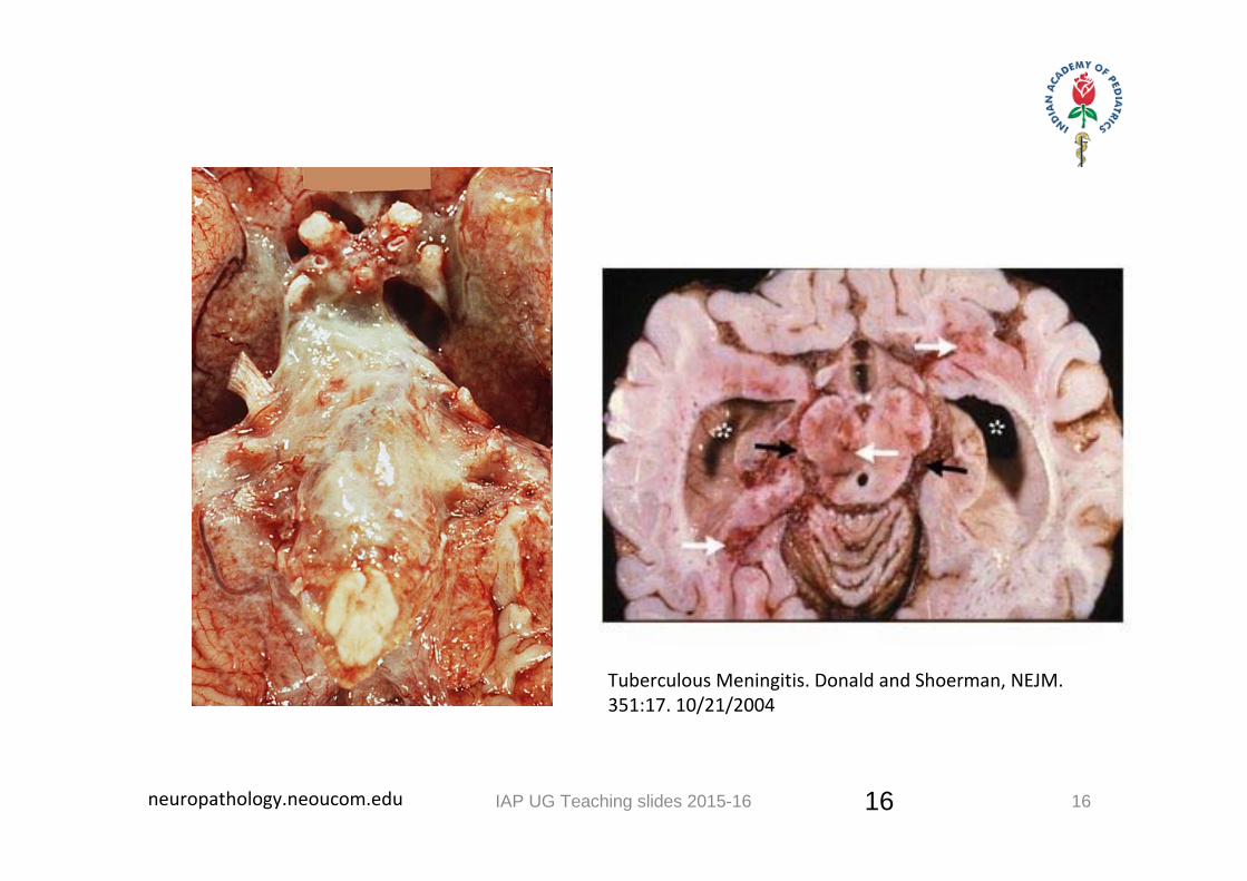
Tuberculous Meningitis. Donald and Shoerman, NEJM. 351:17. 10/21/2004
16
IAP UG Teaching slides 2015-16 17
• Step 1 : Detailed history
• Step 2 : Thorough clinical examination
• Step 3 : Investigation
• Step 4 : Treatment
TB – HOW TO APPROACH
17
IAP UG Teaching slides 2015-16
CLINICAL FEATURES
Always secondary to primary tuberculosis.
•Tuberculous meningitis complicates approximately 0.3% of untreated tuberculosis infections in children.
•Neuro TB occurs in all ages
•Most common between 6 mo‐4 years of age
18
IAP UG Teaching slides 2015-16 19
1ST STAGE ‐ PRODROME STAGE / STAGE OF INVASION
• lasts 1‐2 wk and is characterized by nonspecific symptoms ‐ such as fever, headache, irritability, drowsiness, and malaise. ‐ Anorexia & vomiting may be present. ‐ Child may present with head banging and resents exposure to sunlight. Focal neurologic signs are absent, but infants can experience a
stagnation or loss of developmental milestones.
19
IAP UG Teaching slides 2015-16
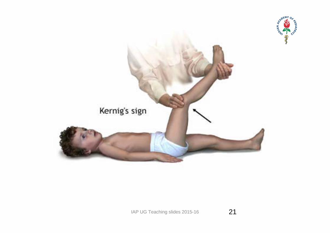
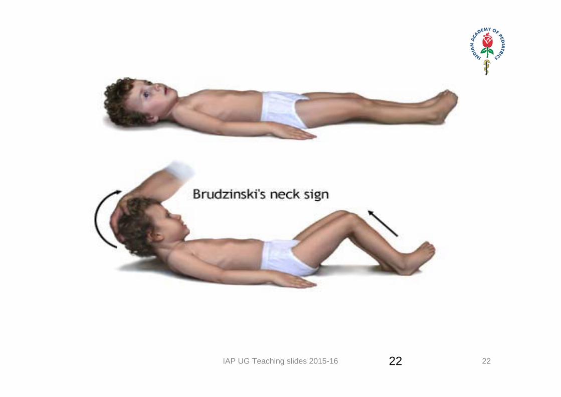
2ND STAGE – STAGE OF MENINGITIS ‐ Begins more abruptly. ‐ The most common features are lethargy, nuchal rigidity,
seizures, positive Kernig and Brudzinski signs, hypertonia, vomiting, cranial nerve palsies, and other focal neurologic signs.
‐ The accelerating clinical illness usually correlates with the development of hydrocephalus, increased intracranial pressure, and vasculitis.
‐ Some children have no evidence of meningeal irritation but can have signs of encephalitis, such as disorientation, movement disorders, or speech impairment.
20
IAP UG Teaching slides 2015-16 21
IAP UG Teaching slides 2015-16 2222
IAP UG Teaching slides 2015-16 23
3RD STAGE – STAGE OF COMA
- Loss of consciousness , rise of temp and altered respiratory pattern
- Pupils are dilated, often unequal with nystagmus and squint.- Ptosis and ophthalmoplegia .- Progression of disease ‐‐ coma deepens , episodic
decerebration , Chyne Stokes breathing , bradycardia and eventually death
23
IAP UG Teaching slides 2015-16
DIAGNOSIS
• Mainly by indirect evidences due to
– Paucibacillary nature of the infection in children
– Requirement of sophisticated instruments
– More false positive results
– Cost factor
24
IAP UG Teaching slides 2015-16
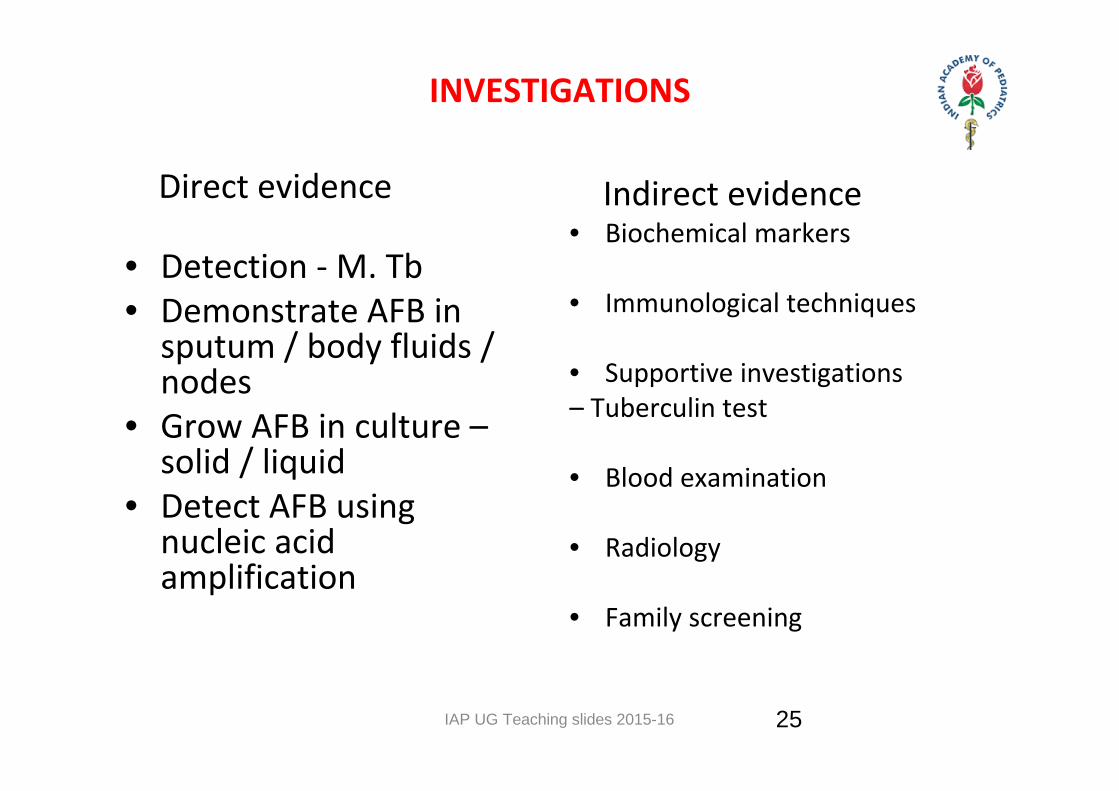
INVESTIGATIONS
Direct evidence
• Detection ‐ M. Tb • Demonstrate AFB in sputum / body fluids / nodes
• Grow AFB in culture – solid / liquid
• Detect AFB using nucleic acid amplification
Indirect evidence• Biochemical markers
• Immunological techniques
• Supportive investigations – Tuberculin test
• Blood examination
• Radiology
• Family screening
25
IAP UG Teaching slides 2015-16
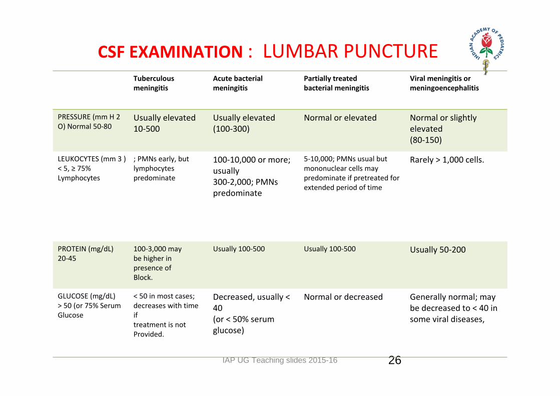
CSF EXAMINATION : LUMBAR PUNCTURE
26
Tuberculous meningitis
Acute bacterialmeningitis
Partially treatedbacterial meningitis
Viral meningitis ormeningoencephalitis
PRESSURE (mm H 2 O) Normal 50‐80
Usually elevated 10‐500
Usually elevated(100‐300)
Normal or elevated Normal or slightlyelevated(80‐150)
LEUKOCYTES (mm 3 ) < 5, ≥ 75% Lymphocytes
; PMNs early, butlymphocytes predominate
100‐10,000 or more; usually300‐2,000; PMNs predominate
5‐10,000; PMNs usual butmononuclear cells maypredominate if pretreated forextended period of time
Rarely > 1,000 cells.
PROTEIN (mg/dL) 20‐45
100‐3,000 maybe higher inpresence ofBlock.
Usually 100‐500 Usually 100‐500 Usually 50‐200
GLUCOSE (mg/dL) > 50 (or 75% Serum Glucose
< 50 in most cases;decreases with time iftreatment is notProvided.
Decreased, usually < 40(or < 50% serumglucose)
Normal or decreased Generally normal; may be decreased to < 40 in some viral diseases,
IAP UG Teaching slides 2015-16 27
CONTRAINDICATION FOR LP
• Increase intracranial pressure.
• Unstable patient.
• Skin infection at site of LP.
• Thrombocytopenia.
• Papilledema.
27
IAP UG Teaching slides 2015-16 28
AFB
• ZN tech ‐ AFB + if > 10,000 bacilli /ml.
• Rapid detection (< 1 hr.)
• Low cost
• High operator dependence
• Labor intensive
• Do not differentiate live/ dead AFB/ NTM
28
IAP UG Teaching slides 2015-16
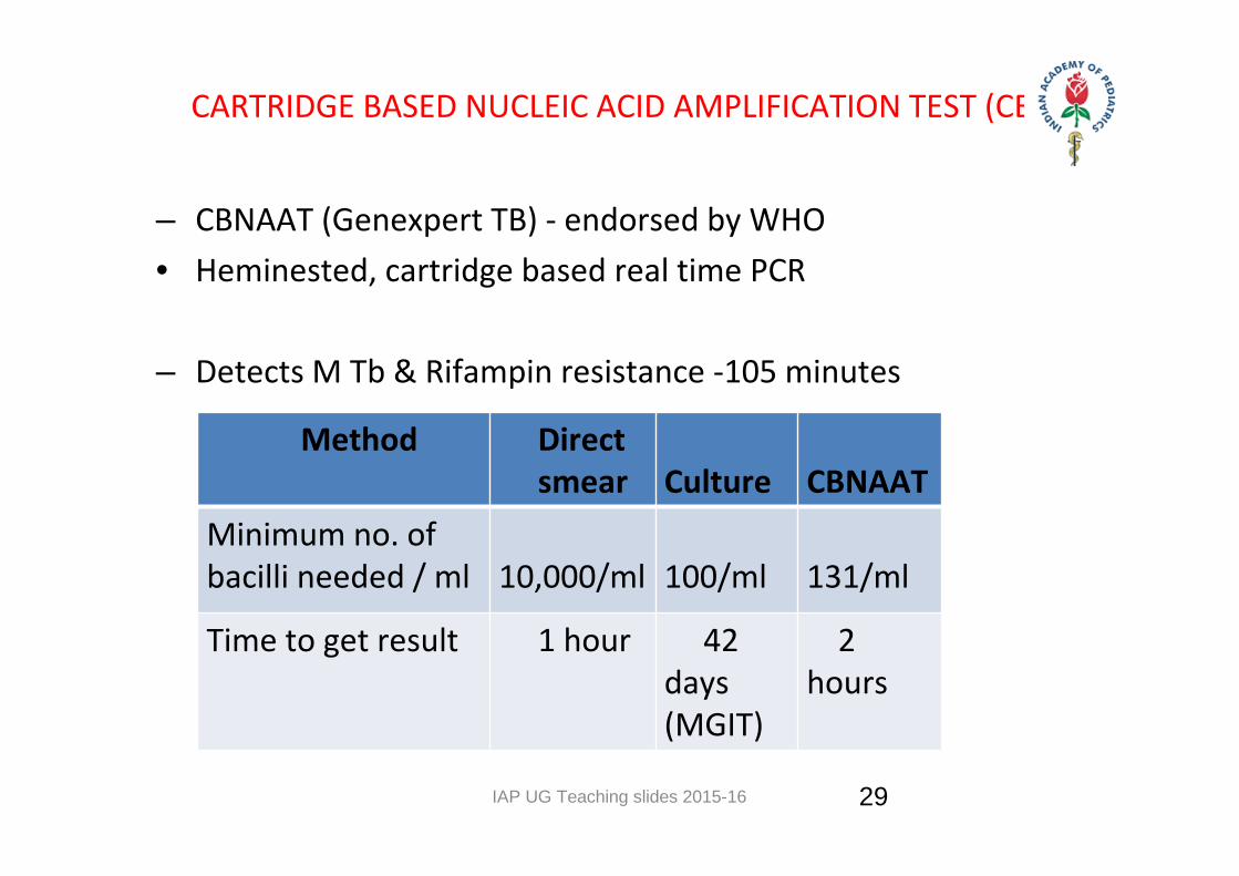
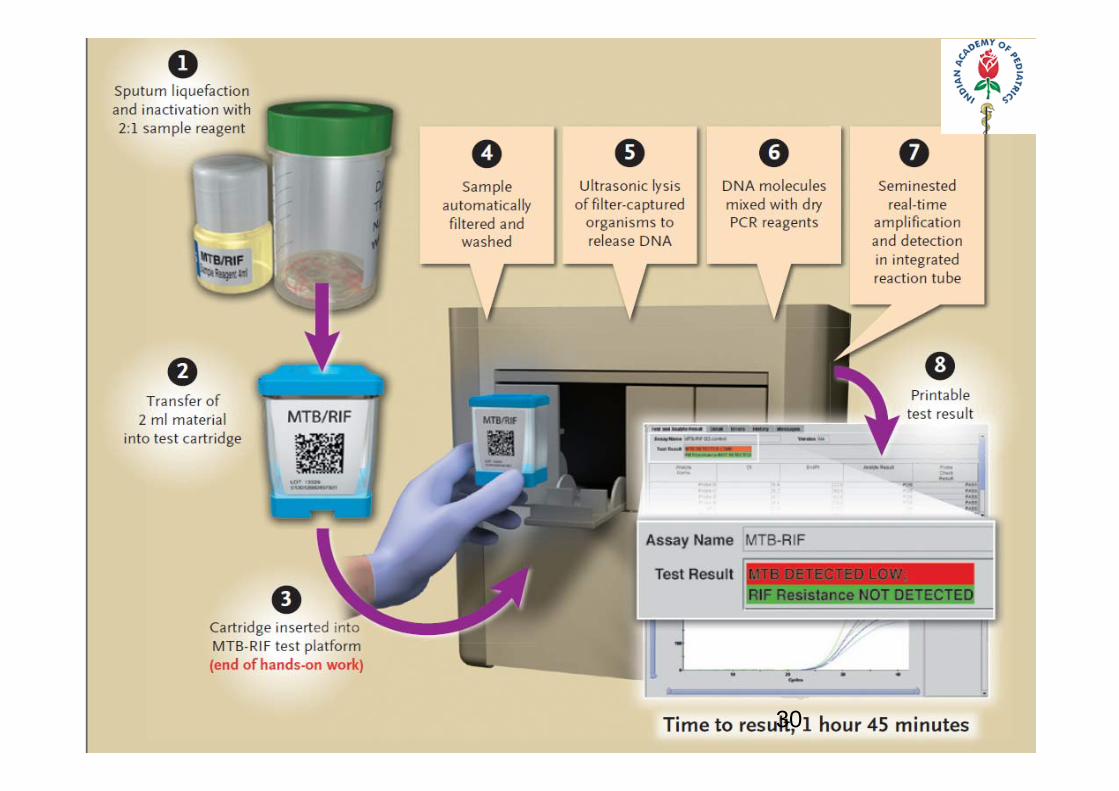
CARTRIDGE BASED NUCLEIC ACID AMPLIFICATION TEST (CBNAAT)
– CBNAAT (Genexpert TB) ‐ endorsed by WHO• Heminested, cartridge based real time PCR
– Detects M Tb & Rifampin resistance ‐105 minutes
Method Direct smear
Culture
CBNAAT
Minimum no. of bacilli needed / ml
10,000/ml
100/ml
131/ml
Time to get result 1 hour 42 days (MGIT)
2 hours
29
IAP UG Teaching slides 2015-16 30
IAP UG Teaching slides 2015-16
IMAGING AND RADIOLOGY
• Brain imaging – ‐ basilar enhancement ‐ communicating hydrocephalus with signs of ‐ cerebral edema or early focal ischemia
‐ Tuberculoma.
• CT SCAN
Helpful with diagnosis of CNS tuberculosis and bone • MRI
Contrast‐ enhanced MRI Considered to be superior to CT in detecting and assessing CNS Tuberculosis.
Limitation is the limited availability and affordability
31
IAP UG Teaching slides 2015-16
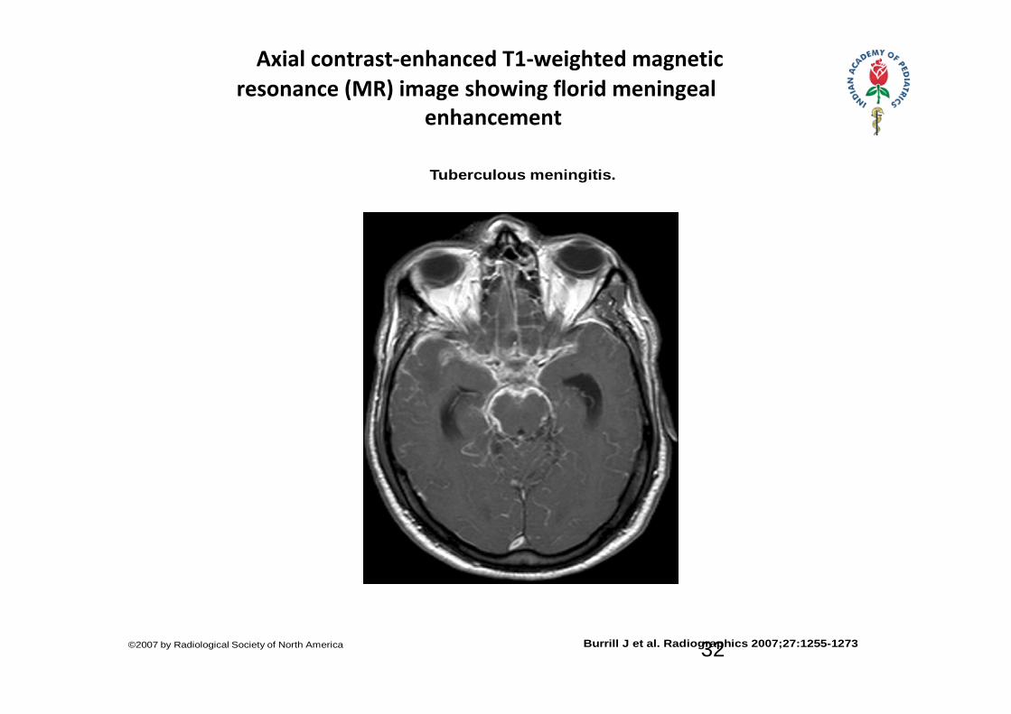
Axial contrast‐enhanced T1‐weighted magnetic resonance (MR) image showing florid meningeal enhancement
Tuberculous meningitis.
Burrill J et al. Radiographics 2007;27:1255-1273©2007 by Radiological Society of North America 32
IAP UG Teaching slides 2015-16
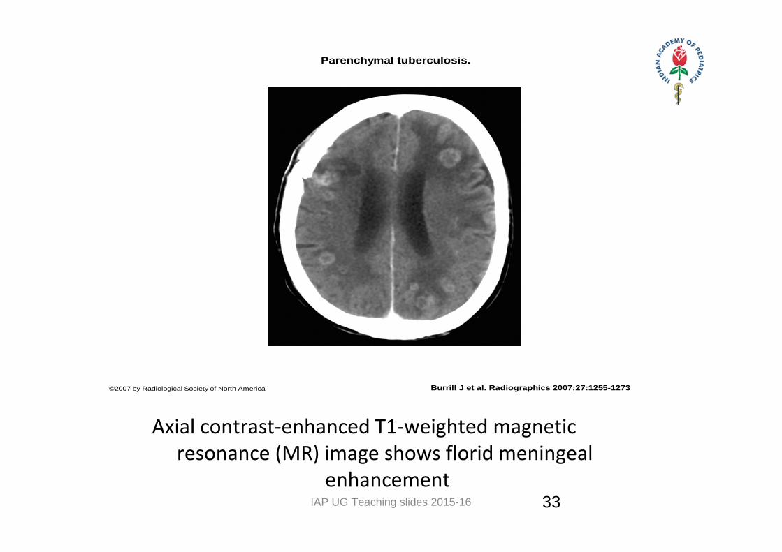
Axial contrast‐enhanced T1‐weighted magnetic resonance (MR) image shows florid meningeal enhancement
Parenchymal tuberculosis.
Burrill J et al. Radiographics 2007;27:1255-1273©2007 by Radiological Society of North America
33
IAP UG Teaching slides 2015-16
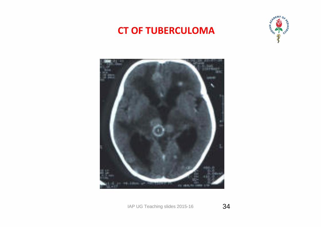
CT OF TUBERCULOMA
34
IAP UG Teaching slides 2015-16
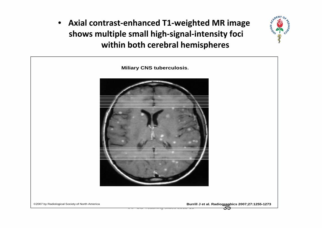
• Axial contrast‐enhanced T1‐weighted MR image shows multiple small high‐signal‐intensity foci
within both cerebral hemispheres
Miliary CNS tuberculosis.
Burrill J et al. Radiographics 2007;27:1255-1273©2007 by Radiological Society of North America
35
IAP UG Teaching slides 2015-16
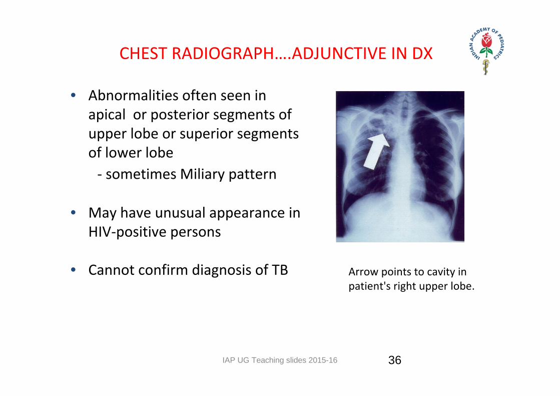
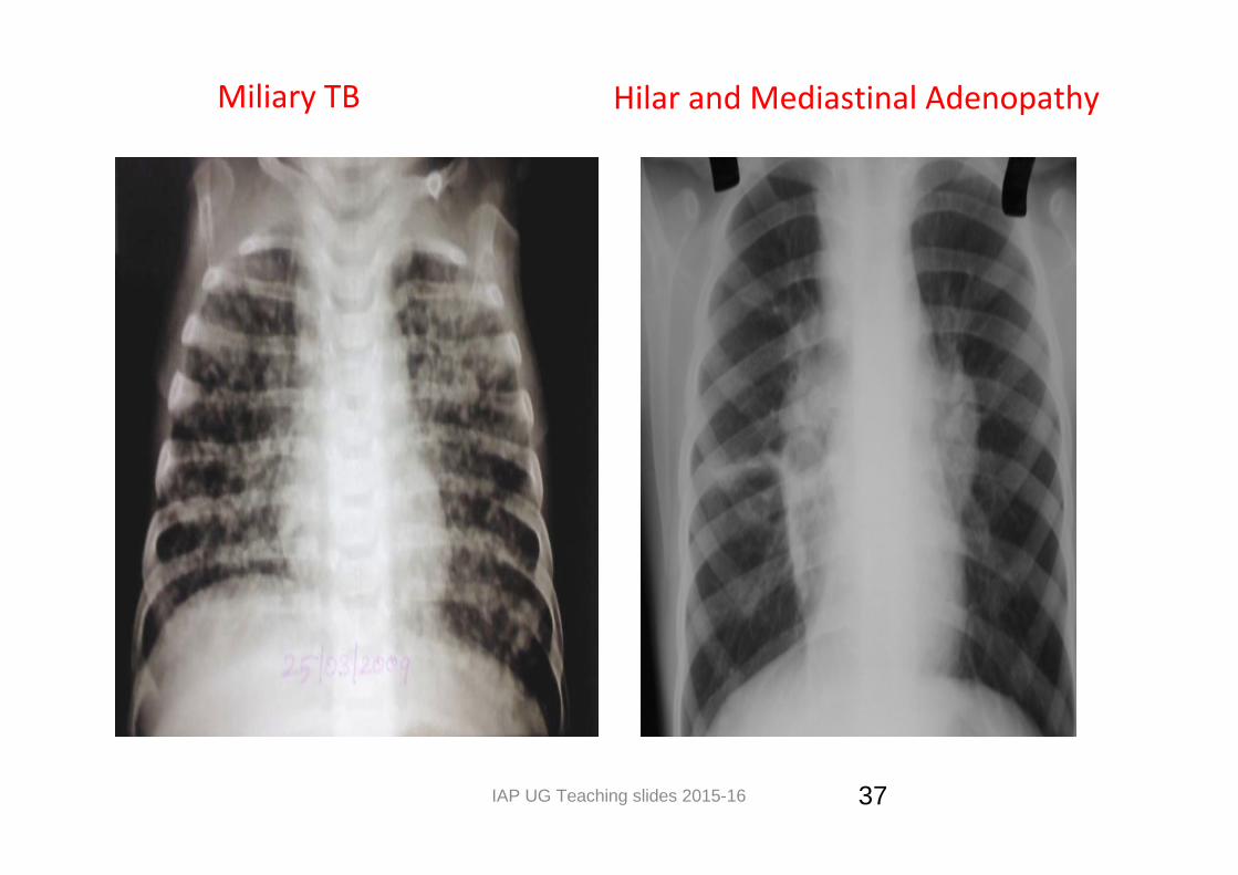
CHEST RADIOGRAPH….ADJUNCTIVE IN DX
• Abnormalities often seen in apical or posterior segments of upper lobe or superior segments of lower lobe‐ sometimes Miliary pattern
• May have unusual appearance in HIV‐positive persons
• Cannot confirm diagnosis of TB Arrow points to cavity in patient's right upper lobe.
36
IAP UG Teaching slides 2015-16 37
Miliary TB Hilar and Mediastinal Adenopathy
IAP UG Teaching slides 2015-16
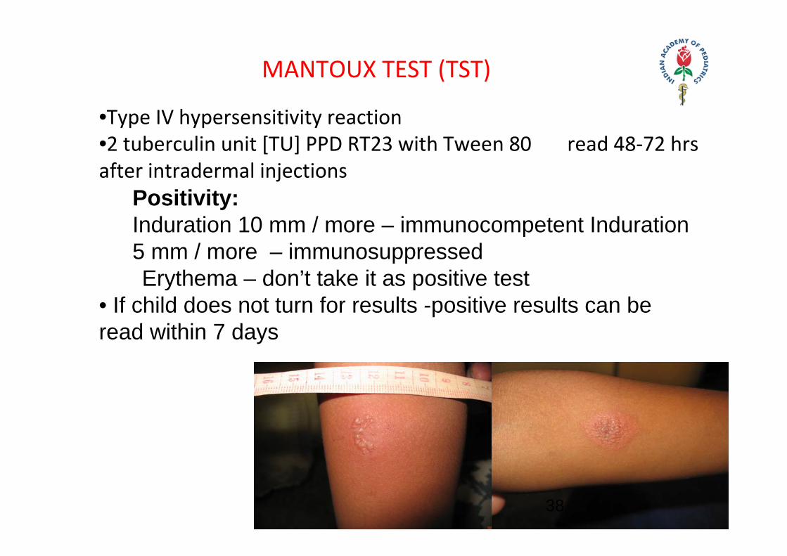
MANTOUX TEST (TST)
•Type IV hypersensitivity reaction•2 tuberculin unit [TU] PPD RT23 with Tween 80 read 48‐72 hrs after intradermal injections
Positivity:Induration 10 mm / more – immunocompetent Induration 5 mm / more – immunosuppressed
Erythema – don’t take it as positive test• If child does not turn for results -positive results can be read within 7 days
38
IAP UG Teaching slides 2015-16
• False negative ‐ infants, HIV positive / immuno‐compromised children, malnutrition extensive or miliary TB.
• False positive – retesting same site, non‐tuberculous (environmental) mycobacteria, higher strengths used
MANTOUX TEST (TST)
39
IAP UG Teaching slides 2015-16
DIAGNOSIS ‐ CONTACT TRACING
• Any person on anti tuberculosis treatment is considered as a
contact for a period of two yearsb) Tuberculin testc) Bacteriological evidence
40
IAP UG Teaching slides 2015-16
BIOCHEMICAL MARKERS
• Adenosine deaminase (ADA) – – level co–relates with proliferation and differentation of lymphocytes.
– Normal levels – 13 – 60 units / ml
• Bromide partition test –
• High performance liquid chromatography
• Tuberculostearic acid detection by gas chromatography
41
IAP UG Teaching slides 2015-16
IMMUNODIAGNOSIS
• Antibody detection– Antibodies to crude antigen/ specific antigen (35 KDa, P 64, P 32, 38 KDa etc)
• Antigen detection– Protein antigens : using polyclonal antibodies / monoclonal antibodies
– ELISA / RIA test used
42
IAP UG Teaching slides 2015-16
CULTURE
• Solid Media – – Lowenstein – Jensen Medium, – Dorsets Medium, – Petroff’s Medium
• Liquid Media – Middle – brooks Medium
• Disadvantages – Difficult to collect CSF & others, – Takes 2 – 8 weeks for result, – Only 5% results come true positive
43
IAP UG Teaching slides 2015-16
RECENT CULTURE TECHNIQUES.`
• Bactec:
– Radiometric culture system– duration time 8 – 14 days– Radiolabelled substrate is used– Growth of AFB is detected radiometrically by measuring the metabolite radiolabelled CO2 that is released
• Septicheck : modified middlebrok broth used• Rapid slide culture method• Mycobacterium growth inhibitor tubes• Remember WHO has cautioned regarding the molecular and genetic
testing.
44
IAP UG Teaching slides 2015-16
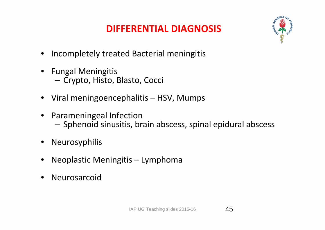
DIFFERENTIAL DIAGNOSIS
• Incompletely treated Bacterial meningitis
• Fungal Meningitis– Crypto, Histo, Blasto, Cocci
• Viral meningoencephalitis – HSV, Mumps
• Parameningeal Infection– Sphenoid sinusitis, brain abscess, spinal epidural abscess
• Neurosyphilis
• Neoplastic Meningitis – Lymphoma
• Neurosarcoid
45
IAP UG Teaching slides 2015-16
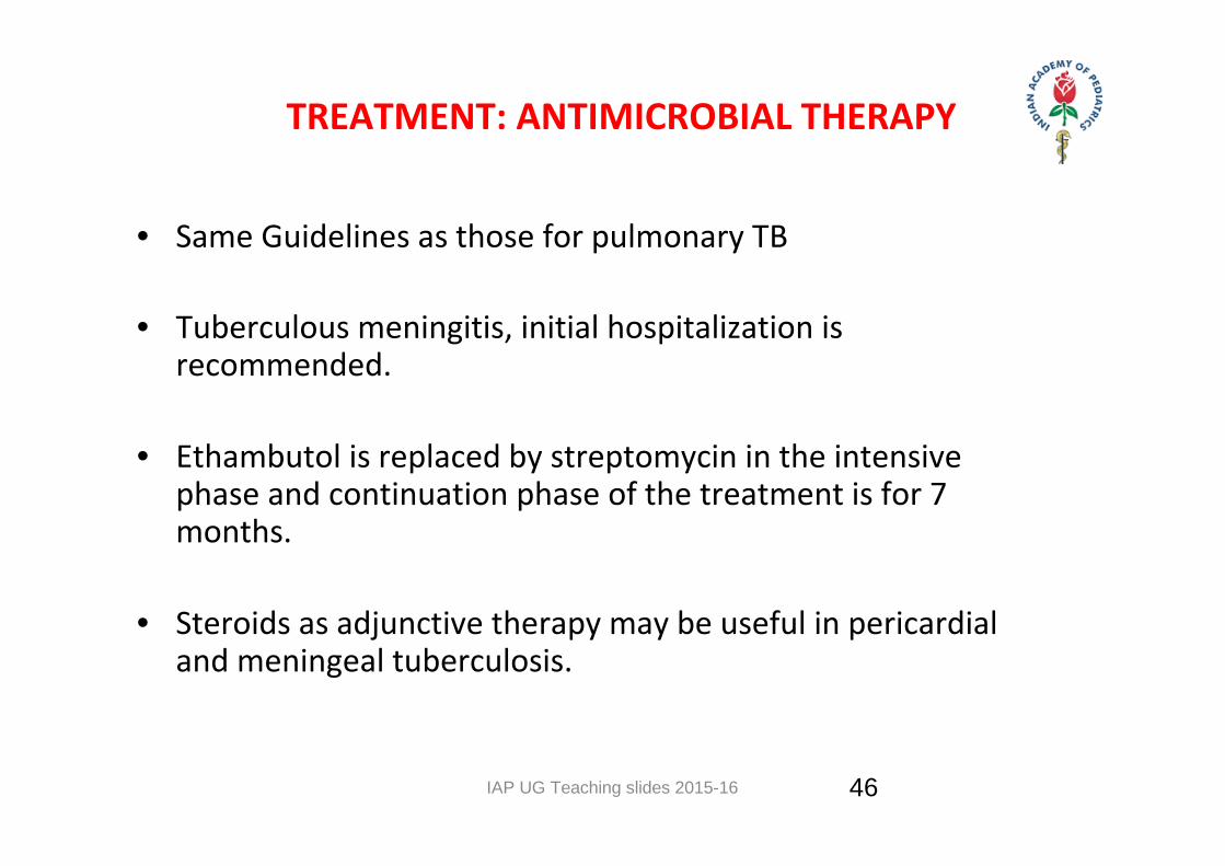
TREATMENT: ANTIMICROBIAL THERAPY
• Same Guidelines as those for pulmonary TB
• Tuberculous meningitis, initial hospitalization is recommended.
• Ethambutol is replaced by streptomycin in the intensive
phase and continuation phase of the treatment is for 7 months.
• Steroids as adjunctive therapy may be useful in pericardial and meningeal tuberculosis.
46
IAP UG Teaching slides 2015-16 47
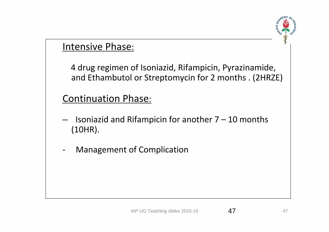
Intensive Phase:
4 drug regimen of Isoniazid, Rifampicin, Pyrazinamide, and Ethambutol or Streptomycin for 2 months . (2HRZE)
Continuation Phase:
– Isoniazid and Rifampicin for another 7 – 10 months (10HR).
‐ Management of Complication
47
IAP UG Teaching slides 2015-16 48
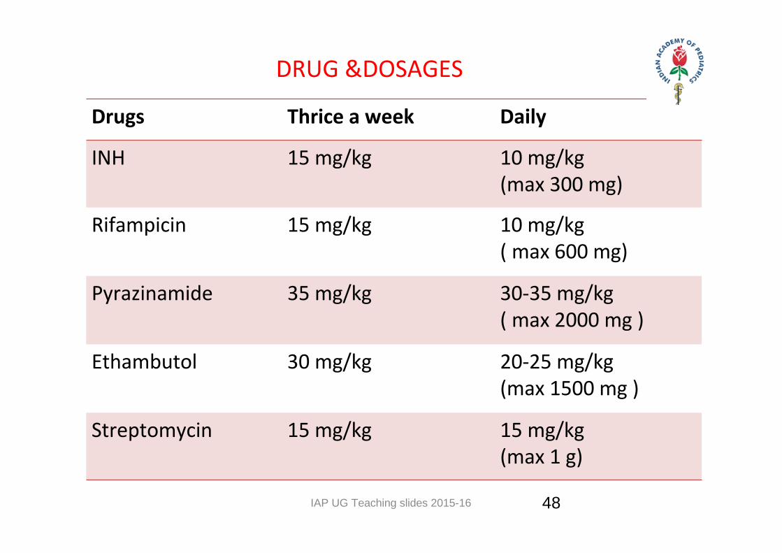
Drugs Thrice a week Daily
INH 15 mg/kg 10 mg/kg (max 300 mg)
Rifampicin 15 mg/kg 10 mg/kg( max 600 mg)
Pyrazinamide 35 mg/kg 30‐35 mg/kg( max 2000 mg )
Ethambutol 30 mg/kg 20‐25 mg/kg(max 1500 mg )
Streptomycin 15 mg/kg 15 mg/kg (max 1 g)
DRUG &DOSAGES
IAP UG Teaching slides 2015-16 49
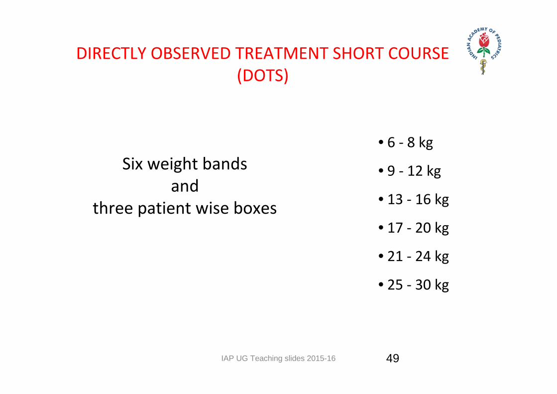
DIRECTLY OBSERVED TREATMENT SHORT COURSE (DOTS)
Six weight bands and
three patient wise boxes
• 6 ‐ 8 kg
• 9 ‐ 12 kg
• 13 ‐ 16 kg
• 17 ‐ 20 kg
• 21 ‐ 24 kg
• 25 ‐ 30 kg
IAP UG Teaching slides 2015-16 50
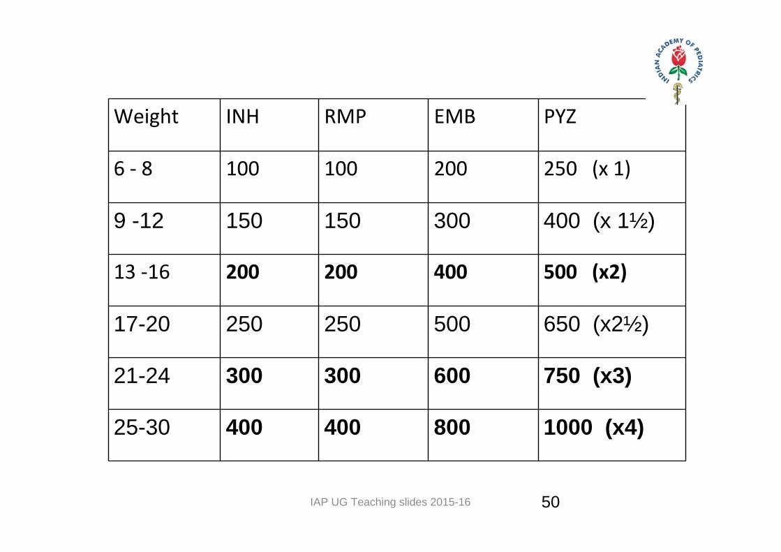
Weight INH RMP EMB PYZ
6 ‐ 8 100 100 200 250 (x 1)
9 -12 150 150 300 400 (x 1½)
13 ‐16 200 200 400 500 (x2)
17-20 250 250 500 650 (x2½)
21-24 300 300 600 750 (x3)
25-30 400 400 800 1000 (x4)
IAP UG Teaching slides 2015-16
INH
• Introduced in the year 1952
• Inhibits the synthesis of mycotic acid in mycobacterial cell wall‐ bactercidal drug
• Dose: 15mg/ kg Max: 300mg/day
• Hepatotoxicity, peripheral neuritis ‐ but rare in pediatric age
51
IAP UG Teaching slides 2015-16
RIFAMPICIN
• Introduced in the year 1962
• It inhibits RNA synthesis in mycobacteria
• Bactericidal drug
• Well absorbed in empty stomach
• Dose ‐ 10mg/kg Max: 450mg/day
• INH and Rifampicin always combined for beneficial effect
• Hepato toxic drug
52
IAP UG Teaching slides 2015-16
PYRIZINAMIDE
• Introduced in the year 1947
• Sterilizing drug
• Bactericidal drug more effective in caseous acidic medium
• Resistance develops quickly
• Dose: 35mg/kg
• Side effects: elevation of uric acid
53
IAP UG Teaching slides 2015-16
ETHAMBUTOL
• Introduced in the year 1952
• Bacteriostatic drug
• Dose: 30mg/kg Max: 800mg/day
• Side effects: Optic neuritis (usually reversible), decreased red‐green color discrimination, gastrointestinal tract disturbances, hypersensitivity.
• The side effects of all these 4 drugs are reversible
54
IAP UG Teaching slides 2015-16
STREPTOMYCIN
• It is an aminoglycoside.
• Bactericidal drug.
• Dose : 15mg/kg ; max : 1g
• Side Effects : Auditory and vestibular toxic effects, nephrotoxic effects, rash
55
IAP UG Teaching slides 2015-16
TREATMENT
Adjunctive steroid therapy
All patients regardless of stage/severity of the disease To decrease neurologic sequelae and mortality
Lowering the intracranial pressure limits tissue damage and favors circulation of anti‐tuberculosis drugs through the brain and
meninges.2mg/kg/24 hours of prednisolone for 6‐8 weeks at the start of treatment starting 3 days after initiation of anti tuberculosis
therapy.
56
IAP UG Teaching slides 2015-16
II LINE OF DRUGS
For MDRTB ,• Six drugs— kanamycin, levofloxacin, ethionamide, pyrazinamide,
ethambutol and cycloserine during 6–9 months of the intensive phase and
• Four drugs—levofloxacin, ethionamide, ethambutol and cycloserine during the 18 months of the continuation phase.
57
IAP UG Teaching slides 2015-16
COMPLICATIONS
• The most common complication is communicating hydrocephalus, which can be seen at both MR imaging and CT and is caused by blockage of the basal cisterns by inflammatory exudates .
• Rarely non‐communicating hydrocephalus occurs due to the mass effect of a tuberculoma causing the obstruction of CSF flow.
• Ischemic infarcts are also a common complication, being seen in 20%–40% of patients at CT , mostly within the basal ganglia or internal capsule regions and resulting from vascular compression and occlusion of small perforating vessels
58
IAP UG Teaching slides 2015-16
TREATMENT OF COMPLICATIONS:
Convulsions: •Benzodiazepines and phenytoinCerebral edema:•Initial therapy is with 20% Mannitol: 5ml/Kg IV over 15 minutes, f/b 3ml/kg 6th hrly till 48 hrs.
•I.V.Dexamethasone can then be used 0.15mg/kg/dose 6 hourly.•Fluids –restricted to 2/3rd maintenance.•Great care to be taken to maintain BP.•Other drugs – Acetazolamide 20‐50mg/kg/day in 3‐4 doses oral glycerol 1ml/kg/dose
59
IAP UG Teaching slides 2015-16 60
PROGNOSIS AND OUTCOME
If left untreated, its course is characterized by confusion and progressively deepening stupor and coma, coupled with cranial nerve palsies, pupillary abnormalities, foci neurologic deficits, raised ICP and decerebrate postures.
Fatal outcome then follows within 4 to 8 weeks at the onset.
60
IAP UG Teaching slides 2015-16
PROGNOSIS
• Overall Poor• Pts presenting in Stage I have 19% mortality• Pts presenting in Stage III have 69% mortality• Only 1/3 ‐ 1/2 of patients demonstrate complete neurologic
recovery• Up to 1/3 of patients have residual severe neurologic deficits
such as hemi paresis, blindness, seizure DO• Most patients in the 3rd stage who survive have permanent
disabilities, including blindness, deafness, paraplegia, diabetes insipidus, or mental retardation.
• Prognosis for young infants is generally worse than for older children
61
IAP UG Teaching slides 2015-16
PREVENTION
• BCG Vaccination provides protection against the CNS Tuberculosis with an efficacy of 75—85%.
REMEMBER
• History of BCG vaccination does not eliminate the need to investigate for CNS Tuberculosis in the right clinical situations.
62
IAP UG Teaching slides 2015-16
REFERENCES
• Nelson textbook• Basic of pediatrics• WHO recommendations• E‐medicine
63
IAP UG Teaching slides 2015-16 64
THANK YOU
64