Embed Size (px)
Citation preview
Research Article
Mona S. Alwhibi, Dina A. Soliman*, Manal A. Awad, Asma B. Alangery, Horiah Al Dehaish, andYasmeen A. Alwasel
Green synthesis of silver nanoparticles:Characterization and its potential biomedicalapplications
https://doi.org/10.1515/gps-2021-0039received May 10, 2021; accepted June 01, 2021
Abstract: In recent times, research on the synthesis ofnoble metal nanoparticles (NPs) has developed rapidlyand attracted considerable attention. The use of plantextracts is the preferred mode for the biological synthesisof NPs due to the presence of biologically active consti-tuents. Aloe vera is a plant endowed with therapeuticbenefits especially in skincare due to its unique curativeproperties. The present study focused on an environ-mental friendly and rapid method of phytosynthesis ofsilver nanoparticles (Ag-NPs) using A. vera gel extract asa reductant. The synthesized Ag-NPs were characterizedby transmission electron microscopy (TEM), UV-Vis spectro-scopy, Fourier transform infrared (FTIR), and dynamiclight scattering (DLS). TEM micrographs showed spherical-shaped synthesized Ag-NPs with a diameter of 50–100 nm.The UV-Vis spectrum displayed a broad absorption peak ofsurface plasmon resonance (SPR) at 450 nm. The meansize and size distribution of the formed Ag-NPs were inves-tigated using the DLS technique. Antibacterial studiesrevealed zones of inhibition by Ag-NPs of A. vera (9 and7mm) against Pseudomonas aeruginosa and Escherichiacoli, respectively. Furthermore, the antifungal activity wasscreened, based on the diameter of the growth inhibition
zone using the synthesized Ag-NPs for different fungalstrains. Anticancer activity of the synthesized Ag-NPs againstthe mouse melanoma F10B16 cell line revealed 100% inhibi-tionwithAg-NPs at a concentration of 100µgmL−1. The phyto-synthesized Ag-NPs demonstrated a marked antimicrobialactivity and also exhibited a potent cytotoxic effect againstmouse melanoma F10B16 cells. The key findings of thisstudy indicate that synthesized Ag-NPs exhibit profoundtherapeutic activity and could be potentially ideal alterna-tives in medicinal applications.
Keywords: Aloe vera, phytosynthesis, silver nanoparticle,antitumor, antimicrobial activities
1 Introduction
Nanotechnology includes the synthesis of nanoparticles(NPs) with varied morphology, size, and controlled dis-persity to be used for their benefits in several ways. It hasthe potential to impact human society for being widelyused in various fields of science and technology [1].
Antimicrobial, dental therapy, wound healing, sur-gery function, catalyst, and biomedical devices are just afew applications of metal NPs [2]. Because of their uniqueoptical and electrical properties, NP-based drugs havebeen found to be more efficacious [3,4]. Surface plasmonresonance (SPR) is a well-known property of NPs thatincreases their effectiveness [5].
Silver nanoparticles (Ag-NPs) remarkably, have anarrow plasmon resonance, a high surface-to-volumeratio, special physicochemical properties, and a widerange of applications in medical research, microelectro-nics, and biological activities [6,7]. AgNPs have garneredconsiderable interest among other metal NPs due to theirwide use in several commercial and pharmacologicallysignificant products [8,9]. Considering the synthesis, thetraditional methods such as the physical, thermal, hydro-thermal, and chemical synthesis modes are expensive,
Mona S. Alwhibi, Horiah Al Dehaish, Yasmeen A. Alwasel:Department of Botany and Microbiology, College of Science, King SaudUniversity, P.O. Box 22452, Riyadh, 11495, Kingdom of Saudi Arabia
* Corresponding author: Dina A. Soliman, Department of Botanyand Microbiology, College of Science, King Saud University,P.O. Box 22452, Riyadh, 11495, Kingdom of Saudi Arabia,e-mail: [email protected]
Manal A. Awad: Department of Physics, College of Science, KingSaud University, P.O. Box 22452, Riyadh, 11495, Kingdom of SaudiArabiaAsma B. Alangery: Department of Chemistry, College of Science,King Saud University, P.O. Box 22452, Riyadh, 11495, Kingdom ofSaudi Arabia
Green Processing and Synthesis 2021; 10: 412–420
Open Access. © 2021 Mona S. Alwhibi et al., published by De Gruyter. This work is licensed under the Creative Commons Attribution 4.0International License.
extremely hazardous and make use of toxic chemicals.Therefore, the emphasis is on a green synthesis approachmaking use of biological resources for the efficient formu-lation of NPs [10–12]. The cornerstone of this sustainablemethod is the use of renewable materials and environmen-tally benign compounds as reducing/capping agents tosynthesize green nanoparticles [13]. Methods of greensynthesis have been effectively utilized to synthesize NPsusing different biomolecules, such as vitamins, yeasts,enzymes, algae, biodegradable polymers, andmicroorgan-isms, and plant parts such as leaf, stem, gum, fruit, bark,shells, roots, buds, and flowers [12,14].
Aloe vera (A. vera) is a succulent plant with thorns inits branches with a waxy coating that grows easily in aridconditions [15,16]. A. vera leaves have three layers: theouter layer is a thick and protective layer which has ahigh proportion of cellulose; the middle layer containsmajor flavanone (aloin A and B), and the inner layerhas a fresh gel that contains an acetylated glucomannan,sugars, vitamins (A, B, C, and E), amino acids, proteins,and anthraquinones [17–19]. A. vera gel is utilized inmany cosmetic products especially skincare. It is alsoconsidered to be effective for treating burns and wounds,which is known for its soothing effect on the affected skin.It can also be used to treat a wide range of health-relateddisorders [20]. The antimicrobial property and biomedicalapplications of AgNPs synthesized with A. vera gel extractare well investigated. However, very few studies havereported and documented the use of A. vera gel to synthe-size AgNPs for biomedical applications.
With this premise, the present study aimed at the synth-esis of A. vera gel extract for phytosynthesis of AgNPs andthe investigation of their promising biomedical applicationsbased on the antimicrobial and anticancer activities.
2 Materials and methods
2.1 Identification of plant
Aloe vera was collected from a local park at King SaudUniversity, girls campus, Riyadh, Saudi Arabia, and identifiedat the Taxonomy Laboratory No. 116, Botany Department,King Saud University, Riyadh.
2.2 Preparation of plant extract
The healthy large outer leaves close to the ground wereselected. They were then cut carefully at a slight angle,
and the leaves were placed upright in a slightly tiltedcontainer for roughly 10 min, allowing much of the sapand the gel to drain out. About 20 g of the collected gelwas weighed and transferred into a 500mL conical flaskcontaining 100mL of deionized water, mixed well, andheated for 45 min in a water bath. The resulting solutionwas centrifuged at 5,000 rpm for 15 min, filtered usingWhatman number 1 filter paper, and then the filtratewas stored at 4°C for further use.
2.3 Synthesis of Ag-NPs
An aqueous solution of silver nitrate (AgNO3) was pre-pared using 2 mM AgNO3 powder dissolved in 100mL ofdeionized water at a fixed ratio. The reaction mixture wasprepared by taking 10mL of the above-prepared filtratedextract obtained from the plant and 40mL of the AgNO3
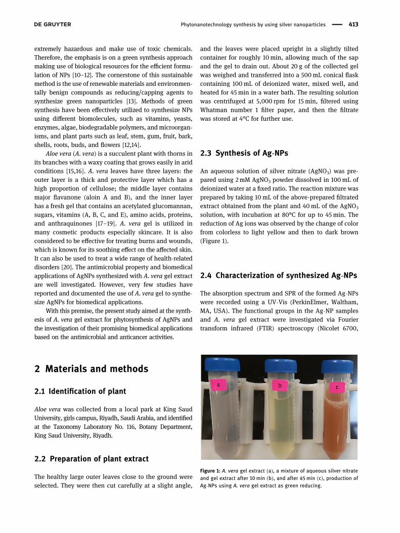
solution, with incubation at 80°C for up to 45 min. Thereduction of Ag ions was observed by the change of colorfrom colorless to light yellow and then to dark brown(Figure 1).
2.4 Characterization of synthesized Ag-NPs
The absorption spectrum and SPR of the formed Ag-NPswere recorded using a UV-Vis (PerkinElmer, Waltham,MA, USA). The functional groups in the Ag-NP samplesand A. vera gel extract were investigated via Fouriertransform infrared (FTIR) spectroscopy (Nicolet 6700,
Figure 1: A. vera gel extract (a), a mixture of aqueous silver nitrateand gel extract after 10min (b), and after 45 min (c), production ofAg-NPs using A. vera gel extract as green reducing.
Phytonanotechnology synthesis by using silver nanoparticles 413
Thermo Scientific, USA), using the potassium bromide(KBr) pellet technique. Transmission electron microscopy(TEM) (JEOL-JEM-1011, Japan) was used to examine thesize and morphology of the phytosynthesized Ag-NPswith an acceleration voltage of 80 kV. The particle sizedistribution of the NPs, hydrodynamic diameter, andpolydispersity index (PdI)were determined andmeasuredby dynamic light scattering (DLS) particle size analyzer(ZEN3600 Malvern, Nano series, HT Laser, Malvern, UK).
2.5 AgNP biomedical activities
2.5.1 Cytotoxicity of synthesized Ag-NPs
The anti-proliferative activity of the Ag-NPs against mousemelanoma B16F10 cell lines was analyzed by the MTTassay using a 96-well plate supplemented with differentconcentrations of A. vera extract and Ag-NPs. The plateswere incubated for 5 h at 37°C. Then, 0.1mL of 1% trypanblue exclusion test was added. Cells were seeded in a96-well plate at a density of 1 × 10⁵ cells/well in a 90 µLDulbecco’s modified Eagle’s medium. The cells were thenallowed to settle before starting treated with different(3.125, 6.25, 12.5, 25, 50, and 100 µgmL−1) concentrationsof the samples. The treated cells were allowed to growfurther for 24 h. The experiment was performed on fourreplicates. Specifically, the non-viable cells adopted ablue color while the live cells did not take on the colorof the dye. Then, the numbers of blue-stained andunstained cells were determined. The cell viability of theuntreated cells was 100% and that of the treated cells wasbelow 100%.
2.5.2 Antibacterial activity
Nutrient agar medium was prepared by dissolving 14 g ofagar powder in 500mL of distilled water and then auto-claved. A total of 20mL of prepared agar was poured intoeach Petri dish, which was left to stand for 15 min for theagar to solidify, then the plates were inoculated overnightwith human pathogens, such as the Gram-negative strain,Escherichia coli ATCC35218, and the gram-positive strains,Staphylococcus aureus ATCC 43300, Enterococcus faecalisATCC 29212, and Bacillus cereus ATCC 11778 (clinical iso-late) obtained from King Khalid University Hospital,Riyadh, Saudi Arabia. All organisms were tested simulta-neously by the disc diffusion method [21]. SynthesizedAg-NPs and pure extract of A. vera were added steadily
until the wells were full, followed by incubation at 37°C for24 h. The diameter of the zone of inhibition was measured.
2.5.3 Antifungal activity
The sampleswere assayed for antifungal activity against bipolarheterothallic, Fusarium oxysporum and Macrophomina.These fungal strains were grown on a potato dextrose agarplate at 28°C. A total of 500mL of the medium, which wasprepared by dissolving 19 g of agar in 500mL of distilledwater, was autoclaved. A volume of 250 μL of synthesizedAg-NPs and pure extract of A. vera, separately, were addedto a sterile petri dish, followed by pouring of the sterilizedmedium with gentle mixing. Fungal discs with a diameterof 6mm, which were grown for 7 days from cultures of theabove-mentioned fungi, were placed aseptically in themiddle of the plate. The plates were incubated for 7−14 daysat 28°C. The antifungal activity was estimated by mea-suring the diameter of the inhibition zone.
3 Results and discussion
3.1 UV-Vis spectral analysis
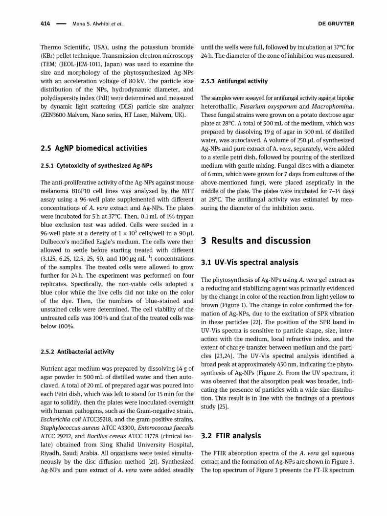
The phytosynthesis of Ag-NPs using A. vera gel extract asa reducing and stabilizing agent was primarily evidencedby the change in color of the reaction from light yellow tobrown (Figure 1). The change in color confirmed the for-mation of Ag-NPs, due to the excitation of SPR vibrationin these particles [22]. The position of the SPR band inUV-Vis spectra is sensitive to particle shape, size, inter-action with the medium, local refractive index, and theextent of charge transfer between medium and the parti-cles [23,24]. The UV-Vis spectral analysis identified abroad peak at approximately 450 nm, indicating the phyto-synthesis of Ag-NPs (Figure 2). From the UV spectrum, itwas observed that the absorption peak was broader, indi-cating the presence of particles with a wide size distribu-tion. This result is in line with the findings of a previousstudy [25].
3.2 FTIR analysis
The FTIR absorption spectra of the A. vera gel aqueousextract and the formation of Ag-NPs are shown in Figure 3.The top spectrum of Figure 3 presents the FT-IR spectrum
414 Mona S. Alwhibi et al.
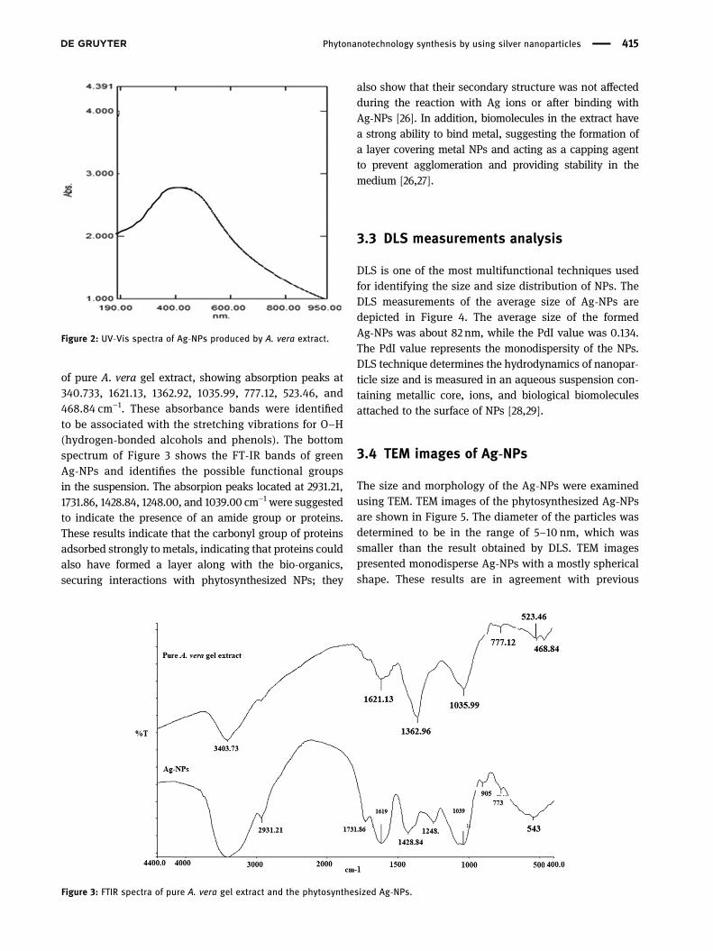
of pure A. vera gel extract, showing absorption peaks at340.733, 1621.13, 1362.92, 1035.99, 777.12, 523.46, and468.84 cm−1. These absorbance bands were identifiedto be associated with the stretching vibrations for O–H(hydrogen-bonded alcohols and phenols). The bottomspectrum of Figure 3 shows the FT-IR bands of greenAg-NPs and identifies the possible functional groupsin the suspension. The absorpion peaks located at 2931.21,1731.86, 1428.84, 1248.00, and 1039.00 cm−1 were suggestedto indicate the presence of an amide group or proteins.These results indicate that the carbonyl group of proteinsadsorbed strongly to metals, indicating that proteins couldalso have formed a layer along with the bio-organics,securing interactions with phytosynthesized NPs; they
also show that their secondary structure was not affectedduring the reaction with Ag ions or after binding withAg-NPs [26]. In addition, biomolecules in the extract havea strong ability to bind metal, suggesting the formation ofa layer covering metal NPs and acting as a capping agentto prevent agglomeration and providing stability in themedium [26,27].
3.3 DLS measurements analysis
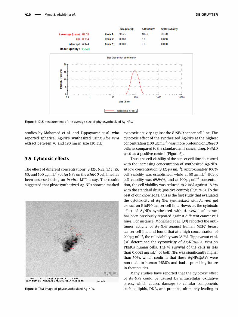
DLS is one of the most multifunctional techniques usedfor identifying the size and size distribution of NPs. TheDLS measurements of the average size of Ag-NPs aredepicted in Figure 4. The average size of the formedAg-NPs was about 82 nm, while the PdI value was 0.134.The PdI value represents the monodispersity of the NPs.DLS technique determines the hydrodynamics of nanopar-ticle size and is measured in an aqueous suspension con-taining metallic core, ions, and biological biomoleculesattached to the surface of NPs [28,29].
3.4 TEM images of Ag-NPs
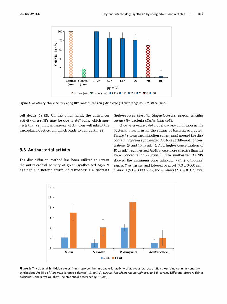
The size and morphology of the Ag-NPs were examinedusing TEM. TEM images of the phytosynthesized Ag-NPsare shown in Figure 5. The diameter of the particles wasdetermined to be in the range of 5–10 nm, which wassmaller than the result obtained by DLS. TEM imagespresented monodisperse Ag-NPs with a mostly sphericalshape. These results are in agreement with previous
Figure 2: UV-Vis spectra of Ag-NPs produced by A. vera extract.
Figure 3: FTIR spectra of pure A. vera gel extract and the phytosynthesized Ag-NPs.
Phytonanotechnology synthesis by using silver nanoparticles 415
studies by Mohamed et al. and Tippayawat et al. whoreported spherical Ag-NPs synthesized using Aloe veraextract between 70 and 190 nm in size [30,31].
3.5 Cytotoxic effects
The effect of different concentrations (3.125, 6.25, 12.5, 25,50, and 100 µgmL−1) of Ag NPs on the B16F10 cell line hasbeen assessed using an in-vitro MTT assay. The resultssuggested that phytosynthesized Ag-NPs showed marked
cytotoxic activity against the B16F10 cancer cell line. Thecytotoxic effect of the synthesized Ag-NPs at the highestconcentration (100 µgmL−1)wasmore profound on B16F10cells as compared to the standard anti-cancer drug, NSAIDused as a positive control (Figure 6).
Thus, the cell viability of the cancer cell line decreasedwith the increasing concentration of synthesized Ag-NPs.At low concentration (3.125 µgmL−1), approximately 100%cell viability was established, while at 50 µgmL−1 (IC50),cell viability was 69.94%, and at 100 µgmL−1 concentra-tion, the cell viability was reduced to 2.14% against 18.5%with the standard drug (positive control) (Figure 6). To thebest of our knowledge, this is the first study that evaluatedthe cytotoxicity of Ag-NPs synthesized with A. vera gelextract on B16F10 cancer cell line. However, the cytotoxiceffect of AgNPs synthesized with A. vera leaf extracthas been previously reported against different cancer celllines. For instance, Mohamed et al. [30] reported the anti-tumor activity of Ag-NPs against human MCF7 breastcancer cell line and found that at a high concentration of200 μgmL−1, the cell viability was 28.7%. Tippayawat et al.[31] determined the cytotoxicity of Ag-NPs@ A. vera onPBMCs human cells. The % survival of the cells in lessthan 0.0025mgmL−1 of both NPs was significantly higherthan 50%, which confirms that these AgNPs@AVs werenon-toxic to human PBMCs and had a promising futurein therapeutics.
Many studies have reported that the cytotoxic effectof Ag-NPs could be caused by intracellular oxidativestress, which causes damage to cellular componentssuch as lipids, DNA, and proteins, ultimately leading to
Figure 4: DLS measurement of the average size of phytosynthesized Ag-NPs.
Figure 5: TEM image of phytosynthesized Ag-NPs.
416 Mona S. Alwhibi et al.
cell death [18,32]. On the other hand, the anticanceractivity of Ag-NPs may be due to Ag+ ions, which sug-gests that a significant amount of Ag+ ions will inhibit thesarcoplasmic reticulum which leads to cell death [33].
3.6 Antibacterial activity
The disc-diffusion method has been utilized to screenthe antimicrobial activity of green synthesized Ag-NPsagainst a different strain of microbes: G+ bacteria
(Enterococcus faecalis, Staphylococcus aureus, Bacilluscereus) G− bacteria (Escherichia coli).
Aloe vera extract did not show any inhibition in thebacterial growth in all the strains of bacteria evaluated.Figure 7 shows the inhibition zones (mm) around the diskcontaining green synthesized Ag-NPs at different concen-trations (5 and 10 μgmL−1). At a higher concentration of10 μgmL−1, synthesized Ag-NPs were more effective than thelower concentration (5 μgmL−1). The synthesized Ag-NPsshowed the maximum zone inhibition (9.1 ± 0.100mm)against P. aeruginosa and followed by E. coli (7.0 ± 0.000mm),S. aureus (4.1 ± 0.100mm), and B. cereus (2.03 ± 0.0577mm)
0
20
40
60
80
100
Control(-ve)
Control(+ve)
3.125 6.25 12.5 25 50 100
Cel
l Via
bilit
y %
µg mL-1
Control (-ve) Control (+ve) 3.125 6.25 12.5 25 50 100
Figure 6: In vitro cytotoxic activity of Ag-NPs synthesized using Aloe vera gel extract against B16F10 cell line.
0
2
4
6
8
10
12
E. coli S. aurous P. aeruginosa Bacillus cereus
5 µL 10 µL
Figure 7: The sizes of inhibition zones (mm) representing antibacterial activity of aqueous extract of Aloe vera (blue columns) and thesynthesized Ag-NPs of Aloe vera (orange columns): E. coli, S. aureus, Pseudomonas aeruginosa, and B. cereus. Different letters within aparticular concentration show the statistical difference (p ≥ 0.05).
Phytonanotechnology synthesis by using silver nanoparticles 417
at a concentration of 100 μgmL−1. The synthesized Ag-NPsat a concentration of 100 μgmL−1 exhibited a significant(p ≥ 0.05) anti-bacterial effect on P. aeruginosa as comparedto the other strains. The antibacterial activity of synthesizedAg-NPs increased in a dose-dependent manner. Similarresults were reported in a study by Anju and Tippayawat[18,31] which showed that Ag-NPs synthesized using Aloevera leaves extract showed similar antibacterial activityagainst various bacterial strains.
Various mechanisms have been proposed for theantibacterial activity of Ag-NPs in previous studies [34].Some studies reported that Ag-NPs exhibited antimicro-bial effects against bacterial cells via (a) membrane dis-ruption due to the association/interaction of Ag-NPs withDNA and other biomolecules, subsequently causing inhi-bition of cell multiplication, and (b) formation of reactiveoxygen species through interaction with enzymes and/orbiomolecules, leading to cellular damage [35].
Gram-positive bacteria possess a thick cell wall of thepeptidoglycan layer composed of linear polysaccharidechains, cross-linked by short peptides, which is a rigidstructure that hinders the penetration of AgNPs into thebacterial cell wall, compared to Gram-negative bacteriawhere the cell wall consists of a thinner peptidoglycanlayer [36–38].
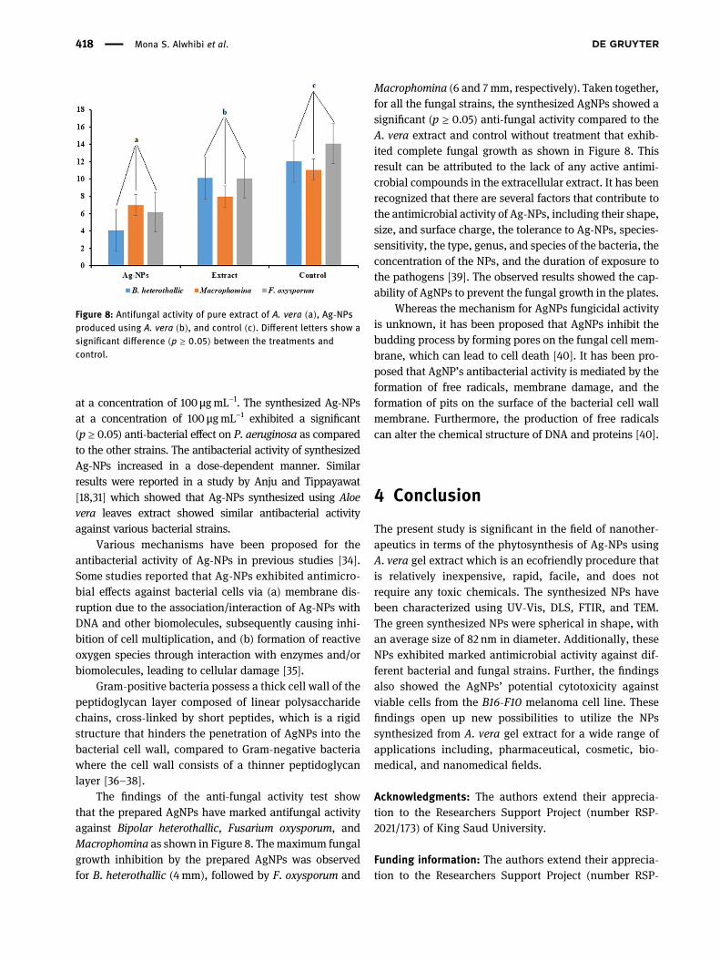
The findings of the anti-fungal activity test showthat the prepared AgNPs have marked antifungal activityagainst Bipolar heterothallic, Fusarium oxysporum, andMacrophomina as shown in Figure 8. Themaximum fungalgrowth inhibition by the prepared AgNPs was observedfor B. heterothallic (4mm), followed by F. oxysporum and
Macrophomina (6 and 7mm, respectively). Taken together,for all the fungal strains, the synthesized AgNPs showed asignificant (p ≥ 0.05) anti-fungal activity compared to theA. vera extract and control without treatment that exhib-ited complete fungal growth as shown in Figure 8. Thisresult can be attributed to the lack of any active antimi-crobial compounds in the extracellular extract. It has beenrecognized that there are several factors that contribute tothe antimicrobial activity of Ag-NPs, including their shape,size, and surface charge, the tolerance to Ag-NPs, species-sensitivity, the type, genus, and species of the bacteria, theconcentration of the NPs, and the duration of exposure tothe pathogens [39]. The observed results showed the cap-ability of AgNPs to prevent the fungal growth in the plates.
Whereas the mechanism for AgNPs fungicidal activityis unknown, it has been proposed that AgNPs inhibit thebudding process by forming pores on the fungal cell mem-brane, which can lead to cell death [40]. It has been pro-posed that AgNP’s antibacterial activity is mediated by theformation of free radicals, membrane damage, and theformation of pits on the surface of the bacterial cell wallmembrane. Furthermore, the production of free radicalscan alter the chemical structure of DNA and proteins [40].
4 Conclusion
The present study is significant in the field of nanother-apeutics in terms of the phytosynthesis of Ag-NPs usingA. vera gel extract which is an ecofriendly procedure thatis relatively inexpensive, rapid, facile, and does notrequire any toxic chemicals. The synthesized NPs havebeen characterized using UV-Vis, DLS, FTIR, and TEM.The green synthesized NPs were spherical in shape, withan average size of 82 nm in diameter. Additionally, theseNPs exhibited marked antimicrobial activity against dif-ferent bacterial and fungal strains. Further, the findingsalso showed the AgNPs’ potential cytotoxicity againstviable cells from the B16-F10 melanoma cell line. Thesefindings open up new possibilities to utilize the NPssynthesized from A. vera gel extract for a wide range ofapplications including, pharmaceutical, cosmetic, bio-medical, and nanomedical fields.
Acknowledgments: The authors extend their apprecia-tion to the Researchers Support Project (number RSP-2021/173) of King Saud University.
Funding information: The authors extend their apprecia-tion to the Researchers Support Project (number RSP-
Figure 8: Antifungal activity of pure extract of A. vera (a), Ag-NPsproduced using A. vera (b), and control (c). Different letters show asignificant difference (p ≥ 0.05) between the treatments andcontrol.
418 Mona S. Alwhibi et al.
2021/173) of King Saud University, Riyadh, Saudi Arabia,for payment of the charge for publishing this manuscript.
Author contributions: Mona S. Alwhibi: funding acquisi-tion, validation, supervision, formal analysis, project admin-istration; Dina A. Soliman: visualization, resources, datacuration, formal analysis, methodology, writing– originaldraft; Manal A. Awad: writing – review and editing,resources, formal analysis; Asma B. Alangery: data cura-tion; Horiah Al Dehaish: resources; Yasmeen A. Alwasel:data curation.
Conflict of interest: The authors declare no competingfinancial interests.
Reference
[1] Siddiquee MA, Ud din Parray M, Mehdi SH, Alzahrani KA,Alshehri AA, Malik MA, et al. Green synthesis of silver nano-particles from Delonix regia leaf extracts: in-vitro cytotoxicityand interaction studies with bovine serum albumin. MaterChem Phys. 2020;242:122493.
[2] Tade RS, Nangare NS, Patil PO. Agro-industrial waste-mediated green synthesis of silver nanoparticles and evalua-tion of its antibacterial activity. Nano Biomed Eng.2020;12(1):57–66.
[3] Smith BR, Gambhir SS. Nanomaterials for in vivo imaging.Chem Rev. 2017;117(3):901–86.
[4] Kumar V, Singh S, Srivastava B, Bhadouria R, Singh R.J Environ Chem Eng. 2019;7(3):103094.
[5] Jabir MS, Hussien AA, Sulaiman GM, Yaseen NY, Dewir YH,Alwahibi MS, et al. Green synthesis of silver nanoparticlesfrom Eriobotrya japonica extract: a promising approachagainst cancer cells proliferation, inflammation, allergic dis-orders and phagocytosis induction. Artif Cells NanomedBiotechnol. 2021;49(1):48–60.
[6] Gul AR, Shaheen F, Rafique R, Bal J, Waseem S, Park TJ. Grass-mediated biogenic synthesis of silver nanoparticles and theirdrug delivery evaluation: a biocompatible anti-cancer therapy.Chem Eng J. 2021;407:127202.
[7] Nouri AF, Yaraki MT, Lajevardi AD, Rezaei ZE, Ghorbanpour MA,Tanzifi M. Ultrasonic-assisted green synthesis of silver nano-particles using Mentha aquatica leaf extract for enhancedantibacterial properties and catalytic activity. ColloidsInterface Sci Commun. 2020;35:100252.
[8] Das G, Shin HS, Kumar A, Vishnuprasad CN, Patra JK. Photo-mediated optimized synthesis of silver nanoparticles using theextracts of outer shell fiber of Cocos nucifera L. fruit anddetection of its antioxidant, cytotoxicity and antibacterialpotential. Saudi J Biol Sci. 2021;28(1):980–7.
[9] Mohammed SS, Lawrance AV, Sampath S, Sunderam V,Madhavan Y. Facile green synthesis of silver nanoparticles fromsprouted Zingiberaceae species: spectral characterization andits potential biological applications. Mater Tech. 2021;35:1–4.
[10] Gul AR, Shaheen F, Rafique R, Bal J, Waseem S, Park TJ. Grass-mediated biogenic synthesis of silver nanoparticles and theirdrug delivery evaluation: a biocompatible anti-cancer therapy.Chem Eng J. 2020;407:127202.
[11] Nasrollahzadeh M, Sajjadi M, Maham M, Sajadi SM,Barzinjy AA. Biosynthesis of the palladium/sodium borosili-cate nanocomposite using Euphorbia milii extract and eva-luation of its catalytic activity in the reduction of chromium(VI),nitro compounds and organic dyes. Mater Res Bull.2018;102:24–35.
[12] Mahdiani M, Soofivand F, Ansari F, Salavati-Niasari M. Graftingof CuFe12O19 nanoparticles on CNT and graphene: eco-friendly synthesis, characterization and photocatalyticactivity. J Clean Prod. 2018;176:1185–97.
[13] Rasli NI, Basri H, Harun Z. Zinc oxide from aloe vera extract:two-level factorial screening of biosynthesis parameters.Heliyon. 2020;6(1):e03156.
[14] Mankodi H. Studies on different type of sutures using aloe veragel coating. Int J Text Fashion Technol. 2013;4:11–6.
[15] Nandal U, Bhardwaj R. Aloe vera: a valuable wonder plant forfood, medicine and cosmetic use – a review. Int J Pharm SciRev Res. 2012;1:59–67.
[16] Chow JT, Williamson DA, Yates KM, Goux WJ. Chemical char-acterization of the immunomodulating polysaccharide of Aloevera L. Carbohydr Res. 2005;340(6):113142.
[17] Reynolds T, Dweck AC. Aloe vera leaf gel: a review update.J Ethnopharmacol. 1999;68:3–37. doi: 10.1016/s0378-8741(99)00085-9.
[18] Anju TR, Parvathy S, Veettil MV, Rosemary J, Ansalna TH,Shahzabanu MM, et al. Green synthesis of silver nanoparticlesfrom Aloe vera leaf extract and its antimicrobial activity. MaterToday Proc. 2021 Mar 12;43:3956–60.
[19] Zaidan MR, Noor A, Badrul AR, Adlin A, Norazah A, Zakiah I. Invitro screening of five local medicinal plants for antibacterialactivity using disc diffusion method. Trop Biomed.2005;22(2):165–70.
[20] Noginov MA, Zhu G, Bahoura M, Adegoke J, Small C, Ritzo BA,et al. The effect of gain and absorption on surface plasmons inmetal nanoparticles. Appl Phys B. 2007;86:455–60.doi: 10.1007/s00340-006-2401-0.
[21] Vidhu VK, Aromal SA, Philip D. Green synthesis of silvernanoparticles using Macrotyloma uniflorum. Spectrochim ActaA Mol Biomol Spectros. 2011;83(1):392–7.
[22] Boken J, Khurana P, Thatai S, Kumar D, Prasad S. Plasmonicnanoparticles and their analytical applications: a review. ApplSpectrosc Rev. 2017;52(9):774–820.
[23] Rajkumar T, Sapi A, Das G, Debnath T, Ansari A, Patra JK.Biosynthesis of silver nanoparticle using extract of Zea mays(corn flour) and investigation of its cytotoxicity effect andradical scavenging potential. JPPBEG. 2019;193:1–7.
[24] Medda S, Hajra A, Dey U, Bose P, Mondal NK. Biosynthesis ofsilver nanoparticles from Aloe vera leaf extract and antifungalactivity against Rhizopus sp. and Aspergillus sp. Appl Nanosci.2015;5(7):875–80.
[25] Awad AM, Salem NM, Abdeen AO. Green synthesis of silvernanoparticles using carob leaf extract and its antibacterialactivity. Int J Ind Chem. 2013;4(1):1–6.
[26] Aslany S, Tafvizi F, Naseh V. Characterization and evaluation ofcytotoxic and apoptotic effects of green synthesis of silver
Phytonanotechnology synthesis by using silver nanoparticles 419
nanoparticles using Artemisia Ciniformis on human gastricadenocarcinoma. Mater Today Commun. 2020;24:101011.
[27] Gupta A, Koirala AR, Gupta B, Parajuli N. Improved method forseparation of silver nanoparticles synthesized using theNyctanthes arbor-tristis shrub. ACMY. 2019;3(1):35–42.
[28] Moteriya P, Chanda S. Green synthesis of silver nanoparticlesfrom Caesalpinia pulcherrima leaf extract and evaluation oftheir antimicrobial, cytotoxic and genotoxic potential (3-in-1system). J Inorg Organomet Polym. 2020;30:3920–32.doi: 10.1007/s10904020-01532-7.
[29] Xia T, Kovochich M, Brant J, Hotze M, Sempf J, Oberley T, et al.Comparison of the abilities of ambient and manufacturednanoparticles to induce cellular toxicity according to an oxi-dative stress paradigm. Nano Lett. 2006;6(8):1794–807.
[30] Mohamed N, El-Masry HM. Aloe Vera gel extract and sunlightmediated synthesis of silver nanoparticles with highly effec-tive antibacterial and anticancer activity. J Nanoanal.2020;7(1):73–82.
[31] Tippayawat P, Phromviyo N, Boueroy P, Chompoosor A. Greensynthesis of silver nanoparticles in aloe vera plant extractprepared by a hydrothermal method and their synergisticantibacterial activity. Peer J. 2016;4:e2589.
[32] Khan Y, Numan M, Ali M, Khali AT, Ali T, Abbas N, et al. Bio-synthesized silver nanoparticles using different plant extracts asanti-cancer agent. J Nanomed Biother Discovery. 2017;7(154):2.
[33] Panda MK, Dhal NK, Kumar M, Mishra PM, Behera RK. Greensynthesis of silver nanoparticles and its potential effect onphytopathogens. Mater Today Proc. 2021;35:233–8.
[34] Panda MK, Singh YD, Behera RK, Dhal NK. Biosynthesis ofnanoparticles and their potential application in food and agri-cultural sector. Green nanoparticles. Cham: Springer; 2020.p. 213–25.
[35] Alwahibi MS, Soliman DA, Alonaizan A, Marraiki NA, El-Zaidy M, Al Subeie MS. Green biosynthesis of silver nanopar-ticle using Commiphora myrrh extract and evaluation of theirantimicrobial activity and colon cancer cells viability. J KingSaud Univ Sci. 2020;32(8):3372–9.
[36] Azizian-Shermeh O, Valizadeh M, Taherizadeh M, Beigomi M.Phytochemical investigation and phytosynthesis of eco-friendly stable bioactive gold and silver nan oparticles usingpetal extract of saffron (Crocus sativus L.) and study of theirantimicrobial activities. Appl Nanosci. 2019;10:1–4.
[37] Arshad H, Muhammad AS, Saima S, Umer H. Salvadora persicamediated synthesis of silver nanoparticles and their anti-microbial efficacy. Sci Rep. 2021;11(1):1–11.
[38] Qais FA, Khan Mohd SA, Ahmed I, Althubiani AS. Potential ofnanoparticles in combating Candida infections. Lett Drug DesDiscov. 2019;16(5):478–91.
[39] Ghojavand S, Madani M, Karimi J. Green synthesis, charac-terization and antifungal activity of silver nanoparticles usingstems and flowers of felty germander. J Inorg OrganometPolym Mater. 2020;30(8):2987–97.
[40] Paul A, Roychoudhury A. Go green to protect plants: repur-posing the antimicrobial activity of biosynthesized silvernanoparticles to combat phytopathogens. NanotechnolEnviron Eng. 2021;6(1):1–22.
420 Mona S. Alwhibi et al.